Isolation by Genomic Subtraction of DNA Probes Specific forErwinia carotovora subsp. atroseptica
ARMELLE DARRASSE, ALAIN KOTOUJANSKY, AND YVES BERTHEAU*Pathologie Vegetale, Institut National Agronomique Paris-Grignon,
Institut National de la Recherche Agronomique, 75231 Paris Cedex 05, France
Received 11 May 1993/Accepted 18 October 1993
Erwinia carotovora subsp. atroseptica is a pathogen of potatoes in Europe because of its ability to induceblackleg symptoms early in the growing season. However, E. carotovora subsp. carotovora is not able toproduce such severe symptoms under the same conditions. On the basis of the technique described by Strausand Ausubel (Proc. Natl. Acad. Sci. USA 87:1889-1893, 1990), we isolated DNA sequences of E. carotovorasubsp. atroseptica 86.20 that were absent from the genomic DNA ofE. carotovora subsp. carotovora CH26. SixDNA fragments ranging from ca. 180 to 400 bp were isolated, cloned, and sequenced. Each fragment was
further hybridized with 130 microorganisms including 87 E. carotovora strains. One probe was specific fortypical E. carotovora subsp. atroseptica strains, two probes hybridized with all E. carotovora subsp. atrosepticastrains and with a few E. carotovora subsp. carotovora strains, and two probes recognized only a subset of E.carotovora subsp. atroseptica strains. The last probe was absent from the genomic DNA ofE. carotovora subsp.carotovora CH26 but was present in the genomes of many strains, including those of other species and genera.This probe is homologous to the putP gene ofEscherichia coli, which encodes a proline carrier. Further use ofthe probes is discussed.
Erwinia carotovora has been particularly studied becauseof its pathogenicity to several crops, of which the potatocrop is the most important (22). The E. carotovora specieshas been divided into the four subspecies atroseptica,carotovora, betavascularum, and wasabiae on the basis ofphysiological and biochemical features and pathogenesis (7,12, 29).
E. carotovora subsp. atroseptica, E. carotovora subsp.betavasculorum, and E. carotovora subsp. wasabiae havenarrow host ranges: E. carotovora subsp. betavasculorumcauses soft rot on sugar beets (33), E. carotovora subsp.wasabiae has been isolated from Japanese horseradish (7),and E. carotovora subsp. atroseptica is usually restricted topotatoes in cool temperate climates (21). Some E. caroto-vora subsp. atroseptica strains not isolated from potatoeshave been called atypical, because they exhibit some partic-ular physiological feature, like the ability to grow at 37°C (16,26). A new subspecies called odorifera has been proposedfor the atypical E. carotovora subsp. atroseptica strainswhich are pathogenic to chicory and produce odorous vola-tile metabolites (6). The other atypical strains have beenidentified as E. carotovora subsp. carotovora strains on thebasis of phenotypic and genotypic characteristics (4, 26). E.carotovora subsp. carotovora is widely distributed in theworld and has a broad host range (24, 28), while E. caroto-vora subsp. carotovora strains may exhibit variation inpathogenicity to plants (28).
E. carotovora subsp. atroseptica is considered the typicalblackleg agent in Europe because of its abilities to bepathogenic at low temperatures (<25°C) and to inducedisease early in the growing season, which increases decay(24). Most E. carotovora subsp. carotovora strains cannotproduce typical blackleg symptoms at low temperatures (26).
* Corresponding author. Mailing address: Pathologie Wgetale,INRA INA P-G, 16 rue Claude Bernard, 75231 Paris Cedex 05,France. Phone: (33.1) 44.08.17.04. Fax: (33.1) 44.08.17.00. Elec-tronic mail address: [email protected].
298
Although French potato seed producers consider E. caroto-vora subsp. carotovora an opportunist rather than a primarydisease causal agent, E. carotovora subsp. carotovora ex-
hibits increased pathogenicity at temperatures greater than25°C and seems to be associated with potato blackleg inArizona and Colorado (30).
Effective controi of bacterial diseases consists mainly ofprophylactic practices that would benefit from tools allowingdetection of a specific pathogen at its minimal level ofinfectivity (13, 19). Specifically, an identification tool isnecessary to discriminate E. carotovora subsp. atrosepticafrom E. carotovora subsp. carotovora. As shown by pheno-typic studies, there are differences in the nutritional andecological features, optimum growth temperatures, hostrange, and pathogenicity of these two subspecies (12, 23).Such characteristics or other differences should appear atthe genomic level.Genomic subtraction (3, 32) was undertaken between E.
carotovora subsp. atroseptica 86.20 (a source of prospectiveprobes) and E. carotovora subsp. carotovora CH26 (sub-tracter DNA). Strain CH26 was isolated from potato and wasunable to induce blackleg under controlled conditions (26).
(Part of this work has been patented [4a].)
MATERIALS AND METHODS
Bacterial strains and plasmids. Bacterial strains and plas-mids used to isolate and clone the specific sequences are
described in Table 1. The microorganisms tested in dotblot hybridization experiments with the generated probesare presented in Table 2. Erwinia and Escherichia colistrains were grown in Luria broth medium (18) at 30 and37°C, respectively. The following antibiotics were used:ampicillin (50 ,ug ml-') and streptomycin (100 ,ug ml-').X-Gal (5-bromo-4-chloro-3-indolyl-3-D-galactopyranoside)(Sigma Chemical Co., St. Louis, Mo.) at a concentration of40 ,ug ml-' was used.
Wild-type strain isolated from potatoes in France in 1986
Wild-type strain isolated from potatoes in Switzerland in 1985
Wild-type strain isolated from Saintpaulia ionantha3937 pelE mutant, fl fragment with Smr and Spcr from R100.1 in BglII
restriction site
Bethesda ResearchLaboratories
B. Jouana
0. Cazellesb
11M. Boccarac
pUC derivative containing pBluescriptpHP45 derivative containing a Smr Spcr fragment from R100.1pUC derivative with T7 RNA polymerase promoterpUBS-3 derivative containing probe A (404 bp) in BamHI site (Apr)pTZ19R derivative containing probe B (303 bp) in BamHI site (Apr)pTZ19R derivative containing probe C (290 bp) in BamHI site (Apr)pUBS-3 derivative containing probe D (233 bp) in BamHI site (Apr)pTZ19R derivative containing probe E (212 bp) in BamHI site (Apr)pTZ19R derivative containing probe F (183 bp) in BamHI site (Apr)
G. Murphyd2517This workThis workThis workThis workThis workThis work
a Pathologie Wgetale, Institut National de la Recherche Agronomique, Le Rheu, France, personal collection.b Station Federale de Recherches Agronomiques, Changins, Switzerland, personal collection.c Pathologie Wgetale, Institut National Agronomique Paris-Grignon, Institut National de la Recherche Agronomique, Paris, France.d Institute for Plant Sciences, Norwich, United Kingdom.
DNA preparation. Total genomic DNA was extracted andpurified by the method of Klotz and Zimm (10).The DNA from which specific probes were desired (target
DNA) was digested to completion with Sau3AI according tothe supplier's instructions. After digestion, DNA was ex-
tracted once with phenol-chloroform, precipitated with eth-anol, washed, and resuspended at 0.1 ,ug pPl` in EE buffer(pH 8.0) (10 mM N-2-hydroxyethylpiperazine-N'-3-propane-sulfonic acid, 1 mM EDTA).Two hundred micrograms of the DNA used to subtract
nonspecific sequences (subtracter DNA) was fragmented bysonication in 3 ml of EE buffer in a Vibra-Cell (Sonics andMaterials, Danbury, Conn.) in the continuous mode and withan output setting of 5. The average size of DNA fragmentswas approximately 1 kb for 20 s. The sheared DNA was
concentrated with 7.5 ml of 2-butanol, ethanol precipitated,washed, and resuspended at 1 pug pI` in water.Genomic DNA for dot blotting was extracted as described
above with 10-fold-lower volumes. DNA purity was deter-mined by spectrophotometry and electrophoresis after en-
donuclease digestion with EcoRI.Large-scale preparations of plasmid DNA were made from
clear lysates (15) followed by two centrifugations in a cesiumchloride and ethidium bromide density gradient (15). Mini-scale preparations were made by the rapid boiling method ofHolmes and Quigley (8).
[k-32PJdATP DNA labeling. DNA (50 ng) was labeled bythe random primed DNA labeling kit (Boehringer GmbH,Mannheim, Germany) according to the supplier's instruc-tions, except that the incubation was for 4 h at room
temperature. Removal of nonincorporated deoxyribonucle-otide triphosphates was performed by chromatography on
0.5-ml column (Sepharose CL-6B; Pharmacia, Uppsala,Sweden).DNA biotinylation. DNA biotinylation was performed in
microtitration plates on ice in a dark room. Fifty microlitersof sheared DNA at 1 pug pI1-1 was distributed into each well,and then 50 pul of photobiotin acetate (Sigma) at 2 pug pI`
was added to each well. The mixture was photoactivated by10 min of illumination (360-nm-wavelength light) from an UVlamp (VL-6LC; Vilbert Fourmat, Maine la Vallee, France)over the plate. After addition of 1 M Tris (pH 9) to a finalconcentration of 100 mM, biotinylated DNA was extractedfour times with water-saturated 1-butanol, ethanol precipi-tated, washed, and resuspended at 2.5 pug pI-' in 2.5x EEbuffer, pH 8.Genomic subtraction. Ten micrograms of biotinylated sub-
tracter DNA and 250 ng of target DNA were used for the firstcycle of genomic subtraction by the protocol of Straus andAusubel (32). Their protocol was unchanged, except thattracer DNA and yeast tRNA were not added to our reactionmixture. The binding of biotinylated DNA was achievedwith a 5% streptavidin solution (Dynabeads M-280; DynalA.S., Oslo, Norway). After each hybridization cycle, biotin-ylated subtracter DNA (10 pig) was added to the samples.Four cycles were performed. At the end of the last cycle, theresulting samples were ethanol precipitated and then resus-
pended in 5 ,ul of TE buffer (pH 8) (15).Ligation of adaptors for PCR amplification. The double-
strand adaptors used were those described by Straus andAusubel (32). Subtracted samples (2.5 pul) were ligated for 24h at 12°C with 150 ng of adaptors, using 4 U of the ligase fromStratagene (La Jolla, Calif.) according to the supplier'sinstructions. After ligation, the samples were purified bychromatography on Sepharose CL-6B to remove small(<100-bp) DNA fragments, particularly the excess adaptorswhich could inhibit PCR. The samples were ethanol precip-itated and resuspended in water for PCR amplification.DNA amplification. The PCR medium used was that rec-
ommended for Taq polymerase from Perkin-Elmer CetusCorp. (Norwalk, Conn.). DNA amplification occurred in a50-pd volume topped with 50 pI of mineral oil (Sigma).Samples contained all the ligated DNA and 100 pmol ofprimer 5'CACTCTCGAGACATCACCG3' (derived fromthat of Straus and Ausubel [32]). The mixture was subjectedto 25 cycles of the following incubations: 1 min at 94°C, 1
min at 65°C, and 1.5 min at 72°C (Pharmacia LKB GeneATAQ Controller). Ten microliters of each PCR sample wasused for a second round of amplification under the sameconditions.
Cloning probes from subtracted library. Plasmids used forcloning are described in Table 1. The ligation protocol wasthe same as the one described above, with an equal concen-tration of vector and insert. E. coli DHSa was transformedby electroporation (1).DNA blotting. DNAs from a collection of microorganisms
(Table 2) were dot blotted by the alkaline method proposedfor Hybond N+ membranes (Amersham International plc,Amersham, England). Genomic DNA for Southern blottingwas cleaved by several endonucleases according to thesupplier's instructions (Boehringer). Reaction mixtures wererun on a 0.8% agarose gel, using 1 ,ug of 1-kb ladder(Bethesda Research Laboratories, Inc., Gaithersburg, Md.)as size markers. Southern blotting was performed on Hy-bond N+ membranes according to the supplier's instruc-tions.Membrane hybridizations. Fifty nanograms of x-32P-la-
beled DNA was used per hybridization. Membranes wereprehybridized for 4 h in a solution containing 6x SSC (lxSSC is 0.15 M NaCl plus 0.015 M sodium citrate), 0.5%sodium dodecyl sulfate (SDS), and 0.01% skim milk (9) at65°C. Hybridizations were performed in a solution of 6xSSC, 0.1% SDS, and 0.01% skim milk incubated overnight at65°C. Membranes were washed for 30 min in 3x SSC-0.5%SDS. Membranes were then washed twice in 0.3x SSC-0.5% SDS and incubated at 65°C for 30 min (low-stringencyconditions) or washed twice in 0.1x SSC-0.5% SDS andincubated at 68°C for 30 min (high-stringency conditions).DNA sequence analysis. Plasmid DNA sequencing was
performed by the dideoxy chain termination method ofSanger et al. (27), using the Sequenase kit (United StatesBiochemical Corporation, Cleveland, Ohio). Sequenceswere analyzed with the programs developed by the GeneticsComputer Group (5), using GenBank and EMBL data basesfor homology searches.
Nucleotide sequence accession numbers. The sequences ofthe six DNA fragments called probes A to F were depositedin GenBank data base and assigned accession numbersL15412 to L15417, respectively.
RESULTS
Calibration of subtraction. Our subtraction protocol wasbased on the method described by Straus and Ausubel (32).Because the material used was not identical, some prelimi-nary tests were necessary to adapt the protocol.
After biotinylation of DNA, binding conditions withstreptavidin were tested: 1 ,ug of sheared o-32P-labeled 3937DNA was mixed with 49 ,ug of sheared 3937 DNA. Themixture was biotinylated as described in Materials andMethods. Samples (10 ,ug) of biotinylated DNA were incu-bated with different concentrations of streptavidin Dyna-beads (0.5, 1, 2.5, and 5% solutions). For the 0.5, 1, and2.5% solutions, some radioactivity remained in the freefraction, while the 5% solution trapped all radioactivity. Thisconcentration was thus determined as the most efficient forbinding.The number of subtraction cycles was chosen to optimize
the two following parameters: (i) binding of homologoussequences and (ii) enrichment in nonhomologous sequences.For this purpose, two subtraction experiments were per-formed with the following pairs: (i) 100 ng of a-32P-labeled
Kb 1 2 3 4 5 6
2 -
1_
0.3 -
0.2 -
0.1 -
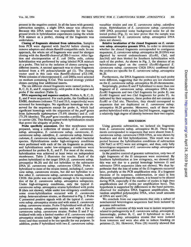
FIG. 1. PCR-amplified fragments selected by genomic subtrac-tion. PCR products were analyzed by electrophoresis (15). Samples(10 ,ul) of PCR mixture were run on the gel. Lane 1, 1-kb DNAladder (Bethesda Research Laboratories); lane 2, E. carotovorasubsp. atroseptica 86.20 and E. carotovora subsp. carotovora CH26(genomic subtraction); lane 3, E. chrysanthemi PMV4071 and E.chrysanthemi 3937 (genomic subtraction); lane 4, E. carotovorasubsp. carotovora CH26 and E. carotovora subsp. carotovora CH26(genomic subtraction); lane 5, PCR-negative control; lane 6, DNAmarker V (Boehringer).
Sau3AI-digested fl fragment, a 2-kb Smr Spcr DNA frag-ment (25), mixed with 150 ng of E. chrysanthemi 3937genomic Sau3AI-digested DNA and subtracted with 10 p,g of3937 biotinylated DNA and (ii) 250 ng of aX-32P-labeledSau3AI-digested 3937 DNA subtracted with 10 p,g of 3937biotinylated DNA. In the first experiment, recovery at eachcycle of the nonhomologous sequences (Q fragment) wasassayed in the free fraction, while in the second experiment,binding of homologous sequences (3937 DNA) was assayedby the radioactivity level in the bound fraction. After fourcycles of subtraction, the heterologous sequence/homolo-gous sequence ratio had increased 18-fold and over 99% ofhomologous sequences had been removed from the solution.However, approximately 40% of heterologous sequenceswere lost at each cycle, resulting in an 83% loss after fourcycles. For these reasons, the best compromise betweenenrichment in heterologous sequences and losses was foursubtraction cycles.Genomic subtraction of E. carotovora subsp. atroseptica
DNA. Four cycles of subtracting hybridizations were donefor each of the following pairs of target and subtracterDNAs: (i) PMV4071 and 3937 strains (differentiated by the flfragment) (positive control), (ii) E. carotovora subsp.atroseptica 86.20 strain and E. carotovora subsp. carotovoraCH26 strain, and (iii) E. carotovora subsp. carotovora CH26and E. carotovora subsp. carotovora CH26 (negative con-trol) (Fig. 1). The positive control yielded only two amplifiedfragments of approximately 230 and 300 bp. Since Sau3AIdigestion of the initial fl fragment generated eight mainfragments of 460, 300, 230, 190, 150, 140, 80, and 60 bp, thiscontrol indicated that not all of the different sequences in thetwo DNA sequences were recovered after genomic subtrac-tion. The subtraction between E. carotovora subsp. atrosep-tica and E. carotovora subsp. carotovora yielded six mainfragments (190, 200, 250, 300, 400, and 410 bp) and a doublediffuse band around 450 bp. No amplified fragment was
PROBES SPECIFIC FOR E. CAROTOVORA SUBSP. ATROSEPTICA 301
present in the negative control. In all the lanes with genomicsubtraction samples, a slight DNA smear was observed.Because this DNA smear was responsible for the back-ground levels in hybridization experiments (using the wholePCR mixture as a probe), cloning of the fragments wasundertaken.
Cloning of fragments obtained from subtraction. Samplesfrom PCR were digested with Sau3AI before cloning toremove adaptors and obtain BamHI-compatible ends. In oneapproach, the whole set of fragments was used for shotguncloning with the BamHI-cleaved pUBS-3 vector. After liga-tion and electroporation of E. coli DH5a cells, colonyhybridization was performed by using labeled PCR mixtureas a probe. This led to the isolation of clones carrying twodifferent inserts. A second approach was to clone fragmentsisolated from a 6% polyacrylamide gel (1). The cloningvector used in this case was BamHI-cleaved pTZ-19R.White colonies of electroporated E. coli DH5a were selectedon medium containing X-Gal. This second strategy yieldedclones carrying four additional inserts.The six different cloned fragments were named probes A,
B, C, D, E, and F, respectively, with probe A the largest andprobe F the smallest (Table 1).DNA sequencing and sequence analysis. Probes A, B, C, D,
E, and F were sequenced. For each probe, the GenBank andEMBL databases (releases 75.0 and 33.0, respectively) werescreened for homologies. No significant homology was ob-served for the sequences except for probe A, which hadsignificant homology with the sequences of the putP genesfrom Salmonella typhimurium (73.8% identity) and E. coli(73.2% identity). The putP gene encodes a proline permeaseor carrier (20). This finding agreed with hybridization resultsthat prove the ubiquity of probe A.Probe specificity assessment. Dot blot membranes were
prepared, using a collection of strains of E. carotovorasubsp. atroseptica, E. carotovora subsp. carotovora, E.carotovora subsp. odonifera, E. carotovora subsp. betavas-culorum, E. carotovora subsp. wasabiae, and other bacterialspecies. Hybridizations under high-stringency conditionswere performed with each of the six fragments as probes,and hybridizations under low-stringency conditions wereperformed for probes B, E, and F. For most of the strains,hybridization was achieved at least twice on independentmembranes. Table 2 illustrates the results. Each of theprobes hybridized to the target DNA (E. carotovora subsp.atroseptica 86.20) and did not hybridize to the subtracterDNA (E. carotovora subsp. carotovora CH26). Probe Ahybridized to most of the strains tested, including E. caroto-vora subsp. carotovora strains, but did not hybridize to afew other E. carotovora subsp. carotovora strains, such asCH26: this probe was not specific enough to be representa-tive of the E. carotovora subsp. atroseptica subspecies.Under high-stringency conditions, only the typical E.carotovora subsp. atroseptica strains hybridized with probeB (data not shown), while under low-stringency conditions,some cross-hybridization occurred with E. carotovorasubsp. betavasculorum strains, as illustrated in Fig. 2. ProbeC presented positive signals with all the typical E. caroto-vora subsp. atroseptica strains and with some E. carotovorasubsp. carotovora strains. Probe D hybridized with all the E.carotovora subsp. atroseptica strains and with a few E.carotovora subsp. carotovora strains. Probes E and F hy-bridized with only a limited number of E. carotovora subsp.atroseptica strains (under high- and low-stringency condi-tions) and thus seemed to be too specific for our purpose. Inaddition, probe F hybridized with two E. carotovora subsp.
wasabiae strains and one E. carotovora subsp. odonferastrain. Hybridization of E. carotovora subsp. carotovora1489 DNA presented some background noise for all thetested probes (Fig. 2); we later prove that the sample wascontaminated by E. carotovora subsp. atroseptica DNA(data not shown).
Relative localization of the cloned fragments on E. caroto-vora subsp. atroseptica genomic DNA. In order to determinewhether the cloned fragments corresponded to contiguoussequences, E. carotovora subsp. atroseptica 86.20 DNA wasdigested by several endonucleases (EcoRI, ClaI, andSau3AI) and then blotted for Southern hybridization witheach of the probes. As shown in Fig. 3, the absence of anhybridization signal on the control (EcoRI-digested E.carotovora subsp. carotovora CH26 DNA) confirmed thatthe probes are specific for E. carotovora subsp. atroseptica86.20.
Furthermore, the DNA fragments revealed by each probewere different, suggesting that the probes are not clusteredon the E. carotovora subsp. atroseptica 86.20 chromosome.
Surprisingly, probes E and F hybridized to more than onefragment of E. carotovora subsp. atroseptica DNA (twoEcoRI fragments and two ClaI fragments for probe E; oneEcoRI fragment and two ClaI fragments for probe F). Asindicated by their sequence, these probes did not contain anEcoRI or ClaI site. Therefore, they should correspond tosequences that are duplicated on E. carotovora subsp.atroseptica 86.20 genome. Moreover, each of these twoprobes hybridized with a single Sau3AI fragment, suggestinga relatively high degree of identity between their two copies.
DISCUSSION
Using genomic subtraction, we isolated six fragmentsfrom E. carotovora subsp. atroseptica 86.20. These frag-ments corresponded to sequences that were absent from E.carotovora subsp. carotovora CH26 DNA, as was shown bySouthern and dot blot hybridizations. This result was ex-pected because the hybridization conditions of subtraction(1M NaCl at 65°C) were not stringent, and thus, only fullyheterologous sequences of E. carotovora subsp. atrosepticaescaped subtraction.
In the positive control of genomic subtraction, only two ofeight fragments of the fl interposon were recovered. BySouthern hybridization at low stringency, we showed thatthis was not due to a partial homology between fQ andsubtracter DNA sequences (data not shown). We thus hy-pothesize that the loss of some of the fl fragments occurredlater, probably at the PCR amplification step. If a fragment(because of its sequence, conformation, or size) is lessefficiently replicated than the others, it is bound to be dilutedduring PCR, especially if the number of cycles is high (twoamplifications and 25 cycles per amplification here). Thishypothesis is supported by differences in the band patterns,observed for multiplex DNA fragment amplification, likerandom amplified polymorphic DNA, when small amountsof DNA are used (34).We conclude from our experiments that only a subset of
nonclustered heterologous sequences had been isolated bygenomic subtraction.A possible outcome of this work was to isolate sequences
related to pathogenicity in E. carotovora subsp. atroseptica.Interestingly, probes B, C, and D hybridized to two E.carotovora subsp. atroseptica strains that were isolatedfrom tomatoes and were also able to induce blackleg onpotatoes (2, 26). However, Priou (26) recently showed that
Potato Argentina, 1989"Water Spain, 1989kWater Spain, 1989"Banana CubafCabbage Malawi, 1986fCarrot United States,' (ATCC 495)"Celery Switzerland, 1988'Chrysanthemum France, 1971"Chrysanthemum United States, 1971"Corn CubafCucumber Italy'Cyclamen GreeceeWitloof chicory France, 1985dIris France, 1973"Leek France, 1982fPotato Denmark, 1952"Potato France, 1988dPotato France, 1988dPotato France, 1988"Potato France, 1987dPotato France, 1986dPotato France, 1977dPotato France, 1977dPotato France, 1976dPotato Malawi, 1986f
a Strains 89.19, 1H, and 40H are atypical E. carotovora subsp. atroseptica strains recently identifed as E. carotovora subsp. carotovora strains by usingphenotypic and genotypic characteristics (4, 26). Superscript T's indicate the type strain of the subspecies. The numbers in brackets are the numbers of strainstested.
b +, hybridization with DNA of the strain; -, no hybridization; ND, data not determined.c Numbers and letters correspond to rows and columns in Fig. 2. NS, data not shown.d Bernard Jouan, Institut National de la Recherch- Agronomique, Rennes, France, personal collection.e Collection Frangaise de Bacteries Phytopathogenes, Angers, France.f R6gine Samson, Institut National de la Recherche Agronomique, Angers, France, personal collection.9 Research Institut for Plant Protection, Wageningen, The Netherlands.h International Potato Center, Lima, Peru.Olivier Cazelles, Station Federale de Recherches Agronomiques, Changins, Switzerland, personal collection.Scottish Corp Research Institute, Dundee, United Kingdom.
k Maria Lopez, Instituto Valenciano de Investigaciones Agrarias, Valencia, Spain, personal collection.Monique Lemattre, Institut National de la Recherche Agronomique, Versailles, France, personal collection.m Claudine Elmerich, Institut Pasteur, Paris, France, personal collection.n Collection Frangaise Informatisee de Souches Microbiennes, Institut National de la Recherche Agronomique, Angers, France.
two E. carotovora subsp. carotovora strains (88.29al andCIPOO9) are able to induce blackleg symptoms identical tothose induced by E. carotovora subsp. atroseptica. Becauseprobes B, C, and D did not detect these two E. carotovorasubsp. carotovora strains, they may not correspond tosequences involved in this pathogenicity trait. It is therefore
A B C D E F G H I J
1234
567
12
FIG. 2. Bacterial strains revealed by probe B under low-strin-gency conditions. Dot blot hybridization was performed on thecollection of bacterial strains (the strains shown in positions aregiven in Table 2) under low-stringency conditions, as described inMaterials and Methods. Rows 1 to 3 contain typical E. carotovorasubsp. atroseptica strains. Rows 4 to 8 contain E. carotovora strains(atypical E. carotovora subsp. atroseptica, E. carotovora subsp.odonifera, E. carotovora subsp. carotovora, and E. carotovorasubsp. betavasculorum). The rest of the membrane is occupied byother bacterial species. The weak signals observed at positions A9,B39, and C9 corresponded to the three E. carotovora subsp. betavas-culorum strains in our collection. The signal at position G8 corre-sponded to E. carotovora subsp. carotovora DNA contaminated byE. carotovora subsp. atroseptica DNA (see Results).
likely that the isolated sequences are related to other phys-iological characteristics specific to E. carotovora subsp.atroseptica.More generally, our data did not show a correlation
between the presence of sequences homologous to theprobes and any physiological feature of E. carotovora subsp.atroseptica or E. carotovora subsp. carotovora strains (26).The exception to this observation was probe A, whichcorresponded to the proline permease gene. Use of probe Arevealed that some E. carotovora subsp. carotovora strainsdo not possess a putP gene. This raised the question ofproline uptake in strains lackingputP: i.e., the question ofwhether such bacteria are unable to efficiently import prolineor whether they possess another carrier, as observed in E.coli and S. typhimurium (14).The results of dot blot hybridizations showed that the
group of E. carotovora subsp. atroseptica strains was fairlyhomogeneous, although some heterogeneity was seen withprobes E and F. Probe B recognized all E. carotovora subsp.atroseptica strains, but not the three atypical strains (89.19,1H, and 40H) that grow at 37°C and do not induce typicalblackleg symptoms on potato and were proposed as E.carotovora subsp. carotovora (4, 26).On the other hand, the group of E. carotovora subsp.
carotovora strains appeared more heterogeneous. This con-clusion is compatible with the results of phylogenic studiesapplying restriction fragment length polymorphism analysisto E. carotovora subsp. atroseptica and E. carotovorasubsp. carotovora pel genes (4). The homogeneity of the E.carotovora subsp. atroseptica group may reflect the adapta-tion of the pathogen to the relatively low genetic diversity ofcultivated potatoes. In contrast, E. carotovora subsp.carotovora is considered an opportunistic pathogen, with awide host range and geographical distribution, which isconsistent with high genetic diversity.One application of genomic subtraction is to isolate DNA
probes for diagnosis, as previously shown for Rhizobium
FIG. 3. Autoradiography of Southern blot membranes hybridized with the a-32P-labeled probes. Hybridization results with DNA probesA to F are shown. Lane L, 1-kb DNA ladder. Lanes 1 to 3, E. carotovora subsp. atroseptica 86.20 DNA digested with EcoRI, ClaI, andSau3AI, respectively; lane 4, E. carotovora subsp. carotovora CH26 DNA digested with EcoRI.
meliloti (3). Probe B could be used to diagnose the presenceof E. carotovora subsp. atroseptica in soil and plant tissuesamples. An improvement in detection sensitivity would bethe development of a PCR test based on this sequence (31).
ACKNOWLEDGMENTS
We thank Sylvie Priou for her collaboration and helpful com-ments.
This work was funded by Institut National de la RechercheAgronomique (INRA), Federation Nationale des Producteurs dePlants de Pomme de Terre (FNPPPT), and Commission of theEuropean Communities.
REFERENCES1. Ausubel, F. M., R. Brent, R. E. Kingston, D. D. Moore, J. G.
Seidman, J. A. Smith, and K. Struhl. 1987. Current protocols inmolecular biology. Greene Publishing Associates, Wiley Inter-science, New York.
2. Barzic, M. R., R. Samson, and A. Trigalet. 1976. Pourriturebacterienne de la tomate cultivee en serre. Ann. Phytopathol.8:237-240.
3. Bjourson, A. J., and J. E. Cooper. 1988. Isolation of Rhizobiumloti strain-specific DNA sequences by subtraction hybridiza-tion. Appl. Environ. Microbiol. 54:2852-2855.
4. Darrasse, A. 1993. Ph.D. thesis. University of Paris VI, Paris.4a.Darrasse, A., A. Kotoujansky, and Y. Bertheau. French patent
93 06072.5. Devereux, J., P. Haeberli, and 0. Smithies. 1984. A comprehen-
sive set of sequence analysis programs for the VAX. NucleicAcids Res. 12:387-395.
6. Gallois, A., R. Samson, E. Ageron, and P. A. D. Grimont. 1992.Erwinia carotovora subsp. odorifera subsp. nov., associatedwith odorous soft rot of chicory (Cichorium intybus L.). Int. J.Syst. Bacteriol. 42:582-588.
7. Goto, M., and K. Matsumoto. 1987. Erwinia carotovora subsp.wasabiae subsp. nov. isolated from diseased rhizomes andfibrous roots of Japanese horseradish (Eutrema wasabi Max-im.). Int. J. Syst. Bacteriol. 37:130-135.
8. Holmes, D. S., and M. Quigley. 1981. A rapid boiling method forthe preparation of bacterial plasmids. Anal. Biochem. 114:193-197.
9. Johnson, D. A. 1984. Improved technique utilizing nonfat drymilk for analysis of proteins and nucleic acids transferred to
nitrocellulose. Gene Anal. Tech. 1:3.10. Klotz, L., and B. H. Zimm. 1972. Size of DNA determined by
viscoelastic measurements: results on bacteriophages, Bacillussubtilis and Escherichia coli. J. Mol. Biol. 72:779-800.
11. Kotoujansky, A., M. Lemattre, and P. Boistard. 1982. Utiliza-tion of a thermosensitive episome bearing transposon TnlO toisolate Hfr donor strains of Erwinia carotovora subsp. chrysan-themi. J. Bacteriol. 150:122-131.
12. Lelliott, R. A., and R. S. Dickey. 1984. Genus VII. ErwiniaWinslow, Broadhurst, Buchanan, Krumwiede, Rogers andSmith 1920, 209AL, p. 469-476. In J. G. Holt and N. R. Krieg(ed.), Bergey's manual of systematic bacteriology, 9th ed.Williams & Wilkins Co., Baltimore.
13. Lelliott, R. A., and D. E. Stead. 1987. Methods for the diagnosisof bacterial diseases of plants. Blackwell Scientific Publications,Ltd., Oxford.
14. Maloy, S. R. 1987. The proline utilization operon, p. 1513-1519.In F. C. Neidhardt, J. L. Ingraham, K. B. Low, B. Magasanik,M. Schaechter, and H. E. Umbarger (ed.), Escherichia coli andSalmonella typhimurium: cellular and molecular biology, vol. 2.American Society of Microbiology, Washington, D.C.
15. Maniatis, T., E. F. Fritsch, and J. Sambrook. 1982. Molecularcloning: a laboratory manual. Cold Spring Harbor Laboratory,Cold Spring Harbor, N.Y.
16. Marti, R., M. M. Lopez, C. Morente, and B. Alargon. 1989.Incidence of erwinia-causing soft rots in irrigation water inValencia (Spain), p. 755-760. In Z. Klement (ed.), Proceedingsof the 7th International Conference on Plant Pathology andBacteriology, Budapest, Hungary. Akademiai Kiad6, Budapest.
17. Mead, D. A., E. S. Skopura, and B. Kemper. 1985. Singlestranded DNA promoter plasmids analysis for engineering mu-tant RNA's and protein: synthesis of a stretched parathyroidhormone. Nucleic Acids Res. 13:1103-1108.
18. Miller, J. H. 1972. Experiments in molecular genetics. ColdSpring Harbor Laboratory, Cold Spring Harbor, N.Y.
19. Miller, S. A., and R. R. Martin. 1988. Molecular diagnosis ofplant disease. Annu. Rev. Phytopathol. 26:409-432.
20. Nelson, K., and R. K. Selander. 1992. Evolutionary genetics ofthe proline permease gene (putP) and the control region of theproline utilization operon in populations of Salmonella andEscherichia coli. J. Bacteriol. 174:6886-6895.
21. Perombelon, M. C. M. 1980. Ecology of the soft rot erwinias.Annu. Rev. Phytopathol. 18:361-387.
22. Perombelon, M. C. M. 1989. Ecology and pathogenicity of soft
rot erwinias: an overview, p. 745-751. In Z. Klement (ed.),Proceedings of the 7th International Conference on Plant Pa-thology and Bacteriology, Budapest, Hungary. Akademiai Ki-ad6, Budapest.
23. Perombelon, M. C. M., and L. J. Hyman. 1986. A rapid methodfor identifying and quantifying soft rot erwinias directly fromplant material based on their temperature tolerances and sensi-tivity to erythromycin. J. Appl. Bacteriol. 60:61-66.
24. Perombelon, M. C. M., and A. Kelman. 1987. Blackleg and otherpotato diseases caused by soft rot erwinias: proposal for revi-sion of terminology. Plant Dis. 71:283-285.
25. Prentki, P., and H. M. Krisch. 1984. In vitro insertional muta-genesis with a selectable DNA fragment. Gene 29:303-309.
26. Priou, S. 1992. Ph.D. thesis. Ecole Nationale SuperieureAgronomique de Rennes, Rennes, France.
27. Sanger, F., S. Nicklen, and A. R. Coulson. 1977. DNA sequenc-ing with chain-terminating inhibitors. Proc. Natl. Acad. Sci.USA 74:5463-5467.
28. Smith, C., and J. A. Bartz. 1990. Variation in the pathogenicityand aggressiveness of strains of Erwinia carotovora subsp.
carotovora isolated from different hosts. Plant Dis. 74:505-509.29. Stanghellini, M. E. 1982. Soft-rot bacteria in the rhizosphere, p.
249-261. In M. S. Mount and G. H. Lacy (ed.), Phytopathogenicprokaryotes, vol. I. Academic Press, Inc., New York.
30. Stanghellini, M. E., and J. C. Meneley. 1975. Identification ofsoft-rot Erwinia associated with blackleg of potato in Arizona.Phytopathology 65:86-87.
31. Steffan, R. J., and R. M. Atlas. 1991. Polymerase chain reaction:applications in environmental microbiology. Annu. Rev. Micro-biol. 45:137-141.
32. Straus, D., and F. M. Ausubel. 1990. Genomic subtraction forcloning DNA corresponding to deletion mutations. Proc. Natl.Acad. Sci. USA 87:1889-1893.
33. Thomson, S. V., D. C. Hildebrand, and M. N. Schroth. 1981.Identification and nutritional differentiation of the erwinia sugarbeet pathogen from members of Erwinia carotovora and Erwiniachrysanthemi. Phytopathology 71:1037-1042.
34. Williams, J. G. K., M. K. Hanafey, J. A. Rafalski, and S. V.Tingey. 1993. Genetic analysis using random amplified polymor-phic DNA markers. Methods Enzymol. 218:704-740.