Abstract In this study, 21 different fungi were isolated from saw dust, sprinkled soil (garden, beach and mud), decaying wood particles, decaying leaf and cultured in the laboratory. Among that, high cellulose degrading varieties were screened and the extracted cellulolytic enzymes were examined for their activity by filter paper assay and DNS reagent method. In addition, fungal cultures were grown on rice straw which is a common agro-waste in Asia. In straw substrate, highest activity of 42.61 FPU/mL was recorded by Helminthosporium sp. where, the saccharification was 3.8% followed by Cladosporium sp. (40.36 FPU/mL). Enzymes of Trichoderma sp. and Aspergillus sp. also showed considerable activity.
Keywords: Cellulose, filter paper assay, rice straw, saccharification, Helminthosporium sp.
Introduction In the kingdom of fungi, most of the individuals are saprophytic and are efficient in degradation of major polymers such as cellulose and lignin. If fungi or their byproducts are used in paper production or the recycling materials, we would be able to eliminate a large source of pollution in the environment. Purified fungal cellulolytic enzymes are used for commercial food processing such as production of coffee. It performs hydrolysis of cellulose during drying of beans. They are also widely used in textile industry and in laundry detergents such as modern washing powders consisting of fungal enzymes, and fermentation of biomass into biofuels. Even in medicine, for e.g. fungal cellulase is used as a treatment for phytobezoars (a form of cellulose bezoar found in the human stomach) (Thorn et al., 1996). It also proved that application of cellulolytic fungi improves the composting potential of cellulose waste where the C: N ratio was not optimal, in addition water holding capacity also improved in fungal inoculated samples (Hart et al., 2002). In a study on rice straw, 25 different species of fungi were extracted belonged to ascomycetes and basidiomycetes. In that study, Trichoderma harzianum showed maximum cellulolytic activity (Lee et al., 2011). Rice straw consists of 37-50% of cellulose which is a common agro-waste in South Asian countries. Its annual production reaches 350 million tons (Han and Anderson, 1974), it is being used for roofing, packing material, feed, fertilizer and as fuel. In addition, by fermentation it can be used in pretreatment during the production of ethanol. To facilitate the ethanol fermentation, enzymatic saccharification is a preparation stage. Fungal cellulolytic enzymes contribute a huge role on this aspect.
This main aim of the present study is to isolate fungal varieties which are found naturally and examine their ability to degrade agricultural wastes. In this study, 21 different fungal cultures were assayed for enzyme activity, among those, topping four varieties were screened and cultured on straw strips and resulting percentage of saccharification was recorded. Materials and methods Source of fungi and initial culturing: Samples were collected from sawdust, straw dust and sprinkled soil (garden, beach and mud), decaying wood particles and decaying leaf collected from the surroundings. Samples were collected into sterile containers and stored separately. Potato dextrose agar medium was used to grow the initial cultures, where samples were cultured by streak plate method and sprinkle method. Methods of inoculation Streak plate method: In order to isolate the fungi this method is best in practice. Initial streak is made with the sample then, all other streaks are continuum of the previous strike using separate sterile tooth peck for each strike. Sprinkled method: This is more suitable for the soil samples and sawdust. Particles were sprinkled on the medium. Well-spaced sprinkled particles would result in separation of colonies. Finally, petri dishes were sealed with para film, labeled and inverted dishes were incubated in a dark place. Visible colonies were observed after 4 d of incubation (Fig. 1a and b).
RESEARCH ARTICLE
Journal of Academia and Industrial Research (JAIR) Volume 2, Issue 8 January 2014 459
Fig. 1. Inoculation of samples on PDA medium by sprinkle method.
a. Sawdust sprinkled.
b. Straw sprinkled.
Media used for sub-culturing Selective water agar medium: To separate the fungi causing cellulose digestion, a medium consisting cellulose as a sole carbon source was prepared. For cellulose Whatman no. 1 filter paper made of 100% cellulose was used. Water agar medium was prepared by dissolving 4 g of agar in 250 mL of distilled water. Autoclaved pieces of filter paper (1 cm × 1 cm) were used for inoculation. After pouring the water agar medium on petri dishes, paper strips were carefully placed on top of the agar bed by a sterile forceps. After the inoculation, sealed dishes were kept for 4 d of incubation. To avoid bacterial growth, antibiotic was added to the medium. This made the medium more selective to fungi. Table 1 depicts the amount of antibiotics that were added for the preparation of selective water agar medium. Sub-culturing continued until the pure culture was available. Culturing fungi in liquid medium containing straw and preparation of crude enzymes: In order to extract the secreted cellulolytic enzymes by each fungal colony, it is important to culture them in a liquid medium.
Cellulolytic basal medium (CBM) was chosen for this purpose. Cellulolytic basal medium (1.5 g; Diammonium tartrate (C4HI2N206), 0.25 g; Potassium dihydrogen phosphate (KH2P04), 0.02 g; yeast extract, 0.15 g; MgS04.7H20, 0.0002 g; CaCl2.2H20 (g/250 mL in distilled water) was prepared (Pointing, 1999). CBM medium was autoclaved and 10 mL aliquots were transferred to sterile 20 mL bottles. Straw strips of same amount were submerged into the CBM medium aseptically inside the laminar airflow. Samples which were previously obtained from pure culture were inoculated by streaking on the submerged straw strip aseptically by sterile tooth peck. Always a control bottle was kept without inoculation. Caps of the bottles were loosely fitted to allow the adequate air exchange. All the bottles were incubated for 4 d at room temperature (25C). After the incubation, observations were made and the liquid medium, which contains the crude fungal enzymes were collected. Each bottle contains the cellulolytic enzymes that were secreted as extracellular enzyme by each fungus colony. At the time of isolation, which is after a period of incubation, it is better to vortex in slow speed in order to ensure the distribution of enzymes all over the liquid medium. About 1 mL of aliquot was taken by sterile micro pipette and placed in 1.5 mL sterile centrifuge tubes. Centrifugation was done at 12,000 rpm for 15 min. The resulted supernatant consists of proteins that are mostly the fungal crude enzymes. Filter paper assay: International Union of Pure and Applied Chemists recommended filter paper assay (FPA) as the standard measure of cellulase activity. Enzymatic reactions often occur in the presence of buffer, which helps to keep the reaction environment stable. This is obtained by maintaining the ionic balance and the pH unchanged. About 2 mL 0.05 M of Trisodium citrate dihydrate (C6H5Na3O7•2H2O) buffer was used with the crude enzymes and filter paper strips (0.5 cm × 0.1 cm) were used as substrate. About 0.1 mL of crude enzyme of each fungus was added with 0.15 mL of Trisodium citrate dihydrate solution, while the pH was maintained at 4.8. Always a blank was maintained in one tube without adding any fungal enzymes instead; it was replaced by same volume of sterilized distilled water. Then Whatman no. 1 filter paper strip (0.5 cm × 0.1 cm) was added as the substrate. Each tube was then incubated in 50C shaking incubator running at 100 rpm (Mandels and Andreotii, 1976). Measuring the activity of cellulolytic enzymes: The activity of extracted fungal enzymes can be measured quantitatively by DNS reagent method and qualitatively by Congo red methods (it can also be quantitative if zone of decolourization is measured accurately). Here, the concentrations of reducing sugars (products of enzyme activity) were measured using DNS reagent test, since the optical absorbance can be more accurately measured using spectrophotometer at 540 m.
Table 1. Antibiotics and their concentrations that used in the culture.
Antibiotic Ampicillin Tetracycline In a mL of stock solution 50 mg/mL 100 mg/mL Final concentration in a mL of water Agar medium 50 µg/mL 100 µg/mL
Journal of Academia and Industrial Research (JAIR) Volume 2, Issue 8 January 2014 460
Dinitrosalicylic acid method: Dinitrosalicylic acid reagent was prepared by adding 1 g 3, 5-dinitrosalicylic acid in 50 mL of distilled water. About 200 mg crystalline phenol (optional) and 30 g of sodium potassium tartrate were added to the solution, which turns the solution into yellow colour. To this, 20 mL of 2N NaOH was added. This turns the colour of the solution into transparent orange yellow. Finally, the stock was made into 200 mL by adding distilled water. Stock was stored at 4C in refrigerator, to prevent deterioration (Miller, 1959). After the incubation, filter paper strips were carefully removed from the tubes using a glass rod. Then, 0.5 mL of DNS reagent was pipetted into each tube. This terminated all enzymatic reactions occurred in the tube. Then, the lids of tubes were tightly closed and placed in a water bath at 95-100C for 10 min. After this, the tubes were immediately transferred into an ice cold bath and kept for few minutes. About 1 mL of distilled water was pipetted into each tube before measuring the optical absorbance. Colour change in each tube was measured by using UV spectrophotometer at 540 nm. Finally, the optical absorbance readings were compared and plotted with the standard glucose curve to find the glucose (product) concentrations (Miller, 1959). From each glucose and buffer mixture, 0.1 mL of solution was added to 0.15 mL of Trisodium citrate dihydrate buffer solution. Then, each centrifuge tube was transferred into a water bath where tubes were incubated at 50C for 1 h, same as the conditions given for the enzyme filter paper assay. After the incubation, 0.5 mL of DNS reagent was pipetted into each tube and the lids of all tubes were tightly closed. Then, the temperature in the water bath was raised to 95-100C and kept for 10 min. Finally, the tubes were immediately transferred into an ice cold bath for few minutes and 1 mL of distilled water was pipetted to each tube before measuring the absorbance of optical absorbance and the samples were examined for the colour change. Colour change in each tube including the control blank was measured using UV spectrophotometer at 540 nm. Finally, the optical absorbance readings were plotted against the concentration of glucose. As given in Table 2, optical absorbance differ according to the concentration of glucose ranging from 1.00 mg/0.5 mL to 3.35 mg/0.5 mL resulted in optical absorbance ranging from 0.228 to 0.766 respectively. A standard curve was used to find the unknown concentrations of reducing sugars in all samples, dilutions used were translated into enzyme concentrations. Concentration of enzyme which would have released exactly 2.0 mg/0.5 mL of glucose by means of a plot of glucose liberated against the logarithm of enzyme concentration was estimated. Filter paper unit was calculated according to IUPAC-FPU.
0.37 FPU = Units/mL
Enzyme conc. to release 2 mg glucose
Congo red method: Congo red has the ability to bind with cellulose and produce bright red colour, if the enzyme acts on the substrate, the cellulose may be used up, so it end with a zone of decolourization. Each pure fungi sample was inoculated aseptically on the Whatman no. 1 filter paper strips of the same size. They were then kept on water agar beds (to maintain the moisture) for a week. After carefully removing the well grown fungus colonies, filter paper strips kept on the lids of the petri dishes were flooded with 0.1% congo red solution. Then it was left for 15 min with intermittent shaking in a mechanical shaker. Finally, strips were dipped in distilled water and washed with 1 M NaCl solution. Efficiency of each fungal enzyme on agricultural waste: Since, Whatman no. 1 filter paper consist of 98% cellulose, substrate concentration in 0.5 mL can be derived as 49 mg (cellulose). By applying the product (glucose) concentration retrieved from the standard glucose curve, the percentage of saccharification was calculated. Similarly agro waste rice straw contains about 32-47% of cellulose (Karimi et al., 2006). When the average 40% the cellulose content in 0.5 mL can be derived as 20%, by applying the product (glucose) concentration retrieved from the standard glucose curve, the saccharification % of straw was calculated. Saccharification % = {Glucose (mg/0.5 mL)/Substrate (mg/0.5 mL)} x 100 Identification of fungi: In order to identify the fungal colonies, colony colour, shape, border and spots (if the spores are available) were recorded. Microscopic visuals were observed under high power oil immersion objective. Spores and the mycelia were observed so clearly (Fig. 2a-e) and the data were recorded and used in classification. Fungi were classified up to the genus level by their morphological features. Classification was based on microscopic observation of mycelia as well as reproductive structures such as spores and fruiting bodies, if available. Characters used in classification were compared by considering mycelial characters such as presence of septa, whether mycelium was branched or not, on mature colonies the presence of reproductive structures such as sporangia, conidia and their morphology, types of spore they generate, whether spores are septate or not and position of rhizoids on the mycelium etc. Effects of pH and Temperature on fungal enzyme activity: Enzyme activity related to variation in temperature and pH were measured separately. Filter paper assays of each fungal crude enzyme were kept in water baths at temperatures of 37, 50 and 60C.
Table 2. Glucose concentration vs. Optical absorption. Glucose conc. Optical absorption at 540 nm
3.35 mg/0.5 mL 0.766 2.50 mg/0.5 mL 0.580 1.65 mg/0.5 mL 0.378 1.00 mg/0.5 mL 0.228
Journal of Academia and Industrial Research (JAIR) Volume 2, Issue 8 January 2014 461
Fig. 2. Spore morphology of isolated fungi (X 40).
a. Cladosporium sp., b. Trichoderma sp., c. Fusarium sp.,
d. Helminthosporium sp., e. Aspergillus sp.
After an hour of incubation, DNS reagent test was done. Similarly, pH of the each buffer solution was changed to 3, 6, 8, and 13 by adding either dilute NaOH or HCl. Then, crude enzyme of each fungus was added and filter paper strips were placed. Assay was incubated at 50C for an hour and products were measured using DNS reagent test. Concentrations of reducing sugars were obtained from the standard glucose curve and finally activity of enzymes was calculated as FPU/mL. Results and discussion Cellulolytic enzyme assay: Results based on the observed readings, which were proportional to the concentration of the product (reducing sugar), clearly demonstrates the potential cellulolytic activity of each fungal crude enzyme tested here. Trichoderma sp. showed the fastest degradation on filter strip in 12 d (Fig. 3). Crude enzyme was extracted from the liquid medium of the cultured fungus. Activity of fungal enzymes in filter paper substrate: Figure 4 depicts the cellulose degrading ability of each fungus tested here.
Fig. 3. Degradation of filter paper strip by Trichoderma sp. after 12 d of incubation and Congo red results.
Trichoderma sp. showed highest activity of 23.54 FPU/mL, while Helminthosporium sp., Aspergillus sp., Cladosporium sp., Fusarium sp. showed filter paper activity of 23.05, 22.98, 20.74, 20.74 FPU/mL respectively (Fig. 4). This result is only relevant to the given physical and chemical conditions because, the activity of each fungal enzyme varies according to its individual preference to the substrate, chemical nature of the medium, optimal temperature, and pH conditions. Such factors are important in determining the survivals of certain fungi on natural environment. If such factors are more suitable for a particular species than another, that species will ultimately win the competition on using all the resources so the other eventually eliminated. However, some fungi can produce specialized structures to survive in such harsh conditions and will germinate when environmental conditions become favourable to them. Congo red strips showed decolourized regions (Fig. 4), however, they are not accurate as DNS reagent method.
Fig. 4. Activity of fungal enzymes on filter paper substrate.
1919.5
2020.5
2121.5
2222.5
2323.5
24
Cla
dosp
oriu
m s
p.
Asp
ergi
llus
sp.
Hel
min
thos
poriu
m s
p.
Tric
hode
rma
sp.
Fusa
rium
sp.
FPU
/mL
b a
c d
e
Journal of Academia and Industrial Research (JAIR) Volume 2, Issue 8 January 2014 462
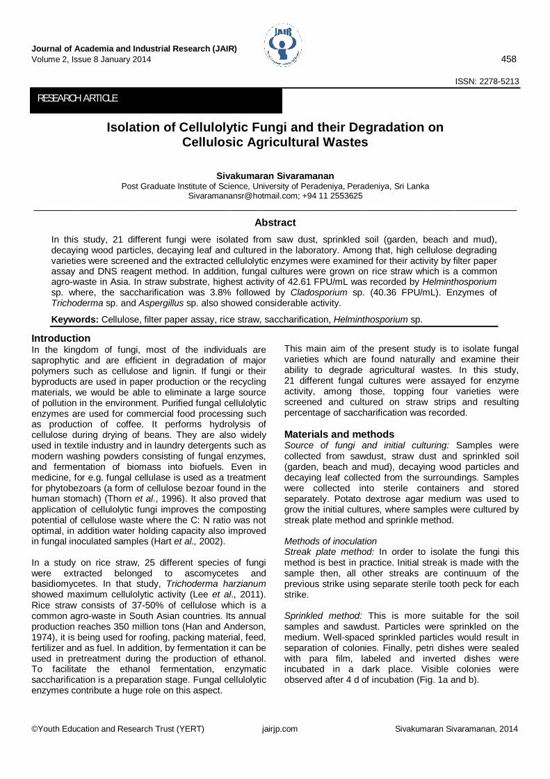
Fig. 5. Sacharification % of fungal enzymes in CBM.
Fig. 6. Activity of fungal enzymes on straw substrate.
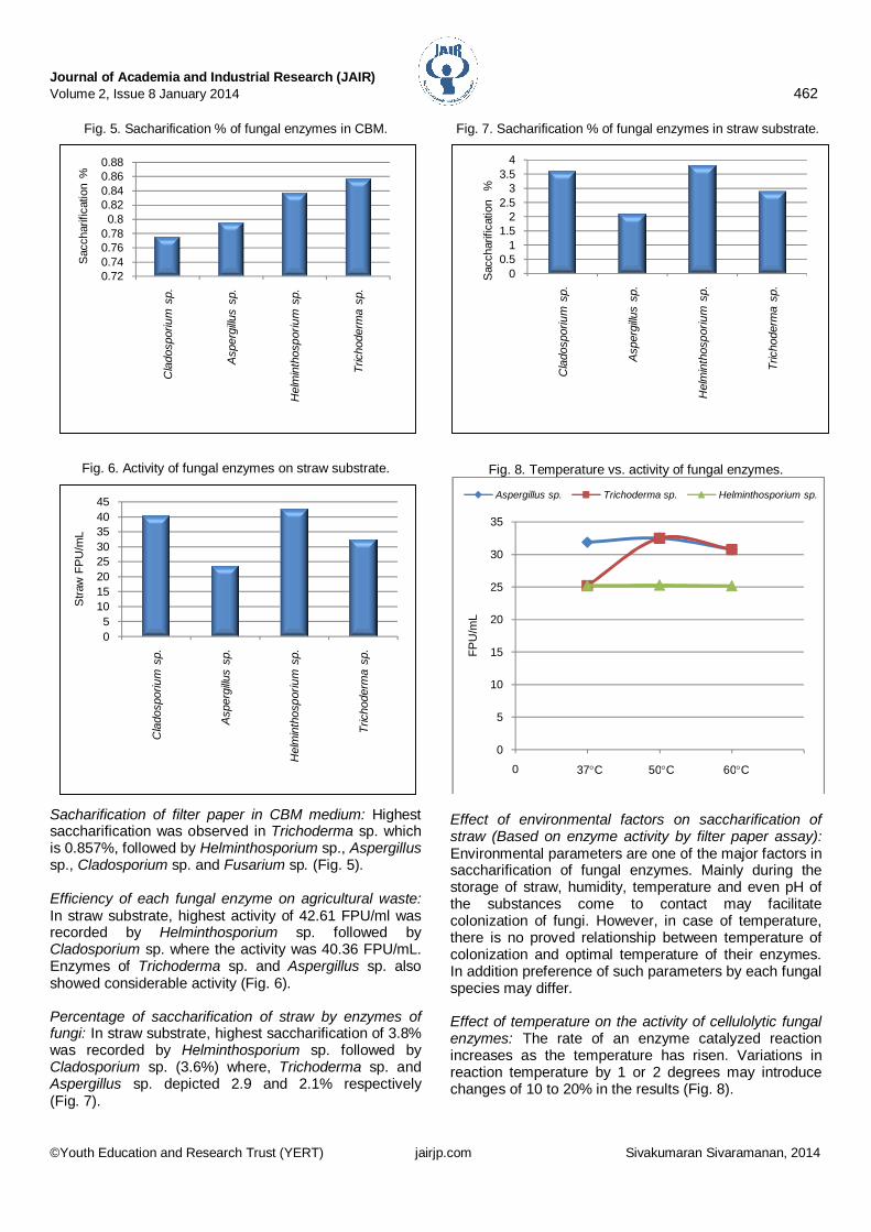
Sacharification of filter paper in CBM medium: Highest saccharification was observed in Trichoderma sp. which is 0.857%, followed by Helminthosporium sp., Aspergillus sp., Cladosporium sp. and Fusarium sp. (Fig. 5). Efficiency of each fungal enzyme on agricultural waste: In straw substrate, highest activity of 42.61 FPU/ml was recorded by Helminthosporium sp. followed by Cladosporium sp. where the activity was 40.36 FPU/mL. Enzymes of Trichoderma sp. and Aspergillus sp. also showed considerable activity (Fig. 6). Percentage of saccharification of straw by enzymes of fungi: In straw substrate, highest saccharification of 3.8% was recorded by Helminthosporium sp. followed by Cladosporium sp. (3.6%) where, Trichoderma sp. and Aspergillus sp. depicted 2.9 and 2.1% respectively (Fig. 7).
Fig. 7. Sacharification % of fungal enzymes in straw substrate.
Fig. 8. Temperature vs. activity of fungal enzymes.
Effect of environmental factors on saccharification of straw (Based on enzyme activity by filter paper assay): Environmental parameters are one of the major factors in saccharification of fungal enzymes. Mainly during the storage of straw, humidity, temperature and even pH of the substances come to contact may facilitate colonization of fungi. However, in case of temperature, there is no proved relationship between temperature of colonization and optimal temperature of their enzymes. In addition preference of such parameters by each fungal species may differ. Effect of temperature on the activity of cellulolytic fungal enzymes: The rate of an enzyme catalyzed reaction increases as the temperature has risen. Variations in reaction temperature by 1 or 2 degrees may introduce changes of 10 to 20% in the results (Fig. 8).
In this experiment, enzymatic reaction of given fungi showed a peak at 50C by reaching a peak of 32.5 FPU/mL. This shows that the temperature for the cellulolytic enzymes of the three tested fungi was 50C. However, if further, high temperatures are tested it is possible to find the point they get denature. Normally animal enzymes get denatured even at 40C. Nevertheless, for fungi it is higher. It is also possible to observe the tolerance as well as the preference of high temperature (since 50C as optimum) by fungal enzymes. According to Fig. 6, the fungus Trichoderma sp. showed an increase in the rate of reaction until 50C followed by decline afterwards. Effect of pH on the activity of cellulolytic fungal enzymes: Since enzymes are proteins, they are very sensitive to changes in pH. Each enzyme has its own optimum range for pH, where it is most active and the result is determined by the effect of pH on a combination of factors such as binding of the enzyme to substrate, catalytic activity of the enzyme, ionization of the substrate and the variation of protein structure. The initial rates for many enzymatic reactions exhibit bell-shaped curves. The most favourable pH value (optimum pH) may vary among enzymes of different fungi. In this study, the optimum pH for fungus Trichoderma sp. and Fusarium sp. was closer to neutral and for Helminthosporium sp. it is 3 (Fig. 9). It means Helminthosporium sp. prefers slightly acidic medium. For Aspergillus sp., the curve was peaking at very low pH that showed the preference of Aspergillus sp. towards acidic environment. Conclusion According to empirical values, among the cultured 21 fungi, Helminthosporium sp., Cladosporium sp., Trichoderma sp. and Aspergillus sp. were screened for their highest enzyme activity. Though there are many synthetic artificial enzymes and chemicals to hydrolyze cellulosic matter, it is always safe to rely on natural fungal enzymes in pretreatment of fermentation.
In addition, they are not so costly and fungal enzymes are reusable proteins that can work efficiently in normal environmental conditions. Saccharification percentage of all screened fungi was above 2, thus, extracted fungal enzymes can be used to digest agro-wastes such as rice straw which can later be used either in fermentation process for the production of biogas or pretreatment of cellulosic wastes. In order to produce better compost, optimized temperature is about 50C for all examined fungal species however pH differs. Acknowledgements Author sincerely thanks Dr. Preminda Samaraweera, Department of Molecular Biology and Biotechnology, University of Peradeniya, Srilanka for supervising this work. References 1. Han, Y.W. and Anderson, A.W. 1974. A problem of rice
straw waste a possible feed through fermentation, from Economic botany, springer for New York botanical gr. Press. 28: 3.
2. Hart, T.D., Leij, F.A.A.M., Kinsey, J. and Lynch, J.M. 2002. Starateges for isolating of fungi for composting wheat straw. World J. Microbiol. Biotechnol. 18: 471-478.
3. Karimi, K., Mkherdmadinia, S.H. and Taherzadeh, M.J. 2006. Conversion of rice straw to sugars by dilute-acid hydrolysis. Biomass Bioenergy. 30: 247-253.
4. Lee, S., Jang, Y., Lee, Y.M., Lee, J., Lee, H., Kim, G.H. and Kim, J.J. 2011. Rice straw-decomposing fungi and their cellulolytic and xylanolytic enzymes. J. Microbiol. Biotechnol. 21(12): 1322-1329.
5. Mandels, M. and Andreotii, R.C. 1976. Measurement of saccharifying cellulase. Biotechnol. Bioeng. Symp. 6: 21-23.
6. Miller, G.L. 1959. Use of Dinitrosalicylic acid for determination of reducing sugar. Anal. Chem. 31: 426-429.
7. Pointing, S.B. 1999. Qualitative methods for the determination of lignocellulolytic enzyme production by tropical fungi. Fungal Diversity. 2: 17-33.
8. Thorn, R.G., Reddy, C.A., Harris, D. and Paul, E.A. 1996. Isolation of saprophytic basidiomycetes from soil. Appl. Environ. Microbiol. 62(11): 4288-4292.