List of Figures Figure 1. Structure of Amylose and Amylopectin ………………………………………..…...... 6 Figure 2. Max.Tensile Strength of Plastic Films made with Starch after Treatment with Culture Supernatant and Fungal Pellet ………………………................................................................. 36 Figure 3. Proteins Isolated from Cultures Grown with or Without Starch by (NH4)2SO4
Figure 4. Proteins Isolated from Cultures Grown with or Without Starch by Anion Exchange Chromatography ……………………………………………………………………………...... 40 Figure 5. Surface Morphology of Starch Granules Observed by SEM ……………………….. 43 Figure 6. Max. Tensile Strength of Plastic Films made with Starch after Treatment with Proteins Isolated from Cultures Grown with or Without Starch ............................................................... 46 Figure 7. Max.Tensile Strength of Plastic Films made with Starch after Treatment with BSA and His-SDH ………………………………………………………………………………………... 49 Figure 8. Max. Tensile Strength of Plastic Films made with Starch after Treatment with His-tagged Protein before and after His-tag cleavage……….……………………………………… 51 Figure 9. Max. Tensile Strength of Plastic Films made with Starch after Treatment with Different Amounts of His-tagged Proteins …………………….................................................. 54 Figure 10. Max.Tensile Strength of Plastic Films made with Starch after Treatment with non-specific proteins of different sizes ………………………..……………………………………. 56 Figure 11. Max.Tensile Strength of Plastic Films made with Starch after treatment with fungal culture, fungal proteins and non-specific proteins …………………………………………….. 59 Figure 12. Water holding capacity of starch treated with 0.06mg/g starch of fungal proteins and non-specific proteins ………………………………………………………………………...…. 63 Figure 13. Water holding capacity of starch treated with 0.4mg/g starch of fungal proteins and non-specific proteins ………………………………………………….................................…... 64
viii
List of Tables Table 1. Amylopectin and amylose composition of various types of starch …………………….4 Table 2. Gelatinization temperatures for various types of starch ………………...……………...9 Table 3. a & b. Tensile strength of starch plastics after treatment with fungal supernatant and fungal pellet ……………………………………………………………………………………. 35 Table 4. High confidence protein matches for O. ulmi proteins ………………………...……. 41 Table 5. a & b. Tensile strength of starch plastics after treatment with fungal protein isolated from cultures grown with and without starch ………………………………………………..… 45 Table 6. a, b & c. Tensile strength of starch plastics after treatment with non-specific proteins, BSA and His-tagged protein ………………………………………………………………….... 48 Table 7. a &b. Tensile strength of starch plastics after treatment with His-tagged protein before and after cleavage of the hexahistidie tag ……………………………………………………… 50 Table 8. a &b. Tensile strength of starch plastics after treatment with different amounts of His-tagged proteins …………………………………………………………………………………. 53 Table 9.a &b Tensile strength of starch plastics after treatment with non-specific proteins of different size …………………………………………………………………………………… 55 Table 10. a, b & c. Tensile strength of starch plastics after treatment with fungal culture, fungal proteins and non-specific proteins …………………………………………………………...… 58 Table 11. Water holding capacity of starch treated with fungal culture, fungal proteins and non-specific proteins ………………………………………………………………………………... 62
ix
List of Abbreviations ASTM - American Society for Testing and Materials
ZnSO4, 0.001% (w/v) FeCl3, 0.00001% (w/v) pyridoxine, and 0.00001% (w/v) thiamine. The
contents were dissolved in deionized water and the pH was adjusted to 6 using concentrated
hydrochloric (HCl) acid. The prepared media was sterilized by heating to 121°C for 20
minutes. 100 mL of fungal stock (approximately 300-400mg on dry matter basis) was
inoculated into one liter of media and was grown on a shaker at 180 rpm at 25°C for three
days.
2.2 Isolation of Secreted Proteins
2.2.1 Ammonium Sulfate Precipitation
Fungal cultures were centrifuged at 5000rpm and 4˚C for 30 minutes to remove fungal cells.
The supernatant was vacuum filtered using 0.35µm pore size to remove excess spores and
any other unwanted matter that might have accumulated in the sample. Proteins from the
sample were precipitated using a 95% saturation of ammonium sulfate (65.9g/100ml) which
was added gradually. The sample was spun down at 8000 rpm for 30 min to obtain the
precipitate. The pellet was resuspended in a buffer containing 50mM Tris pH 7.5 and
24
100mM NaCl along with 0.01% protease inhibitor (PI; PMSF and benzamadine cocktail).
The protein samples were dialyzed for several hours at 4˚C to remove ammonium sulfate
(dialysis buffer contained 50mM Tris pH 7.5, 100mM NaCl). The proteins were
subsequently concentrated by ultracentrifugation using Millipore membrane with a 10kD cut-
off. Sample volume was reduced by about 95% of initial volume. Unused portions of the
protein samples were stored at -20˚C.
2.2.2 Anion Exchange Chromatography
The pH of fungal culture supernatant was adjusted to 7.5 (from an average of about 4) using
HCl. Proteins were isolated using DEAE-Sepharose anion exchange. All steps were carried
out at 4˚C unless otherwise specified; this was done to reduce protein degradation.
Approximately 5mL of the DEAE-Sepharose resin (GE Healthcare) was applied to a 50mL
column and equilibrated with 5 column volumes of 50mM TRIS-HCl buffer pH 7.5. The
culture supernatant was run through the columns. 10 Volumes of the 50mM Tris-HCl buffer
pH 7.5 were used to wash the columns. Proteins were eluted from the column with about 15
mL of 800mM KCl in 50mM Tris-HCl pH 7.5 containing 0.01% protease inhibitor. Samples
were collected on ice in 1.5 ml fractions; the fractions with the most protein were pooled and
stored at -20˚C until further analysis.
2.3 Protein Quantification
Protein quantification was carried out using a method adapted from Bradford (1976). The
concentration of isolated protein samples was estimated using a BSA standard curve. The
curve was made using absorbance data from different concentrations of Bovine Serum
25
Albumin (BSA) ranging from 5 to 30µg. Bradford Assay Reagent (Biorad) was used as per
manufacturer instructions. Absorbance readings were taken at a 595nm wavelength, using
the Cary UV-Vis spectrophotometer (Varian) and the Cary WinUV Simple Reads
application. The spectrophotometer was blanked with buffer before the proteins.
2.4 SDS-PAGE - Visualization of Isolated Proteins
Proteins were separated and visualized by one dimensional polyacrylamide gel
electrophoresis (SDS-PAGE) using the Bio-Rad Mini-PROTEAN 3 system. 12.5% Tris-HCl
gels were run at 200V for about 45 min, using a slightly modified discontinuous SDS buffer
system from Laemmli, 1970. Samples were mixed with loading dye and boiled for a few
minutes before loading. The gels were stained with 0.1% Coomassie Brilliant Blue G-250
(Bio-Rad) while shaking gently for at least a few hours and de-stained using 10% acetic acid,
40% ethanol till bands became visible. Gel images were captured using a UV gel box.
2.5 Identification of Isolated Proteins
2.5.1 Tryptic Digestion
The trypsin digest protocol was adapted from Shevchenko et al. (1996). Protein bands from
de-stained gels were cut into the 1mm pieces and were incubated with 100mM NH4CO3 for
10 minutes. NH4CO3 was removed and acetonitrile (ACN) was added to the gel plugs for 10
minutes. ACN was removed and 10mM DTT in 100mM NH4CO3 was added and incubated
at 56˚C for one hour. The solution was removed and 50mM IAA in 100mM NH4CO3 was
added and incubated at room temperature (RT) for 45 minutes in the dark with occasional
vortexing. This solution was removed and the gel plugs were washed with 100mM NH4CO3
26
for 10 minutes. The solution was removed and of ACN was added to dehydrate the gel plugs
and was incubated at RT for 10 minutes. The last two washing and drying steps were
repeated once. The gel plugs were swelled in 10μL of a trypsin buffer (4ng/μL in 50mM
NH4CO3) on ice for 10 minutes. The trypsin buffer was removed and was incubated with
50mM NH4CO3 at 37˚C for a minimum of 4 hours. The gel plugs were spun down and the
supernatant was collected. 20mM NH4CO3 was added to the gel plugs, vortexed for 2
minutes and the supernatant was transferred to the same tube. 50% ACN with 5% formic
acid was added to the gel plugs, vortexed for 2 minutes and the supernatant was saved in the
same tube. The supernatant was spun in the speed-vac until dryness and was stored at -20˚C
until analysis.
2.5.2 Mass Spectrometry
For mass spectrometric (MS) analysis, the sample was re-suspended in 50% CAN with 0.1%
TFA. It was mixed in a 1:1 ratio with a saturated solution of α-cyano-4-hydrocinnamic acid
and 1μL was spotted on an Opti-TOF 384 well MALDI plate and air dried. An Applied
Biosystems 4800 MALDI TOF/TOFTM Analyzer (Framingham, MA, USA) mass
spectrometer was used in this study to acquire MALDI and MS/MS spectra. This TOF/TOF
instrument is equipped with a Nd:YAG laser with 355-nm wavelength of <500 ps pulse and
200 Hz repetition rate in both MS and MS/MS modes. The precursor ion selector of the
analyzer has a mass resolution of about 400. All measurements were performed in automatic
mode. For MS/MS experiments, the potential difference between the source acceleration
voltage and the collision cell was set at 1 kV. MALDI-TOF/TOF-MS and MALDI-
TOF/TOF-MS/MS spectra were recorded in reflector positive ion mode. MS and MS/MS
27
data were processed using Data Explorer 4.4 (Applied Biosystems).The MS/MS spectra were
subjected to a database search via the Mascot (Matrixscience, UK) database search engine.
The search parameters were: tryptic enzyme specificity, 100 ppm mass tolerance for the
parent mass and 0.8 Da for the fragment masses. S-carbamidomethyl was selected as the
fixed modification and no variable modifications were selected. The NCBInr database was
used for the search, using fungi as the taxonomic restriction.
2.6 Treatment of Starch with Isolated Protein
For testing the effect of 300µg of protein, solutions were prepared by using distilled water, at
room temperature, to disperse 5g of starch and then heating in the microwave for about 2
min. The starch was visibly gelatinized and turned viscous and translucent. Water was added
to make up a 100ml sample volume and was mixed thoroughly. For the later experiments
where 4mg protein was used, solutions were prepared similarly using 10g starch and 1L Tris-
HCl buffer at pH 7.5.
Dialyzed proteins were added to cooled starch solutions in the desired amounts. The
samples were kept overnight (unless otherwise specified) at 25ºC while shaking at 150rpm.
After treatment, starch was precipitated from the treated samples by adding 1:1 (v/v) 95%
ethanol. The precipitate formed was strained and centrifuged at low speed to remove the
excess liquid and then frozen, and dried to constant mass under a vacuum.
28
2.7 Scanning Electron Microscopy (SEM) of Starch Precipitate
Untreated starch precipitate as well as that obtained after fungus or protein treatment was
thoroughly dried under a vacuum and ground to a fine powder. For each sample, a few
milligrams of powder was applied to sticky tape and mounted onto an SEM stub. Each
sample was gold coated for 25 seconds with two stub rotations, and a voltage of 15 kV was
used for imaging.
2.8 Solution Casting of Starch Film
The starch precipitates were freeze-dried, ground to a fine powder with a mortar and pestle
and used to cast films. 4 grams of ground starch powder was first dispersed in a small
volume of distilled water and mixed with 1.8g of glycerol (45% w/w of starch). Distilled
water was added to make up to 100ml and the mixture heated at 80-90ºC in a water bath.
Throughout the heating process, the mixture was stirred with a mechanical stirrer and the
heating continued until the volume was reduced by 25% (about 45 min). A polytron machine
was used to homogenize the mixture prior to heating, in order to remove clumps that formed
once ethanol-precipitated starch comes into contact with water. The solutions were poured
into 15cm Petri plates and placed in a 50ºC oven to dry till films could be peeled off without
feeling sticky (usually 2 days).
2.9 Tensile Testing of Plastic Films
Strips (ASTM D638, type I) were cut from the starch-films using a standard die and
mechanical press. The strips were kept at 50°C for at least a few hours or till the test was
performed. The gage length was fixed at 25.4mm. Each strip had a width of 3mm and the
29
minimum thickness of the specimens was measured with a digital caliper gauge. The strips
were inserted into the Instron Universal Testing Machine to measure tensile properties of the
films. The crosshead was manually returned to initial position after each test.
2.10 Water Holding Capacity Measurement of Starch
The water holding capacity (WHC) of treated starch was determined by the method of
Mishra and Rai (2006) with minor modifications. Starch was obtained from fungal culture,
treated with 300 µg of protein or buffer as described previously. 1% starch (on dry matter
basis) was added to 10ml deionized water and vortexed intermittently for an hour. The
mixture was centrifuged at 15,000rpm at 25C and the free water was decanted and the wet
starch was weighed. The WHC was calculated as follows:
WHC (g H2O g-1 starch) = mass of wet starch - mass of dry starch mass of dry starch 2.11 Fourier Transform- Infrared Spectroscopy
This involved preparing a pellet of the starch sample in a potassium bromide (KBr) matrix
using a pelletizing device and mechanical press. The pellet was kept in a dessicator till it was
ready to be inserted in the FT-IR machine resulting in a spectrum of absorbance peaks
indicating the presence or absence of functional groups.
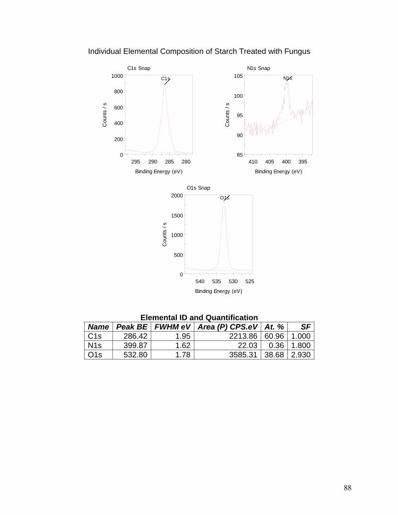
2.12 X-ray Photoelectron Spectroscopy – Surface analysis of treated starch
XPS was performed at the Surface Interface Ontario facility, using the Thermo Scientific K-
Alpha spectroscope and an Al monochromatic source (1486.6 eV). Untreated, fungus-treated
30
and protein treated starch samples were used for the analysis after being thoroughly dried and
ground. A 400 µm area was scanned. The following parameters were used: Survey: Scanned
– 150 eV pass energy, LR:Snap-mode – 150 eV pass energy and C1s HR: Scanned – 20 eV
pass energy. Peaks were shifted to place main C1s peak at 286.5 eV (ie. C-O bonding).
Charge compensation was applied.
2.13 Statistical Analysis
Statistical significance was calculated by a 2-tailed, unpaired t-test using Microsoft Excel. A
95% confidence level was used; differences with p values less than 0.05 were considered
statistically significant.
2.14 Expression and Purification of non-specific proteins
Glycerol stocks of His-tagged constructs transformed into E.coli strain BL21 were obtained
from different members of the lab. HopF3 and HopZ (effector proteins from Pseudomonnas
syringae pv phaseolicola and syringae respectively) were provided by Van Quach and
Shikimate dehydrogenase (SDH) (shikimate pathway enzyme from Arabidopsis thaliana)
was obtained from James Peek. His-SDH was used in all assays involving His-tagged
proteins unless otherwise specified.
The glycerol stocks were used to grow overnight starter cultures in 50 ml Luria-
Bertani (LB) media supplemented with appropriate antibiotics. The starters were used to
inoculate 1L LB media (with suitable antibiotics) that was incubated in a 37°C shaker until
optical density at 600nm (OD600) reached between 0.6 and 0.8. To induce protein expression,
31
isopropyl β-D-thiogalactopyranoside (IPTG) was added to a final concentration of 0.4mM.
The cultures continued to grow at 37°C for 4 hours, after which the temperature was reduced
to 16°C overnight. The bacterial cells were pelleted at 3500 rpm for 10 minutes. The cells
were resuspended in binding buffer (50mM Tris-HCl pH7.5, 5mM imidazole, 5% glycerol
and 500mM NaCl) and then sonicated on ice. A duty cycle of 90, output of 5 and 30 sec on/
30 sec off cycle was used for 10 min. 0.01% PI was added to the lysate which was spun
down at 14000 rpm at 4°C for 30 min.
Ni-NTA resin (Qiagen) (about 4ml) was applied to a glass chromatography column,
equilibrated using 2 column volumes of binding buffer and cell lysate was passed through.
The column was washed and eluted with buffer that had the same composition as binding
buffer except 30mM imidazole and 100mM imidazole respectively. 1mM EDTA was added
to the elution fraction, followed by 0.33mM DTT. The eluted protein was then dialyzed at
4°C against buffer containing 10mM Tris-HCl pH7.5, 500mM NaCl, 5% glycerol and β-
mercaptoethanol. TEV was added to the protein sample during dialysis if the His-tag was to
be cleaved. Upon cleavage of the His-tag, the protein sample was run through another Ni-
NTA column as previously described. The flow-through was collected which contained the
cleaved protein.
Pure BSA (Bio-Rad) was dissolved in Tris-HCl buffer pH 7.5 to make a 1mg/ml
stock. Appropriate volumes of the stock were used to obtain desired quantity of protein for
starch treatment.
32
3. Results
3.1 Establishing the Effect of O. ulmi on Starch
The first step in the study was to establish that O. ulmi could improve properties of
thermoplastic starch such as tensile strength (TS) and water absorption, under the conditions
used in our study. This involved culturing the fungus in presence of starch (10g/L) for 3 days
after which the starch was precipitated out to generate plastic films. Standard tensile tests
were performed on strips cut from the plastic films, using an Instron machine. The film
thickness was relatively uniform; in addition to ensure consistencies in our analysis, strips of
similar thickness +/- 0.03 were used. Repeated analysis at ambient conditions revealed that
the fungus significantly increase the maximum tensile strength of plastic, with values ranging
from 8MPa to 36MPa. This represented an increase of about 400% (figures 2, 6 and 11),
relative to the control, which was usually from 1.76MPa to 8.43MPa (tables 3, 5 and 10).
Water absorption of treated starch precipitates was assessed by a commonly used
method involving measurement of water holding capacity (WHC). The samples were soaked
in water and the increase in mass was determined after the excess water had been removed.
WHC was calculated as mass of water per unit mass of starch (g H2O/g starch). At ambient
conditions, the WHC of the untreated starch was found to be between 20.1 to 25.1g H2O/g of
starch whereas after fungus treatment, the WHC was between 0.7 to 1.3g H2O/g of starch
(table 11). The fungal treatment therefore resulted in a striking decrease in WHC of about
96% relative to the control (figures 12 and 13).
33
3.2 Effect of O. ulmi Secreted Molecules on Starch Properties
In order to test the hypothesis that the extracellular proteins of O. ulmi are mediating the
modifying effects on starch, the first consideration was whether the fungus itself was
required to produce the changes in starch or if the secreted matter was sufficient for the
observed effects. For this purpose, the fungal culture was separated into a pellet that
consisted of fungal debris (including cells and spores) and supernatant, which contained all
extracellular molecules secreted by the fungus. The pellet and supernatant were tested
separately for their effect on tensile properties of thermoplastic starch.
The untreated and fungal treated samples had a tensile strength (TS) that ranged from
1.76 to 8.43 MPa and 7 to 36 MPa respectively (table 3). The pellet and supernatant
treatment resulted in a range of TS from 4.84 to 10.24 MPa and from 24.2 to 33.17 MPa
respectively (table 3). As shown in Figure 2, the fungal culture and supernatant treatments
both resulted in a comparable increase in maximum tensile strength relative to the untreated
control, about 376 and 350% respectively (P > 0.05). By comparison, pellet treatment
resulted in less than 50% increase as compared to the control. The pellet-treated starch had
significantly inferior tensile strength (P < 0.05) relative to the fungal culture treatment.
34
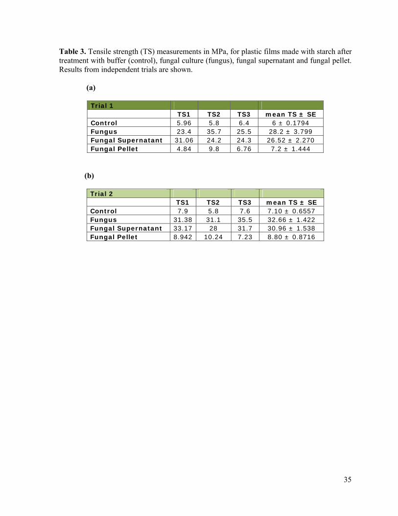
Table 3. Tensile strength (TS) measurements in MPa, for plastic films made with starch after treatment with buffer (control), fungal culture (fungus), fungal supernatant and fungal pellet. Results from independent trials are shown. (a)
Figure 2. Maximum tensile strength (TS) of plastic films made with starch treated with buffer (control), fungal culture (fungus), fungal supernatant and fungal pellet respectively. Each bar represents the mean TS in MPa (n = 3); results from two independent trials are shown.
36
3.3 Isolation of Secreted Proteins from O. ulmi
A key step in the study was to obtain proteins from the liquid culture and to determine
whether there was any difference in protein composition obtained from cultures grown with
starch compared to without it. Total protein precipitation was achieved using ammonium
sulfate precipitation. Secreted proteins isolated in the presence and absence of starch had a
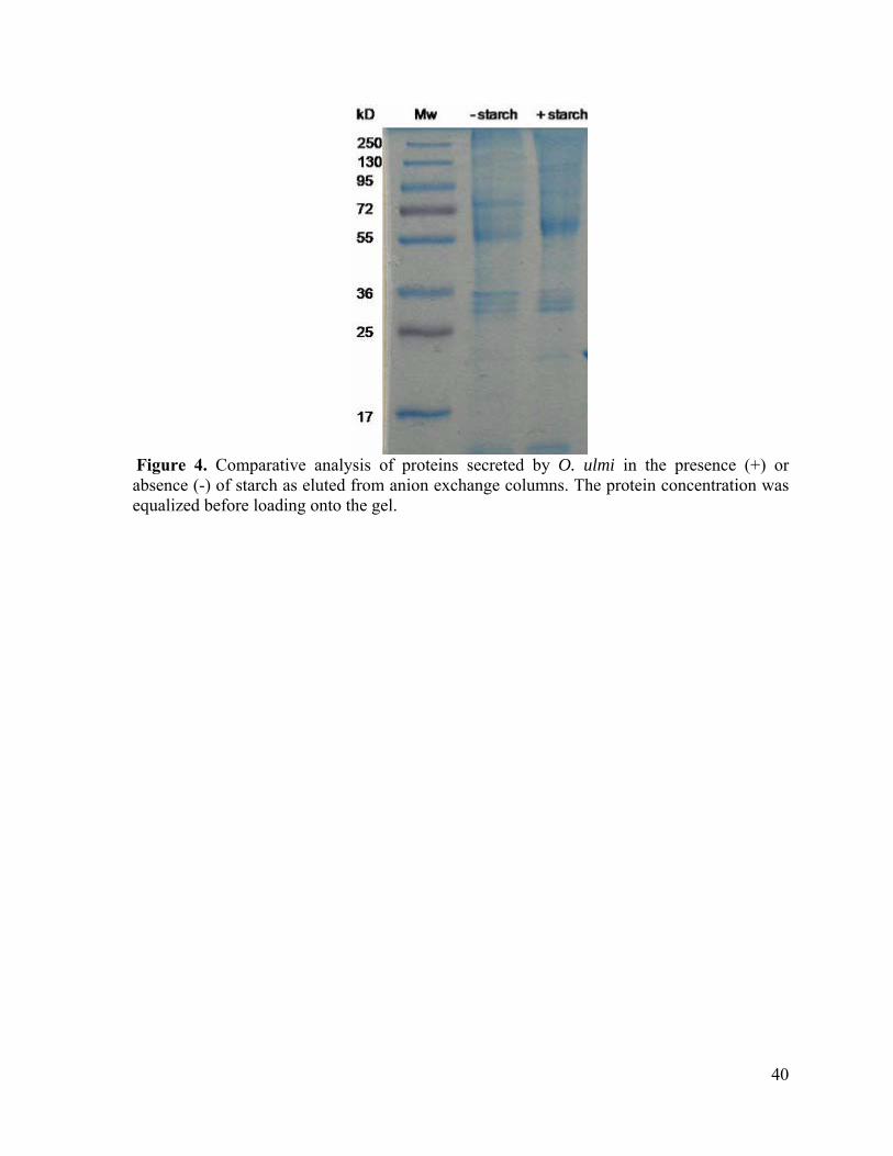
different profile as visualized on SDS-PAGE (figure 3). The presence of starch in the culture
seemed to cause a few proteins to be expressed and/or secreted that were not seen in its
absence. Bands that were approximately 20kDa (F23), 32kDa (F20) and 80kDa (F13) in size
were only seen in the presence of starch. Also, the abundance of certain proteins was seen to
increase. The SDS-PAGE suggested that starch could induce protein production and/or
secretion in O. ulmi cultures.
Proteins eluted from the column were quantified and it was found that the culture
without starch yielded about 10 times more protein than the starch-containing culture. The
proteins from the starch-containing culture were concentrated to normalize the amount
loaded onto the SDS-PAGE (figure 3). It was seen that all the proteins that were obtained by
(NH4)2SO4 precipitation were not present in elution samples from anion-exchange columns
(figures 3 and 4). The profile of eluted proteins from cultures grown with or without starch
was very similar as opposed to what was observed with (NH4)2SO4 precipitation.
3.4 Identification of Secreted Proteins
The protein bands visualized by SDS-PAGE were subjected to MALDI-TOF MS/MS. The
analysis did not generate high confidence matches to entries in the MS database; however,
37
some of the higher scoring matches are included in the table 4. The highest scoring matches
were mostly hypothetical proteins; therefore, no insight could be obtained into the function
associated with each protein. DeNovo sequencing was also performed following MS/MS, but
sequence tags generated were too short to generate any identification hits.
38
Figure 3. Comparative analysis of proteins secreted by O. ulmi in the presence (+) or absence (-) of starch in Tchernoff medium. The bands labeled F3 to 25 were subjected to MS , the “*” indicates the bands for which MS data is included in table 4.
39
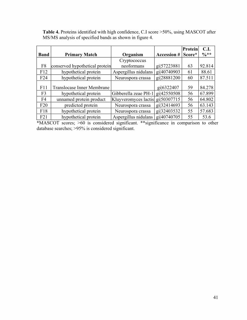
Figure 4. Comparative analysis of proteins secreted by O. ulmi in the presence (+) or absence (-) of starch as eluted from anion exchange columns. The protein concentration was equalized before loading onto the gel.
40
Table 4. Proteins identified with high confidence, C.I score >50%, using MASCOT after MS/MS analysis of specified bands as shown in figure 4.
Band Primary Match Organism Accession # Protein Score*
F12 hypothetical protein Aspergillus nidulans gi|40740903 61 88.61 F24 hypothetical protein Neurospora crassa gi|28881200 60 87.511
F11 Translocase Inner Membrane gi|6322407 59 84.278F3 hypothetical protein Gibberella zeae PH-1 gi|42550508 56 67.899F4 unnamed protein product Kluyveromyces lactis gi|50307715 56 64.802F20 predicted protein Neurospora crassa gi|32414693 56 63.143F18 hypothetical protein Neurospora crassa gi|32403532 55 57.683F21 hypothetical protein Aspergillus nidulans gi|40740705 55 53.6
*MASCOT scores; >60 is considered significant. **significance in comparison to other database searches; >95% is considered significant.
41
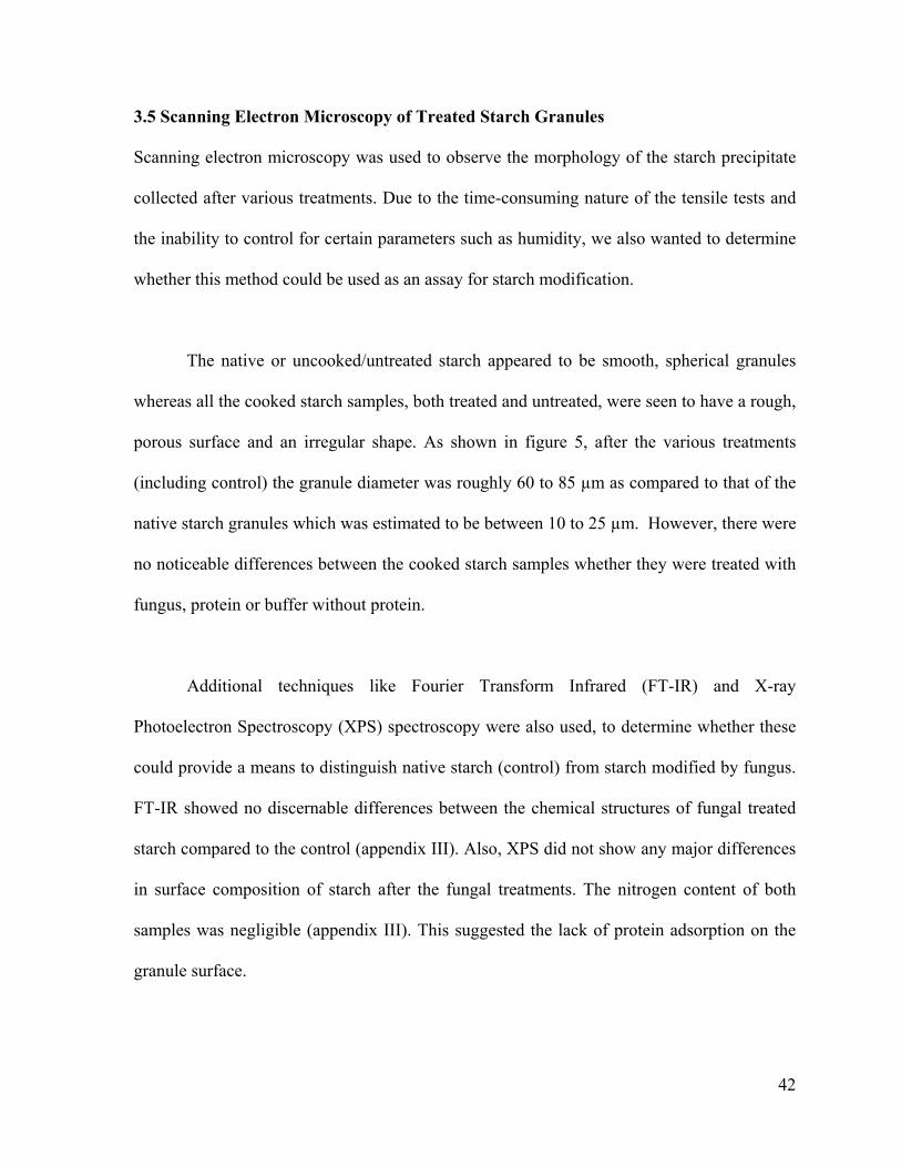
3.5 Scanning Electron Microscopy of Treated Starch Granules
Scanning electron microscopy was used to observe the morphology of the starch precipitate
collected after various treatments. Due to the time-consuming nature of the tensile tests and
the inability to control for certain parameters such as humidity, we also wanted to determine
whether this method could be used as an assay for starch modification.
The native or uncooked/untreated starch appeared to be smooth, spherical granules
whereas all the cooked starch samples, both treated and untreated, were seen to have a rough,
porous surface and an irregular shape. As shown in figure 5, after the various treatments
(including control) the granule diameter was roughly 60 to 85 µm as compared to that of the
native starch granules which was estimated to be between 10 to 25 µm. However, there were
no noticeable differences between the cooked starch samples whether they were treated with
fungus, protein or buffer without protein.
Additional techniques like Fourier Transform Infrared (FT-IR) and X-ray
Photoelectron Spectroscopy (XPS) spectroscopy were also used, to determine whether these
could provide a means to distinguish native starch (control) from starch modified by fungus.
FT-IR showed no discernable differences between the chemical structures of fungal treated
starch compared to the control (appendix III). Also, XPS did not show any major differences
in surface composition of starch after the fungal treatments. The nitrogen content of both
samples was negligible (appendix III). This suggested the lack of protein adsorption on the
granule surface.
42
a) b) c)
d) e) f)
Figure 5. Scanning electron micrographs of different types of starch granules; native (a) or treated with buffer (control) (b) fungus (c) fungal protein (d) His-SDH (non-specific protein) (e) and BSA (non-specific protein) (f).
43
3.6 Effect of Proteins on Tensile Strength of Thermoplastic Starch 3.6.1 Effect of O. ulmi Secreted Fungal Proteins It was further tested whether the proteins isolated from the supernatant had any effect on the
tensile properties of starch plastics (table 5 and figure 6). This was done by treating a
precooked starch sample (5g starch/100ml buffer) overnight, with a known amount of protein
isolated from starch-containing fungal cultures. Repeated trials carried out with fungal
extract containing 300 µg of fungal protein (0.06mg protein/g starch) resulted in TS values
ranging from 2.33 to 11.89MPa, this translated to a consistent increase of about 110%
relative to the control, which ranged from 1.76 to 4.5MPa. This was repeated with proteins
isolated from cultures without starch. The resulting tensile strength measurements ranged
from 3.24 to 10.62MPa, an increase of about 130% compared to the untreated control. The
increase caused by 0.06 mg protein/g starch treatment was intermediate to the effect of the
fungus which produced a TS between 6.54 to 18MPa and a relative increase of about 370%.
44
Table 5. Tensile strength (TS) measurements in MPa, for plastic films made with starch after treatment with buffer (control), fungus and fungal proteins isolated from cultures grown with or without starch. Results from independent trials are shown. (a) Trial 1 TS1 TS2 TS3 mean TS ± SE Control 2 1.76 1.9 1.89 ± 0.0696 Fungus 6.54 10.26 9.8 8.87 ± 1.171 Fungal Protein Extract (Starch Culture) 5.06 4.5 2.33 3.96 ± 0.8338 Fungal Protein Extract Protein (Non-starch Culture) 4.68 5.1 3.24 4.34 ± 0.5639 (b) Trial 2 TS1 TS2 TS3 mean TS ± SE Control 3.7 2.6 2.89 3.06 ± 0.3292 Fungus 13.2 18 13.83 15.01 ± 1.506 Fungal Protein Extract (Starch Culture) 6.4 8.98 6.22 7.2 ± 0.8922 Fungal Protein Extract Protein (Non-starch Culture) 9 7.8 6.18 7.66 ± 0.8186 (c)
Trial 3 TS1 TS2 TS3 mean TS ± SE Control 4.5 4 2.3 3.6 ± 0.6658 Fungus 17.8 13 14.59 15.13 ± 1.412 Fungal Protein Extract (Starch Culture) 7 6.7 11.89 8.53 ± 1.682 Fungal Protein Extract Protein (Non-starch Culture) 8.1 7.8 10.62 8.84 ± 0.8942
45
Figure 6. Maximum tensile strength (TS) of plastic films made with starch treated with buffer (control), fungal protein isolated from cultures grown with or without starch and the fungal culture. 0.06mg protein/g starch was used. Each bar represents a mean TS measurement in MPa (n = 3); results from three independent trials are shown.
46
3.6.2 Effect of Non-specific Proteins
Since fungal proteins had a favorable effect on properties of thermoplastic starch, we wanted
to test the effect of randomly selected proteins to establish whether these changes could be a
result of non-specific interaction between starch and protein. For this purpose, BSA and a
histidine-tagged protein, His-SDH, previously expressed recombinantly in our lab, were
tested. A precooked starch sample (5g starch/100ml buffer) was treated with 300µg of
protein (i.e. 0.06 mg protein/g starch). Following overnight treatment, starch was precipitated
and subjected to tensile tests. BSA caused a TS of 6.89 to 11.95 MPa whereas the His-tagged
protein resulted in TS ranging from 8.4 to 14.16 MPa (table 5). This was a respective
increase of about 65 and 100%, relative to the untreated sample (figure 7).
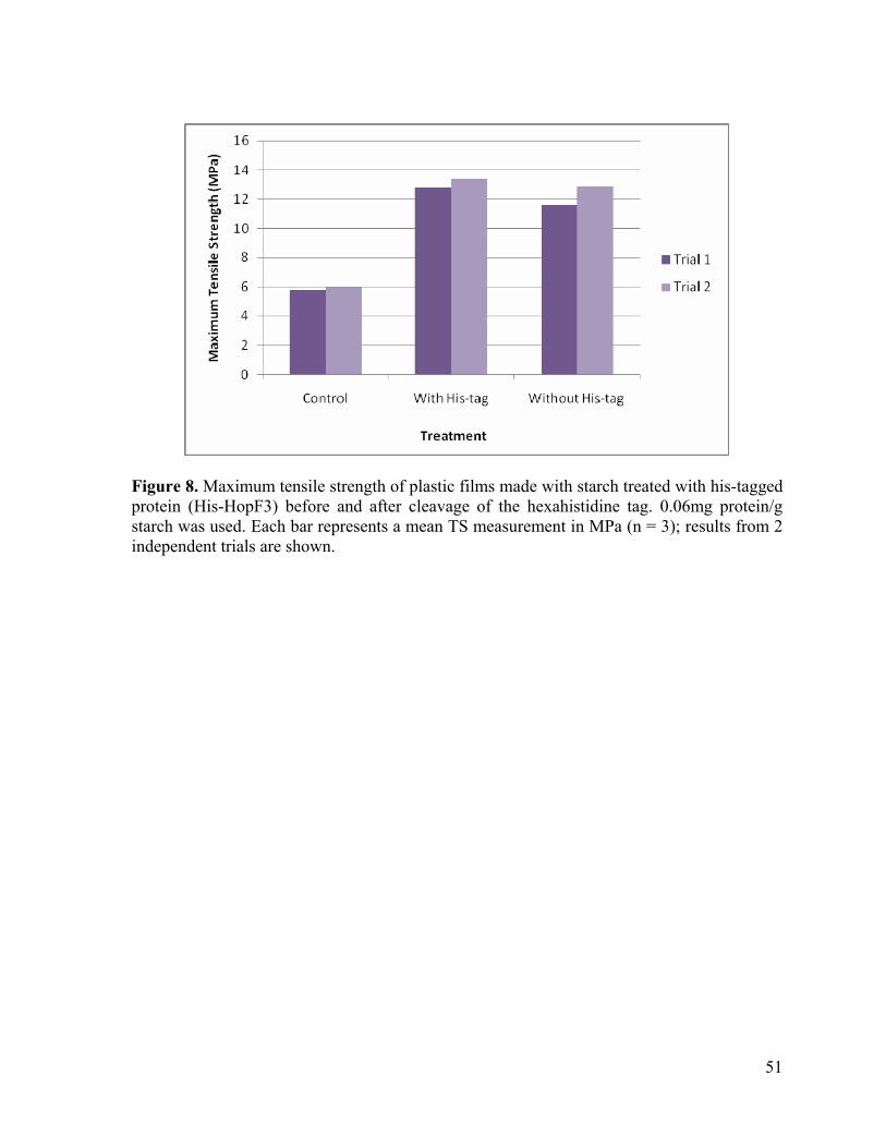
3.6.2.1 Effect of Hexahistidine Tag
It was subsequently tested whether the effect of the His-tagged proteins was due to the
imidazole ring contained in the hexahistidine tag. To determine this, starch was treated with
the intact His-tagged protein and the same protein with the His-tag cleaved off. The
subsequently made films were tested. The TS measurements for treatments with buffer, with
His-tag and without His-tag were in the ranges 5.3 to 6.3 MPa, 11.8 to 14.73 MPa and 9.7 to
16.45, respectively (table 6). Therefore, both the proteins caused a comparable relative
increase in TS of about 120% and 100% respectively (P > 0.05) (figure 7). It should be
noted that the His-tag alone could not be tested separately as there was no means to verify
whether the His-tag had been successfully isolated.
47
Table 6. Tensile strength (TS) measurements in MPa, for plastic films made with starch after treatment with buffer (control) and non-specific proteins BSA and His-SDH. Results from independent trials are shown. (a)
Trial 1 T1 T2 T3 mean T ± SE Control 4.2 4.8 4.59 4.53 ± 0.1758 BSA 6.97 7.89 9.33 8.06 ± 0.6868 His-tagged Protein 8.4 10.23 11.68 10.10 ± 0.9478
(b)
Trial 2 T1 T2 T3 mean T ± SE Control 6.2 5.46 5.34 5.67 ± 0.2689 BSA 8.76 10.2 9.26 9.41 ± 0.4221 His-tagged Protein 9.54 12 14.16 11.9 ± 1.335
(c)
Trial 3 T1 T2 T3 mean T ± SE Control 4.87 5.5 5.44 5.27 ± 0.20 BSA 7.65 6.89 11.55 8.7 ± 1.44 His-tagged Protein 13 9.45 9.17 10.54 ± 1.23
48
Figure 7. Maximum tensile strength of plastic films made with starch treated with buffer (control) and non-specific proteins, BSA and His-SDH. 0.06mg protein/g starch was used. Each bar represents a mean TS measurement in MPa (n = 3); results from three independent trials are shown.
49
Table 7. Tensile strength (TS) measurements in MPa, for plastic films made with starch after treatment with buffer (control) and His-HopF3 before and after cleavage of the hexahistidine tag. Results from independent trials are shown. (a)
Trial 1 T1 T2 T3 mean T ± SE Control 5.9 5.23 6.27 5.8 ± 0.3044 With His-tag 11.8 13 13.6 12.8 ± 0.5292 Without His-tag 9.7 13.4 11.67 11.59 ± 1.069
(b)
Trial 2 T1 T2 T3 mean T ± SE Control 6.3 5.6 6.1 6 ± 0.2082 With His-tag 12.2 14.73 13.27 13.4 ± 0.7332 Without His-tag 11.8 10.45 16.45 12.9 ± 1.817
50
Figure 8. Maximum tensile strength of plastic films made with starch treated with his-tagged protein (His-HopF3) before and after cleavage of the hexahistidine tag. 0.06mg protein/g starch was used. Each bar represents a mean TS measurement in MPa (n = 3); results from 2 independent trials are shown.
51
3.6.2.2 Role of Protein Concentration in Non-Specific Protein-Mediated Effect
It was further tested if the amount of protein used in this assay had any effect on the results
obtained. Different quantities of His-SDH protein were used in the assay; 0.3mg, 2mg and
4mg (0.03mg, 0.2mg and 0.4mg protein per g starch) were used to treat 10g starch/L. The TS
values lay in the following ranges: 10.8 to 12.34MPa, 10.54 to 11.86MPa and 10.45 to
12.3MPa respectively while the control had a TS between 8 to 8.43MPa (table 8). 0.06mg
protein produced an increase of about 38% in tensile strength compared to the control while
0.2mg and 0.4mg produced an increase of about 34% and 39% respectively (figure 9).
Therefore the treatments were statistically equivalent (P > 0.05).
3.6.2.3 Role of Protein Size on Non-specific Protein-Mediated Effect
It is conceivable that proteins with a different molecular weight would have a different effect
on the mechanical properties of starch. We therefore investigated this dependency as well.
For this purpose, three proteins with a difference of about 15-20kDa were selected to treat
starch. These were His-SDH (25kDa), His-HopZ (42kDa) and BSA (66kDa). Each of these
protein treatments led to TS values of 7.97 to 8.9MPa, 7.98 to 9MPa and 7.9 to 8.6MPa
(table 9). While all protein treatments caused an increase in tensile strength of about 40%,
38% and 35% respectively (figure 10), all treatments were statistically equivalent (P > 0.05).
This indicated that the treatment of starch with any non-specific protein might produce a
small increase in maximum tensile strength.
52
Table 8. Tensile strength (TS) measurements in MPa, for plastic films made with starch after treatment with buffer (control) and different amounts (mg/g of starch) of His-SDH as indicated. Results from independent trials are shown.
(a)
Trial 1 T1 T2 T3 mean T ± SE Control 8.156 8.32 8.43 8.302 ± 0.0761 0.03 mg 12.2 11.33 10.84 11.46 ± 0.3977 0.2 mg 11.48 11 10.89 11.12 ± 0.1811 0.4 mg 11.8 10.45 12.3 11.52 ± 0.5525
(b)
Trial 2 T1 T2 T3 mean T ± SE Control 8.1 8.02 8.35 8.16± 0.0994 0.03 mg 10.9 12 12.34 11.75 ± 0.4338 0.2 mg 10.536 11.86 10.85 11.08 ± 0.3993 0.4 mg 12 11.364 11.38 11.58 ± 0.2088
53
Figure 9. Maximum tensile strength (TS) of plastic films made with starch treated with different amounts (/10g starch) of His-SDH as indicated on the horizontal axis.. Each bar represents a mean TS measurement in MPa (n = 3); results from 2 independent trials are shown.
54
Table 9. Tensile strength (TS) measurements in MPa, for plastic films made with starch after treatment with buffer (control) and non-specific proteins of different sizes as indicated. Results from independent trials are shown.
(a) Trial 1 T1 T2 T3 mean T ± SE Buffer 6.12 6.2 6.15 6.16 ± 0.0233 25kDa 8.1 8.56 8.64 8.43 ± 0.1691 40kDa 9 8.78 9 8.93 ± 0.0739 60kDa 8.34 8.6 7.99 8.31 ± 0.1754
(b)
Trial 2 T1 T2 T3 mean T ± SE Buffer 6.2 5.9 5.9 6 ± 0.1000 25kDa 8.9 7.97 8.33 8.4 ± 0.2707 40kDa 8.76 8.1 7.98 8.28 ± 0.2425 60kDa 8.54 7.8 7.96 8.1 ± 0.2248
55
Figure 10. Maximum tensile strength (TS) of plastic films made with starch treated with proteins of different sizes as indicated on the horizontal axis. 0.4mg protein/g starch was used. Each bar represents a mean TS measurement in MPa (n = 3); results from 2 independent trials are shown.
56
3.7 Comparative Effect of Fungal Protein Extract and Non-Specific Proteins
To allow for a better comparison between the effect of proteins and fungus, a consolidated
experiment was carried out where 10g/L starch was treated with either buffer, 0.4 mg of
protein/g starch or fungus. This was especially desirable since the fungal treatment was
carried out over a period of 3 days in a sample volume of 1L with 10g starch/L whereas
protein treatment were carried out overnight with 5g starch in 100ml sample volume.
Tensile strength values from 4.3 to 7.4MPa were obtained for the control. BSA and
His-protein treatments resulted in TS values of 5.92 to 10.04MPa and 5.81 to 11.17MPa
(table 10). Fungal protein and fungus caused TS values that ranged from 19.65 to 33.44MPa
and 18.74 to 30MPa. The tensile strength measurements were plotted as percentages that
were normalized against the buffer-treated sample (control). Figure 12 is representative of
the effects of the fungus and various protein treatments on the tensile properties of starch
plastic. The fungal extract treatment resulted in about 350% increase in maximum tensile
strength and the starch from the fungal culture showed an approximately 310% increase
compared to control. Both BSA and the His-tagged protein resulted in a respective modest
increase of about 35% and 45% relative to the buffer-treated control.
57
Table 10. Tensile strength (TS) measurements in MPa, for plastic films made with starch after treatment with buffer (control), fungal culture (fungus) and fungal proteins or non-specific proteins, BSA and His-SDH (0.4 mg protein/g starch). Results from independent trials are shown. (a)
Trial 1 T1 T2 T3 mean T ± SE Control 6.2 6.13 6.19 6.17 ± 0.0219 BSA 8.03 8.3 8.1 8.14 ± 0.0806 His-tagged protein 8.198 9.3 8.97 8.82 ± 0.3366 Fungal protein 27.34 25.16 27.83 26.78 ± 0.8213 Fungus 25.21 23.722 26.03 24.99 ± 0.6764
(b)
Trial 2 T1 T2 T3 mean T ± SE Control 6.87 7.4 6.76 7.01 ± 0.1976 BSA 9.21 10.04 9.26 9.5 ± 0.2675 His-tagged protein 11.17 9.76 9.77 10.23 ± 0.4677 Fungal protein 29 33.44 32.2 31.55 ± 1.3215 Fungus 29.2 27.65 30 28.95 ± 0.6908
(c)
Trial 3 T1 T2 T3 mean T ± SE Control 4.3 4.58 4.92 4.6 ± 0.1793 BSA 6.1 7.3 5.92 6.44 ± 0.4331 His-tagged protein 7.21 5.81 6.99 6.67 ± 0.4347 Fungal protein 23.45 20.1 19.65 21.07 ± 1.1979 Fungus 21.2 18.74 22.16 20.7 ± 1.0184
58
Figure 11. Maximum tensile strength (TS) of plastic films made with starch treated as indicated on the horizontal axis. 0.4mg protein/g of starch was used. Each bar represents a mean TS measurement in MPa (n = 3); results from three independent trials are shown.
59
3.8 Temporal Effect of Fungal Protein Extract
In order to see if the effect of fungal proteins was enzymatic, we wanted to determine if the
effect of the fungal proteins on tensile properties of thermoplastic starch was time-dependent.
For this purpose, an assay was performed by treating a large volume of cooked starch with
4mg of protein/10g of starch. Starch was extracted at different time points. The resulting
starch precipitate was used to make films for tensile tests.
After 5 hours of treatment, average TS measurement was 17.84MPa. After 1, 2, 3 and
4 days of treatment, mean TS was found to be 10.71, 5.875, 17.94 and 14.1MPa respectively
while the buffer-treated control had mean TS of 3MPa. The TS measurements for all
treatments were variable nonetheless all durations caused a significant increase (P < 0.05) in
TS compared to the control. The relative increase in TS was 494, 350, 95, 498 and 370%
respectively (appendix II). It should be noted that only preliminary data for the time-course
was collected and reported here and further verification is necessary by independent
replication of the assay. However, 5 hours of treatment had the same outcome as 3 days of
treatment, which suggests that the effect of the fungal protein extract on mechanical
properties of starch plastic, might be independent of the time duration. It may also suggest
that the change in mechanical properties of starch plastic is not enzymatic. Nonetheless, this
experiment needs to be repeated in order to obtain more conclusive results.
3.9 Effect of Proteins on Water Holding Capacity of Starch
We wanted to determine if protein treatment could change water barrier properties of starch
to decrease water absorption. The water holding capacity was measured as the mass of water
60
absorbed as a function of the dry mass of starch. Taking the WHC of the untreated sample as
the maximum water that can be absorbed by starch (i.e. 100% WHC), relative percentage
WHC was calculated. Three independent trials were carried out with 300 µg of fungal
protein as well as non-specific proteins. The fungal protein treatment resulted in a WHC of
about 58% with absolute values ranging from 11.6 to 14.4 g H2O/g of starch. However this
was similar to the outcome of the non-specific protein treatments. BSA-treated starch had a
WHC between 14.3 and 15.6 g H2O/g starch (table 11), which translated to almost 69%
relative WHC. His-tagged protein (His-SDH) treatment resulted in a 67% WHC with values
ranging from 12.9 to 16.5 g H2O/g starch (figure 12).
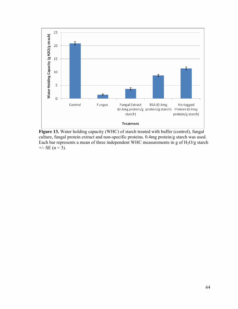
The assay was repeated with starch samples (10g/L) that had been treated with 4 mg
of each protein (0.4 mg protein/ g starch). Treatment with fungal extract, BSA and His-
tagged protein resulted in WHC values that ranged from 3.1 to 4.6, 8.1 to 9.2 and 10.5 to
12.3 g H2O/g starch, respectively (table 10). A 17% WHC was seen after treatment with
fungal extract. Both BSA and His-tagged protein caused much higher water absorption, with
a WHC of approximately 40-50% relative to the control (figure 13).
61
Table 11. Water holding capacity (WHC) of starch, precipitated from fungal culture (fungus), treated with buffer (control) and specified amounts of fungal protein, BSA and His-SDH WHC1 WHC2 WHC3 mean WHC ± SE Control 20.1 25.1 22.1 22.43 ± 1.453 Fungus 0.7 1.1 1.3 1.03 ± 0.1764 Fungal Protein (0.06mg/g starch) 11.6 14.4 13.4 13.33 ± 0.8192 Fungal Protein (0.4mg/g starch) 3.1 4.6 3.4 3.7 ± 0.4583 BSA (0.06mg/g starch) 14.3 16.4 15.6 15.43 ± 0.6119 BSA (0.4mg/g starch) 8.1 9.2 8.9 8.73 ± 0.3283 His-tagged Protein (0.06mg/g starch) 12.9 16.5 15.6 15 ± 1.082 His-tagged Protein (0.4mg/g starch) 10.5 11.5 12.3 11.43 ± 0.5207 WHC 1, 2 and 3 represent measurements from three independent trials.
62
Figure 12. Water holding capacity (WHC) of starch treated with buffer (control), fungal culture, fungal protein extract and non-specific proteins. 0.06mg protein/g starch was used. Each bar represents a mean of three independent WHC measurements in g of H2O/g starch +/- SE (n = 3).
63
Figure 13. Water holding capacity (WHC) of starch treated with buffer (control), fungal culture, fungal protein extract and non-specific proteins. 0.4mg protein/g starch was used. Each bar represents a mean of three independent WHC measurements in g of H2O/g starch +/- SE (n = 3).
64
4. Discussion Our hypothesis was that starch modification by O. ulmi, is mediated by secreted proteins.
Therefore the goal of this study was to investigate the effect of secreted proteins on starch as
well as to identify them. We observed and compared the tensile properties and water holding
capacity of untreated starch, fungus treated starch and starch treated with fungal extract with
proteins. The protein-treated sample consistently produced an improvement in plastic
properties. Even though positive identification of proteins could not be achieved, our
findings supported our hypothesis and led us to conclude that the O. ulmi extracellular
proteins can modify starch and improve plastic properties. However, our findings have also
potentiated investigation into the role of extracellular polysaccharide (EPS) in modification
of starch properties.
4.1 Establishing Effect of O. ulmi and Secreted Molecules on Starch
This study established that under our experimental conditions, O. ulmi is able to alter starch
properties such that plastic films with a significantly higher tensile strength and water barrier
properties can be obtained, relative to native starch. Once the effect of the fungus was
determined, it was investigated whether the starch modification effect, and thereby
improvement in tensile properties, could be achieved by exposing starch to the culture
supernatant (after the fungus was removed). It was seen that the culture supernatant, and
therefore, the secreted molecules, had the same effect on starch tensile properties as the
whole culture whereas the fungal pellet did not have much of an effect (figure 5). This
confirmed that the factors that mediate the modification are extracellular. Fungal exudates
have not been previously shown to improve tensile properties of thermoplastic starch as
65
demonstrated in this study. However, as described previously, fungi secrete polysaccharides
as well as lipids in culture, in addition to proteins (Wu, 2007). Therefore, it remained to be
tested whether the modifying factors were fungal proteins.
4.2 Protein Purification
To confirm the involvement of secreted proteins in the modification process, the proteins had
to first be purified from the fungal culture. Using neutral salts in a process known as “salting
out” is a well established method for protein purification. The most commonly used salt for
this is ammonium sulfate as it is cheap and sufficiently soluble. Different degrees of salt
saturation may be required to purify different proteins, depending on their surface
hydrophobicity. The propensity for aggregation and hence precipitation is directly
proportional to the degree of surface hydrophobicity (Roe, 2001). Since complete protein
precipitation was desired, 95% saturation was used. When the precipitated proteins were
dialyzed and run on an SDS-PAGE, the bands appeared diffuse. This may be ascribed to
protein size heterogeneity due to glycosylation. The majority of the secreted proteins from
filamentous fungi are known to be highly glycosylated (Oda et al., 2006).
It was suspected that starch modification may be specific to factors induced by the
starch in cultures. Proteins were therefore collected and compared from cultures grown in the
presence and absence of starch. It was interesting to see that the secretion profile was slightly
different in both cases, as seen by bands F13, 20, 21, 22 and 23 in figure 4. Attempts were
made to identify all the proteins visualized on the SDS-PAGE by mass spectrometry.
Differentially expressed proteins (figure 4) could be of particular interest since the presence
66
of starch might induce specific proteins that modify starch. However, the presence of starch
in the fungal cultures is known to induce production of starch hydrolyzing enzymes (Ventura
et al., 1995). Thus, some of the additional or more intense bands in figure 4 might be starch
hydrolyzing enzymes that could confound results.
A concern was that using ammonium sulfate precipitation was extremely time-
consuming due to the large sample volume and the high saturation of ammonium sulfate
(95%) used could cause functional denaturation of the proteins. This is especially undesirable
if the starch modification occurs through enzymatic action of these proteins. There was also a
frequent problem of starch precipitating out of solution with the protein, making the samples
viscous and therefore difficult to run on the SDS-PAGE. Ion exchange chromatography
(IEC) is another straightforward, non-denaturing method for protein purification. Most
proteins are positively charged at physiological pH; therefore, anion exchange is more useful
for isolating a wider range of proteins (Simpson, 2003). Thus IEC, using DEAE-Sepharose
(positive resin), became the method of choice for protein isolation. This reduced the risk of
functional denaturation as well as starch precipitation.
Some bands visible in the SDS-PAGE after (NH4)2SO4 precipitation (figure 3) were missing
from ion exchange elution fractions (figure 4). This was not surprising as 95% saturation of
ammonium sulfate would most likely cause total protein precipitation whereas the ion-
exchange conditions used would be more suitable for some proteins over others. The profile
of eluted proteins from cultures grown with or without starch was very similar as opposed to
what was observed with ammonium sulfate precipitation. A possible explanation could be
67
that certain proteins formed associations with starch which ware then unable to pass through
the column. The elution conditions used may not have been suitable for breaking those
interactions, thus preventing some proteins from passing through the column. Nevertheless,
this may not be a major problem since the eluted proteins were sufficient to cause a
significant increase in maximum tensile strength of thermoplastic starch. In fact, this may
suggest that the missing proteins are not essential for the desired effect on starch and they
could potentially be ruled out.
4.3. Protein Identification
Upon repeated mass spectrometric analysis by MALDI-TOF MS/MS, only a few of the
proteins were matched to existing proteins in the database with some confidence.
Furthermore, these matches were only made with hypothetical protein with unknown
functions, from various organisms. The identification process was therefore largely
unsuccessful. The major obstacle was the absence of O. ulmi proteomic information in the
present databases. Though several Expressed Sequence Tags (ESTs) for Ophiostomatoid
fungi exist in the NCBI database including 561 from O.novo-ulmi and 9589 from O.piliferum
(redundant sequences included), none of the proteins analyzed by MS were matched to them.
All hits obtained were from non-Ophiostomatoid organisms. This sort of cross-species
protein identification can be accomplished by partially aligning the analyzed protein from an
unsequenced organism to a homologous sequence of a related organism present in the
database. The previously described study by Medina et al., (2005) successfully used this
approach to identify A. flavus proteins because complete genomes sequences from other
Aspergilli were available. However, there are no Ophiostomatoid fungi with complete
68
genomic sequences and as organisms become phylogenetically distant, their homologous
gene sequences and corresponding proteins are unlikely to retain a high percent identity
(Shevchenko, 1996). Thus it is imperative to such studies that sequence information is
available for the specific organism of interest.
4.4 Non-specific Effect of Proteins on Starch
During the course of this research, we tested some random proteins to investigate whether
the effects of the fungal proteins is specific in nature. Upon testing BSA, it became apparent
that non-specific interactions might play some role in causing the effects of fungal protein.
This opened up the exciting possibility of developing an alternate method to improve starch
plastics without the complexity of O. ulmi protein identification.
The phenomenon of non-specific interaction was further tested by using histidine-
tagged proteins. The rationale was that a hexahistidine tag containing the charged imidazole
ring might further enhance physical interaction between starch and protein. Preliminary
findings showed that these proteins had a similar effect to fungal protein which was higher
than the BSA-mediated effect. It was further investigated whether the effects of the histidine-
tagged proteins were in fact dependent upon the hexahistidine residues. However, the relative
increase in tensile strength using the his-tagged protein was about the same, with or without
the his-tag (figure 8). Thus, the His-tag or imidazole ring was not responsible for the
improved tensile properties.
69
This led the investigation towards testing if the effects were dependent on the size of
protein used in the assay. This was warranted by the fact that in initial testing, two
individually tested His-tagged proteins of similar size (25-28kD) had a similar effect on
starch whereas BSA (66kD) had a lesser effect (figure 11). It was thought that perhaps a
smaller sized protein could form a more effective association with starch molecules and may
act as a crosslink between two starch molecules. The smaller size of the protein may allow
the starch molecules to remain in closer proximity, thus enabling more chances for bonding
between neighboring molecules. Furthermore, different amounts of the His-tagged protein
had a similar effect on tensile properties (figure 10). Therefore, it seems that non-specific
protein-mediated improvement observed in starch films may not be easily enhanced in an
amount or size-dependent fashion.
4.5 Specific Effect of Secreted Fungal Proteins on Starch
Although using an increasing amount of non-specific protein did not have an incremental
effect on the tensile strength of the plastic (figure 9), the maximum improvement in tensile
strength of about 450% was observed when the highest amount of fungal protein (4 mg/g of
starch) was used (figure 11). However, these 4 mg protein treatments were carried out over a
period of 3 days instead of overnight as is the case for the previous experiments with lesser
protein. Therefore an experiment was done to test the temporal effect of 0.4mg of fungal
protein/ g of starch and investigate whether treatment duration was a factor in the observed
increase in tensile strength (figure 16). Due to time constraints only preliminary data could
be collected. The data showed that the tensile strength after 5 hours was the same as that after
70
3 days of treatment. This suggested that the modification effect was not time-dependent and
therefore may not be enzymatic. However, this experiment needs to be repeated in order to
confirm the findings. Therefore, a non-enzymatic, physical interaction between starch and
protein may occur to improve starch tensile properties. This interaction may be amount-
dependent and this needs to be investigated.
The 4 mg treatment involved the maximum volume of fungal extract used in this
study and it should be noted that the volume used in the experiments varied with the amount
of protein that was desired for each treatment. Therefore, any non-protein component and its
concentration were unaccounted for in the treatments. Interestingly, the fungus is known to
produce an extracellular polysaccharide (Jeng et al., 2006) and lack of enzymatic
modification could point to a potential role of the EPS in enhancing starch properties.
Fractions eluted off the anion exchange columns always had a yellow or brownish
coloration. A darker color of the eluent coincided with more protein, as detected by Bradford
reagent. Interestingly, Binz and Canevascini (1997) describe a yellow sample being obtained
upon lacasse purification from O.novo-ulmi, using ion exchange chromatography. They
associate the yellow coloration with heavy contamination with EPS. The presence of EPS in
the eluted protein sample and its presence during fungal protein treatment could explain the
variability between treatment outcomes. It would also explain the huge increase in maximum
tensile strength upon addition of a larger volume of fungal eluted sample (hence a larger
amount of EPS) as compared to treatment with an equivalent amount of non-fungal protein.
Furthermore, Jeng et al. (2007) observed that O. ulmi strain W9, which is the strain used in
71
our study, produces the highest amount of EPS compared to O. novo-ulmi as well as other
strains of O. ulmi. It is therefore reasonable to postulate that EPS could be a key factor in
starch modification and its effects on tensile properties.
The individual effect of the EPS and the protein can be determined by separating the
two from the eluted sample after ion-exchange chromatography. The molecular weight of
EPS is estimated to be around 2-8MDa (Jeng et al., 2006), whereas most of the visualized
proteins are between 10kDa to 200kDa (around 10 orders of magnitude smaller than the
EPS), therefore their separation can be done by size exclusion. Gel filtration could be used;
however, since larger volumes might be involved, a concentrating process using a filter
membrane with a MWCO of around 300kD may be used. This would allow the two fractions
to be individually rich in either protein or EPS. The effect of these fractions can then be
tested on starch to determine which component can be used more effectively to produce the
desired properties in starch plastics.
4.6 Surface Properties of Starch
Native starch granules had a smooth surface while the buffer-treated as well as protein-
treated starch samples were swelled up, rough and porous. The porosity and increase in
granule size is therefore an effect of the gelatinization process and not the protein treatment.
Since the fungus or protein treatment did not result in any noticeable morphological
differences compared to the control, (figure 8), SEM could not be used as a means to
evaluate the modification effect.
72
Proteins were detected on SDS-PAGE after the starch precipitate from fungal culture
was washed with 10% SDS (appendix I). However, based on XPS results, it seems that
neither fungal proteins nor the other proteins tested are adsorbed onto the starch. One reason
could be that 0.06mg of protein/g of starch is an insufficient amount to be detected by XPS
even though the same amount is able to bring about a change in the physical properties of the
material. It may also be that all the starch granules are not able to form interactions with the
protein and since only a miniscule amount of the sample is scanned, the proteins remain
undetected. Another reason for not being able to detect protein on the treated starch may be
that the XPS only provides surface analysis with a 10nm depth whereas from the SEM
micrographs it is evident that the starch granules are quite rough and porous. It is possible
that the proteins used in the treatment get lodged inside the crevices of the granules and
cannot be detected. This is supported by the fact that proteins were detected using Bradford
reagent on fungus or protein-treated starch granules that were not precooked (results not
shown).
Hydrophobin assembly could occur on the surface of starch as the fungal culture
grow as well as upon treatment with fungal protein. This would explain the decrease in water
absorption (figures 12 and 13). Future investigation will be needed to confirm the
hydrophobin-mediated effect on starch. This could include isolating hydrophobins from
Ophiostoma cultures, based on their small size (about 10kD). Also, known hydrophobins
from other filamentous fungi, for example Trichoderma or Aspergillus, can be expressed and
tested for their effect on the water absorption capacity of starch. Furthermore, the fungal
73
extract should be investigated for the presence of lipids, which may cause the increase in
hydrophobicity.
One method of determining hydrophobicity is measurement of the contact angle. This
is the angle that is formed at the interface between a liquid and a solid surface. A water
droplet can be placed onto the film and the contact angle can be measured using a
goniometer. The contact angle depends on the attractive forces between the solid and the
droplet, if the film is hydrophilic the water droplet will spread out and the contact angle
would be close to zero whereas, on a hydrophobic surface, the droplet will bead up and the
contact angle will be higher. In this way the hydrophobicity of the films can be determined
quantitatively.
4.7 Water Absorption
The fungus was seen to reduce water absorption by almost 96%. A small amount of protein
(0.06 mg/g starch) led to a more modest but consistent decrease in water holding capacity of
about 40% and non-specific proteins led to a decrease of around 20-30%. Increasing the
amount of protein used for the treatment caused a further decrease in water absorption, even
for non-specific proteins; this suggested that part of the effect of fungal proteins may also be
non-specific. The fact that 83% reduction in water holding capacity occurred 0.4mg protein/g
of starch suggested that this reduction in hydrophilicity was dependent on amount of protein
used to treat starch. Presumably, as more protein molecules are available to interact with
starch, less hydroxyl groups might be available for interaction with water.
74
4.8 Limitations
The major challenge faced in this study was the lack of background information on the
subject and the lack of established protocols. The lack of sequence information has already
been discussed; moreover, very few studies have been published on the subject of proteins
secreted by Ophiostoma; these include Binz and Canevascini (1997) and Wu et al (2007).
However, these studies involved isolation and characterization of specific proteins like
laccasses and serine proteases respectively and did not take a holistic proteomic approach.
Also, we have no current knowledge on the manner in which O. ulmi modifies starch, and
therefore, no reasonable predictions can be made regarding the nature of proteins that may be
involved.
It is curious that despite its ability to produce hydrolyzing enzymes, Ophiostoma ulmi
is able to improve certain properties of starch instead of breaking it down completely. One
possibility is that the growth medium contained glucose, which would presumably be more
readily absorbed than starch, thereby reducing the need for starch degradation. Another
reason may be that O. ulmi is like certain other filamentous fungi that retain some major
hydrolyzing enzymes like α-amylases and β-glucosidases in their cell wall and do not secrete
them into liquid culture (Oda et al., 2006).
4.8.1 Protein Yield
Small amounts of fungal proteins were used for the treatments; this was mainly due to low
protein recovery from purification procedures. This was not a major concern for MS analysis
as only minute quantities of protein were required. However for the starch treatment, larger
75
quantities of protein were needed. Initial protein concentration was also low and the proteins
in the cultures could not be detected with Bradford reagent or spectrometry ( = 280nm).
Concentrating the culture supernatant repeatedly enabled a rough estimation of the total
protein content in the 1 liter culture to be about 20-30mg on average. The purification
methods used resulted in further reduced amounts of protein. The ammonium sulfate method
involves several different steps and each subsequent step can lead to protein loss, while the
anion exchange method is not specific and therefore is likely to have a diminished yield. In
fact, the highest amount of total protein obtained from a 1L culture off the column was about
8 mg. For future studies and large-scale application, initial protein yield or protein secretion
capacity of the fungus should be improved (discussed later in 4.10.2).
4.8.2 Sources of Variance in Tensile Test Data
Several problems were faced when trying to optimize the solution casting and testing
procedure for the plastic films. These included air bubbles entrapped within the films,
shrinkage and cracking as well as brittleness while cutting specimens for testing. Many of
these were resolved over the course of the study for example using shear to reduce viscosity
allowed air bubbles to be removed from the casting mixture completely while adjusting
drying time and temperature (reducing temperature to 50C and drying for longer periods)
reduced issues with cracking and brittleness. This reduced in-sample variance nonetheless
some issues still remained.
Atmospheric conditions like relative humidity (RH) could not be regulated at the
instron facility or during the drying process. In order to effectively minimize variance both
76
within replicates of one sample and between different samples, the RH must be controlled.
This is especially true for thin plastic films because even small fluctuations in RH could
upset the moisture equilibrium within the film. While the data collected in this study are a
good means for preliminary evaluation, any further research done in this area, particularly for
industrial application, should take this into consideration. This may be achieved by storing
films in a controlled humidity chamber prior to testing and ideally performing the tests at a
facility that also has controlled humidity. Furthermore, the RH conditions used should be in
accordance with specific application intended for the material.
A phenomenon known as anisotropy may also lead to variance in tensile strength
measurements. Anisotropy refers to the lack of homogenous directionality or orientation of
starch molecules within the film. If the films are anisotropic, or if test specimens are cut in
different directions,, the mechanical properties and measurements may not be accurate
(Wang, J., Lu, Y., Yuan, H and Dou, P., 2008). It is difficult to control anisotropy in manual
casting procedures therefore, mechanical methods like extrusion casting or compression
molding would allow for more homogenous, isotropic films. Such methods can also
eliminate residual starch granules that are sometimes left insoluble and can affect tensile
measurements (van Soest and Vliegenthart, 1997). Furthermore, high glycerol content was
used in this study (45%) to prevent brittleness and facilitate film handling and mechanical
casting methods may also reduce the amount of glycerol needed. Low plasticizer content
leads to higher tensile strength (Thunwall, Boldizar and Rigdahl, 2006)
77
Phase separation, which entails formation of separate amylose and amylopectin-rich
regions within a film, can also result in inconsistencies in strength and barrier properties
(Paes, Yakimets and Mitchell, 2008). Linear amylose chains can intertwine more effectively
by hydrogen-bonding and crystallization to form a cohesive matrix with high strength and
stiffness whereas, amylopectin cannot form such strong bonds due to its branched nature and
leads to more flexible structures (van Soest and Vliegenthart, 1997). Therefore, use of either
pure amylose or amylopectin would result in more consistent properties. Starches with a high
amylopectin such as that from tapioca result in lower strength properties while amylose rich
starches are reported to have higher tensile strength (Paes, Yakimets and Mitchell, 2008).
The use of the polytron to homogenize starch mixtures, may also explain the large standard
deviation in data since high shear rates have been shown to cause data scattering and reduced
reproducibility (Paes, Yakimets and Mitchell, 2008).
4.9 Suggestions for Improved Protein Identification
This study has implicated O. ulmi proteins in starch modification and has further necessitated
their identification. MALDI-TOF MS is reported as being most effective when the genome
sequence of the organism is known (Medina, et al, 2005). The proteins in the complex fungal
extracts might be more successfully identified using ion-trap MS/MS. This is a useful
method for microorganisms that do not have a completely sequenced genome (Oda et al.,
2006). It has also been demonstrated that LC-MS/MS is efficient in high-throughput protein
identification in samples where limited genomic sequence data are present and identifications
are based on databases containing homologous protein sequences (Medina et al., 2005).
78
Alternatively, fractionation of isolated protein samples would allow for less complex
protein mixtures to be used for starch treatment. A comparative analysis of the effects of
different fractions could simplify the identification process if only one or few fractions can
bring about the desired effects. Reducing the complexity of sample could increase the
success of positive identification of proteins. Also, the individual proteins in such fractions
can be subjected to N-terminal sequencing using degenerate primers. This was the approach
used by Wu et al. (2006) to clone an α-amylase gene from O.floccosum. The sequences
obtained could be used to individually express the proteins which could then be tested for
their effect on starch. This could help bypass the major issue of O. ulmi being unsequenced.
However, this may not be very easy given that preliminary attempts to fractionate proteins by
using different ionic strengths for elution failed. Even when different saturation levels of
(NH4)2SO4 were used, there was significant overlap between the proteins found in each
fraction (appendix I). Gel filtration could be considered as it would enable separation of
proteins based on their size and will allow for fractions with more or less individual proteins
to be collected. The samples would have to be concentrated beforehand given that only small
volumes can be processed by this method.
4.10 Future Directions and Considerations for Large-Scale Application
Once the gene sequence is determined, proteins of interest can be expressed in appropriate
hosts, in order to investigate function and activity through kinetic and structural studies.
Furthermore, the biochemical properties of the amino acid residues that make up these
proteins will give insight into the type of interaction and/or bonding that may occur between
79
them and starch. Ultimately, the proteins may be engineered to produce more robust forms,
or new proteins can be designed such that they are more suitable for industrial processes.
4.10.1 Overexpression of Proteins
Protein identification strategies have been discussed earlier but another foreseeable challenge
in accomplishing the above is finding a suitable host to express the O. ulmi proteins. E.coli is
the most commonly used host for recombinant overexpression of proteins; however, fungal
proteins are reported to have best yields when recombinantly expressed in fungal hosts. For
example, secretion of Rhizomucor miehei aspartic proteinase was up to 3g/l in A. oryzae in a
controlled fermentation (Christensen et al., 1988). Given that fungal proteins will have
several unique post-translational modifications (PTMs), E.coli may not be a viable option. In
this event, yeast could be considered, as it is a well-established industrial microorganism and
can be used conveniently and cost-effectively for industrial production of enzymes (Ahmed
et al., 2009). In light of recent literature, filamentous fungi themselves or even Ophiostoma
can be explored (Wu et al., 2006). T. reesei, A. niger and A. oryzae are already being used as
fungal expression systems while Ophiostoma is in its earlier stages of being developed as a
recombinant host (Nevalainen et al., 2005 and Wu et al., 2006). Ahmed et al. (2009)
reviewed various studies on recombinant expression of xylanases in different expression
systems; E.coli did not give optimal expression of the enzyme while yeast and filamentous
fungi were most effective. As mentioned previously, dominant enzymes secreted by
Ophiostoma spp include proteases, lipases and amylases (Brush et al., 1999; Gao and Breuil,
1998; Wu et al., 2006). Genes encoding these efficiently secreted proteins provide a potential
source for strong promoters for high level gene expression which is an essential requirement
80
for an efficient expression system. Furthermore, Ophiostoma can be cultivated on cheap
medium, thus reducing the cost for large-scale production of commercially important
proteins (Wu et al., 2006).
4.10.2 Improving Protein Secretion
While efforts for protein identification are ongoing, strategies to improve protein secretion
would be helpful especially for large-scale use. It has been found that filamentous fungi
primarily secrete proteins through their hyphae at actively growing tips (Moukha et al.,
1993). Lee et al. (1998) reported an increase in the amount of extracellular proteins in
response to a mutation in N. crassa that caused increased growth surface area. Furthermore,
filamentous fungi have been found to secrete quantities of enzymes in solid-state that far
exceed submerged cultures, for example A. oryzae (Oda et al., 2006). This is probably due
to the fact that dimorphic fungi grow exclusively with mycelia on solid media and are
therefore able to secrete more protein through their hyphae (Wu, 2007). Therefore, if
mycelial growth of the fungus is encouraged, protein secretion could be improved. One
method to improve protein secretion could be to grow the fungus on solid media instead of
liquid culture. Another way is to alter the contents of the growth medium; for example use of
nitrogen sources like ammonium, asparagine and arginine promotes mycelial growth
whereas, proline is known to lead to yeast-like growth (Wu, 2007). The presence of calcium
has also been shown to be required for hyphal growth (Gadd and Brunton, 1992).
4.11 Conclusion
The results from this study show that extracellular proteins from O. ulmi can be used to
improve tensile strength and water barrier properties. The fungal protein extract appeared to
81
act in a specific, time-independent manner in order to mediate these effects and did not
require starch for induction. These findings are novel in that (to the best of our knowledge)
fungal exudates have not been used for improvement of tensile strength of thermoplastic
starch. This study is crucial to our understanding of how O. ulmi can be used in a cost-
effective and efficient method for the production of starch plastics with enhanced properties.
The secreted proteins and their effect on starch should be further investigated and other
molecules secreted by O ulmi should also be studied.
82
Appendix I
Figure 14. Coomassie Blue stained SDS-PAGE of proteins removed from starch, using 10% SDS, after fungal treatment.
Figure 15. Silver-stained SDS-PAGE of proteins from fungal cultures grown with starch, fractionated using 30%, 60% and 95% saturation of ammonium sulfate saturation.
83
Appendix II
Figure 16. Tensile strength measurement of plastic films made with starch treated with fungal protein extract for various time durations as indicated on the horizontal axis. 0.4mg protein/g of starch was used. Each bar represents a mean TS measurement in MPa (n = 3); results from a single trial are shown.
The measurements used to generate the above graph are as follows:
Trial 1 TS1 TS2 TS3 mean TS ± SE
Control 2.21 3.14 3.67 3.01 ± 0.4267 5 hours 17.32 18.22 17.97 17.84 ± 0.2682 1 Day 10.57 10.5 11.07 10.71 ± 0.1795 2 Days 6.35 5.52 6.12 5.10 ± 0.2131 3 Days 18.87 17.75 17.21 17.94 ± 0.4889 4 Days 14.9 13.16 14.25 14.10 ± 0.5076
84
Appendix III
FT-IR
Figure 17. FT-IR spectra of native starch and fungus-treated starch showing only one additional peak in the native starch spectrum indicating CO2.
XPS XPS: Thermo Scientific K-Alpha Al monochromatic source (1486.6 eV) Area: 400 µm Survey: Scanned – 150 eV pass energy LR: Snap-mode – 150 eV pass energy C1s HR: Scanned – 20 eV pass energy Peaks shifted to place main C1s peak at 286.5 eV (ie. C-O bonding “C2”) Charge Compensation applied The spectra shown are in the following order:
1. Starch treated with buffer (control) 2. Starch treated with fungus 3. Starch treated with protein
85
Overall Elemental Spectrum of Control Treated with Buffer
0.00E+00
2.00E+04
4.00E+04
6.00E+04
8.00E+04
1.00E+05
1.20E+05
1.40E+05
1.60E+05
0100200300400500600700800900100011001200
Cou
nts
/ s
Binding Energy (eV)
Survey1 Scan, 1 m 3.1 s, 400µm, CAE 150.0, 1.00 eV
C1s
N1s
O1s
86
85
Individual Elements for Control Starch Treated with Buffer
0
100
200
300
400
500
600
280285290295
Cou
nts
/ s
Binding Energy (eV)
C1s Snap
65
70
75
80
85
395400405410
Cou
nts
/ s
Binding Energy (eV)
N1s Snap
N1s
0
200
400
600
800
1000
1200
525530535540
Cou
nts
/ s
Binding Energy (eV)
O1s Snap
O1s
Elemental ID and Quantification Name Peak BE FWHM eV Area (P) CPS.eV At. % SF C1s 286.53 1.99 1690.57 64.95 1.000 N1s 399.96 1.38 22.67 0.51 1.800 O1s 532.97 1.83 2294.95 34.54 2.930
Overall Elemental Composition of Starch Treated with Fungus
0.00E+00
2.00E+04
4.00E+04
6.00E+04
8.00E+04
1.00E+05
1.20E+05
1.40E+05
1.60E+05
1.80E+05
2.00E+05
2.20E+05
0100200300400500600700800900100011001200
Cou
nts
/ s
Binding Energy (eV)
Survey1 Scan, 1 m 3.1 s, 400µm, CAE 150.0, 1.00 eV
C1s
N1s
O1s
87
Individual Elemental Composition of Starch Treated with Fungus
0
200
400
600
800
1000
280285290295
Cou
nts
/ s
Binding Energy (eV)
C1s Snap
C1s
85
90
95
100
105
395400405410
Cou
nts
/ s
Binding Energy (eV)
N1s Snap
N1s
0
500
1000
1500
2000
525530535540
Cou
nts
/ s
Binding Energy (eV)
O1s Snap
O1s
Elemental ID and Quantification Name Peak BE FWHM eV Area (P) CPS.eV At. % SF C1s 286.42 1.95 2213.86 60.96 1.000 N1s 399.87 1.62 22.03 0.36 1.800 O1s 532.80 1.78 3585.31 38.68 2.930
88
Overall Elemental Composition of Starch Treated with Fungal Protein Extract
0.00E+00
2.00E+04
4.00E+04
6.00E+04
8.00E+04
1.00E+05
1.20E+05
1.40E+05
1.60E+05
1.80E+05
2.00E+05
0100200300400500600700800900100011001200
Cou
nts
/ s
Binding Energy (eV)
Survey1 Scan, 1 m 3.1 s, 400µm, CAE 150.0, 1.00 eV
C1s
N1s
O1s
90
Individual Elemental Composition of Starch Treated with Fungal Proteins
0
200
400
600
800
1000
280285290295
Cou
nts
/ s
Binding Energy (eV)
C1s Snap
C1s
78
80
82
84
86
88
90
395400405410
Cou
nts
/ s
Binding Energy (eV)
N1s Snap
N1s
0
500
1000
1500
2000
525530535540
Cou
nts
/ s
Binding Energy (eV)
O1s Snap
O1s
Elemental ID and Quantification Name Peak BE FWHM eV Area (P) CPS.eV At. % SF C1s 286.47 1.89 1951.90 59.92 1.000 N1s 400.08 0.92 8.93 0.16 1.800 O1s 532.85 1.80 3319.95 39.92 2.930
91
References
Aguileta, G., Hood, M. E., Refregier, G and Giraud, T., (2008). Genome evolution in plant pathogenic and symbiotic fungi. Advances in Botanical Research 49:151-
193. Ahmed, S., Riaz, S and Jamil, A., (2009). Molecular cloning of xylanases: an overview.
Appl Microbiol Biotechnol 84:19–35. Atwell, W.A., Hood, L. F., Lineback, D. R., Varriano-Marston, E., and Zobel, H. F.,
(1988). The Terminology and Methodology Associated with Basic Starch Phenomena. Cereal Foods World 33:306-311.
Bernier et al., (2004). The Canadian Ophiostoma Genome Project. Invest Agrar: Sist
Recur For 13 (1), 105-11. Binz, T and Canevascini,G., (1997). Purification and Partial Characterization of the
Extracellular Laccase from Ophiostoma novo-ulmi. Current Microbiology 35: 278–281
Boels I.C., Ramos A., Kleerebezem M., de Vos W.M., (2001). Functional analysis of the
Lactococcus lactis galU and galE genes and their impact on sugar nucleotide and exopolysaccharide biosynthesis. Appl Environ Microbiol. 67(7):3033-40.
Bouws, H., Wattenberg, A and Zorn, H (2008) Fungal scretomes-nature’s toolbox for
white technology. Appl Microbial Biotechnol, 80:381-388. Bradford, M. 1976 "A Rapid and Sensitive Method for the Quantitation of Microgram
Quantities of Protein Utilizing the Principle of Protein-Dye Binding" Anal. Biochem. 72:248-254.
Brush, T.S., Chapman, R., Kurzman, R. and Williams, D.P. (1999) Purification and
characterization of extracellular lipases from Ophiostoma piliferum. Bioorg. Med. Chem. 7: 2131-2138.
Cherry, J.R. and Fidantsef, A.L. (2003) Directed evolution of industrial enzymes: an
update. Curr. Opin. Biotechnol. 14: 438-443. Christensen, T., Woeldike, H., Boel, E., Mortensen, S.B., Hjortshoej, K., Thim, K. and
Hansen, M.T. (1988) High level expression of recombinant genes in A. oryzae. Biotechnology 6: 1419-1422
Cornish, E.H., (1987). Materials and their Designer. NY. USA: Cambridge University
J. (1989) Protein secretion in Aspergillus niger. In Hershberger, C.L., Queenerand, S.W. and Hegeman, G. (Eds.), Genetics and Molecular Biology of Industrial Microorganisms. (pp, 295-300). Washington, DC: American Society for Microbiology
Flieger, M., Kantorova, M., Prell, A., Rezanka, T and Votruba, J., (2003). Biodegradable Plastics from Renewable Sources. Folia Microbiol. 48(1):27-44. Gadd, G.M. and Brunton, A.H. (1992) Calcium involvement in dimorphism of
Ophiostoma ulmi, the Dutch Elm disease fungus, and characterization of calcium uptake by yeast cells and germ tubes. J. Gen. Microbiol. 138:1561-1571.
Gao, Y. and Breuil, C. (1998) Properties and substrate specificities of an extracellular
Lipase purified from Ophiostoma piceae. World J. Microbiol. Biotechnol.14: 421-429.
Griffiths, W and and Wang, Y., (2009) Mass spectrometry: from proteomics to
metabolomics and lipidomics. Chem. Soc. Rev. 88:1882-1896. Grinyer, J., Kautto, L., Traini, M., Te’o, Junior, Bergquist, P. and Nevalainen, H. (2007)
Proteome mapping of the Trichoderma reesei 20S proteasome. Curr. Genet. 51:79-88.
Gross, R.A. and Kalra, B., (2002) Biodegradable Polymers for the Environment. Science.
297:803. Harding, K.G., Dennis, J.S,, von Blottnitz, H. and Harrison, S.T. (2007) Environmental
analysis of plastic production processes: comparing petroleum-based polypropylene and polyethylene with biologically-based poly-beta-hydroxybutyric acid using life cycle analysis. J Biotechnol. 130(1):57-66.
Heudorf, U., Mersch-Sundermann, V., Angerer J., (2007) Phthalates: toxicology and
94
exposure. Int J Hyg Environ Health. 210(5):623-34. Herrero-Martinez, J.M., Schoenmakers, P.J. and Kok, W.T., (2004). Determination of the
amylose-amylopectin ratio of starches by iodine-affinity capillary electrophoresis. Journal of Chromatography A, 1053:227-234.
Huang, C. et al., (2006). Production, characterization and mechanical properties of Starch
Modified by Ophiostoma spp. BioResources. 1(2): 257-269. Hui, Y.H. (2006) Handbook of Food Science Technology and Engineering. Volume 1
Boca Raton, Florida: CRC Press. Jeng, R et al., (2007). Starch-like exopolysaccharide produced by the filamentous fungus
Ophiostoma ulmi and O. novo-ulmi. For. Path. 37:80–95. Keon, J.P.R., Byrde, R.J.W., Cooper, R.M., (1990). Some aspects of fungal enzymes that
degrade cell walls. In: Fungal Infection of Plants. Ed. By Pegg, G.F., Ayres, P.G. Cambridge: Cambridge University Press: 133-158.
Kim, Y., Nandakumar, M.P. and Marten, M.R. (2007) Proteomics of filamentous fungi.
Trends in Biotechnology, 25(9):395-400. Laemmli, U.K. (1970). Cleavage of structural proteins during the assembly of the head of
bacteriophage T4. Nature 227 (5259): 680–685. Lee, C et al., (2007). Enzymatic Synthesis and Properties of Highly Branched Rice starch
amylose and amylopectin cluster. Journal of Agricultural and Food Chemistry. 56(1): 126-131.
Lee, I.H., Walline, R.G. and Plamann, M., (1998). Increased secretion of extracellular
proteins. Molecular Microbiology. 29(1): 209-218. Lozano, J.E. (2000). Food Preservation and Technology Series: Trends in Food
Engineering, Pennsylvania, USA: CRC Press. Lutzoni, F., (2004) Assembling the Fungal Tree of Life: Progress, Classification, and
Evolution of Subcellular Traits. American Journal of Botany 91(10): 1446-1480. MacDonald, T.H. (2009) Removing the Barriers to Global Health Equity. Oxon, UK:
Radcliffe Publishing Machovic, M and Janecek, S., (2006) Starch-binding domains in the post genomic era.
Cell. Mol. Life Sci. 63: 2710–2724. Medina, M.L., Haynes, P.A., Breci, L. and Francisco, W.A., (2005) Analysis of secreted
proteins from Aspergillus flavus. Proteomics 5: 3153-3161.
95
Meeker J.D., Sathyanarayana S., Swan S.H., (2009) Phthalates and other additives in
plastics: human exposure and associated health outcomes.Philos Trans R Soc Lond B Biol Sci. 27;364(1526):2097-113.
Mooney, B.P., (2009). The Second Green Revolution? Production of Plant-based
Biodegradable Plastics. Biochemistry Journal. 418:219-232. Moukha, S.M., Wösten, H.A., Asther, M., Wessels, J.G., (1993) In situ localization of the
secretion of lignin peroxidases in colonies of Phanerochaete chrysosporium using a sandwiched mode of culture. J Gen Microbiol. 139(5):969-78.
Nawarth, C., Poirer, Y., and Somerville, C., (1995) Plant Polymers for Biodegradable Plastics: Cellulose, Starch and Polyhydroxyalkanoates. Molecular Breeding 1: 105-122. Oda, K., Kakizono, D., Yamada, O., Iefuji, H., Akita, O and Iwashita, K. (2006)
Proteomic analysis of extracellular proteins from Aspergillus oryzae grown under submerged and solid-state culture conditions. 72(5):3448-3457.
Paes, S. S., Yakimets, I., and Mitchell, J.R. (2008) Influence of gelatinization process on functional properties of cassava starch films. Food Hydrocolloids 22:788-797
Ratanyake, W.S. and Jackson, D.S., (2009) Starch Gelatinization. Advances in Food and
Nutrition Research, Volume 55 Chapter 5 Roe, S., (2001) Protein purification techniques: a practical approach. 2nd ed. New York:
Oxford University Press. pg 138.
Roos, Y.H., (1995) Phase Transitions in Food. San Diego, CA: Academic Press.
Scheffer, R.J., Liem, J.I., Elgersma, D.M. (1987) Production in vitro of phytotoxic
compounds by non-aggressive and aggressive isolates of Ophiostoma ulmi, the Dutch elm disease pathogen. Physiological Molecular Plant Pathology 30:321-325.
Scholtmeijer, K., Wessels, J.G.H.,Wösten, H.A.B., (2001). Fungal hydrophobins in
medical and technical applications. Appl Microbiol Biotechnol 56:1–8
Shevchenko A., Wilm M., Vorm O., and Mann M., (1996). Mass spectrometric sequencing of proteins from silver-stained polyacrylamide gels. Anal Chem 68:850-8.
Simpson, R.J., (2003). Purifying Proteins for Proteomics: A Laboratory manual. Cold Spring Harbor, New York: Cold Spring Harbor Press. Page 123.
Stringer, M. A. and Timberlake, W. E., (1993) Cerato-ulmin, a Toxin Involved in Dutch Elm Disease, Is a Fungal Hydrophobin. Plant Cell 5:145-146.
Szilvary, G.R., Nakari-Setala, T and Lindar, M.B. (2006) Behavior of Trichoderma reesi
hydrophobins in solution:Interactions, dynamics, and multimer formation. Biochemistry 45(28):8590-8598.
Takai, S. (1974) Pathogenicity and cerato-ulmin productions in Ceratostomella ulmi.
Nature 252:124-126. Tang, H., Mitsunaga, T., and Kawamura, Y., (2006) Molecular Arrangement in Blocklets
and Starch Granule Architecture. Carbohydrate Polymers 63:555-560.
Tchernoff, V., (1965). Methods for screening and for the rapid selection of elms for resistance to Dutch elm disease. Acta bot neerl. 14: 409–452.
Temple, B., Horgen, P.A. (2000) Biological roles for ceratoulmin a hydrophhobin
secreted by the elm pathogens, Ophiostoma ulmi and O. novo-ulmi, Mycologia 92:1-9.
Thunwall, M, Boldizar, A and Rigdahl, M (2006). Compression molding and tensile
properties of thermoplastic potato starch materials. Biomacromolecules 7:981-986 van Beilen, J.B. and Poirier, Y., (2007) Prospects for Biopolymer Production in Plants.
Adv Biochem Engin/Biotechnol. 107:133-151. van Soest, J. J. G., and Vilegenthart, J. F.G., (1997) Crystallinity in Starch Plastics:
Consequences for Material Properties. TIBTECH 15:208-212. Ventura, L., González-Candelas, L. Pérez- González, J.A. and Ramón, D. (1995)
Molecular cloning and transcriptional analysis of the Aspergillus terreus gla1 gene encoding a glucoamylase. Appl. Environ. Microbiol. 61:399-402.
Wang, J., Lu, Y., Yuan, H and Dou, P. (2008). Crystallization, Orientation Morphology,
and Mechanical Properties of Biaxially Oriented Starch/PolyvinylAlcohol Films. Journal of Applied Polymer Science, 110:523–530
Whistler, R.L., BeMiller, J.N. (2009) Starch: Chemistry and Technology. New York,
NY:Academic Press. Yang, J-S, Kang, S-Y and Lee, D.N., (1997). Changes in the Tensile Strength and
97
98
Fracture Mode with Thickness of Electrodeposited Copper Foil. Metal and Materials. 3(2):130-136.
Wu, C (2007) Developing Ophiostoma floccosum as a novel expression system. (Doctoral Dissertation, Macquarie University, 2007.) Australian Digital Thesis
Program. Wu, C., Xu, Q., Liu, F and Nevalainen, K.M.H., (2007) Activity-based identification of
secreted serine proteases of the filamentous fungus, Ophiostoma. Biotechnol. Lett. 29:937-943.
Yu J, Chen L.X., (2008) The greenhouse gas emissions and fossil energy requirement of
bioplastics from cradle to gate of a biomass refinery. Environ Sci Technol. 42(18):6961-6.