27 INTRODUCTION Clover (Marsilea crenata Presl.) is a type of aquac plant that is widely distributed in Southeast Asia and grows wild in rice fields, dams, waterways, guers, and areas with standing water (Ma’arif & Agil, 2018). Although it has been known and used for decades by the people of Surabaya, the existence of M. crenata is now very rare, even though M. crenata has excellent potenal to be developed into tradional Indonesian medicine. Several studies have been carried out on M. Crenata and its leaves are known to contain saponin, terpenoids, steroids, polyphenols, and anoxidant compounds (Yacoeb, Arifin, Sulisono & Krisono, 2010; Nurjanah & Abdullah, 2010). The ethanol extract of M. crenata leaves has been tested through the radioimmunoassay (RIA) technique and shows that the concentraon of estrogen-like substance detected is quite high (Laswa, 2011). The results of in vivo acvity ARTICLE INFO ABSTRACT Arcle History Received October 21, 2019 Revised Dedember 27, 2019 Accepted January 5, 2020 Keywords Marsilea crenata Presl. Isolaon Terpenoid Compounds Doi 10.22219/farmasains.v4i2.10717 Isolaon, idenficaon, and structure elucidaon of terpenoid compounds from an n-hexane extract of Marsilea crenata Presl., had been done. The leaves of M. crenata was extracted using n-hexane solvent. The extract then separated with vacuum column chromatography and open column chromatography to obtain the isolate. Furthermore, the isolate was idenfied and elucidated using UV-Vis, FT-IR, 1 H-NMR, 13 C- NMR, and 2D- NMR (COSY, HMQC, and HMBC). The idenficaon and elucidaon of the isolated structure from an n-hexane extract of M. crenata leaves indicate the isolate was a pentacyclic triterpenoid. Research Article Isolation of terpenoid compound of n-hexane extract of Marsilea crenata Presl. Burhan Ma’arif [1]* , Mangestuti Agil [2] , Retno Widyowati [2] 1 Department of Pharmacy, Faculty of Medicine and Health of Science, Islamic State University Maulana Malik Ibrahim, Malang, Indonesia. 2 Department of Pharmacognosy and Phytochemistry, Faculty of Pharmacy, Airlangga University, Surabaya, Indonesia * Email: [email protected] Phone : +62 813 3555 5725 FARMASAINS: JURNAL FARMASI DAN ILMU KESEHATAN Volume 4, Number 2, 2019. p-ISSN : 2086-3373 | e-ISSN : 2620-987X https: ejournal.umm.ac.id/index.php/farmasains (Ma’arif, Agil & Widyowati, 2019) Farmasains : Jurnal Farmasi dan Ilmu Kesehatan 4(2), 27-31. Open Access This arcle is licensed under a Creave Commons Aribuon 4.0 Internaonal License, which permits use, sharing, adaptaon, distribuon and reproducon in any medium or format, as long as you give appropriate credit to the original author(s) and the source, as described at hp://creavecommons.org/licenses/by/4.0/.
Transcript
27
INTRODUCTION
Clover (Marsilea crenata Presl.) is a type of aquatic plant that is widely distributed in Southeast Asia and grows wild in rice fields, dams, waterways, gutters, and areas with standing water (Ma’arif & Agil, 2018).
Although it has been known and used for decades by the people of Surabaya, the existence of M. crenata is now very rare, even though M. crenata has excellent potential to be developed into traditional Indonesian medicine. Several studies have been carried out on M. Crenata and its leaves are known to contain saponin, terpenoids, steroids, polyphenols, and antioxidant compounds (Yacoeb, Arifin, Sulistiono & Kristiono, 2010; Nurjanah & Abdullah, 2010). The ethanol extract of M. crenata leaves has been tested through the radioimmunoassay (RIA) technique and shows that the concentration of estrogen-like substance detected is quite high (Laswati, 2011). The results of in vivo activity
ARTICLE INFO ABSTRACT
Article History Received October 21, 2019 Revised Dedember 27, 2019 Accepted January 5, 2020 Keywords Marsilea crenata Presl. Isolation Terpenoid Compounds Doi 10.22219/farmasains.v4i2.10717
Isolation, identification, and structure elucidation of terpenoid compounds from an n-hexane extract of Marsilea crenata Presl., had been done. The leaves of M. crenata was extracted using n-hexane solvent. The extract then separated with vacuum column chromatography and open column chromatography to obtain the isolate. Furthermore, the isolate was identified and elucidated using UV-Vis, FT-IR, 1H-NMR, 13C- NMR, and 2D-NMR (COSY, HMQC, and HMBC). The identification and elucidation of the isolated structure from an n-hexane extract of M. crenata leaves indicate the isolate was a pentacyclic triterpenoid.
Research Article
Isolation of terpenoid compound of n-hexane extract of
Marsilea crenata Presl.
Burhan Ma’arif[1]*
, Mangestuti Agil[2]
, Retno Widyowati[2]
1 Department of Pharmacy, Faculty of Medicine and Health of Science, Islamic State University Maulana Malik
Ibrahim, Malang, Indonesia. 2 Department of Pharmacognosy and Phytochemistry, Faculty of Pharmacy, Airlangga University, Surabaya,
tests on vertebral and femur trabecular bones of female mice also showed that 96% ethanol extract of M. crenata leaves could inhibit osteoporosis by increasing the mechanism of bone remodeling at the stage of bone formation (Laswati, 2011; Adityara, 2017; Widiasari, 2017; Agil, Ma’arif & Aemi, 2018). Another in vitro study of MC3T3-E1 osteoblast cells showed that n-hexane extracts and fractions resulting from the separation of M. crenata leaves could increase the proliferation and differentiation of MC3T3-E1 osteoblast cells (Ma’arif, Agil & Laswati, 2018). Other studies using Gas Chromatography-Mass Spectrometry (GC-MS) analysis show that M. crenata leaves contain several types of compounds that can enhance the process of bone formation (Ma’arif, Agil & Laswati, 2019). These benefits arise because of the effects of secondary metabolites contained in M. crenata (Manitto, 1992). Steroid and triterpenoid compounds in M. crenata are known to have an estrogenic activity (Ososki & Kennelly, 2003).
Based on these data, further research is needed regarding phytoestrogen compounds contained in M. crenata by isolation and identification of the structure using UV-Vis, FT-IR, ¹H-NMR, and ¹³C-NMR so that they can provide useful scientific data on their use as medicinal plants.
RESEARCH METHOD
Materials
Plants Material
M. crenata Presl., used in this study was obtained from the Benowo area, Surabaya, East Java, and was determined at LIPI Purwodadi Pasuruan East Java with a determination code 1a-17b-18a-1
Chemical Material
N-hexane, petroleum ether, toluene, chloroform, methanol, ethyl acetate, potassium hydroxide, anhydrous sodium sulfate, silica gel 60 GF254, anisaldehyde, sulfuric acid, anhydrous acetic acid, and distilled water (Merck.)
Methods
Extraction and Fractionation
M. crenata leaf powder was extracted using the n-hexane solvent. Phytochemical screening was then performed on n-hexane extract by thin-layer chromatography (TLC) method. Sulfuric acid and anisaldehyde were used as stain visualization. The extract was then prepared to be separated by a vacuum column chromatography (VCC) method, the GF254 silica gel stationary phase, and n-hexane:ethyl
acetate with gradient elution as mobile phase. TLC tested the fraction separated by VCC, and selected fractions that produced red-purple or purple stains with specific H2SO4 anisaldehyde stain appearance showed terpenoid class compounds. The selected fraction is then combined, prepared, and separated using slow column chromatography (MPA) with a diameter of 1.5 cm and a height of 50 cm, Merck 60 silica gel stationary phase (0,200-0,500 mm), and the mobile phase of n-hexane:ethyl acetate with isocratic elution (4:1) (Sarker, Latief & Gray, 2006).
Compound Identification
The isolates obtained were dissolved with chloroform and identified by the HP 8452 A UV-Vis spectrophotometer single beam diode array using a wavelength of 200-500 nm. The isolate is mixed with anhydrous KBr and pressed to form a transparent round plate, and then observed with Jasco FTIR 5300 Spectrophotometer. Elucidation of the structure carried out with 500 MHz 1H-NMR and 13C-NMR (Sarker et al., 2006).
RESULTS AND DISCUSSION
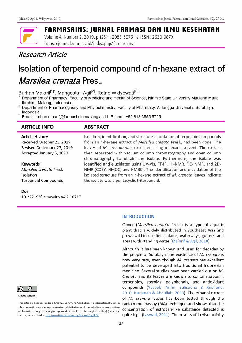
1.6 kg M. crenata leaf powder was extracted with n-hexane by maceration method to obtain an extract weighing 80 g with a yield of 5%. The maceration method was chosen because it is a relatively safer and effective method of extracting compounds from plants. The n-hexane extract stain profile was identified using TLC with an H2SO4 anisaldehyde stain appearance, and several specific purple, red, pink, and blue stains showed a terpenoid group (Figure 1).
The 11 g n-hexane extract was separated by the VCC method with the 150 g stationary silica gel GF254 stationary phase and the gradient mobile phase n-hexane:ethyl acetate. The weight of n-hexane extract used in separation with VCC is based on the number of stationary phases in the column. The VCC column used has a diameter of 7.5 cm and a height of 7.5 cm, the size of this column has a maximum quiescent capacity of 70 g, so the amount of extract taken to be separated is 5% of 70 g which is 3.5 g of n- extract hexane. Determination of the weight of this extract aims to produce good separation with VCC, and if the amount of extract is excessive, it will produce unfavorable separation. The results of the n-hexane extract of M. crenata leaves with VCC resulted in 11 fractions (Table 1).
The fractions are then grouped into three large fraction groups based on the proximity of Rf and the color similarity of the stain. Fraction 2 (a combination
(Ma’arif, Agil & Widyowati, 2019) Farmasains : Jurnal Farmasi dan Ilmu Kesehatan 4(2), 27-31.
29
of fractions 4 and 5) was then selected and separated by the MPA method with the Merck 60 silica gel powder and the n-hexane:ethyl acetate (4:1) mobile phase. The fractions produced from the MPA are further grouped based on the TLC profile, and six fraction groups are obtained (Table 2)
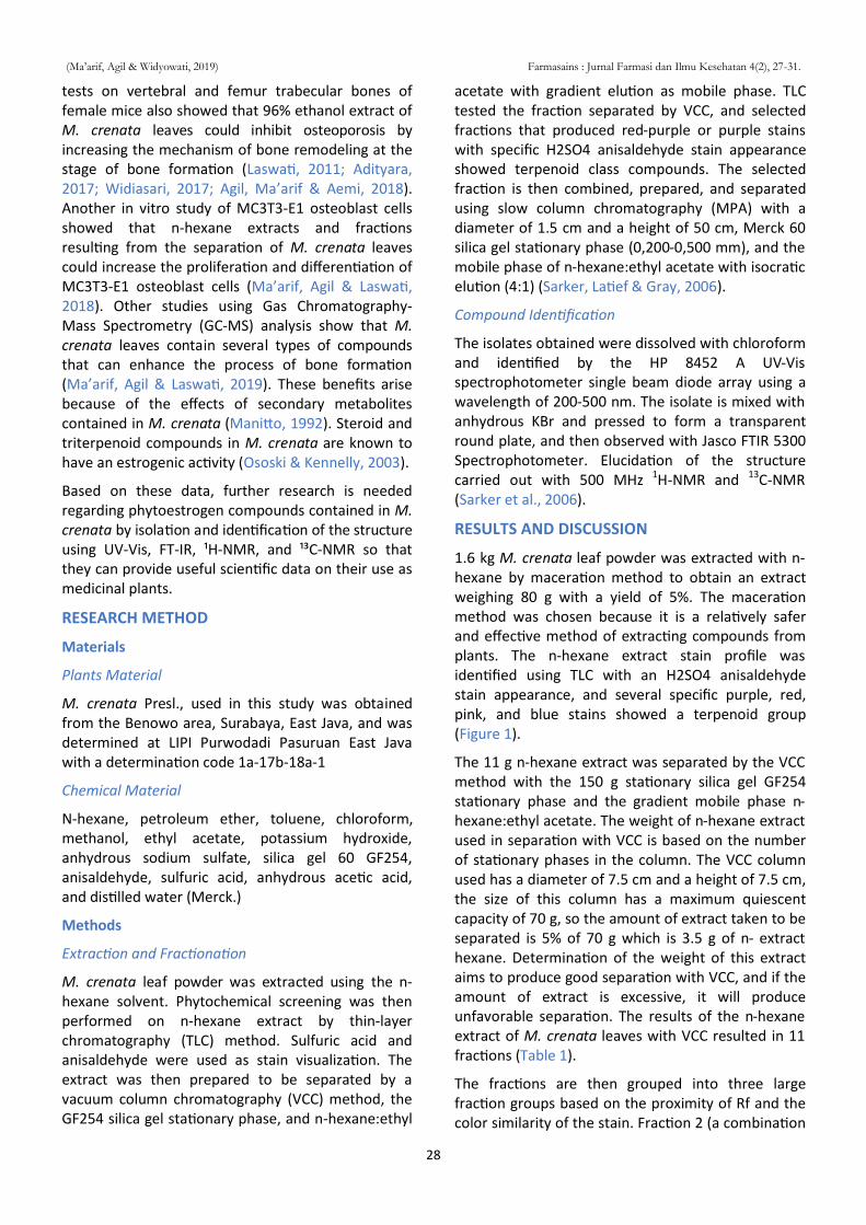
Of these fractions, 2.4 fractions were selected for further analysis by spectrophotometry, fractions 2.4 were chosen because they had spot on Rf 0.38 in intensive red and purple color corresponding to the characteristics of triterpenoid group compounds (Figure 2).
The isolate (fraction 2.4) was dissolved with
chloroform solvent and analyzed with a wavelength UV-Vis (λ) spectrophotometer 200-450 nm. Isolates showed maximum absorption at λ 232 nm and 242 nm, this indicates the existence of double bonds as chromophore groups in isolates, so that it can be detected in UV light. The isolate has a maximum of 2 λ allegedly due to the presence of impurities in the isolate, which gives low absorbance at 232 nm.
Analysis of isolates with FT-IR spectrophotometer showed a widening absorption in the 3434 cm-1 regions, which was thought to be absorption from the OH group (3500-3200 cm-1). In the area of 2956, 2917, and 2849 cm-1, there was an absorption that was thought to be a stretching vibration of the C-H group at CH3. The absorption band at 1631 cm-1 shows the stretching vibration of the C=C group (1700-1600 cm-1), which shows the double bond in the isolate, while the absorption at 1472, 1462, and 1379 cm-1 shows the stretching vibration of the C-H group at CH3. In the 1094 cm-1 region, there was a strong absorption, which appeared as stretching vibrations from the CO group (1320-1000 cm-1), and in regions 800, 730, and 719 cm-1, there was an absorption which showed a stretching vibration = CH (1000-650 cm-1). From the interpretation of FT-IR spectrophotometer data above, it is suspected that the isolate has hydroxy (OH), alkyl (CH3), alkenyl (C = C), =C-H, and C-O groups.
The next step was to isolate analysis using NMR spectrometers. Analysis of the 1H-NMR spectrum of the isolates showed that there were 11 types of H atoms, three metin at δ 1.1398 (H-25), δ 1.5795 m (H-18) and δ 1.9894 m (H-26), three ethers at δ 4,1595 d (6.5) (H-27), δ 3,6380 t (7,15; 6,45) (H-28), and δ 5,4099 t (7,15; 1, 3) (H-29), one methoxy group at δ 1.2527 s (H-14), and several methyl at δ 0.8493 m (H-1), δ 0.8584 m (H-3), δ 0.8635 m (H-5), and δ 1.0672 m (H-2). Analysis of the 13C-NMR spectrum of the isolates showed that as many as 30 C atoms were
Table 1. The TLC profile of the fraction resulted from the separation of fractions 2 of M. crenata leaf with anisaldehyde H2SO4 stain appearance and the mobile phase of n-hexane:ethyl acetate (4:1) .
Figure 1. Stain profile of M. crenata Presl n-hexane extract with
anisaldehyde H2SO4 stain appearance and the mobile
phase of n-hexane:ethyl acetate (4:1) .
Figure 2. Stain profile of fraction 2.4 with anisaldehyde H2SO4
stain appearance and the mobile phase of n-
hexane:ethyl acetate (4:1).
Fraction
Stain Spot Colour Rf Weight (mg)
1 - - - 159.1
2 1 Grenish blue 0.75 -
3 1 Purple 0.75 1958.9
4 1 Purplish red 0.38 4164.9
5 1 Purplish red 0.38 1901.6
6 1 Purple 0.23 454.9
7 1 Purple 0.00 79.4
8 1 Brown 0.00 32.1
9 1 Brown 0.00 24.7
10 1 Brown 0.00 23
11 - - - -
Fraction Stain Spot Color Rf Weight (mg)
2.1 - - - -
2.2 1 Grenish blue 0.85 513
2.3 1 Purple 0.77 -
2.4 1 Red 0.38 482
2.5 1 Brownish purple 0.25 391
2.6 1 Brown 0.31 -
Table 1. TLC profile of the fraction resulted from the separation of n-hexane extract of M. crenata leaves with anisaldehyde H2SO4 stain appearance and the mobile phase of n-hexane:ethyl acetate (4:1).
(Ma’arif, Agil & Widyowati, 2019) Farmasains : Jurnal Farmasi dan Ilmu Kesehatan 4(2), 27-31.
30
making up the isolate. The atoms are identified based on their chemical shift region and among them form methoxy, methyl groups, and several quaternary carbon atoms.
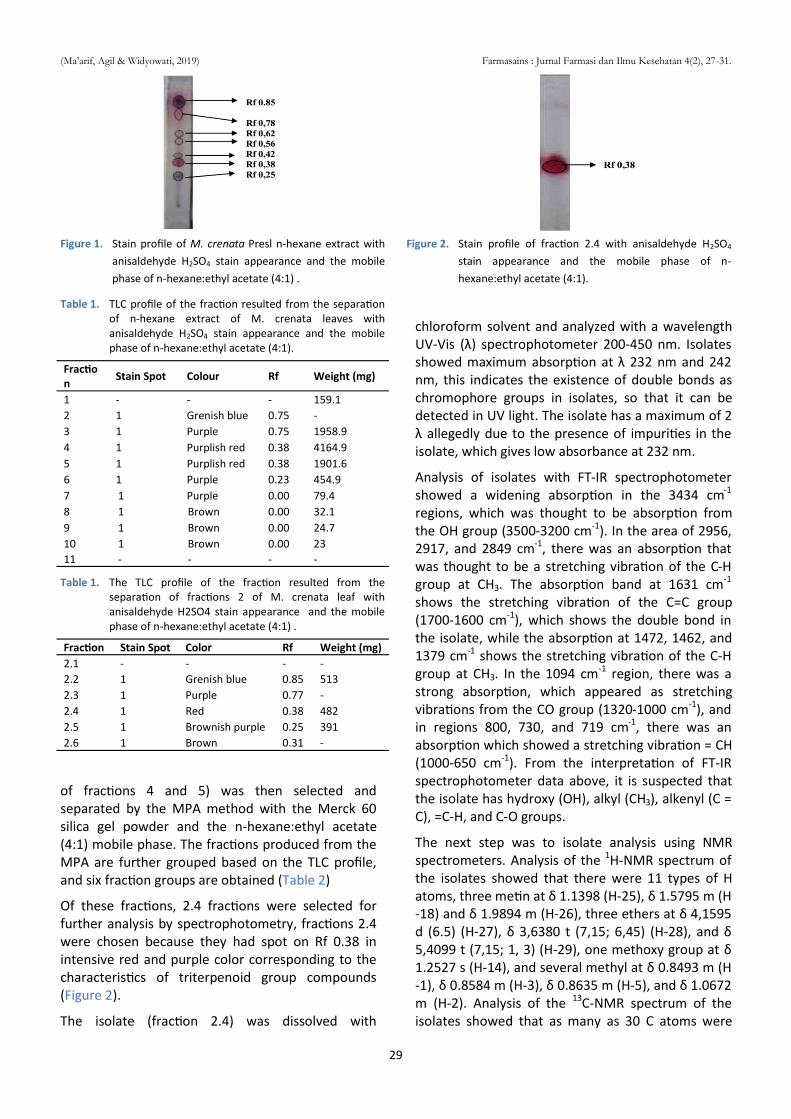
The 2D-NMR was then used to complete the isola-tion structure isolation using Heteronuclear Multiple Quantum Coherence (HMQC), Heteronuclear Corre-lated Spectroscopy (COSY), and Heteronuclear Multi-ple Bond Connectivity (HMBC). From a series of isolation and identification processes, obtained terpenoid group compounds, namely triterpenoid pentacyclic with a total of 30 C atoms, 11 O atoms, and 35 H atoms, with the chemical name 14-(1- hydroxyethyl) - 3 - isopropyl - 8 - methoxy - 6,11 - Dimethyl - 1 , 2 , 3 , 4 , 4a , 5 , 6 , 6a , 6b , 7 , 9 , 10 , 11 , 12b , 13 , 14 , 14a , 14b - octadecahydropicene - 1,2,4,5,7,9,10,12,13-nonaol (Figure 3).
CONCLUSION
The process of separating and isolating the n-hexane extract of M. crenata leaves produced isolates that were included in the pentacyclic triterpenoid com-pounds. Although further studies need to be carried out on the estrogenic activity of isolates, the isolated structure obtained is included in phytoestrogen group compounds.
REFERENCES
Nurjanah, A. A., Abdullah, A. (2012). Aktivitas antioksidan dan komponen bioaktif semanggi air (Marsilea crenata). Jurnal Inovasi dan Kewirausahaan, 1(3), 152-158.
Laswati, H. (2011). Green Clover Potentiates Delaying the Increment of Imbalance Bone Remodeling Process in Postmenopausal Women. Folia Medica Indonesiana, 47(2), 112-117.
Agil, M., Ma'arif, B., Aemi, N. Y. (2018). Antiosteoporotic activity of n-hexane fraction from Marsilea crenata Presl. leaves in increasing trabecular vertebrae bone density of female mice. Jurnal Tumbuhan Obat Indonesia, 11(2), 1-7.
Adityara, R. A. (2017). Uji aktivitas antiosteoporosis fraksi etil asetat daun Marsilea crenata Presl. dalam meningkatkan kepadatan tulang trabekula femur mencit betina. (Undergraduate’s Thesis). University of Airlangga, Surabaya, Indonesia.
Widiasari, F. A. (2017). Uji aktivitas antiosteoporosis fraksi etil asetat daun Marsilea crenata Presl. dalam meningkatkan kepadatan tulang trabekula vertebra mencit betina (Undergraduate’s Thesis). University of Airlangga, Surabaya, Indonesia.
Ma’arif, B., Agil, M., & Laswati, H. (2016). Phyto-chemical Assessment On n-Hexane Extract And Fractions Of Marsilea crenata Presl. Leaves Through GC-MS. Majalah Obat Tradi-sional, 21(2), 77-85.
Ma'arif, B., Agil, M., & Laswati, H. (2018). Alkaline phosphatase activity of Marsilea crenata Presl. extract and fractions as marker of MC3T3-E1 osteoblast cell differentiation. Journal of Applied Pharmaceutical Science, 8(3), 55-59.
Ma’arif, B., & Agil, M. (2018). Metabolite Profiling of Ethyl Acetate Extract from Marsilea crenata Presl. Using UPLC-QToF-MS/MS. In Prajogo, B., Wolfender, J., Ismail, N. H., Calderon, A., Wahyuni, T. S., Suciati., ... & Widiandhani, T (Eds.). Bromo Conference. Proceedings of Bromo Conference (pp. 50-57). Surabaya, Indonesia.
Manitto, P. (1992). Biosintesis Metabolit Sekunder. Semarang, Indonesia: IKIP Semarang Press.
Ososki, A. L., & Kennelly, E. J. (2003). Phytoestrogens: a review of the present State of Research. Phytotherapy Research, 17(8), 845-869.
Sarker, S. D., Latif, Z., & Gray, A. I. (2006). An Overview of Natural Product Isolation. Natural Product Isolation (2nd Ed). Totowa, NJ: Humana Press Inc.
Yacoeb, A.M., Arifin, M., Sulistiono, W., & Kristiono, S. S. (2010). Deskripsi histologis dan perubahan komposisi kimia daun dan tangkai
Figure 1. HMBC spectrum of isolate from M. crenata leaves
(Ma’arif, Agil & Widyowati, 2019) Farmasains : Jurnal Farmasi dan Ilmu Kesehatan 4(2), 27-31.
31
semanggi (Marsilea crenata Presl., Marsileaceae) akibat perebusan. Jurnal Pengolahan Hasil Perikanan Indonesia, 13(2), 81-95.
(Ma’arif, Agil & Widyowati, 2019) Farmasains : Jurnal Farmasi dan Ilmu Kesehatan 4(2), 27-31.


![Practice Topic 11 questions [55 marks] - PBworksschultz915.pbworks.com/w/file/fetch/125246906/KEY... · Bromine was added to hexane, hex-1-ene and benzene. Identify the compound(s)](https://static.documents.pub/doc/80x56/5f03bb097e708231d40a7d5c/practice-topic-11-questions-55-marks-bromine-was-added-to-hexane-hex-1-ene.jpg)


