+ All Categories
Home > Documents > Isolation of the Virulence Gene(s) in the New, Live, Oral ...

Isolation of the Virulence Gene(s) in the New, Live, Oral ...

Date post: 02-Dec-2021
Category:
Upload: others
View: 1 times
Download: 0 times
Share this document with a friend
56
University of Rhode Island University of Rhode Island DigitalCommons@URI DigitalCommons@URI Open Access Master's Theses 1998 Isolation of the Virulence Gene(s) in the New, Live, Oral Vaccine Isolation of the Virulence Gene(s) in the New, Live, Oral Vaccine Candidate Candidate Salmonella typhimurium SR-11 Fad SR-11 Fad James H. Allen University of Rhode Island Follow this and additional works at: https://digitalcommons.uri.edu/theses Recommended Citation Recommended Citation Allen, James H., "Isolation of the Virulence Gene(s) in the New, Live, Oral Vaccine Candidate Salmonella typhimurium SR-11 Fad" (1998). Open Access Master's Theses. Paper 1097. https://digitalcommons.uri.edu/theses/1097 This Thesis is brought to you for free and open access by DigitalCommons@URI. It has been accepted for inclusion in Open Access Master's Theses by an authorized administrator of DigitalCommons@URI. For more information, please contact [email protected].
Transcript

University of Rhode Island University of Rhode Island

DigitalCommons@URI DigitalCommons@URI

Open Access Master's Theses

1998

Isolation of the Virulence Gene(s) in the New, Live, Oral Vaccine Isolation of the Virulence Gene(s) in the New, Live, Oral Vaccine

Candidate Candidate Salmonella typhimurium SR-11 Fad SR-11 Fad

James H. Allen University of Rhode Island

Follow this and additional works at: https://digitalcommons.uri.edu/theses

Recommended Citation Recommended Citation Allen, James H., "Isolation of the Virulence Gene(s) in the New, Live, Oral Vaccine Candidate Salmonella typhimurium SR-11 Fad" (1998). Open Access Master's Theses. Paper 1097. https://digitalcommons.uri.edu/theses/1097

This Thesis is brought to you for free and open access by DigitalCommons@URI. It has been accepted for inclusion in Open Access Master's Theses by an authorized administrator of DigitalCommons@URI. For more information, please contact [email protected].

ISOLATION OF THE VIRULENCE GENE(S) IN THE NEW, LIVE, ORAL

VACCINE CANDIDATE Salmonella typhimurium SR-11 Fad-

BY

JAMES H. ALLEN

A THESIS SUBMITTED IN PARTIAL FULFILLMENT OF THE

REQUIREMENTS FOR THE DEGREE OF

MASTER OF SCIENCE

IN

MICROBIOLOGY

UNIVERSITY OF RHODE ISLAND

1998

MASTER OF SCIENCE THESIS

OF

JAMES H. ALLEN

APPROVED:

Thesis Committee

UNIVERSITY OF RHODE ISLAND

1998

MASTER OF SCIENCE THESIS

OF

JAMES H. ALLEN

APPROVED:

Thesis Committee

UNIVERSITY OF RHODE ISLAND

1998

Abstract

A DNA fragment containing the defective virulence gene(s) in the live oral

vaccine candidate Salmonella typhimurium SR-11 Fad· was isolated. Genomic DNA

fragments from the vaccine candidate, constructed by TnlOd::cam transposon

mutagenesis, were probed with the 1.4 Kbp chloramphenicol resistance gene contained in

TnlOd::cam and analyzed by Southern hybridization. Two of the fragments, an 8.0 Kbp

Hind III fragment and a 4.5 Kbp Pst I :fragment, were isolated, cloned into E. coli HB 101,

and mapped. The Pst I fragment, containing approximately 2.9 Kbp of flanking genomic

DNA, was subcloned into the sequencing vector pBluescript II SK + (Stratagene, La

Jolla, CA) and mapped.

11

Acknowledgement

* Dr. Paul S. Cohen

*Dr. Richard E. Koske

*Dr. David R. Nelson

*Dr. Alison Roberts

* Dr. David C. Laux

* Maryjane Utley

* Paul W. Johnson

* Carolyn Lunt

* The Nelson Lab

* The Norris Lab

* Michael Bono

* Aimee Hebert

* Ian Lodovice

iii

Preface

This research is presented here in the standard format for thesis.

IV

Contents

Abstract. .............................................................................................................................. .ii

Acknowledginent ........................................................................................ ....................... .iii

Preface ................................................................................... ............................................ .iv

Contents ............................................................................................................................... v

List of Tables .................................................................................................................... vii

List ofFigures ............................................................ ............................ ........................... viii

Introduction ........................... ............................................................................... ................ 1

The Vaccine Candidate: Salmonella typhimurium SR-11 Fad-........................................... 2

Materials and Methods ......................................................................................................... 4

Agarose gel electrophoresis ..................................................................................... 4

Bacterial strains ........................................................................................................ 4

Calf intestinal alkaline phosphatase ................. .-...................................................... .4

DIG-labeled probes ................................................... ............................................... 4

Endo nuclease digests ............................................................................................... 5

Incubation of bacterial cultures and plates ............................................................... 5

Isolation of DNA ..................................................................................................... 5

Ligase reactions ................................. ........................ .......... .................................. 10

Media and antibiotics ........................ ..... .. .............................................................. 10

v

Southern blotting .................................................................................................... 10

Transformations ........................................................................ ............................. 12

Vectors ................................................................................................................... 13

Results ................................................................................................................................ 15

Isolation of the chloramphenicol resistance cassette ............................................. 15

Southern blot of Salmonella typhimurium SR-11 Fad-........................... .............. 16

Cloning of the Hind III and Pst I fragments of Salmonella typhimurium

SR-11 Fad- genomic DNA ..................................................................................... 17

Cloning the 4.5 Kbp Pst I fragment using a sequencing vector ............................. 19

Discussion .......................................................................................................................... 21

Tables ................................................................................................................................. 24

Figures ................................................................................................................................ 25

References .................................................................... : ..................................................... 38

Bibliography ....................................................................................................................... 41

vi

List of Tables

Bacterial strains .................................................................................................................. 24

vii

List of Figures

The unique Bam ID, Eco Rl, Hind III, and Pst I restriction enzyme sites in

the cloning vector pBR322 ................................................................................................ 25

The construction of pJHA I ................................................................................................ 26

The interruption of the tetracycline resistance gene in the plasmid pJHA 1 ....................... 27

Agarose (0.9%) gel analysis of the plasmid pJHAl.. ........................................................ 28

The plasmid pJHAl mapped with the restriction enzymes BamH I and EcoR 1.. ............. 29

Unique restriction enzyme sites in the plasmid pJHAl.. ................................................. .30

Southern hybridization analysis of SR-11 genomic DNA ............................................... .31

Agarose (0.5%) gel analysis of the plasmid pJHA6 ......................................................... .32

Agarose (0.7%) gel analysis of the plasmid pJHA6 ......................................................... .33

The plasmid pJHA6 mapped with BamH I, EcoR I, and Pst I ......................................... .34

Map of the sequencing vector pBluescript II SK+ ..... : .................................................... .35

Agarose (0.7%) gel analysis of the plasmid pJHA7 ......................................................... .36

The plasmid pJHA 7 mapped with BamH I, EcoR I, and Pst 1.. ........................................ .37

viii

Introduction

Poultry production is reduced significantly by Salmonella bacteria. The U.S.

poultry industry hatches approximately 7.5 billion eggs annually in incubation facilities,

and nearly $77 million is lost to the poultry industry each year due to Salmonella

outbreaks in poultry farms (1 ). The health care costs associated with human

salmonellosis caused by eating contaminated eggs and poultry is estimated at $4 billion

annually (1 ). Vaccination of chickens against Salmonella would both reduce losses to the

poultry industry and the poultry-associated human salmonellosis.

Typhoid fever, an infectious disease caused by the bacterium Salmonella typhi,

remains a serious public health problem in developing countries causing an estimated

600,000 deaths annually (2). Two Salmonella typhi vaccines, the live oral S. typhi Ty21 a

and a vaccine based on the Vi polysaccharide capsule of S. typhi, currently are licensed

in several countries (3). Each of these vaccines, while effective, still suffers from

drawbacks such as fevers after administration, incomplete protection, and a loss of

protection with time (3). Therefore biotechnology is being used to develop new

attenuated S. typhi live oral vaccines (3).

Live oral vaccines, based on deletions into genes necessary for Salmonella

pathogenesis, have shown great promise (4, 5). For example, the live oral S. typhimurium

~cya (adenylate cyclase) ~crp (cAMP receptor protein) vaccine is based on deletions in a

Salmonella global regulatory system and is effective in animals and humans ( 4, 5).

Another S. typhimurium live oral vaccine, avirulent and immunogenic in mice and cattle,

is based on aro (aromatic compounds) deletions; this strain is auxotrophic for p-

aminobenzoic acid and 2,3-dihydroxybenzoate which are unavailable in mammalian cells

(6, 7). In addition, Salmonella strains containing deletions in genes of the phoP-phoQ

regulon (regulation of virulence) are effective mouse and human vaccines (8, 9).

The use of attenuated Salmonella strains as live vaccines is a safe and effective

means of inducing significant humoral and secretory antibody responses in animal

species: humans, cattle, sheep, rabbits, fowl, and mice (10). Live Salmonella invade the

Peyer's patches, where they present their numerous antigens directly to the T and B

lymphocytes of the mucosal-associated lymphoid tissues (10). This elicits the mucosal

immune system to produce antigen-specific immune responses (10). Because of the

significant humoral and secretory antibody responses, live attenuated Salmonella strains

show great promise for carriers of antigenic determinants from other pathogenic

microorganisms (10).

The Vaccine Candidate: Salmonella typhimurium SR-11 Fad·. The live oral

vaccine candidate, Salmonella typhimurium SR-11 Fad·, was constructed by TnlOd::cam

transposon mutagenesis of the SR-11 parent strain (11). The mutant strain was selected

for its inability to catabolize oleate and citrate as sole carbon sources (11 ). It was

phenotypically designated Fad- (Fatty acid) for its inability to catabolize fatty acids as a

sole carbon source (11 ). The SR-11 Fad- strain also was unable to utilize acetate and

isocitrate as carbon sources, but could utilize glucose, glycerol, pyruvate, and succinate as

sole carbon sources (11 ).

Salmonella typhimurium SR-11 Fad·, administered peroraly, was completely

avirulent in BALB/c mice at a dose as high as 109 colony forming units (cfu) (11). In

2

contrast, the SR-11 parent strain proved lethal at doses of greater than 104 cfu (11 ). The

vaccine candidate also was found to be protective in BALB/c mice (11) as well as

avirulent and protective in chickens (Dr. Paul S. Cohen: personal communication). A

single oral dose of 107 SR-11 Fad· cells protected BALB/c mice against a lethal dose of

109 cells of the virulent SR-11 Salmonella typhimurium parent strain (11).

In order for a vaccine to be useful, it must be safe. Therefore is should not

contain antibiotic resistance genes. Since the SR-11 Fad· vaccine candidate was

constructed by TnJOd::cam transposon mutagenesis (11), it contains a chloramphenicol

resistance gene. This chloramphenicol cassette is interrupting a virulence gene(s) and is

blocking the expression of that virulence gene(s). Further development of this vaccine

candidate required the isolation of this interrupted virulence gene(s). The virulence

gene(s) and the chloramphenicol resistance cassette can then be permanently deleted,

yielding a safer live oral vaccine candidate.

3

Materials and Methods

Agarose gel electrophoresis. Agarose gels were made with concentrations

between 0.50% and 0.90% of electrophoresis grade agarose (Fisher Scientific, Fair Lawn,

NJ) in TAE buffer (40 mM Tris-acetate; 1.0 mM EDTA, 5 Prime~ 3 Prime, Inc.,

Boulder, CO) and ethidium bromide (final concentration: 0.2-0.4 µg/ml; Sigma Chemical

Co., St. Louis, MO) (12). Several DNA standard markers were used: phage lambda cut

with Hind III (Gibco BRL, Gaithersburg, MD) and 1 Kbp DNA ladders (Gibco BRL,

Gaithersburg, MD and Promega Corporation, Madison, WI).

Bacterial strains. See Table 1.

Calf intestinal alkaline phosphatase. Calf intestinal alkaline phosphatase

(CIAP) (Promega Corporation, Madison, WI) was employed for the hydrolysis of 5'­

phosphate groups from vector DNA, following incubation with a restriction enzyme.

Three successive CIAP reactions, following the manufacturer's protocol, were performed

on vectors prior to ligation to an insert.

DIG-labeled probe. A DIG-labeled (digoxigenin) probe was made from the 1.4

Kbp chloramphenicol cassette using A DIG High Prime DNA Labeling & Detection

Starter Kit II (Boehringer-Mannheim, Germany): 750 ng of template DNA

( chloramphenicol cassette) was denatured by boiling for 10 minutes and quickly cooled in

an ice/ethanol bath; the now single-stranded template DNA was added to a labeling

reaction mixture of deoxynucleoside triphosphates (dATP, dGTP, dCTP, dTTP and DIG­

dUTP) and the Kienow fragment of DNA polymerase III; labeling of the template

occurred, in this DNA synthesis reaction, by the random incorporation ofDIG-dUTP (a

4

steroid hapten covalently bound to deoxycytidine trip hosp hate) over a 25 hour period at

37°C. The labeling reaction was stopped by heat inactivation at 65°C for 10 minutes.

Endonuclease digests. The restriction endonucleases, Bam HI, Eco RI, Hind III,

Pst I, Pvu II, and Xho I were purchased from Gibco BRL (Gaithersburg, MD) and/or

Promega Corporation (Madison, WI). The manufacturer's protocols for restriction

endonuclease digestions were modified for volumes of20-50 µl using nuclease-free,

deionized, distilled water (Sigma Chemical Co., St. Louis, MO), and DNA was digested

overnight with an excess of enzyme (>5U/µg DNA).

Incubation of bacterial cultures and plates. All liquid bacterial cultures, except

where noted, were grown overnight at 37°C with aeration in the appropriate antibiotic.

All plates inoculated with bacteria, except where noted, were incubated at 37°C for 12-18

hours.

Isolation of DNA. (Note: volume of reagents used and/or centrifugation speed

and time varied according to the initial volume of the bacterial cultures and/or bacterial

stain.) Genomic DNA was isolated. by a modified protocol from Current Protocols in

Molecular Biology (13). Bacterial cells from an overnight culture were pelleted by

centrifugation, resuspended in TE Buffer, gently lysed with sodium dodecyl sulfate (SOS)

(BioRad Laboratories, Richmond, CA), and incubated at 37°C for one hour in proteinase

K (Sigma Chemical Co., St. Louis, MO) to degrade the proteins.

Hexadecyltrimethylammonium bromide (10% CTAB in 0.7 M NaCl, Sigma Chemical

Co., St. Louis, MO) and NaCl was added to the lysate and incubated at 65°C for 15

minutes: the CTAB solution complexes with denatured protein and polysaccharides; the

5

salt neutralizes macromolecules. An emulsion was formed by the addition of an equal

volume of chloroform/isoamyl (24: 1) (Sigma Chemical Co., St. Louis, MO) to the lysate.

The aqueous phase, containing the DNA, was extracted after centrifugation leaving

behind an organic phase of cell wall debris, denatured protein, and polysaccharides

complexed to CTAB. A second extraction with phenol/chloroform/isoamyl (25:24:1)

(Sigma Chemical Co., St. Louis, MO), removed residual CT AB precipitate. The DNA

was precipitated in 100% isopropanol (Sigma Chemical Co., St. Louis, MO) and pelleted

by centrifugation. The DNA pellet was rinsed in 70% ethanol to remove excess salt,

lyophilized, and resuspended in TE buffer. Incubation (37°C for greater than 1 hour) of

the genomic DNA in RNase A (Sigma Chemical Co., St. Louis, MO) degraded most of

the contaminating RNA. The DNA underwent a final phenol/chloroform/isoamyl

extraction to remove the RNase A and any residual proteins. After precipitation in 100%

ethanol, the genomic DNA was ready for manipulation.

Genomic DNA also was isolated by using the G NOME® genomic DNA isolation

kit (BIO 101, Vista, CA). The protocol from the manufacturer was modified by

increasing the initial volume of overnight liquid bacterial cell culture to 10 ml per

miniprep. The kit used a "Cell Suspension Solution," "RNase Mixx," "Cell

Lysis/Denaturing Solution," "Protease Mixx," and "Salt-Out Mixture" of unspecified

composition and replaced the chloroform steps of the standard protocol (13) with

incubation at 4°C followed by centrifugation. A final precipitation of the genomic DNA

in 100% ethanol completed the manufacturer's protocol. Genomic DNA isolated by the

6

kit contained less RNA and protein contaminants than genomic DNA isolated by the

standard protocol (13).

Quantities of plasmid DNA greater than 10 µg were isolated by modified alkaline

lysis (14). A minimum of 100 ml of bacterial cells from an overnight liquid culture were

pelleted by centrifugation and resuspended in GTE Buffer (50 mM glucose; 10 mM

EDTA [ethylenediaminetetraacetic acid]; 25 mM Tris-HCl

[Tris[hydroxymethyl]aminomethane hydrochloride], Fisher Scientific, Fair Lawn, NJ).

The resuspended cells were added to a lysis solution, containing 0.2 N NaOH (Sigma

Chemical Co., St. Louis, MO) and 1.0% SDS (wt/vol), and incubated on ice for IO

minutes. An appropriate volume of potassium acetate solution (5 M KOAc, pH 4.8) was

mixed into the cell lysate and centrifuged. The supernatant, containing the plasmid DNA,

was extracted with phenol/chloroform/isoamyl alcohol and precipitated in 100%

isopropanol at -20°C for 30 minutes. The plasmid DNA was pelleted by centrifugation,

rinsed in 70% ethanol to remove excess salt, lyophilized, and resuspended in TE buffer.

Incubation (37°C for greater than 1 hour) of the plasmid DNA in RNase A degraded most

of the contaminating RNA. The plasmid DNA underwent a final

phenol/chloroform/isoamyl extraction to remove the RNase A and any residual proteins.

After a final concentration by precipitation in 100% ethanol, the plasmid DNA was ready

for manipulation.

Quantities of plasmid DNA less than 10 µg were isolated by Wizard™ Plus

Minipreps DNA Purification Systems (Promega Corporation, Madison, WI). The

protocol from the manufacturer was modified by increasing the initial volume of

7

overnight liquid bacterial cell culture to 25 ml per miniprep. The bacterial cells were

pelleted by centrifugation and resuspended in Cell Resuspension Solution (50 mM Tris,

pH 7.5; 10 mM EDTA; 100 µg/ml RNase A). Cell Lysis Solution (0.2 N NaOH; 1.0%

SDS) was added to the resuspended cells, mixed by inversion of the microcentrifuge

tubes, and incubated at room temperature for 5 minutes. Neutralization Solution (1.32 M

KO Ac) was mixed into the cell lysate and centrifuged. The supernatant, containing the

plasmid DNA, was pipetted into a 3 ml syringe (Becton Dickenson and Co., Franklin

Lakes, NJ) attached to a Wizard™ Minicolumn. Wizard™ DNA Purification Resin was

added to the lysate and plunged through the minicolumn. The minicolumn was rinsed

with Column Wash Solution (80 mM KOAc; 8.3 mM Tris-HCl, pH 7.5; 40 µM EDTA;

55% ethanol), removed from the syringe and dried by centrifugation. Plasmid DNA was

eluted off the minicolumn by the addition of TE buffer and centrifugation. The plasmid

DNA was then ready for manipulation.

DNA was purified, when necessary, by extraction with

phenol/chloroform/isoamyl alcohol (15). Phase Lock Gel™ (5 Prime~ 3 Prime, Inc.,

Boulder, CO) microcentrifuge tubes were used during the extraction process according to

the manufacturer's protocol: an equal volume of DNA sample (genomic or plasmid) and

phenol/chloroform/isoamyl alcohol were added to a Phase Lock Gel™ microcentrifuge

tube, mixed, and centrifuged (14,000 x g) for two minutes. A thick density barrier was

formed between the upper aqueous phase, containing the DNA, and the protein-laden

interface and the lower organic phase. The upper aqueous phase was then simply poured

off into sterile microcentrifuge tubes. A second extraction of the trapped organic phase

8

with TE buffer and phenol/chlorofonn/isoamyl alcohol removed any remaining DNA.

The DNA samples were pooled, concentrated by ethanol precipitation, lyophilized, and

resuspended in an appropriate volume of TE buffer.

DNA was concentrated, when necessary by ethanol precipitation (15).

GenElute™ (Supelco, Bellefonte, PA) LPA (linear polyacrylamide DNA carrier) was

used in all ethanol precipitations of plasmid DNA. The protocol from the manufacturer

was modified by increasing the sodium acetate (Sigma Chemical Co., St. Louis, MO)

concentration to 0.3 M to facilitate a more complete precipitation of the DNA in 100%

ethanol (Quantum Chemical CO, Tuscola, IL). Precipitated DNA was pelleted by

centrifugation (12,000 x g), lyophilized, and resuspended in TE buffer (I 0 mM Tris-Cl;

1.0 mM EDTA, 5 Prime~ 3 Prime, Inc., Boulder, CO). DNA was frozen at -20°C for

short-term storage and -78°C for long-term storage.

Concentrations of DNA were estimated by ethidium bromide dot quantitation

(16). Sample DNA, in a series of dilutions in TE Buffer and ethidium bromide (1.0

µg/ml final concentration), was spotted onto Parafilm®-M (American Can Company,

Greenwich, CT) and viewed on a ultra-violet transilluminator. A series of standards,

lambda DNA cut with Hind III (Gibco BRL, Gaithersburg, MD), was spotted above the

sample DNA. Comparison of the fluorescence of the sample to these standards provided

an estimate of the DNA concentration in the sample (16).

DNA was also recovered from agarose gels. Bands of electrophoresed genomic

or plasmid DNA were cut from agarose gels and added to GenElute™ Minus EtBr Spin

Columns (Supelco, Bellefonte, PA) prewashed in TE Buffer. The columns were inserted

9

onto microcentrifuge tubes and centrifuged (12,000 x g) for 10 minutes. The columns

retained the agarose and ethidium bromide while the DNA was eluted by the TE buffer.

The DNA was then concentrated by ethanol precipitation.

Ligase reactions. T4 DNA ligase (Promega Corporation, Madison, WI) reactions

were performed according to the manufacturers protocol for greater than 12 hours at

l 6°C. T 4 DNA ligase catalyzed the formation of a 3' to 5' phosphodiester bonds between

the 3'-hydroxyl group of one restriction fragment and the 5'-phosphate group of another

restriction fragment while their sticky ends are transiently base-paired (17).

Media and antibiotics. Luria broth (Lennox formulation: 5g NaCl IL, Sigma

Chemical Co., St. Louis, MO; 10 g tryptone IL, Difeo Laboratories, Detroit, MI; 5g yeast

extract IL, Difeo Laboratories, Detroit, MI; pH 7.5) and Luria agar plates (Lennox

formulation for Luria broth plus 15 g Bacto agar IL, Difeo Laboratories, Detroit, MI; pH

7.5) were used exclusively. Antibiotics were purchased from Sigma Chemical Co. (St.

Louis, MO) and added to the media at a final concentration as follows: 100 µg/ml

ampicillin; 30 µg/ml chloramphenicol; 50 µg/ml nalidiXic acid; 100 µg/ml streptomycin

sulfate; 10 µg/ml tetracycline hydrochloride. MacConkey agar (Difeo Laboratories,

Detroit, MI) and XLD agar (Difeo Laboratories, Detroit, MI) were used to characterize

initial strains and all transformants.

Southern blotting. DNA, digested with restriction enzymes, was electrophoresed

in a 0.85% agarose gel and photographed on an ultra-violet transilluminator (18). The

DNA was partially depurinated by gently shaking the agarose gel in 0.25 M HCl bath for

30 minut~s (19). The partial depurination cleaved DNA strands into fragments 1 to 2 Kb

IO

in length improving their mobility in the gel (19). The gel was rinsed with distilled water

and bathed in 0.4 M NaOH on an orbital shaker for 20 minutes. The DNA within the gel

was denatured at this step yielding single-stranded DNA (19). The single-stranded DNA

was then transferred overnight to a Magna Charge Nylon (Micron Separations, Inc.,

Westboro, MA) membrane (positively-charged) by upward capillary action using 0.4 M

NaOH as the transfer buffer (19). The negatively-charged DNA was immobilized on the

positively-charged nylon membrane during the transfer. The membrane was rinsed in 2x

SSC (0.3 M NaCl; 30 mM sodium citrate; pH 7.0; 5 Prime-+ 3 Prime, Inc., Boulder, CO)

twice to remove excess salt and agarose fragments (19).

A DIG High Prime DNA Labeling & Detection Starter Kit II (Boehringer­

Mannheim, Germany) was used to further hybridize and develop the blot. The DIG­

labeled 1.4 Kbp chloramphenicol cassette probe, denatured by boiling for 5 minutes and

quickly cooled in an ice/ethanol bath, was diluted in prewarmed standard hybridization

buffer (5x SSC; 0.1 % [wt/vol] N-lauroylsarcosine; 0.02% [wt/vol] SDS; 1 % [vol/vol]

Blocking reagent) to a final concentration of 15 ng/ml.. The probe was hybridized to the

membrane for 12.5 hours at 68°C with gentle agitation. At this point, the single-stranded

DNA probe in solution hybridized to homologous single-stranded DNA bound to the

membrane. The membrane was then rinsed in post-hybridization solutions: twice with

2x SSC, 0.1 % SDS at room temperature and twice with 0.1 % (vol/vol) SSC, 0.1 %

(wt/vol) SDS at 68°C with gentle agitation. The post hybridization washes (stringency

washes) removed any non-specifically bound probe from the membrane.

11

To detect the probe, the membrane was rinsed for 5 minutes in washing buffer

(0.1 M maleic acid, Sigma Chemical Co., St. Louis, MO; 0.15 M NaCl; 0.3% [vol/vol]

Tween 20®, Bio-Rad Laboratories, Hercules, CA; pH 7.5). The membrane was then

incubated for 30 minutes in blocking solution (0.1 M maleic acid; 0.15 M NaCl; 10%

[vol/vol] Blocking reagent). The membrane was then incubated for 30 minutes in anti­

DIG-AP conjugate antibodies (anti-digoxigenin Fab fragments conjugated to alkaline

phosphatase) diluted to 75 mU/ml in blocking solution. The anti-DIG-AP conjugate

antibodies bound the DIG-labeled 1.4 Kbp chloramphenicol cassette probe via the hapten

digoxigenin coupled to dUTP. Non-specific binding of the antibodies to the membrane

was decreased by the blocking solution. The membrane was then washed twice for 15

minutes in washing buffer and equilibrated for 5 minutes in detection buffer (0.1 M Tris­

HCl; 0.1 M NaCl; 50 mM MgCh; pH 9.5). The substrate CSPD® was added to the

membrane in a development folder and incubated at room temperature for 5 minutes.

Enzymatic dephosphorylation of the substrate CSPD® by alkaline phosphatase (anti-DIG­

AP conjugate antibodies bound to the DIG-labeled probe) emitted light at a maximum

wavelength of 4 77 run. The light emissions were recorded on Hyperfilm-ECL High

Performance Chemiluminescence Film (Amersham Life Science, Buckinghamshire,

England) with exposure times ranging from 30 seconds to 5 minutes.

Transformations. Competent recipient cells are required for high-efficiency

transformation by electroporation (20). Escherichia coli HBlOl competent recipient cells

were prepared for all transformations according to modified protocol from Current

Protocols in Molecular Biology (20). An overnight culture of Escherichia coli HBlOl

12

cells were grown in Luria broth containing 100 µg/ml streptomycin sulfate; the fresh

overnight culture was diluted (1: 100) into Luria broth lacking the antibiotic and grown to

an optical density (absorbance @600 nm) of 0.5 to 1.0. The cells were chilled on ice and

pelleted by centrifugation (4000 x g) for 15 minutes in a cold rotor. The supernatant was

poured off and the cells were resuspended in the same volume of cold, sterile, distilled,

deionized water (ddiH20). The bacterial cells were pelleted by centrifugation, washed

again in sterile ddiH20, and washed once in 20 ml of 10% glycerol (vol/vol) (Sigma

Chemical Co., St. Louis, MO). The washed cells were resuspended in 10% glycerol

(vol/vol) to a final concentration of approximately 1010 cells/ml. These competent

recipient cells were used immediately and/or frozen at -78°C for up to 6 months.

Electroporation with high voltage was utilized to introduce plasmid DNA into the

competent recipient cells (20). Each transformation contained 40 µl of chilled competent

recipient cells and 1-2 µl of heat-inactivated ligase reaction. These were electroporated in

0.2 cm electrode gap cuvettes (Bio-Rad Laboratories, Hercules, CA) using a Bio-Rad

Pulse Controller and Bio-Rad Gene Pulser (Bio-Rad Laboratories, Hercules, CA) at 2.5

kV, 25µF, and 2000. Time constants ranged between 4.4 msec to 4.8 msec. The

electroporated cells were then incubated, shaking, for 1 hour at 3 7°C in Luria broth.

During this time, CIAP-treated plasmids (when used) were repaired as they replicated

within the cell. After incubation, 200 µl aliquots of the transformation mixture were

plated onto Luria agar containing the appropriate antibiotic.

Vectors. The vector, pBR322 (Figure 1), was isolated from Escherichia coli

HBlOl (pBR322). The vector, pBluescript II SK(+), was purchased from Stratagene (La

13

Jolla, CA).

14

Results

Isolation of the chloramphenicol resistance cassette. A live oral vaccine

candidate, Salmonella typhimurium SR-11 Fad·, had been constructed previously by

TnlOd::cam transposon mutagenesis of the SR-11 parent strain (11). The location of the

inserted chloramphenicol cassette within the chromosome of SR-11 Fad· was unknown.

As a first step in determining the location of the chloramphenicol cassette inserted into

SR-11 Fad·, it was necessary to isolate the cassette for future use as a probe. It was

known that the 1.4 Kbp chloramphenicol resistance gene is flanked by Barn HI

endonuclease restriction sites (21). Therefore, genomic DNA from SR-11 Fad· was

isolated and digested with Barn HI. The vector pBR322, which contains a unique Barn HI

site (Figure 1), was also digested with the endonuclease Barn HI. The cut vector was

then dephosphorylated with calf intestinal alkaline phosphatase (CIAP) to prevent self­

ligation. A ligase reaction (Figure 2) using a 3:1 insert to vector molar ratio was

performed.

The ligation reaction was electroporated into E. ·coli HBIOI competent cells.

Transformants were selected on Luria agar plates containing chloramphenicol. One of

the chloramphenicol-resistant transformants was isolated and found to be resistant to

ampicillin. The clone was also determined to be sensitive to tetracycline which suggested

an interruption of the tetracycline resistance gene in the vector pBR322 by the

chloramphenicol cassette (Figures 2, 3). The transformant was designated: E. coli HBlOl

(pJHAI ). The plasmid pJHAl was digested with Barn HI and electrophoresed on a 0.9%

agarose gel. Two bands were observed: a band at approximately 4.3 Kbp which

15

represents the vector pBR322 and a 1.4 Kbp band corresponding to the chloramphenicol

resistance gene (Figure 4: lane 4). The plasmid pJHAl was also mapped with Eco RI

since the chloramphenicol cassette and the vector pBR322 were known to contain a

unique Eco RI restriction sites (21) (Figure 1). Bands at approximately 4.6 Kbp and 1.2

Kbp (Figure 4: lane 3) confirmed the predicted map of pJHAl (Figure 5).

Southern blot of Salmonella typhimurium SR-11 Fad-. The goal of the

southern hybridization was to determine the sizes of fragments of SR-11 Fad- genomic

DNA containing an intact chloramphenicol cassette. When hybridized to a labeled

chloramphenicol cassette probe, different sized fragments will correspond to different

restriction endonucleases employed to digest the SR-11 Fad- genomic DNA. In order to

reach this goal, the chloramphenicol cassette must be digested with various restriction

endonucleases to determine which enzymes leave the chloramphenicol cassette intact.

Since the plasmid pJHAl now contained the 1.4 Kbp chloramphenicol cassette (Figure 3),

it was mapped with the restriction endonucleases Eco RI, Hind III, Pst I, Pvu II, and Xho

I. Restriction enzymes which left the chloramphenicol cassette intact appeared on an

electrophoresed agarose gel as a singie band: the circular plasmid being cut and linearized

at a unique restriction site in pBR322 (Figure 1), the vector used in constructing pJHAl.

The restriction endonucleases Hind III and Pst I digests of pJHA 1 each revealed a single

band indicating an intact chloramphenicol cassette (Figure 6: lanes 3, 5).

Genomic DNA from Salmonella typhimurium SR-11 Fad- was digested seperately

with Hind III and Pst I, electrophoresed on 0.85% agarose gel, and blotted to a

positively-charged nylon membrane. The membrane was hybridized with the 1.4 Kbp

16

chloramphenicol cassette DIG- labeled chemiluminescent probe and developed (see

Materials and Methods). The blot revealed a Hind III fragment of approximately 8.0 Kbp

(Figure 7: lane 6) and a Pst I fragment of approximately 4.2 Kbp (Figure 7: lane 7). The

Hind III fragment should contain approximately 6.6 Kbp of Salmonella typhimurium SR-

11 Fad- genomic DNA flanking the chloramphenicol cassette. Likewise, the Pst I

:fragment should contain approximately 2.8 Kbp of Salmonella typhimurium SR-11 Fad­

genomic DNA flanking the chloramphenicol cassette. Equally important, the southern

blot revealed that the virulent parent strain of Salmonella typhimurium SR-11 did not

contain the chloramphenicol cassette (Figure 7: lanes 1, 5), as did the SR-11 Fad- vaccine

candidate strain.

Cloning of the Hind III and Pst I fragments of Salmonella typhimurium SR-

11 Fad- genomic DNA. Genomic DNA from the SR-11 Fad- strain was isolated and

digested with Hind III or Pst I . The vector pBR322 also was digested with Hind III or

Pst I (Figure 1). Ligase reactions using a 3:1 insert to vector molar ratio were performed

(see Materials and Methods). The ligation reactions were electroporated into E. coli

HB101 competent cells. Transformants were selected on Luria agar plates containing

chloramphenicol.

Plasmid DNA from two of the Hind III chloramphenicol resistant transformants

was isolated, and the clones were characterized. Both clones were determined to be

ampicillin-resistant and tetracycline-resistant. Also, both clones contained plasmids.

These were designated E.coli HB101 (pJHA3) and E.coli HB101 (pJHA4) since they

may or may not have the same Hind III insert orientation.

17

Subsequent mapping with the restriction endonuclease Eco RI revealed E. coli

HB101 (pJHA3) as a spontaneous chloramphenicol mutation in the chromosome of E.

coli HBlOl. The clone also contained the insertless, self-ligated vector pBR322. This

insertless vector conferred the observed resistance to ampicillin and tetracycline.

Better results were observed with E.coli HB101 (pJHA4): mapping with Eco RI

revealed three bands greater than 4.0 Kbp. One band represented the pPR322 vector with

its unique Eco RI site (Figures 1 ). The Eco RI site in the 1.4 Kbp chloramphenicol

resistance gene is 600 hp into the cassette (21). This suggested that fragments of SR-11

Fad- genomic DNA at least 3.2 Kbp in size flanked the chloramphenicol cassette.

However, the flanking genomic DNA was much larger than the ideal 1 Kbp (financial

consideration), and pJHA4 was not sequenced.

Plasmid DNA from two of the Pst I chloramphenicol resistant transformants also

were isolated, and the clones were characterized. Both clones were determined to be

ampicillin-sensitive and tetracycline-resistant. This suggested an interruption of the

ampicillin resistance gene in the vector pBR322 by the chloramphenicol cassette (Figure

1). These clones were designated E. coli HBlOl (pJHA5) and E. coli HB101 (pJHA6)

since they may or may not have the same Pst I insert orientation.

Subsequent mapping of the plasmid pJHA6 with Bam HI, Eco RI, and Pst I

revealed it to be approximately 9 .4 Kbp in size. The Eco RI digest of pJHA6 revealed

two bands: approximately 6.9 Kbp and 2.5 Kbp (Figure 8: lane 2). The Pst I digest of

pJHA6 revealed one tall band between approximately 4.2 Kbp and 4.5 Kbp (Figure 9:

lane 3) representing the vector (pBR322 is 4.361 Kbp in size) and the Pst I insert; a

18

second band at approximately 600 hp was also observed, but due to its low intensity, is

not visible in the duplicate photograph (Figure 9: lane 3). The Bam HI digest ofpJHA6

showed three bands: approximately 5.7 Kbp, 2.3 Kbp, and a 1.4 Kbp band corresponding

to the chloramphenicol cassette (Figure not shown). Five bands were observed with the

Bam HI-Pst I double digest ofpJHA6 (Figure 9: lane 5): approximately 3.2 Kbp, 1.9

Kbp, 1.4 Kbp, I.I Kbp and 0.6 Kbp (the 0.6 Kbp band is not visible in the duplicate

photograph due to its low intensity). The bands at 1.1 Kbp appear to represent two bands:

one on top of the other. Five bands also were observed with the Eco RI-Pst I double

digest ofpJHA6: approximately 3.6 Kbp, 2.7 Kbp, 1.7 Kbp, 0.8 Kbp and 0.6 Kbp (Figure

8: lane 4). Initially, the 600 hp fragment (a very low intensity band in 0.5% and 0.7%

agarose gels) were puzzling because they were not observed when the plasmid pJHA6

was double digested with Bam HI and Pst I (Figure 9: lane 5). The answer was in the

construction ofpJHA6: the 5.1 Kbp Pst I insert was not isolated from a gel. Instead, SR-

11 Fad- genomic DNA was digested with Pst I and this digested DNA was used in the

ligation reaction. The Pst I digest of SR-11 Fad- genomic DNA either was incomplete or

the small 600 hp fragment was simply ligated to the end of the fragment containing the

chloramphenicol cassette (Figure 10).

Cloning the 4.5 Kbp Pst I fragment using a sequencing vector. Thirty-six

micrograms of plasmid DNA from E. coli HB 10 I (pJHA6) was isolated, digested with

Pst l , and electrophoresed on a 0. 7% agarose gel. The 4.5 Kbp band was cut from the

gel, purified, and ethanol-precipitated (see Materials and Methods). This removed the

unwanted 600 hp artifact fragment (Figure 10). The 4.5 Kbp fragment (insert) was

19

ligated into the multiple cloning site of the cloning vector, pBluescript II SK+

(Stratagene, La Jolla, CA) (Figure 11 ). The ligation reactions were electroporated into E.

coli HB101 competent cells. Transformants were selected on Luria agar plates containing

ampicillin and chloramphenicol. Three of the thousands of transformants were

characterized and plasmid DNA from each was isolated from two of the clones. After

preliminary mapping with Bam HI, Eco RI, and Pst I, one of the clones was designated E.

coliHBlOl (pJHA7).

Plasmid DNA from E. coli HB 101 (pJHA 7) was again isolated and digested with

Bam HI, Eco RI, and Pst I. All the digests confirmed the size of the plasmid:

approximately 7.4 Kbp (Figure 12). The Bam HI-Eco RI double digest ofpJHA7 (Figure

12: lane 9) was identical the Bam HI-Eco RI-Pst I triple digest of pJHA 7 (Figure 12: lane

10) since they are only several base pairs apart in the multiple cloning site of the

sequencing vector pBluescript II SK+ (Stratagene, La Jolla, CA) (Figurel 1). Both

digests ofpJHA7 revealed five bands: approximately 2.9 Kbp, 2.0 Kbp, 1.1Kbp,0.8 Kbp

and 0.6 Kbp (Figure 12: lanes 9, 10). These double and. triple digests confirmed the

remaining single and double digests ofpJHA7 (Figure 12, lanes 3, 4, 5, 7, 8). The

virulence gene(s) interrupted by the chloramphenicol cassette has been isolated. The

chloramphenicol cassette is flanked by approximately 1.0 Kbp and approximately 1.9

Kbp fragments of Salmonella typhimurium SR-11 Fad· genomic DNA (Figure 13).

Since primers for the T3 and T7 promoter sites (Figures 11, 13) in this sequencing

vector have already been made, this step will facilitate the sequencing of the unknown

gene(s). The plasmid, pJHA 7, has been sent out for sequencing.

20

Discussion

Salmonella typhimurium SR-11 Fad-, the vaccine candidate, was phenotypically

designated Fad- (Fatty acid) for its inability to metabolize fatty acids as a sole carbon

source (11 ). The SR-11 Fad- strain also is unable to utilize acetate, citrate, and isocitrate

as carbon sources, but can utilize glucose, glycerol, pyruvate, and succinate as sole

carbon sources (11 ). Since SR-11 Fad· can utilize succinate and pyruvate but not citrate

and isocitrate, there appear to be at least two blocks in the tricarboxylic acid cycle (TCA):

one prior to succinate and one in the glyoxylate bypass (11 ). Both blocks result in a

reduction in the synthesis or activity of enzymes within the TCA cycle (11 ). These data

suggest that the mutation is regulatory in nature.

S. typhimurium SR-11 Fad· is a worthy live oral vaccine candidate since it is both

avirulent and immunogenic in BALB/c mice (11) and chickens (Dr. Paul S. Cohen:

personal communication). Since SR-11 Fad- grows on several sugars subject to catabolite

repression, it is not the same live oral vaccine candidate as those based on ~cya

(adenylate cyclase) and ~crp (cyclic 3', 5'-AMP receptor protein) mutations (4, 16).

Furthermore, since SR-11 Fad· grows on glucose supplemented minimal media without

the addition of aromatic compounds, it is not same as live oral vaccine candidates based

on AaroC, AaroD mutations (11 ).

In order for a vaccine to be useful, it must be safe. Therefore it should not contain

antibiotic resistance genes. Since the SR-11 Fad- vaccine candidate was constructed by

TnlOd::cam transposon mutagenesis (11), it contains a chloramphenicol resistance gene.

This chloramphenicol cassette appears to interrupt the expression of a regulatory gene

21

necessary for the expression of virulence. The goal of this research was to isolate the

gene(s) interrupted by the chloramphenicol cassette. This virulence gene(s) and the

chloramphenicol resistance cassette could then be permanently deleted yielding a safer

live oral vaccine candidate.

The first step toward that goal was to isolate the chloramphenicol cassette and use

it as a probe in Southern hybridization analysis of genomic DNA. This step revealed the

size of the restriction enzyme-digested DNA fragments containing the chloramphenicol

cassette. The goal was reached when the 4.5 Kbp Pst I-digested fragment was cloned

using the sequencing vector pBluescript II SK+ (Stratagene, La Jolla, CA) (Figures 11,

13). The resulting plasmid, pJHA7, contains 1.0 Kbp and 1.9 Kbp fragments of SR-11

Fad- genomic DNA flanking the chloramphenicol cassette (Figure 13). The flanking

DNA is the virulence gene(s).

The virulence gene(s) in pJHA 7 can now be sequenced (22) taking advantage of

primers for the T3 and T7 promoters and the known sequences of the 70 hp inverted

repeats flanking the chloramphenicol cassette (Figure 13). If the results of the sequencing

reveal that the virulence gene(s) lies within a large operon, it may exhibit a polar effect on

downstream genes in the operon (23). If this is the case, then the larger 8.0 Kbp Hind III

fragment in the plasmid pJHA4 can be sub-cloned into the sequencing vector and the

entire operon can be sequenced.

Either fragment of SR-11 Fad- genomic DNA (1.0 Kbp or 1.9 Kbp) in the

plasmid pJHA 7 (Figure 13) can now be made into a probe. With this probe, other genera

and species of intracellular bacteria such as Salmonella cholerasuis (swine pathogen),

22

Salmonella dub/in (bovine pathogen), Salmonella gallinarum (poultry pathogen),

Escherichia coli, and Yersinia can be screened for virulence gene(s) by Southern

hybridization (19). If they contain the virulence gene(s), then other chloramphenicol

resistant vaccine candidates (easily selected) can be made by homologous recombination

of the 4.5 Kbp Pst I fragment utilizing an allelic exchange vector (24, 25).

Cardenas and Clements (10) noted that, "The use of live attenuated Salmonella

strains as delivery vectors of heterologous antigens to the secretory immune system

constitutes a promising approach for the development of new vaccines against a number

of diseases." It is now recognized that live oral Salmonella vaccines stimulate significant

humoral and secretory antibody responses en route (10). Thus, the SR-11 Fad- vaccine

candidate shows great promise as a delivery vehicle ofheterologous antigens.

23

Tables

Table 1. Bacterial strains used in this study.

Strain

Salmonella typhimurium SR-11

Salmonella typhimurium SR-11 Fad·

Escherichia coli HB 101

Relevant Genotype

gyrA1816

gyrA1816

TnJOd::cam

rpsL20

24

Relevant Phenotype

nalidixic acid resistant

nalidixic acid resistant

chloramphenicol resistant

streptomycin resistant

Figures

ampicillin resistance

ori

Hind IH(29)

I Eco RI (4359)

pBR322 4361 hp

Bam Ill (375)

tetracycline resistance

Figure 1. The unique Ram HI, Eco RI, Hind III, and Pst I restriction enzyme

sites in the cloning vector pBR322. The blue section represents the ampicillin resistance

gene (bla) and the purple section represents the tetracycline resistance gene (tet). The

arrows designate the direction of transcription. The gray arrow depicts the origin and

direction of replication. The numbers in parentheses denote base pairs of DNA.

25

ampicillin resistance

ori

BamHI

pBR322 4361 bp

1.4 Kbp chloramphenicol resistance cassette

Bamm

tetracycline resistance

Figure 2. The construction ofpJHAl. The Bam HI-digested chloramphenicol

cassette (red) was ligated into the unique Bam HI restriction enzyme site in the cloning

vector pBR322. The blue section represents the ampicillin resistance gene (bla) and the

purple section represents the tetracycline resistance gene (tet). The arrows designate the

direction of transcription. The gray arrow depicts the origin and direction of replication.

The numbers in parentheses denote base pairs of DNA.

26

ampicillin resistance

ori

pJHAl -5761 hp

tetracycline resistance

1.4 Kbp chloramphenicol / resistance cassette

Bam m (- 1775)

tetracycline resistance

Figure 3. The interruption of the tetracycline resistance gene (purple) by the 1.4

Kbp chloramphenicol resistance cassette (red) in the plasmid pllIAl. The ampicillin

resistance gene (blue) remains intact. The arrows designate the direction of transcription.

The gray arrow depicts the origin and direction of replication. The numbers in

parentheses, which denote base pairs of DNA, are approximate.

27

Lanes: 1 2 3 4

6.1 _/ .,~~ 5.0 '1-.0 - "'"~

3.0

2-0 ·

'·' ""'-+ t.O ~L.2..

Figure4. Agarose(0.9%)gelanalysisoftheplasmidpJHA1. Lane 1: 1 Kbp

ladder; lane 2: Eco RI-digested pBR322; lane 3: Eco RI-digested pJHAl; lane 4: Barn HI­

digested pJHA 1.

28

ampicillin resistanc

ori

tetracycline / resistance

Eco RI (5759)

pJHAl -5761 hp

~1200 bp

Eco RI (1175)

1.4 Kbp chloramphenicol resistance cassette

Bamm (1775)

tetracycline resistance

Figure 5. The plasmid pJHAI mapped with the restriction endonucleases Barn HI

and Eco RI. The tetracycline resistance gene (purple) is interrupted by the

chloramphenicol resistance cassette (red). The ampicillin resistance gene (blue) remains

intact. The arrows in these genes designate the direction of transcription. The gray arrow

( ori) depicts the origin and direction of replication. The numbers in parentheses, which

denote base pairs of DNA, are approximate.

29

Lanes: 1 2 3 4 5 6 7 8 9 10 11

~p

\ 2.. 2.

7.1 s.o ... 0

'3.0

".2,.0

'·'

Figure 6. Agarose (0.7%) gel analysis of the plasmid pJHAl revealing the

unique Hind III and Pst I restriction enzyme sites (lanes 3, 5). Lanes 7-11 are control

digests to determine and/or confirm unique restriction endonuclease sites in the vector

pBR322. Unique sites (Lanes 3, 5, 8, 9, 11) are represented by single a band oflinearized

DNA. Lane 1: undigested pJHAl; lane 2: Pvu II-digested pJHAl; lane 3: Hind 111-

digested pJHAl; lane 4: Xho I-digested pJHAl (either pJHAl does not contain anXho I

restriction site or this was an ineffective digest); lane 5: Pst I-digested pJHAl; lane 6: Eco

RI-digested pJHAl (a band at approximately 1.2 Kbp was also observed, but due to its

low intensity, it is not visible in this duplicate photograph); lane 7: undigested pBR322;

lane 8: Pvu II-digested pBR322; lane 9: Hind III-digested pBR322; lane 10: Xho !­

digested pBR322 (pBR322 does not contain an Xho I restriction site); lane 11: Pst !­

digested pBR322.

30

Lanes: 1 2 3 4 5 6 7 8

23.l -q.+ _

.-Js.o~b

~. S-

+.3 - rv 5 .0 Kb ,...... +.2 l<b

2."3 -2.0 -

I · ~-~ 1.4Kb

{.0 -

Figure 7. Southern hybridization analysis of Salmonella typhimurium SR-11

genomic DNA using the 1.4 Kbp chloramphenicol cassette as a probe. Lane 1: undigested

SR-11 wild-type DNA; lane 2: undigested SR-11 Fad· DNA; lane 3: Barn HI-digested

pJHAl; lane 4: Barn HI-digested SR-11 Fad- DNA; lane 5: Barn HI-digested SR-11 wild­

type DNA; lane 6: Hind III-digested SR-11 Fad· DNA; lane 7: Pst I-digested SR-11 Fad·

DNA; lane 8: Eco RI-digested SR-11 Fad· DNA.

31

Lanes: 1 2 3 4 5 6 7

10 .0 \0. 0

8.o 8.0

6.0 b.O

5.0 S .o

4 .0 4.0

.3.0 3.0

'2. 5 ?.. 5

:2.a :2. 0

1 · S /. !.)

\.0 1.0 0.1-5 0.15 0.5"0 o.so

Figure 8. Agarose (0.5%) gel analysis of the plasmid pJHA6. Lane 1: 1 Kbp

ladder; lane 2: Eco RI-digested pJHA6; lane 3: Eco RI, Pst I-digested pBR322; lane 4:

Eco RI, Pst I-digested pJHA6; lane 5: 1 Kbp ladder; lane 6: undigested pJHA6; lane 7:

undigested pBR322.

32

Lanes: 1 2 3 4 5 6

~p ~ \ 0 .0 \O. O s .c e.o

h .O E..O .s.o .5.0 +.o 4\. 0 ao 2 .5' 'J. .O

l · s \. O

~ o .1-S' o.50

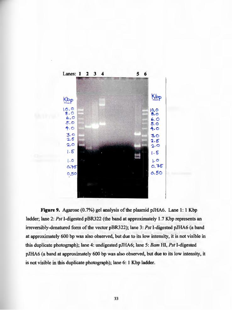
Figure 9. Agarose (0. 7%) gel analysis of the plasmid pJHA6. Lane 1: 1 Kbp

ladder; lane 2: Pst I-digested pBR322 (the band at approximately 1.7 Kbp represents an

irreversibly-denatured form of the vector pBR322); lane 3: Pst I-digested pJHA6 (a band

at approximately 600 bp was also observed, but due to its low intensity, it is not visible in

this duplicate photograph); lane 4: undigested pJHA6; lane 5: Bam HI, Pst I-digested

pJHA6 (a band at approximately 600 bp was also observed, but due to its low intensity, it

is not visible in this duplicate photograph); lane 6: 1 Kbp ladder.

33

ampicillin resistanc~ Eco RI (9459)

Pst1 (8706) (O) Bam ID(375)

SR-11 Fad- genomic DNA

1.4 Kbo chloramohenicol resistance cassette

tetracycline resistance

pJHA6 --9461 bp

SR-11 Fad- genomic DNA

ori

ampicillin resistance

Pstl(3606)

Figure 10. The plasmid pJHA6 mapped with the restriction enzymes Bam m, Eco RI, and Pst I. The ampicillin resistance gene (blue) is interrupted by the 4.5 Kbp Pst

I insert and a 600 bp Pst I artifact. The tetracycline resistance gene (purple) remains

intact. The arrows designate the direction of transcription. The gray arrow depicts the

origin and direction of replication. The numbers in parentheses, which denote base pairs

of DNA, are approximate.

34

(0)

pBluescrlpt II SK (+) 2961 bp

c~f El origin

lacZ

T7 promoter multiple

cloning site T3 promoter

Figure 11. Map of the sequencing vector pBluescript II SK+ (Stratagene, La

Jolla, CA). The multiple cloning site contains the Bam HI, Eco RI, and Pst I restriction

endonuclease sites. The blue arrow represents the ampicillin resistance gene ( bla) and its

direction of transcription. Two origins of replication (gray arrows) are included in this

sequencing vector. The lac Z gene is represented by the fuchsia arrow.

35

Lanes:

~p to. o 8 - 0 ~. o ~. o

; 4.0 3,0 ·-2.!i "l_.O

\. 5

\. C o.i-s a.so 0 .'25'

1 2 3 4 5 6 7 8 9 10 11

~p

10 .0 e.,o ~o 5.0 4.0 :!5. 0 2. 5 2.0

\. S'

1.0 0.15 o. 5o o. 25

Figure 12. Agarose (0. 7%} gel analysis of the plasmid pJHA 7. Lane 1: 1 Kbp

ladder; lane 2: undigested pJHA 7; lane 3: Pst I-digested pJHA 7 (the upper two bands

reveal that this digest was incomplete); lane 4: Eco RI-digested pJHA7; lane 5: Barn HI­

digested pJHA 7; lane 6: 1 Kbp ladder; lane 7: Eco RI, Pst I- digested pJHA 7; lane 8: Barn

HI, Pst I-digested pJHA 7 (the upper band at approximately 4 .1 Kbp reveals that this

digest was incomplete); lane 9: Barn HI, Eco RI-digested pJHA7; lane 10: Barn HI, Eco

RI, Pst I-digested pJHA 7; lane 11: 1 Kbp ladder.

36

SR-11 Fad_ genomic DNA

70 bp from right tenninus ofTn JOd

Bamm (5461)

1.4 Kbp chloramphenicol resistance cassette

Pstl(O)

BamID(13) multiple cloning site T3 promoter lacZ

pJHA7 -7461 bp

co!El origin

ampicillin resistance

fl (+)origin

EcoRf (4661) BamID(4061)

. I T7 promoter

multiple cloning site Eco RI (2954)

Pstl(2961)

70 bp from right terminus ofTnJOd

SR-11 Fad­genomic DNA

Figure 13. The plasmid pJHA 7 mapped with the restriction enzymes Ram HI,

Eco RI, and Pst I. The ampicillin resistance gene (blue arrow) remains intact. The

sequences of the T3 and T7 promoters (orange-brown diagonals) and the 70 bp inverted

repeats (orange-brown diagonals) flanking the chloramphenicol cassette (red) are known.

The numbers in parentheses, which denote base pairs of DNA, are approximate.

37

References

1. Todd, E. C. D. 1989. Preliminary estimates of costs offoodbome disease in the United States. J. Food. Prod. 52:595-601.

2. Edelman, R., and M. Levine. 1986. Summary of an international workshop on typhoid fever. Rev. Infect. Dis. 8:329-347.

3. Ivanoff, B., M. M. Levine, and P.H. Lambert. 1994. Vaccination against typhoid fever: present status. Bull. WHO. 72:957-971.

4. Curtiss III, R., and S. M. Kelly. 1987. Salmonella typhimurium deletion mutants lacking adenylate cyclase and cyclic AMP receptor protein are avirulent and immunogenic. Infect. Immun. 55:3035-3043.

5. Tacket, C. 0., D. M. Hone, R. Curtiss III, S. M. Kelly, G. Losonsky, L. Guers, A. M. Harris, R. Edelman, and M. M. Levine. 1992. Comparison of the safety and immunogenicity of &iroC &iroD and &cya &crp Salmonella typhi strains in adult volunteers. Infect. Immun. 60:536-541.

6. Jones, P. W., G. Dougan, C. Hayward, N. Mackensie, P. Collins and S. N. Chatfield. 1991. Oral vaccination of calves against experimental salmonellosis using a double aro mutant of Salmonella typhimurium. Vaccine. 9:29-34.

7. Stocker, B. A. D., S. K. Hoiseth, and B. P. Smith. 1983. Aromatic-dependent Salmonella sp. as live vaccine in mice and calves. Dev. Biol. Stand. 53:47-54.

8. Miller, S. I., W. P. Loomis, C. Alpuche-Aranda, I. Behlau, and E. Hohmann. 1993. The PhoP virulence regulon and live oral Salmonella vaccines. Vaccine. 11: 122-125.

9. Hohmann, E. L., C. A. Oletta, K. P. Killeen, and S. I. Miller. 1996. PhoP/ PhoQ deleted-Salmonella typhi (Ty800) is a safe and immunogenic single-dose typhoid fever vaccine in volunteers. J. Infect. Dis. 173:1408-1414.

38

10. Cardenas, L., and J. D. Clements. 1992. Oral immunization using live attenuated Salmonella spp. as carriers of foreign antigens. Clin. Microbiol. Rev. 5:328-342.

11. Utley, M., D. P. Franklin, K. A. Krogfelt, D. C. Laux, and P. S. Cohen. 1998. A Salmonella typhimurium mutant unable to utilize fatty acids and citrate is avirulent and immunogenic in mice. Submitted to: FEMS Microbiol. Letters on 2/24/98.

12. Ausubel, F. M., R. Brent, R. E. Kingston, D. D. Moore, J. G. Seidman, J. A. Smith, and K. Strobl. 1988. Agarose gel electrophoresis. Cur. Prot. Mol. Biol. 2.5:1-9.

13. Ausubel, F. M., R. Brent, R. E. Kingston, D. D. Moore, J. G. Seidman, J. A. Smith, and K. Strobl. 1994. Preparation of genomic DNA from bacteria. Cur. Prot. Mol. Biol. 2.4:1-5.

14. Ausubel, F. M., R. Brent, R. E. Kingston, D. D. Moore, J. G. Seidman, J. A. Smith, and K. Strobl. 1991. Alkaline lysis miniprep. Cur. Prot. Mol. Biol. 1.6: 1-2.

15. Ausubel F. M., R. Brent, R. E. Kingston, D. D. Moore, J. G. Seidman, J. A. Smith, and K. Strobl. 1996. Purification and concentration of DNA from aqueous solution. Cur. Prot. Mol. Biol. 2.1:1-3.

16. Ausubel, F. M., R. Brent, R. E. Kingston, D. D. Moore, J. G. Seidman, J. A. Smith, and K. Strobl. 1993. Rapid estimation of DNA concentration by ethidium bromide dot quantitation. Cur. Prot. Mol. Biol. 2.6:9.

17. Lodisb, H., D. Baltimore, A. Berk, S. L. Zipursky, P. Matsudaira, and J. Darnell. 1995. DNA ligase covalently links restriction fragments, p. 227. In Molecular cell biology. Scientific American Books, New York, NY.

18. Ausubel, F. M., R. Brent, R. E. Kingston, D. D. Moore, J. G. Seidman, J. A. Smith, and K. Strobl. 1988. Photography of DNA in agarose gels. Cur. Prot. Mol. Biol. 2.5:4.

39

19. Ausubel, F. M., R. Brent, R. E. Kingston, D. D. Moore, J. G. Seidman, J. A. Smith, and K. Struhl. 1993. Southern Blotting. Cur. Prot. Mol. Biol. 2.9:1-15.

20. Ausubel, F. M., R. Brent, R. E. Kingston, D. D. Moore, J. G. Seidman, J. A. Smith, and K. Strohl. 1997. High-efficiency transformation by electroporation. Cur. Prot. Mol. Biol. 1.8:4-10.

21. Elliot T ., and J. R. Roth. 1988. Characterization of Tnl Od Cam: A transposition-defective Tnl 0 specifying chloramphenicol resistance. Mol. Gen. Genet. 213:332-338.

22. Ausubel, F. M., R. Brent, R. E. Kingston, D. D. Moore, J. G. Seidman, J. A. Smith, and K. Strobl. 1994. Dideoxy (Sanger) Sequencing. Cur. Prot. Mol. Biol. 7.0:3-7.

23. Snyder, S., and W. Champness. 1997. Effects on genes adjacent to the insertion site, p. 208. In Molecular genetics of bacteria. ASM Press, Washington, D.C.

24. Schweizer, H.P. 1992. Allelic exchange in Pseudomonas aeruginosa using novel Co IE I-type vectors and a family of cassettes containing a portable orzT and the counter-selectable Bacillus subtilis sacB marker. Mol. Microbiol. 6: 1195-1204.

25. Blomfield, I. C., V. Vaughn, R. F. Rest, and B. I. Eisenstein. 1991. Allelic exchange in Escherichia coli using the Bacillus subtilis sacB gene and a temperature-sensitive pSC 101. Mol. Microbiol. 5: 144 7-1457.

40

Bibliography

Ausubel, F. M., R. Brent, R. E. Kingston, D. D. Moore, J. G. Seidman, J. A. Smith, and

K. Struhl. 1988. Agarose gel electrophoresis. Current Protocols in Molecular

Biology. 2.5:1-9.

Ausubel, F. M., R. Brent, R. E. Kingston, D. D. Moore, J. G. Seidman, J. A. Smith, and

K. Struhl. 1994. Preparation of genomic DNA from bacteria. Current Protocols in

Molecular Biology. 2.4:1-5.

Ausubel, F. M., R. Brent, R. E. Kingston, D. D. Moore, J. G. Seidman, J. A. Smith, and

K. Struhl. 1991. Alkaline lysis miniprep. Current Protocols in Molecular Biology.

1.6: 1-2.

Ausubel F. M., R. Brent, R. E. Kingston, D. D. Moore, J. G. Seidman, J. A. Smith, and

K. Struhl. 1996. Purification and concentration of DNA from aqueous solution.

Current Protocols in Molecular Biology. 2.1:1-3.

Ausubel, F. M., R. Brent, R. E. Kingston, D. D. Moore, J. G. Seidman, J. A. Smith, and

K. Struhl. 1993. Rapid estimation of DNA concentration by ethidium bromide dot

quantitation. Current Protocols in Molecular Biology. 2.6:9.

41

Ausubel, F. M., R. Brent, R. E. Kingston, D. D. Moore, J. G. Seidman, J. A. Smith, and

K. Struhl. 1988. Photography of DNA in agarose gels. Current Protocols in

Molecular Biology. 2.5:4.

Ausubel, F. M., R. Brent, R. E. Kingston, D. D. Moore, J. G. Seidman, J. A. Smith, and

K. Struhl. 1993. Southern Blotting. Current Protocols in Molecular Biology.

2.9:1-15.

Ausubel, F. M., R. Brent, R. E. Kingston, D. D. Moore, J. G. Seidman, J. A. Smith, and

K. Struhl. 1997. High-efficiency transformation by electroporation. Current

Protocols in Molecular Biology. 1.8:4-10.

Ausubel, F. M., R. Brent, R. E. Kingston, D. D. Moore, J. G. Seidman, J. A. Smith, and

K. Struhl. 1994. Dideoxy (Sanger) Sequencing, Current Protocols in Molecular

Biology. 7.0:3-7.

Blomfield, I. C., V. Vaughn, R. F. Rest, and B. I. Eisenstein. 1991. Allelic exchange in

Escherichia coli using the Bacillus subtilis sacB gene and a temperature-sensitive

pSClOl. Molecular Microbiology. 5:1447-1457.

42

Cardenas, L., and J. D. Clements. 1992. Oral immunization using live attenuated

Salmonella spp. as carriers of foreign antigens. Clinical Microbiology Reviews.

5:328-342.

Curtiss III, R., and S. M. Kelly. 1987. Salmonella typhimurium deletion mutants lacking

adenylate cyclase and cyclic AMP receptor protein are avirulent and immunogenic.

Infection and Immunity. 55:3035-3043.

Edelman, R., and M. Levine. 1986. Summary of an international workshop on

typhoid fever. Reviews oflnfectious Disease. 8:329-347.

Elliot T., and J. R. Roth. 1988. Characterization ofTnlOd Cam: A transposition

defective Tnl 0 specifying chloramphenicol resistance. Molecular and General

Genetics. 213:332-338.

Hohmann, E. L., C. A. Oletta, K. P. Killeen, and S. I. Miller. 1996. PhoP/PhoQ

deleted-Salmonella typhi (Ty800) is a safe and immunogenic single-dose typhoid

fever vaccine in volunteers. Journal oflnfectious Disease. 173: 1408-1414.

43

Ivanoff, B., M. M. Levine, and P.H. Lambert. 1994. Vaccination against typhoid

fever: present status. Bulletin of the World Health Organization. 72:957-971.

Jones, P. W., G. Dougan, C. Hayward, N. Mackensie, P. Collins and S. N. Chatfield.

1991. Oral vaccination of calves against experimental salmonellosis using a

double aro mutant of Salmonella typhimurium. Vaccine. 9:29-34.

Lodish, H., D. Baltimore, A. Berk, S. L. Zipursky, P. Matsudaira, and J. Darnell. 1995.

DNA ligase covalently links restriction fragments, p. 227. In Molecular Cell

Biology. Scientific American Books, New York, NY.

Miller, S. I., W. P. Loomis, C. Alpuche-Aranda, I. Behlau, and E. Hohmann. 1993.

The PhoP virulence regulon and live oral Salmonella vaccines. Vaccine.

11:122-125.

Schweizer, H.P. 1992. Allelic exchange in Pseudomonas aeruginosa using novel

ColE 1-type vectors and a family of cassettes containing a portable orff and the

counter-selectable Bacillus subtilis sacB marker. Molecular Microbiology.

6:1195-1204.

44

Snyder, S., and W. Champness. 1997. Effects on genes adjacent to the insertion site,

p. 208. In Molecular Genetics of Bacteria. ASM Press, Washington, D.C.

Stocker, B. A. D., S. K. Hoiseth, and B. P. Smith. 1983. Aromatic-dependent

Salmonella sp. as live vaccine in mice and calves. Developmental Biology

Standards. 53:47-54.

Tacket, C. 0., D. M. Hone, R. Curtiss III, S. M. Kelly, G. Losonsky, L. Guers, A. M.

Harris, R. Edelman, and M. M. Levine. 1992. Comparison of the safety and

immunogenicity of &iroC &lroD and ~cya ~crp Salmonella typhi strains in adult

volunteers. Infection and Immunity. 60:536-541.

Todd, E. C. D. 1989. Preliminary estimates of costs offoodbome disease in the United

States. Journal of Food Production. 52:595-601.

Utley, M., D. P. Franklin, K. A. Krogfelt, D. C. Laux, and P. S. Cohen. 1998. A

Salmonella typhimurium mutant unable to utilize fatty acids and citrate is avirulent

and immunogenic in mice. Submitted to: FEMS Microbiology Letters on 2/24/98.

45


Recommended