+ All Categories
Home > Documents > ISOLATION, PARTIAL PURIFICATION, AND CHARACTERIZATION OF … · ISOLATION, PARTIAL PURIFICATION,...

ISOLATION, PARTIAL PURIFICATION, AND CHARACTERIZATION OF … · ISOLATION, PARTIAL PURIFICATION,...

Date post: 27-May-2020
Category:
Upload: others
View: 21 times
Download: 4 times
Share this document with a friend
82
ISOLATION, PARTIAL PURIFICATION, AND CHARACTERIZATION OF GOAT ALPHA-ONE PROTEASE INHIBITOR BY PARVEEN SALAHUDDIN •PARTMENT OF BIOCHEMISrRY ^. N. MEDICAL COLLEGpf ALI&AJlH MUSLIM UNIVER'^SITY ALIGARH (INDIA) Date Approved:- M. Abnl Qadm, Supervisor A Dissertation submitted in partial fulfilment of the requirements for tlie degree of A/aster of Philosophy in BunUcmistry in the faculty of Medicine oj the /iligarn Musi; in Un.^'eiMty AUG AH,; 1986
Transcript

ISOLATION, PARTIAL PURIFICATION, AND CHARACTERIZATION OF

GOAT ALPHA-ONE PROTEASE INHIBITOR

BY PARVEEN SALAHUDDIN

•PARTMENT OF BIOCHEMISrRY ^. N. MEDICAL COLLEGpf

ALI&AJlH MUSLIM UNIVER'^SITY ALIGARH (INDIA)

Date

Approved:-

M. Abnl Qadm, Supervisor

A Dissertation submitted in partial fulfilment of the requirements for tlie degree of A/aster of Philosophy in BunUcmistry in the

faculty of Medicine oj the /iligarn Musi; in Un.^'eiMty AUG AH,;

1986

i

DS1301

ChRTIFIGATE

I cer t i fy t ha t the work presented in the following pages

has been carr ied out by Miss Perveen Salahuddin and t h a t i t i s

su i table for the award of M.Phil, degree in Biochemistry of the

Aligarh Muslim Universi ty, Aligerh.

/

(M. Abul Qasim) Lecturer in Biochemistry, Department of Biochemistry, J.N, Medical College, Aligarh Muslim Universi ty, Aligarh,

I I

In t h i s d i s s e r t a t i o n , i s o l a t i o n , p a r t i a l pur i f i ca t ion and

brief charac te r iza t ion of c - i - p r o t e a s e i n h i b i t o r from goat serum

i s described.

cx- i -proteese i nh ib i to r from goat serum was Isola ted by

subjecting i t to amrionium su l fa te f r ac t iona t ion , a f f in i ty chroma­

tography and ion-exchange chrcMnatography. F i r s t a crude preparation

of the i nh ib i t o r was obtained by col lec t ing a 50-80/^ ammonium

sulfa te fract ion which contained most of the inhibi tory a c t i v i t y .

This f ract ion was passed through a Cibacron-blue i?epharose 48 column

equi l ibrated with .01 M sodium phosphate buffer, pH 6 .8 , Ihis s tep

removed almost a l l of the albumin end ce r t a in other prote ins v*iich

had an af f in i ty for Cibecron blue. Finally the i nh ib i to r was pur i ­

fied by ion-exchange column diromatography on a i^i=At-cellulose

column ec^ i l ib ra ted with 0.005 M sodium phosphate ixiffer, pH 6 .5 .

The bound proteins were eluted using stepwise e lu t ion with 0,005 M

sodium phosi:*ate buffer containing 0.07 M NaCl and 0 ,1 M NaCl respec­

t i v e l y . Two peeks were obtained, c< -1-pro tease i nh ib i to r was

located mainly in the second peak. The purity of th i s preparation

was checked by polyacrylamide ge l e lec t rophores i s . One major and

two ve:cy f a in t bands were obtained, suggesting t h a t the i nh ib i to r

preparation was f a i r l y pure. bDS-polyacrylaroide gel e lect rophores is

gave two bands corresponding to molecular weight values of 67,000

and 56,000. The inh ib i to r thus i so la ted showed strong inhibi tory

ac t iv i ty against t ryps in i r r e s p e c t i v e of the fac t whether synthet ic

i l l

substrate like BANA or BAPNA was used or a protein substrate like

casein was used. Further it was found that one molecule of inhibi­

tor inhibited nearly three molecajles of trypsin indicating that the

inhibitor has multiple binding sites. The influence of two proteins

namely bovine serum albumin and porcine gamma globulin on the inhi­

bitory activity of the inhibitor was investigated at different

protein concentration. Interestingly, albumin caused marked

increase in the inhibitor/ activity of inhibitor whereas gamma

globulin had no effect. The reason for the enhancements of inhibi­

tory activity is not clear at present.

iv

I an greatly indebted to my supervisor, Dr* M. AbuX Qasin

for helping me at each and every ciucial stage of my research work.

I am also thankful to Prof, Salahudiih, Ghainnan, for provi­

ding me fac i l i t i e s for my research wozic.

I have no words to thank to my senior colleagues Mr« Saad

Tayyab, Mrs. Kinsshtar Salman, Mrs. Ra^eedunnisa, Dr. Sudhir Kimar

Agaxwal and Mr. Tauseef Saeed Khan.

I also appreciate the cooperation of my colleagues

Miss, Renu Tyagi, A^ss Najma M i , Miss Manjeet Kair, Miss Nausheen

Haleem Khan, Mr. Kri^nan Hajela, Mr. Mir A^zaffar, Mr. Khalid

Majid Fazli, Miss Sadhana Shazma and Miss. Seeraa Hassan.

I am also thankful to non-teaching staff Mr. Behzad Khan

and Mr. Mohd. Nasir.

I also acknowledge to Indian Council of Medical Research for

assistance as Junior Research Fellowship to st»jdy this project.

. . . . (Parveen Salahuddin) Aiigarn May, 1986.

D E D I C A T E D

T O

My l a t e ma te rna l Grand F a t h e r

Hi rza Aiwil Haaan Beg.

v l

Pag«

ABSTRACT III

ACaCNOWLEDGEJlENTS V

DEDICATIC2J VI

LIST OF TABLES IX

LIST OF FIGURES X

LIST OP ABBREVIATICM3S XII

I, INTRODUCTION 2

II, EXPERIMENTAL %$

A, Materials, 25

B, Methods, 29

1) Meastir«nent of pH^ 29

2) O o t i c a l measurement 29

3) Determlnatlcm of p r o t e i n coiic«ntrati<»». 29

4) Ion-exchange chr<Mnatography on DEAE- 31

c e l l u l o s e .

5) Preparat l<»i of a f f i n i t y coltam 32

6) Polyacry lamide g e l e l e c t r o p h o r e s i s , 33

7) Soditsn do-decy l s u l p h a t e e l e c t r o p h o r e s i s ^ 3

8) I s o l a t i o n and p u r i f i c a t i c m of g o a t o C - 1 - 37

P r o t e a s e i n h i b i t o r ,

i ) ^imonium s u l f a t e f r a c t i o n a t i o n , 37

i i ) A f f i n i t y chrcwnatography on Cibacrcai 38 b l u e Sepharose-4B.

i l l ) DEAE c e l l u l o s e chrc»natography ,jj

v i i

Page

9) Measurement of Inhibitory activity of 39

C7^-1-protease inhibitor.

i) Using Casein as substrate, 39

ii) Using BAWJA as substrate. 40

iii) Using BANA as substrate, 41

III. RESULTS AND DISCUSSION 43

1) Isolation and p\irification of goatc<:-l- 43

protease inhibitor,

2) PAGE 55

3) SDS-PAGE 55

4) Influence of serton albumin and gamma 58

globulin on inhibitory activity.

5) Effect of inhibitor concentration on its 58

inhibitory activity,

IV, REFERENCES 59

V, BIOGRAPHY 64

VI, LIST OP PRESENTATIONS 65

viii

LIST OP TABLES

Paqm

Table I / nlnoacld and carbohydrate conqpositicms of hvBian« 5

rat, isotise and rabbit (Fast and Slow cooEponent)

oc-1-protease and mouse ccmtrapsin.

Table II Half time of association of different proteinases 19

with htanan plasma inhibitors.

Table III Coinparison of physico-ch«aical properties of 21

htsnan, rabbit, rheusus intmlcey and rat <^-1-protease

inhibitor.

Table IV List of chemicals used in this study, 25

Table V Isolati<»j and purification ofc^ -1-protease inhi- 47

bitor fran goat plasma. Inhibitory activity was

assayed using BAPNA as std:>strate.

Table VI Isolation and purification ofc^-1-protease inhi- 48

bitor from goat plasma. Inhibitory activity was

assayed using SANA as sijbstrate.

Table VII IsolaticHi and ptirificaticai of <3 -1-protease inhi- 49

bitor frc»n goat plasma. Inhibitory activity was

assayed using Casein as substsate.

Table VIII Molecular weight and relative iwability for diff- 53

erent proteins in sodium do-decyl sulfate polyac-

rylamide gel electrophoresis.

ix

I4»7 OF FX6URES

Fig, 1 fmiRO acid mmtemnoB of Inasaiicx -l-protcafttt inhibitor. 4

Fig, 2 StrtKsttirs of oligosacchari(!te coi^»oii«nt of oc -l«|>rot- 10 ease i sh ib i tor .

f i g , 3 Hypothetical structtara of inhibitor. 14

Fig. 4 Cos^^arisioii of inhibitory ac t iv i ty i» aiff«racit 42

nasRalat goat, s h a ^ buffalo «id rsdobit.

Fig, 5 Affinity chromatography &a Cib«srao bliMi sephartMMi 44

-4B and locatiem of inhibitory ac t iv i ty in diff«»

rtmt fracti<»i9 tasiag BAH4A, BmA «8id Casein aa

substrates.

Fig, 6 Iaa««xch«ige chromatography on DE E .. ce l lu lose 45

«ttJd location *f inhibitory ac t iv i ty in different

fractions ttsing«BM^A, BMA mad Casein as subst­

rates*

Fig, 7 PolyiK;ryl«Ride gel eleetrophcnresis of oC-l-ilirotease, 50

inhibi tor .

Fig,8(A) Sodium do-diKsyl sulphate poly«^ryl«Ride ge l e l s e - 51

tz^::^hore8is of cX-l-protease inhibitor*

Fig,8(B) Sodium do-decyl sulphate polywsrylisnide gel e l e c - 52

trophoresis of >x-l-f>rot«aee inhibitc»r and marker

proteins*

Fig, 9 Plot of logrsthims of molecular iraight verstui 54

re la t ive mobility.

Fig. 10 Sffact of 8SA and IgC5 on aZbunto a^l«t«*oC- l - 56

protease inhibitor .

Fig. 11 Effect of diff«r«nt inhibitor coiic«atrati«ai cm 57

i t s inhibitory a c t i v i t y .

3ei

LIST GF ABBREVIAnONS

AT-III

e\ -l-Achy

c< -i-Ac

< -.2-AP

BAHA

BAPNA

C.-Inh.

DEAE-cellulose

HCl

NeCl

NaOH

A - i - P I

I - ^ - I

RBC

SDS

TEMED

TGfe

Tris

Antlthrorabin-III

(^ - i-Antichymotrypsin

oC-i-Anticoliagenas#

oc -2-Antiplaan in

c/:-N-Benzoylargine napthylaroide

(^-N-Bpnzoyl arginine-DJUp-nitroermilide HZl

Cj^-irihibitor,

Diethyl aminoethyi c e l l u l o s e

Hydrochloric acid

Sodijirj chlor ide

Sddium hydroxide

c^ - i - P r o t e a s e i n h i b i t o r

In t e r -oC- i - an t i t ryps in i r t i i b i t o r

Red blood corpuscles

Sodium dodecyl s u l f a t e .

N,N,N» ,N»-tetraraethylene athy lenedianine

Trichloroacet ic acid

Tris (hydroxy raethyl)methyl amine.

x i i

ISOLATION, PARTIAL PURIFICATION AtJD CHAR^TERIZATION

OF GOAT ALPHA ONE PROTEASE INHIBITOR.

I,NTR(yXJgTIQN

i^rotease i nh ib i to r s ere present in p lants micro-organians

(Sves and mcmmals. Their presence was f i r s t demonstrated by Fertni

ouu jt ernoss i beck in 1894. In mamtnals protease inh ib i to r s are

believed to perform several important functions such as connective

t i s s u e turnover, coagulation, f i b r i n o l y s i s , complement f ixa t ion e t c .

They are found in various c e l l s as well as in d i f fe ren t body f lu ids

including plasma. In plasme t h e i r concentrat ion i s about lO,o of

the t o t a l plasma protein , i t cons t i t u t e s the l a rges t group of plasma

protein a f te r albumin and gamii a qlobul in . Though various types of

protease i n h i b i t o r s have been recognized the exact physiological

ro le of most of the protease i n h i b i t o r s i s unknown. The reason i s

t h i s t h a t not many pathological condit ions a re known where j u s t one

of the protease inh ib i to r i s missirwf or de f i c i en t end secondly most

of the i nh ib i to r s inh ib i t several d i f fe ren t proteases . The physio­

log ica l ro le in such cases can only be derived ind i rec t ly from the

k ine t i c s of inh ib i t ion of various proteases with the i n h i b i t o r .

In the following few pages a br ief desc r ip t ion of molecular

proper t ies of x - l - p r o t e a s e i nh ib i to r from human are presented.

Typ?$ 9f Pypt?^^? |nh4W9ys

Protease inhibitors are classified according to the type of

protease which they inhibi t . The four major classes of proteases

are as follows:-

1. Thiol proteases

2. Metallo proteases

3, Aspartic proteases

4, Serine proteases.

Inh ib i to rs for a l l the four c lasses of proteases are known.

For example A -1-protease i nh ib i t o r (^ - i - P I ) group and the i n t e r

/ - t r y p s i n inh ib i to r ( I - T C - I ) i n h i b i t s only serine proteases , x - 1 -

cysteine protease inh ib i to r ( ^ -1-C3*I) i n h i b i t s only cysteine pro te ­

ases \fchereas ?<- i -an t i -co l l agenase {.\~iAC) i n h i b i t s only collegeno-

l y t i c enzymes of the metalloenzyme c l a s s . In cont ras t t o these c lass

specif ic i n h i b i t o r s < -2-Mecrogiobulin (-^-2-M) i n h i b i t s members of

a l l the four protease c l a s s e s .

Jf various c lasses of protease i n h i b i t o r s , ' ^ 4 - P I i s the

best understood. I t s physiological ro le has been es tab l i shed . The

primary function of i nh ib i to r i s to control the turnover of lur^

connective t i s s u e proteins by inh ib i t ing the e l a s t a se enzyme. The

deficiency of t h i s enzyme causes increased turnover of lung connec­

t ive t i s s u e causing the development of pulmonary emphysema, U>mtt

of the important protease i n h i b i t o r s which belong to t h i s c lass are

A- i -p ro tease i nh ib i t o r , Antithrombin I I I (AT- I i l ) , A -2-Antipiasmin

(c<-2-AP), .Tv-i-Antichymotrypsin ( ^ - i - A c h y ) end CI- inhib i tor

( Cl-Inh, ).

Primary St ruc ture ;

The primary s t ruc ture of human c;C-i«protease inh ib i to r has

P i g . l , Amino acid seqiience of human oC-1-antitrypsin ( C a r r e l l . e t . a l , , 1982.)

AMINO ACID SEaUENCE OF HUM ANC^-1-ANTITRYPSIN

n

« u

« n

g

« u

g

o •

s ^ o « •p «

n 4>

P o «

«B « 3 +» O «S E

Is o « e o ^

•a (4

+> •o c •H 3 O I

t

C ^ •H +> 6 -H

o u

sg !3

II o o

U E

«

1 ^ O

m u a

« o n C C

O « a « ^

c 0 « V U

0 o

©f-4 00

•» o •

14

ff) a

05 E S * « •^ %

T 4 ' ^

•rl B ^

2 2

C CO t> -P •H ©r-* O

M O M ' - ' E CU

GO

tfl

fO

N

• • • • • • • • • • • • g

• • • • • • • • • • • *• s

« ^

o

G o u

m • • • • • • • • •

«*> M M »0 rH »-« N

w •

00 m

• o*

«o •

a>

IT)

o* «nt^oc^»0'*oo<or*•a)<^ • • • • • • • • • • • •

o> o o o o o r ^ o ^ o D « o t ^ » - « ^ t * -

CM

C4 m

O « M 0 0 0 ' * 0 0 0 0 0 0 • • • • • • • • • • • •

o* r 4 « - i t ^ o o ^ t n e o < * « M C 4 0 o

o o o o o o o o o o o o o o

O O O O O O O O O O O O O O • • • • • • • • • • • • • •

\D M t * ' T « r « - O t * - 0 0 » - » C O p « M 0 0 O

•H •p

o

II s

§ «

« •

« •rl

« c c

c e c u « d „ -H -ri •a c -H M P O > 1 « r - ( r - i 4 J

r-l 1,4 iH i-l « 0 e 0, O < S > £

M M P

t t CO O

« C

B rH

o

CO CO

tn

« o • %0

• o

c* •

m o •

tfl • Vd

Q

o> • Ov ^

m • «>

» • o e*

e g s

M • O •-I

««» • t"

o • o\

in • <fi

«0 rH • o ^

'* • r

in • 0* rt

« • w g O

t«4

<8

CD

in

£5

m w •

o • » m

w •

tn

w • CO

in •

o •

r4 W

o> • C4

O •

^

P • 2

m • If •-I

o • 10

o • in

M • in

« • ^

« •

• in < — » «

Q • S

m • o

tn • o

« c 0 z

m

c<

o •

tn '»

o • w ^

o t <«-*

o • m w

o •

«-i «

o • "* «

Q • Z

o • m

o • o

o • Ov t-l

o • m

o • CD

O • «

o • to

o •

Oi

o • l>

o •

T4

«n • o

2

g 2;

c

I e 5 e o ^

^

1 s 1 >« -P § g & B

4» B « O •

III ao i

0 c s £

• « 0 4* o e m o

g o •H «-<

(Q

o o 3

o

« « 0

^

been sequenced by c l a s s i c a l sequencing techniques (Johnson and

Travis, 1978;TroVls e t a l , 1978; Shochat e t a i . , 1978; Garrel i

e t a l , , 1979) cloning and sequencing of c *»A for baboon end human

Ck - l- i^I (Keruchl and Chandra e t a l . , 1981).

Humantx -1-pro teese i n h i b i t o r i s a r e l a t i v e l y sraail and polar

prote in . Thus, i t eas i ly passes through extravoscular spaces

( C a r r e l l e t a l . , 1982), The amino acid composition of ^ - l - p r o t e a s e

inh ib i to r from various animal species , such a s , human (Travis , 1974),

rhesus monkey (Berninger e t a l . , 1976), r a t (Rol l and Glew, 1981),

mouse ( Takahara and ^inohara, 1982) r abb i t (Koj e t a l , , 1978) has

beef) determined. Ihe amino acid composition from these animals i s

sumriarized in Table I . All of them have s ingle polypeptide chain

end are glycoprotein in na ture . The human < -1«PI vdiich i s by far

the best studied of a l l the Inh ib i to r s contains 394 amino ac ids . I t

has glutamic acid a t i t s I^-teitninal and lysine a t G-terminai. I t

contains 12% carbohydrate by weight, cDNA s tudies reveal tha t ^ - 1 -

protease inh ib i to r do not have pro-peptide but has a 24 residue

hydrophobic or pre-s ignei peptide (Carlson and i,tenflo, 1981). The

i nh ib i t o r molecule has e s ingle r e r c t i v e centre methionine loccted

a t posi t ion 358 in the amino acid sequence. Acidic amino acid

residues are concentrated a t N-terminal and basic amino acids are

more abundant at G-terminal.

ex - l - p r o t e a s e i nh ib i to r lacks d i su l f ide bonds. Instead i t

has a single cyste lnyl residue which forms d i su l f ide bond to e i t he r

cysteine or g lu ta th ione . The reason for t h i s pecul iar bond i s s t i l l

6

a mystery (Jeppson et a l . , 1976). However, i t i s thought t h a t in

nat ive s t a t e ^ - i - p r o t e a s e i n h i b i t o r ex i s t s in reduced s t a t e having

free t h i o l groups which perhaps pro tec t X - l - p r o t e a s e i nh ib i t i on

for being oxidized by chemical o r b io log ica l oxidants (Travis and

Salveson, 1983),

Reactive Centre:

The reac t ive centre of CT^-l-protease i n h i b i t o r has been

debatable. Due to i t s o r i g i n a l name x - i - a n t i t r y p s i n , lysine and

arginine were thought t o be present a t i t s r eac t ive cen t r e . On

ace ty la t ion of lys ine and arginine by maleic anhydride (Heimberger

e t a l . , 1971), c r i t acon ic anhydride (Johnson, 1978) and acet ic anhyd­

r ide (Fretz and Gan, 1978) the i n h i b i t o r a c t i v i t y was abolished.

On modifying with less bulky methyl group, inh ib i tory ac t iv i ty

was not l o s t (Busby and Gan, 1976). However, various s tudies on

chemical modification of methionine residue suggest t h a t t h i s residue

i s primarily involved in the i n t e r a c t i o n with t a r g e t pro tease .

.< - i - p r o t e a s e i n h i b i t o r i s capable of i nh ib i t i ng an e n t i r e

ser ine c lass although with d i f fe ren t r a t e s . The presence of mult iple

reac t ive cen t re was therefore suggested (Feste e t a l , , 1981; James,

1978; Cohen, 1979; Martodam, 1981). But l a t e r on i t was revealed

t h a t on d i s soc ia t ing the complex of o^- i -p ro tease i r tn ibi tor to any

of these ser ine proteases i . e . t r yps in , chymotrypsin, plasmin or

e l as tase by nucleophiles , and the same peptide was cleaved in each

case

9

Third controversial debate bout the reactive eentre was

the position of the reactive c«itre methionine. Most of the workers

cited that methionine is located near N«.terminal (Panell et.al;

1974? Mor*i et.al., 1978f Crawford, 1973| Cohen et.al., 1978). This

ccmfusion arose because on coRiplex dissociation the cleaved peptide

binds throt^h hydrc hofoic forces to N-terminal. This jjeptide can

however, be dissociated frcrni .terminal by strong detergents like

sodium dodecyl sulfate ^ca^rell et*alf 1979f Morii et.al, 1979).

C^ analyzing the amino acid sequence of both nidified inhibitor and

the peptide fragment, the overlapping amino acid seqpience was obtained

(Travis and Johnscm^ 1978 ) thi» proving that reactive centre

methionine is located near Ct^rmixial.

Another question which was asked by workers (Del Hars et.al;

1979| Nakajima et.al., 1978f Mc Rae et.al., 1980) in this area is

that y an easily oxidizable group like methi(^ine is present at

the reactive centre. Apparently the presence of methionine at the

reactive centre appears to be disadvantageous, since this methionine

group is very prone to oxidation and thereby inactivating the inhi­

bitor.

In fact, it has been found that replac^R^t of methionine by

valine at the reactive centre prodxices an alnwst equally effective

inhibitor which has the added advantage that it cannot be inactivated

by the various cocidissing agents which are produced in the cell.

Three explanations have be«n given sti fgesting the importance of

n«thi<»iine residue at the active sitet-

10

SIALIC ACID SIALIC ACID SIALIC ACID

GALACTOSE

GLcNAc

MANNOS

MANNOSE I

GLcNAc

GLc U Ac

GALACTOSE I

GLc NAc

ASPARANGINE

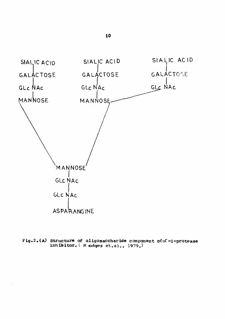
F i g . 2 . ( A ) Structure of ol igosaccharide component ofoC-i-protease I n h i b i t o r . ( Hodges e t . a l . , 1979.)

11

SIAL

MANNOSE-

GLcNAt

GUN Ac

ASPARANGINE

C ACID

GALACTOSE

GLc NAc

MAN NOSE

Fig.2 . (B) Structure of oligosaccharide component of (^C-l-protease i n h i b i t o r . ( Hodges e t . a l . , 1919,)

12

i) Rate of inactivaticm of neutrophil elastase Is faster when

methicmyl residue is present at its active centre?

ii) Methionine reactive centre provides lung tissue remodelling

to occur dxiring oxidatiem. Had there been any other amino

acid residue it would have been very difficult to remodel

the Itsig tissue.

iii) The process of phagocytosis requires an active participation

of proteolytic enzymes. Inactivaticm of these cmsBymes by

the inhibitor wDuld have deleterious effect on the whole

process. The control over the inhibitory activity of inhibi­

tor is provided by simultaneous secretion of oxidants by

phagocytising cells which inactivate inhibitor in the variety

of phagocytizing cell. This process would be possible only

if methionine residues were present at reactive centre,

Caybffhydfftt ig

Several workers have studied the structtire of carbohydrate of

htananoC-l-protease inhibitor. Two tyxses of oligosaccharide units

are proposed to be attached at a total of four sites in the inhibi­

tor molecule. The oligosaccharide structure was proposed by Hodges

«t.ai,(l979) (see Fig, 2), However, there is sc«ne dispute about

the type of oligosaccharides and the nuinber of attachment points to

the protein molecule. For example. Roll et.al.Cl978) have proposed

four different types of oligosaccharide units attached at 3-4 points

in the protein ttralecule. The oligosaccharide units are composed of

mannose, galactose, N-acetylglucoseamine and sialic acid.

13

Effect of Chemical and 3ioIo:jiceI Jxidents on .<-L^Protease Inhibitor

The presence of methionine residue at the reactive centre

generated an intense investigation of chemical and bioiogicei

oxidants effect on X-i-protease inhibitor (Johnson et al., 1978;

Laskowski et ai., 1979; Jannof, 1979; Travis, 1979; Cohen et si.,

1979). Jn oxidizing chemically with iM~chlorosuccinamide or N-bromo

succinamide the biological activity was abolished, aiological

systems generate oxidants by utilizing H2J2, CI" (Carp, 1978;

Matheson et al., 1976; Matheson et al., 1979, 1980; Clerk et al.,

1981) and myeloperoxidase. Ihe biological oxididants thus generated

also inactivate the inhibitor.

Secondary Structure

The secondary structure of cZ-l-PI has been studied by

Jirgenson (1977) by C.D. spectral measurement. His result suggests

the presence of 30 --< -helix, 40; P> -pleated structure and 30/o

random coil. Thus the protein contains significant amount of secon­

dary structure. In the absence of disulfide bond in this protein

the major stabilizing forces for the nctive structure are believed

to be hydrophobic and electrostatic interactions.

Teritarv Structure

Molecular organization of oC-i-protease inhibitor reveals it

to be highly ordered structure. u>berman et al. (1984) have proposed

a three dimensional structure of modified inhibitor through crystal-

14

Met358

394

Z342

P i g . 3 . (a) The reac t ive cent re of oc- i -an t i t ryps in i s exposed on residue 342, ( s i t e of the z-mutation.)

Ser359

Met

358

Fig.3*(l9) Cleavage by t a r g e t enzyn» r e l ea se s the loop to give the s table post complex form with met 358 seprated by 69 AOfrara ser ine 359.

IS

io^raphic s tud ies . The protein molecule has dimension of 67/. x

32 A^ X 32 A®. / c id ic and basic amino acids are present ot oppo­

s i t e ends of e i i i p s o i d e l molecule, thus making molecule polar in

character . This highly ordered s t ruc tu re primarily serves to fix

the reac t ive centre on the exposed s i t e ,

Carrel l et e l . (1985) hypothesized t h a t in modified i nh io i -

to r the s t ruc ture of reac t ive s i t e i s 'orung with methionine 35B dt

one end of the molecule separated by 69 A^ from serine 559 at the

other (see Fig. 3 ) . in the in t ac t i nh ib i t o r methionine 358 must

be plucked out from a P - sheet to give s t rained loop ( see Fig. 3)

joining with ser ine 359. Thus the cleaved i nh ib i t o r is thermodyna-

miceily s table and the i n t a c t i n h i b i t o r i s me t e s t a b l e , which expic^ins

the d i f f i cu l ty in c r y s t a l l i z i n g the native >:-1-protease inh io i to r .

This view of molf^cule with an exposed react ive centre s u i t s with

the observation such as , vu lnerab i l i ty of a reac t ive centre to

chemiccl and b io logica l oxidants .

Role of Carbohydrate

Recently (i-eter et a l , , 1985} revealed the fac t , tha t the

function of carbohydrate moiety i s to s t a b i l i z e the native conforma­

t ion of-^' -1-protease i nh ib i t o r . This was p rac t i ca l ly demonstrcted

by these workers tha t the half l i f e of unglycosylated -^^-i-protecce

inh ib i to r i s shorter in comparison to glycosylated < - i -pro tecse

i nh ib i t o r . The protein synthesized in yeast or :. Coli

(Rosenberg e t a l . , 1984; Hil l e t a l . , 1984) by DNA-recombinant

16

technolo^ is devoid of carbohydrate moiety, -ach an inhibitor

nnolecul^ is likely to be cleared rapidly from the blood circuletion.

Standard Mechanism

Interaction between protein end protease inhibitor follows

the standard mechanism. The general criterion for the standard

mechanism ere the following (Travis et ai, 1983).

i) Rate of association between an enzyme and inhibitor should

be faft.

2) Rate of dissociation of enzyme and inhibitor complex should

be slow.

The x-ray data (i-askowski, 1980; Robert et ai., 1972)

revealed that the geometry of an active site should be optimal i.e.

it should act as a good substrate. Now the question arises what

makes an inhibitor act as an inhibitor rather than act as a good

substrate. In terms of structural analysis this problem has not

been solved but kinetically it can be explained as follows

The Kcat/Km value for the hydrolysis of the peptide bond in

the inhioitor following the standard mechanism is very high but the

individual value of Kcat and Km alone is very low ( Laskowski, i980J.

Therefore, peptide bond hydrolysis is very slow. This hydrolysis

of peptide bond is not irreversible but rather reversible. Hence,

an equilibrium near jnity is formed. Consider the general standard

17

mechenism.

I + 'i > EI^ > C >£!-• -rh + I*

K-i

where t. is th€- protease and I and I ere virgin and modified inhioi-

tors respectively and C is the stable intermediate complex. The

stable complex C can be formed from either virgin or modified inhi­

bitors. 3ut the apparent rate of association of modified inhioitor

with c- nzyme is rather slow in comparison to virgin inhioitor.

Deviation from the ;:.tandard Mechanism;

o<-l-protease inhibitor follows the standard mechanism

partially. The rate of association between enzyme and inhibitor

is very fast and the resultant complex so formed contains one mole-

culdi of each enzyme and reactants, aut it deviates from the stand­

ard mechanism in other aspects (Travis et al,, 1983).

1) Modified ^-l-proteese inhibitor is inactive and cannot

recombinc with protein. The inhibition mechanism of

^-i-proteese inhibitor obeys the following scheme:

K. K^ K^

L + i - > tL ^ I * >b + 1*

K-i

where I cannot reassociate with enzyme i.e. reaction mechanism is

not reversible; hence, modified inhibitor is inactive.

18

2) The strength of the in t e rac t ion i s so strong, therefore i t

has t o be covalently s t ab i l i zed .

E f f e c t 9 f Ui 9He ffp ^^yym o^- j -Fyp^^g?? ^nh4^3,^y

Pulmonary emphysema i s associated with low level of c i r cu l a ­

t ing iA -1-protease i nh ib i t o r . On the other hand c i g a r e t t e smoking

increase the r i sk fac tor of emphysema. Cigaret te smoke a t t r a c t s

neut rophi ls in lung which in turn r e l ea se b io logica l oxidants , and

thereby, oxidize the <-X: - i - p r o t e a s e i nh ib i t o r . The r a t e of i n t e r ­

action of oxidized c< - i - p r o t e a s e inh ib i to r i s 3CX)0 times slower

than na t ive i nh ib i to r and therefore i t f a i l s to control the e las tase

a c t i v i t y ,

Another group of workers (Beith e t a l . 1985) suggested that

smoke does not oxidize methionine as already claimed by several

workers. Bather a compound present in c i g a r e t t e modifies the basic

amino acids of^^ -1 -PI . This a l t e r s the nat ive conformation, which

in turn reduces the r a t e of i n t e r ac t ion of enzyme and inh ib i to r ,

int^y^gtion 9f^~i-i^irqt???e ;nh4'JU9y wj,t!) knV^ Pr9^?^s$

In terac t ion of -X - i - p r o t e a s e inh ib i to r with serine proreases

has been studied in human and in mouse ( Takahara and oinohara, i9S3j.

In terac t ion of human X -1-protease inh ib i to r with d i f fe ren t

ser ine protease such as pancreatic and neutrophil e las tase (bchwic.

e t a l . , 1966; Jannof e t a l . , 1972; daugh and Travis, 1976, Trypsin,

u O +» •H

(0

(0

c

(A (0 c

+> o M

a o c o

•H • P •0 o o (0 (0 <D

'H O

(1>

I

ID

to

(4 O

O « n>

M O

o

I (0 (D C

a, e

^5

I •H C ,-4 0) (0 (

i (0

6i^

I

£ C

a

19

3 I

(0 « »-4 «) UJ-f*

5

in

o -4 » N • -4

1

1

1

1

1

o o -< X »n •

CO

t

1

0*

3 X •

o X o

+

1

1

«n~ q -4 X cn •

<N

i

t

ID

X -H CM

in

o

CO

00

o

8

tf)

X

m

•o CM

tf> CO

>o • o

M 0. 1 ^ 1 <0 JC

a

1

>» x: o < i -1 i «0 JC

a

CM • N

s u (D =! CM i <0 x: sx

CM

fO CO en

MD 00

JC

o

CM

8 o CM

1

IS

c •H

1

1

8 o :4

r«-NO

a <

I CM

i ID

00

c o

•H

O to u +» c o 2

(0

o •H

t c •H

C o

• P to >

•H +» U c

20

chymotrypsin (SchiwicX e t*a l l 1976)* cathepsln G (Beith and Travis#

1930)I thrcmdsin (Matl^son et:«al««i976)« plasnin (Sharpio e t . a l . ' 1966;

Harcz et .al«# 1966)* acrosin (Frltas et«al .* 1970}* t i ssue kalllkrein

(Fritz et .a l« ' i969)« Factor Xa (£ l i l san at«ai.« 1962)« F4K;tor Xia

(Scott e t . al .» 1982). skin synovial collafenase (Ibkoro e t . a l * ' 1972)

an^ Urokinase (Harris et* al*« 1969) has l»een stu^ie^.

In pet i t ion to t h i s saicraliial serine proteai^ (Berfvist*

1963; i^icher* 1973; Sasaki* 1975) i s a lso inactivated tey^-l-pro-

tea^» intiD»itor. aut ti^e inactivatiem i t s e l f cannot c lar i fy the

physiological role ofc^^l'-protease inhi]»itor rather the rate of

ine^tivation i s of priaary importance* Meamiresaent of tlie rate of

aasociati(»i shows that the rate of in^:tivatioR i s fa s t e s t with

neutrojE^il e lastas^ (see Talnle ZZ) ssiffestinf that pr^ary function

ofc/-1-protease inhibitor Is to control td^ e l a s t o l y t i c ac t iv i ty .

"nrypsiii inhi)»itory capacity in mouse i s greater in coraperison

to humancT^-l-Pl due to ttie presence of two isofozm inhU^itors. One

of the isof orni i s ccmtrapsin which i s a)i«»nt i s man and t^e other

isoform iso<-l-antitrypsin ti^ich i s homologous to huaanoc^-l-

protease inhi]»itor. In mouse these two isoforms have different

inhibitory spectrum. Mouse ^-1 -protease inhi)»itcr inactivates

chymotrypsin* e l a s t a ^ and thrOBri»in« where as mcmse ccmtrapsin inhi­

bited trypsin* plaanin* and trypsin-l ike protests from sub-maxillary

glands. Ibe dif ferent inhibitory spectrum of Uiese two isofax^jts

suffest t^at these two isoinhibitors d i f fer in their physiolofical

r o l e s .

(0

>.

o B 3

X)

C

§ x:

I u

u • •^ IB to V

f|

O 4*

«

C

o • H

CO

a o

CO

o

(2 S

•a ^^ O , o*

(A a> • 3 C M 119 " H ^ <i> Cx:

•f» • H 4 J jQ «

I '^ - I «

c 10 (0 e "H

8 •H

a 8

a

u

Q

D

Z

00

CM

in I

• • o fo

o

Q 2

Q

e 3 o >

O '2 ' "^ JQ4J0O <V < Crg a

0) (0

•ri U O «^ ^ t H - 4 "H O H ' *>

9 ®^ li o. o< «o

21

c

8

o B «0

4 * * 0

-« o t3 n

Q

2

a 2

§

*

I

o o

•H O

•H C

x: •H

H (0

3

o S

o (0

g 6 <0

to

c

g •2

^

i ^ _ 3 O N «in OB -*

o 4*

> o

•-« o

o.

o

«

a

&

o

• • • Q Q Q

• » • 2 2 2

K Q • •

•H o 2

^ 1 5 . ^

00

H I • - • >0 '(T

CO

^ 00 I

CM

4 *

M o •p «» >» x: u

u 4» u

O JO 2 C O -H -4 •H<H X3w4 X i fH Q) 4 ) O B <0 H «0 O

22

Genetics end Variants;

Human c^-1-protease i n h i b i t o r occurs in a t l e e s t twenty

d i f fe ren t forms in general population. This protein i s encoded by

two independent a l l e l e at a s ingle locus r e s j i t i n g in autosomal

codominant inher i t ance . The va r i an t s are c l a s s i f i ed according to

t h e i r e lec t rophore t ic mobil i ty . For excm^le, PiBB (where ^ • i s

protease i nh ib i to r ) i s horoozygote for anodal va r i an t s and txZi i s

hoiaozygote for cathodal var ian ts and PiMM represents the horoozygote

for normal a l l e l e . These two genetic var ian ts i . e , b and Z, c<use

decrease in X -1-protease i nh ib i t o r concentration in plasma. This

can be explained as fo l lows:-

in i, var ian ts mutation of glutamic acid occurs at amino acid

residue 264 to Val ( Carrel l e t a l , , 1976), whereas in Z. var ian ts

subs t i tu t ion occurs a t glutamic acid 342 to lysine (Jeppson, 1976).

These two giutonic acid in normal M-allele forms s a l t bridge and

s t a b i l i z e s rX- l -p ro tease i n h i b i t o r molecule. Subs t i tu t ion of these

two glutamic acid cause decrease in s t a b i l i t y which was also shown

by heat s t r e s s method. This increases the turnover of v>^-i-piotease

i nh ib i to r and thus resu l t ing in decrease in ^ ' - l»pro tease i nh ib i to r

concentration in plasma from iOl-130 mg/lOO ml to 15 m^/lOO ml.

Physical Properties;

Physical proper t ies of ^X- i -pro tea te i nh io i to r s of d i f f . r t .n t

mararals are t eoa l t t ed in Table 111,

23

Isolation of c:<-I-protease inhibitor has been performed by

several workers using a variety of techniques. Norm ally isolat ion

of (X - i -protease inhibitor i s performed in the following s teps: -

( A ) E^DOval of Albunin

(a) Salt Fractionation

( Z) Ion-exchange Chrotaatography.

( A ) fieffi9Yg4 9 l AA^fflJ^n

Since albumin and o< - l -protease inhibitor have seme molecular

weights and i soe lec tr ic points» therefore, they caiuiot be separated

by gel f i l t r a t i o n or by ion exchange chromatography. However, they

can be separated in two wayst-

( i ) Affinity QiroffiatoaraDhv on Cibacron»blue SeDharQse-4B

It binds albumin through bil irubin binding s i t e and to those

proteins having dlnucleotide fold but not cx-i-proteese inhibitor

from plasma.

(2) Affinity O ffflgtMr^p^Y gn ffgn A- ep^gfpgg 4P

It binds (j< -1-protease inhibitor and most of the glyco­

proteins but not albumin.

Albumin depleted plasma, isolated from previous step i s

subjected to sa l t fractionation. The inhibitor fraction i s used

for further purification.

24

(C) Ign E?^c^^nq» (;^r9ii|i^^oqraphY

Fiirther pur i f ica t ion can be carr ied out by ion-exchange chro­

matography on DEAE-celiulose or DEAE-sephadex using l i n e a r gradient .

S i t e s of o^ . l - p r o t e a s e Inh ib i to r Svnthes is t -

The s i t e of i nh ib i to r biosynthesis has been studied by

using three techniques:

i ) Radio-labelled amino acid techniouei -

By t h i s technique c< -1-pro tease i n h i b i t o r synthesis was

detected in macrophages, monocyte and lymphocyte (Wilson e t a l . ,

1981).

2} |mipyp9flv9y^?geot g"d p$p9XJld?g^ St^l^l^nq ^?ghr^J^qMgg:~

By these two techniques synthesis of ex -1-pro tease inh ib i ­

tor i s found to occur in kupffer c e l l s , b i l i a r y epithelium - i s l e t of

Langerhans, i n t e s t i n a l epithelium, macrophages, neutrophi ls and

hepatocytes (Eriksson e t a 1. , 1981; Geobes, 1982, Ray e t a l . , 1977).

Recently Franklin and Baumann (1985) discovered t h a t in

mouse species (M.crota l i ) (^ -1 -pro tease inh ib i to r expresses i t s

mRNA in kidney ra ther than in l i v e r which i s then secreted in ur ine .

The physiological ro le of o^ -1 -p ro tease inh ib i to r so synthesized

i s not c lear a t present , however, i t i s capable of inh ib i t ing

t rypsin and chymotrypsin.

25

A* MataertaXs

The chemicals an€ ot lwr mater ia l s use^ in ^ I s study are

suiranar i ze i here •

TABUB IV

L i s t of chemical used in t i l l s s tudy.

S.NO.

1 .

2 .

3 .

4 .

5 .

6 .

7 .

8 .

9 .

1 0 .

1 1 .

1 2 .

1 3 .

1 4 .

1 5 .

Chemica l s

A c e t i c a c i d

Acetone

Acrylamide

Amldo schwao^tz

Ammonium p e r s u l f a t e

Ammoniion s u l f a t e

Annmoniuro sulphamate

BAPMA

BAKA

Blue i:textran-200

Bovine serum albumin

Brcmiophenol b l u e

Br<»ilne

Calcium c h l o r i d e

Chloroform

S p e c i f i c a t i o n

AR

lA

X«R

LR

LR

LR

AR

Lot

Lot

Lot

Lot

LR

LR

LR

LR

No.

No.

No.

No.

119 C-

98 C-

1388

,19 C-(

-045

-055

3115

Source

BDFL I n d i a .

BDH* Ind l a .

M}H« England.

E.Merck* Germany,

Mitu in<? l a .

- do -

E.Merck* Germany

Sigma Chero.Co./

USA.

do -

Pharmacia f i n e .

Slfraa Che.Co«

USA.

BDH« Enf land.

BDH# I n d i a .

Sarabhal* Ind l a .

BDH# Ind l a

16. c* -Chymotryps Inofen^ M t N0.986-317P Sigma. CheiB.Co,9SA»

26

S*HO« Chemical

17 • CiJsacron-blue

18* Cooraassle b r i l l i a n t blue

19, Copper s u l f a t e

2 0. Dia lyzer tubing

2 1 . Dichlorod iinethyl-s i l a n e

S p e c i f i c a t i o n

AR

LR

LR

1/100* diameter

LR

2 2 , D i e t h y l ©nino e t h y l c e l l u l o s e

2 3 . Disodium hyelrogen phosphate

2 4 . Ep ich lo rohydr in

2 5 . E thano l

2 6 . Glycero l

2 7 . Glycine

2 8 . Hydrochlor ic ac id

2 9 . linmunoglobulin Type-G

30. Lithium s u l f a t e

3 1 . ^- raexxaptoethanol

32. «k - l -naph ty l - emine 'el i h y d r o g e n c h l o r i i e

3 3 . N-N*-methylene b i s a c r y l ^nide

34. N , N # N ' # N ' - t - t e t r a methylene^laraine

35 . Orthophosphor ic ac id

36 . Ovalbtnain

3 7 . pH paper

Lot NO.59C-0243

Source

Sipaa Chem.Co.,USA .

- do -

sm» I n d i a .

Arthur H. Thcxnas' USA.

E.Merklc* Germany.

Sigma Chem.Co;USA.

AR

AR

Q i e m i c a l l y pure sample

LR

LR

AR

AR

LR

LR

LR

l&

LR

LR

Lot

i^H-:

/

HO.A-^503

L-10

BDH# I n d i a .

BDH# I n d i a .

Bloger^ India*

Serva* Germany.

BDH, I n d i a .

Sigma Chem.Co;USA.

E,Merck» Germany.

Ca lb iochem.Cal . USA,

BDH# England.

Koch-Light -Lab. England.

Fluka* AG* S w i t z e r ­l a n d .

BDH# I n d i a .

Sigma (%em.Co.USA.

E.ttexrek. Germanv.

27

S.NO.

38,

39 ,

4 0 .

41*

4 2 .

4 3 .

4 4 .

4 5 .

46 .

4 7 .

4 8 .

4 9 .

5 0 .

5 1 .

5 2 .

53*

54 .

5 5 .

5 6 .

5 7 .

5 8 .

59 .

QieraicaXs

Potassium ^ichronste

PotassitsQ hy^rofen* phtha la te . Potass i i ^ pemansanatis

Ril»oflavin

RiJaonuc l e a ^ -A

depharose-4B

Sodium azicie

sodium i»icaxliionats

Sodium carbcmate

sodium chXoride

sodiiaa djUiydrofen phospiiate

sodiura dodecyl mi l fate

sodium h/droxlde

sodium molykdate

soditua n i t r i t e

sodium potassium t a r -tara te

Sodiuoa tetralaorate

Sodiisn tung s t a t e

soya Isean tryps in inhi)>itor.

SDcrose

Sulfuric aci^

Tr lch loro-ace t i c acid

S f e c i f i c a t i o n

AR

AR

AR

LR

l o t . No. 184916

hB.

IM

LR

W.

LR

AR

LR

I«R

AR

X^

LR

AR

Iot.No.29C-8040

LR

LR

AR

Source

SDH, l ^ i a .

- do -

- do -

B.!4erck« Gez^any.

Sisco* Ind i a .

Pharm«M:ia fiiva Chem. tJpsala* SMsden.

PKi FblasHie* Poland,

BDH* India.

- do -

Glaxo-Lab.« Indla.

- do «

BDfe Englead.

3DH# India*

- do -

S. Merck# Germany.

QDH* Ii%3 i a .

Sara^a i« M. I i^ l a .

ffl3H« India .

Sigma chem.Co.f USA.

mM* India.

SaraMiai M.Chem.m India.

Rei<i e t De Haen» Germany.

28

S.No. Chanlcals Specification Source

60, Tris(hydroxy-m ethyl) LR methyl amine

61. Whatman f i l t e r paper No. 1

BDH, England.

W and R Balston Ltd., England.

All glass distilled water was used throughout the experi­

ments.

29

pH measurements were made either on ELIOO U-iO pH-«ieter

using EXieo glass and calomel electrode or on EC digital pH-meter

model pH 5691 in conjunction with EC eonbination electrode. The

{^•Hoeters were routinely standardized with 0«05 M potassium hydro"*

gen-pthalate, pH 4,01 (at 23®c), in the acidic range and 0.01 M

sodium tetraborate buffer pH 9.2 (at 25^C) in the basic range.

Light absorption measurement in the visible range were made

on Carl Zeiss Jena spectrocolorimeter, Speckol.

Protein concentration was deteirmined by the method of JU ry

et al . (1951). Bovine sexum allMimin was used as standard.

The Folin-phenol reagent was prepared according to the method

rec(^mended by Folin and Ciocalteau (1927). In a round bottmn flask

50 grams of sodium tungstate, 12.5 grams of sodiwn molybdate* 25 ml

of 85^ orthof^os|:^oric acid and 50 ml of hydrochloric acid along

with ^ 0 ml of water were taken and refluxed for about ten hours.

Then 75 grans of litiiium sulfate, 25 ml of dist i l led water and few

drops of liquid bromine were added to i t . The mixture was heated

30

vdthout a condenser t o reiBov« excess of bromin3. After cooling the

br ight yellow coloured reagent was d i lu ted t o 300 ml by adding

d i s t i l l e d weter. Then the contents were f i l t e r e d and stored in an

amber colo^ired b o t t l e . This reagent was d i lu ted four times before

use .

(>3) Pyep^raU9n yf cPPP^y yggflgPlf

Ihe copper reagent was prepared using the following solu-

tionsj-

i ) 4% sodium carbonate (W/V)

i i ) 4^ sodium potassium t a r t a r a t e (w/v)

i i i ) 2% copper su l fa te (w/V)

Solutions ( i ) , ( i i ) and ( i i i ) were mixed in the r a t i o of

100 j l : l i n t h i s sequence to avoid p r ec ip i t a t i on . The br ight blue

coloured reagent was f i l t e r e d before u se .

(c ) Ii!,y9,gedarff.

To one ml of protein solution, five ml of freshly prepared

copper reagent was added and mixed well and was kept for ten minutes

at room temperature. To this was added one ml of diluted Folin-

phenol reagent. Colour was allowed to develop, for 30 minutes then

the colour intensity was read at 700 nm. aiank was also prepared in

a similar fashion except that in place of protein buffer was taken.

The protein concentration was computed from the standard plot of

bovine serum albumin.

31

Twenty grams of DEAE ce l l u lo se was f i r s t soaked over night

in d i s t i l l e d water. Then the f in 6 s wer e removed thoroughly by

decantat ion. After t h i s the r e s in was regenerated according to the

method recotnraended by Bailey (1967). F i r s t the res in was equ i l i b r ­

ated with 0 .1 M NaQH to bring the res in in sodium cycle. Then the

res in was washed on a bucchner funnel t i l l the pH of the s lurry was

cbout 8, After t h a t the res in was t rea ted with 0 .1 M Hydrochloric

acid. Again the res in was washed extensively with water t i l l i t

was free from acid and f ina l ly equi l ibra ted with the s t a r t i ng

kwffer.

Packing of column

Column was f i r s t washed with detergent and was then rinsed

with d i s t i l l e d water. Lower end of the column was f i t t e d with l a ­

tex tubing and the column was mounted v e r t i c a l l y in vibrant f ree

pos i t ion . Lower end of the tubing was closed by pinch cock and

glass wool was inserted a t the lower end and few glass beads were

added. One th i rd of the column was f i l l e d with operating buffer.

Moderately d i l u t e slurry was poured with the help of glass rod. when

the res in had s e t t l ed t o few centimetre the lower end of the tubing

was opened slowly to half of the operating flow r a t e . Then flow

r a t e was increased gradually t i l l i t was s l i gh t ly higher than

required.

32

Epichlorohydrin cross-blinked desuiphated sepharose 43 was

prepared according to the method of Porath e t a i , ( i 9 7 i ) . 20 grao

of Sephcrose-4B suspension was t rea ted with 18 ml of IM NaOH and

2 ml of epichlorohydrin e t room toaaperature in tho presence of

40 mg of sodium borohydride. The suspension was heated to 60^c with

s t i r r i n g on water bath and the react ion was stopped a f te r two hours*

The gel was washed extensively with d i s t i l l e d water on a laiucchner

funnel t i l l i t was free from a l k a l i ,

aiue-sepharose 43 was prepared by coupling cibacron blue to

epichlorohydrin cross linked sepharose 43 according to the procedure

described by Bohme e t a l . (1972). A solut ion of 200 mg of cibacron

blue in 50 ml of water was added dropwise with vigorous s t i r r i n g t o

a 10 gm suspension of epichlorohydrin cross- l inked sepharose-.43 in

350 ml of water a t 60*^C. After s t i r r i n g for 30 minutes, 45 j r im^of

sodium chloride was added and s t i r r i n g was continued for about one

hour. After t ha t the mixture was heated to SO^C and 4 grams of

sodium carbonate was added to the mixture and s t i r r i n g c3ntinued

for another two hours a t t h i s temperature. After coo l i i^ the

mixture was brought to room temperature and the gel wes washed

extensively with hot boil ing d i s t i l l e d water on a bucchner funnel.

I t was f ina l ly washed with the equ i l ib ra t ing buffer.

33

PolyacrylaBitelc gel electro^Bhoregls

Polyacrylaml^« f s l electrofA^orests of - l - p ro t ea se Inh ib i ­

t o r was car r ied out In 7»0% ecryleaaliie gel a t pH 8.3 according to

the procedure recoiamenBled by Davis (1964), Stock so lu t ions for

polyacrylsBRi«ie ge l e l ec t rophores i s %»re prepared as given Iselow.

l ) Stock Solutions

Solution A : -

Porty e igh t i n i l l i l i t r e of 1 N |4C1, 36.6 grams of t r i s

and 0,2 3 ml of 1EMED were taken. Tlie volxjane was maie up to 100 ml

with water and pH was adjusted t o 8.9»

Solution Bi -

Forty e i g h t m i l l i l i t r e of 1 N HCl, 5.98 graos of t r i s#

0.46 ral of TSMED weire taken in water . The pH was adjusted to pH 6,7

and the volume was made up to 100 ml with d i s t i l l e d water.

Solution C i -

Twenty e igh t grams of scrylami^e and 0,735 graaa of N-N*-

methyleiw bis-acrylamide %#ere dissolved in water anci f i na l volume

was m ^ e up to 100 ml.

Solution D : -

Ten grams of acrylamide an^ 2,5 grams of N-N*methylene

his acryl&ai&e were dissolved in water anA then the volume was made

up to 100 ml.

34

F i r s t of a l l gel tubes ( 0 . 5 x i i cm) were throughly washed

end dr ied . The tubes were s i l iconized with a 1% dichlorodimethyl

s i iane in chloroforra. After drying, the tubes were fixed ve r t i ca l ly

in a stand by inser t ing t h e i r lower end in to the well of rubber

stopper. Each tube was f i l l e d with 2.2 ml of small pore solut ion.

The surface of gel solut ion was careful ly layered with a few drops

of water with the help of a syringe t o get uniform s j r face . The

polymerization was complete within twenty miruite. After polymeri­

zat ion water layer was removed with the help of w.hatman f i l t e r

paper. Then 0 .5 ml of large pore solut ion was t ransfer red in to each

gel tube and the surface was covered with a few drops of water

before keeping the tubes under f luorescent l i gh t for photppolymeriza-

t ion . After the la rge pore gel had polymerized the water layer was

again removed. The gel tubes were then fixed in e lec t rophore t ic

tank such t h a t the lower ends of the gel tubes were in contact with

the buffer in the lower chamber and the upper ends were in the upper

chamber. Now the assembly was connected t o the power supply^

Protein sample in glycerol was poured gently to each tube. Then the

remaining space in the tube was f i l l e d with e lec t rophores i s buffer.

F ins l ly , the buffer was poured in the upper chamber such t h a t the

upper end of the gel tube i s completely dipped in e lect rophores is

buffer. After t h a t , few drops of 0.02% broroophenol blue dye was

mixed in the upper chamber. Electrophoresis was carr ied out a t 210

vo l t s with a current of 4 mA per tube. When bromophenol blue dye had

moved more than three fourth of the t o t a l gel length, the power

'.^

supply was turned off and the ge ls were removed from gel tubes by

squir t ing water between gel and the ge l tube with t h e help of a

syringe f i l l e d with a long needle. The ge ls were stained with IA>

amidoSchwartz (Stock solut ion G) solut ion for on© hour. Destaining

was done mechanically by shaking the gels with 7. (V/V) ace t ic acid.

Sodium dodecvl su l fa te DOlyacivlamide oel e lect roohores ia

SDS-PAQE was carr ied out according t o the method of i «eber

and Osborn (1969).

The small pore so lu t ion was prepared by mixing 7 ml of

stock solut ion C, 10 ml of gel buffer (0 .2 M sodium phosphate buffer,

pH 7.2 , containing 2X> SDS), 0 .5 ml of ammonium persul fe te (1.5^) and

0.02 ml of TEMED and 2 .5 ml of d i s t i l l e d water.

The solut ion mbcture was then, careful ly poured in s i l iconized

gel tubes end with the help of a syringe and then few drops of water

was layered cereful ly over the surface of t h i s so lu t ion . The solu­

t ion was then allowed to polymerize a t room temperature for 30

minutes a f te r t ha t water was r^aoved by soaking : t with wiatman

f i l t e r paper.

(b) PreoareUon of protein sample

Ten milligrams of prote in in two ml of 0.2 M sodium phosphate

buffer containing ij> SDS and IX> f' -merceptoethanol was taken in e

3 6

t e s t tube and heated in a boi l ing water bath for 5 minutes. The

protein solut ion was then cooled to room t«nperature and a few

drops of bromophenol blue and 0.4 ml of glycerol were added. Thirty

to f i f ty mic ro l i t e r of protein sample containing nearly 60-100 ug

protein was then applied over the surface of small pore gel in each

tube. The tubes were then placed in e lec t rophores i s assembly in the

manner described above. The e lec t rophores is was performed using

0.2 M sodium phosphate buffer contcining 0 .2^ i^S, pH 7 .2 , as

e lec t rophores is buffer. A current of 8 mA/tube was passed t i l l the

dye front had migrated to a d i s tance of about 5 cm from the point of

sample appl ica t ion . After e lec t rophores is the gels were taken out

from the gel tubes and stained for protein by coomessie b r i l l i a n t

blue R-250.

( c ) Staining

The s taining of the pro te ins and desta ining of the gel was

performed with coomassie b r i l l i a n t blue R-250 by the method of

Fairbank et a l . (1971), The following solut ions were prepared for

stc'ining and destaining of ge l s .

i>olution A

I t contains 25/& (V/V) isopropyl alcohol , lD,o (V/V) ace t ic

acid end 0.05% (W/V) cjomas&ie b r i l l i a n t blue R-250.

Solution B

It contains 10^ (V/V) isopropyl alcohol, lO,o (V/V) acetic acid

and 0.05^ ( w/V) oommassie brilliant blue R-250.

37

Solution Cs

This solut ion contains 0.0028^ (W/V) cjomassie br i i i ian 'c

blue and iO;*5 (V/V) ace t ic acid .

With the above three solut ions s ta ining and desteining takes

place siimilteneously (Fairbank e t a l , , 1971). immediately a f te r

e lect rophores is each gel was dipped in about 25 ml of solution A

at room temperature for overnight . After t ha t the gels were t r ea ted

with solut ion 3 for a t o t a l period of 8 hours and f ina l ly ge ls were

t rea ted with solut ion C for 6 hours. The destaining process was

complete with another 5-6 washings w-ith 10;^ (V/V) ace t i c acid.

I so la t ion and pur i f i ca t ion of ^ - l - a n t i t r v p s i n from goat plasma

- / -1 -p ro t ea se i nh ib i to r was i so la ted using three techniques:

1) Ammonium su l fa te f r ac t iona t ion .

2) Affinity chromatography on Cibacron blue s.epharose-4B.

3) Ion exchange chromatography on DEAE-cellulose a t pH 6 . 5 .

For the i so l a t i on of ^ - l - p r o t e a s e i nh ib i t o r fresh samples

of blood were col lected from the local s laughter house in t he prese­

nce of appropriate concentration of anticoacpjlant l ike tr i-sodium

c i t r a t e . R8C from the c i t r a t e d blood was removed by centr i fugat ion

at 4000 rpm for 10 minutes and the plasma thus^pto|p^^l;M»6S^fcde

^^ c/ V - ' s.'' .

38

50% saturated with awionijura s u l f a t e lay adding jneh^ul^ite amounts o£

s o l i d aBsnonium s u l f a t e . Ihe preci4pitati(»3i was allowed t o take place

o v e m i f h t at 4°C» The contents were then centrlfu^ad at 6000 rpai for

half an hour and thm p r e c i p i t a t e which contained very l i t t l e i n h i b i ­

tory a c t i v i t y was discarded and ^ e supernatant was l»r(»ight t o 80%

aRsnonium s u l f a t e saturation* Afain* i t was Icept overnight at 4°C,

Next day the s o l u t i o n was centr i fuged a t 12000 rpra f o r half an hour

in a r e f r i f e r a t e d c e n t r i f u f e . Prec ip i ta te so obtained was d i s so lved

in minimum volxime of O.Ol M sodium ^oapnatM buf fer i ^ 6«8 and was

dialyased e x t e n s i v e l y against the saeie buf fer .

(2) AEf i n i t v chromatography <Ma Cibacron blue ^pharo8e-4B

serum albumin and(^-l-*protease i n h i b i t o r have nearly stfoe

molecular weights and i s o e l e c t r i c points* Iherefore* the ir separa­

t i o n i s d i f f i c u l t e i t h e r on g e l f i l t r a t i o n or on ion exchange. This

can however be separated on Cibacron blue a^pharose 4B* s ince t h i s

t r i a z i n e dye binds albumin* <:?c-l-antichymotrypsin# coagulat ion f a c ­

t o r s and to those prote ins having d inuc leot ide fold# l i k e dehydro­

genases*

Itie prote in preparation obtained by 80% airanonitxn s u l f a t e

p r e c i p i t a t i o n (about 50 mf/40 ml) was applied on a cibacron blue

Se{^aros«-4B (5 cm x 7 cm) column equi l ibrated with O.Ol M sodium

phosphate buf fer pH 6.8* Column was operated a t the rate of 40 « l / h r .

Unbound prote ins were e luted In a s i n g l e p r o f i l e , Ant i trypt ic a c t i v i t y

was assayed in each fr^:ti(H}s and inh ib i tor r i c h f r a c t i o n s were pooled

for further pur i f i ca t ion*

39

(3) DEAE'-celluloae chrxaraatOTaiBhv

Fract ions r ich in antitry]»tic a c t i v i t y were d ia lyze*

a f a i n s t 0.005 M sodium phOFhate buffer/ pH 6.5 an«l applieei on

D£AE-ceIlulose column ( 7 cm x 1.66 cm ) . The coliamn was washed

with two l»ei volumes of t h i s buffer t o remove unbound ]»roteins.

Bound pro te ins were e luted by incorporatinf 0.07 M NaCl and O.lOO M

NaCl in b u f f e r . Anti trypt ic r i ch f rac t ions were pooled.

III.Maasurement of inhib i tory a c t i v i t y of " l -nrotease inh ib i tor

( A ) The inhib i tory a c t i v i t y of -1-protease i n h i b i t o r

was assayed accordinf to the methocH of Anson (1938) us inf case in

as subs tra te .

Tfen mi l l i frams of tryps in in 10 ml of 0. Oi M Tris-HCl

(jrti 7 .7 ) conta in inf O.Ol N caiciurn chlor ide was tftk;en» case in so lu ­

t ion was prepared by d i s s o l v i n f 2 §m of c a s e i n in 0,06 M sodium

phophate buf fer pH 7.7 in 100.0 ml.

To one ml of protein* 0.1 ml of tzrypsln was added an t h i s

was incubated a t 37°C for 2 0 minutes. Then 3 ml of 10% TCA was added

and the reac t ion mixture was afain incxibated a t 37'* C for one hour.

A white p r e c i p i t a t e was obtained which was f i l t e r e d o f f . Control

was prepared in s imi lar fashion also* except that c a s e i n was ad^ed

a f t e r stoppinf the reac t ion with 10?4 TCA. The f i t r a t e so obtained

from the sample and contro l were assayed f o r t h e i r hyerolyzed pro­

duct by the method of Lowry e t . a l . # (1951) .

The inhibitory activity of CK-I-protease inhibitor towards

the S3^thetic stjbstrate was measured using BARN A and BANA,

(l) Assay of ex -1-protease inhibitor using BAPNA as si±>strate»

The inhibitory activity ofc<-t-protease inhitor using

B4PNA as substrate was assayed according to the method of

Waheed and Salahuddin (1975),

Ten milligrams of BAENA was dissolved in 0,15 ml of dimet­

hyl sulfoxide and the voliaroe was made to 10 ml with 0,01 M

Tris-*iCl, (pH 8,0) . Trypsin (10 mg/10 ml) was dissolved in the

same buffer containing 0.01 M calcixan chloride.

To one ml of proteiir, 0,1 ml of trypsin was added and the

reaction mixture was incubated at 37* C for ten minutes, Th«i

erne ml of BAPNA was added to this reacticm mixture and was

incubated for five minutes, Th®r» the reaction was stopped with

one ml of 30% acetic acid, TRe colotir intensity was measured at

410 nm,

(ii) Assay of A-1-protease ii dJsitor using BANA as substratex

The inhibitory activity of -1-protease inhibitor was

measured according to ^je method of Martineck et,al. (1964)

with slight nradificaticm. Ten milligrams of BANA was dissolved

in 0,5 ml of dimethyl sulfoxide and the voltime was made to

10 ml with 0,05 M sodiian phosphate buffer pH 6,2. Ten milli­

grams of trypsin was dissolved in 10 ml of 0,01 M Tris-HCl

buffer pH 8,0 containing lOmH of calcitim chloride.

41

"^ one n l of lnhiJ»itor« O.l »1 of trypsin was aided and

Inculiated for 30 minutes at 37 C« After addlnQ 0.5 naX of SANA i t

was afain lncid»ated for 30 nlnutaes at 37°b. fhe reaction was

stopped liy the addition of 0.5 n l of 4 N HCl. llhe ccmtrol was

prepared in the s ^ ^ manner* except that ttim mlistrate was added

after the addition of 4 Ij HC1«

Ihe ^Rcnint of 2-nap^ylaBii» fozswd during tkm hydrolysis

of SANA at pH 6 .3 was determined toy couplinf and dias^t lsat ion.

This was done hy addinf 0«S ml of maiiiam n i t r i t e (0*2^ w/v) to

t^e solut ion. Ihe contents were i^aken thoroughly and ex&ctly

after three minutes* one ml o£ aamxaiAjm sulj^^aate (0«5^/v) were

added. Exactly after three mJUnutes two ml of couplinf dye i . e .

N-l-Napthylethylene diamine dihydrochloride (0,<B^w/v) in etha-

nol was added, The colour was allowed to develop for about 40

minutes and the intensity oi v i o l e t blue colour was read at 540 rm

42

PROTEIN CONCE/MTPATION LY\ W 3

P i g . 4 . Comparison o^ Inhibi tory a c t i v i t y in d i f f e ren t mamals; goat C O ) ' sheep ( # ) ' >^uffalo ( ( J >' r a b b i t ( • ) .

Inhibi tory a c t i v i t y was assayed according to the method of Waheed & Salahuddin (1975J

43

Before s t a r t i ng these s tud ies , preliroinaiy experiments on

the concentration of -X^-i«protease i nh ib i to r from the sera of

various animais such as goat, buffalo, sheep and rabbi t were perfor­

med in order to s e l e c t a source which contains highest concentration

of i n h i b i t o r . For inhibi tory ac t iv i ty meaairement from these sera

30-80% ammonium su l fa te f rac t ion was obtained and inhibi tory ac t iv i ty

was measured against t r yps in . The r e s u l t s are shown in Fig. 4 . I t

can be seen t h a t a l l sera contain inh ib i to ry a c t i v i t y . However,

there are considerable quan t i t a t i ve differences , whereas in tiie

CtiQ of rabb i t serum 2.74 mg prote in was required t o cause 50%

inh ib i t i on , the goat serum was several time more ef fec t ive and

only 0.4 mg of protein was requiired to causa 50;^ inh ib i t ion under

iden t i ca l condi t ions . In the case of buffalo and sheep 0.74 mg and

1.25 mg was required to cause 50!^ i nh ib i t i on . These preliminary

s tudies suggested t h e t of the four sera t r i e d highest concentrexion

of inh ib i to r was present in the esse of goat serum. For t h i s reason

goat serum was selected as the source of i n h i b i t o r in these s tud ies .

n . l§9igU<?n end ourWggtlOT Qi ^9?\ ^ "I'pygtttgs^ inhi^^tgf

The 50.80^ ammonium su l fa te f rac t ion obtained during ammonium

sulphate f rac t iona t ion of goat serum was subjected to a f f i n i t y

chromatography on Cibacron blue Sepharose 4B. This step was essent ia l

44

40 80 120 160 200

ELUTION VOLUME.tT\4

Pig,5, Affinity chrcmiatography of 80% ammonium sulphate fraction on Cibacron blue sepharose- 4B,

The column (5cm x 7 cm) was equilibrated with ,01 M sodiiBTi phosphate buffer, pH 6.8. About 30mg of 80% ammonium sulphate fraction was applied on the coltimn and the unbound protein was collected in 10ml fraction at a flow rate of 40ml per hour. Inhibitory activity was located in different fractions using BAPNA ( ^ ) , BANA ( • ) , and Casein ( f) ) as sxdbstrate.

45

NOIi iaiHNIdO •/.

UJ

Z ID _l O >

UJ

1-0-; m u o o i IV 3 0 N V i m s N V a i » / ,

§d 10

46

since albumin, which i s the major plasma protein present, i s similar

to '^ -1-proteese inhibitor in molecular weight and electxo phoretic

rK>bllity and without prior removal of albumin the i so lat ion of the

inhibitor free from aliMmin contamination i s very d i f f i c u l t . The

aff inity column would bind albumin as well as a l l other proteins

which contain dinucleotide fold such as dehydrogenases, but not

oc - l -proteese inhibitor among other proteins. The aff inity coiuotfi

chromatography result i s shown in Fig, 5, Almost a l l of the inhibi­

tory act iv i ty was accounted for by the unbound fractions. The bound

fractions (mostly albumin) which could be isolated by eluting the

column with 2 M potassium thiocyanate in buffer was found to be

virtually free from the inhibitory ac t iv i ty .

Finally cx- l -protease inhibitor was purified by ion exchange

chromatography on a DEAE-cellulose column \AAiich was previCMJsly equi­

librated with 0.005 M sodium pdiosphate buffer, pH 6 .5 . About 36 mg

of protein in 40 ml of 0.005 N sodium phosphate buffer, pH 6.5, was

applied to the ion exchange colunff). The unbound proteins were remo­

ved by washing the column with 0,005 M sodium phosphate buffer, pri

6 .5 . The bound proteins were eluted by stepwise incre&se in ionic

strength. Two peaks eluting at ionic strength 0.07 and 0.1 were

obtained ( see Fig. 6 ) . The protein fractions obtained frc»n 0I:>.£-

ce l lu lose column were also monitored for inhibitory act iv i ty against

trypsin using SAPNA, SANA and Casein as substrates ( see Tables V, VI

end VII respect ive ly) . Both the protein peaks showed signif icant

inhibitory ac t iv i ty . However, the inhibitory act ivi ty was compara­

t ively more in the peak obtained at 0.1 M ionic s t r ^ g t h . Active

47

4J

s u

c <

• u 0

•H

a 45

« t» e ® 4J 0 U 0> T

r* 1

+> o QL 01

m 0

6 0

•H *J « o "H

^ P.

•0

i c o •H

4J ra o CO

M

• 0 •p « b 4> e

^ 10

10 «0

< z 0 .

SI

0> c •Hi

m 0 •0 ®

^ n to

1

> t

•* •H >

«

1

28 •O-ri 'H

M

<

^ > - » m to «

--« © ^

4J 0 6 0 M-*

S4 a

ffi Hf i ^ « 9»-( •»r-» g 0 O-^ n >

m

43 01

•o \0

o o

«0 o o

in

1-1 in «

CM

M r*

• o

M in o

• <ri

m 00

• w

fO

w •

tfi

o N •

VD

<M 1 ^ n fO

in «o m ^

CO T ^

w

00 o rH

o o o in

o o

E

&§ S () E » « 0 ^ «1» M-H 0

J>^0 0, •p ^ © •H >iCO

»M M'HW *« CPA**

gci ^g O Q

« c

x: JC n U 0 . 0

8 M :3 C 0'-* 0 . P «

M «D O

0 z

• CO

• H

• H H

• H M M

• > H

48

o

u n. U O

Xi •H

o a

I

m o

c

o •H

c

c o «H •f* «0 '^ o

|2

(A 3

"S 3 (ff « e iff

s >.

4J

O

, - 5

»4

^ ^ «M ^ CT •H > 6

CO m o

•+» p o>

« 3'-«! 4> r-i M|)

8 >o O

C4 •

n in

« h-

« -« •

03

8 ^ » •

o o o

00 H CM

tM

9 CM

3

s O 14

COQ

O

« 4» <0

(0

AS

94* •H C C «0 Q 4 J E m | g

6)

^ a ^ (0

«M

3

•H

c

O N COM-

M >

U)

49

a

<

u $ •H JO •H

•g

to •

a m

I m

(0

O "H

cn « 10

O O

•o

^ • H O

Co

10 8 8

C 0) o c •H ^ 4-* (0 « 3 O

r* >< M <9 3 M a (A m •o c <n flS CO -. » c o >, •H 4» • * •H <0 > •H ^ O -P M «

• M M > «

Xi .« H

H !

8. « •» U)

s • W)

5 P s

8 CM

S Ch

-* o

s o

s o

-I « >o isT

a •

o

pi ^

« •

o

s IQ

-« •

-4

If)

9 3 N

Pi •

-4

8 •H

v:> •

•H

:1

s

<o a* a ^ «o -* Q 2 O **

Q

«

II « 3

O Q> M U

M

50

P l f . 7 , Poly aery lamide f e l e l e c t r o p h o r e s i s of goat cx- i -protease Inh ib i tor .

E lec trophores i s was performed in Tr i s - f lye i i i e buffer pH 8.3* 7.0% polyacrylenaide g e l . About 60uf px^tein was applied on § e l tube and e l e c t r o p h o r e s i s was perforraeg f o r ^ o u t 2 hour« with anodic current of 4 inA/tube. itie g e l s were stained wit^ cooraassie b r i l l i a n t blue and dest-ained with 1% a c e t i c ac id .

51

F i g . 8 , ( A ) Sodium io^ecyl sulphate polyacrylamlde gel e lec t rophores i s of goat JC-1-protease i n h i b i t o r .

Electrophoresis was performed using 0,2M sodium phosphate buffer pH 7,4 containinq 0, iJi SDS on lO)i poiyacrylaniide g e l . About lOO g containing 0,1% -mercaptoethnol wcs performed for about 3 hours with an anodic current of 8 raVtube. Itie ge ls were stained with coamassie hXne and destained with 10% ace t ic ac id .

'mm

52

Fig«8» ( B ) Soaivan oLodecyl s t i lphate p o l y a c r y l a n t d e g e l e l e c t x o p h o r e s i s of (A) Goat "^^-i-protease i n h i b i t o r , (B) Ovalbumin. (C) <^-Chymotrypsinogen (D) Soya bean t r y p s i n i n h i b i t o r (E) Ribonuclease (F) Bovine ^srum albumin.

E l e c t r o p h o r e s i s was performed us ing 0,2M sodium phosphate b u f f e r pH 7,4 c o n t a i n i n g 0,1^4 SDS on lOjS poly aery lam ide g e l . About 70 g c o n t a i n i n f 0.1% -mercap toe thno l was performed f o r about 3 hours with an anodic c u r r e n t of 8 mA/tube, Ihe g e l s were s t a i n e d wi th coamassie b lue and d e s t a i n e d witli 10% a c e t i c a c i d .

ff

%t

T«ia« vni. mimmiMV wi^ ani xtUtiiNi Mk&Uty for mtlfw pxottlni tfStd in todiwi dodttf X •itifat* pcil.yaeryl«ild«

Pfotttn U l M

Bovifi* t«n» allMiviii

OvelbuMin

oc«>«hyaotzyp«ia8^Hii

s<iv«lMan tiyptin if«ilbit«t

RlboauelMM

oc •i->»ntitiy|paiii

land • X

Band • ZX

m^ooQ 49^000

a ^ ^

^m i i » « i

«•

-

##838iF

i*«33i

4«4tf»

4*312

4 a 3 i

0.23

0*399

0 3 4

0#10

0,04

0«2i

0.2T

M

02 04 OS OS

RELATIVE MOBILITY —*

Fi«,'9;. Plot •£ Mm •«lta»« of iMjrIwr pmtmimm iwrstas Xofrsthln 0i mol««til«r iraifht.

Th0 »«r)c«r protoiiui w«r« (1) Bovln* ••ran «lbuRii» M«Mt.69»000l. (2) Chralbumin (M.Mt«4S.000} . (3) • Chymotrypmifk9^^ti (M,Wt.25,500) . (4) Soya !>••& trypsin, inhibitor (M,lft,21,500) . (5)Ribonucl«a»« (II.Wt.l3,6SD •

55

fractions un<«r thia peak were pooled*

m * Parity of g^-^-»rot«aee tohiiittort

The purity of the inhibitor preparation ol»taii)«l after

ion*exchanf« chromatofraphy was checXedl l y poXyeeryl«ilie f e l e l e c ­

trophoresis in Tris-f lycine lauffer* pH 8*3 (Davis* 1964 )• As shown

in Fif• 7# one major Itand ami two veiy fa int liants were obtained in

eXectrophoreatOfr«a. These resul t s suffest l^at ^ e inhibitor pre­

paration was fa i r ly homofeneous on eharfe liasis*

2V. ao^ija #oAecyl sulfate Polvacryl«iide aal elaetroBhoreaist

SDS-polyacrylamKe f e l electroiritoresis of t^e inhUtoitor

an<l the marlcer proteins was performed in 0,2 M soiiuin phophate buf­

f er f>a 7«2# accoriinf to the procedure of weiier ani Glrt»om (1969),i!tM

resu l t s of sDs-electrophoresis of marker proteins anal the inhJJkitor

are shown in Fif . 8. In the case of inhibitor two protein bands were

(Stained, "me relat ive mobility of SEDS-protein complex for different

marker proteins was plotted molecular weifht. A s trai fht line was

obtained <see Fi f . 9) which af ter l eas t squsrs analysis f i t s into

the followinf equation.

Lof M m -1.236 -¥ 5.16

The re lat ive mobility of th« inhibitor-SDs-complax was

calculated to be 0.21 and 0.27 which accordinf to above equation

cozresponds to molecular weifht values of 67#000 and 5d«000 xespec-

t i v e l y .

56

I N H 1 B I T 0 R \ B S A (MoUr rat io )

0-93 186 3 0 0 372 4-^48 558 6-^8

0-86 172 25 3 4 4'i 4-8 BSA OY I9G I INHIBITOR (MoUr yatio )

Fig,j_(y Effect of BSA ( # ) and Ig.G ( O ) on albtimin depleted -1 protease inhibitor.

The BSA was incubated with different concentration of inhibitor from 2,89xliy®M to LSxlO-^M) ( O ) and similaryly the inhibitor was incub^ed with different concentration of BSA and IgG from 3.7x10"^ to 3.0xlO-=M ( # ) and 9.4xlO-6M to 9,0xl0-6M ( O ) respectively. Inhibitory activity was assayed using BAPNA as substrate according to the method of Waheed & Salahuddin (1975) .

St

o

X

z u. o

0-5 152 2-54 357 4-59

MOLAH RATIO INHIBITOR ENZYME

?ig.ll, Sff«ct of inhibitor concentration oa its inhibitory acitivity.

EiisBYBio trypsin (4,2x10"' was incubated.with different ccmc^atration of inhibitor from (8,4xl0~"^ to 1,07x10 M> Inhibitory activity was assayed using BAPNA as a stibstrate according to the method of Waheed & Salahuddin (1975) ,

58

Influence of tvvo proteins namely serum albuinln and gamma glo­

bulin on the inhibitory activity of the inhibitor was studied at

different canc^itrations of these two proteins as well as the inhi­

bitor. Ihe results are shown in Fig.10 * Qaania globulin had no

effect on the inhibitory ectivity of the ir^ibitor at least up to

five fold molar excess over inhibitor. Under the same conditions

bovine serum albumin cajsed percent inhibition to increase from 118

to 200. In fact there were almost a linear increase in inhibitory

activity upto a molar ratio of 4«8. Interestingly, vi en serum albu­

min concentration was maintained constant and the inhibitor concen­

tration was increased, a decrease In percent inhibition takes place.

Both of these results suggest that serum albumin increases 'ttie

inhibitory activity of the inhibitor.

Effect of different concentration of irUiibitor as shown in

Fig. 11 on inhibitory activity suggest that inhibitory activity

increases as inhibitor concentration i s increased. However, at

higher inhibitor concentration (molar ratio 3) inhibitory activity

remains constant suggesting that 3 molecules of enzyme had completely

inactivated one molecule of oc -1-protease inhibitor. This would

mean thct inhibitor has multiple binding s i t e s .

5f

Anson«M»L. (1938) . J , Oon, Phy«lol* 2 i ' 79«83*

Bailoy* J«L, (1967}» VBChnlqu* in pzrotatn cheinistxy^ 2n4 •4 i t lon« £ lMivi«r Aibliirtiiim CoRiiNBny Anstsriam# p« 300*

8a(Ufh« R*« Travis* J« (1976)* Biochemistry j^# 836-e42«

B«am«ci< H*« Franklin* S* (1985)* J« B i o l , Ch0iB,« 260* l l (0»1165*

Beith* G.j . f P h i l l i p s <198S)« Biochan* Biophys* ites* Comntm 126* 257«28i .

aer fv i s t* ft* (19C3). Jvrta* Chemica. Scan«« J^# a2 39-224i«

aaminfar* R*W«« Mathis* R«K. (1976) . Biocham. J . 159*798*804.

Bloon* J.W.* Huntar* M.J* (1970)* J* B i o l - CHan* 253* 547*559«

Bohma* H«J«« Kopperschlaser* G** schults* J ; Hofman* B* ( i972)«

J* Chromat* |^« 209'^14»

Bundy* H*F«* Mthl* J*W« (1959)• J* Biol* ChMi* g^^* 1124-1128*

Busiiy* J*F*« Oan* J*C« (1976)* Arch* Blocham* Bioiphys* 184* 267«275i

Carlson* J«* s t snf lo* B*C. i l 9 8 l ) * Aon* Bav* aasp* Dissase* 113* 617-621.

Carp* H.* Janoff* A. (1979) . J . C l i n . Xnvast* Sg* 793*797.

Carrell* R.* Owan* M.* Brennan* S.* Vaugham* L. (1979) . Biochem.

BioiAiys. ites. ComtRun. 2X* 1032*1037.

Carrall* R.* Bo8«fell.* R** Joppaon* J .C.* Brannan* S.O. (1983) .

Biocham. Blophys. Acta. 744* 212*213*

Carrall* R.* ffavia* J . (198S) . TIBS V6l. 10* Ko. 1 .

Chan* S.K.* Haates* I«.C. (1979) . J . B i o l . Chan. 2i4# 8202*8212.

Clarka* R.A.* stcme* P.J.* Hat* A.E** Jamaa* D.C.* Fraaaiti»lori* C« J . B i o l . Cheat. 250* 3248*3353.

ClaiwwnatMR* J«* Ghriatanaan* U. ( 1 9 7 6 ) . Biochim* Blophys. fctm. ^JiJL* 541*549.

Cohsn* A.B. (1973) . J . B i o l . Cham. JJf 7055-7059.

60

Q>hen, A.B., Geczy, D«» James* H.L, (19'^) . Biochemistry, 17. 392-400.

Cohen» A.B. (1979). Ann» Rev. Resp. Oiseas* 4J2., 953-960.

Crawford, I.P. (1973). Arch. Biochem. Biophys. jy2§.» 215-222.

David, E.R., Robert, H.Q. (1984). J. Bioehem., i S&f 6190-8196.

Davis, B, S. (1964). Annals of New York, Academy of Sciences, | 2 1 . 404-427.

Del Mar, E.G., Brodrich, J.Vii., Qeokas, M.C., Lergman, C. (19790). Biochem. Biophys. Res. , Cooinun. fj^ 346-350.

Fairbanks, G., Steek, J . , Viallach, D.F.H, (1971). Biochemistry, i a . 2606-2617.

Fermi, C , Pemossi , L. (1897). Z^eear Hy9. J^, 23.

Feste, A., Gan, J.C. ( i 9 8 l ) . J. Biol. Chem. gSk> 6347-6380.

Folin, 0 . , Ciocalteau, V. (1927). J. Biol . Chem. 2 ^ 627-650.

Fretz, C , Gen, J.C. (1978). Biochim. Biophys. Acta 537. 226-

Fritz, H., Brucy, S.B., Scteal, A.E. (1969). Hoppe, Seylers, Z, Physiol, Chem. aSSt 1551-1557.

Fritz, H., Heimberger, M., Meier, M., Amhold, M., Zanneveld, L.J.D., Schumacker, G.P. (1971). Hoppe Seylers . Z.. Hiysiol, Chem. 253. 1953-1956.

Geobes, K., Ray, AA.B., Rutgerts, P., Called, F. , Desalt, V.J., Van Kappen, G, (1982). Histopathology, ^, 55-60.

Harris, E.D., Dibone, D.R., Krane, S.M. (1969). J. Clin. Invest. ^ 3104-211.

Heimberger, M., Haupt, H., Schiwick, H. (1971), Proc. First Internl. Res. Conference 'Protease Inhibitors' ed. pp. 112, Berlin.

Hidenari, Takahara, Hyogo, Sinohara (1982), J. Biol. Chem. g JLt 2438-2446.

Hidenari, Takahara, Hyogo, Sinohara (1983). J. Biochesa. 9^t 1411-1419.

61

Hi l l s , R.E., Shaw, P.H., Boyd, P.A., Brennam, H., Hastie, N.D. (1984) t Nature 5^4, 175«1T7.

Hodges, L . C , Laine, R«, Chen, S.K. (1979). J . Biol . Chen. 254. 8207-8212,

Janes, H.L,, Cohen, A.3. (1978). J. Clin. Invest. ^ , 1344-1357.

Jeppson, J.O., FEBS Lett . , (1976). ^ 195-197,

Johnson, D.A,, Travis, J. (1975), Protides Biol . Fluid Proc. Colloq. ^ t Z&.

Johnson, D,, Travis, J, (1977). fiiochea, J . , 163. 639-642.

Johnson, D. , Travis, J . (1978). J. Biol . Chen. ^^g. 7141-7144.

Johnson, D., Travis, J, (1979). J. Biol. Chen. 25i« 4012-4026.

Jirgenson, B. (1977). Biochia. Biophys. Aeta 49^. 352-357.

Koj, A., Hatton, M.W.C., Wong, K.L., Begoeezi , E. (1978), Bioehen. J. i ^ » 589-597.

Lobeznan^, H., Tokuoka, R., Delsenhofet, J . , Huber, R. (1964), J. Mol. B io l . , i2I» 531-536.

Lowry, O.H., Rosenbxcnigh, N.J. , Farr, A.L., R&ndall, R,J. (1951). J. Biol . Chcra. ^ f j . 265-275.

Martineck, R.G., Berger, L., Broide, D. (1964). Clin. Chem. Aeta. A2., 1087-1091.

Martodan, R., U.oner, I .E. (1981). Biochia. B i o f ^ s . Acta. 67. 328-340.

Matheson, N.R., Travis, J. (1976), Biochera. J. 159t 495-502.

Matheson, N.R., Wong, P .S . , Travis, J. (1979), Bioehen. Biophys. Res. Cotm, 8g,i 402-409.

Matheson, N.R,, Wong, P .S . , Schenyier, M., Trevis, J. (1980), Biochenistry 2g , 325-330.

McRae, B., Nakajima, K. , Travis, J . , Fower, J.C. (1980). Bioche­nistry i 2 , 3973-3970.

Morri, M., Odani, S. , Ikenaka, T. (1979), J. Bioehen. ^, 915-921.

Morri, M., Odeni, S. , Koide, T., Ikenaka, T. (1978). J. Bioehen. aa, 269-277.

62

Ntoxii, M., Odani* S,, Ikenakat T. (1977). J. BioX. Chen:, 2S4, 4027-4032.

Nakajima* K., Pow«r, J . C , Atha, B.N., Zlaneziian, M, (1979). J. BioX. Cham, g^i, 4027-4032.

Osbozn, N., Wabar, K. (1976). Proe. NatX. Aead. Sei. J ^ 867-87X.

PaneXX» R., Johnaon, D.» Travla* J, (X974). Biochamlstzy 13, 5439-

Pater, C.H., Wabar, W., KXaus* S,« Golkar, a«« lb. A.T.T.« KarX» D. WoXfars, G. (1985). Sloehaai. Biophyt. Has. Com. 126, 630^35.

Porath, J«, Janaon, J . C , i.asa» T. (1971). J. GhztNuat. g ^ 167-177.

Ray, M.B., Daaait, V.J., Qapts, W. (1977), Cell and Tisaua Rasaareh iga>, 63-68.

Robertua, J.O., Kraut, J . , Alden, R.A., Blrkhoff, J.J. (1972), Bloch«8i8tzy, jy , 293-304.

Roll, D.E., Aguanno, J.T. , Goffat. C.J., GXew , R.H. (1978). J, BioX. Chaa. g^^ 6992-6996.

Roaanbarg, S., Barr, P.S., Najarja, R., HaXXawaX, R.A. (1984). Natura, 31^. 77-80.

Saaakl, N,, Yanamoto, H,, Yamoaioto, H., Lidaa, S. (1975). J« Bioehan. a t , 171-177.

Schlwick, H.Q., Haiatbergar, M., Haupt, H. (1966), Z. FesMiba, Int. Mad. Ihri Gaamgab, g ^ 1-3.

Scott, C.F., Seharpia, N., James, H.L., Cohan, A.B., Cohen, A.P. (1982). J, Clin. Invest. §SL^ 844-852,

Schonhat, D., Staples, 5 . , Hargrove, K., KoiaX, J .S . , Chan, S.K. (1978). J. Biol. CheiD, 5630-5634,

Tokoro, Y., Eisan, A.Z., Jaffary, J.J. (1972). Bioehia. Biophyt. ^cta 28^8 289-302.

Travis, J . , Browen, J . , Baugh, R. (1977). Bioehasiistry, 17, 5651-5656.

Travis, J , , Gamer, D,, Bowen, J. (1978). Biochanistsy, 17, 5647-5650.

63

Travis, J.« Johnson, D. (1978), Bioehen. Biophys. Hss,, Cosiimin, 84, 219*224.

Travis, J , , Salvsson (X983}. Am). Rsv. of Bloehem. *^i 655»709«

Waheed, A., Salahuddln, A. (J.975), Biochen. J . , 147. 139-144.

Wieher, V,, Dolovieh, J. (1973). Xmminoehemlstxy ^ 239-249.

Wilson, G.B., Walker, J.H., Watkins, J.H., Wolgrooh, B. (1980). Proceeding of Experinental Biology of Medicine, 174i 105»114.

$4

BIOGRAPHY

Parveen SaXahuddin was born in Azaiagaih on Novamber 17,

1961. She passed High School Examination in 1977 from Aligaxh

Muslim University, Aligarh. She received her B.Sc. (Hons.) degree

in Chemistry in 1981 and M.Sc. degre in Biochemistry in 1984. She

was enrolled as Ph.D. student on 17th December 1984 in the

Department of Biochemistry, Jawaharlal Nehru Medical College, Aligaih

Muslim University. She is a member of the Society of Biological

Chemists (India).

es

LIST OF PRESENlATIONS/PUBLICAnONS

1. Isolation purification and partial characterization of goat

oC-l-protease inhibitor.

(Miss) Parveen Salahuddin

Proceeding of the 54 Annual Meeting of the Society of

Biological Chemist (India) held at Pantnagar fron Deeember

7-9, 1985.


Recommended