Vol. 167, No. 1, 1990 BIOCHEMICAL AND BlOPHYSlCAL RESEARCH COMMUNICATIONS February 28, 1990 Pages 89-95 ISOLATION, PRELIMINARY CHEMICAL CHARACTERIZATION, AND FdOLOGICAL ACTIVITY OF Borrelia burgdorferi PEPTIDOGLYCANl Gregory Beck2, Jorge L. Benach2.3, and Gail S. Habicht2.4 2Department of Pathology, SUNY at Stony Brook and ‘New York State Department of Health, Stony Brook, NY 11794 Received January 15, 1990 Peptidoglycan (PG), an essential cell wall polymer of most bacteria, has been isolated from many species of spirochetes. Gur interest in the host response to Borrelia burgdorfri led us to isolate and characterize its PG. Extracted cells were solubilized with warm 1% SDS followed by digestion with proteases. Amino acid analysis of the isolated PG demonstrated the presence of alanine, glycine, glutamic acid, and omithine as occurs in other spirochetes and bacteria. Intense erythematous reactions were observed after id injection of 10 pg of PG into normal human skin. PG was not mitogenic for human peripheral blood mononuclear cells. Murine splenocytes of certain strains responded to the PG. but only at concentrations of 25 pg/ml or more. PG stimulated macrophages to produce interlelukin 1. Sixteen pg of PG injected iv into rabbits produced biphasic fevers, These observations on the in vitro and in vivo activities associated with the cellular components of the B. burgdorferi spirochete give further insight to how a small number of invading organisms can cause a multisystemic disease such as Lyme disease. @ 1990 Academic Press, Inc. Lyme disease, first recognized as a focal outbreak of arthritis in Old Lyme, Connecticut, is caused by the bite of an Ixodes tick infected with the spirochete Borreliu burgdorferi (1,2). This spirochete has been isolated from blood, skin lesions, spinal fluid, and synovial fluid from patients with Lyme disease (2,3,4). Evidence to date indicates that a relatively few spirochetes in the infected host produce chronic, systemic disease (5,6). This suggests that the spirochetes possessor produce a potent mediator that can amplify the effect of this small number of invading organisms. We have previously isolated a lipopolysaccharide (LPS) from this organism that possesses all the chemical characteristics and biologic activities of other LPS (7). Another bacteria1 component with potent biological activities which might account for the pathogenicity of a small number of spirochetes is peptidoglycan (PG). For this reason we wished to characterize the peptidoglycan from B. burgdorferi and compare it to the PG from related species (8). Peptidoglycan is an important component of bacterial cells. It is an essential cell wall polymer and has been isolated from many species of spirochetes (8). Spirochetal PG is associated with the cytoplasmic membrane and it is thought to be involved in maintaining cell rigidity and shape (9). It also serves to confer the coiled configuration of the spirochete (9). In most species studied, PG consists of a glycan backbone with alternating p 1-4-linked residues of N-ace@-D-glucosamine and muramic acid (10,ll). The biological activities of PG are diverse and complex (12). Its immunopotentiating activities have made it the subject of intense scrutiny. Some of the varied properties of PG include induction of antibody formation, tInformed consent was obtained from all donors, and the study was conducted according to the guidelines of the Committee on Research in Human subjects SUNY at Stony Brook Health Sciences Center. 4To whom requests for reprints should be addressed at the Department of Pathology, SUNY at Stony Brook Health Sciences Center, Stony Brook, NY 11794. 89 0006-291)(/90 $1.50 Copyright 0 1990 by Academic Press, Inc. All rights of reproduction in any form reserved.
Transcript
Vol. 167, No. 1, 1990 BIOCHEMICAL AND BlOPHYSlCAL RESEARCH COMMUNICATIONS
February 28, 1990 Pages 89-95
ISOLATION, PRELIMINARY CHEMICAL CHARACTERIZATION, AND FdOLOGICAL ACTIVITY OF Borrelia burgdorferi PEPTIDOGLYCANl
Gregory Beck2, Jorge L. Benach2.3, and Gail S. Habicht2.4
2Department of Pathology, SUNY at Stony Brook and ‘New York State Department of Health, Stony Brook, NY 11794
Received January 15, 1990
Peptidoglycan (PG), an essential cell wall polymer of most bacteria, has been isolated from many species of spirochetes. Gur interest in the host response to Borrelia burgdorfri led us to isolate and characterize its PG. Extracted cells were solubilized with warm 1% SDS followed by digestion with proteases. Amino acid analysis of the isolated PG demonstrated the presence of alanine, glycine, glutamic acid, and omithine as occurs in other spirochetes and bacteria. Intense erythematous reactions were observed after id injection of 10 pg of PG into normal human skin. PG was not mitogenic for human peripheral blood mononuclear cells. Murine splenocytes of certain strains responded to the PG. but only at concentrations of 25 pg/ml or more. PG stimulated macrophages to produce interlelukin 1. Sixteen pg of PG injected iv into rabbits produced biphasic fevers, These observations on the in vitro and in vivo activities associated with the cellular components of the B. burgdorferi spirochete give further insight to how a small number of invading organisms can cause a multisystemic disease such as Lyme disease. @ 1990 Academic Press, Inc.
Lyme disease, first recognized as a focal outbreak of arthritis in Old Lyme, Connecticut, is caused by the
bite of an Ixodes tick infected with the spirochete Borreliu burgdorferi (1,2). This spirochete has been isolated from
blood, skin lesions, spinal fluid, and synovial fluid from patients with Lyme disease (2,3,4). Evidence to date
indicates that a relatively few spirochetes in the infected host produce chronic, systemic disease (5,6). This suggests
that the spirochetes possess or produce a potent mediator that can amplify the effect of this small number of invading
organisms. We have previously isolated a lipopolysaccharide (LPS) from this organism that possesses all the
chemical characteristics and biologic activities of other LPS (7). Another bacteria1 component with potent
biological activities which might account for the pathogenicity of a small number of spirochetes is peptidoglycan
(PG). For this reason we wished to characterize the peptidoglycan from B. burgdorferi and compare it to the PG
from related species (8).
Peptidoglycan is an important component of bacterial cells. It is an essential cell wall polymer and has
been isolated from many species of spirochetes (8). Spirochetal PG is associated with the cytoplasmic membrane
and it is thought to be involved in maintaining cell rigidity and shape (9). It also serves to confer the coiled
configuration of the spirochete (9). In most species studied, PG consists of a glycan backbone with alternating p
1-4-linked residues of N-ace@-D-glucosamine and muramic acid (10,ll).
The biological activities of PG are diverse and complex (12). Its immunopotentiating activities have made
it the subject of intense scrutiny. Some of the varied properties of PG include induction of antibody formation,
tInformed consent was obtained from all donors, and the study was conducted according to the guidelines of the Committee on Research in Human subjects SUNY at Stony Brook Health Sciences Center.
4To whom requests for reprints should be addressed at the Department of Pathology, SUNY at Stony Brook Health Sciences Center, Stony Brook, NY 11794.
89
0006-291)(/90 $1.50 Copyright 0 1990 by Academic Press, Inc.
All rights of reproduction in any form reserved.
Vol. 167, No. 1, 1990 BIOCHEMICAL AND BIOPHYSICAL RESEARCH COMMUNICATIONS
immunomodulation, complement activation, release of mediators, induction of inflammation, and increased
macrophage phagocytosis (10).
Many of the symptoms of Lyme disease could be explained by the host response to spirochetal PG. These
include fever, arthritis, erythema chronicum migrans, and malaise. Our interest in the host response in Lyme disease
led us to study its PG. We therefore extracted and characterized the PG from Borreliu burgdorferi.
MATERIALS AND METHODS
Materials. Pyrogen free water and pymgen free saline were obtained from Travenol (Deerfield, IL) and were used to prepare all solutions. Fetal calf serum (FCS, lot no. 1111610; endotoxin level co.008 @ml) was obtained from HyClone Labs (Logan, UT). Sterile pyrogen free syringes and needles were obtained from Becton-Dickinson (Rutherford, NJ). All plasticware was obtained from Falcon (Oxnard, CA). All tissue culture media were obtained from Flow Laboratories (McLean, VA). All other reagents were of analytical grade or belter and were obtained from Sigma (St. Louis, MO) or Fisher (Springfield, NJ).
Cell culture. The murine macrophage cell line P388Dl was grown in RPM1 1640 containing 10% heat-inactivated FCS, penicillin (100 U/ml), and streptomycin (100 @ml). The Shelter Island isolate of the Lyme disease spirochete Borreliu burgdorferi was grown and passaged as described (7). Before use, spirochetes were washed in sterile pyrogen free saline three times by resuspension after centrifugation at 9,000 X g at 4°C for 20 min.
Preparation of spirochete lipopolysaccharide (LPS). LPS was isolated from Borrelia burghferi by the petroleum-ether:chloroform:liquid-phenol (PCP) extraction procedure as previously described (7). We have since modified the procedure to include a final treatment step with proteinase-K to remove any residual protein. Twenty five pg of proteinase-K was added to the LPS (1 mg/ml) for 5 hrs at 37’C. The LPS was then dialyzed (3-4,000 dalton cut off, Spectrum Medical, Inc., Los Angeles, CA) in phosphate-buffered saline (PBS). When electrophoresed on 15% SDS-PAGE gels and stained for protein with silver (7), there were no silver slaining protein bands. Also, no protein was detected in the LPS when assayed by the method of Spector (13).
Preparation of spirochete peptidoglycan (PC). We modified the method of Umemoto et al. Lo extract the peptidoglycan (14). Briefly, after the second extraction step of the isolation of the LPS (7), we extracted the recovered pellet in 1% SDS for 18 hrs at 37°C. This mixture was centrifuged at 110,000 x g for 60 min and the recovered pellet was resuspended in 1% SDS. After incubation, again at 37°C for 18 hrs, and another centrifugation the pellet was washed with 6 M urea to remove the residual SDS. The pellet was put into sterile pyrogen free distilled water and the mixture was centrifuged at 1,500 x g to remove debris. This supematant was centrifuged at 110,000 x g and the pellet lyophilized. The resulting material was suspended in .Ol M Tris pH 7.4 (14.5 ml) and incubated with trypsin (1.46 mg) and stirred for 18 hrs at 37°C. This mixture was washed twice with sterile pyrogen free distilled water by centrifugation at 110,000 x g for 90 min. The pellet was resuspended in the Tris buffer (14.5 ml) and Pronase P (1.46 mg) was added and the mixture was stirred for 18 hrs at 37°C. The pellet obtained by centrifugation at 110,000 x g for 90 min was washed twice with sterile pyrogen free distilled water. The final pcllcl was lyophilized and designated PG.
Preparation of radioiodinated albumin. Bovine serum albumin (BSA) was radioiodinated using Iodo-Beads (Pierce Chem. Co., Rockford, IL) and Iodine-125 (1 mCi, >350 mCi/ml; New England Nuclear, Boston, MA) according to the method of Markwell (15). The iodinated albumin was passed over a Dowex-l-chloride (Sigma) column to remove unbound iodine. The [‘251]-albumin ([ i25 II-BSA) had a specific activity of 1 pCi/pg.
Measurement of vascular permeability. Preparation and handling of rabbits and intradermal injections of experimental agents and controls were as described in detail previously (16). Briefly, test materials were injected intradermally in a volume of 0.1 ml into shaved rabbit skin at various times before sacrifice. Fifteen minutes before sacrifice by lethal injection (T61: Hoechst Corp., Somerville, NJ), [t25I]-BSA was injected into the marginal ear vein. The skin on the backs was removed, and the blood in the large veins was expelled by pushing it to the edge. The skin was frozen at -70°C and the lesions were punched out with a 1.5 cm steel punch. Radioactivity in the tissue sample was counted in a well type gamma counter. A sample of blood removed 5 min before sacrifice was used to measure the amount of [ 1*51]-BSA in 1 ~1 of serum. All agents were diluted in pyrogcn free saline and sterilized by filtration (0.22 pm; Millipore, Bedford, MA) before injection.
Interleukin 1 (IL-l) assay: Thymocyte proliferation assay. Samples were assayed for lymphocyte activation factor (LAF) activity as a measure of IL-l activity with thymocytes from 4 to 8 week old BALB/c mice, as described (16). For all assays, significance of differences was assessed by Student’s t test.
Mitogenesis assays. PG and control fractions were assayed for mitogenic activity with murim splenocytes as described in detail previously (7). Spleens from four-to eight-week-old C3H/HeJ, BALB/c, AKR, and C57BL/6 mice were used. PG and control fractions were assayed for mitogenic activity using human peripheral blood mononuclear cells as described in detail previously (7).
90
Vol. 167, No. 1, 1990 BIOCHEMICAL AND BIOPHYSICAL RESEARCH COMMUNICATIONS
Pyrogen assay. New Zealand albino rabbits weighing 4-6 kg. were used as previously described in detail (7). Materials to be assayed and control fractions were diluted in pyrogen free saline. A rise in temperature of <0.6’C was not considered significant.
Detec,tion of endotoxin. A limulus amebocyte lysate (IAL) assay using a chromogenic substrate was employed as described previously (7). The kit was obtained from M.A. Bioproducts (Walkersville, MD) and used according to the manufacturer’s instructions.
Characterization of the isolated PG. The amino acid composition of the isolated PG was analyzed on a PICO-TAG amino acid analysis system (Waters Assoc., Milford, MA). Samples were hydrolysed with 6 N-HCl in a sealed tube at 105’C for 18 hrs. The manufacturer’s amino acid standard was spiked with ornithine in order to determine its elution time.
RESULTS
Preparation and chemical characterization of spirochete peptidoglycan. Peptidoglycan (PG)
was extracted from Borreliu burgdorferi by a modification of the technique described by Umemoto and coworkers (14).
Spirochetes were extensively washed and extracted with PCP to remove the LPS. This LPS has been described
previously (7), and has characteristics of a “rough” LPS and therefore partitions to the organic phase. The pellet that
remains contains cellular debris and it is with this pellet that we started the extraction of the PG. This modification
of the Umemoto technique allowed us to separate the LPS from the PG early in the extraction process. From the
amount of material recovered we estimate that the PG accounted for approximately 0.01% of the dry weight of the
spirochete.
Many of the biologic properties of the PG and LPS are shared (10). We employed a chromogenic LAL test
to determine the degree of LPS contamination in the PG preparation. This is an important consideration, since any
contaminating LPS activity could also give positive results in our assays. Any LPS from outside sources would
also give false positive results. Analysis of the purified PG showed a negligible amount of LPS (<0.009 ng/mg
PG) in our PG preparation.
The amino acid composition of the PG is shown in Table 1. The relative molar amounts of the
predominant amino acids involved in the PG of Borrelia burgdorferi, glycine, alanine, ornithine, and glutamic acid
were approximately 1.5: 1:l: 1. Serine, leucine, and lysine were detected, but accounted for a very small percentage of
the amino acids and are probably not associated with the peptide bridge of the PG.
Mitogenic activity of spirochete PG. The mitogenic activity of the PG was first determined in
spleen cell cultures of several strains of mice. The effect of PG on in vitro incorporation of [sH]-thymidinc by
murine splenocytes is shown in Table 2. All strains of mice responded to the PG, with the response of some being
Table 1 Relative molar ratios of amino acids of B. burgdorferi peptidoglycan
Vol. 167, No. 1, 1990 BIOCHEMICAL AND BIOPHYSICAL RESEARCH COMMUNICATIONS
Table 2 The peptidoglycan has a mitogenic effect on murine splenocytes of certain strains
Tritiated Thymidine Incorporation (DPM i SEM)
Strain
Concentration @g/ml) c3HmcJ BALE/C AKR C5lBLJ6
0 4,152 f 622 10,351 f 525 4,006 f 288 6,000 f 467 1 3,944 f 335 12,463 f 1,246 5,034 f 503 9,511 + 952a 5 4,518 f 632 14,292 f 1,479 6,404 f 81Oa 19,256 f 2,902~
10 4,855 + 485 12,271 f 1,727 8,672 + 687~ 24,282 f 2,428~ 25 6,396 f 350a 14,581 + 3,353 9,125 2 914~ 25,884 i 3,346~ 50 6,911 f 1,109 22,484 f 1,913~ 15,192 f 2,122~ 35,161 f 7,735=
loo 8,562 f 250~ 26,471 f 1,805~ 19,376 + 408~ 47,201 f 4,720~
a p ~0.01 as compared to control. b p ~0.02 as compared to control. c p <O.OOl as compared to control.
more sensitive than others. C3H/HeJ and BALB/c mice responded, but only with high concentrations of PG. The
AKR and C57BL/6 mice responded with a doubling of the background at concentrations in the 5-10 pg/ml range.
The mitogenic activity of the spirochete PG was also determined by using normal human peripheral blood
mononuclear cells. The PG had no effect on the uprake of [3H]-thymidine by the mononuclear cells. When some
preparations of mononuclear cells were further purified by resetting to isolate T and B cells, these individual
populations of lymphocytes did not respond to the PG even at concentrations of >.500 pg/ml (data not shown).
Stimulation of murine macrophages to produce interleukin 1 (IL-l). When PG was added
to the murine macrophage cell line P388Dl the supematants collected after 24 hrs contained LAF activity that was
detected in the murine thymocyte proliferation assay. Concentrations of PG as small as 0.5 pg/ml were capable of
stimulating the P388Dl cells to release interleukin 1 (data not shown).
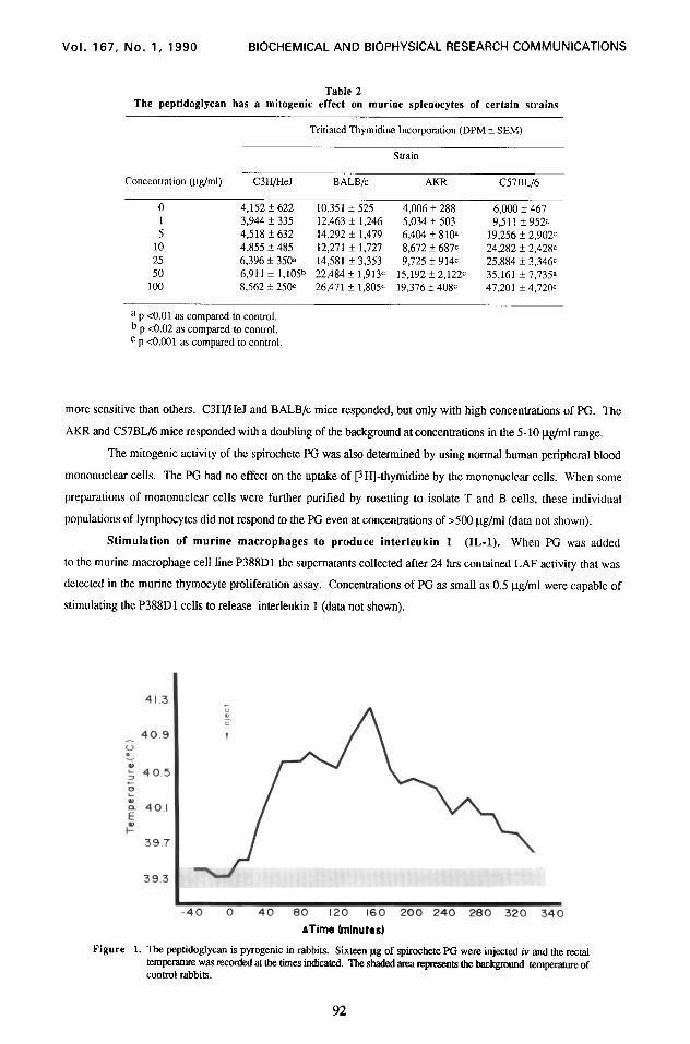
Figure 1. The peptidoglycan is pyrogenic in rabbits. Sixteen pg of spirochete PG were injected iv and the rectal temperatllre was reuxded at the times indicated. The shaded area represents the background temperatore of control rabbits.
92
Vol. 167, No. 1, 1990 BIOCHEMICAL AND BIOPHYSICAL RESEARCH COMMUNICATIONS
Figure 2. ‘lhepeptidogJycancausesanircrea% in vascular permeability. samples as indicated WeC injected sobcutaaeously into shaved rabbit b&s. The amount of vascular leakage was quantilied by measuring the amount of [lsr]-BSA in the injection sites.
Pyrogenicity of spirochete PG. To measure pyrogenicity, we injected New Zealand albino rabbits
iv with the spirochete PG and measured rectal temperatures. Figure 1 shows the results of a representative
experiment in which 16 pg were injected. As can be seen, a classic biphasic fever profile was observed.
In vlivo inflammatory effects of spirochete PG. Two experimental models were chosen to
investigate the in viva inflammatory properties of the spirochete PG. In the first model PG was injected
intradermally into shaved rabbit backs and increases in vascular permeability were measured by quantifying the
Figure 3. The peptidoglycan causes an intense skin reaction when injected inaadennally. Ten pg of F’G were injected intradermally into the faearm of a normal volunteer (right side). Saline was injected into a control site (left side). ‘lk sites were observed and meawwl for the duration of the inflammatory reaction (appmximatcly 72 hrs). The photograph was taken 24 hrs after the injection.
93
Vol. 167, No. 1, 1990 BIOCHEMICAL AND BIOPHYSICAL RESEARCH COMMUNICATIONS
amount of [ t2sI]-BSA leakage at the injection site. As can be seen in Figure 2 the change in vascular permeability
could be seen in as little as 20 min, with leakage still occurring at 135 min. In this representative experiment 15 pg
of PG were used.
In the second model, PG was injected subcutaneously into the forearm of a human volunteer (GB). Ten pg
(in 25 pl sterile pyrogen-free saline) were injected and the area was observed for any changes. A skin reaction
characterized by erythema and induration was observed. The reaction started a few minutes after injection and reached
a peak by 36 hrs and then subsided. The lesion disappeared after a few days and no permanent damage was seen.
Figure 3 shows the skin lesion at 24 hrs. At this time the lesion was red, indurated, and warm to the touch. The
site on the left was injected with saline (25 l.tl).
DISCUSSION
In this paper we report on the isolation and characterization of a PG extracted from the Lyme disease
spirochete Bon&a burgdo@?. The isolation procedure was fast and separated the PG from any contaminating
spirochetal LPS.
The amino acid analysis of the PG revealed the major amino acids to be alanine, glycine, glutamic acid, and
omithine. These results are in agreement with Yanagihara el al. (8), who isolated PG from a similar spirochete
(Borreliu herti). They found the major amino acids to be alanine, glycine, and glutamic acid, and omithine. In a
preliminary examination of B. burgahferi Johnson et al. (17) reported that the PG diamino amino acid is omithine.
The biologic activities associated with this PG are similar to those described for other isolated PG.
Pyrogenicity is a major activity of PG (10,18). Our PG was a potent pyrogen, and it is unlikely that the activity
was due to contaminating endotoxin in our preparations since the LAL test was negative. Also, the amount of LPS
present in the injected PG (co.14 pg) could not account for the fever observed. Stimulation of macrophages by PG
to produce IL-1 has been observed by several groups (19,20). A biphasic fever may result from both direct
pyrogenicity and the in vivo induction of IL-1 by PG (7). The inflammatory properties of PG include increases in
capillary permeability (21). This may be caused by the ability of PG to induce the release of endogenous mediators
of inflammation (e.g., histamine) (10). Our experiments at this time cannot answer this question. Acute
inflammation of the skin has been observed as a major activity of PG in both man and animals (10,18).
Many of the symptoms of Lyme disease could be explained by the activities of the PG. Fever, the
characteristic skin rash erythema chronicum migrans, arthritis, and malaise (22) could all be caused by direct action
of the PG. Taken with our data on the isolation and biologic characterization of the LPS from this spirochete it is
easy to speculate that both these cellular components contribute to the multisystemic course of Lyme disease. That
both agents induce the production of IL-l could contribute to the “biologic amplification” of a small number of
invading spirochetes.
In conclusion, the role of PG and LPS in discussions of the pathogenesis of Lyme disease must now be
taken into consideration. That B. burgdorferi PG and LPS are both powerful immunomodulators and stimulators of
IL-1 gives credence to the importance of this cytokine in the pathogenesis of Lyme disease.
ACKNOWLEDGMENTS
This work was supported by a grant (AR 36028) (to G.S.H.) from the National Institutes of Health. The amino acid analysis was performed by the Center for Analysis and Synthesis of Macromolecules at SUNY Stony Brook which is supported by NIH grant RR02427, and the Center for Biotechnology.
94
Vol. 167, No. 1, 1990 BIOCHEMICAL AND BIOPHYSICAL RESEARCH COMMUNICATIONS
1. 2.
3.
4.
5.
6. 7. 8.
9. 10.
11. 12. 13. 14.
15. 16. 17. 18.
19. 20. 21. 22.
REFERENCES
Habicht, G.S., Beck, G., and Benach, J.L. (1987) Scientific American. July 257,78-83. Steere, A.C., Grodzicki, R.L., Kornblatt, A.N., Craft, J.E., Barbour, A.G., Burgdorfer, W., Schmid, G.P., Johnson, I!., and Malawista, SE. (1983) N. Engl. J. Med. 308,733-740. Benach, J.L., Bosler, E.M., Hanrahan, J.P., Coleman, J.L., Habicht, G.S., Bast, T.F., Cameron, D.J., Ziegler, J.L., Barbour, A.G., Burgdorfer, W., Edelman, R., and Kaslow, R.A. (1983) N. Engl. J. Med. 308, 74&742. Snyderman, D.R., Schenkein, D.P., Berardi, V.P., Lastavica, CC., and Pariser, K.M. (1986) Ann, Intern. Med. 104.798-800. Johnson, Y.E., Duray, P.A., Steere, A.C., Kashgarian, M., Buza, J., Malawista, SE., and Askenase, P.W. (1985) Am. J. Pathol. 118, 26-34. Komblatt, A.N., Steere, A.C., and Brownstein, D.G. (1984) Infect. Immun. 46,220-223. Beck, G., Habicht, G.S., Benach, J.L., and Coleman, J.L. (1985) J. Infect. Dis. 152, 108-117. Yanagihara, Y., Kei-ichi, K., Yasuda, S., Kobayashi, S., Mifuchi, I., Azuma, I., Yamamura, Y., and Johnson, R.C. (1984) Microbial. Immunol. 28, 535-544. Joseph, R., Holt, S.E., and Canale-Parola., E. (1973) J. Bacterial. 115.426435. Heymer, B., Seidl, P.H., and Schleifer, K.H. (1985) In Immunology of the Bacterial Cell Envelope (D.E.S. Stewart-Tall, and M. Davies, Eds), pp. 1 l-25. John Wiley and Sons, Ltd., New York. Kolenbrander, P.E., and Ensign, J.C. (1968) J. Bacterial. 95,201-210. Stewart-Tull, D.E.S. (1980) Ann. Rev. Microbial. 34.311-340. Spector, T. (1978) Anal. B&hem. 86, 142-146. Umemoto, T., Ota, T., Sagawa, H., Kato, K., Takada, H., Tsujimoto, M., Kawasaki, A., Ogawa, T., Harada, K., and Kotani, S. (1981) Infect. Immun. 31, 767-774. Markwell, M. (1982) Anal. B&hem. 125,427-432. Beck, G., Habicht, G.S., Benach, J.L., and Miller, F. (1986) J. Immunol. 136.3025-3031. Johnson, F:.C., Hyde, F.W., and Rumpel., CM. (1984) Yale J. Biol. and Med. 57, 529-537. Stewart-Tull, D.E.S. (1985) In Immunology of the Bacterial Cell Envelope (D.E.S. Stewart-Tull, and M. Davies, Eds), pp. 47-65. John Wiley and Sons, Ltd., New York. Gppenheim, J.J., Togawa, A., Chedid, L., and Mizel, S.B. (1980) Cell. Immunol. 50, 71-81. Vacheron, F., Guenounou, M., and Nauciel, C. (1983) Infect. Immun. 42, 1049-1054. Otha, M. (1981) Nippon Ika Daigaku Zasshi. 48.402-409. Beck, G., Habicht, G.S., Benach, J.L., Coleman, J.L., Lysik, R.M., and O’Brien, R.F. (1986) Zbl. Bakt. Hyg. A. 2.63, 133-136.