Page 1
Ashok Mateti*et al. /International Journal of Pharmacy & Technology
IJPT| Sep-2017| Vol. 9 | Issue No.3 | 30834-30849 Page 30834
ISSN: 0975-766X
CODEN: IJPTFI
Available Online through Research Article
www.ijptonline.com PRO VESICULAR BASED COLLOIDAL CARRIERS FOR TRANSDERMAL DRUG
DELIVERY Ashok Mateti
1*, Mohammed Habibuddin
2, Raju Jukanti
3
1Department of Pharmaceutics, Trinity College of Pharmaceutical Sciences,
Peddapalli, Karimnagar, Telangana, India. 2Department of Pharmaceutics, Shadan College of Pharmacy, Hyderabad, Telangana, India.
3Department of Pharmaceutics, Drugs Inspector, Karimnagar, Telangana, India.
Email: [email protected]
Received on: 12-08-2017 Accepted on: 20-09-2017
Abstract
The present investigation aimed at formulation development and performance evaluation of proniosomal gel as a
vesicular drug carrier system. Domperidone (DOM) is a dopamine- receptor (D2) antagonist, which is widely used in
the treatment of motion-sickness. The pharmacokinetic parameters (absolute bioavailability about 10-20 % and log P,
3.11) make DOM a suitable candidate for transdermal delivery. The purpose of the present investigation was to
develop transdermal delivery systems for DOM and to evaluate their physicochemical characteristics, in vitro release
an ex vivo permeation through rat abdominal skin. Proniosome drug delivery was preferred due to improved stability
of the system than niosomes. A proniosome based transdermal drug delivery system of domperidone was developed
and extensively characterized both in-vitro and ex-vivo. Proniosomal gel formulations of domperidone were
characterized for vesicular shape & size, entrapment efficiency, permeation study. The effects of cholesterol, lecithin
and different non-ionic surfactants on transdermal permeability profile of domperidone were performed. The stability
studies were performed at 2°C and at room temperature. Optimized formulation showed maximum cumulative
percentage of drug release (96.551%), permeation (4.119 mg) in 24 hrs, flux (0.173 μg / cm2/ hr), and permeation
coefficient of 0.029 cm2/hr and enhancement ratio 2.351. FTIR studies showed no evidence of interaction between
the drug and polymers. Penetration enhancers, non-ionic surfactants and vesicle-skin interaction may contribute to the
enhanced domperidone permeation. Thus Proniosome was found to be a promising carrier system for domperidone
because of ease in preparation and stability for prolonged period. The study demonstrated the utility of proniosomal
transdermal patch bearing domperidone for emesis.
Keywords: Transdermal, Domperidone, Proniosomes, Permeation, Span 60, Lecithin.
Page 2
Ashok Mateti*et al. /International Journal of Pharmacy & Technology
IJPT| Sep-2017| Vol. 9 | Issue No.3 | 30834-30849 Page 30835
Introduction
The transdermal route is widely used as it is convenient, safe and offers numerous advantages over conventional ones
that includes evading GI incompatibility, variable GI absorption, bypassing first pass metabolism, enhanced
bioavailability, decreased frequency of administration, improved patient compliance, rapid cessation of drug input
and can maintain a suitable plasma concentration. One of the major disadvantages in transdermal drug delivery is the
low penetration rate of substances through the skin (Schreier and Bouwstra, 1994). Several techniques have been
explored to increase the drug penetration rate across skin including iontophoresis and penetration enhancement,
particularly for the delivery of peptides and proteins. Third alternative method, the encapsulation of drugs in lipid
vesicles prepared from phospholipids (liposomes) or nonionic surfactants (niosomes) which have been shown to
facilitate transport of drugs into and across skin. Niosomes have a problem of degradation by hydrolysis or oxidation,
demands special storage and handling shows sedimentation, aggregation or fusion on storage.
Proniosomes a versatile delivery system is their potential of entrapping a wide range of active compounds without
showing any problems of physical stability (aggregation, fusion, leaking). Proniosomes provide the convenience of
the transportation, distribution, storage, and dosing. Proniosomes upon hydration with water from skin after topical
application get readily converted into niosomes of uniform size. Proniosomes exists in two forms, i.e. semi solid
liquid crystal gel and dry granular powder, depending on their method of preparation. Out of these two forms, the
proniosome gel is mainly used for topical/transdermal applications. Proniosomal gel is a compact semi-solid liquid
crystaline (gel) product of non-ionic surfactants easily formed on dissolving the surfactant in minimal amount of
acceptable solvent and the least amount of aqueous phase. This compact liquid crystalline gel can be readily
converted into niosomes on hydration. Proniosomal gel offers a great potential to reduce the side effects of drugs and
increased therapeutic effectiveness.
Proniosomes can entrap both hydrophilic and hydrophobic drugs. Domperidone is a dopamine- receptor (D2)
antagonist, widely used in the treatment of motion-sickness. In humans, peak plasma levels of domperidone occur
within 10 to 30 min following intra-muscular injection and 30 min after oral (fasted) administration. It has been
reported that it is rapidly absorbed after oral administration, but undergoes extensive first pass metabolism; leading to
poor bioavailability of 15%. From both, physicochemical (low molecular weight 425.9g/mol, low dose 10 mg) and
pharmacokinetic (absolute bioavailability about 10-20 % and log P, 3.11) perspective, DOM was considered to be a
suitable candidate for transdermal delivery (Madishetti et al., 2010).
Page 3
Ashok Mateti*et al. /International Journal of Pharmacy & Technology
IJPT| Sep-2017| Vol. 9 | Issue No.3 | 30834-30849 Page 30836
Materials and Methods
Span 20 (Ozone chemicals), 40 (Central Drug House pvt Ltd., New Delhi), Span 60, Span 80, phospholipoid
(Germany), cholesterol (S.D. fine chemicals), Ethanol (Changshu yangyuan chemical, china), Methanol, Isopropyl
alcohol, 0.1% Glycerol (Merck, Mumbai), other chemicals were of analytical grade. Distilled water was used for all
experiments.
Preparation of proniosomes
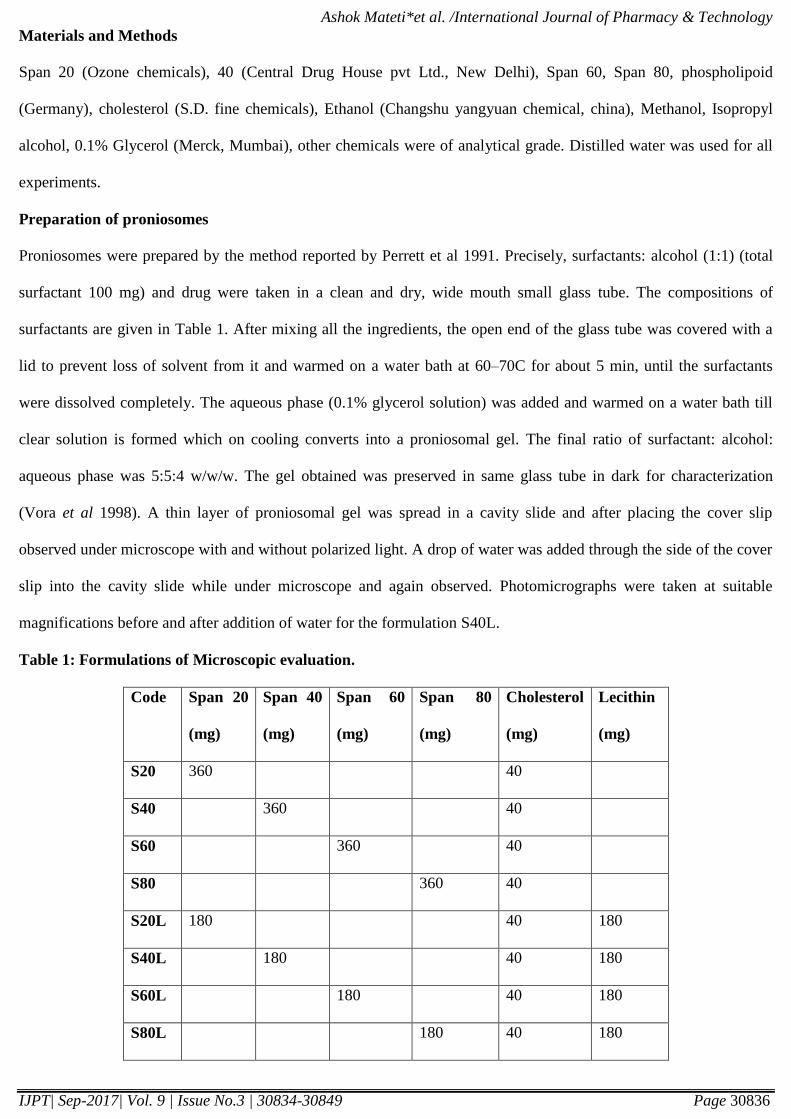
Proniosomes were prepared by the method reported by Perrett et al 1991. Precisely, surfactants: alcohol (1:1) (total
surfactant 100 mg) and drug were taken in a clean and dry, wide mouth small glass tube. The compositions of
surfactants are given in Table 1. After mixing all the ingredients, the open end of the glass tube was covered with a
lid to prevent loss of solvent from it and warmed on a water bath at 60–70C for about 5 min, until the surfactants
were dissolved completely. The aqueous phase (0.1% glycerol solution) was added and warmed on a water bath till
clear solution is formed which on cooling converts into a proniosomal gel. The final ratio of surfactant: alcohol:
aqueous phase was 5:5:4 w/w/w. The gel obtained was preserved in same glass tube in dark for characterization
(Vora et al 1998). A thin layer of proniosomal gel was spread in a cavity slide and after placing the cover slip
observed under microscope with and without polarized light. A drop of water was added through the side of the cover
slip into the cavity slide while under microscope and again observed. Photomicrographs were taken at suitable
magnifications before and after addition of water for the formulation S40L.
Table 1: Formulations of Microscopic evaluation.
Code Span 20
(mg)
Span 40
(mg)
Span 60
(mg)
Span 80
(mg)
Cholesterol
(mg)
Lecithin
(mg)
S20 360 40
S40 360 40
S60 360 40
S80 360 40
S20L 180 40 180
S40L 180 40 180
S60L 180 40 180
S80L 180 40 180
Page 4
Ashok Mateti*et al. /International Journal of Pharmacy & Technology
IJPT| Sep-2017| Vol. 9 | Issue No.3 | 30834-30849 Page 30837
Size and size distribution
Size and size distribution studies were done for niosomes prepared from proniosomes hydration. The proniosomal
gel (100 mg) was hydrated in a small glass test tube using 10 ml of pH 7.4 phosphate buffer solution. The dispersion
was observed under optical microscope at 40X magnification. Size and size distribution of 200–300 niosomes were
noted using calibrated stage and ocular micrometers (Elico Instruments, Hyderabad). Similarly, size was noted for
niosomes formed spontaneously from proniosomes after hydration without agitation in a cavity slide [4].
Entrapment efficiency
To 0.2 g of proniosome gel, weighed in a glass tube, 10 ml phosphate buffer pH 7.4 were added. The aqueous
suspension was then sonicated.
Niosomes containing domperidone were separated from untrapped drug by centrifugation at 9000rpm for 45 min at 4
◦C. The supernatant was recovered and assayed spectrophotometrically using UVspectrophotometer (UV-1800
Shimadzu, Japan), at 283nm Ammara et al., 2011). The encapsulation percentage of drug (EP) was calculated by the
following equation Alsarra et al., 2005).
EP = [(Ct – Cr)/ Ct] * 100
Where, Ct, concentration of total domperidone, Cr, concentration of free domperidone.
Vesicle physical analysis
The shape, surface characteristics, and size of the niosomes were observed by scanning electron microscopy. Once
again, 0.2 g of the proniosome gel in a glass tube was diluted with 10 ml of pH 7.4 phosphate buffer. The niosomes
were mounted on an aluminium stub using double-sided adhesive carbon tape. Then the vesicles were sputter-coated
with gold palladium (Au/Pd) using a vacuum evaporator (Edwards) and examined using a scanning electron
microscope (Hitachi 3700N, Germany) equipped with a digital camera, at 10 kV accelerating voltage.
Fourier transform infrared spectroscopy
Fourier transform IR spectra were obtained on Shimadzu FT-IR spectrometer. Samples were prepared in KBr disks
(2mg sample in 200mg KBr). The scanning range was 450-4000 cm-1
and the resolution was 4 cm-1
.
Fabrication of transdermal patch:
The circular aluminium foil of diameter 2.5 cm was used as backing membrane. On this backing membrane a plastic
sheet of same size with 1.0 mm thickness was stuck with adhesive. The circle of diameter 1.32 cm (corresponding to
1.369cm2 area) was cut centrally on a plastic sheet of the same size and thickness used previously and stuck with
Page 5
Ashok Mateti*et al. /International Journal of Pharmacy & Technology
IJPT| Sep-2017| Vol. 9 | Issue No.3 | 30834-30849 Page 30838
adhesive. The proniosomal gel was evenly spread over this area and covered with the fine nylon mesh. The liquid
crystalline proniosomal gel acts as a reservoir for the transdermal delivery of domperidone.
In Vitro Release Study
In vitro release studies were carried out using unjacketed vertical franz diffusion cells with a diffusional surface area
of 6.154 cm2 and 20 mL of receptor cell volume. Prior to the study, the dialysis membrane (Himedia laboratories Pvt
Ltd., Mumbai) was soaked in phosphate buffer pH 7.4 Formulation equivalent to 5mg of Domperidone was placed in
the donor compartment.
The receptor compartment consisting of PB pH 7.4 (containing 0.02% w/v of ethanol to retard microbial growth) was
maintained at 37±2°C under constant stirring upto 24 hrs . The donor chamber and the sampling port were covered
with lid to prevent evaporation during the study. Aliquots of 5 mL were withdrawn periodically at different time
intervals (0.25, 0.5, 1, 1.5, 2, 3, 4, 6, 8, 10, 12, and 24 hrs) and replaced with equal volume to maintain constant
receptor phase volume. At the end of the study, the samples were suitably diluted and the amount of drug was
determined spectrophotometrically at 283 nm.
In Vitro Permeation Study Using Excised Rat Abdominal Skin
Preparation of Rat Abdominal Skin
Male albino rats (150-200 g) were used for the experiment. The rats were sacrificed by using excess amount of
anesthetic ether. Before surgical removal of the skin, hair on dorsal side was removed with hair clipper taking
extreme precautions not to damage the skin. The epidermis was prepared by a heat separation technique, which
involved soaking of the entire abdominal skin in water at 60°C for 45 seconds, followed by careful removal of the
epidermis. The epidermis was washed with water, wrapped in aluminium foil and stored at -20°C till further use
(used within 2 weeks of preparation [6].
Permeation Study
In vitro permeation studies were carried out using unjacketed vertical franz diffusion cells with a diffusional surface
area of 6.154 cm2 and 20 mL of receptor cell volume. The skin was brought to the room temperature and mounted
between the donor and receiver compartment of the franz diffusion cell, where the stratum corneum side faced the
donor compartment. Before being dosed the skin was allowed to equilibrate for 1 h and 0.5 g of gel
formulation/reference formulation equivalent to 5 mg of domperidone was placed in the donor compartment. The
receptor compartment consisting of PB pH 7.4 (containing 0.02% w/v of ethanol to retard microbial growth) was
Page 6
Ashok Mateti*et al. /International Journal of Pharmacy & Technology
IJPT| Sep-2017| Vol. 9 | Issue No.3 | 30834-30849 Page 30839
maintained at 37±2°C under constant stirring upto 24 hrs. The donor chamber and the sampling port were covered
with lid to prevent evaporation during the study. Aliquots of 5 mL were withdrawn periodically at different time
intervals (0.25, 0.5, 1, 1.5, 2, 3, 4, 6, 8, 10, 12, and 24 hrs) and replaced with equal volume to maintain constant
receptor phase volume. At the end of the study, the samples were suitably diluted and the amount of drug was
determined spectrophotometrically at 283 nm.
Permeation Data Analysis
The cumulative amount of drug permeated through a unit area of skin was plotted as a function of time.
Flux
The Steady state Flux was calculated by using the slope of the graph where J=Flux(µg/cm2/hr), A=Surface area,
dQ/dt = Cummulative amount permeated per unit area per unit time.containing cumulative amount permeated
through unit area (CAP) Vs Time.
Jss = (dQ/dt)*(1/A)
Permeability co efficient (Kp)
Permeability co efficient which represents the correlation between the flux and initial drug load was calculated using
the following equation [6].
Kp = Jss/C
where, Kp=Permeability co efficient (cm/hr); J= transdermal flux; C= Initial concentration of acyclovir sodium in the donor
compartment.
Enhancement Ratio
The penetration enhancing effect of various formulations containing acyclovir sodium gels were calculated in terms
of Enhancement Ratio (ER) by using the following equation.
ER = Jss of formulation/ Jss of reference
Evaluation of Optimised Formulation
Formulation F4 was optimised on the basis of permeation studies and evaluated for following parameters
Stability studies
Stability studies were carried out by storing the optimized formulation (F4) at various temperature conditions as per
ICH guidelines i.e. at refrigeration temperature (2°-8°C), room temperature (25°± 0.5°C) for a period of two months.
Drug content and variation in the average vesicle diameter were determined before and after the completion of 2
Page 7
Ashok Mateti*et al. /International Journal of Pharmacy & Technology
IJPT| Sep-2017| Vol. 9 | Issue No.3 | 30834-30849 Page 30840
months. Surface morphology of optimized formulation (F4) by SEM image is shown in Figure 9&10. and the results
are reported in Table 5 ( Raju Jukanti et al., 2011).
Skin irritancy test
The skin irritancy potential of the proniosome formulations was evaluated in albino rats. The hair was removed on the
back of the animal and the formulations were applied, and the animals were examined for any signs of skin irritation
and erythema for a period of 1 week.
Statistical Analysis
Significance of difference between formulations was calculated by one way analysis of variance using Newman
Keuls (compare all pairs) with Instant Graph Pad Prism software. The difference was considered to be statistically
significant at p<0.05.
Results and discussion
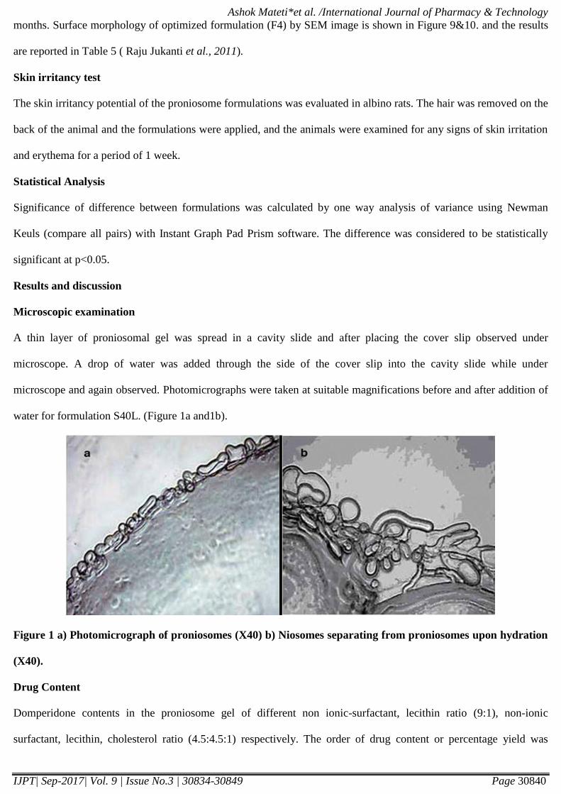
Microscopic examination
A thin layer of proniosomal gel was spread in a cavity slide and after placing the cover slip observed under
microscope. A drop of water was added through the side of the cover slip into the cavity slide while under
microscope and again observed. Photomicrographs were taken at suitable magnifications before and after addition of
water for formulation S40L. (Figure 1a and1b).
Figure 1 a) Photomicrograph of proniosomes (X40) b) Niosomes separating from proniosomes upon hydration
(X40).
Drug Content
Domperidone contents in the proniosome gel of different non ionic-surfactant, lecithin ratio (9:1), non-ionic
surfactant, lecithin, cholesterol ratio (4.5:4.5:1) respectively. The order of drug content or percentage yield was
Page 8
Ashok Mateti*et al. /International Journal of Pharmacy & Technology
IJPT| Sep-2017| Vol. 9 | Issue No.3 | 30834-30849 Page 30841
S40L>S20L>S60L>S80L>S20>S40>S60>S80. Uniformity of content present in proniosomal gel confirmed to assure
uniformity in dosages. The results are reported in Table. 2.
Particle size analysis
The results were reported in Table no. 2. Size range of the all formulations was found to be is 13.84μm(S20),
15.99μm(S20L), 12.36μm(S40), 11.22μm(S40L), 13.24μm(S60), 10.14μm(S60L), 12.79μm(S80), 10.05μm(S80L)
respectively. The proniosome vesicle were formed to be uniform in size 11.22±0.76µm (S40L) and 10.14μm (S60L).
The differences of vesicle size among all niosomes with span were not great. The relationship observed between
niosome size and span hydrophobicity has been attributed to the decrease in surface energy with increasing
hydrophobicity, resulting in the small vesicles. Vesicles with small diameter are believed to better permeate through
the skin. Niosomes of S60L were smaller in size than S40L. In corporation of lecithin leads to vesicles of smaller size
due to increase in hydrophobicity which results in reduction of vesicles size . There is probably formation of more
compact and well organized bilayers which prevents the leakage of drug (Hemant et al., 2012). Isopropanol results in
vesicles of smallest size, may be due to branched chain present in it.
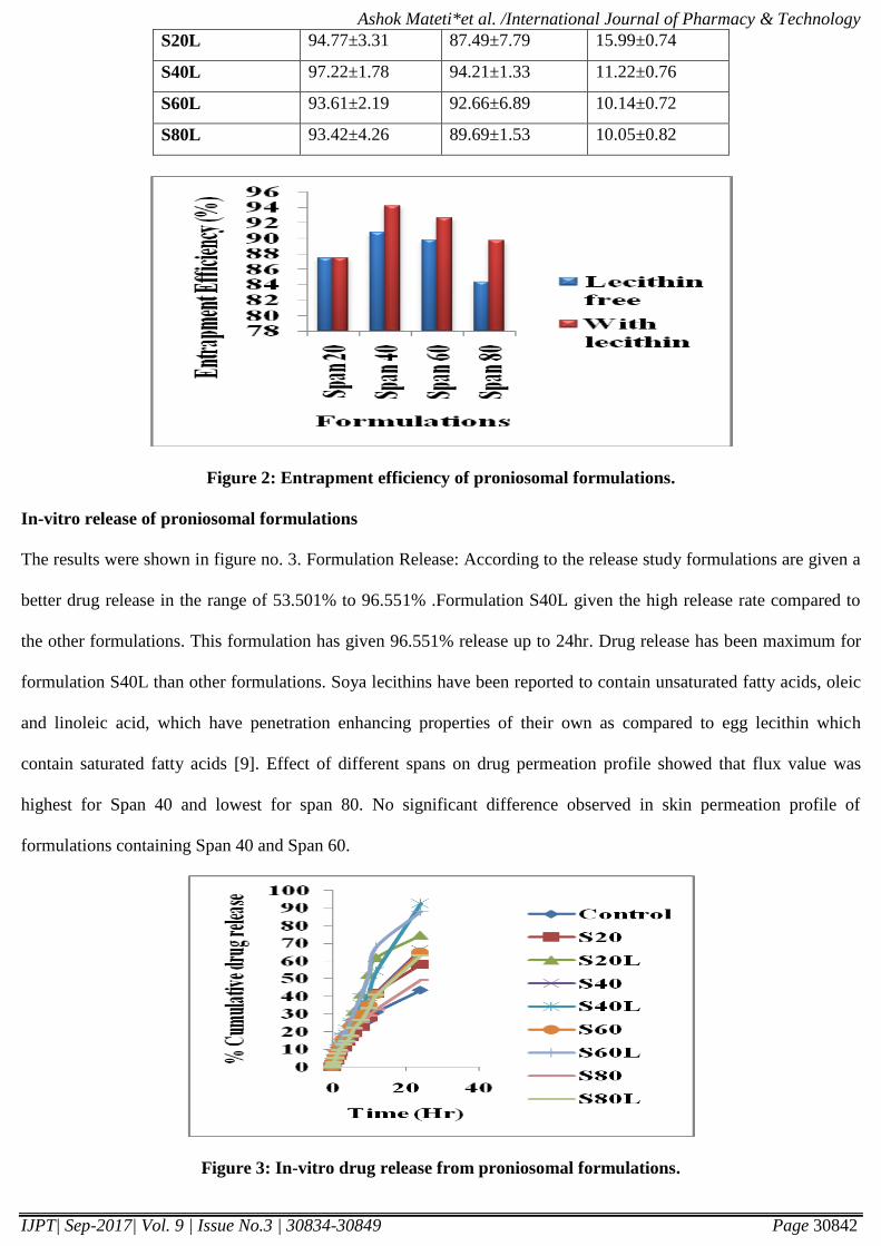
Entrapment efficiency (EE)
The results were reported in Table no. 2. & Figure no. 2. The entrapment efficiency was found to be (84.37% -
94.21%) for formulations. Encapsulation studies were carried out on all formulations. In these studies the higher
entrapment efficiency is for S40L formulation 94.21%. Incorporation of lecithin is also justified as it acts as
permeation enhancers. Incorporation of lecithin further enhanced the percent drug entrapment to 94.21%. Entrapment
efficiencies in span 40 and span 60 formulations were higher than those in other span formulations. Span 40 and Span
60 are solid at room temperature and showed the higher phase transition temperature (Tc). This result was consistent
of Levonorgestrel in proniosomes incorporated with span 40.
Table 2: % of drug content, entrapment efficiency, particle size of domperidone proniosome gel, mean ± S.D.
(n=3).
Formulation
code
% Drug content Entrapment
Efficiency (%)
Particle Size
(μm)
S20 93.37±2.74 87.41±3.35 13.84±0.68
S40 92.43±3.02 90.75±1.04 12.36±0.54
S60 91.73±2.15 89.84±2.74 13.24±0.37
S80 90.32±3.26 84.37±2.42 12.79±0.47
Page 9
Ashok Mateti*et al. /International Journal of Pharmacy & Technology
IJPT| Sep-2017| Vol. 9 | Issue No.3 | 30834-30849 Page 30842
S20L 94.77±3.31 87.49±7.79 15.99±0.74
S40L 97.22±1.78 94.21±1.33 11.22±0.76
S60L 93.61±2.19 92.66±6.89 10.14±0.72
S80L 93.42±4.26 89.69±1.53 10.05±0.82
Figure 2: Entrapment efficiency of proniosomal formulations.
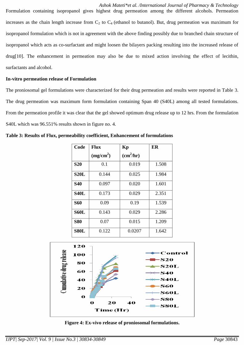
In-vitro release of proniosomal formulations
The results were shown in figure no. 3. Formulation Release: According to the release study formulations are given a
better drug release in the range of 53.501% to 96.551% .Formulation S40L given the high release rate compared to
the other formulations. This formulation has given 96.551% release up to 24hr. Drug release has been maximum for
formulation S40L than other formulations. Soya lecithins have been reported to contain unsaturated fatty acids, oleic
and linoleic acid, which have penetration enhancing properties of their own as compared to egg lecithin which
contain saturated fatty acids [9]. Effect of different spans on drug permeation profile showed that flux value was
highest for Span 40 and lowest for span 80. No significant difference observed in skin permeation profile of
formulations containing Span 40 and Span 60.
Figure 3: In-vitro drug release from proniosomal formulations.
Page 10
Ashok Mateti*et al. /International Journal of Pharmacy & Technology
IJPT| Sep-2017| Vol. 9 | Issue No.3 | 30834-30849 Page 30843
Formulation containing isopropanol gives highest drug permeation among the different alcohols. Permeation
increases as the chain length increase from C2 to C4 (ethanol to butanol). But, drug permeation was maximum for
isopropanol formulation which is not in agreement with the above finding possibly due to branched chain structure of
isopropanol which acts as co-surfactant and might loosen the bilayers packing resulting into the increased release of
drug[10]. The enhancement in permeation may also be due to mixed action involving the effect of lecithin,
surfactants and alcohol.
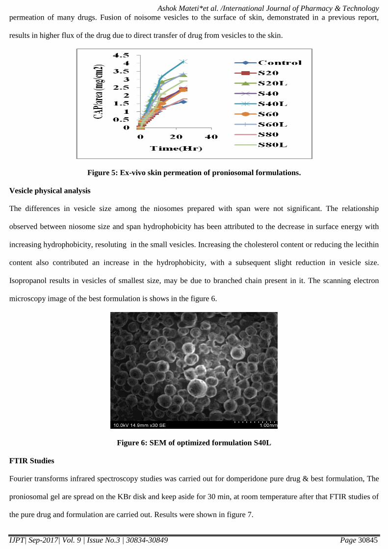
In-vitro permeation release of Formulation
The proniosomal gel formulations were characterized for their drug permeation and results were reported in Table 3.
The drug permeation was maximum form formulation containing Span 40 (S40L) among all tested formulations.
From the permeation profile it was clear that the gel showed optimum drug release up to 12 hrs. From the formulation
S40L which was 96.551% results shown in figure no. 4.
Table 3: Results of Flux, permeability coefficient, Enhancement of formulations
Code Flux
(mg/cm2)
Kp
(cm2/hr)
ER
S20 0.1 0.019 1.508
S20L 0.144 0.025 1.984
S40 0.097 0.020 1.601
S40L 0.173 0.029 2.351
S60 0.09 0.19 1.539
S60L 0.143 0.029 2.286
S80 0.07 0.015 1.209
S80L 0.122 0.0207 1.642
Figure 4: Ex-vivo release of proniosomal formulations.
Page 11
Ashok Mateti*et al. /International Journal of Pharmacy & Technology
IJPT| Sep-2017| Vol. 9 | Issue No.3 | 30834-30849 Page 30844
Permeation release study is performed on all formulations. Permeation study is needed for flux, enhancement ratio.
The range of flux is (0.07µg/cm2/hr - 0.173µg/cm
2/hr). Formulation S40L has given high flux value. According to
this study we considered that S40L is best formulation. The permeation data fitted in first order drug release. It was
but obvious that the smaller vesicular size of S40L enabled it to penetrate easily through the skin as smaller vesicles
tend to fuse readily with the skin. Thus proniosome gel prepared by using Span 40 exhibited better permeation and
optimum entrapment efficiency.
This could also be due to the emulsification effect of the surfactant after the hydration of the proniosome by the
dissolution medium and formation of elution channels within the gel structure due to loss of lipid bilayers that
resulted in higher flux value [11]. The results were shown in Table 3 & 5. Hence formulation S40L had selected as
optimum formulation and stability testing studies were carried out. These observations were in accordance with
earlier reports saying that incorporation of cholesterol was known to influence vesicle stability, permeability and
entrapment efficiency. Increase in the cholesterol content resulted in a more intact and ordered lipid bilayer as a
barrier for drug release and helped as a controlled release polymer and also decreased drug leakage by improving the
fluidity of the bilayer membrane and reducing its permeability [12].
Table 5: Results of stability studies of optimized formulation (S40L)
S. No Temp Initial drug
content
Initial vesicle
size
After 2months
drug content
After 2months
vesicle size
1 2°C 97.22±1.78 11.22±0.76 96.72±1.08 11.89±0.68
2 25°C 97.22±1.78 11.22±0.76 96.08±1.12 11.64±0.82
Proniosomes should be hydrated to form niosomal vesicles before the drug is released and permeates across the skin.
Several mechanisms could explain the ability of niosomes to modulate drug transfer across skin [13], including (i)
adsorption and fusion of niosomes onto the surface of skin would facilitate drug permeation, (ii) the vesicles act as
penetration enhancers to reduce the barrier properties of the stratum corneum, and (iii) the lipid bilayers of niosomes
act as a rate-limiting membrane barrier for drugs.
One of the possible mechanisms for niosomal enhancement of the permeability of drugs is structure modification of
the stratum corneum. It has been reported that the intercellular lipid barrier in the stratum corneum would be
dramatically looser and more permeable following treatment with liposomes and niosomes. Both phospholipids and
nonionic surfactants in the proniosomes can act as penetration enhancers, which are useful for increasing the
Page 12
Ashok Mateti*et al. /International Journal of Pharmacy & Technology
IJPT| Sep-2017| Vol. 9 | Issue No.3 | 30834-30849 Page 30845
permeation of many drugs. Fusion of noisome vesicles to the surface of skin, demonstrated in a previous report,
results in higher flux of the drug due to direct transfer of drug from vesicles to the skin.
Figure 5: Ex-vivo skin permeation of proniosomal formulations.
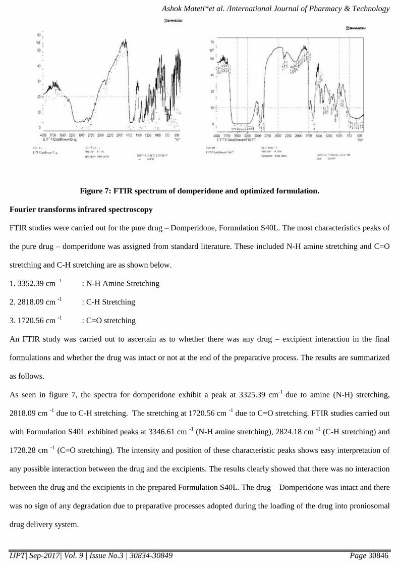
Vesicle physical analysis
The differences in vesicle size among the niosomes prepared with span were not significant. The relationship
observed between niosome size and span hydrophobicity has been attributed to the decrease in surface energy with
increasing hydrophobicity, resoluting in the small vesicles. Increasing the cholesterol content or reducing the lecithin
content also contributed an increase in the hydrophobicity, with a subsequent slight reduction in vesicle size.
Isopropanol results in vesicles of smallest size, may be due to branched chain present in it. The scanning electron
microscopy image of the best formulation is shows in the figure 6.
Figure 6: SEM of optimized formulation S40L
FTIR Studies
Fourier transforms infrared spectroscopy studies was carried out for domperidone pure drug & best formulation, The
proniosomal gel are spread on the KBr disk and keep aside for 30 min, at room temperature after that FTIR studies of
the pure drug and formulation are carried out. Results were shown in figure 7.
Page 13
Ashok Mateti*et al. /International Journal of Pharmacy & Technology
IJPT| Sep-2017| Vol. 9 | Issue No.3 | 30834-30849 Page 30846
Figure 7: FTIR spectrum of domperidone and optimized formulation.
Fourier transforms infrared spectroscopy
FTIR studies were carried out for the pure drug – Domperidone, Formulation S40L. The most characteristics peaks of
the pure drug – domperidone was assigned from standard literature. These included N-H amine stretching and C=O
stretching and C-H stretching are as shown below.
1. 3352.39 cm -1
: N-H Amine Stretching
2. 2818.09 cm -1
: C-H Stretching
3. 1720.56 cm -1
: C=O stretching
An FTIR study was carried out to ascertain as to whether there was any drug – excipient interaction in the final
formulations and whether the drug was intact or not at the end of the preparative process. The results are summarized
as follows.
As seen in figure 7, the spectra for domperidone exhibit a peak at 3325.39 cm-1
due to amine (N-H) stretching,
2818.09 cm -1
due to C-H stretching. The stretching at 1720.56 cm -1
due to C=O stretching. FTIR studies carried out
with Formulation S40L exhibited peaks at 3346.61 cm -1
(N-H amine stretching), 2824.18 cm -1
(C-H stretching) and
1728.28 cm -1
(C=O stretching). The intensity and position of these characteristic peaks shows easy interpretation of
any possible interaction between the drug and the excipients. The results clearly showed that there was no interaction
between the drug and the excipients in the prepared Formulation S40L. The drug – Domperidone was intact and there
was no sign of any degradation due to preparative processes adopted during the loading of the drug into proniosomal
drug delivery system.
Page 14
Ashok Mateti*et al. /International Journal of Pharmacy & Technology
IJPT| Sep-2017| Vol. 9 | Issue No.3 | 30834-30849 Page 30847
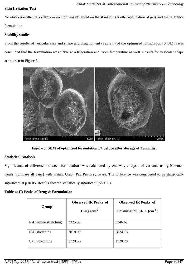
Skin Irritation Test
No obvious erythema, oedema or erosion was observed on the skins of rats after application of gels and the reference
formulation.
Stability studies
From the results of vesicular size and shape and drug content (Table 5) of the optimised formulation (S40L) it was
concluded that the formulation was stable at refrigeration and room temperature as well. Results for vesicular shape
are shown in Figure 8.
Figure 8: SEM of optimized formulation F4 before after storage of 2 months.
Statistical Analysis
Significance of difference between formulations was calculated by one way analysis of variance using Newman
Keuls (compare all pairs) with Instant Graph Pad Prism software. The difference was considered to be statistically
significant at p<0.05. Results showed statistically significant (p<0.05).
Table 4: IR Peaks of Drug & Formulation
Group
Observed IR Peaks of
Drug (cm-1)
Observed IR Peaks of
Formulation S40L (cm-1
)
N-H amine stretching 3325.39 3346.61
C-H stretching 2818.09 2824.18
C=O stretching 1720.56 1728.28
Page 15
Ashok Mateti*et al. /International Journal of Pharmacy & Technology
IJPT| Sep-2017| Vol. 9 | Issue No.3 | 30834-30849 Page 30848
Conclusion
Proniosomal formulations of domperidone were prepared with different non-ionic surfactants with phosphotidyl
choline and without phosphotidyl choline and evaluated. The formulation S40L is found to be the best formulation.
The cumulative drug release was found to be 96.55% at the end of 24 hr study and the formulation F4 drug release
followed first order kinetics with Hixon Crowell mechanism and non-fickian diffusion (n <1). From ex-vivo
abdominal rat skin absorption studies formulation S40L showed good release of 96.55% up to 24 hr. The entrapment
efficiency was between 86.66±2.89 and 94.21±1.33%. The flux was found to be 0.173µg/cm2/hr. Statistical analysis
showed results were significant (p<0.05). Scanning electron microscopy (SEM) studies revealed that the proniosome
were spherical in shape with smooth surface morphology and had a mean particle diameter between 10.05±0.82 and
15.99±0.74 µm. The formulation is easy to scale up as the procedure is simple and does not involve lengthy
procedures and unnecessary use of pharmaceutically unacceptable additives. Hence it offers direct fabrication of a
transdermal patch and does not require dispersion of vesicles into a polymer matrix.
Acknowledgements
The authors acknowledge the Central Drug House Pvt Ltd., New Delhi, India for gift samples.
References
1. Schreier H and Bouwstra J., Liposomes and niosomes as topical drug carriers: dermal and transdermal drug
delivery. J. Control. Rel., (1994) 30, 1–15
2. Madishetti S.K., Palem C.R., Gannu R., Thatipamula R.P., Panakanti P.k., Yamsani M.R., Development of
domperidone bilayered matrix type transdermal patches: physicochemical, in-vitro and ex-vivo characterization,
DARU., (2010) 18, 221-229.
3. S. Perrett, M. Golding, W.P. Williams, A simple method for the preparation of liposomes for pharmaceutical
application and characterization of liposomes, J. Pharm. Pharmacol. 43 (1991) 154–161.
4. Vora B, Khopade AJ, Jain NK. Proniosome based transdermal delivery of levonorgesterel for effective
contraception. J. Control. Rel., 1998; 54: 149-165
5. Ammara HO, Ghorabb M, El-Nahhasc SA, Higazya IM. Proniosomes as a carrier system for transdermal
delivery of tenoxicam. Int J Pharm. 2011; 405(1-2):142-52.
6. Alsarra IA., Bosela AA., Ahmed SM, Mahrous GM., Proniosomes as a drug carrier for transdermal delivery of
ketorolac. Eur. J. Pharm. Biopharm., (2005) 59, 485-490.
Page 16
Ashok Mateti*et al. /International Journal of Pharmacy & Technology
IJPT| Sep-2017| Vol. 9 | Issue No.3 | 30834-30849 Page 30849
7. Raju Jukanti, Ashok Mateti, Suresh Bandari and Prabhakar R Veerareddy, Transdermal Delivery of Acyclovir
Sodium Via Carbopol Gels: Role of Chemical Permeation Enhancers, Letters in Drug Design & Discovery, 2011.
8. Hemant n. Patil, sharwaree r. Hardikar, ashok v. Bhosale, Formulation development & evaluation of proniosomal
gel of carvedilol, International Journal of Pharmacy and Pharmaceutical Sciences, Vol 4, Issue 1, 2012.
9. Jia-You Fang, Song-Yih Yu, Pao-Chu Wu, Yaw- Bin Huang, Yi-Hung Tsai. In vitro skin permeation of estradiol
from various proniosome formulations. Int J Pharm. 2001; 215:91–99.
10. M. Ghyczy, J. Greiss, Liposomes from vegetable phosphatidyl choline. Their production and effects on skin,
Cosmet. Toil. 109 (1994) 75–80.
11. D. Friend, P. Catz, J. Heller, J. Reid, R. Baker, Transdermal delivery of levonorgestrel I: Alkanols as permeation
enhancers in vitro, J. Controlled Release 7 (1988) 243–250.
12. S. Vemuri, C.D. Yu, J.S. deGroot, N. Roosdorp, In vitro interaction of sized and unsized liposome vesicles with
high density lipoproteins, Drug Dev. Ind. Pharm. 16 (1990) 1579–1584.
13. Bernsdorff, C., Wolff, A., Winter, R., Gratton, E., 1997. Effect of hydrostatic pressure on water penetration and
rotational dynamics in phospholipids–cholesterol bilayers. Biophys. J. 72, 1264–1277.
14. Barry, B.W., 2001. Novel mechanisms and devices to enable successful transdermal drug delivery. Eur. J.
Pharm. Sci. 14, 101–114.
Corresponding Author:
Ashok Mateti*,
Email: [email protected]