150
World Journal of Gastroenterology World J Gastroenterol 2017 October 14; 23(38): 6923-7058 ISSN 1007-9327 (print) ISSN 2219-2840 (online) Published by Baishideng Publishing Group Inc

World Journal of GastroenterologyWorld J Gastroenterol 2017 October 14; 23(38): 6923-7058
ISSN 1007-9327 (print)ISSN 2219-2840 (online)
Published by Baishideng Publishing Group Inc
The World Journal of Gastroenterology Editorial Board consists of 1353 members, representing a team of worldwide experts in gastroenterology and hepatology. They are from 68 countries, including Albania (1), Algeria (1), Argentina (7), Australia (31), Austria (9), Belgium (10), Brazil (20), Brunei Darussalam (1), Bulgaria (2), Cambodia (1), Canada (25), Chile (4), China (161), Croatia (1), Cuba (1), Czech (6), Denmark (2), Egypt (9), Estonia (2), Finland (6), France (17), Germany (56), Greece (31), Guatemala (1), Hungary (14), Iceland (1), India (33), Indonesia (2), Iran (10), Ireland (9), Israel (18), Italy (195), Japan (151), Jordan (1), Kuwait (1), Lebanon (7), Lithuania (1), Malaysia (1), Mexico (10), Morocco (1), Netherlands (5), New Zealand (4), Nigeria (3), Norway (6), Pakistan (6), Poland (12), Portugal (8), Puerto Rico (1), Qatar (1), Romania (10), Russia (3), Saudi Arabia (2), Singapore (7), Slovenia (2), South Korea (64), Spain (51), Sri Lanka (1), Sudan (1), Sweden (12), Switzerland (5), Thailand (7), Trinidad and Tobago (1), Tunisia (2), Turkey (56), United Kingdom (47), United States (173), Venezuela (1), and Vietnam (1).
Editorial Board2014-2017
EDITORS-IN-CHIEFStephen C Strom, StockholmSaleh A Naser, OrlandoAndrzej S Tarnawski, Long BeachDamian Garcia-Olmo, Madrid
GUEST EDITORIAL BOARD MEMBERSJia-Ming Chang, TaipeiJane CJ Chao, TaipeiKuen-Feng Chen, TaipeiTai-An Chiang, TainanYi-You Chiou, TaipeiSeng-Kee Chuah, KaohsiungWan-Long Chuang, KaohsiungHow-Ran Guo, TainanMing-Chih Hou, TaipeiPo-Shiuan Hsieh, TaipeiChing-Chuan Hsieh, Chiayi countyJun-Te Hsu, TaoyuanChung-Ping Hsu, TaichungChien-Ching Hung, TaipeiChao-Hung Hung, KaohsiungChen-Guo Ker, KaohsiungYung-Chih Lai, TaipeiTeng-Yu Lee, Taichung CityWei-Jei Lee, TaoyuanJin-Ching Lee, KaohsiungJen-Kou Lin, TaipeiYa-Wen Lin, TaipeiHui-kang Liu, TaipeiMin-Hsiung Pan, TaipeiBor-Shyang Sheu, TainanHon-Yi Shi, KaohsiungFung-Chang Sung, TaichungDar-In Tai, Taipei
Jung-Fa Tsai, KaohsiungYao-Chou Tsai, New Taipei CityChih-Chi Wang, KaohsiungLiang-Shun Wang, New Taipei CityHsiu-Po Wang, TaipeiJaw-Yuan Wang, KaohsiungYuan-Huang Wang, TaipeiYuan-Chuen Wang, TaichungDeng-Chyang Wu, KaohsiungShun-Fa Yang, TaichungHsu-Heng Yen, Changhua
MEMBERS OF THE EDITORIAL BOARD
Albania
Saadi Berkane, Algiers
Algeria
Samir Rouabhia, Batna
Argentina
N Tolosa de Talamoni, CórdobaEduardo de Santibanes, Buenos AiresBernardo Frider, Capital FederalGuillermo Mazzolini, PilarCarlos Jose Pirola, Buenos AiresBernabé Matías Quesada, Buenos AiresMaría Fernanda Troncoso, Buenos Aires
Australia
Golo Ahlenstiel, WestmeadMinoti V Apte, SydneyJacqueline S Barrett, MelbourneMichael Beard, AdelaideFilip Braet, SydneyGuy D Eslick, SydneyChristine Feinle-Bisset, AdelaideMark D Gorrell, SydneyMichael Horowitz, AdelaideGordon Stanley Howarth, RoseworthySeungha Kang, BrisbaneAlfred King Lam, Gold CoastIan C Lawrance, PerthFremantleBarbara Anne Leggett, BrisbaneDaniel A Lemberg, SydneyRupert W Leong, SydneyFinlay A Macrae, VictoriaVance Matthews, MelbourneDavid L Morris, SydneyReme Mountifield, Bedford ParkHans J Netter, MelbourneNam Q Nguyen, AdelaideLiang Qiao, WestmeadRajvinder Singh, AdelaideRoss Cyril Smith, StLeonardsKevin J Spring, SydneyDebbie Trinder, FremantleDaniel R van Langenberg, Box HillDavid Ian Watson, AdelaideDesmond Yip, GarranLi Zhang, Sydney
March 26, 2014IWJG|www.wjgnet.com
Austria
Felix Aigner, InnsbruckGabriela A Berlakovich, ViennaHerwig R Cerwenka, GrazPeter Ferenci, WienAlfred Gangl, ViennaKurt Lenz, LinzMarkus Peck-Radosavljevic, ViennaMarkus Raderer, ViennaStefan Riss, Vienna
Belgium
Michael George Adler, BrusselsBenedicte Y De Winter, AntwerpMark De Ridder, JetteOlivier Detry, LiegeDenis Dufrane Dufrane, BrusselsNikos Kotzampassakis, LiègeGeert KMM Robaeys, GenkXavier Sagaert, LeuvenPeter Starkel, BrusselsEddie Wisse, Keerbergen
Brazil
SMP Balzan, Santa Cruz do SulJLF Caboclo, Sao jose do rio pretoFábio Guilherme Campos, Sao PauloClaudia RL Cardoso, Rio de JaneiroRoberto J Carvalho-Filho, Sao PauloCarla Daltro, SalvadorJosé Sebastiao dos Santos, Ribeirao PretoEduardo LR Mello, Rio de JaneiroSthela Maria Murad-Regadas, FortalezaClaudia PMS Oliveira, Sao PauloJúlio C Pereira-Lima, Porto AlegreMarcos V Perini, Sao PauloVietla Satyanarayana Rao, FortalezaRaquel Rocha, SalvadorAC Simoes e Silva, Belo HorizonteMauricio F Silva, Porto AlefreAytan Miranda Sipahi, Sao PauloRosa Leonôra Salerno Soares, NiteróiCristiane Valle Tovo, Porto AlegreEduardo Garcia Vilela, Belo Horizonte
Brunei Darussalam
Vui Heng Chong, Bandar Seri Begawan
Bulgaria
Tanya Kirilova Kadiyska, SofiaMihaela Petrova, Sofia
Cambodia
Francois Rouet, Phnom Penh
Canada
Brian Bressler, Vancouver
Frank J Burczynski, WinnipegWangxue Chen, OttawaFrancesco Crea, VancouverMirko Diksic, MontrealJane A Foster, HamiltonHugh J Freeman, VancouverShahrokh M Ghobadloo, OttawaYuewen Gong, WinnipegPhilip H Gordon, QuebecRakesh Kumar, EdmontonWolfgang A Kunze, HamiltonPatrick Labonte, LavalZhikang Peng, WinnipegJayadev Raju, OttawaMaitreyi Raman, CalgaryGiada Sebastiani, MontrealMaida J Sewitch, MontrealEldon A Shaffer, AlbertaChristopher W Teshima, EdmontonJean Sévigny, QuébecPingchang Yang, HamiltonPingchang Yang, HamiltonEric M Yoshida, VancouverBin Zheng, Edmonton
Chile
Marcelo A Beltran, La SerenaFlavio Nervi, SantiagoAdolfo Parra-Blanco, SantiagoAlejandro Soza, Santiago
China
Zhao-Xiang Bian, Hong Kong San-Jun Cai, ShanghaiGuang-Wen Cao, ShanghaiLong Chen, NanjingRu-Fu Chen, GuangzhouGeorge G Chen, Hong KongLi-Bo Chen, WuhanJia-Xu Chen, BeijingHong-Song Chen, BeijingLin Chen, BeijingYang-Chao Chen, Hong KongZhen Chen, ShanghaiYing-Sheng Cheng, ShanghaiKent-Man Chu, Hong KongZhi-Jun Dai, Xi’anJing-Yu Deng, TianjinYi-Qi Du, ShanghaiZhi Du, TianjinHani El-Nezami, Hong KongBao-Ying Fei, HangzhouChang-Ming Gao, NanjingJian-Ping Gong, ChongqingZuo-Jiong Gong, WuhanJing-Shan Gong, ShenzhenGuo-Li Gu, BeijingYong-Song Guan, ChengduMao-Lin Guo, LuoyangJun-Ming Guo, NingboYan-Mei Guo, ShanghaiXiao-Zhong Guo, ShenyangGuo-Hong Han, Xi’anMing-Liang He, Hong KongPeng Hou, Xi’anZhao-Hui Huang, Wuxi
Feng Ji, HangzhouSimon Law, Hong KongYu-Yuan Li, Guangzhou Meng-Sen Li, HaikouShu-De Li, Shanghai Zong-Fang Li, Xi’anQing-Quan Li, ShanghaiKang Li, LasaHan Liang, TianjinXing’e Liu, HangzhouZheng-Wen Liu, Xi’anXiao-Fang Liu, YantaiBin Liu, TianjinQuan-Da Liu, BeijingHai-Feng Liu, BeijingFei Liu, ShanghaiAi-Guo Lu, ShanghaiHe-Sheng Luo, WuhanXiao-Peng Ma, ShanghaiYong Meng, ShantouKe-Jun Nan, Xi’anSiew Chien Ng, Hong KongSimon SM Ng, Hong KongZhao-Shan Niu, QingdaoBo-Rong Pan, Xi’anDi Qu, ShanghaiRui-Hua Shi, NanjingBao-Min Shi, ShanghaiXiao-Dong Sun, HangzhouSi-Yu Sun, ShenyangGuang-Hong Tan, HaikouWen-Fu Tang, ChengduAnthony YB Teoh, Hong KongWei-Dong Tong, ChongqingEric Tse, Hong KongHong Tu, ShanghaiRong Tu, HaikouJian-She Wang, ShanghaiKai Wang, JinanXiao-Ping Wang, XianyangDao-Rong Wang, YangzhouDe-Sheng Wang, Xi’anChun-You Wang, WuhanGe Wang, ChongqingXi-Shan Wang, HarbinWei-hong Wang, BeijingZhen-Ning Wang, ShenyangWai Man Raymond Wong, Hong KongChun-Ming Wong, Hong KongJian Wu, ShanghaiSheng-Li Wu, Xi’anWu-Jun Wu, Xi’anBing Xia, WuhanQing Xia, ChengduYan Xin, ShenyangDong-Ping Xu, BeijingJian-Min Xu, ShanghaiWei Xu, ChangchunMing Yan, JinanXin-Min Yan, KunmingYi-Qun Yan, ShanghaiFeng Yang, ShanghaiYong-Ping Yang, BeijingHe-Rui Yao, GuangzhouThomas Yau, Hong KongWinnie Yeo, Hong KongJing You, KunmingJian-Qing Yu, WuhanYing-Yan Yu, ShanghaiWei-Zheng Zeng, ChengduZong-Ming Zhang, Beijing
March 26, 2014IIWJG|www.wjgnet.com

Dian-Liang Zhang, QingdaoYa-Ping Zhang, ShijiazhuangYou-Cheng Zhang, LanzhouJian-Zhong Zhang, BeijingJi-Yuan Zhang, BeijingHai-Tao Zhao, BeijingJian Zhao, ShanghaiJian-Hong Zhong, NanningYing-Qiang Zhong, GuangzhouPing-Hong Zhou, ShanghaiYan-Ming Zhou, XiamenTong Zhou, NanchongLi-Ming Zhou, ChengduGuo-Xiong Zhou, NantongFeng-Shang Zhu, ShanghaiJiang-Fan Zhu, ShanghaiZhao-Hui Zhu, Beijing
Croatia
Tajana Filipec Kanizaj, Zagreb
Cuba
Damian Casadesus, Havana
Czech
Jan Bures, Hradec KraloveMarcela Kopacova, Hradec KraloveOtto Kucera, Hradec KraloveMarek Minarik, PraguePavel Soucek, PragueMiroslav Zavoral, Prague
Denmark
Vibeke Andersen, OdenseE Michael Danielsen, Copenhagen
Egypt
Mohamed MM Abdel-Latif, AssiutHussein Atta, CairoAshraf Elbahrawy, CairoMortada Hassan El-Shabrawi, CairoMona El Said El-Raziky, CairoElrashdy M Redwan, New Borg AlrabZeinab Nabil Ahmed Said, CairoRagaa HM Salama, AssiutMaha Maher Shehata, MansouraMostafa Sira, Menofiya
Estonia
Margus Lember, TartuTamara Vorobjova, Tartu
Finland
Marko Kalliomäki, TurkuThomas Kietzmann, Oulu
Kaija-Leena Kolho, HelsinkiEija Korkeila, TurkuHeikki Makisalo, HelsinkiTanja Pessi, Tampere
France
Armando Abergel Clermont, FerrandElie K Chouillard, PolssyPierre Cordelier, ToulousePascal P Crenn, GarchesCatherine Daniel, LilleFanny Daniel, ParisCedric Dray, ToulouseBenoit Foligne, LilleJean-Noel Freund, StrasbourgNathalie Janel, ParisMajid Khatib, BordeauxJacques Marescaux, StrasbourgJean-Claude Marie, ParisHang Nguyen, Clermont-FerrandHugo Perazzo, ParisAlain L Servin, Chatenay-MalabryChang Xian Zhang, Lyon
Germany
Stavros A Antoniou, MonchengladbachErwin Biecker, SiegburgHubert E Blum, FreiburgThomas Bock, BerlinKatja Breitkopf-Heinlein, MannheimElke Cario, EssenGüralp Onur Ceyhan, MunichAngel Cid-Arregui, HeidelbergMichael Clemens Roggendorf, MünchenChristoph F Dietrich, Bad MergentheimValentin Fuhrmann, HamburgNikolaus Gassler, AachenAndreas Geier, WuerzburgMarkus Gerhard, MunichAnton Gillessen, MuensterThorsten Oliver Goetze, OffenbachDaniel Nils Gotthardt, HeidelbergRobert Grützmann, DresdenThilo Hackert, HeidelbergJoerg Haier, MuensterClaus Hellerbrand, RegensburgHarald Peter Hoensch, DarmstadtJens Hoeppner, FreiburgRichard Hummel, MuensterJakob Robert Izbicki, HamburgGernot Maximilian Kaiser, EssenMatthias Kapischke, HamburgMichael Keese, FrankfurtAndrej Khandoga, MunichJorg Kleeff, MunichAlfred Koenigsrainer, TuebingenPeter Christopher Konturek, SaalfeldMichael Linnebacher, RostockStefan Maier, KaufbeurenOliver Mann, HamburgMarc E Martignoni, MunicThomas Minor, BonnOliver Moeschler, OsnabrueckJonas Mudter, EutinSebastian Mueller, HeidelbergMatthias Ocker, Berlin
Andreas Ommer, EssenAlbrecht Piiper, FrankfurtEsther Raskopf, BonnChristoph Reichel, Bad BrückenauElke Roeb, GiessenUdo Rolle, FrankfurtKarl-Herbert Schafer, ZweibrückenAndreas G Schreyer, RegensburgManuel A Silva, PenzbergGeorgios C Sotiropoulos, EssenUlrike S Stein, BerlinDirk Uhlmann, LeipzigMichael Weiss, Halle Hong-Lei Weng, MannheimKarsten Wursthorn, Hamburg
Greece
Alexandra Alexopoulou, AthensNikolaos Antonakopoulos, AthensStelios F Assimakopoulos, PatrasGrigoris Chatzimavroudis, ThessalonikiEvangelos Cholongitas, ThessalonikiGregory Christodoulidis, LarisaGeorge N Dalekos, LarissaMaria Gazouli, AthensUrania Georgopoulou, AthensEleni Gigi, ThessalonikiStavros Gourgiotis, AthensLeontios J Hadjileontiadis, ThessalonikiThomas Hyphantis, IoanninaIoannis Kanellos, ThessalonikiStylianos Karatapanis, RhodesMichael Koutsilieris, AthensSpiros D Ladas, AthensTheodoros K Liakakos, AthensEmanuel K Manesis, AthensSpilios Manolakopoulos, AthensGerassimos John Mantzaris, AthensAthanasios D Marinis, PiraeusNikolaos Ioannis Nikiteas, AthensKonstantinos X Papamichael, AthensGeorge Sgourakis, AthensKonstantinos C Thomopoulos, PatrasKonstantinos Triantafyllou, AthensChristos Triantos, PatrasGeorgios Zacharakis, AthensPetros Zezos, AlexandroupolisDemosthenes E Ziogas, Ioannina
Guatemala
Carlos Maria Parellada, Guatemala
Hungary
Mihaly Boros, SzegedTamás Decsi, PécsGyula Farkas, SzegedAndrea Furka, DebrecenY vette Mandi, SzegedPeter L Lakatos, BudapestPal Miheller, BudapestTamás Molnar, SzegedAttila Olah, GyorMaria Papp, DebrecenZoltan Rakonczay, Szeged
March 26, 2014IIIWJG|www.wjgnet.com
Ferenc Sipos, BudapestMiklós Tanyi, DebrecenTibor Wittmann, Szeged
Iceland
Tryggvi Bjorn Stefánsson, Reykjavík
India
Brij B Agarwal, New DelhiDeepak N Amarapurkar, Mumbai Shams ul Bari, SrinagarSriparna Basu, VaranasiRunu Chakravarty, KolkataDevendra C Desai, Mumbai Nutan D Desai, MumbaiSuneela Sunil Dhaneshwar, PuneRadha K Dhiman, ChandigarhPankaj Garg, MohaliUday C Ghoshal, LucknowKalpesh Jani, VadodaraPremashis Kar, New DelhiJyotdeep Kaur, ChandigarhRakesh Kochhar, ChandigarhPradyumna K Mishra, MumbaiAsish K Mukhopadhyay, KolkataImtiyaz Murtaza, SrinagarP Nagarajan, New DelhiSamiran Nundy, DelhiGopal Pande, HyderabadBenjamin Perakath, VelloreArun Prasad, New DelhiD Nageshwar Reddy, HyderabadLekha Saha, ChandigarhSundeep Singh Saluja, New DelhiMahesh Prakash Sharma, New DelhiSadiq Saleem Sikora, BangaloreSarman Singh, New DelhiRajeev Sinha, JhansiRupjyoti Talukdar, HyderabadRakesh Kumar Tandon, New DelhiNarayanan Thirumoorthy, Coimbatore
Indonesia
David Handojo Muljono, JakartaAndi Utama, Jakarta
Iran
Arezoo Aghakhani, TehranSeyed Mohsen Dehghani, ShirazAhad Eshraghian, ShirazHossein Khedmat, TehranSadegh Massarrat, TehranMarjan Mohammadi, TehranRoja Rahimi, TehranFarzaneh Sabahi, TehranMajid Sadeghizadeh, TehranFarideh Siavoshi, Tehran
Ireland
Gary Alan Bass, Dublin
David J Brayden, DublinRonan A Cahill, DublinGlen A Doherty, DublinLiam J Fanning, CorkBarry Philip McMahon, DublinRossMcManus, DublinDervla O’Malley, CorkSinead M Smith, Dublin
Israel
Dan Carter, Ramat GanJorge-Shmuel Delgado, MetarEli Magen, AshdodNitsan Maharshak, Tel AvivShaul Mordechai, Beer ShevaMenachem Moshkowitz, Tel AvivWilliam Bahij Nseir, NazarethShimon Reif, JerusalemRam Reifen, RehovotAriella Bar-Gil Shitrit, JerusalemNoam Shussman, JerusalemIgor Sukhotnik, HaifaNir Wasserberg, Petach TiqwaJacob Yahav, RehovotDoron Levi Zamir, GederaShira Zelber-Sagi, HaifaRomy Zemel, Petach-Tikva
Italy
Ludovico Abenavoli, CatanzaroLuigi Elio Adinolfi, NaplesCarlo Virginio Agostoni, MilanAnna Alisi, RomePiero Luigi Almasio, PalermoDonato Francesco Altomare, BariAmedeo Amedei, FlorencePietro Andreone, BolognaImerio Angriman, PadovaVito Annese, FlorencePaolo Aurello, RomeSalavtore Auricchio, NaplesGian Luca Baiocchi, BresciaGianpaolo Balzano, MilanAntonio Basoli, RomeGabrio Bassotti, San SistoMauro Bernardi, BolognaAlberto Biondi, RomeEnnio Biscaldi, GenovaMassimo Bolognesi, PaduaLuigi Bonavina, MilanoAldo Bove, ChietiRaffaele Bruno, PaviaLuigi Brusciano, NapoliGiuseppe Cabibbo, PalermoCarlo Calabrese, BolognaDaniele Calistri, MeldolaVincenza Calvaruso, PalermoLorenzo Camellini, Reggio EmiliaMarco Candela, Bologna Raffaele Capasso, NaplesLucia Carulli, ModenaRenato David Caviglia, RomeLuigina Cellini, ChietiGiuseppe Chiarioni, VeronaClaudio Chiesa, RomeMichele Cicala, RomaRachele Ciccocioppo, Pavia
Sandro Contini, ParmaGaetano Corso, FoggiaRenato Costi, ParmaAlessandro Cucchetti, BolognaRosario Cuomo, NapoliGiuseppe Currò, MessinaPaola De Nardi, MilanoGiovanni D De Palma, NaplesRaffaele De Palma, NapoliGiuseppina De Petro, BresciaValli De Re, AvianoPaolo De Simone, PisaGiuliana Decorti, TriesteEmanuele Miraglia del Giudice, NapoliIsidoro Di Carlo, CataniaMatteo Nicola Dario Di Minno, NaplesMassimo Donadelli, VeronaMirko D’Onofrio, VeronaMaria Pina Dore, SassariLuca Elli, MilanoMassimiliano Fabozzi, AostaMassimo Falconi, AnconaEzio Falletto, TurinSilvia Fargion, MilanMatteo Fassan, VeronaGianfranco Delle Fave, RomaAlessandro Federico, NaplesFrancesco Feo, SassariDavide Festi, BolognaNatale Figura, SienaVincenzo Formica, RomeMirella Fraquelli, MilanMarzio Frazzoni, ModenaWalter Fries, MessinaGennaro Galizia, NaplesAndrea Galli, FlorenceMatteo Garcovich, RomeEugenio Gaudio, RomePaola Ghiorzo, GenoaEdoardo G Giannini, GenovaLuca Gianotti, MonzaMaria Cecilia Giron, PadovaAlberto Grassi, RiminiGabriele Grassi, TriesteFrancesco Greco, BergamoLuigi Greco, NaplesAntonio Grieco, RomeFabio Grizzi, RozzanoLaurino Grossi, PescaraSalvatore Gruttadauria, PalermoSimone Guglielmetti, MilanTiberiu Hershcovici, JerusalemCalogero Iacono, VeronaEnzo Ierardi, BariAmedeo Indriolo, BergamoRaffaele Iorio, NaplesPaola Iovino, SalernoAngelo A Izzo, NaplesLoreta Kondili, RomeFilippo La Torre, RomeGiuseppe La Torre, RomeGiovanni Latella, L’AquilaSalvatore Leonardi, CataniaMassimo Libra, CataniaAnna Licata, PalermoC armela Loguercio, NaplesAmedeo Lonardo, ModenaCarmelo Luigiano, CataniaFrancesco Luzza, CatanzaroGiovanni Maconi, MilanoAntonio Macrì, MessinaMariano Malaguarnera, Catania
March 26, 2014IVWJG|www.wjgnet.com
Francesco Manguso, NapoliTommaso Maria Manzia, RomeDaniele Marrelli, SienaGabriele Masselli, RomeSara Massironi, MilanGiuseppe Mazzarella, AvellinoMichele Milella, RomeGiovanni Milito, RomeAntonella d’Arminio Monforte, MilanFabrizio Montecucco, GenoaGiovanni Monteleone, RomeMario Morino, TorinoVincenzo La Mura, MilanGerardo Nardone, NaplesRiccardo Nascimbeni, BresciaGabriella Nesi, FlorenceGiuseppe Nigri, RomeErica Novo, TurinVeronica Ojetti, RomeMichele Orditura, NaplesFabio Pace, SeriateLucia Pacifico, RomeOmero Alessandro Paoluzi, RomeValerio Pazienza, San Giovanni RotondoRinaldo Pellicano, TurinAdriano M Pellicelli, RomeNadia Peparini, CiampinoMario Pescatori, RomeAntonio Picardi, RomeAlberto Pilotto, PadovaAlberto Piperno, MonzaAnna Chiara Piscaglia, RomeMaurizio Pompili, RomeFrancesca Romana Ponziani, RomeCosimo Prantera, RomeGirolamo Ranieri, BariCarlo Ratto, TomeBarbara Renga, PerugiaAlessandro Repici, RozzanoMaria Elena Riccioni, RomeLucia Ricci-Vitiani, RomeLuciana Rigoli, MessinaMario Rizzetto, TorinoBallarin Roberto, ModenaRoberto G Romanelli, FlorenceClaudio Romano, MessinaLuca Roncucci, ModenaCesare Ruffolo, TrevisoL ucia Sacchetti, NapoliRodolfo Sacco, PisaLapo Sali, FlorenceRomina Salpini, RomeGiulio Aniello, Santoro TrevisoArmando Santoro, RozzanoEdoardo Savarino, PaduaMarco Senzolo, PaduaAnnalucia Serafino, RomeGiuseppe S Sica, RomePierpaolo Sileri, RomeCosimo Sperti, PaduaVincenzo Stanghellini, BolognaCristina Stasi, FlorenceGabriele Stocco, TriesteRoberto Tarquini, FlorenceMario Testini, BariGuido Torzilli, MilanGuido Alberto Massimo, Tiberio BresciaGiuseppe Toffoli, AvianoAlberto Tommasini, TriesteFrancesco Tonelli, FlorenceCesare Tosetti Porretta, TermeLucio Trevisani, Cona
Guglielmo M Trovato, CataniaMariapia Vairetti, PaviaLuca Vittorio Valenti, MilanoMariateresa T Ventura, BariGiuseppe Verlato, VeronaAlessandro Vitale, PadovaMarco Vivarelli, AnconaGiovanni Li Volti, CataniaGiuseppe Zanotti, PaduaVincenzo Zara, LecceGianguglielmo Zehender, MilanAnna Linda Zignego, FlorenceRocco Antonio Zoccali, MessinaAngelo Zullo, Rome
Japan
Yasushi Adachi, SapporoTakafumi Ando, NagoyaMasahiro Arai, TokyoMakoto Arai, ChibaTakaaki Arigami, KagoshimaItaru Endo,YokohamaMunechika Enjoji, FukuokaShunji Fujimori, TokyoYasuhiro Fujino, AkashiToshiyoshi Fujiwara, OkayamaYosuke Fukunaga, TokyoToshio Fukusato, TokyoTakahisa Furuta, HamamatsuOsamu Handa, KyotoNaoki Hashimoto, OsakaYoichi Hiasa, ToonMasatsugu Hiraki, SagaSatoshi Hirano, SapporoKeiji Hirata, FukuokaToru Hiyama, HigashihiroshimaAkira Hokama, NishiharaShu Hoteya, TokyoMasao Ichinose, WakayamaTatsuya Ide, KurumeMasahiro Iizuka, AkitaToshiro Iizuka, TokyoKenichi Ikejima, TokyoTetsuya Ikemoto, TokushimaHiroyuki Imaeda, SaitamaAtsushi Imagawa, Kan-onjiHiroo Imazu, TokyoAkio Inui, KagoshimaShuji Isaji, TsuToru Ishikawa, NiigataToshiyuki Ishiwata, TokyoSoichi Itaba, KitakyushuYoshiaki Iwasaki, OkayamaTatehiro Kagawa, IseharaSatoru Kakizaki, MaebashiNaomi Kakushima, ShizuokaTerumi Kamisawa, TokyoAkihide Kamiya, IseharaOsamu Kanauchi, TokyoTatsuo Kanda, ChibaShin Kariya, OkayamaShigeyuki Kawa, MatsumotoTakumi Kawaguchi, KurumeTakashi Kawai, TokyoSoo Ryang Kim, KobeShinsuke Kiriyama, GunmaTsuneo Kitamura, UrayasuMasayuki Kitano, OsakasayamaHirotoshi Kobayashi, TokyoHironori Koga, Kurume
Takashi Kojima, SapporoSatoshi Kokura, KyotoShuhei Komatsu, KyotoTadashi Kondo, TokyoYasuteru Kondo, SendaiYasuhiro Kuramitsu, YamaguchiYukinori Kurokawa, OsakaShin Maeda, YokohamaKoutarou Maeda, ToyoakeHitoshi Maruyama, ChibaAtsushi Masamune, SendaiHiroyuki Matsubayashi, SuntogunAkihisa Matsuda, InzaiHirofumi Matsui, TsukubaAkira Matsumori, KyotoYoichi Matsuo, NagoyaY Matsuzaki, AmiToshihiro Mitaka, SapporoKouichi Miura, AkitaShinichi Miyagawa, MatumotoEiji Miyoshi, SuitaToru Mizuguchi, SapporoNobumasa Mizuno, NagoyaZenichi Morise, NagoyaTomohiko Moriyama, FukuokaKunihiko Murase, Tusima Michihiro Mutoh, TsukijiAkihito Nagahara, TokyoHikaru Nagahara, TokyoHidenari Nagai, TokyoKoichi Nagata, Shimotsuke-shiMasaki Nagaya, KawasakiHisato Nakajima, Nishi-ShinbashiToshifusa Nakajima, TokyoHiroshi Nakano, KawasakiHiroshi Nakase, KyotoToshiyuki Nakayama, NagasakiTakahiro Nakazawa, NagoyaShoji Natsugoe, Kagoshima CityTsutomu Nishida, SuitaShuji Nomoto, NaogyaSachiyo Nomura, TokyoTakeshi Ogura, TakatsukishiNobuhiro Ohkohchi, TsukubaToshifumi Ohkusa, KashiwaHirohide Ohnishi, AkitaTeruo Okano, TokyoSatoshi Osawa, HamamatsuMotoyuki Otsuka, TokyoMichitaka Ozaki, SapporoSatoru Saito, YokohamaChouhei Sakakura, KyotoNaoaki Sakata, SendaiKen Sato, MaebashiToshiro Sato, TokyoTomoyuki Shibata, ToyoakeH Shimada, TokyoTomohiko Shimatani, KureYukihiro Shimizu, NantoTadashi Shimoyama, HirosakiMasayuki Sho, NaraIkuo Shoji, KobeAtsushi Sofuni, TokyoTakeshi Suda, NiigataM Sugimoto, HamamatsuKen Sugimoto, HamamatsuHaruhiko Sugimura, HamamatsuShoichiro Sumi, KyotoHidekazu Suzuki, TokyoMasahiro Tajika, NagoyaHitoshi Takagi, TakasakiToru Takahashi, Niigata
March 26, 2014VWJG|www.wjgnet.com
Yoshihisa Takahashi, TokyoShinsuke Takeno, FukuokaAkihiro Tamori, OsakaKyosuke Tanaka, TsuShinji Tanaka, HiroshimaAtsushi Tanaka, TokyoYasuhito Tanaka, NagoyaShinji Tanaka, TokyoMinoru Tomizawa, Yotsukaido CityKyoko Tsukiyama-Kohara, KagoshimaTakuya Watanabe, NiigataKazuhiro Watanabe, SendaiSatoshi Yamagiwa, NiigataTakayuki Yamamoto, YokkaichiHiroshi Yamamoto, OtsuKosho Yamanouchi, NagasakiIchiro Yasuda, GifuYutaka Yata, Maebashi-cityShin-ichi Yokota, SapporoNorimasa Yoshida, KyotoHiroshi Yoshida, Tama-CityHitoshi Yoshiji, KashiharaKazuhiko Yoshimatsu, TokyoKentaro Yoshioka, ToyoakeNobuhiro Zaima, Nara
Jordan
Khaled Ali Jadallah, Irbid
Kuwait
Islam Khan, Kuwait
Lebanon
Bassam N Abboud, BeirutKassem A Barada, BeirutMarwan Ghosn, BeirutIyad A Issa, BeirutFadi H Mourad, BeirutAIa Sharara, BeirutRita Slim, Beirut
Lithuania
Antanas Mickevicius, Kaunas
Malaysia
Huck Joo Tan, Petaling Jaya
Mexico
Richard A Awad, Mexico CityCarlos R Camara-Lemarroy, MonterreyNorberto C Chavez-Tapia, Mexico CityWolfgang Gaertner, Mexico CityDiego Garcia-Compean, MonterreyArturo Panduro, GuadalajaraOT Teramoto-Matsubara, Mexico CityFelix Tellez-Avila, Mexico CityOmar Vergara-Fernandez, Mexico CitySaúl Villa-Trevino, Cuidad de México
Morocco
Samir Ahboucha, Khouribga
Netherlands
Robert J de Knegt, RotterdamTom Johannes Gerardus Gevers, NijmegenMenno Hoekstra, LeidenBW Marcel Spanier, ArnhemKarel van Erpecum, Utrecht
New Zealand
Leo K Cheng, AucklandAndrew Stewart Day, ChristchurchJonathan Barnes Koea, AucklandMax Petrov, Auckland
Nigeria
Olufunmilayo Adenike Lesi, LagosJesse Abiodun Otegbayo, IbadanStella Ifeanyi Smith, Lagos
Norway
Trond Berg, OsloTrond Arnulf Buanes, KrokkleivaThomas de Lange, RudMagdy El-Salhy, StordRasmus Goll, TromsoDag Arne Lihaug Hoff, Aalesund
Pakistan
Zaigham Abbas, KarachiUsman A Ashfaq, FaisalabadMuhammad Adnan Bawany, HyderabadMuhammad Idrees, LahoreSaeed Sadiq Hamid, KarachiYasir Waheed, Islamabad
Poland
Thomas Brzozowski, CracowMagdalena Chmiela, LodzKrzysztof Jonderko, SosnowiecAnna Kasicka-Jonderko, SosnowiecMichal Kukla, KatowiceTomasz Hubert Mach, KrakowAgata Mulak, WroclawDanuta Owczarek, KrakówPiotr Socha, WarsawPiotr Stalke, GdanskJulian Teodor Swierczynski, GdanskAnna M Zawilak-Pawlik, Wroclaw
Portugal
Marie Isabelle Cremers, Setubal
Ceu Figueiredo, PortoAna Isabel Lopes, LIsbonM Paula Macedo, LisboaRicardo Marcos, PortoRui T Marinho, LisboaGuida Portela-Gomes, EstorilFilipa F Vale, Lisbon
Puerto Rico
Caroline B Appleyard, Ponce
Qatar
Abdulbari Bener, Doha
Romania
Mihai Ciocirlan, BucharestDan LucianDumitrascu, Cluj-NapocaCarmen Fierbinteanu-Braticevici, BucharestRomeo G Mihaila, SibiuLucian Negreanu, BucharestAdrian Saftoiu, CraiovaAndrada Seicean, Cluj-NapocaIoan Sporea, TimisoaraLetiţia Adela Maria Streba, CraiovaAnca Trifan, Iasi
Russia
Victor Pasechnikov, StavropolVasiliy Ivanovich Reshetnyak, MoscowVitaly Skoropad, Obninsk
Saudi Arabia
Abdul-Wahed N Meshikhes, DammamM Ezzedien Rabie, Khamis Mushait
Singapore
Brian KP Goh, SingaporeRichie Soong, SingaporeKer-Kan Tan, SingaporeKok-Yang Tan, SingaporeYee-Joo Tan, SingaporeMark Wong, SingaporeHong Ping Xia, Singapore
Slovenia
Matjaz Homan, LjubljanaMartina Perse, Ljubljana
South Korea
Sang Hoon Ahn, SeoulSoon Koo Baik, WonjuSoo-Cheon Chae, IksanByung-Ho Choe, Daegu
March 26, 2014VIWJG|www.wjgnet.com
Suck Chei Choi, IksanHoon Jai Chun, SeoulYeun-Jun Chung, SeoulYoung-Hwa Chung, SeoulKi-Baik Hahm, SeongnamSang Young Han, BusanSeok Joo Han, SeoulSeung-Heon Hong, IksanJin-Hyeok Hwang, SeoungnamJeong Won Jang, SeoulJin-Young Jang, SeoulDae-Won Jun, SeoulYoung Do Jung, KwangjuGyeong Hoon Kang, SeoulSung-Bum Kang, SeoulKoo Jeong Kang, DaeguKi Mun Kang, JinjuChang Moo Kang, Seodaemun-guGwang Ha Kim, BusanSang Soo Kim, Goyang-siJin Cheon Kim, SeoulTae Il Kim, SeoulJin Hong Kim, SuwonKyung Mo Kim, SeoulKyongmin Kim, SuwonHyung-Ho Kim, SeongnamSeoung Hoon Kim, GoyangSang Il Kim, SeoulHyun-Soo Kim, WonjuJung Mogg Kim, Seoul Dong Yi Kim, GwangjuKyun-Hwan Kim, SeoulJong-Han Kim, AnsanJa-Lok Ku, SeoulKyu Taek Lee, SeoulHae-Wan Lee, ChuncheonInchul Lee, SeoulJung Eun Lee, SeoulSang Chul Lee, DaejeonSong Woo Lee, Ansan-siHyuk-Joon Lee, SeoulSeong-Wook Lee, YonginKil Yeon Lee, SeoulJong-Inn Lee, SeoulKyung A Lee, SeoulJong-Baeck Lim, SeoulEun-Yi Moon, SeoulSH Noh, SeoulSeung Woon Paik, SeoulWon Sang Park, SeoulSung-Joo Park, IksanKyung Sik Park, DaeguSe Hoon Park, SeoulYoonkyung Park, GwangjuSeung-Wan Ryu, DaeguDong Wan Seo, SeoulIl Han Song, CheonanMyeong Jun Song, DaejeonYun Kyoung Yim, DaejeonDae-Yeul Yu Daejeon
Spain
Mariam Aguas, ValenciaRaul J Andrade, MálagaAntonio Arroyo, ElcheJosep M Bordas, BarcelonaLisardo Boscá, MadridRicardo Robles Campos, Murcia
Jordi Camps, ReusCarlos Cervera BarcelonaAlfonso Clemente, Granada Pilar Codoner-Franch, ValenciaFernando J Corrales, PamplonaFermin Sánchez de Medina, GranadaAlberto Herreros de Tejada, MajadahondaEnrique de-Madaria, AlicanteJE Dominguez-Munoz, Santiago de CompostelaVicente Felipo, ValenciaCM Fernandez-Rodriguez, MadridCarmen Frontela-Saseta, MurciaJulio Galvez, GranadaMaria Teresa García, VigoMI Garcia-Fernandez, MálagaEmilio Gonzalez-Reimers, La LagunaMarcel Jimenez, BellaterraAngel Lanas, ZaragozaJuan Ramón Larrubia, GuadalajaraAntonio Lopez-Sanroman, MadridVicente Lorenzo-Zuniga, BadalonaAlfredo J Lucendo, TomellosoVicenta Soledad Martinez-Zorzano, VigoJosé Manuel Martin-Villa, MadridJulio Mayol, MadridManuel Morales-Ruiz, BarcelonaAlfredo Moreno-Egea, MurciaAlbert Pares, BarcelonaMaria Pellise, BarcelonaJosé Perea, MadridMiguel Angel Plaza, ZaragozaMaría J Pozo, CáceresEnrique Quintero, La LagunaJose M Ramia, MadridFrancisco Rodriguez-Frias, BarcelonaSilvia Ruiz-Gaspa, BarcelonaXavier Serra-Aracil, BarcelonaVincent Soriano, MadridJavier Suarez, PamplonaCarlos Taxonera, MadridM Isabel Torres, JaénManuel Vazquez-Carrera, BarcelonaBenito Velayos, ValladolidSilvia Vidal, Barcelona
Sri Lanka
Arjuna Priyadarsin De Silva, Colombo
Sudan
Ishag Adam, Khartoum
Sweden
Roland G Andersson, LundBergthor Björnsson, LinkopingJohan Christopher Bohr, ÖrebroMauro D’Amato, StockholmThomas Franzen, NorrkopingEvangelos Kalaitzakis, LundRiadh Sadik, GothenburgPer Anders Sandstrom, LinkopingErvin Toth, MalmöKonstantinos Tsimogiannis, Vasteras
Apostolos V Tsolakis, Uppsala
Switzerland
Gieri Cathomas, LiestalJean Louis Frossard, GeneveChristian Toso, GenevaStephan Robert Vavricka, ZurichDominique Velin, Lausanne
Thailand
Thawatchai Akaraviputh, BangkokP Yoysungnoen Chintana, PathumthaniVeerapol Kukongviriyapan, MuangVijittra Leardkamolkarn, BangkokVarut Lohsiriwat, BangkokSomchai Pinlaor, Khaon KaenD Wattanasirichaigoon, Bangkok
Trinidad and Tobago
B Shivananda Nayak, Mount Hope
Tunisia
Ibtissem Ghedira, SousseLilia Zouiten-Mekki, Tunis
Turkey
Sami Akbulut, DiyarbakirInci Alican, IstanbulMustafa Altindis, SakaryaMutay Aslan, AntalyaOktar Asoglu, IstanbulYasemin Hatice Balaban, IstanbulMetin Basaranoglu, AnkaraYusuf Bayraktar, Ankara Süleyman Bayram, AdiyamanAhmet Bilici, IstanbulAhmet Sedat Boyacioglu, AnkaraZüleyha Akkan Cetinkaya, Kocaeli Cavit Col, BoluYasar Colak, IstanbulCagatay Erden Daphan, KirikkaleMehmet Demir, HatayAhmet Merih Dobrucali, IstanbulGülsüm Ozlem Elpek, AntalyaAyse Basak Engin, AnkaraEren Ersoy, AnkaraOsman Ersoy, AnkaraYusuf Ziya Erzin, IstanbulMukaddes Esrefoglu, IstanbulLevent Filik, AnkaraOzgur Harmanci, AnkaraKoray Hekimoglu, AnkaraAbdurrahman Kadayifci, GaziantepCem Kalayci, IstanbulSelin Kapan, IstanbulHuseyin Kayadibi, AdanaSabahattin Kaymakoglu, IstanbulMetin Kement, IstanbulMevlut Kurt, BoluResat Ozaras, Istanbul
March 26, 2014VIIWJG|www.wjgnet.com
Elvan Ozbek, AdapazariCengiz Ozcan, MersinHasan Ozen, AnkaraHalil Ozguc, BursaMehmet Ozturk, IzmirOrhan V Ozkan, SakaryaSemra Paydas, AdanaOzlem Durmaz Suoglu, IstanbulIlker Tasci, AnkaraMüge Tecder-ünal, AnkaraMesut Tez, AnkaraSerdar Topaloglu, TrabzonMurat Toruner, AnkaraGokhan Tumgor, AdanaOguz Uskudar, AdanaMehmet Yalniz, ElazigMehmet Yaman, ElazigVeli Yazisiz, AntalyaYusuf Yilmaz, IstanbulOzlem Yilmaz, IzmirOya Yucel, IstanbulIlhami Yuksel, Ankara
United Kingdom
Nadeem Ahmad Afzal, SouthamptonNavneet K Ahluwalia, StockportYeng S Ang, LancashireRamesh P Arasaradnam, CoventryIan Leonard Phillip Beales, NorwichJohn Beynon, SwanseaBarbara Braden, OxfordSimon Bramhall, BirminghamGeoffrey Burnstock, LondonIan Chau, SuttonThean Soon Chew, LondonHelen G Coleman, BelfastAnil Dhawan, LondonSunil Dolwani, CardiffPiers Gatenby, LondonAnil T George, LondonPasquale Giordano, LondonPaul Henderson, EdinburghGeorgina Louise Hold, AberdeenStefan Hubscher, BirminghamRobin D Hughes, LondonNusrat Husain, ManchesterMatt W Johnson, LutonKonrad Koss, MacclesfieldAnastasios Koulaouzidis, EdinburghSimon Lal, SalfordJohn S Leeds, AberdeenHongxiang Liu, CambridgeMichael Joseph McGarvey, LondonMichael Anthony Mendall, LondonAlexander H Mirnezami, SouthamptonJ Bernadette Moore, GuildfordClaudio Nicoletti, NorwichSavvas Papagrigoriadis, LondonDavid Mark Pritchard, LiverpoolJames A Ross, EdinburghKamran Rostami, WorcesterXiong Z Ruan, LondonDina Tiniakos, Newcastle upon TyneFrank I Tovey, LondonDhiraj Tripathi, Birmingham Vamsi R Velchuru, Great YarmouthNicholas T Ventham, EdinburghDiego Vergani, LondonJack Westwood Winter, Glasgow
Terence Wong, LondonLing Yang, Oxford
United States
Daniel E Abbott, CincinnatiGhassan K Abou-Alfa, New YorkJulian Abrams, New YorkDavid William Adelson, Los AngelesJonathan Steven Alexander, ShreveportTauseef Ali, Oklahoma CityMohamed R Ali, SacramentoRajagopal N Aravalli, MinneapolisHassan Ashktorab, WashingtonShashi Bala, WorcesterCharles F Barish, RaleighP Patrick Basu, New YorkRobert L Bell, Berkeley HeightsDavid Bentrem, ChicagoHenry J Binder, New HavenJoshua Bleier, PhiladelphiaWojciech Blonski, Johnson CityKenneth Boorom, CorvallisBrian Boulay, ChicagoCarla W Brady, DurhamKyle E Brown, Iowa CityAdeel AButt, PittsburghWeibiao Cao, ProvidenceAndrea Castillo, CheneyFernando J Castro, WestonAdam S Cheifetz, BostonAdam S Cheifetz, BostonXiaoxin Luke Chen, DurhamRamsey Cheung, Palo AltoParimal Chowdhury, Little RockEdward John Ciaccio, New YorkDahn L Clemens, OmahaYingzi Cong, GalvestonLaura Iris Cosen-Binker, BostonJoseph John Cullen, LowaMark J Czaja, BronxMariana D Dabeva, BronxChristopher James Damman, SeattleIsabelle G De Plaen, ChicagoAbhishek Deshpande, ClevelandPunita Dhawan, NashvilleHui Dong, La JollaWael El-Rifai, NashvilleSukru H Emre, New HavenPaul Feuerstadt, HamdenJosef E Fischer, BostonLaurie N Fishman, BostonJoseph Che Forbi, AtlantaTemitope Foster, AtlantaAmyEFoxx-Orenstein, ScottsdaleDaniel E Freedberg, New YorkShai Friedland, Palo AltoVirgilio George, IndianapolisAjay Goel, DallasOliver Grundmann, GainesvilleStefano Guandalini, ChicagoChakshu Gupta, St. JosephGrigoriy E Gurvits, New YorkXiaonan Han, CincinnatiMohamed Hassan, JacksonMartin Hauer-Jensen, Little RockKoichi Hayano, BostonYingli Hee, AtlantaSamuel B Ho, San Diego
Jason Ken Hou, HoustonLifang Hou, ChicagoK-Qin Hu, OrangeJamal A Ibdah, ColumbiaRobert Thomas Jensen, BethesdaHuanguang “Charlie” Jia, GainesvilleRome Jutabha, Los AngelesAndreas M Kaiser, Los AngelesAvinash Kambadakone, BostonDavid Edward Kaplan, PhiladelphiaRandeep Kashyap, RochesterRashmi Kaul, TulsaAli Keshavarzian, ChicagoAmir Maqbul Khan, MarshallNabeel Hasan Khan, New OrleansSahil Khanna, RochesterKusum K Kharbanda, OmahaHyun Sik Kim, PittsburghJoseph Kim, DuarteJae S Kim, GainesvilleMiran Kim, ProvidenceTimothy R Koch, WashingtonBurton I Korelitz, New YorkBetsy Kren, MinneapolisShiu-Ming Kuo, BuffaloMichelle Lai, BostonAndreas Larentzakis, BostonEdward Wolfgang Lee, Los AngelesDaniel A Leffler, BostonMichael Leitman, New YorkSuthat Liangpunsakul, IndianapolisJoseph K Lim, New HavenElaine Y Lin, BronxHenry C Lin, AlbuquerqueRohit Loomba, La JollaJames David Luketich, PittsburghMohammad F Madhoun, Oklahoma CityThomas C Mahl, BuffaloAshish Malhotra, BettendorfPranoti Mandrekar, WorcesterJohn Marks, WynnewoodWendy M Mars, PittsburghJulien Vahe Matricon, San AntonioCraig J McClain, LouisvilleGeorge K Michalopoulos, PittsburghTamir Miloh, PhoenixAyse Leyla Mindikoglu, BaltimoreHuanbiao Mo, DentonKlaus Monkemuller, BirminghamJohn Morton, StanfordAdnan Muhammad, TampaMichael J Nowicki, JacksonPatrick I Okolo, BaltimoreGiusepp Orlando, Winston SalemNatalia A Osna, OmahaVirendra N Pandey, NewarkMansour A Parsi, Cleveland Michael F Picco, JacksonvilleDaniel S Pratt, BostonXiaofa Qin, NewarkJanardan K Reddy, ChicagoVictor E Reyes, GalvestonJon Marc Rhoads, HoustonGiulia Roda, New YorkJean-Francois Armand Rossignol, TampaPaul A Rufo, BostonMadhusudana Girija Sanal, New York Miguel Saps, ChicagoSushil Sarna, GalvestonAnn O Scheimann, BaltimoreBernd Schnabl, La Jolla
March 26, 2014VIIIWJG|www.wjgnet.com

Matthew J Schuchert, PittsburghEkihiro Seki, La JollaChanjuan Shi, NashvilleDavid Quan Shih, Los AngelesWilliam B Silverman, Iowa CityShashideep Singhal, New YorkBronislaw L Slomiany, NewarkSteven F Solga, BethlehemByoung-Joon Song, BethesdaDario Sorrentino, RoanokeScott R Steele, Fort LewisBranko Stefanovic, TallahasseeArun Swaminath, New YorkKazuaki Takabe, RichmondNaoki Tanaka, BethesdaHans Ludger Tillmann, Durham
George Triadafilopoulos, StanfordJohn Richardson Thompson, NashvilleAndrew Ukleja, WestonMiranda AL van Tilburg, Chapel HillGilberto Vaughan, AtlantaVijayakumar Velu, AtlantaGebhard Wagener, New YorkKasper Saonun Wang, Los AngelesXiangbing Wang, New BrunswickDaoyan Wei, HoustonTheodore H Welling, Ann ArborC Mel Wilcox, BirminghamJacqueline Lee Wolf, BostonLaura Ann Woollett, CincinnatiHarry Hua-Xiang Xia, East HanoverWen Xie, Pittsburgh
Guang Yu Yang, ChicagoMichele T Yip-Schneider, IndianapolisKezhong Zhang, DetroitHuiping Zhou, RichmondXiao-Jian Zhou, CambridgeRichard Zubarik, Burlington
Venezuela
Miguel Angel Chiurillo, Barquisimeto
Vietnam
Van Bang Nguyen, Hanoi
March 26, 2014IXWJG|www.wjgnet.com
S
EDITORIAL6923 EvolvingroleofFDG-PET/CTinprognosticevaluationofresectablegastriccancer
De Raffele E, Mirarchi M, Cuicchi D, Lecce F, Cola B
6927 Stagingchronicpancreatitiswithexocrinefunctiontests:Arewebetter?
Sperti C, Moletta L
MINIREVIEWS
6931 Howtoperformgastrointestinalultrasound:Anatomy and normal findings
Atkinson NSS, Bryant RV, Dong Y, Maaser C, Kucharzik T, Maconi G, Asthana AK, Blaivas M, Goudie A, Gilja OH,
Nuernberg D, Schreiber-Dietrich D, Dietrich CF
6942 Dysphagia:Thinkingoutsidethebox
Philpott H, Garg M, Tomic D, Balasubramanian S, Sweis R
6952 Roleofendoscopicultrasoundinidiopathicpancreatitis
Somani P, Sunkara T, Sharma M
ORIGINAL ARTICLE
Basic Study
6962 Delayedandshortcourseofrapamycinpreventsorganrejectionafterallogeneiclivertransplantationin
rats
Hamdani S, Thiolat A, Naserian S, Grondin C, Moutereau S, Hulin A, Calderaro J, Grimbert P, Cohen JL, Azoulay D,
Pilon C
6973 Adipose-derivedstromalcellsresemblebonemarrowstromalcellsinhepatocytedifferentiationpotential
invitro andin vivo
Xu LJ, Wang SF, Wang DQ, Ma LJ, Chen Z, Chen QQ, Wang J, Yan L
6983 Fecalmicrobiotatransplantationpreventshepaticencephalopathyinratswithcarbontetrachloride-
inducedacutehepaticdysfunction
Wang WW, Zhang Y, Huang XB, You N, Zheng L, Li J
6995 Mitofusin-2mediatedmitochondrialCa2+uptake1/2inducedliverinjuryinratremoteischemic
perconditioninglivertransplantationandalphamouseliver-12hypoxiacelllinemodels
Liang RP, Jia JJ, Li JH, He N, Zhou YF, Jiang L, Bai T, Xie HY, Zhou L, Sun YL
Contents Weekly Volume 23 Number 38 October 14, 2017
� October 14, 2017|Volume 23|�ssue 38|WJG|www.wjgnet.com

ContentsWorld Journal of Gastroenterology
Volume 23 Number 38 October 14, 2017
7009 ExpressionofannexinIIingastriccarcinomaanditsroleingastriccancermetastasis
Han F, Shrestha S, Huang H, Lv HY, Nie C, Lin L, Lu ML
Retrospective Study
7016 RiskfactorsforpostoperativerecurrenceafterprimarybowelresectioninpatientswithCrohn’sdisease
Yang KM, Yu CS, Lee JL, Kim CW, Yoon YS, Park IJ, Lim SB, Park SH, Ye BD, Yang SK, Kim JC
7025 Trendsandoutcomesofpancreaticoduodenectomyforperiampullarytumors:A25-yearsingle-center
studyof1000consecutivecases
EI Nakeeb A, Askar W, Atef E, Hanafy EE, Sultan AM, Salah T, Shehta A, Sorogy ME, Hamdy E, Hemly ME, El-Geidi AA,
Kandil T, El Shobari M, Allah TA, Fouad A, Zeid MA, El Eneen AA, El-Hak NG, El Ebidy G, Fathy O, Sultan A, Wahab MA
Prospective Study
7037 TestingforhepatitisBvirusalonedoesnotincreasevaccinecoverageinnon-immunizedpersons
Boyd A, Bottero J, Carrat F, Gozlan J, Rougier H, Girard PM, Lacombe K
CASE REPORT
7047 Gastricadenocarcinomaoffundicglandtypespreadingtoheterotopicgastricglands
Manabe S, Mukaisho K, Yasuoka T, Usui F, Matsuyama T, Hirata I, Boku Y, Takahashi S
7054 High-grade myofibroblastic sarcoma in the liver: A case report
Wen J, Zhao W, Li C, Shen JY, Wen TF
�� October 14, 2017|Volume 23|�ssue 38|WJG|www.wjgnet.com

NAMEOFJOURNALWorld Journal of Gastroenterology
ISSNISSN 1007-9327 (print)ISSN 2219-2840 (online)
LAUNCHDATEOctober 1, 1995
FREQUENCYWeekly
EDITORS-IN-CHIEFDamian Garcia-Olmo, MD, PhD, Doctor, Profes-sor, Surgeon, Department of Surgery, Universidad Autonoma de Madrid; Department of General Sur-gery, Fundacion Jimenez Diaz University Hospital, Madrid 28040, Spain
Stephen C Strom, PhD, Professor, Department of Laboratory Medicine, Division of Pathology, Karo-linska Institutet, Stockholm 141-86, Sweden
Andrzej S Tarnawski, MD, PhD, DSc (Med), Professor of Medicine, Chief Gastroenterology, VA Long Beach Health Care System, University of Cali-fornia, Irvine, CA, 5901 E. Seventh Str., Long Beach,
CA 90822, United States
EDITORIALBOARDMEMBERSAll editorial board members resources online at http://www.wjgnet.com/1007-9327/editorialboard.htm
EDITORIALOFFICEJin-Lei Wang, DirectorYuan Qi, Vice DirectorZe-Mao Gong, Vice DirectorWorld Journal of GastroenterologyBaishideng Publishing Group Inc7901 Stoneridge Drive, Suite 501, Pleasanton, CA 94588, USATelephone: +1-925-2238242Fax: +1-925-2238243E-mail: [email protected] Desk: http://www.f6publishing.com/helpdeskhttp://www.wjgnet.com
PUBLISHERBaishideng Publishing Group Inc7901 Stoneridge Drive, Suite 501, Pleasanton, CA 94588, USATelephone: +1-925-2238242Fax: +1-925-2238243E-mail: [email protected] Desk: http://www.f6publishing.com/helpdesk
Contents
EDITORS FOR THIS ISSUE
Responsible Assistant Editor: Xiang Li Responsible Science Editor: Ke ChenResponsible Electronic Editor: Yu jie Ma Proofing Editorial Office Director: Jin-Lei WangProofing Editor-in-Chief: Lian-Sheng Ma
http://www.wjgnet.com
PUBLICATIONDATEOctober 14, 2017
COPYRIGHT© 2017 Baishideng Publishing Group Inc. Articles pub-lished by this Open-Access journal are distributed under the terms of the Creative Commons Attribution Non-commercial License, which permits use, distribution, and reproduction in any medium, provided the original work is properly cited, the use is non commercial and is otherwise in compliance with the license.
SPECIALSTATEMENTAll articles published in journals owned by the Baishideng Publishing Group (BPG) represent the views and opin-ions of their authors, and not the views, opinions or policies of the BPG, except where otherwise explicitly indicated.
INSTRUCTIONSTOAUTHORSFull instructions are available online at http://www.wjgnet.com/bpg/gerinfo/204
ONLINESUBMISSIONhttp://www.f6publishing.com
World Journal of GastroenterologyVolume 23 Number 38 October 14, 2017
EditorialboardmemberofWorldJournalofGastroenterology ,JorgKleeff,MD,Professor,DepartmentofVisceral,VascularandEndocrineSurgery,UniversityHospitalHalle(Saale),Halle(Saale)06120,Germany
World Journal of Gastroenterology (World J Gastroenterol, WJG, print ISSN 1007-9327, online ISSN 2219-2840, DOI: 10.3748) is a peer-reviewed open access journal. WJG was estab-lished on October 1, 1995. It is published weekly on the 7th, 14th, 21st, and 28th each month. The WJG Editorial Board consists of 1375 experts in gastroenterology and hepatology from 68 countries. The primary task of WJG is to rapidly publish high-quality original articles, reviews, and commentaries in the fields of gastroenterology, hepatology, gastrointestinal endos-copy, gastrointestinal surgery, hepatobiliary surgery, gastrointestinal oncology, gastroin-testinal radiation oncology, gastrointestinal imaging, gastrointestinal interventional ther-apy, gastrointestinal infectious diseases, gastrointestinal pharmacology, gastrointestinal pathophysiology, gastrointestinal pathology, evidence-based medicine in gastroenterol-ogy, pancreatology, gastrointestinal laboratory medicine, gastrointestinal molecular biol-ogy, gastrointestinal immunology, gastrointestinal microbiology, gastrointestinal genetics, gastrointestinal translational medicine, gastrointestinal diagnostics, and gastrointestinal therapeutics. WJG is dedicated to become an influential and prestigious journal in gas-troenterology and hepatology, to promote the development of above disciplines, and to improve the diagnostic and therapeutic skill and expertise of clinicians.
World Journal of Gastroenterology (WJG) is now indexed in Current Contents®/Clinical Medicine, Science Citation Index Expanded (also known as SciSearch®), Journal Citation Reports®, Index Medicus, MEDLINE, PubMed, PubMed Central and Directory of Open Access Journals. The 2017 edition of Journal Citation Reports® cites the 2016 impact factor for WJG as 3.365 (5-year impact factor: 3.176), ranking WJG as 29th among 79 journals in gastroenterology and hepatol-ogy (quartile in category Q2).
I-IX EditorialBoard
ABOUT COVER
INDEXING/ABSTRACTING
AIMS AND SCOPE
FLYLEAF
��� October 14, 2017|Volume 23|�ssue 38|WJG|www.wjgnet.com

Evolving role of FDG-PET/CT in prognostic evaluation of resectable gastric cancer
Emilio De Raffele, Mariateresa Mirarchi, Dajana Cuicchi, Ferdinando Lecce, Bruno Cola
Emilio De Raffele, Dajana Cuicchi, Ferdinando Lecce, Bruno Cola, Unità Operativa di Chirurgia Generale, Dipartimento dell’Apparato Digerente, Azienda Ospedaliero-Universitaria di Bologna, Policlinico S. Orsola-Malpighi, 40138 Bologna, Italy
Mariateresa Mirarchi, U.O. di Chirurgia Generale, Dipartimento Strutturale Chirurgico, Ospedale “SS Antonio e Margherita”, 15057 Tortona, Italy
ORCID number: Emilio De Raffele (0000-0003-1743-7471); Mariateresa Mirarchi (0000-0003-1896-2438); Dajana Cuicchi (0000-0002-1504-4888); Ferdinando Lecce (0000-0003-2042-0339); Bruno Cola (0000-0002-3568-9835).
Author contributions: De Raffele E conceived of and designed the study, and wrote the manuscript; De Raffele E, Mirarchi M, Cuicchi D and Lecce F contributed to acquisition, analysis and interpretation of data; Cola B made critical revisions on and provided final approval of the paper. Conflict-of-interest statement: None of the authors have any conflict of interest related to this publication.
Open-Access: This article is an open-access article which was selected by an in-house editor and fully peer-reviewed by external reviewers. It is distributed in accordance with the Creative Commons Attribution Non Commercial (CC BY-NC 4.0) license, which permits others to distribute, remix, adapt, build upon this work non-commercially, and license their derivative works on different terms, provided the original work is properly cited and the use is non-commercial. See: http://creativecommons.org/licenses/by-nc/4.0/
Manuscript source: Invited manuscript
Correspondence to: Emilio De Raffele, MD, PhD, Unità Operativa di Chirurgia Generale, Dipartimento dell’Apparato Digerente, Azienda Ospedaliero-Universitaria di Bologna, Policlinico S. Orsola-Malpighi, Via Massarenti 9, 40138 Bologna, Italy. [email protected]: +39-51-6364235Fax: +39-51-6363412
Received: July 4, 2017Peer-review started: July 5, 2017
First decision: July 28, 2017Revised: August 28, 2017Accepted: September 19, 2017 Article in press: September 19, 2017 Published online: October 14, 2017
AbstractGastric cancer (GC) remains a leading cause of cancer death worldwide. Radical gastrectomy is the only potentially curative treatment, and perioperative adjuvant therapies may improve the prognosis after curative resection. Prognosis largely depends on the tumour stage and histology, but the host systemic inflammatory response (SIR) to GC may contribute as well, as has been determined for other malignancies. In GC patients, the potential utility of positron emission tomography/computed tomography (PET/CT) with the imaging radiopharmaceutical 18F-fluorodeoxyglucose (FDG) is still debated, due to its lower sensitivity in diagnosing and staging GC compared to other imaging modalities. There is, however, growing evidence that FDG uptake in the primary tumour and regional lymph nodes may be efficient for predicting prognosis of resected patients and for monitoring tumour response to perioperative treatments, having prognostic value in that it can change therapeutic strategies. Moreover, FDG uptake in bone marrow seems to be significantly associated with SIR to GC and to represent an efficient prognostic factor after curative surgery. In conclusion, PET/CT technology is efficient in GC patients, since it is useful to integrate other imaging modalities in staging tumours and may have prognostic value that can change therapeutic strategies. With ongoing improvements, PET/CT imaging may gain further importance in the management of GC patients.
Key words: Gastric cancer; Prognosis; 18F-fluorode-oxyglucose; Positron emission tomography-computed tomography; Bone marrow
EDITORIAL
6923 October 14, 2017|Volume 23|Issue 38|WJG|www.wjgnet.com
Submit a Manuscript: http://www.f6publishing.com
DOI: 10.3748/wjg.v23.i38.6923
World J Gastroenterol 2017 October 14; 23(38): 6923-6926
ISSN 1007-9327 (print) ISSN 2219-2840 (online)

© The Author(s) 2017. Published by Baishideng Publishing Group Inc. All rights reserved.
Core tip: Gastric cancer (GC) is still a leading cause of cancer death worldwide. Prognosis depends on surgical curability, response to adjuvant therapies, tumour stage and histology, but also on the systemic inflammatory response to malignancy. While the diagnostic role of positron emission tomography with 18F-fluorodeoxyglucose (FDG) in GC is still debated, due to unsatisfactory sensitivity, there is growing evidence that FDG uptake, either at the tumour sites or in the bone marrow, may represent an efficient tool for predicting prognosis of resected patients and for monitoring tumour response to adjuvant treatments, and may have prognostic value in directing therapeutic strategies.
De Raffele E, Mirarchi M, Cuicchi D, Lecce F, Cola B. Evolving role of FDG-PET/CT in prognostic evaluation of resectable gastric cancer. World J Gastroenterol 2017; 23(38): 6923-6926 Available from: URL: http://www.wjgnet.com/1007-9327/full/v23/i38/6923.htm DOI: http://dx.doi.org/10.3748/wjg.v23.i38.6923
INTRODUCTIONGastric cancer (GC) remains a leading cause of cancer death worldwide, with poor prognosis despite significant advances in diagnosis and treatment. Survival rates are progressively increasing in western countries[1-3], and are highest in Japan, due to focused management of preventive and prognosis-related factors (i.e. infection and smoking, respectively)[2]. Prognostic factors related to GC are quite well established, such as local extension, lymph node involvement and presence of distant metastases, and can be adequately defined by the conventional imaging modalities, including endoscopic ultrasound (EUS), computed tomography (CT) and magnetic resonance imaging (MRI). However, some emerging prognostic factors related to the metabolism of tumour cells, such as the glucose avidity, or to the systemic inflammatory response (SIR) to the tumour can be better evaluated through the metabolic information that are provided by positron emission tomography (PET) integrated with CT, even though the role of PET/CT imaging in the evaluation of GC is still controversial.
CLASSIFICATION, THERAPEUTIC STRATEGIES AND PROGNOSISGC can be categorized according to anatomical location, as either true GC (non-cardia) or gastro-oesophageal-junction (cardia) cancer (GEJ)[1,2]. In general, GC are predominantly adenocarcinomas, classified according to the World Health Organization
(WHO) classification into tubular, papillary, mucinous (MAC), poorly cohesive and rare variants[1-3]. The Lauren classification distinguishes GC according to intestinal type, diffuse type (including signet ring cell carcinoma (SRC)), mixed type and indeterminate type[1-3]. Classification of GC based on molecular subtyping has been proposed recently[1] and is promising for helping to improve the accuracy of prediction of individual prognosis and for providing individually-tailored therapies.
Radical surgical resection is the only potentially curative therapeutic option for resectable GC presently. Adequate surgery includes complete resection of the primary tumour and appropriate lymphadenectomy. Tumours of the lower two-thirds of the stomach can be selectively treated with distal subtotal gastrectomy; otherwise, total gastrectomy is recommended[2-4]. This approach has contributed in part to the amelioration of cure rates from 30% to over 50% in selected series over the past decade[1]. Early GC (EGC) is defined as limited to the mucosa or submucosa (T1 stage or lower), regardless of nodal status. Endoscopic resection is considered appropriate for small (≤ 20 mm), non-ulcerated, superficial GC that are well differentiated and limited to the mucosa (T1a), because the incidence of regional lymph node metastases is very low[3]. If, however, the tumour has invaded the submucosa (T1b), radical gastrectomy with lymphadenectomy is required, since lymph node involvement is observed in up to 20% of cases[1,2].
Locally advanced GC (AGC; invading the muscularis propria and beyond (T2 stage or higher)) presents in most cases with metastases to lymph nodes, distant organs, or both. Patients without distant metastases are candidates for potentially radical surgery, either conventional or minimally invasive by laparoscopy[1-4]. Perioperative therapies for resectable GC include chemotherapy (CHT), radiotherapy and chemoradiotherapy, performed before and/or after surgery. Even though adjuvant and neoadjuvant therapies have been demonstrated to improve prognosis after potentially curative resection of locally AGC, the optimal strategy is still debated[1-3].
Despite substantial advances in the staging procedures, imaging techniques and treatment options, prognosis of GC remains poor, with postoperative 5-year survival rates of 25%-30% in western countries, because of the high incidence of advanced tumours[3]. Cardia GC and diffuse-type non-cardia GC have the worst prognosis. For resectable locally AGC, outcome depends on the surgical disease stage. Resection of EGC provides excellent 5-year survival rates, up to 90%. However, at the time of diagnosis GC is usually advanced, with reported involvement of the regional lymph nodes in 70% to 80% of cases. If the tumour invades the subserosa (T3 stage), 5-year survival decreases to less than 50%. Moreover, the presence of nodal involvement in T3 lesions further decreases 5-year overall survival to less than 30%[2].
6924 October 14, 2017|Volume 23|Issue 38|WJG|www.wjgnet.com
De Raffele E et al. FDG-PET/CT in gastric cancer
Besides tumour-related factors, the survival of GC patients, as for other malignancies, is also dependent on the host’s reaction to the cancer. SIR plays a critical role in carcinogenesis and tumour diffusion[5]. Several host SIR markers (SIRMs) have been identified as prognostic factors. Neutrophil-to-lymphocyte ratio (NLR), platelet-to-lymphocyte ratio (PLR), albumin and C-reactive protein (CRP) have been indicated, alone or in combination, as significant factors for predicting postoperative prognosis of GC patients[6,7].
ROLE OF PET/CT IN DIAGNOSIS AND PROGNOSTIC EVALUATIONClinical evaluation of GC has greatly improved with the availability of EUS, CT, MRI, PET/CT and laparoscopic staging. PET/CT using 18F-fluorodeoxyglucose (FDG) has proven useful for staging, detecting recurrence, evaluating treatment response and predicting prognosis[1-4,8,9]. However, the overall sensitivity of FDG-PET/CT for detecting GC is lower than for most other malignancies, so that its effective role in GC patients is still controversial[8,9]. FDG-PET may have different sensitivities for different histotypes, with better sensitivity for GEJ tumours, but significantly lower sensitivities for diffuse type adenocarcinoma, including SRC, or for MAC[8,9]. Since tumour size and depth of invasion are significant factors influencing FDG-PET detection of GC, sensitivity is low for EGC and far higher for AGC. Altogether, the role of PET/CT is limited in T staging due to its low spatial resolution[9]. For N staging in GC, the sensitivity and specificity of FDG-PET/CT range between 33.3%-64.6% and 85.7%-97.0%, respectively[8]. The low sensitivity in detecting lymph node metastases may be related to the histotype of the primary tumour, or even to the size of the metastatic lymph nodes; some small lymph nodes may be difficult to visualize because of the radioactive volume effect generated by the nearby primary cancer[8,9]. Nonetheless, FDG-PET/CT is considered to have higher specificity than CT and MRI in the N staging of GC, especially for the N2 and N3 groups[9]. FDG-PET/CT has lower sensitivity than CT for the diagnosis of peritoneal seeding, while being more efficient in the detection of solid organ metastases, including those involving the lung, liver, bone or adrenal gland, with near 100% sensitivity and specificity[8,9].
Despite these limitations, FDG-PET/CT is emerging as an effective tool for therapeutic and prognostic evaluation of AGC. Preoperative FDG uptake has been demonstrated as an independent, significant prognostic factor following curative gastrectomy[8,9]; although, the collective data are not in full agreement. Patients with lower preoperative FDG uptake in the GC have shown significantly lower incidence of recurrence and better recurrence-free survival after surgery[8,9]. Lower preoperative FDG uptake has been reported as
a predictor of tumour curability at the time of surgery, since higher FDG uptake in the primary tumour and positive FDG uptake in local lymph nodes have been significantly associated with non-curative resection, suggesting that these patients should be candidates for neoadjuvant CHT[9].
Neoadjuvant treatments have been increasingly used for AGC to reduce tumour stage, plan the optimal surgical timing and strategies, and improve the overall prognosis[9]. About 30% to 60% of histologically partial or even total responders have been reported with different therapeutic regimens[8]. Since patients with clinical and pathological response to neoadjuvant therapies are considered to gain significant survival benefit, the prompt identification of responders seems to be essential. FDG uptake in PET/CT scans is actually considered an early and sensitive indicator of response to treatment[2,3,8,9], concordant with histopathological analysis for tumour response. Changes in FDG uptake soon after the initiation of treatment have been related to final outcome also. In some studies, metabolic responders have shown better prognosis than non-responders, while FDG non-avid tumours seem to have poor response rates to CHT and unfavourable prognosis, indicating that neoadjuvant therapies may be ineffective in metabolic non-responders and in patients with low FDG uptake at baseline PET imaging[8].
In neoplastic patients, FDG uptake in bone marrow (BM) on PET/CT has been shown to be significantly associated with SIRMs, suggesting that this imaging finding has a significant relationship with SIR to malignancy[7]. In non-small cell lung cancer patients with curative surgical resection, Lee et al[7] have recently shown that the FDG uptake in BM and the BM to liver uptake ratio (BLR) were significantly correlated with albumin and CRP levels, white blood cell count, NLR and PLR; moreover, the BLR was identified as an independent prognostic factor of recurrence-free survival. The authors concluded that the FDG uptake in BM for non-small cell lung cancer patients reflects the degree of SIR and can be used as a prognostic factor after curative surgery[7].
In a recent retrospective series of 309 GC patients undergoing curative surgical resection, Lee et al[10] demonstrated that the preoperative BM FDG uptake, and BLR especially, are correlated with SIRMs of GC. In addition, patients with AGC, recurrence and positive FDG uptake of primary cancer were shown to have higher BM FDG uptake than those with EGC, no recurrence and negative FDG uptake, respectively; thus, GC patients with advanced stage and aggressive features might have higher degrees of SIR. BLR was identified as an independent prognostic factor for predicting survival, along with T4 stage, lymph node metastasis and positive resection margin. The authors conclude that for GC, both tumour factors and SIR could play important roles in long-term prognosis of resectable patients, and that BM FDG uptake could
6925 October 14, 2017|Volume 23|Issue 38|WJG|www.wjgnet.com
De Raffele E et al. FDG-PET/CT in gastric cancer
6926 October 14, 2017|Volume 23|Issue 38|WJG|www.wjgnet.com
4 Waddell T, Verheij M, Allum W, Cunningham D, Cervantes A, Arnold D; European Society for Medical Oncology (ESMO); European Society of Surgical Oncology (ESSO); European Society of Radiotherapy and Oncology (ESTRO). Gastric cancer: ESMO-ESSO-ESTRO Clinical Practice Guidelines for diagnosis, treatment and follow-up. Ann Oncol 2013; 24 Suppl 6: vi57-vi63 [PMID: 24078663 DOI: 10.1093/annonc/mdt344]
5 Elinav E, Nowarski R, Thaiss CA, Hu B, Jin C, Flavell RA. Inflammation-induced cancer: crosstalk between tumours, immune cells and microorganisms. Nat Rev Cancer 2013; 13: 759-771 [PMID: 24154716 DOI: 10.1038/nrc3611]
6 Liu J, Geng Q, Chen S, Liu X, Kong P, Zhou Z, Zhan Y, Xu D. Nomogram based on systemic inflammatory response markers predicting the survival of patients with resectable gastric cancer after D2 gastrectomy. Oncotarget 2016; 7: 37556-37565 [PMID: 27121054 DOI: 10.18632/oncotarget.8788]
7 Lee JW, Na JO, Kang DY, Lee SY, Lee SM. Prognostic Significance of FDG Uptake of Bone Marrow on PET/CT in Patients With Non-Small-Cell Lung Cancer After Curative Surgical Resection. Clin Lung Cancer 2017; 18: 198-206 [PMID: 27495385 DOI: 10.1016/j.cllc.2016.07.001]
8 Wu CX, Zhu ZH. Diagnosis and evaluation of gastric cancer by positron emission tomography. World J Gastroenterol 2014; 20: 4574-4585 [PMID: 24782610 DOI: 10.3748/wjg.v20.i16.4574]
9 Yun M. Imaging of Gastric Cancer Metabolism Using 18 F-FDG PET/CT. J Gastric Cancer 2014; 14: 1-6 [PMID: 24765531 DOI: 10.5230/jgc.2014.14.1.1]
10 Lee JW, Lee MS, Chung IK, Son MW, Cho YS, Lee SM. Clinical implication of FDG uptake of bone marrow on PET/CT in gastric cancer patients with surgical resection. World J Gastroenterol 2017; 23: 2385-2395 [PMID: 28428718 DOI: 10.3748/wjg.v23.i13.2385]
P- Reviewer: Abadi ATB, Amiri M, Cheng H, Tsunoda S S- Editor: Qi Y L- Editor:Filipodia E- Editor: Ma YJ
reflect the degree of SIR to cancer and provide information on prognosis after curative surgery.
CONCLUSIONIn conclusion, PET/CT technology represents an efficient tool for use in GC patients, since it is useful to integrate other imaging modalities in staging tumours. Moreover, it can be effective in monitoring tumour response to treatments and may have prognostic value with the potential to change therapeutic strategies. Although some problems still persist, PET/CT imaging remains promising, and with ongoing improvements may gain further importance in the evaluation and treatment of GC patients.
REFERENCES1 Van Cutsem E, Sagaert X, Topal B, Haustermans K, Prenen H.
Gastric cancer. Lancet 2016; 388: 2654-2664 [PMID: 27156933 DOI: 10.1016/S0140-6736(16)30354-3]
2 Ahmad SA, Xia BT, Bailey CE, Abbott DE, Helmink BA, Daly MC, Thota R, Schlegal C, Winer LK, Ahmad SA, Al Humaidi AH, Parikh AA. An update on gastric cancer. Curr Probl Surg 2016; 53: 449-490 [PMID: 27671911 DOI: 10.1067/j.cpsurg.2016.08.001]
3 de Mestier L, Lardière-Deguelte S, Volet J, Kianmanesh R, Bouché O. Recent insights in the therapeutic management of patients with gastric cancer. Dig Liver Dis 2016; 48: 984-994 [PMID: 27156069 DOI: 10.1016/j.dld.2016.04.010]
De Raffele E et al. FDG-PET/CT in gastric cancer

Staging chronic pancreatitis with exocrine function tests: Are we better?
Cosimo Sperti, Lucia Moletta
Cosimo Sperti, Lucia Moletta, Department of Surgery, Oncology and Gastroenterology, 3rd Surgical Clinic, University of Padua, 35128 Padua, Italy
ORCID number: Cosimo Sperti (0000-0002-7869-8715); Lucia Moletta (0000-0003-4041-5721).
Author contributions: Sperti C and Moletta L conceived the study and drafted the manuscript; both authors approved the final version of the article.
Conflict-of-interest statement: The authors have no conflict of interest to declare.
Open-Access: This article is an open-access article which was selected by an in-house editor and fully peer-reviewed by external reviewers. It is distributed in accordance with the Creative Commons Attribution Non Commercial (CC BY-NC 4.0) license, which permits others to distribute, remix, adapt, build upon this work non-commercially, and license their derivative works on different terms, provided the original work is properly cited and the use is non-commercial. See: http://creativecommons.org/licenses/by-nc/4.0/
Manuscript source: Invited manuscript
Correspondence to: Cosimo Sperti, Professor, Department of Surgery, Oncology and Gastroenterology, 3rd Surgical Clinic, University of Padua, via giustiniani 2, 35128 Padua, Italy. [email protected]: +39-49-8218845Fax: +39-49-8218821
Received: July 26, 2017Peer-review started: July 27, 2017First decision: August 30, 2017Revised: September 7, 2017Accepted: September 19, 2017Article in press: September 19, 2017Published online: October 14, 2017
AbstractChronic pancreatitis (CP) is an inflammatory disease of the pancreas evolving in progressive fibrotic disruption of the gland with exocrine and endocrine pancreatic insufficiency. Although imaging features of CP are well known, their correlation with exocrine pancreatic function tests are not obvious, particularly in the early stage of the disease. There are many clinical classification of CP, all suggested for better distinguish and manage different forms based on etiological and clinical factors, and severity of the disease. Recently, a new classification of CP has been suggested: the M-ANNHEIM multiple risk factor classification that includes etiology, stage classification and degree of clinical severity. However, more accurate determination of clinical severity of CP requires a correct determination of exocrine function of the pancreas and fecal fat excretion. Recently, Kamath et al demonstrated that the evaluation of exocrine pancreatic function by acid steatocrit and fecal elastase-1 (EF-1) was helpful, but EF-1 was able to detect exocrine pancreatic insufficiency in more patients, upgrading some patients in higher stage of disease according to M-ANNHEIM classification. So, EF-1 is a more accurate test to determine exocrine pancreatic insufficiency and to stage chronic pancreatitis in the M-ANNHEIM classification. On the contrary, EF-1 determination shows low sensitivity in detecting exocrine pancreatic insufficiency in early stage of the disease.
Key words: Chronic pancreatitis; Exocrine pancreatic insufficiency; Fecal elastase-1; Pancreatic function tests; Steathorrea
© The Author(s) 2017. Published by Baishideng Publishing Group Inc. All rights reserved.
EDITORIAL
6927 October 14, 2017|Volume 23|Issue 38|WJG|www.wjgnet.com
Submit a Manuscript: http://www.f6publishing.com
DOI: 10.3748/wjg.v23.i38.6927
World J Gastroenterol 2017 October 14; 23(38): 6927-6930
ISSN 1007-9327 (print) ISSN 2219-2840 (online)
Core tip: Classification of chronic pancreatitis is useful for planning adequate diagnosis and management of the disease, particularly in the early detection and prevention of related-complications. Recognition of pancreatic exocrine insufficiency is useful for graduating severity of chronic pancreatitis in modern classification systems, and fecal elastase determination appears the better method in term of simplicity and sensitivity to stage exocrine function of the pancreas. However, sensitivity of elastase-1 is low in early stage of chronic pancreatitis, and new diagnostic tools or combination of different procedures are needed to better stage pancreatic function.
Sperti C, Moletta L. Staging chronic pancreatitis with exocrine function tests: Are we better? World J Gastroenterol 2017; 23(38): 6927-6930 Available from: URL: http://www.wjgnet.com/1007-9327/full/v23/i38/6927.htm DOI: http://dx.doi.org/10.3748/wjg.v23.i38.6927
INTRODUCTIONChronic pancreatitis (CP) is the most commonly known cause of pancreatic exocrine insufficiency (PEI)[1]. Every patient with a new diagnosis of CP should be screened for PEI and in order to detect maldigestion prior to the occurrence of overt clinical symptoms, the presence of PEI should be evaluated annually in patients with CP. PEI can lead to poor quality of life, steathorrea, abdominal pain and malabsorption, and early diagnosis of PEI is important to prevent malnutrition-related complications. Many tests are nowadays available for the diagnosis of PEI, but each one has some diagnostic limitations. As a consequence, PEI is still underdiagnosed and undertreated. In addition to its clinical relevance, PEI represents an helpful method for staging CP in different classification systems. The M-ANNHEIM classification is a new system for staging and grading the severity of CP[2]. The M-ANNHEIM classification system is based on the categorization determined by etiological factors, clinical stage and severity of CP. This system constitutes a simple, objective, accurate and non-invasive method for clinicians which combines the influence and interaction of several risk factos on the course of the disease. These multiple (M) risk factors included the subsets of alcohol consumption (A), nicotin consumption (N), nutritional factors (N), hereditary factors (H), efferent pancreatic duct factors (E), immunological factors (I), and various rare miscellaneous and metabolic (M) factors[2]. The M-ANNHEIM staging of CP is divided into an asymptomatic phase (stage 0) and a symptomatic phase (stages I, II, III, IV) of the disease[2]. The latter phase represents the period of clinically evident chronic inflammation of he pancreas. The evaluation
of the symptomatic phases of CP is based on pain and on the presence and degree of PEI[2]. An accurate evaluation of PEI is therefore necessary to validate the diagnosis and allow the proper treatment of the disease. In fact, a correct diagnosis of PEI suggests the pancreatic enzyme replacement therapy (PERT) and it is essential to monitor the efficacy of treatment in order to avoid complications of CP. Only few studies in the literature have compared the different diagnostic power of the tests available to evaluate PEI, and only few data are available for determining their role in the present staging system of CP.
STUDY ANALYSISIn the recent issue of the World Journal of Gastroenterology, Kamath et al[3] reported a prospective analysis comparing two tests for PEI to use in M-ANNHEIM staging for pancreatitis. In this study, 116 patients with CP were included. PEI was analyzed by faecal elastase-1 (FE-1) value and fecal fat excretion by the acid steatocrit method. Based on the results of the two tests, the patients were separately categorized as per M-ANNHEIM stages. Among the 116 patients with CP, the presence of PEI was evident in 61 (52.5%) and 79 (68.1%) by the acid steatocrit method and FE-1, respectively. A statistically significant difference was seen between the M-ANNHEIM stages as classified separately by acid steatocrit and the FE-1. The Authors concluded that FE-1 estimation permits better staging of pancreatitis by the M-ANNHEIM classification, since it diagnosed a higher number of patients with exocrine pancreatic insufficiency. They recommend the use of FE-1 test for staging CP by the M-ANNHEIM classification. The study of Kamath et al[3] is interesting because it deals with the assessment of PEI, which still remains a diagnostic challenge in patients affected by CP.
The ideal test for the diagnosis of PEI should be accurate, non-invasive, widely available and easy to perform. In our Center, in twenty years of experience, we observed 325 patients with a diagnosis of CP, and among them 253 received surgery[4,5]. In this period, different tests were used to diagnose PEI (fecal fat excretion, p-aminobenzoic acid test, coefficient of fat absorption, fecal chymotrypsin)[4-6], and in more recent years they have been replaced by FE-1 test. Nowadays, two different groups of tests (direct and indirect tests) are available for the diagnosis of PEI[1] (Table 1). Among direct tests, the most sensitive method is derived from the aspiration of the pancreatic secretions during secretin-cholecystokinin/cerulein administration[7]. However, this test is invasive and it is available only in few specialized centers[7]. Among indirect test, the coefficient of fat absorption (CFA), the fecal-elastase-1 determination and the acid steatocrit test are the most frequently used[7]. FDA have approved treatments for PEI based on randomized
6928 October 14, 2017|Volume 23|Issue 38|WJG|www.wjgnet.com
Sperti et al. Pancreatic function tests
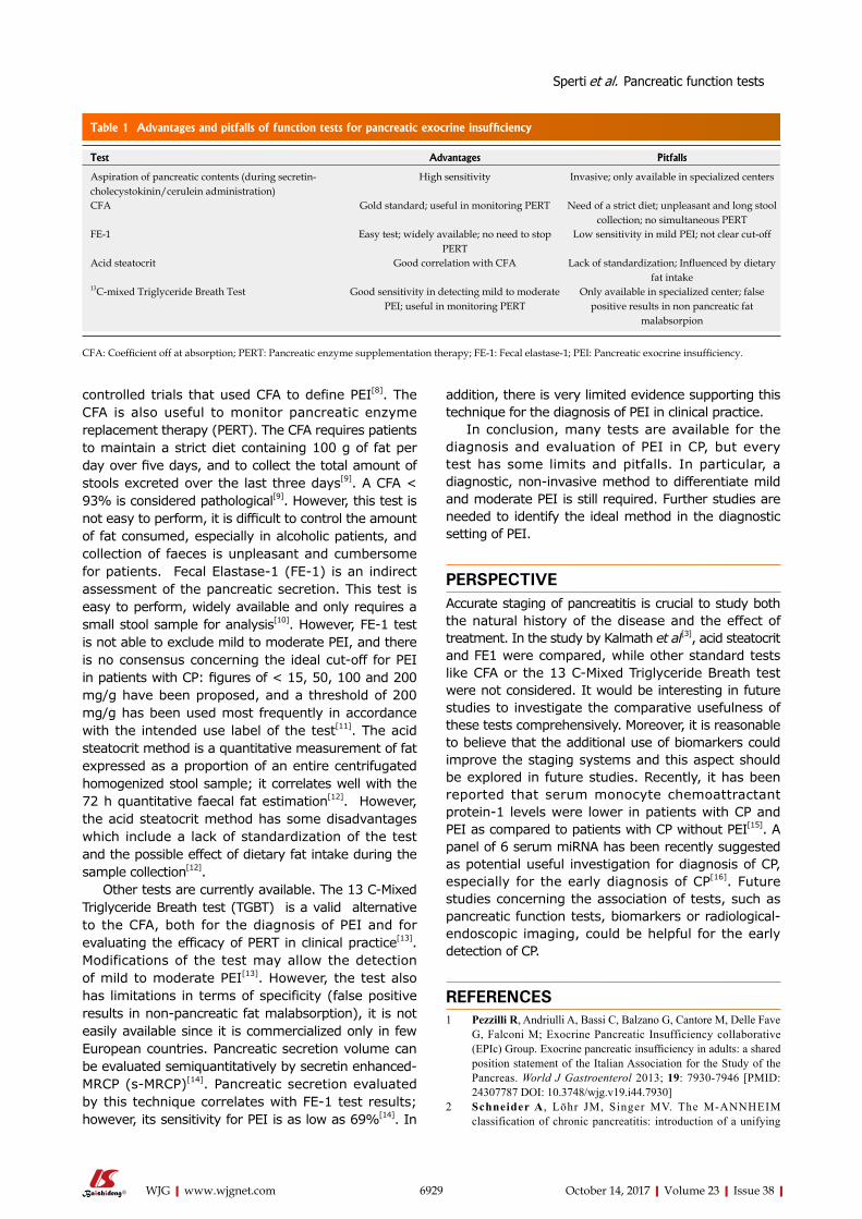
controlled trials that used CFA to define PEI[8]. The CFA is also useful to monitor pancreatic enzyme replacement therapy (PERT). The CFA requires patients to maintain a strict diet containing 100 g of fat per day over five days, and to collect the total amount of stools excreted over the last three days[9]. A CFA < 93% is considered pathological[9]. However, this test is not easy to perform, it is difficult to control the amount of fat consumed, especially in alcoholic patients, and collection of faeces is unpleasant and cumbersome for patients. Fecal Elastase-1 (FE-1) is an indirect assessment of the pancreatic secretion. This test is easy to perform, widely available and only requires a small stool sample for analysis[10]. However, FE-1 test is not able to exclude mild to moderate PEI, and there is no consensus concerning the ideal cut-off for PEI in patients with CP: figures of < 15, 50, 100 and 200 mg/g have been proposed, and a threshold of 200 mg/g has been used most frequently in accordance with the intended use label of the test[11]. The acid steatocrit method is a quantitative measurement of fat expressed as a proportion of an entire centrifugated homogenized stool sample; it correlates well with the 72 h quantitative faecal fat estimation[12]. However, the acid steatocrit method has some disadvantages which include a lack of standardization of the test and the possible effect of dietary fat intake during the sample collection[12].
Other tests are currently available. The 13 C-Mixed Triglyceride Breath test (TGBT) is a valid alternative to the CFA, both for the diagnosis of PEI and for evaluating the efficacy of PERT in clinical practice[13]. Modifications of the test may allow the detection of mild to moderate PEI[13]. However, the test also has limitations in terms of specificity (false positive results in non-pancreatic fat malabsorption), it is not easily available since it is commercialized only in few European countries. Pancreatic secretion volume can be evaluated semiquantitatively by secretin enhanced-MRCP (s-MRCP)[14]. Pancreatic secretion evaluated by this technique correlates with FE-1 test results; however, its sensitivity for PEI is as low as 69%[14]. In
addition, there is very limited evidence supporting this technique for the diagnosis of PEI in clinical practice.
In conclusion, many tests are available for the diagnosis and evaluation of PEI in CP, but every test has some limits and pitfalls. In particular, a diagnostic, non-invasive method to differentiate mild and moderate PEI is still required. Further studies are needed to identify the ideal method in the diagnostic setting of PEI.
PERSPECTIVEAccurate staging of pancreatitis is crucial to study both the natural history of the disease and the effect of treatment. In the study by Kalmath et al[3], acid steatocrit and FE1 were compared, while other standard tests like CFA or the 13 C-Mixed Triglyceride Breath test were not considered. It would be interesting in future studies to investigate the comparative usefulness of these tests comprehensively. Moreover, it is reasonable to believe that the additional use of biomarkers could improve the staging systems and this aspect should be explored in future studies. Recently, it has been reported that serum monocyte chemoattractant protein-1 levels were lower in patients with CP and PEI as compared to patients with CP without PEI[15]. A panel of 6 serum miRNA has been recently suggested as potential useful investigation for diagnosis of CP, especially for the early diagnosis of CP[16]. Future studies concerning the association of tests, such as pancreatic function tests, biomarkers or radiological-endoscopic imaging, could be helpful for the early detection of CP.
REFERENCES1 Pezzilli R, Andriulli A, Bassi C, Balzano G, Cantore M, Delle Fave
G, Falconi M; Exocrine Pancreatic Insufficiency collaborative (EPIc) Group. Exocrine pancreatic insufficiency in adults: a shared position statement of the Italian Association for the Study of the Pancreas. World J Gastroenterol 2013; 19: 7930-7946 [PMID: 24307787 DOI: 10.3748/wjg.v19.i44.7930]
2 Schneider A , Löhr JM, Singer MV. The M-ANNHEIM classification of chronic pancreatitis: introduction of a unifying
6929 October 14, 2017|Volume 23|Issue 38|WJG|www.wjgnet.com
Table 1 Advantages and pitfalls of function tests for pancreatic exocrine insufficiency
Test Advantages Pitfalls
Aspiration of pancreatic contents (during secretin-cholecystokinin/cerulein administration)
High sensitivity Invasive; only available in specialized centers
CFA Gold standard; useful in monitoring PERT Need of a strict diet; unpleasant and long stool collection; no simultaneous PERT
FE-1 Easy test; widely available; no need to stop PERT
Low sensitivity in mild PEI; not clear cut-off
Acid steatocrit Good correlation with CFA Lack of standardization; Influenced by dietary fat intake
13C-mixed Triglyceride Breath Test Good sensitivity in detecting mild to moderate PEI; useful in monitoring PERT
Only available in specialized center; false positive results in non pancreatic fat
malabsorpion
CFA: Coefficient off at absorption; PERT: Pancreatic enzyme supplementation therapy; FE-1: Fecal elastase-1; PEI: Pancreatic exocrine insufficiency.
Sperti et al. Pancreatic function tests

6930 October 14, 2017|Volume 23|Issue 38|WJG|www.wjgnet.com
10 Lankisch PG. Function tests in the diagnosis of chronic pancreatitis. Critical evaluation. Int J Pancreatol 1993; 14: 9-20 [PMID: 8409575 DOI: 10.1007/BF02795225]
11 Löhr JM, Dominguez-Munoz E, Rosendahl J, Besselink M, Mayerle J, Lerch MM, Haas S, Akisik F, Kartalis N, Iglesias-Garcia J, Keller J, Boermeester M, Werner J, Dumonceau JM, Fockens P, Drewes A, Ceyhan G, Lindkvist B, Drenth J, Ewald N, Hardt P, de Madaria E, Witt H, Schneider A, Manfredi R, Brøndum FJ, Rudolf S, Bollen T, Bruno M; HaPanEU/UEG Working Group. United European Gastroenterology evidence-based guidelines for the diagnosis and therapy of chronic pancreatitis (HaPanEU). United European Gastroenterol J 2017; 5: 153-199 [PMID: 28344786 DOI: 10.1177/2050640616684695]
12 Ramakrishna BS. The steatocrit as a measure of fecal fat excretion: uses and pitfalls. Indian J Gastroenterol 2009; 28: 195-197 [PMID: 20177864 DOI: 10.1007/s12664-009-0076-2]
13 González-Sánchez V, Amrani R, González V, Trigo C, Picó A, de-Madaria E. Diagnosis of exocrine pancreatic insufficiency in chronic pancreatitis: 13C-Mixed Triglyceride Breath Test versus Fecal Elastase. Pancreatology 2017; 17: 580-585 [PMID: 28291656 DOI: 10.1016/j.pan.2017.03.002]
14 Madzak A, Olesen SS, Haldorsen IS, Drewes AM, Frøkjær JB. Secretin-stimulated MRI characterization of pancreatic morphology and function in patients with chronic pancreatitis. Pancreatology 2017; 17: 228-236 [PMID: 28162928 DOI: 10.1016/j.pan.2017.01.009]
15 Kamath MG , Pai CG, Kamath A, Kurien A. Monocyte chemoattractant protein-1, transforming growth factor-beta1, nerve growth factor, resistin and hyaluronic acid as serum markers: comparison between recurrent acute and chronic pancreatitis. Hepatobiliary Pancreat Dis Int 2016; 15: 209-215 [PMID: 27020638 DOI: 10.1016/S1499-3872(15)60029-7]
16 Xin L, Gao J, Wang D, Lin JH, Liao Z, Ji JT, Du TT, Jiang F, Hu LH, Li ZS. Novel blood-based microRNA biomarker panel for early diagnosis of chronic pancreatitis. Sci Rep 2017; 7: 40019 [PMID: 28074846 DOI: 10.1038/srep40019]
P- Reviewer: Machado M, Fujino Y, Peng SY, Sharma V S- Editor: Gou SX L- Editor: A E- Editor: Ma YJ
classification system based on a review of previous classifications of the disease. J Gastroenterol 2007; 42: 101-119 [PMID: 17351799 DOI: 10.1007/s00535-006-1945-4]
3 Kamath MG, Pai CG, Kamath A, Kurien A. Comparing acid steatocrit and faecal elastase estimations for use in M-ANNHEIM staging for pancreatitis. World J Gastroenterol 2017; 23: 2217-2222 [PMID: 28405150 DOI: 10.3748/wjg.v23.i12.2217.]
4 Pedrazzoli S, Pasquali C, Guzzinati S, Berselli M, Sperti C. Survival rates and cause of death in 174 patients with chronic pancreatitis. J Gastrointest Surg 2008; 12: 1930-1937 [PMID: 18766421 DOI: 10.1007/s11605-008-0620-x]
5 Seiler CM, Izbicki J, Varga-Szabó L, Czakó L, Fiók J, Sperti C, Lerch MM, Pezzilli R, Vasileva G, Pap A, Varga M, Friess H. Randomised clinical trial: a 1-week, double-blind, placebo-controlled study of pancreatin 25 000 Ph. Eur. minimicrospheres (Creon 25000 MMS) for pancreatic exocrine insufficiency after pancreatic surgery, with a 1-year open-label extension. Aliment Pharmacol Ther 2013; 37: 691-702 [PMID: 23383603 DOI: 10.1111/apt.12236]
6 Pasquali C., Fogar P., Sperti C, Basso D., De Paoli M., Plebani M., Pedrazzoli S.: Efficacy of a pancreatic enzyme formulation in the treatment of steatorrhea in patients with chronic pancreatitis. Curr Ther Res 1996; 57: 358-365 [DOI: 10.1016/S0011-393X(96)80044-0]
7 O’Keefe SJ, Stevens S, Lee R, Zhou W, Zfass A. Physiological evaluation of the severity of pancreatic exocrine dysfunction during endoscopy. Pancreas 2007; 35: 30-36 [PMID: 17575542 DOI: 10.1097/mpa.0b013e3180646775]
8 Trapnell BC, Maguiness K, Graff GR, Boyd D, Beckmann K, Caras S. Efficacy and safety of Creon 24,000 in subjects with exocrine pancreatic insufficiency due to cystic fibrosis. J Cyst Fibros 2009; 8: 370-377 [PMID: 19815466 DOI: 10.1016/j.jcf.2009.08.008]
9 Hart PA, Conwell DL. Diagnosis of Exocrine Pancreatic Insufficiency. Curr Treat Options Gastroenterol 2015; 13: 347-353 [PMID: 26077487 DOI: 10.1007/s11938-015-0057-8]
Sperti et al. Pancreatic function tests

How to perform gastrointestinal ultrasound: anatomy and normal findings
Nathan S S Atkinson, Robert V Bryant, Yi Dong, Christian Maaser, Torsten Kucharzik, Giovanni Maconi, Anil K Asthana, Michael Blaivas, Adrian Goudie, Odd Helge Gilja, Dieter Nuernberg, Dagmar Schreiber-Dietrich, Christoph F Dietrich
Nathan S S Atkinson, Translational Gastroenterology Unit, Oxford University Hospitals NHS Foundation Trust, Oxford OX3 9DU, United Kingdom
Robert V Bryant, School of Medicine, University of Adelaide, Adelaide, South Australia, 5005, Australia
Robert V Bryant, Department of Gastroenterology and Hepatology, Royal Adelaide Hospital, Adelaide 5000, Australia
Yi Dong, Department of Ultrasound, Zhongshan Hospital, Fudan University, Shanghai 200032, China
Christian Maaser, Ambulanzzentrum Gastroenterologie, Klinikum Lüneburg, 21339 Lüneburg, Germany
Torsten Kucharzik, Department of Gastroenterology, Städtisches Klinikum Luneburg gGmbH, 21339 Lüneburg, Germany
Giovanni Maconi, Gastrointestinal Unit, Department of Biomedical and Clinical Sciences, “L.Sacco” University Hospital, 20157 Milan, Italy
Anil K Asthana, Department of Gastroenterology and Hepatology, The Alfred Hospital, Melbourne, Vic, Australia; Monash University, Melbourne 3004 Vic, Australia
Michael Blaivas, Piedmont Hospital, Department of Emergency Medicine, Atlanta, GA 30076, United States
Adrian Goudie, Fremantle Hospital and Health Service, Emergency Department, Fremantle, WA 6160, United States
Odd Helge Gilja, National Centre for Ultrasound in Gastroenterology, Haukeland University Hospital, Bergen N5021, Norway
Odd Helge Gilja, Department of Clinical Medicine, University of Bergen, 5021 Bergen, Norway
Dieter Nuernberg, Department of Gastroenterology, Brandenburg Medical School, 16816 Neuruppin, Germany
Dagmar Schreiber-Dietrich, Christoph F Dietrich, Med. Klinik 2, CaritasKrankenhaus Bad Mergentheim, D97980 Bad Mergentheim, Germany
Author contributions: All authors contributed to this manuscript.
Conflict-of-interest statement: No potential conflicts of interest. No financial support.
Open-Access: This article is an openaccess article which was selected by an inhouse editor and fully peerreviewed by external reviewers. It is distributed in accordance with the Creative Commons Attribution Non Commercial (CC BYNC 4.0) license, which permits others to distribute, remix, adapt, build upon this work noncommercially, and license their derivative works on different terms, provided the original work is properly cited and the use is noncommercial. See: http://creativecommons.org/licenses/bync/4.0/
Manuscript source: Invited manuscript
Correspondence to: Christoph F Dietrich, MD, PhD, Med. Klinik 2, CaritasKrankenhaus Bad Mergentheim, Uhlandstr. 7, D97980 Bad Mergentheim, Germany. [email protected]: +4979315822012200Fax: +497931582290
Received: February 9, 2017 Peer-review started: February 11, 2017First decision: April 25, 2017Revised: May 30, 2017 Accepted: June 18, 2017 Article in press: June 19, 2017 Published online: October 14, 2017
AbstractGastrointestinal ultrasound is a practical, safe, cheap and reproducible diagnostic tool in inflammatory
MINIREVIEWS
6931 October 14, 2017|Volume 23|Issue 38|WJG|www.wjgnet.com
Submit a Manuscript: http://www.f6publishing.com
DOI: 10.3748/wjg.v23.i38.6931
World J Gastroenterol 2017 October 14; 23(38): 6931-6941
ISSN 1007-9327 (print) ISSN 2219-2840 (online)

bowel disease gaining global prominence amongst clinicians. Understanding the embryological processes of the intestinal tract assists in the interpretation of abnormal sonographic findings. In general terms, the examination principally comprises interrogation of the colon, mesentery and small intestine using both low-frequency and high-frequency probes. Interpretation of findings on GIUS includes assessment of bowel wall thickness, symmetry of this thickness, evidence of transmural changes, assessment of vascularity using Doppler imaging and assessment of other specific features including lymph nodes, mesentery and luminal motility. In addition to B-mode imaging, transperineal ultrasonography, elastography and contrast-enhanced ultrasonography are useful adjuncts. This supplement expands upon these features in more depth.
Key words: Ultrasound; Intestinal; Inflammatory bowel disease; Guidelines; Teaching
Core tip: In general terms, gastrointestinal ultrasound examination principally comprises interrogation of the colon, mesentery and small intestine using both low-frequency and high-frequency probes. In addition to B-mode imaging, transperineal ultrasonography, elastography and contrast-enhanced ultrasonography are useful adjuncts.
Atkinson NSS, Bryant RV, Dong Y, Maaser C, Kucharzik T, Maconi G, Asthana AK, Blaivas M, Goudie A, Gilja OH, Nuernberg D, SchreiberDietrich D, Dietrich CF. How to perform gastrointestinal ultrasound: Anatomy and normal findings. World J Gastroenterol 2017; 23(38): 69316941 Available from: URL: http://www.wjgnet.com/10079327/full/v23/i38/6931.htm DOI: http://dx.doi.org/10.3748/wjg.v23.i38.6931
INTRODUCTIONGastrointestinal ultrasound (GIUS) is an accurate diagnostic imaging tool for inflammatory bowel disease[1-3]. Utilisation has steadily increased in different global regions including Asia-Pacific[4]. To correctly interpret GIUS findings, it is necessary to have a firm grounding in intestinal anatomy, the fundamentals of ultrasonography, as well as the examination techniques and approach. The indications for intestinal ultrasound are wide-ranging including inflammatory bowel disease, assessment of functional aspects and general gastroenterological conditions such as diverticular disease. Various educational theories are relevant in the process of learning intestinal ultrasonography including the learning process itself, using formative assessments such as DOPS (direct observation of procedural skills), adopting the apprenticeship or core competencies model and formulation of a GIUS curriculum. These principles and a process of learning GIUS have been recently proposed[1].
The purpose of this paper is to review our know
ledge of intestinal embryology relevant to GIUS. Understanding the origins and evolution of abdominal structures during the embryological process can assist in highlighting the reasoning behind abnormalities found on GIUS. We then expand upon examination techniques relevant to different segments of and structures around the intestine, including an overview of transperineal ultrasonography. We describe specific intestinal luminal parameters to be assessed in GIUS including special techniques such as contrastenhanced ultrasound.
REVIEW OF INTESTINAL EMBRYOLOGY In the 6th and 8th week of intrauterine life, the primitive mid-gut intestinal tube elongates on the mesentery around the superior mesenteric artery (SMA), herniating into the umbilical cord. As the gut grows and returns into the peritoneal cavity, it eventually rotates 270 degrees counterclockwise, such that the duodenum rests behind the SMA[1]. The caecum, initially in the upper abdomen, descends to the right lower quadrant. Thus the mesentery attachment of the small bowel takes an oblique course from the duodenojejunal junction at the level of the left L2 process, over the 3rd part of the duodenum, down to the level of the right sacroiliac joint[5]. Though the mesentery attachment is only 1520 cm long, it supplies a length of small intestine approximately 40 times its length, a feat achieved through progressive fanlike ruffles. Seen with traditional barium enterography, each curve of the intestine has a concave and convex aspect, the concave generally pointing towards the mesentery whilst the convex aspect representing the antimesenteric border[6]. These can be viewed in real time with GIUS.
Towards the end of the first trimester, the peritoneum of the newly forming ascending colon and the hindgut derived left colonic segments, begin to fuse with the posterior abdominal wall. Although traditionally described as retroperitoneal structures, modern postmortem studies have found that two thirds of the ascending and a third of the descending colonic segments have mobile portions of elongated mesentery[7]. Nonetheless, peritoneal attachments have significance for the flow of free fluid within the abdomen as fluid tends to flow caudally, medial to the ascending colon towards the ileocaecal junction and thus metastatic deposits may become lodged in the mesenteric ruffles en route.
Relative thickenings of the mesocolon provide ligamental support to the colonic flexures; the nephrocolic ligament runs from the inferior aspect of the right kidney to the hepatic flexure which then becomes intimately related to the descending duodenum before the transverse mesocolon begins; the splenic flexure is suspended by (1) the phrenicocolic ligament which runs from the diaphragm and also supports the spleen; (2) the splenocolic; and (3) the pancreaticocolic ligaments which are essentially extensions of the transverse
6932 October 14, 2017|Volume 23|Issue 38|WJG|www.wjgnet.com
Atkinson NSS et al . How to perform gastrointestinal ultrasound
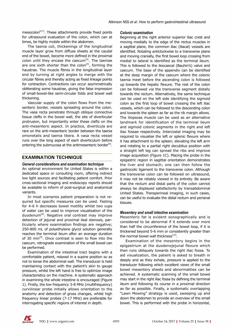
mesocolon[5,7]. These attachments provide fixed points for ultrasound evaluation of the colon, which can at times, be highly mobile within the abdomen.
The taenia coli, thickenings of the longitudinal muscle layer grow from diffuse sheets at the caudal end of the bowel, become more defined in the proximal colon until they encase the caecum[6]. The taeniae are one sixth shorter than the colon[5], forming the haustrae. The muscle fibres in the longitudinal layer end by turning at right angles to merge with the circular fibres and thereby acting as fixed linkage points for contraction. Contractions can occur asymmetrically obliterating some haustrae, giving the false impression of smallbowellike semicircular folds and bowel wall thickening.
Vascular supply of the colon flows from the me-senteric border, vessels spreading around the colon. The vasa recta penetrate through oblique connective tissue clefts in the bowel wall, the site of diverticular protrusion, but importantly enter these clefts on the antimesenteric aspect. In practice, diverticula are rare on the anti-mesenteric border between the taenia omonetalis and taenia libera. A vasa recta vessel runs over the long aspect of each diverticulum before entering the submucosa at the antimesenteric border[6].
EXAMINATION TECHNIQUEGeneral considerations and examination techniqueAn optimal environment for United States is within a dedicated space or consulting room, offering indirect low light sources and facilitating patient comfort. Prior cross-sectional imaging and endoscopy reports should be available to inform of postsurgical and anatomical variants.
In most scenarios patient preparation is not re-quired but specific measures can be used. Fasting for 4-6 h decreases bowel motility whilst two cups of water can be used to improve visualisation of the duodenum[8]. Negative oral contrast may improve detection of jejunal and proximal ileal stenosis, particularly where examination findings are negative; 250-800 mL of polyethylene glycol solution generally reaches the terminal ileum after an average duration of 30 min[9]. Once contrast is seen to flow into the caecum, retrograde examination of the small bowel can be performed.
Examination of the intestinal tract begins with a comfortable patient, relaxed in a supine position so as not to tense the abdominal wall. The transducer is held maintaining contact with the patient’s skin to gauge pressure, whilst the left hand is free to optimize image characteristics on the machine. A systematic approach in examining the whole intestine is encouraged (Figure 1). Firstly, the low frequency 38 MHz (multifrequency) curvilinear probe initially allows orientation to the anatomy and detection of gross changes, whilst high frequency linear probes (717 MHz) are preferable for interrogating specific regions of interest in depth.
Colonic examinationBeginning at the right anterior superior iliac crest and moving medially to the edge of the rectus muscles in a sagittal plane, the common iliac (iliacal) vessels are identified. Rotating anticlockwise to a transverse plane and moving cranially, the first bowel loop crossing from medial to lateral is identified as the terminal ileum. This is followed to the ileocaecal (Bauhin’s) valve and caecum. The base of the appendix can be identified at the deep margin of the caecum where the colonic taenia meet before the ascending colon is followed up towards the hepatic flexure. The rest of the colon can be followed via the transverse segment distally towards the rectum. Alternatively, the same technique can be used on the left side identifying the sigmoid colon as the first loop of bowel crossing the left iliac vessels, which can be followed to the descending colon and towards the spleen as far as the rib margin allows. The iliopsoas muscle can be used as an alternative landmark for identification of the terminal ileum and sigmoid colonic segments in the right and left iliac fossae respectively. Intercostal imaging may be required to visualise the left or splenic flexure where it has attachment to the spleen; elevating the left arm and rotating to a partial right decubitus position with a straight left leg can spread the ribs and improve image acquisition (Figure 1C). Placing the probe in the epigastric region in sagittal orientation demonstrates the liver and stomach; one can then follow the gastrocolic ligament to the transverse colon. Although the transverse colon can be followed on ultrasound, it may not be reliably viewed in its entirety. Be aware that the rectum and distal parts of the colon cannot always be displayed satisfactorily by transabdominal United States. Transperineal imaging, in such cases, can be useful to evaluate the distal rectum and perianal tissues.
Mesentery and small intestine examinationMesenteric fat is evident sonographically and is considered to be abnormal if it extends over more than half the circumference of the bowel loop, if it is thickened beyond 56 mm or consistently greater than the normal bowel wall thickness[10].
Examination of the mesentery begins in the epigastrium at the duodenojejunal flexure which then runs obliquely towards the right iliac fossa. To aid visualization, the patient is asked to breath in deeply and as they exhale, pressure is applied to the transducer following which excellent views of the small bowel mesentery sheets and abnormalities can be achieved. A systematic scanning of the small bowel may start in the right iliac fossa by defining the terminal ileum and following its course in a proximal direction as far as possible. Finally, a systematic overlapping “Lawn Mowing” strategy is used, sweeping up and down the abdomen to provide an overview of the small bowel. This is performed with the probe in horizontal,
6933 October 14, 2017|Volume 23|Issue 38|WJG|www.wjgnet.com
Atkinson NSS et al . How to perform gastrointestinal ultrasound

6934 October 14, 2017|Volume 23|Issue 38|WJG|www.wjgnet.com
Perineal ultrasound TPUS allows visualisation of the perianal tissues, anal canal, sphincters, the distal 57 cm section of the rectum, vagina and a part of urinary bladder. A point for orientation is the symphysis. Knowledge of the pelvic anatomy is essential[12,13] (EFSUMB Case of the Month). No specific patient preparation is required.
sagittal and oblique (parallel to the mesenteric attachment) orientations in order to allow one’s eye to follow structures and detect abnormalities. A full video explanation of abdominal and intestinal ultrasound examination technique is freely available on the EFSUMB website[11] (www.efsumb.org/education/cfd-videos001.asp).
a
B
Rectus
TIIAV
Pelvic wall
Rectus
Sigmoid
IAV
C
Rectus
Spleen SF
D
Rectus
SMA
Aorta
Figure 1 A systematic approach in examining the whole intestine. A: Examination begins in a relaxed ventral position; B: Beginning medial to the right anterior superior iliac spine, the iliacal vessels (IAV) are identified and the first bowel loop crossing medial-to-lateral is the terminal ileum (TI). The same technique on the right identifies the sigmoid colon; C: Elevating the arm spreads the rib spaces to improve visualisation of the splenic flexure (SF); D: Gentle pressure as the patient breaths out improves visualization of the mesentery and superior mesenteric artery (SMA) to exclude lymphadenopathy. The videos can be accessed via the efsumb website [www.efsumb.org/education/cfd-videos001.asp].
Atkinson NSS et al . How to perform gastrointestinal ultrasound

6935 October 14, 2017|Volume 23|Issue 38|WJG|www.wjgnet.com
The patient is placed in the left lateral position as for a digital rectal examination. The probe is covered in either a sterile cover or an examination glove with ultrasound gel between the layers.
Examination begins in the midline just above the anus with the probe in a sagittal plane. The ultrasound probe can be moved laterally, however angulated views have reduced sensitivity for identification of pathology. Fistulous tracts can be followed by first placing the probe over the external opening. If necessary, the probe can be placed in a coronal angle, although this is usually less comfortable for the patient. It is useful to start with an abdominal convex probe (lower frequency) for the deeper structures and then continue with a higher frequency probe (715 MHz). Examination is also possible after rectal amputation (e.g., QuénuOperation).
The anal canal, sphincter complexes, hemorrhoidal plexus (Figure 2), rectovaginal plus anovaginal septums, the walls of the vagina and distal rectum can be defined. Fistulae should be classified as per Parks’ classification[14] although TPUS has reduced sensitivities for sphincteric relationships and therefore the American Gastroenterological Association (AGA)[15] distinction of “simple” from “complex” is of more clinical utility; the former including low fistulae (superficial, intersphincteric or intrasphincteric) below the dentate line, with a single external opening and without perianal complications or active proctitis[16]. Fistula and abscesses visibility can be improved with ultrasound contrast agents (UCA) using contrast enhanced ultra-sound techniques[17,18]. Colour Doppler improves the differentiation of inflammatory reactions.
INTESTINAL LUMINAL FEATURES Bowel wall thicknessBowel wall thickness (BWT) is the measure most consistently reported in diagnostic and activity trials. Wall thickness of the alimentary tract differs by region and depends on the degree of distension
and contraction[19,20]. The overall thickness should be measured under mild compression from just above an air-mucosal interface to the outside of the outer muscularis propria layer border, including the whole bowel wall[21]. Under these standardised conditions, the stomach wall thickness measures 36 mm; terminal ileum 13 mm; and colon 0.52 mm. In fact, the normal range is likely to be even lower than this[22]. Values in children can be reliably obtained without the need for sedation[22] but bear in mind that values do increase over childhood[23] whilst still remaining < 2 mm. The optimal threshold for abnormal thickness is debatable, as specificity improves with increasing wall thickness at the cost of sensitivity (Figures 3 and 4).
Bowel wall layersThe GI wall has five layers that usually can be visualized with ultrasound. The sonographic layers are a combination of interface echoes and the echo characteristics of the histological layers[24-26]. When imaged in the anterior wall of a bowel loop starting from the lumen the hyperechoic layer 1 corresponds to the interface between the mucosa and the lumen and is not a part of the actual GI wall. The hypoechoic layer 2 corresponds to the mucosa without the interface between the submucosa and mucosa, the hyperechoic layer 3 to the submucosa including this interface echo, the hypoechoic layer 4 to most of the proper muscle and layer 5 to the hyperechoic interface echo between the proper muscle and the serosa.
Interface echoes are always hyperechoic and located distally to the actual tissue interface. Therefore, the correspondence between histology and sonographic layers differ slightly in the dorsal wall. Specifically, the interface between lumen and mucosa (layer 1) is a part of the actual mucosa and layer 2 represents the rest of the mucosa without muscularis mucosae, which normally is covered by an interface echo and add thickness to layer 3. Moreover, the interface between submucosa and the proper muscle adds thickness to layer 3 and reduces the thickness of layer 4. Finally, the interface between the proper muscle and serosa (layer 5) extends beyond the actual serosa[27,28].
The interface from the serosa is hard to delineate. Accordingly, the measurement should be made from the start of the hypoechoic layer of the proper muscle to the end of the hypoechoic layer of the mucosa. Transducer-compression of the bowel wall will reduce thickness and can make it challenging to distinguish wall layers[29,30]. However, some operators practice mild compression suggesting that this improves reproducibility of measurements[21,22,3133]. The examiner should also be aware of interpretation difficulties due to mucosal folds and haustrations and to keep the probe angle perpendicular to the bowel wall to avoid tangential measurements. In conclusion, dosed compression is a prerequisite for a reproducible examination for some authors whereas others use it with caution.
Figure 2 Example on the use of color doppler imaging and continous duplex scanning. Perineal ultrasound showing the hemorrhoidal pleaxus using color doppler imaging and continous duplex scanning with the typical spectrum of the hemorrhoids.
Atkinson NSS et al . How to perform gastrointestinal ultrasound
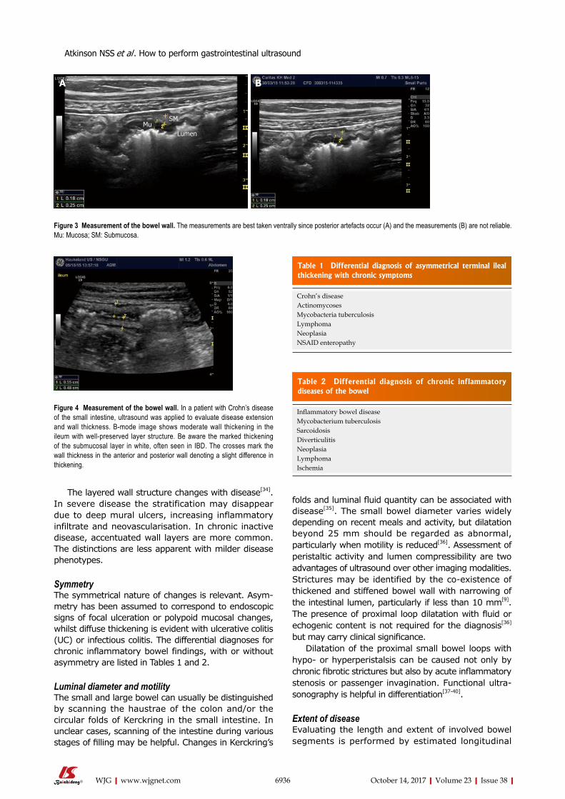
6936 October 14, 2017|Volume 23|Issue 38|WJG|www.wjgnet.com
The layered wall structure changes with disease[34]. In severe disease the stratification may disappear due to deep mural ulcers, increasing inflammatory infiltrate and neovascularisation. In chronic inactive disease, accentuated wall layers are more common. The distinctions are less apparent with milder disease phenotypes.
SymmetryThe symmetrical nature of changes is relevant. Asymmetry has been assumed to correspond to endoscopic signs of focal ulceration or polypoid mucosal changes, whilst diffuse thickening is evident with ulcerative colitis (UC) or infectious colitis. The differential diagnoses for chronic inflammatory bowel findings, with or without asymmetry are listed in Tables 1 and 2.
Luminal diameter and motilityThe small and large bowel can usually be distinguished by scanning the haustrae of the colon and/or the circular folds of Kerckring in the small intestine. In unclear cases, scanning of the intestine during various stages of filling may be helpful. Changes in Kerckring’s
folds and luminal fluid quantity can be associated with disease[35]. The small bowel diameter varies widely depending on recent meals and activity, but dilatation beyond 25 mm should be regarded as abnormal, particularly when motility is reduced[36]. Assessment of peristaltic activity and lumen compressibility are two advantages of ultrasound over other imaging modalities. Strictures may be identified by the coexistence of thickened and stiffened bowel wall with narrowing of the intestinal lumen, particularly if less than 10 mm[9]. The presence of proximal loop dilatation with fluid or echogenic content is not required for the diagnosis[36] but may carry clinical significance.
Dilatation of the proximal small bowel loops with hypo- or hyperperistalsis can be caused not only by chronic fibrotic strictures but also by acute inflammatory stenosis or passenger invagination. Functional ultrasonography is helpful in differentiation[37-40].
Extent of diseaseEvaluating the length and extent of involved bowel segments is performed by estimated longitudinal
a B
MuSM
Lumen
Figure 3 Measurement of the bowel wall. The measurements are best taken ventrally since posterior artefacts occur (A) and the measurements (B) are not reliable. Mu: Mucosa; SM: Submucosa.
Figure 4 Measurement of the bowel wall. In a patient with Crohn’s disease of the small intestine, ultrasound was applied to evaluate disease extension and wall thickness. B-mode image shows moderate wall thickening in the ileum with well-preserved layer structure. Be aware the marked thickening of the submucosal layer in white, often seen in IBD. The crosses mark the wall thickness in the anterior and posterior wall denoting a slight difference in thickening.
Crohn’s diseaseActinomycosesMycobacteria tuberculosisLymphomaNeoplasiaNSAID enteropathy
Table 1 Differential diagnosis of asymmetrical terminal ileal thickening with chronic symptoms
Inflammatory bowel diseaseMycobacterium tuberculosisSarcoidosisDiverticulitisNeoplasiaLymphomaIschemia
Table 2 Differential diagnosis of chronic inflammatory diseases of the bowel
Atkinson NSS et al . How to perform gastrointestinal ultrasound
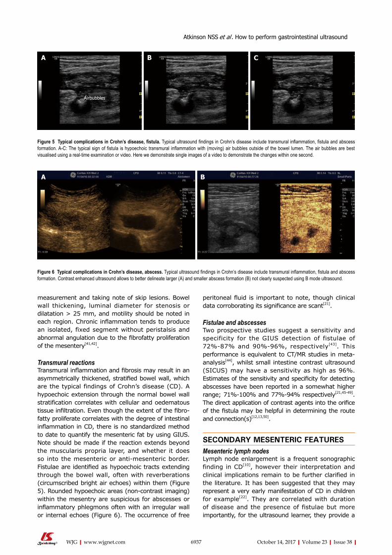
6937 October 14, 2017|Volume 23|Issue 38|WJG|www.wjgnet.com
measurement and taking note of skip lesions. Bowel wall thickening, luminal diameter for stenosis or dilatation > 25 mm, and motility should be noted in each region. Chronic inflammation tends to produce an isolated, fixed segment without peristalsis and abnormal angulation due to the fibrofatty proliferation of the mesentery[41,42].
Transmural reactionsTransmural inflammation and fibrosis may result in an asymmetrically thickened, stratified bowel wall, which are the typical findings of Crohn’s disease (CD). A hypoechoic extension through the normal bowel wall stratification correlates with cellular and oedematous tissue infiltration. Even though the extent of the fibrofatty proliferate correlates with the degree of intestinal inflammation in CD, there is no standardized method to date to quantify the mesenteric fat by using GIUS. Note should be made if the reaction extends beyond the muscularis propria layer, and whether it does so into the mesenteric or anti-mesenteric border. Fistulae are identified as hypoechoic tracts extending through the bowel wall, often with reverberations (circumscribed bright air echoes) within them (Figure 5). Rounded hypoechoic areas (non-contrast imaging) within the mesentry are suspicious for abscesses or inflammatory phlegmons often with an irregular wall or internal echoes (Figure 6). The occurrence of free
peritoneal fluid is important to note, though clinical data corroborating its significance are scant[21].
Fistulae and abscessesTwo prospective studies suggest a sensitivity and specificity for the GIUS detection of fistulae of 72%87% and 90%96%, respectively[43]. This performance is equivalent to CT/MR studies in metaanalysis[44], whilst small intestine contrast ultrasound (SICUS) may have a sensitivity as high as 96%. Estimates of the sensitivity and specificity for detecting abscesses have been reported in a somewhat higher range; 71%100% and 77%94% respectively[21,4549]. The direct application of contrast agents into the orifice of the fistula may be helpful in determining the route and connection(s)[12,13,50].
SECONDARY MESENTERIC FEATURESMesenteric lymph nodes Lymph node enlargement is a frequent sonographic finding in CD[10], however their interpretation and clinical implications remain to be further clarified in the literature. It has been suggested that they may represent a very early manifestation of CD in children for example[22]. They are correlated with duration of disease and the presence of fistulae but more importantly, for the ultrasound learner, they provide a
Airbubbles
a B C
Figure 5 Typical complications in Crohn’s disease, fistula. Typical ultrasound findings in Crohn’s disease include transmural inflammation, fistula and abscess formation. A-C: The typical sign of fistula is hypoechoic transmural inflammation with (moving) air bubbles outside of the bowel lumen. The air bubbles are best visualised using a real-time examination or video. Here we demonstrate single images of a video to demonstrate the changes within one second.
a B
Figure 6 Typical complications in Crohn’s disease, abscess. Typical ultrasound findings in Crohn’s disease include transmural inflammation, fistula and abscess formation. Contrast enhanced ultrasound allows to better delineate larger (A) and smaller abscess formation (B) not clearly suspected using B mode ultrasound.
Atkinson NSS et al . How to perform gastrointestinal ultrasound

6938 October 14, 2017|Volume 23|Issue 38|WJG|www.wjgnet.com
marker of procedural competence and interpretation. B mode characteristics of lymph nodes to consider include their length and particularly for those < 15 mm; their short axis dimension should be less than half their longitudinal diameter. Furthermore, the normal lymph node architecture and hilum should be preserved in normal or inflammatory nodes[51].
Mesenteric inflammation, oedema and vascularityThe supporting structures of the intestine run within the sheets of mesentery, seen as layers of mixed echogenicity with hyperechoic serosal layers on either side, which does not have peristaltic movement and appears similar in both transverse and sagittal planes. Fat wrapping has long been recognised by surgeons as a common and specific feature of CD. Socalled (creeping) fat, extending from the mesenteric attachment to partially cover the small or large intestine resulting in loss of bowel mesentery angle, is seen as an early event in the disease course and plausibly plays a role in the inflammatory milieu[52]. In practical terms the serosal planes on either side of the mesentery may be detected and should cover less than half of the bowel circumference. It is also the most common cause of bowel loop separation[41]. A subjective impression of increased thickening and echogenicity has been applied in the literature[10], correlating with clinical severity and primary luminal findings; although in long standing disease it can become more heterogeneous and hypoechoic[41].
TECHNIQUES FOR INTERROGATION OF FEATURES OF INTERESTDoppler imaging of tissue and SMA parametersColour Doppler imaging of the bowel wall is part of standard assessment of the intestine and mesenterial vessels (Figure 7). Hyperaemia is associated with inflammation, usually seen in the submucosal layer and the penetrating vessels of the muscularis propria. Use of Doppler evaluation increases the sensitivity of US for evaluating disease activity[33,39,5359]. The degree
of vascularity can be graded by the Limberg scale, a semiquantitative assessment[60] that lacks routine practical relevance. Other more complex quantitative measurements of Doppler parameters have been proposed, however a standardised protocol to compensate for confounders has not yet become widely used. Power Doppler assessment of the arterial inflow, in particular the inferior mesenteric artery (IMA) for left sided colonic disease and SMA for proximal colon and small bowel activity, can be assessed in the majority of patients and correlates with other ultrasound markers of disease activity. It should therefore be interpreted in the overall context of ultrasound findings[61]. A prognostic role for Doppler parameters was previously proposed[62] but awaits further study and validation.
Contrast-enhanced USSecond generation contrast agents such as SonoVue®, produce harmonic frequencies from microbubbles approximately the size of a single red blood cell, and are stable within the circulation[17,63]. Imaging systems thereafter allow visualisation of individual blood vessels through a tissue and thereby improve the accuracy of Doppler US in evaluating bowel wall vascularity. This technique has been shown to be useful in the assessment of disease activity in CD, in particular differentiating inflammatory masses from abscesses and may help to distinguish inflammatory from fibrotic strictures in certain situations[64-66]. Use of Contrast-Enhanced US (CEUS) during GIUS has been standardized, does not requires specific expertise, and its use in IBD presently is increasing[67].
ElastographySimilar to palpation, the elastic properties of a tissue can be evaluated by assessing the speed of a sheer wave through tissue or the amount of deformity created by the sheer stress (strain imaging). Various sonographic approaches to generating and measuring these parameters are available[68], which may compliment standard B-mode assessment of a lesion. Fibrotic lesions may appear stiff and inflammatory lesions soft using elastography, which can help to characterise intestinal lesions and has been correlated with endoscopic findings (Figure 8)[69,70].
CONCLUSIONUnderstanding the anatomy and embryology of the intestinal tract is highly relevant in identifying sonographic abnormalities relevant to GIUS. The general principles of examination involve specific interrogation of the colon, small intestine and mesentery. Further work is required to validate and understand the significance of certain sonographic parameters where understanding is limited; this includes further evaluation of abnormal wall thicknesses and quantifying this to a higher degree of accuracy, understanding the significance of peritoneal fluid present as a reflection of transmural reactions,
Figure 7 Complications of inflammatory bowel disease. Thrombosis of the superior mesenteric vein. Partial recanalisation is shown by the markers.
Atkinson NSS et al . How to perform gastrointestinal ultrasound

6939 October 14, 2017|Volume 23|Issue 38|WJG|www.wjgnet.com
and the accurate interpretation and implications of lymph nodes. Despite the presence of semiquantitative measures, such as the Limberg score, they lack practical relevance and so there is a need for further multicentre prospective studies.
Various sonographic abnormalities can be detected and interpreted currently but a standardized scoring system for GIUS in inflammatory bowel disease, akin to validated endoscopic scores (such as the Ulcerative Colitis Endoscopic Index of Severity) is lacking. Ultimately, formulating a reproducible and validated scoring system integrating different sonographic parameters to reflect severity will be highly relevant; this will require agreement amongst GIUS experts and validation in multicentre prospective studies. Finally, a standardized method of documentation, including how to capture images, needs to be developed.
REFERENCES1 Atkinson NS, Bryant RV, Dong Y, Maaser C, Kucharzik T,
Maconi G, Asthana AK, Blaivas M, Goudie A, Gilja OH, Nolsøe C, Nürnberg D, Dietrich CF. WFUMB Position Paper. Learning Gastrointestinal Ultrasound: Theory and Practice. Ultrasound Med Biol 2016; 42: 27322742 [PMID: 27742140 DOI: 10.1016/j.ultrasmedbio.2016.08.026]
2 Nylund K, Maconi G, Hollerweger A, Ripolles T, Pallotta N, Higginson A, Serra C, Dietrich CF, Sporea I, Saftoiu A, Dirks K, Hausken T, Calabrese E, Romanini L, Maaser C, Nuernberg D, Gilja OH. EFSUMB Recommendations and Guidelines for Gastrointestinal Ultrasound. Ultraschall Med 2017; 38: e1e15 [PMID: 27604052 DOI: 10.1055/s0042115853]
3 Nylund K, Maconi G, Hollerweger A, Ripolles T, Pallotta N, Higginson A, Serra C, Dietrich CF, Sporea I, Saftoiu A, Dirks K, Hausken T, Calabrese E, Romanini L, Maaser C, Nuernberg D, Gilja OH. EFSUMB Recommendations and Guidelines for Gastrointestinal Ultrasound. Ultraschall Med 2017; 38: 273284 [PMID: 27604051 DOI: 10.1055/s0042115410]
4 Asthana AK, Friedman AB, Maconi G, Maaser C, Kucharzik T, Watanabe M, Gibson PR. Failure of gastroenterologists to apply intestinal ultrasound in inflammatory bowel disease in the AsiaPacific: a need for action. J Gastroenterol Hepatol 2015; 30: 446452 [PMID: 25529767 DOI: 10.1111/jgh.12871]
5 Jorge JMN, Habr Gama A. Anatomy and Embryology of the Colon, Rectum, and Anus. In: Wolff BG, Flechman JW, Beck DE, Pemberton JH, Wexner SD, Church JM, editors. The ASCRS
Textbook of Colon and Rectal Surgery. New York: Springer, 2007: 122
6 Meyers MA. Dynamic Radiology of the Abdomen. New York: Springer, 2005
7 Phillips M, Patel A, Meredith P, Will O, Brassett C. Segmental colonic length and mobility. Ann R Coll Surg Engl 2015; 97: 439444 [PMID: 26274737 DOI: 10.1308/003588415X14181254790527]
8 Taylor S, Mallett S, Bhatnagar G, Bloom S, Gupta A, Halligan S, Hamlin J, Hart A, Higginson A, Jacobs I, McCartney S, Morris S, Muirhead N, Murray C, Punwani S, RodriguezJusto M, Slater A, Travis S, Tolan D, Windsor A, Wylie P, Zealley I. METRIC (MREnterography or ulTRasound in Crohn‘s disease): a study protocol for a multicentre, nonrandomised, singlearm, prospective comparison study of magnetic resonance enterography and small bowel ultrasound compared to a reference standard in those aged 16 and over. BMC Gastroenterol 2014; 14: 142 [PMID: 25110044 DOI: 10.1186/1471230X14142]
9 Parente F, Greco S, Molteni M, Anderloni A, Sampietro GM, Danelli PG, Bianco R, Gallus S, Bianchi Porro G. Oral contrast enhanced bowel ultrasonography in the assessment of small intestine Crohn‘s disease. A prospective comparison with conventional ultrasound, x ray studies, and ileocolonoscopy. Gut 2004; 53: 16521657 [PMID: 15479688 DOI: 10.1136/gut.2004.041038]
10 Maconi G, Greco S, Duca P, Ardizzone S, Massari A, Cassinotti A, Radice E, Porro GB. Prevalence and clinical significance of sonographic evidence of mesenteric fat alterations in Crohn‘s disease. Inflamm Bowel Dis 2008; 14: 15551561 [PMID: 18521926 DOI: 10.1002/ibd.20515]
11 Dietrich CF. EFSUMB Course Book on Ultrasound. Examination technique (videos). London: European Federation of Societies for Ultrasound in Medicine and Biology (EFSUMB), 2016
12 Dietrich CF, Barreiros AP, Nuernberg D, SchreiberDietrich DG, Ignee A. [Perianal ultrasound]. Z Gastroenterol 2008; 46: 625630 [PMID: 18537090 DOI: 10.1055/s20081027518]
13 Maconi G, Tonolini M, Monteleone M, Bezzio C, Furfaro F, Villa C, Campari A, DellʼEra A, Sampietro G, Ardizzone S, de Franchis R. Transperineal perineal ultrasound versus magnetic resonance imaging in the assessment of perianal Crohn‘s disease. Inflamm Bowel Dis 2013; 19: 27372743 [PMID: 24193154 DOI: 10.1097/01.MIB.0000436274.95722.e5]
14 Parks AG, Gordon PH, Hardcastle JD. A classification of fistula-inano. Br J Surg 1976; 63: 112 [PMID: 1267867]
15 Sandborn WJ, Fazio VW, Feagan BG, Hanauer SB. AGA technical review on perianal Crohn‘s disease. Gastroenterology 2003; 125: 15081530 [PMID: 14598268]
16 Sandborn WJ, Fazio VW, Feagan BG, Hanauer SB, American Gastroenterological Association Clinical Practice Committee. AGA technical review on perianal Crohn‘s disease. Gastroenterology 2003; 125: 15081530 [PMID: 14598268 DOI: 10.1016/j.gastro.2003.08.025]
17 Ignee A, Jenssen C, Cui XW, Schuessler G, Dietrich CF. Intracavitary contrastenhanced ultrasound in abscess drainagefeasibility and clinical value. Scand J Gastroenterol 2016; 51: 4147 [PMID: 26166454 DOI: 10.3109/00365521.2015.1066423]
18 Ignee A, Cui X, Schuessler G, Dietrich CF. Percutaneous transhepatic cholangiography and drainage using extravascular contrast enhanced ultrasound. Z Gastroenterol 2015; 53: 385390 [PMID: 25965985 DOI: 10.1055/s00341398796]
19 Nylund K, Hausken T, Odegaard S, Eide GE, Gilja OH. Gastrointestinal wall thickness measured with transabdominal ultrasonography and its relationship to demographic factors in healthy subjects. Ultraschall Med 2012; 33: E225E232 [PMID: 22504939 DOI: 10.1055/s00311299329]
20 Cantisani V, Dietrich CF, Badea R, Dudea S, Prosch H, Cerezo E, Nuernberg D, Serra AL, Sidhu PS, Radzina M, Piscaglia F, Bachmann Nielsen M, Calliada F, Gilja OH. EFSUMB statement on medical student education in ultrasound [short version]. Ultraschall Med 2016; 37: 100102 [PMID: 26871409 DOI: 10.1055/s00351566959]
Figure 8 The evaluation of bowel wall stiffness. Elastography is helpful to determine stiff tissue as shown in this patient with colorectal carcinoma and infiltration of the abdominal wall.
Atkinson NSS et al . How to perform gastrointestinal ultrasound
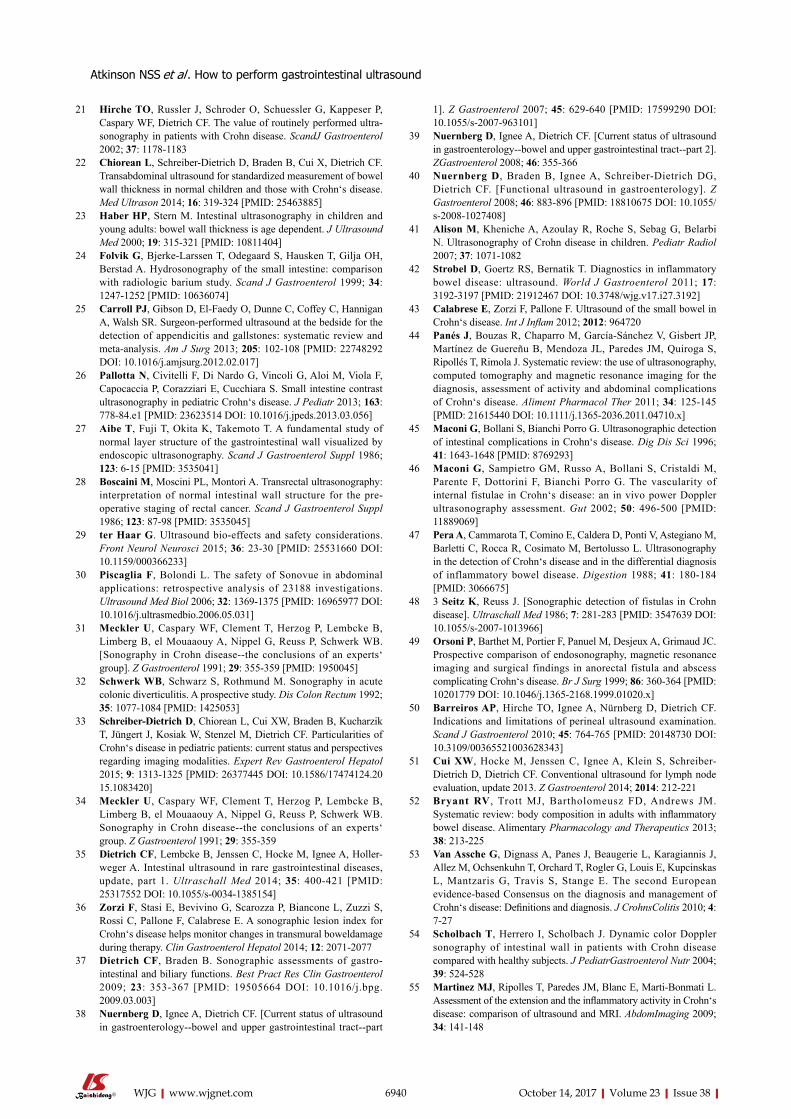
6940 October 14, 2017|Volume 23|Issue 38|WJG|www.wjgnet.com
21 Hirche TO, Russler J, Schroder O, Schuessler G, Kappeser P, Caspary WF, Dietrich CF. The value of routinely performed ultrasonography in patients with Crohn disease. ScandJ Gastroenterol 2002; 37: 11781183
22 Chiorean L, SchreiberDietrich D, Braden B, Cui X, Dietrich CF. Transabdominal ultrasound for standardized measurement of bowel wall thickness in normal children and those with Crohn‘s disease. Med Ultrason 2014; 16: 319324 [PMID: 25463885]
23 Haber HP, Stern M. Intestinal ultrasonography in children and young adults: bowel wall thickness is age dependent. J Ultrasound Med 2000; 19: 315321 [PMID: 10811404]
24 Folvik G, BjerkeLarssen T, Odegaard S, Hausken T, Gilja OH, Berstad A. Hydrosonography of the small intestine: comparison with radiologic barium study. Scand J Gastroenterol 1999; 34: 12471252 [PMID: 10636074]
25 Carroll PJ, Gibson D, ElFaedy O, Dunne C, Coffey C, Hannigan A, Walsh SR. Surgeonperformed ultrasound at the bedside for the detection of appendicitis and gallstones: systematic review and metaanalysis. Am J Surg 2013; 205: 102108 [PMID: 22748292 DOI: 10.1016/j.amjsurg.2012.02.017]
26 Pallotta N, Civitelli F, Di Nardo G, Vincoli G, Aloi M, Viola F, Capocaccia P, Corazziari E, Cucchiara S. Small intestine contrast ultrasonography in pediatric Crohn‘s disease. J Pediatr 2013; 163: 77884.e1 [PMID: 23623514 DOI: 10.1016/j.jpeds.2013.03.056]
27 Aibe T, Fuji T, Okita K, Takemoto T. A fundamental study of normal layer structure of the gastrointestinal wall visualized by endoscopic ultrasonography. Scand J Gastroenterol Suppl 1986; 123: 615 [PMID: 3535041]
28 Boscaini M, Moscini PL, Montori A. Transrectal ultrasonography: interpretation of normal intestinal wall structure for the preoperative staging of rectal cancer. Scand J Gastroenterol Suppl 1986; 123: 8798 [PMID: 3535045]
29 ter Haar G. Ultrasound bioeffects and safety considerations. Front Neurol Neurosci 2015; 36: 2330 [PMID: 25531660 DOI: 10.1159/000366233]
30 Piscaglia F, Bolondi L. The safety of Sonovue in abdominal applications: retrospective analysis of 23188 investigations. Ultrasound Med Biol 2006; 32: 13691375 [PMID: 16965977 DOI: 10.1016/j.ultrasmedbio.2006.05.031]
31 Meckler U, Caspary WF, Clement T, Herzog P, Lembcke B, Limberg B, el Mouaaouy A, Nippel G, Reuss P, Schwerk WB. [Sonography in Crohn diseasethe conclusions of an experts‘ group]. Z Gastroenterol 1991; 29: 355359 [PMID: 1950045]
32 Schwerk WB, Schwarz S, Rothmund M. Sonography in acute colonic diverticulitis. A prospective study. Dis Colon Rectum 1992; 35: 10771084 [PMID: 1425053]
33 Schreiber-Dietrich D, Chiorean L, Cui XW, Braden B, Kucharzik T, Jüngert J, Kosiak W, Stenzel M, Dietrich CF. Particularities of Crohn‘s disease in pediatric patients: current status and perspectives regarding imaging modalities. Expert Rev Gastroenterol Hepatol 2015; 9: 13131325 [PMID: 26377445 DOI: 10.1586/17474124.2015.1083420]
34 Meckler U, Caspary WF, Clement T, Herzog P, Lembcke B, Limberg B, el Mouaaouy A, Nippel G, Reuss P, Schwerk WB. Sonography in Crohn diseasethe conclusions of an experts‘ group. Z Gastroenterol 1991; 29: 355359
35 Dietrich CF, Lembcke B, Jenssen C, Hocke M, Ignee A, Hollerweger A. Intestinal ultrasound in rare gastrointestinal diseases, update, part 1. Ultraschall Med 2014; 35: 400421 [PMID: 25317552 DOI: 10.1055/s00341385154]
36 Zorzi F, Stasi E, Bevivino G, Scarozza P, Biancone L, Zuzzi S, Rossi C, Pallone F, Calabrese E. A sonographic lesion index for Crohn‘s disease helps monitor changes in transmural boweldamage during therapy. Clin Gastroenterol Hepatol 2014; 12: 20712077
37 Dietrich CF, Braden B. Sonographic assessments of gastrointestinal and biliary functions. Best Pract Res Clin Gastroenterol 2009; 23: 353367 [PMID: 19505664 DOI: 10.1016/j.bpg. 2009.03.003]
38 Nuernberg D, Ignee A, Dietrich CF. [Current status of ultrasound in gastroenterologybowel and upper gastrointestinal tractpart
1]. Z Gastroenterol 2007; 45: 629640 [PMID: 17599290 DOI: 10.1055/s2007963101]
39 Nuernberg D, Ignee A, Dietrich CF. [Current status of ultrasound in gastroenterologybowel and upper gastrointestinal tractpart 2]. ZGastroenterol 2008; 46: 355366
40 Nuernberg D, Braden B, Ignee A, SchreiberDietrich DG, Dietrich CF. [Functional ultrasound in gastroenterology]. Z Gastroenterol 2008; 46: 883896 [PMID: 18810675 DOI: 10.1055/s20081027408]
41 Alison M, Kheniche A, Azoulay R, Roche S, Sebag G, Belarbi N. Ultrasonography of Crohn disease in children. Pediatr Radiol 2007; 37: 10711082
42 Strobel D, Goertz RS, Bernatik T. Diagnostics in inflammatory bowel disease: ultrasound. World J Gastroenterol 2011; 17: 31923197 [PMID: 21912467 DOI: 10.3748/wjg.v17.i27.3192]
43 Calabrese E, Zorzi F, Pallone F. Ultrasound of the small bowel in Crohn‘s disease. Int J Inflam 2012; 2012: 964720
44 Panés J, Bouzas R, Chaparro M, GarcíaSánchez V, Gisbert JP, Martínez de Guereñu B, Mendoza JL, Paredes JM, Quiroga S, Ripollés T, Rimola J. Systematic review: the use of ultrasonography, computed tomography and magnetic resonance imaging for the diagnosis, assessment of activity and abdominal complications of Crohn‘s disease. Aliment Pharmacol Ther 2011; 34: 125145 [PMID: 21615440 DOI: 10.1111/j.13652036.2011.04710.x]
45 Maconi G, Bollani S, Bianchi Porro G. Ultrasonographic detection of intestinal complications in Crohn‘s disease. Dig Dis Sci 1996; 41: 16431648 [PMID: 8769293]
46 Maconi G, Sampietro GM, Russo A, Bollani S, Cristaldi M, Parente F, Dottorini F, Bianchi Porro G. The vascularity of internal fistulae in Crohn‘s disease: an in vivo power Doppler ultrasonography assessment. Gut 2002; 50: 496500 [PMID: 11889069]
47 Pera A, Cammarota T, Comino E, Caldera D, Ponti V, Astegiano M, Barletti C, Rocca R, Cosimato M, Bertolusso L. Ultrasonography in the detection of Crohn‘s disease and in the differential diagnosis of inflammatory bowel disease. Digestion 1988; 41: 180184 [PMID: 3066675]
48 3 Seitz K, Reuss J. [Sonographic detection of fistulas in Crohn disease]. Ultraschall Med 1986; 7: 281283 [PMID: 3547639 DOI: 10.1055/s20071013966]
49 Orsoni P, Barthet M, Portier F, Panuel M, Desjeux A, Grimaud JC. Prospective comparison of endosonography, magnetic resonance imaging and surgical findings in anorectal fistula and abscess complicating Crohn‘s disease. Br J Surg 1999; 86: 360364 [PMID: 10201779 DOI: 10.1046/j.13652168.1999.01020.x]
50 Barreiros AP, Hirche TO, Ignee A, Nürnberg D, Dietrich CF. Indications and limitations of perineal ultrasound examination. Scand J Gastroenterol 2010; 45: 764765 [PMID: 20148730 DOI: 10.3109/00365521003628343]
51 Cui XW, Hocke M, Jenssen C, Ignee A, Klein S, SchreiberDietrich D, Dietrich CF. Conventional ultrasound for lymph node evaluation, update 2013. Z Gastroenterol 2014; 2014: 212221
52 Bryant RV, Trott MJ, Bartholomeusz FD, Andrews JM. Systematic review: body composition in adults with inflammatory bowel disease. Alimentary Pharmacology and Therapeutics 2013; 38: 213225
53 Van Assche G, Dignass A, Panes J, Beaugerie L, Karagiannis J, Allez M, Ochsenkuhn T, Orchard T, Rogler G, Louis E, Kupcinskas L, Mantzaris G, Travis S, Stange E. The second European evidencebased Consensus on the diagnosis and management of Crohn‘s disease: Definitions and diagnosis. J CrohnsColitis 2010; 4: 727
54 Scholbach T, Herrero I, Scholbach J. Dynamic color Doppler sonography of intestinal wall in patients with Crohn disease compared with healthy subjects. J PediatrGastroenterol Nutr 2004; 39: 524528
55 Martinez MJ, Ripolles T, Paredes JM, Blanc E, MartiBonmati L. Assessment of the extension and the inflammatory activity in Crohn‘s disease: comparison of ultrasound and MRI. AbdomImaging 2009; 34: 141148
Atkinson NSS et al . How to perform gastrointestinal ultrasound
6941 October 14, 2017|Volume 23|Issue 38|WJG|www.wjgnet.com
56 Spalinger J, Patriquin H, Miron MC, Marx G, Herzog D, Dubois J, Dubinsky M, Seidman EG. Doppler US in patients with crohn disease: vessel density in the diseased bowel reflects disease activity. Radiology 2000; 217: 787791
57 Rapaccini GL, Pompili M, Orefice R, Covino M, Riccardi L, Cedrone A, Gasbarrini G. Contrastenhanced power doppler of the intestinal wall in the evaluation of patients with Crohn disease. ScandJ Gastroenterol 2004; 39: 188194
58 Fraquelli M, Colli A, Casazza G, Paggi S, Colucci A, Massironi S, Duca P, Conte D. Role of US in detection of Crohn disease: metaanalysis. Radiology 2005; 236: 95101
59 Dietrich CF, Ignee A, Seitz KH, Caspary WF. [Duplex sonography of visceral arteries]. Ultraschall Med 2001; 22: 247257
60 Limberg B. [Colon sonographya new method in the diagnosis of Crohn disease and ulcerative colitis]. Monatsschr Kinderheilkd 1990; 138: 422426 [PMID: 2215503]
61 Dietrich CF, Jedrzejczyk M, Ignee A. Sonographic assessment of splanchnic arteries and the bowel wall. Eur J Radiol 2007; 64: 202212 [PMID: 17923366 DOI: 10.1016/j.ejrad.2007.06.034]
62 Ludwig D, Wiener S, Brüning A, Schwarting K, Jantschek G, Fellermann K, Stahl M, Stange EF. Mesenteric blood flow is related to disease activity and risk of relapse in ulcerative colitis: a prospective follow up study. Gut 1999; 45: 546552
63 Ignee A, Boerner N, Bruening A, Dirks K, von Herbay A, Jenssen C, Kubale R, Sattler H, Schuler A, Weiss H, Schuessler G, Dietrich CF. Duplex sonography of the mesenteric vesselsa critical evaluation of interobserver variability. Z Gastroenterol 2016; 54: 304311 [PMID: 27056458 DOI: 10.1055/s0041107544]
64 Braden B, Ignee A, Hocke M, Palmer RM, Dietrich C. Diagnostic value and clinical utility of contrast enhanced ultrasound in intestinal diseases. Dig Liver Dis 2010; 42: 667674 [PMID: 20598952 DOI: 10.1016/j.dld.2010.05.018]
65 Piscaglia F, Nolsøe C, Dietrich CF, Cosgrove DO, Gilja OH, Bachmann Nielsen M, Albrecht T, Barozzi L, Bertolotto M, Catalano O, Claudon M, Clevert DA, Correas JM, D‘Onofrio M, Drudi FM, Eyding J, Giovannini M, Hocke M, Ignee A, Jung EM,
Klauser AS, Lassau N, Leen E, Mathis G, Saftoiu A, Seidel G, Sidhu PS, ter Haar G, Timmerman D, Weskott HP. The EFSUMB Guidelines and Recommendations on the Clinical Practice of Contrast Enhanced Ultrasound (CEUS): update 2011 on nonhepatic applications. Ultraschall Med 2012; 33: 3359 [PMID: 21874631 DOI: 10.1055/s00311281676]
66 Ripollés T, Rausell N, Paredes JM, Grau E, Martínez MJ, Vizuete J. Effectiveness of contrastenhanced ultrasound for characterisation of intestinal inflammation in Crohn‘s disease: a comparison with surgical histopathology analysis. J Crohn‘s & colitis 2013; 7: 120128
67 Allgayer H, Ignee A, Dietrich CF. Endosonographic elastography of the anal sphincter in patients with fecal incontinence. Scand J Gastroenterol 2010; 45: 3038 [PMID: 20001748 DOI: 10.3109/00365520903383251]
68 Bamber J, Cosgrove D, Dietrich CF, Fromageau J, Bojunga J, Calliada F, Cantisani V, Correas JM, D‘Onofrio M, Drakonaki EE, Fink M, FriedrichRust M, Gilja OH, Havre RF, Jenssen C, Klauser AS, Ohlinger R, Saftoiu A, Schaefer F, Sporea I, Piscaglia F. EFSUMB guidelines and recommendations on the clinical use of ultrasound elastography. Part 1: Basic principles and technology. Ultraschall Med 2013; 34: 169184 [PMID: 23558397 DOI: 10.1055/s00331335205]
69 Ishikawa D, Ando T, Watanabe O, Ishiguro K, Maeda O, Miyake N, Nakamura M, Miyahara R, Ohmiya N, Hirooka Y, ElOmar EM, Goto H. Images of colonic realtime tissue sonoelastography correlate with those of colonoscopy and may predict response to therapy in patients with ulcerative colitis. BMC Gastroenterol 2011; 31: 29
70 Cosgrove D, Piscaglia F, Bamber J, Bojunga J, Correas JM, Gilja OH, Klauser AS, Sporea I, Calliada F, Cantisani V, D‘Onofrio M, Drakonaki EE, Fink M, FriedrichRust M, Fromageau J, Havre RF, Jenssen C, Ohlinger R, Săftoiu A, Schaefer F, Dietrich CF; EFSUMB. EFSUMB guidelines and recommendations on the clinical use of ultrasound elastography. Part 2: Clinical applications. Ultraschall Med 2013; 34: 238253 [PMID: 23605169 DOI: 10.1055/s00331335375]
P- Reviewer: Capasso R S- Editor: Ma YJ L- Editor: A E- Editor: Li D
Atkinson NSS et al . How to perform gastrointestinal ultrasound

Dysphagia: Thinking outside the box
Hamish Philpott, Mayur Garg, Dunya Tomic, Smrithya Balasubramanian, Rami Sweis
Hamish Philpott, Mayur Garg, Dunya Tomic, Smrithya Balasubramanian, Department of Gastroenterology, Eastern Health Clinical School Monash University, Melbourne 3128, Australia
Rami Sweis, University College London, London NW1 2BU, United Kingdom
Author contributions: Philpott H conceptualized the review; Philpott H and Sweis R wrote the paper; Garg M and Sweis R edited the paper; Tomic D and Balasubramanian S contributed content.
Conflict-of-interest statement: Nil to declare.
Open-Access: This article is an open-access article which was selected by an in-house editor and fully peer-reviewed by external reviewers. It is distributed in accordance with the Creative Commons Attribution Non Commercial (CC BY-NC 4.0) license, which permits others to distribute, remix, adapt, build upon this work non-commercially, and license their derivative works on different terms, provided the original work is properly cited and the use is non-commercial. See: http://creativecommons.org/licenses/by-nc/4.0/
Manuscript source: Invited manuscript
Correspondence to: Dr. Hamish Philpott, Department of Gastroenterology, Eastern Health Clinical School Monash University, 5 Arnold St Box Hill 3128, Melbourne 3128, Australia. [email protected]: +61-3-421227551Fax: +61-3-98396733
Received: February 8, 2017Peer-review started: February 8, 2017First decision: April 21, 2017Revised: August 29, 2017Accepted: September 26, 2017Article in press: September 26, 2017Published online: October 14, 2017
AbstractDysphagia is a common symptom that is important
to recognise and appropriately manage, given that causes include life threatening oesophageal neoplasia, oropharyngeal dysfunction, the risk of aspiration, as well as chronic disabling gastroesophageal reflux (GORD). The predominant causes of dysphagia varies between cohorts depending on the interplay between genetic predisposition and environmental risk factors, and is changing with time. Currently in white Caucasian societies adopting a western lifestyle, obesity is common and thus associated gastroesophageal reflux disease is increasingly diagnosed. Similarly, food allergies are increasing in the west, and eosinophilic oesophagitis is increasingly found as a cause. Other regions where cigarette smoking is still prevalent, or where access to medical care and antisecretory agents such as proton pump inhibitors are less available, benign oesophageal peptic strictures, Barrett’s oesophagus, adeno- as well as squamous cell carcinoma are endemic. The evaluation should consider the severity of symptoms, as well as the pre-test probability of a given condition. In young white Caucasian males who are atopic or describe heartburn, eosinophilic esophagitis and gastroesophageal reflux disease will predominate and a proton pump inhibitor could be commenced prior to further investigation. Upper gastrointestinal endoscopy remains a valid first line investigation for patients with suspected oesophageal dysphagia. Barium swallow is particularly useful for oropharyngeal dysphagia, and oesophageal manometry mandatory to diagnose motility disorders.
Key words: Dysphagia; Oesophagus; Eosinophilic; Manometry; Gastroesophageal reflux; Aspiration; Food bolus impaction
© The Author(s) 2017. Published by Baishideng Publishing Group Inc. All rights reserved.
Core tip: Dysphagia may represent serious and l ife-threatening pathology such as oesophageal neoplasia or oropharyngeal dysfunction capable of causing aspiration. In young white Caucasian males, eosinophilic esophagitis and gastroesophageal reflux
MINIREVIEWS
6942 October 14, 2017|Volume 23|Issue 38|WJG|www.wjgnet.com
Submit a Manuscript: http://www.f6publishing.com
DOI: 10.3748/wjg.v23.i38.6942
World J Gastroenterol 2017 October 14; 23(38): 6942-6951
ISSN 1007-9327 (print) ISSN 2219-2840 (online)

disease are predominant causes, and a trial of proton pump inhibitor is suggested whilst awaiting endoscopy.
Philpott H, Garg M, Tomic D, Balasubramanian S, Sweis R. Dysphagia: Thinking outside the box. World J Gastroenterol 2017; 23(38): 6942-6951 Available from: URL: http://www.wjgnet.com/1007-9327/full/v23/i38/6942.htm DOI: http://dx.doi.org/10.3748/wjg.v23.i38.6942
INTRODUCTIONDysphagia is a common symptom in patients presenting to both generalist and specialist medical practitioners[1,2]. The key initial consideration for clinicians is to rule out lifethreatening pathology such as neoplasia, and to identify patients who are at risk of aspiration such that immediate intervention can occur[3]. Dysphagia is most commonly due to chronic benign disorders, although it can be associated with appreciable morbidity and impaired quality of life (QOL)[4]. Gastroesophageal reflux disease (GORD) is the major cause in younger individuals, whilst oropharyngeal dysphagia (OD) secondary to cerebrovascular disease is more frequent in the elderly[1,4]. Dysphagia as a symptom attributable to GORD in younger adults does not automatically mandate investigation, rather a trial of a proton pump inhibitor (PPI) and strict followup to ensure symptomatic resolution might be sufficient[5]. OD and oesophageal dysphagia can often be reliably differentiated symptomatically and might help reduce the risk of aspiration in OD[6]. A comprehensive awareness of the multitude of potential causes of dysphagia, including common medications and rheumatological conditions, is necessary to allow for efficient and thorough evaluation. Judicious investigation with the appropriate choice of tests in order to consider both common and unusual aetiologies is required; high quality endoscopy with care to take sufficient oesophageal biopsies even if mucosa looks normal [e.g., eosinophilic oesophagitis (EoE)], high resolution oesophageal manometry (HRM) in suspected motility disturbance, barium swallow and where appropriate, crosssectional imaging [e.g., computed tomography (CT) scan]. The escalating frequency of GORD and EoE, and the increase in mean age (and associated conditions such as CVA) will ensure that dysphagia remains a common presentation in the future[7,8]. This review attempts to present a contemporary understanding and approach to patients with dysphagia.
EPIDEMIOLOGYDysphagia is the subjective awareness of an impairment in swallowing of saliva, liquid or solid
food. The prevalence varies by age of cohort, method of data collection, and if the dysphagia is classified as acute or chronic[1,4,9]. A community survey of 1000 individuals in Sydney Australia, demonstrated a prevalence of dysphagia of 16% (defined as ever having the symptom), although few (1%) reported dysphagia as chronic or severe[4]. GORD, hypertension and psychological symptoms (anxiety and depression) were independently associated with dysphagia[4]. Subjectivity in symptom reporting and the contribution of GORD have emerged as important themes in contemporary studies of dysphagia[1]. A study of patients with GORD reported resolution of dysphagia with PPI[10]. General practice cohorts have found a frequency of dysphagia of up to 23%, thus reflecting the significant health care burden taken up by this patient group[2]. Selfreported dysphagia is often variably defined[11]; an isolated globus sensation can erroneously be considered a form of dysphagia.
In a large survey of an adult United States population (n = 7640, 3669 respondents), 17% of respondents reported “infrequent” (< 1 episode per week) and 3% “frequent” (> 1 episode per week) dysphagia[1]. On interrogation of these respondents’ medical files, symptoms of heartburn and endoscopic diagnosis of GORD were significantly associated with both the “frequent” and “infrequent” cohorts[1]. Perhaps due to the limited sample size and old age of the respondents (mean 62 years), other typical conditions were not statistically associated with dysphagia. Primary oesophageal conditions (EoE, achalasia, cancer) and systemic conditions (scleroderma, rheumatoid arthritis) were only reported amongst the “frequent” dysphagia group[1]. Therefore, clinically significant (or frequent) dysphagia should alert the clinician to serious underlying pathology, whilst infrequent dysphagia is most likely to represent GORD and a trial of acid suppression may be worthwhile in the first instance provided there are no alarm symptoms. This view is supported by several experts in the field (see below).
AETIOLOGY AND PATHOGENESISDysphagia is the subjective awareness of impairment in the passage of food from the oropharynx to the stomach, and therefore may signify an actual delay in bolus transit, or merely the sensation thereof[12]. It is useful to consider the anatomical structures implicated in swallowing (including innervation), and the location of pathology within (Figure 1)[10]. The process of swallowing, whereby food or liquid travels from the mouth to the stomach, involves a complex sequence of muscular contraction and relaxation, involving striated (oropharynx and upper 1/3rd of the oesophagus) and smooth muscle (lower 2/3rd of the oesophagus) that is controlled by motor neurons of the brainstem, and autonomic innervation (of the myenteric plexus) respectively[10,11]. The oropharyngeal component is
6943 October 14, 2017|Volume 23|Issue 38|WJG|www.wjgnet.com
Philpott H et al. Approach to the patient with dysphagia

initially voluntary (including chewing). This is followed by an involuntary phase initiated by food entering the pharynx, whereby the swallowing reflex causes simultaneous relaxation and contraction of the soft palate, upper oesophageal sphincter, oesophagus and lower oesophageal sphincter[6].
Causes of dysphagia vary across the world, and there is evidence of a change in the relative proportion of the various disease states in the last few decades[8]. In the United States, benign oesophageal stricture has become less common, whilst EoE is increasing, along with oesophageal adenocarcinoma[8]. This pattern is mirrored in other Western nations[18]. Oesophageal squamous cell cancer remains a common condition in Asia, whilst Chagas disease still is prevalent is some regions of South America[18].
The oesophageal lumen can become narrowed by inflammation (e.g., erosive reflux disease or EoE), neoplasia or fibrous stricture, or can be compressed externally by lymph nodes or degenerative disease of the cervical spine[8,1215].
Luminal stenosisNarrowing of the oesophageal lumen by inflammation, stricture, web or tumour can result in dysphagia[16]. Distal oesophageal inflammation from GORD can result in dysphagia due to transient decrement in peristaltic vigour, and if sustained can lead to fibrous stricture formation[8,15]. Barrett’s oesophagus and/or sliding hiatus hernia (HH) are present in > 80% of
those with chronic severe GORD and there is some evidence that both may independently influence oesophageal peristalsis[17,18]. Treatment of Barrett’s oesophagus with endoscopic mucosal resection (EMR) and radiofrequency ablation can lead to fibrosis[19]. The Schatzki ring (a mucosal ring of the distal oesophagus), occurs just proximal to the gastroesophageal sphincter, often within a hiatus hernia, and is relatively common[20,21]. Barium swallow reveals Schatzki ring in 5%15% of patients with HH[8]. The association with dysphagia and and its cause (congenital, secondary to GERD or even EoE) are debated[22]. Proximal oesophageal webs have been described in individuals with dysphagia. An association with iron deficiency anaemia (the PlummerVinson syndrome) was apparently common in the first half of last century, although is rarely reported today[23,24]. EoE is perhaps the most important, benign cause of dysphagia behind GORD. It is increasingly common and may be responsible for some cases previously diagnosed as fibrous strictures or webs[7]. This is an important consideration amongst Caucasian patients in western countries, and is readily treatable (see below)[8,25]. The prevalence of EoE is estimated at between 1 in 500 and 1 in 1000 adult Caucasian males[26]. The importance of EoE is redoubled by the frequent occurrence of food bolus obstruction, a complication that is associated with significant morbidity including the need for hospitalisation, upper gastrointestinal endoscopy (UGE) and (rarely but catastrophically) oesophageal perforation[27].
Non-obstructive gastro-oesophageal reflux diseaseMany patients that describe dysphagia will have normal investigations including UGE and highresolution manometry (HRM), suggesting that a dysfunction of the somatosensory as opposed to neuromuscular apparatus might be present[28,29]. A unifying hypothesis to explain this apparent dichotomy is that a minor disturbance in peristalsis (caused for example by GORD, or nonerosive reflux disease NERD) may be below the threshold of detection even by sensitive HRM, yet be experienced by the patient as dysphagia. Indeed, many people will describe a resolution of symptoms with proton pump inhibition (PPI). Interventions aimed at modulating the somatosensory system have hence been advocated[28,29].
Primary motility disturbanceAchalasia is the prototypical condition classified as a primary (isolated to the oesophagus) disorder of oesophageal motility, yet is quite rare, with an estimated prevalence of 0.17% in individuals undergoing endoscopy[30]. Patients typically present with a chronic history of dysphagia for liquids and/or solids, and often with retrosternal discomfort, heartburn and weight loss[31]. The underlying pathophysiology is of an imbalance between excitatory
6944 October 14, 2017|Volume 23|Issue 38|WJG|www.wjgnet.com
Oral cavity Upper oesophageal sphincter
Cervical spine
Upper 1/3 oesophagusstriated muscle
Lower 2/3 oesophagus smooth muscle
Lower oesophageal sphincter
Stomach
Figure 1 Anatomy of the oesophagus. Disease of the upper 1/3 of the oesophagus causing dysphagia may include extrinsic compression (e.g., cervical osteophytes), or dysfunction secondary to rheumatological conditions (e.g., Sjogrens’s syndrome) or in eosinophilic oesophagitis (along with the lower oesophagus). The lower oesophagus can be afflicted in scleroderma, gastroesophageal reflux disease and in eosinophilic oesophagitis.
Philpott H et al. Approach to the patient with dysphagia
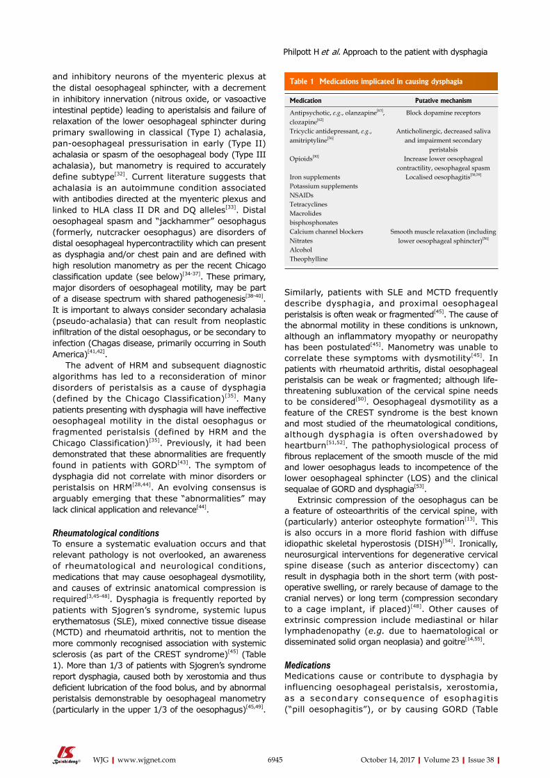
6945 October 14, 2017|Volume 23|Issue 38|WJG|www.wjgnet.com
Similarly, patients with SLE and MCTD frequently describe dysphagia, and proximal oesophageal peristalsis is often weak or fragmented[45]. The cause of the abnormal motility in these conditions is unknown, although an inflammatory myopathy or neuropathy has been postulated[45]. Manometry was unable to correlate these symptoms with dysmotility[45]. In patients with rheumatoid arthritis, distal oesophageal peristalsis can be weak or fragmented; although lifethreatening subluxation of the cervical spine needs to be considered[50]. Oesophageal dysmotility as a feature of the CREST syndrome is the best known and most studied of the rheumatological conditions, although dysphagia is often overshadowed by heartburn[51,52]. The pathophysiological process of fibrous replacement of the smooth muscle of the mid and lower oesophagus leads to incompetence of the lower oesophageal sphincter (LOS) and the clinical sequalae of GORD and dysphagia[53].
Extrinsic compression of the oesophagus can be a feature of osteoarthritis of the cervical spine, with (particularly) anterior osteophyte formation[13]. This is also occurs in a more florid fashion with diffuse idiopathic skeletal hyperostosis (DISH)[54]. Ironically, neurosurgical interventions for degenerative cervical spine disease (such as anterior discectomy) can result in dysphagia both in the short term (with postoperative swelling, or rarely because of damage to the cranial nerves) or long term (compression secondary to a cage implant, if placed)[48]. Other causes of extrinsic compression include mediastinal or hilar lymphadenopathy (e.g. due to haematological or disseminated solid organ neoplasia) and goitre[14,55].
MedicationsMedications cause or contribute to dysphagia by influencing oesophageal peristalsis, xerostomia, as a secondary consequence of esophagitis (“pill oesophagitis”), or by causing GORD (Table
and inhibitory neurons of the myenteric plexus at the distal oesophageal sphincter, with a decrement in inhibitory innervation (nitrous oxide, or vasoactive intestinal peptide) leading to aperistalsis and failure of relaxation of the lower oesophageal sphincter during primary swallowing in classical (Type I) achalasia, panoesophageal pressurisation in early (Type II) achalasia or spasm of the oesophageal body (Type III achalasia), but manometry is required to accurately define subtype[32]. Current literature suggests that achalasia is an autoimmune condition associated with antibodies directed at the myenteric plexus and linked to HLA class II DR and DQ alleles[33]. Distal oesophageal spasm and “jackhammer” oesophagus (formerly, nutcracker oesophagus) are disorders of distal oesophageal hypercontractility which can present as dysphagia and/or chest pain and are defined with high resolution manometry as per the recent Chicago classification update (see below)[3437]. These primary, major disorders of oesophageal motility, may be part of a disease spectrum with shared pathogenesis[3840]. It is important to always consider secondary achalasia (pseudoachalasia) that can result from neoplastic infiltration of the distal oesophagus, or be secondary to infection (Chagas disease, primarily occurring in South America)[41,42].
The advent of HRM and subsequent diagnostic algorithms has led to a reconsideration of minor disorders of peristalsis as a cause of dysphagia (defined by the Chicago Classification)[35]. Many
patients presenting with dysphagia will have ineffective oesophageal motility in the distal oesophagus or fragmented peristalsis (defined by HRM and the Chicago Classification)[35]. Previously, it had been demonstrated that these abnormalities are frequently found in patients with GORD[43]. The symptom of dysphagia did not correlate with minor disorders or peristalsis on HRM[28,44]. An evolving consensus is arguably emerging that these “abnormalities” may lack clinical application and relevance[44].
Rheumatological conditionsTo ensure a systematic evaluation occurs and that relevant pathology is not overlooked, an awareness of rheumatological and neurological conditions, medications that may cause oesophageal dysmotility, and causes of extrinsic anatomical compression is required[3,4548]. Dysphagia is frequently reported by patients with Sjogren’s syndrome, systemic lupus erythematosus (SLE), mixed connective tissue disease (MCTD) and rheumatoid arthritis, not to mention the more commonly recognised association with systemic sclerosis (as part of the CREST syndrome)[45] (Table 1). More than 1/3 of patients with Sjogren’s syndrome report dysphagia, caused both by xerostomia and thus deficient lubrication of the food bolus, and by abnormal peristalsis demonstrable by oesophageal manometry (particularly in the upper 1/3 of the oesophagus)[45,49].
Table 1 Medications implicated in causing dysphagia
Medication Putative mechanism
Antipsychotic, e.g., olanzapine[63], clozapine[62]
Block dopamine receptors
Tricyclic antidepressant, e.g., amitriptyline[56]
Anticholinergic, decreased saliva and impairment secondary
peristalsisOpioids[90] Increase lower oesophageal
contractility, oesophageal spasmIron supplements Localised oesophagitis[58,59]
Potassium supplementsNSAIDsTetracyclinesMacrolidesbisphosphonatesCalcium channel blockers Smooth muscle relaxation (including
lower oesophageal sphincter)[56]NitratesAlcoholTheophylline
Philpott H et al. Approach to the patient with dysphagia

6946 October 14, 2017|Volume 23|Issue 38|WJG|www.wjgnet.com
2)[47,5659]. Sedative agents can lead to compromise of airway maintenance and thus aspiration is a risk. Finally, medications that have a local or systemic immunosuppressant effect can predispose to infective oesophagitis[60]. Among individuals with a psychiatric diagnosis, dysphagia may relate, at least in part, to the use of psychotropic medication[61]. The anticholinergic and antidopaminergic effects of antipsychotics and tricyclic antidepressants have been implicated, not only in causing a dry mouth (anticholinergic) thus limiting lubrication of the food bolus, but also in decreasing the coordination and strength of peristalsis and the timing of lower oesophageal sphincter relaxation[62,63]. It is also possible that serotonergic pathways may be responsible for the dysphagia caused by clozapine,
although this hypothesis is perhaps refuted by the absence of case reports relating SSRIs to dysphagia[63]. Individuals with psychiatric diagnoses may also have an increased rate of neurological disorders (such as Parkinson’s disease), particularly among older individuals in residential care, that can cause dysphagia[3,61]. The use of opiates, even if low dose and over the counter, can lead to hypercontractile or hypertensive sequelae of the oesophagus and/or LOS, sometimes even mimicking Type III achalasia.
Neurological disordersNeurogenic dysphagia, a term loosely applied to cases where the pathology involves the central, autonomic or peripheral nervous system (and not isolated to the oesophagus such as achalasia) is more often characterised by an oropharyngeal component, with neurologists and speech pathologists primarily involved in management[64,65]. Dysphagia is common after cerebrovascular accidents (CVA) involving the cortex often causing hemiplegia[66]. Rapid improvement in swallowing function occurs in the majority[67]. Brainstem infarcts with cranial nerve involvement is another (albeit less common) lesion, but can be severe and sustained[3]. The basal ganglia are the major site of pathology in Parkinson’s disease, and oropharyngeal involvement (with difficulty initiating swallowing and impairment in bolus transit between the pharynx and proximal oesophagus) demonstrable at video fluoroscopy[68].
Table 2 Neurogenic dysphagia - Common causes
Condition Description
Parkinson’s disease[68] Oropharyngeal and oesophageal dysphagia is possible
Multiple sclerosis[64] Oropharyngeal and oesophageal dysphagia is possible
Cerebrovascular disease[3,67] Oropharyngeal dysphagia typicalMotor neuron disease[11,64] Features of bulbar and pseudobulbar
palsy possibleMyasthenia gravis[64,91]
Myopathy (various, including inflammatory)[45,64]
Oropharyngeal and proximal oesophageal
Cerebellar pathology (various)[64]
Philpott H et al. Approach to the patient with dysphagia
Figure 2 Proposed management algorithm for patients presenting with dysphagia.
Clinical assessment to determine risk of aspiration or if suitable for a PPI
Oropharyngeal dysphagia likely:Consider Barium swallow andreferral to e.g. neurologist,
otolaryngologist or speech pathologist
Age < 50 yr, non-smoker and EoEconsidered unlikely: Empirical trial of
PPI
When no cause is found consider oesophageal manometry and/or cross-sectional imaging (e.g. CT scan)
Eosinophilic oesophagitis possible:Gastroscopy and biopsies of the
mid and lower oesophagus
Clinical Review: If no improvement in 2 weeks consider endoscopy
Dysphagia
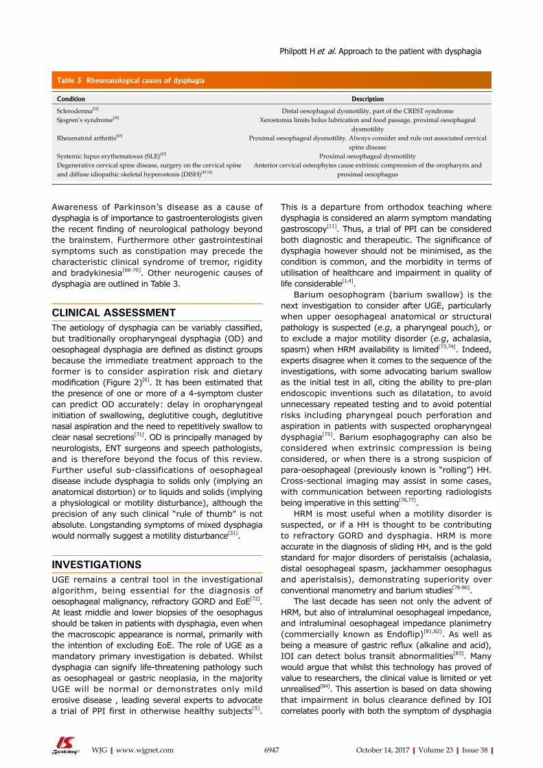
6947 October 14, 2017|Volume 23|Issue 38|WJG|www.wjgnet.com
Awareness of Parkinson’s disease as a cause of dysphagia is of importance to gastroenterologists given the recent finding of neurological pathology beyond the brainstem. Furthermore other gastrointestinal symptoms such as constipation may precede the characteristic clinical syndrome of tremor, rigidity and bradykinesia[6870]. Other neurogenic causes of dysphagia are outlined in Table 3.
CLINICAL ASSESSMENTThe aetiology of dysphagia can be variably classified, but traditionally oropharyngeal dysphagia (OD) and oesophageal dysphagia are defined as distinct groups because the immediate treatment approach to the former is to consider aspiration risk and dietary modification (Figure 2)[6]. It has been estimated that the presence of one or more of a 4symptom cluster can predict OD accurately: delay in oropharyngeal initiation of swallowing, deglutitive cough, deglutitive nasal aspiration and the need to repetitively swallow to clear nasal secretions[71]. OD is principally managed by neurologists, ENT surgeons and speech pathologists, and is therefore beyond the focus of this review. Further useful subclassifications of oesophageal disease include dysphagia to solids only (implying an anatomical distortion) or to liquids and solids (implying a physiological or motility disturbance), although the precision of any such clinical “rule of thumb” is not absolute. Longstanding symptoms of mixed dysphagia would normally suggest a motility disturbance[31].
INVESTIGATIONSUGE remains a central tool in the investigational algorithm, being essential for the diagnosis of oesophageal malignancy, refractory GORD and EoE[72]. At least middle and lower biopsies of the oesophagus should be taken in patients with dysphagia, even when the macroscopic appearance is normal, primarily with the intention of excluding EoE. The role of UGE as a mandatory primary investigation is debated. Whilst dysphagia can signify lifethreatening pathology such as oesophageal or gastric neoplasia, in the majority UGE will be normal or demonstrates only mild erosive disease , leading several experts to advocate a trial of PPI first in otherwise healthy subjects[5].
This is a departure from orthodox teaching where dysphagia is considered an alarm symptom mandating gastroscopy[11]. Thus, a trial of PPI can be considered both diagnostic and therapeutic. The significance of dysphagia however should not be minimised, as the condition is common, and the morbidity in terms of utilisation of healthcare and impairment in quality of life considerable[1,4].
Barium oesophogram (barium swallow) is the next investigation to consider after UGE, particularly when upper oesophageal anatomical or structural pathology is suspected (e.g, a pharyngeal pouch), or to exclude a major motility disorder (e.g, achalasia, spasm) when HRM availability is limited[73,74]. Indeed, experts disagree when it comes to the sequence of the investigations, with some advocating barium swallow as the initial test in all, citing the ability to preplan endoscopic inventions such as dilatation, to avoid unnecessary repeated testing and to avoid potential risks including pharyngeal pouch perforation and aspiration in patients with suspected oropharyngeal dysphagia[75]. Barium esophagography can also be considered when extrinsic compression is being considered, or when there is a strong suspicion of paraoesophageal (previously known is “rolling”) HH. Crosssectional imaging may assist in some cases, with communication between reporting radiologists being imperative in this setting[76,77].
HRM is most useful when a motility disorder is suspected, or if a HH is thought to be contributing to refractory GORD and dysphagia. HRM is more accurate in the diagnosis of sliding HH, and is the gold standard for major disorders of peristalsis (achalasia, distal oesophageal spasm, jackhammer oesophagus and aperistalsis), demonstrating superiority over conventional manometry and barium studies[7880].
The last decade has seen not only the advent of HRM, but also of intraluminal oesophageal impedance, and intraluminal oesophageal impedance planimetry (commercially known as Endoflip)[81,82]. As well as being a measure of gastric reflux (alkaline and acid), IOI can detect bolus transit abnormalities[83]. Many would argue that whilst this technology has proved of value to researchers, the clinical value is limited or yet unrealised[84]. This assertion is based on data showing that impairment in bolus clearance defined by IOI correlates poorly with both the symptom of dysphagia
Table 3 Rheumatological causes of dysphagia
Condition Description
Scleroderma[52] Distal oesophageal dysmotility, part of the CREST syndromeSjogren’s syndrome[49] Xerostomia limits bolus lubrication and food passage, proximal oesophageal
dysmotilityRheumatoid arthritis[45] Proximal oesophageal dysmotility. Always consider and rule out associated cervical
spine diseaseSystemic lupus erythematosus (SLE)[45] Proximal oesophageal dysmotilityDegenerative cervical spine disease, surgery on the cervical spine and diffuse idiopathic skeletal hyperostosis (DISH)[48,54]
Anterior cervical osteophytes cause extrinsic compression of the oropharynx and proximal oesophagus
Philpott H et al. Approach to the patient with dysphagia

6948 October 14, 2017|Volume 23|Issue 38|WJG|www.wjgnet.com
and with manometric findings[28]. Endoflip has only recently become available and can measure the hitherto unknown parameter of resistance to distension (distensability)[85] .This technology has shown promise in assessing reduced distensiblity in EoE and in postoperative measurement of achalasia response. On the other hand, inclusion of Impedance sensors with HRM has helped provide a visual impression of bolus transport or interruption, which is akin to radiology.
TREATMENTOnce a malignant process is excluded, the aims of treatment are to avoid aspiration or food bolus impaction, and minimise the morbidity associated with ongoing symptoms. Effective management of dysphagia obviously starts with early identification of the cause with judicious use of investigations. Requests for endoscopy for patients with oropharyngeal dysphagia where the risk of periprocedural aspiration is considerable and the utility questionable should ideally be preceded by an assessment of cricopharyngeal function and speech pathologist evaluation of oral intake.
Proton pump inhibitors should be used when GORD is suspected as well as in cases of EoE to determine if this might be proton pump Inhibitor responsive oesophageal eosinophilia (PPIREE)[25]. Patients with persistent eosinophilia will require swallowed topical corticosteroids (e.g., budesonide 1mg orally twice daily administered as an oral viscous solution) or an elimination diet[25]. Specific treatment for oesophageal neoplasia is beyond the scope of this review and will depend on the extent of local invasion or occurrence of metastasis. The treatment of hypercontractile disorders of peristalsis may entail botulinum toxin injection of the distal oesophagus, and of achalasia might include pneumatic dilatation, or myotomy (surgical or peroral endoscopic myotomy (POEM)][32,38]. Botulinum toxin injection will last only for a few months, and commonly requires repeated therapy[86,87]. Surgical myotomy can result in durable improvement, whilst POEM may offer a less invasive approach, although longterm data is currently unavailable[88,89]. Medications such as oral nitrates or calcium channel blockers lack efficacy but can be used as a holding measure while definitive therapy is being considered[87].
CONCLUSIONDysphagia is a common symptom with a diverse range of aetiologies. In young healthy patients that do not describe additional red flag symptoms, a trial of PPI is reasonable and will result in symptomatic resolution in many cases. GORD can cause not only a decrement in peristaltic vigour, but may contribute to hyperalgesia, such that whilst food passes freely into the stomach, the patient experiences dysphagia indicating a somatosensory dysfunction. When further investigation
is required, reconsideration of the site and the nature of symptoms (e.g., oropharyngeal vs oesophageal), and early recourse to upper gastrointestinal endoscopy is suggested, particularly given the increasing frequency of EoE and thus the need for oesophageal biopsies. HRM is required to exclude major disorders of peristalsis (achalasia, jackhammer oesophagus, distal oesophageal spasm and aperistalsis), although the finding of minor disorders of peristalsis is of unclear significance. Finally, gastroenterologists require a comprehensive knowledge of conditions that may cause or worsen dysphagia so that timely and efficient management occurs.
REFERENCES1 Cho SY, Choung RS, Saito YA, Schleck CD, Zinsmeister
AR, Locke GR 3rd, Talley NJ. Prevalence and risk factors for dysphagia: a USA community study. Neurogastroenterol Motil 2015; 27: 212-219 [PMID: 25376877 DOI: 10.1111/nmo.12467]
2 Morris H. Dysphagia in a general practice population. Nurs Older People 2005; 17: 20-23 [PMID: 16294767 DOI: 10.7748/nop2005.11.17.8.20.c2393]
3 Takizawa C, Gemmell E, Kenworthy J, Speyer R. A Systematic Review of the Prevalence of Oropharyngeal Dysphagia in Stroke, Parkinson's Disease, Alzheimer's Disease, Head Injury, and Pneumonia. Dysphagia 2016; 31: 434-441 [PMID: 26970760 DOI: 10.1007/s00455-016-9695-9]
4 Eslick GD, Talley NJ. Dysphagia: epidemiology, risk factors and impact on quality of life--a population-based study. Aliment Pharmacol Ther 2008; 27: 971-979 [PMID: 18315591 DOI: 10.1111/j.1365-2036.2008.03664.x]
5 Sifrim D, Blondeau K, Mantillla L. Utility of non-endoscopic investigations in the practical management of oesophageal disorders. Best Pract Res Clin Gastroenterol 2009; 23: 369-386 [PMID: 19505665 DOI: 10.1016/j.bpg.2009.03.005]
6 Cook IJ, Kahrilas PJ. AGA technical review on management of oropharyngeal dysphagia. Gastroenterology 1999; 116: 455-478 [PMID: 9922328 DOI: 10.1016/S0016-5085(99)70144-7]
7 Van Rhijn BD, Verheij J, Smout AJ, Bredenoord AJ. Rapidly increasing incidence of eosinophilic esophagitis in a large cohort. Neurogastroenterol Motil 2013; 25: 47-52.e5 [PMID: 22963642 DOI: 10.1111/nmo.12009]
8 Kidambi T, Toto E, Ho N, Taft T, Hirano I. Temporal trends in the relative prevalence of dysphagia etiologies from 1999-2009. World J Gastroenterol 2012; 18: 4335-4341 [PMID: 22969196 DOI: 10.3748/wjg.v18.i32.4335]
9 Kuhlemeier KV. Epidemiology and dysphagia. Dysphagia 1994; 9: 209-217 [PMID: 7805418 DOI: 10.1007/BF00301912]
10 Vakil NB, Traxler B, Levine D. Dysphagia in patients with erosive esophagitis: prevalence, severity, and response to proton pump inhibitor treatment. Clin Gastroenterol Hepatol 2004; 2: 665-668 [PMID: 15290658 DOI: 10.1016/S1542-3565(04)00289-7]
11 Taft TH, Riehl M, Sodikoff JB, Kahrilas PJ, Keefer L, Doerfler B, Pandolfino JE. Development and validation of the brief esophageal dysphagia questionnaire. Neurogastroenterol Motil 2016; 28: 1854-1860 [PMID: 27380834 DOI: 10.1111/nmo.12889]
12 Cook IJ. Diagnostic evaluation of dysphagia. Nat Clin Pract Gastroenterol Hepatol 2008; 5: 393-403 [PMID: 18542115 DOI: 10.1038/ncpgasthep1153]
13 Goyal RK, Chaudhury A. Physiology of normal esophageal motility. J Clin Gastroenterol 2008; 42: 610-619 [PMID: 18364578 DOI: 10.1097/MCG.0b013e31816b444d]
14 Philpott H, Nandurkar S, Thien F, Gibson PR, Royce SG. Eosinophilic esophagitis: a clinicopathological review. Pharmacol Ther 2015; 146: 12-22 [PMID: 25200122 DOI: 10.1016/j.pharmthera.2014.09.001]
15 Starmer HM, Riley LH 3rd, Hillel AT, Akst LM, Best SR, Gourin
Philpott H et al. Approach to the patient with dysphagia

6949 October 14, 2017|Volume 23|Issue 38|WJG|www.wjgnet.com
CG. Dysphagia, short-term outcomes, and cost of care after anterior cervical disc surgery. Dysphagia 2014; 29: 68-77 [PMID: 23943072 DOI: 10.1007/s00455-013-9482-9]
16 Inayat F, Hussain Q, Shafique K. Dysphagia Caused by Extrinsic Esophageal Compression From Mediastinal Lymphadenopathy in Patients With Sarcoidosis. Clin Gastroenterol Hepatol 2017; 15: e119-e120 [PMID: 27840183 DOI: 10.1016/j.cgh.2016.11.010]
17 Oda K, Iwakiri R, Hara M, Watanabe K, Danjo A, Shimoda R, Kikkawa A, Ootani A, Sakata H, Tsunada S, Fujimoto K. Dysphagia associated with gastroesophageal reflux disease is improved by proton pump inhibitor. Dig Dis Sci 2005; 50: 1921-1926 [PMID: 16187198 DOI: 10.1007/s10620-005-2962-5]
18 Malagelada JR, Bazzoli F, Boeckxstaens G, De Looze D, Fried M, Kahrilas P, Lindberg G, Malfertheiner P, Salis G, Sharma P, Sifrim D, Vakil N, Le Mair A. World gastroenterology organisation global guidelines: dysphagia--global guidelines and cascades update September 2014. J Clin Gastroenterol 2015; 49: 370-378 [PMID: 25853874 DOI: 10.1097/MCG.0000000000000307]
19 Roman S, Kahrilas PJ. The diagnosis and management of hiatus hernia. BMJ 2014; 349: g6154 [PMID: 25341679 DOI: 10.1136/bmj.g6154]
20 Savarino E, Gemignani L, Pohl D, Zentilin P, Dulbecco P, Assandri L, Marabotto E, Bonfanti D, Inferrera S, Fazio V, Malesci A, Tutuian R, Savarino V. Oesophageal motility and bolus transit abnormalities increase in parallel with the severity of gastro-oesophageal reflux disease. Aliment Pharmacol Ther 2011; 34: 476-486 [PMID: 21671968 DOI: 10.1111/j.1365-2036.2011.04742.x]
21 Haidry RJ, Dunn JM, Butt MA, Burnell MG, Gupta A, Green S, Miah H, Smart HL, Bhandari P, Smith LA, Willert R, Fullarton G, Morris J, Di Pietro M, Gordon C, Penman I, Barr H, Patel P, Boger P, Kapoor N, Mahon B, Hoare J, Narayanasamy R, O’Toole D, Cheong E, Direkze NC, Ang Y, Novelli M, Banks MR, Lovat LB. Radiofrequency ablation and endoscopic mucosal resection for dysplastic barrett’s esophagus and early esophageal adenocarcinoma: outcomes of the UK National Halo RFA Registry. Gastroenterology 2013; 145: 87-95 [PMID: 23542069 DOI: 10.1053/j.gastro.2013.03.045]
22 Pezzullo JC, Lewicki AM. Schatzki ring, statistically reexamined. Radiology 2003; 228: 609-613 [PMID: 12869689 DOI: 10.1148/radiol.2283021162]
23 Jamieson J, Hinder RA, DeMeester TR, Litchfield D, Barlow A, Bailey RT Jr. Analysis of thirty-two patients with Schatzki’s ring. Am J Surg 1989; 158: 563-566 [PMID: 2589590 DOI: 10.1016/0002-9610(89)90193-1]
24 Hendrix TR. Schatzki ring, epithelial junction, and hiatal hernia--an unresolved controversy. Gastroenterology 1980; 79: 584-585 [PMID: 7429120]
25 Bakari G, Benelbarhdadi I, Bahije L, El Feydi Essaid A. Endoscopic treatment of 135 cases of Plummer-Vinson web: a pilot experience. Gastrointest Endosc 2014; 80: 738-741 [PMID: 25085338 DOI: 10.1016/j.gie.2014.05.332]
26 Bakshi SS. Plummer-Vinson Syndrome. Mayo Clin Proc 2016; 91: 404 [PMID: 26944249 DOI: 10.1016/j.mayocp.2015.11.002]
27 Philpott H, Nandurkar S, Royce SG, Thien F, Gibson PR. A prospective open clinical trial of a proton pump inhibitor, elimination diet and/or budesonide for eosinophilic oesophagitis. Aliment Pharmacol Ther 2016; 43: 985-993 [PMID: 26939578 DOI: 10.1111/apt.13576]
28 Philpott H, Nandurkar S, Royce SG, Thien F, Gibson PR. Risk factors for eosinophilic esophagitis. Clin Exp Allergy 2014; 44: 1012-1019 [PMID: 24990069 DOI: 10.1111/cea.12363]
29 Philpott HL, Nandurkar S, Thien F, Bloom S, Lin E, Goldberg R, Boyapati R, Finch A, Royce SG, Gibson PR. Seasonal recurrence of food bolus obstruction in eosinophilic esophagitis. Intern Med J 2015; 45: 939-943 [PMID: 25871330 DOI: 10.1111/imj.12790]
30 Xiao Y, Kahrilas PJ, Nicodème F, Lin Z, Roman S, Pandolfino JE. Lack of correlation between HRM metrics and symptoms during the manometric protocol. Am J Gastroenterol 2014; 109: 521-526 [PMID: 24513804 DOI: 10.1038/ajg.2014.13]
31 Aziz Q, Fass R, Gyawali CP, Miwa H, Pandolfino JE, Zerbib F. Functional Esophageal Disorders. Gastroenterology 2016; 150: 1368–1379 [PMID: 27144625 DOI: 10.1053/j.gastro.2016.02.012]
32 Enestvedt BK, Williams JL, Sonnenberg A. Epidemiology and practice patterns of achalasia in a large multi-centre database. Aliment Pharmacol Ther 2011; 33: 1209-1214 [PMID: 21480936 DOI: 10.1111/j.1365-2036.2011.04655.x]
33 Howard PJ, Maher L, Pryde A, Cameron EW, Heading RC. Five year prospective study of the incidence, clinical features, and diagnosis of achalasia in Edinburgh. Gut 1992; 33: 1011-1015 [PMID: 1398223 DOI: 10.1136/gut.33.8.1011]
34 Pandolfino JE, Gawron AJ. Achalasia: a systematic review. JAMA 2015; 313: 1841-1852 [PMID: 25965233 DOI: 10.1001/jama.2015.2996]
35 Becker J, Haas SL, Mokrowiecka A, Wasielica-Berger J, Ateeb Z, Bister J, Elbe P, Kowalski M, Gawron-Kiszka M, Majewski M, Mulak A, Janiak M, Wouters MM, Schwämmle T, Hess T, Veits L, Niebisch S, Santiago JL, de León AR, de la Serna JP, Urcelay E, Annese V, Latiano A, Fumagalli U, Rosati R, Laghi L, Cuomo R, Lenze F, Sarnelli G, Müller M, von Rahden BH, Wijmenga C, Lang H, Czene K, Hall P, de Bakker PI, Vieth M, Nöthen MM, Schulz HG, Adrych K, Gąsiorowska A, Paradowski L, Wallner G, Boeckxstaens GE, Gockel I, Hartleb M, Kostic S, Dziurkowska-Marek A, Lindblad M, Nilsson M, Knapp M, Thorell A, Marek T, Dąbrowski A, Małecka-Panas E, Schumacher J. The HLA-DQβ1 insertion is a strong achalasia risk factor and displays a geospatial north-south gradient among Europeans. Eur J Hum Genet 2016; 24: 1228-1231 [PMID: 26733285 DOI: 10.1038/ejhg.2015.262]
36 Roman S, Pandolfino JE, Chen J, Boris L, Luger D, Kahrilas PJ. Phenotypes and clinical context of hypercontractility in high-resolution esophageal pressure topography (EPT). Am J Gastroenterol 2012; 107: 37-45 [PMID: 21931377 DOI: 10.1038/ajg.2011.313]
37 Kahrilas PJ, Bredenoord AJ, Fox M, Gyawali CP, Roman S, Smout AJ, Pandolfino JE; International High Resolution Manometry Working Group. The Chicago Classification of esophageal motility disorders, v3.0. Neurogastroenterol Motil 2015; 27: 160-174 [PMID: 25469569 DOI: 10.1111/nmo.12477]
38 Gockel I, Lord RV, Bremner CG, Crookes PF, Hamrah P, DeMeester TR. The hypertensive lower esophageal sphincter: a motility disorder with manometric features of outflow obstruction. J Gastrointest Surg 2003; 7: 692-700 [PMID: 12850684 DOI: 10.1016/S1091-255X(03)00043-X]
39 Kahrilas PJ, Ghosh SK, Pandolfino JE. Esophageal motility disorders in terms of pressure topography: the Chicago Classification. J Clin Gastroenterol 2008; 42: 627-635 [PMID: 18364587 DOI: 10.1097/MCG.0b013e31815ea291]
40 Vaezi MF, Pandolfino JE, Vela MF. ACG clinical guideline: diagnosis and management of achalasia. Am J Gastroenterol 2013; 108: 1238-1249; quiz 1250 [PMID: 23877351 DOI: 10.1038/ajg.2013.196]
41 Huang L, Rezaie A. Progression of Jackhammer Esophagus to Achalasia. J Neurogastroenterol Motil 2016; 22: 348-349 [PMID: 27032546 DOI: 10.5056/jnm16021]
42 Jia Y, McCallum RW. Jackhammer Esophagus Based on the New Chicago Classification. J Clin Gastroenterol 2017; 51: 88 [PMID: 27661972 DOI: 10.1097/MCG.0000000000000707]
43 Hirano T, Miyauchi E, Inoue A, Igusa R, Chiba S, Sakamoto K, Sugiura H, Kikuchi T, Ichinose M. Two cases of pseudo-achalasia with lung cancer: Case report and short literature review. Respir Investig 2016; 54: 494-499 [PMID: 27886865 DOI: 10.1016/j.resinv.2016.04.006]
44 Freitas EC, Oliveira Mde F, Andrade MC, Vasconcelos AS, Silva Filho JD, Cândido Dda S, Pereira Ldos S, Correia JP, Cruz JN, Cavalcanti LP. PREVALENCE OF CHAGAS DISEASE IN A RURAL AREA IN THE STATE OF CEARA, BRAZIL. Rev Inst Med Trop Sao Paulo 2015; 57: 431-433 [PMID: 26603232 DOI: 10.1590/S0036-46652015000500011]
45 Bredenoord AJ. Minor Disorders of Esophageal Peristalsis: Highly Prevalent, Minimally Relevant? Clin Gastroenterol
Philpott H et al. Approach to the patient with dysphagia

6950 October 14, 2017|Volume 23|Issue 38|WJG|www.wjgnet.com
Hepatol 2015; 13: 1424-1425 [PMID: 25796576 DOI: 10.1016/j.cgh.2015.03.013]
46 Sheehan NJ. Dysphagia and other manifestations of oesophageal involvement in the musculoskeletal diseases. Rheumatology (Oxford) 2008; 47: 746-752 [PMID: 18326539 DOI: 10.1093/rheumatology/ken029]
47 Stiefelhagen P. [Dysphagia, constipation, impacted feces: Parkinson disease is also a gastrointestinal disease]. MMW Fortschr Med 2010; 152: 13 [PMID: 20509252 DOI: 10.1007/BF03372577]
48 Philpott HL, Nandurkar S, Lubel J, Gibson PR. Drug-induced gastrointestinal disorders. Postgrad Med J 2014; 90: 411-419 [PMID: 24942356 DOI: 10.1136/postgradmedj-2013-100316rep]
49 Reinard KA, Cook DM, Zakaria HM, Basheer AM, Chang VW, Abdulhak MM. A cohort study of the morbidity of combined anterior-posterior cervical spinal fusions: incidence and predictors of postoperative dysphagia. Eur Spine J 2016; 25: 2068-2077 [PMID: 26972082 DOI: 10.1007/s00586-016-4429-0]
50 Anselmino M, Zaninotto G, Costantini M, Ostuni P, Ianniello A, Boccú C, Doria A, Todesco S, Ancona E. Esophageal motor function in primary Sjögren’s syndrome: correlation with dysphagia and xerostomia. Dig Dis Sci 1997; 42: 113-118 [PMID: 9009125 DOI: 10.1023/A:1018845323765]
51 Medrano M. Dysphagia in a patient with rheumatoid arthritis and iron deficiency anemia. MedGenMed 2002; 4: 10 [PMID: 12466753]
52 Tang DM, Pathikonda M, Harrison M, Fisher RS, Friedenberg FK, Parkman HP. Symptoms and esophageal motility based on phenotypic findings of scleroderma. Dis Esophagus 2013; 26: 197-203 [PMID: 22590983 DOI: 10.1111/j.1442-2050.2012.01349.x]
53 Carlson DA, Hinchcliff M, Pandolfino JE. Advances in the evaluation and management of esophageal disease of systemic sclerosis. Curr Rheumatol Rep 2015; 17: 475 [PMID: 25475597 DOI: 10.1007/s11926-014-0475-y]
54 Mainie I, Tutuian R, Patel A, Castell DO. Regional esophageal dysfunction in scleroderma and achalasia using multichannel intraluminal impedance and manometry. Dig Dis Sci 2008; 53: 210-216 [PMID: 17549634 DOI: 10.1007/s10620-007-9845-x]
55 McCafferty RR , Harrison MJ, Tamas LB, Larkins MV. Ossification of the anterior longitudinal ligament and Forestier’s disease: an analysis of seven cases. J Neurosurg 1995; 83: 13-17 [PMID: 7782829 DOI: 10.3171/jns.1995.83.1.0013]
56 Bhansali A, Bhadada S, Kochhar R, Muralidharan R, Dash RJ. Multinodular goitre, dysphagia and nodular shadows in the lung. Postgrad Med J 1999; 75: 627-629 [PMID: 10621912 DOI: 10.1136/pgmj.75.888.627]
57 Stoschus B, Allescher HD. Drug-induced dysphagia. Dysphagia 1993; 8: 154-159 [PMID: 8467724 DOI: 10.1007/BF02266997]
58 Bashford G, Bradd P. Drug-induced Parkinsonism associated with dysphagia and aspiration: a brief report. J Geriatr Psychiatry Neurol 1996; 9: 133-135 [PMID: 8873877 DOI: 10.1177/089198879600900305]
59 Brewer AR, Smyrk TC, Bailey RT Jr, Bonavina L, Eypasch EP, Demeester TR. Drug-induced esophageal injury. Histopathological study in a rabbit model. Dig Dis Sci 1990; 35: 1205-1210 [PMID: 2209288 DOI: 10.1007/BF01536408]
60 Bonavina L, DeMeester TR, McChesney L, Schwizer W, Albertucci M, Bailey RT. Drug-induced esophageal strictures. Ann Surg 1987; 206: 173-183 [PMID: 3606243 DOI: 10.1097/00000658-198708000-00010]
61 Philpott-Howard JN, Wade JJ, Mufti GJ, Brammer KW, Ehninger G. Randomized comparison of oral fluconazole versus oral polyenes for the prevention of fungal infection in patients at risk of neutropenia. Multicentre Study Group. J Antimicrob Chemother 1993; 31: 973-984 [PMID: 8360134 DOI: 10.1093/jac/31.6.973]
62 Aldridge KJ, Taylor NF. Dysphagia is a common and serious problem for adults with mental illness: a systematic review. Dysphagia 2012; 27: 124-137 [PMID: 22120763 DOI: 10.1007/s00455-011-9378-5]
63 Sagar R, Varghese ST, Balhara YP. Dysphagia due to olanzepine, an antipsychotic medication. Indian J Gastroenterol 2005; 24: 37-38 [PMID: 15778537]
64 Osman M, Devadas V. Clozapine-induced dysphagia with secondary substantial weight loss. BMJ Case Rep 2016; 2016: [PMID: 27543610 DOI: 10.1136/bcr-2016-216445]
65 Bakheit AM. Management of neurogenic dysphagia. Postgrad Med J 2001; 77: 694-699 [PMID: 11677277 DOI: 10.1136/pmj.77.913.694]
66 Malandraki GA, Rajappa A, Kantarcigil C, Wagner E, Ivey C, Youse K. The Intensive Dysphagia Rehabilitation Approach Applied to Patients With Neurogenic Dysphagia: A Case Series Design Study. Arch Phys Med Rehabil 2016; 97: 567-574 [PMID: 26711168 DOI: 10.1016/j.apmr.2015.11.019]
67 Cohen DL, Roffe C, Beavan J, Blackett B, Fairfield CA, Hamdy S, Havard D, McFarlane M, McLauglin C, Randall M, Robson K, Scutt P, Smith C, Smithard D, Sprigg N, Warusevitane A, Watkins C, Woodhouse L, Bath PM. Post-stroke dysphagia: A review and design considerations for future trials. Int J Stroke 2016; 11: 399-411 [PMID: 27006423 DOI: 10.1177/1747493016639057]
68 Perry L, Love CP. Screening for dysphagia and aspiration in acute stroke: a systematic review. Dysphagia 2001; 16: 7-18 [PMID: 11213249 DOI: 10.1007/PL00021290]
69 Jones CA, Ciucci MR. Multimodal Swallowing Evaluation with High-Resolution Manometry Reveals Subtle Swallowing Changes in Early and Mid-Stage Parkinson Disease. J Parkinsons Dis 2016; 6: 197-208 [PMID: 26891176 DOI: 10.3233/JPD-150687]
70 Stokholm MG, Danielsen EH, Hamilton-Dutoit SJ, Borghammer P. Pathological α-synuclein in gastrointestinal tissues from prodromal Parkinson disease patients. Ann Neurol 2016; 79: 940-949 [PMID: 27015771 DOI: 10.1002/ana.24648]
71 Holmqvist S, Chutna O, Bousset L, Aldrin-Kirk P, Li W, Björklund T, Wang ZY, Roybon L, Melki R, Li JY. Direct evidence of Parkinson pathology spread from the gastrointestinal tract to the brain in rats. Acta Neuropathol 2014; 128: 805-820 [PMID: 25296989 DOI: 10.1007/s00401-014-1343-6]
72 Cook IJ. Investigative techniques in the assessment of oral-pharyngeal dysphagia. Dig Dis 1998; 16: 125-133 [PMID: 9618131 DOI: 10.1159/000016856]
73 Dellon ES, Gonsalves N, Hirano I, Furuta GT, Liacouras CA, Katzka DA; American College of Gastroenterology. ACG clinical guideline: Evidenced based approach to the diagnosis and management of esophageal eosinophilia and eosinophilic esophagitis (EoE). Am J Gastroenterol 2013; 108: 679-92; quiz 693 [PMID: 23567357 DOI: 10.1038/ajg.2013.71]
74 Cho YK, Choi MG, Oh SN, Baik CN, Park JM, Lee IS, Kim SW, Choi KY, Chung IS. Comparison of bolus transit patterns identified by esophageal impedance to barium esophagram in patients with dysphagia. Dis Esophagus 2012; 25: 17-25 [PMID: 21668570 DOI: 10.1111/j.1442-2050.2011.01212.x]
75 Lin Z, Yim B, Gawron A, Imam H, Kahrilas PJ, Pandolfino JE. The four phases of esophageal bolus transit defined by high-resolution impedance manometry and fluoroscopy. Am J Physiol Gastrointest Liver Physiol 2014; 307: G437-G444 [PMID: 24970774 DOI: 10.1152/ajpgi.00148.2014]
76 Abdel Jalil AA, Katzka DA, Castell DO. Approach to the patient with dysphagia. Am J Med 2015; 128: 1138.e17-1138.e23 [PMID: 26007674 DOI: 10.1016/j.amjmed.2015.04.026]
77 Kaul BK, DeMeester TR, Oka M, Ball CS, Stein HJ, Kim CB, Cheng SC. The cause of dysphagia in uncomplicated sliding hiatal hernia and its relief by hiatal herniorrhaphy. A roentgenographic, manometric, and clinical study. Ann Surg 1990; 211: 406-410 [PMID: 2322035 DOI: 10.1097/00000658-199004000-00005]
78 Kahrilas PJ, Kim HC, Pandolfino JE. Approaches to the diagnosis and grading of hiatal hernia. Best Pract Res Clin Gastroenterol 2008; 22: 601-616 [PMID: 18656819 DOI: 10.1016/j.bpg.2007.12.007]
79 Saleh CM, Smout AJ, Bredenoord AJ. The diagnosis of gastro-esophageal reflux disease cannot be made with barium esophagograms. Neurogastroenterol Motil 2015; 27: 195-200
Philpott H et al. Approach to the patient with dysphagia

6951 October 14, 2017|Volume 23|Issue 38|WJG|www.wjgnet.com
[PMID: 25327284 DOI: 10.1111/nmo.12457]80 Weijenborg PW, van Hoeij FB, Smout AJ, Bredenoord AJ.
Accuracy of hiatal hernia detection with esophageal high-resolution manometry. Neurogastroenterol Motil 2015; 27: 293-299 [PMID: 25620528 DOI: 10.1111/nmo.12507]
81 Carlson DA, Ravi K, Kahrilas PJ, Gyawali CP, Bredenoord AJ, Castell DO, Spechler SJ, Halland M, Kanuri N, Katzka DA, Leggett CL, Roman S, Saenz JB, Sayuk GS, Wong AC, Yadlapati R, Ciolino JD, Fox MR, Pandolfino JE. Diagnosis of Esophageal Motility Disorders: Esophageal Pressure Topography vs. Conventional Line Tracing. Am J Gastroenterol 2015; 110: 967-77; quiz 978 [PMID: 26032151 DOI: 10.1038/ajg.2015.159]
82 Bredenoord AJ, Tutuian R, Smout AJ, Castell DO. Technology review: Esophageal impedance monitoring. Am J Gastroenterol 2007; 102 : 187-194 [PMID: 17100961 DOI: 10.1111/j.1572-0241.2006.00966.x]
83 Lin Z, Kahrilas PJ, Xiao Y, Nicodème F, Gonsalves N, Hirano I, Pandolfino JE. Functional luminal imaging probe topography: an improved method for characterizing esophageal distensibility in eosinophilic esophagitis. Therap Adv Gastroenterol 2013; 6: 97-107 [PMID: 23503784 DOI: 10.1177/1756283X12470017]
84 Gao F, Gao Y, Hobson AR, Huang WN, Shang ZM. Normal esophageal high-resolution manometry and impedance values in the supine and sitting positions in the population of Northern China. Dis Esophagus 2016; 29: 267-272 [PMID: 25516299 DOI: 10.1111/dote.12320]
85 Carlson DA, Lin Z, Hirano I, Gonsalves N, Zalewski A, Pandolfino JE. Evaluation of esophageal distensibility in eosinophilic esophagitis: an update and comparison of functional lumen imaging probe analytic methods. Neurogastroenterol Motil 2016; 28: 1844-1853 [PMID: 27311807 DOI: 10.1111/nmo.12888]
86 Herbella FA. Critical analysis of esophageal multichannel intraluminal impedance monitoring 20 years later. ISRN Gastroenterol 2012; 2012: 903240 [PMID: 23150831 DOI: 10.5402/2012/903240]
87 Crespin OM, Liu LWC, Parmar A, Jackson TD, Hamid J, Shlomovitz E, Okrainec A. Safety and efficacy of POEM for treatment of achalasia: a systematic review of the literature. Surg Endosc 2017; 31: 2187-2201 [PMID: 27633440 DOI: 10.1007/s00464-016-5217-y]
88 O’Neill OM, Johnston BT, Coleman HG. Achalasia: a review of clinical diagnosis, epidemiology, treatment and outcomes. World J Gastroenterol 2013; 19: 5806-5812 [PMID: 24124325 DOI: 10.3748/wjg.v19.i35.5806]
89 El Khoury R, Teitelbaum EN, Sternbach JM, Soper NJ, Harmath CB, Pandolfino JE, Kahrilas PJ, Hungness ES. Evaluation of the need for routine esophagram after peroral endoscopic myotomy (POEM). Surg Endosc 2016; 30: 2969-2974 [PMID: 26487213 DOI: 10.1007/s00464-015-4585-z]
90 Hungness ES, Teitelbaum EN, Santos BF, Arafat FO, Pandolfino JE, Kahrilas PJ, Soper NJ. Comparison of perioperative outcomes between peroral esophageal myotomy (POEM) and laparoscopic Heller myotomy. J Gastrointest Surg 2013; 17: 228-235 [PMID: 23054897 DOI: 10.1007/s11605-012-2030-3]
91 Ratuapli SK, Crowell MD, DiBaise JK, Vela MF, Ramirez FC, Burdick GE, Lacy BE, Murray JA. Opioid-Induced Esophageal Dysfunction (OIED) in Patients on Chronic Opioids. Am J Gastroenterol 2015; 110: 979-984 [PMID: 26032150 DOI: 10.1038/ajg.2015.154]
92 Cook IJ. Oropharyngeal dysphagia. Gastroenterol Clin North Am 2009; 38: 411-431 [PMID: 19699405 DOI: 10.1016/j.gtc.2009.06.003]
P- Reviewer: Demirhan E S- Editor: Gong ZM L- Editor: A E- Editor: Ma YJ
Philpott H et al. Approach to the patient with dysphagia
Role of endoscopic ultrasound in idiopathic pancreatitis
Piyush Somani, Tagore Sunkara, Malay Sharma
Piyush Somani, Malay Sharma, Department of Gastroenterology, Jaswant Rai Speciality Hospital, Meerut 25001, India
Tagore Sunkara, Department of Gastroenterology and Hepatology, The Brooklyn Hospital Center, Clinical Affliate of The Mount Sinai Hospital, Brooklyn, NY 11201, United States
ORCID number: Piyush Somani (0000000254737265); Tagore Sunkara (0000000195369027); Malay Sharma (0000000324789117).
Author contributions: Somani P wrote the manuscript; Sunkara T and Sharma M edited the manuscript; Somani P, Sunkara T and Sharma M designed the study.
Conflict-of-interest statement: Authors declare no conflict of interests for this article
Open-Access: This article is an openaccess article which was selected by an inhouse editor and fully peerreviewed by external reviewers. It is distributed in accordance with the Creative Commons Attribution Non Commercial (CC BYNC 4.0) license, which permits others to distribute, remix, adapt, build upon this work noncommercially, and license their derivative works on different terms, provided the original work is properly cited and the use is noncommercial. See: http://creativecommons.org/licenses/bync/4.0/
Manuscript source: Invited manuscript
Correspondence to: Piyush Somani, MD, DM, Department of Gastroenterology, Jaswant Rai Speciality Hospital, Saket, Uttar Pradesh, Meerut 25001, India. [email protected]: +919320740518Fax: +911212657154
Received: August 12, 2017Peer-review started: August 13, 2017First decision: August 30, 2017Revised: September 11, 2017Accepted: September 20, 2017 Article in press: September 19, 2017Published online: October 14, 2017
AbstractRecurrent acute pancreatitis (RAP) is defined based on the occurrence of two or more episodes of acute pancreatitis. The initial evaluation fails to detect the cause of RAP in 10%-30% of patients, whose condition is classified as idiopathic RAP (IRAP). Idiopathic acute pancreatitis (IAP) is a diagnostic challenge for gastroenterologists. In view of associated morbidity and mortality, it is important to determine the aetiology of pancreatitis to provide early treatment and prevent recurrence. Endoscopic ultrasound (EUS) is an investigation of choice for imaging of pancreas and biliary tract. In view of high diagnostic accuracy and safety of EUS, a EUS based management strategy appears to be a reasonable approach for evaluation of patients with a single/recurrent idiopathic pancreatitis. The most common diagnoses by EUS in IAP is biliary tract disease. The present review aims to discuss the role of EUS in the clinical management and diagnosis of patients with IAP. It elaborates the diagnostic approach to IAP in relation to EUS and other different modalities. Controversial issues in IAP like when to perform EUS, whether to perform after first episode or recurrent episodes, comparison among different investigations and the latest evidence significance are detailed.
Key words: Endoscopic ultrasound; Pancreatitis; ERCP; MRCP; USG; CT; Idiopathic recurrent pancreatitis; Ascariasis; Pancreatic cancer; Biliary sludge; Gallstones; Microlithiasis; Common bile duct stones
© The Author(s) 2017. Published by Baishideng Publishing Group Inc. All rights reserved.
Core tip: The initial evaluation fails to detect the cause of recurrent acute pancreatitis (RAP) in 10%-30% of patients, whose condition is classified as idiopathic RAP. Idiopathic acute pancreatitis (IAP) is a diagnostic challenge for gastroenterologists. In view of high diagnostic accuracy and safety of endoscopic ultrasound
MINIREVIEWS
6952 October 14, 2017|Volume 23|Issue 38|WJG|www.wjgnet.com
Submit a Manuscript: http://www.f6publishing.com
DOI: 10.3748/wjg.v23.i38.6952
World J Gastroenterol 2017 October 14; 23(38): 6952-6961
ISSN 1007-9327 (print) ISSN 2219-2840 (online)

(EUS), a EUS based management strategy appears to be a reasonable approach for evaluation of patients with a single/recurrent idiopathic pancreatitis. The most common diagnoses by EUS in IAP is biliary tract disease. This review aims to discuss the role of EUS in the clinical management and diagnosis of patients with IAP.
Somani P, Sunkara T, Sharma M. Role of endoscopic ultrasound in idiopathic pancreatitis. World J Gastroenterol 2017; 23(38): 69526961 Available from: URL: http://www.wjgnet.com/10079327/full/v23/i38/6952.htm DOI: http://dx.doi.org/10.3748/wjg.v23.i38.6952
INTRODUCTIONAcute pancreatitis (AP) is an inflammatory process of the pancreas affecting peripancreatic tissues and distant sites. In most patients after an attack of AP, aetiology can be found with gallstones and alcohol use being most often implicated. Approximately 20% of patients will develop subsequent attacks, defined as recurrent acute pancreatitis (RAP). Most aetiologies of AP can lead to recurrent attacks, if the underlying etiology persists[1,2,3]. RAP is a challenging condition, because it leads to significant patient morbidity, can progress to chronic pancreatitis (CP), and has limited treatment options[3]. In up to 10% of patients with a single episode of AP and in up to 30% of patients with RAP, the aetiology is not identified after the initial evaluation. This condition is known as idiopathic pancreatitis[4]. Evaluation and treatment is important as 50% of untreated patients with IRAP experience current episodes that may lead to CP[2]. Endoscopic ultrasound (EUS) is minimally invasive, highly accurate imaging modality for studying the pancreas and the biliary tree[5]. The goal of this review is to discuss the role of EUS in idiopathic pancreatitis, focusing on the methodology, findings, and limitations of the available literature.
WHAT IS IDIOPATHIC PANCREATITIS?RAP has various definitions. RAP is defined as the occurrence of two or more episodes of AP without concurrent clinical or imaging evidence suggestive of CP[3]. Another definition is two or more episodes of AP with or without complete or near complete resolution of symptoms between episodes[6]. AP or RAP can be from an identifiable etiology or idiopathic (unidentifiable etiology). Initial evaluation of a patient with AP/RAP can identify the aetiology in 70%-90% of cases. When the etiology could not be identified on initial evaluation as it happens in 10%-30% of the
patients, AP/RAP can be defined as IAP (idiopathic acute pancreatitis)/IRAP[2,3,7]. The initial evaluation includes a thorough history (including medication review and family history of pancreatitis), physical examination, laboratory studies (serum liver tests, calcium and triglyceride levels), transabdominal right upper quadrant ultrasound, contrast-enhanced abdominal computerized tomography (CT), or magn-etic resonance cholangiopancreatography (MRCP). Various explanations for non-detection of aetiology of AP includes microlithiasis as the cause being difficult to diagnose using standard imaging analysis; pancreatic inflammation/necrosis, preventing visualization of pancreatic solid or cystic tumours; and biological abnormalities occurring during the initial days of AP, making difficult to diagnose lipid- or calcium metabolism abnormalities[8]. There are limited studies regarding the natural history following a single attack of IAP. These studies suggest relapse rates varying from 14% to 24%[9-11]. According to the latest prospective study by Wilcox et al[12], the relapse rate is 24. Various diagnostic studies to investigate the remaining 10%-30% cases includes microscopic bile examination (MBE), pancreatic function testing, genetic testing for mutations associated with pancreatitis, secretin-enhanced MRCP (MRCP-S), EUS, EUS elastography, and endoscopic retrograde cholangiopancreatography (ERCP) with sphincter of oddi manometry (SOM). This additional workup usually leads to the diagnosis of microlithiasis or biliary sludge, sphincter of oddi dysfunction (SOD), pancreas divisum (PD), hereditary pancreatitis, cystic fibrosis, a choledochocele, annular pancreas, an anomalous pancreatobiliary junction, pancreatobiliary tumours, duodenal duplication cyst, periampullary diverticulum or pancreatic-biliary asca-riasis (Figures 1 and 2)[2,4,13-16]. After a complete additional advanced work-up, the aetiology remains unknown in no more than 10% of RAP, which can then be defined as true IRAP[2,3]. However, which test(s) to perform is not standardized. EUS is emerging as a diagnostic tool of choice for the patients with idiopathic pancreatitis[12].
Diagnostic yield of endoscopic ultrasound in idiopathic acute pancreatitis and its advantagesSince the early 1980s, EUS has been in use and is a safe, minimally invasive diagnostic procedure in patients with RAP. Various advantages includes close proximity of EUS probe to the pancreas and availability of high frequency EUS probes leading to high resolution images, and non-interference of the intestinal gases with image acquisition. EUS has few complications[17,18]. The diagnostic yield of EUS in various studies varies from 29%-80%. In a recent systemic review of 13 studies evaluating the role of EUS in IAP, the most frequent aetiology was
6953 October 14, 2017|Volume 23|Issue 38|WJG|www.wjgnet.com
Somani P et al . Role of EUS in idiopathic pancreatitis
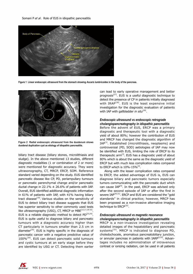
Figure 2 Radial endoscopic ultrasound from the duodenum shows duodenal duplication cyst as etiology of idiopathic pancreatitis.
biliary tract disease (biliary stones, microlithiasis and sludge). In the above mentioned 13 studies, different diagnostic modalities (1 or combination of 2 or more) were mentioned for diagnostic accuracy. They were ultrasonography, CT, MRCP, ERCP, SOM. Reference standard varied depending on the study. EUS identified pancreatic disease like CP, PD, periampullary tumours or pancreatic parenchymal change and/or pancreatic ductal change in 22.1% ± 26.6% of patients with IAP. Overall, EUS identified additional diagnostic information in 61% of patients with IAP, with 41% having biliary tract disease[19]. Various studies on the sensitivity of EUS to detect biliary tract disease suggests that EUS has superior sensitivity to other commonly used tests like ultrasonography (USG), CT, MRCP or MBE[12,17,18-22]. EUS is a reliable diagnostic method to detect PD[23,24].
EUS is quite useful to diagnose biliary and pancreatic tumours with a diagnostic accuracy higher than CT particularly in tumours smaller than 2.5 cm in diameter[25]. EUS is highly specific in the diagnosis of pancreatic cancer with a negative predictive value of 100%[26]. EUS can detect pancreatic solid tumours and cystic tumours at an early stage before they are identified by USG or CT. Detecting them earlier
can lead to early operative management and better prognosis[27]. EUS is a useful diagnostic technique to detect the presence of CP in patients initially diagnosed with IRAP[28]. EUS is the least expensive initial investigation for the diagnostic evaluation of patients with IAP with gallbladder in situ[29].
Endoscopic ultrasound vs endoscopic retrograde cholangiopancreatography in idiopathic pancreatitisBefore the advent of EUS, ERCP was a primary diagnostic and therapeutic tool with a diagnostic yield of about 80%; however the contribution of EUS and MRCP has changed the diagnostic algorithm of IAP[3]. Established (microlithiasis, neoplasms) and controversial (PD, SOD) aetiologies of IAP may now be identified with EUS, limiting the role of ERCP to its therapeutic arm[3]. EUS has a diagnostic yield of about 80% which is about the same as the diagnostic yield of ERCP but with much less complication rates compared to ERCP which is 10%-15%[4].
Along with the lesser complication rates compared to ERCP, the added advantage of EUS is, EUS can diagnose biliary and pancreatic tumors especially tumors communicating with the pancreatic duct which can cause IAP[4]. In the past, ERCP was advised only after the second episode of IAP or after the first in severe IAP[30,31]. ERCP and EUS are considered the “gold standards” in clinical practice; however, MRCP has been proposed as a non-invasive alternative imaging technique to ERCP[32].
Endoscopic ultrasound vs magnetic resonance cholangiopancreatography in idiopathic pancreatitisMRCP is a non-invasive investigation revealing detailed images of the hepatobiliary and pancreatic systems[32]. MRCP is indicated to diagnose PD, choledochocele, anomalous pancreatobiliary junction, or annular pancreas in patients with IARP[2,33]. Advan-tages includes no administration of intravenous contrast or ionizing radiation, can be used in all patients
6954 October 14, 2017|Volume 23|Issue 38|WJG|www.wjgnet.com
Figure 1 Linear endoscopic ultrasound from the stomach showing Ascaris lumbricoides in the body of the pancreas.
LUMEN
CYST
Somani P et al . Role of EUS in idiopathic pancreatitis
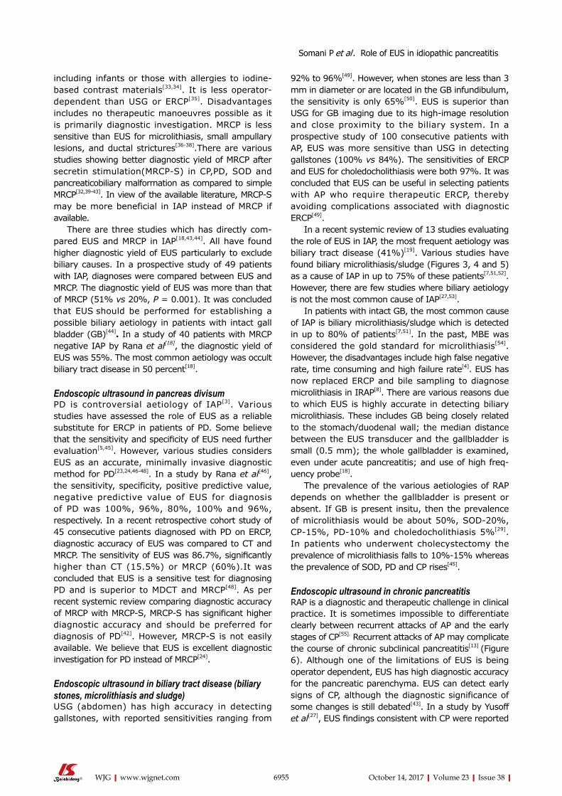
6955 October 14, 2017|Volume 23|Issue 38|WJG|www.wjgnet.com
92% to 96%[49]. However, when stones are less than 3 mm in diameter or are located in the GB infundibulum, the sensitivity is only 65%[50]. EUS is superior than USG for GB imaging due to its high-image resolution and close proximity to the biliary system. In a prospective study of 100 consecutive patients with AP, EUS was more sensitive than USG in detecting gallstones (100% vs 84%). The sensitivities of ERCP and EUS for choledocholithiasis were both 97%. It was concluded that EUS can be useful in selecting patients with AP who require therapeutic ERCP, thereby avoiding complications associated with diagnostic ERCP[49].
In a recent systemic review of 13 studies evaluating the role of EUS in IAP, the most frequent aetiology was biliary tract disease (41%)[19]. Various studies have found biliary microlithiasis/sludge (Figures 3, 4 and 5) as a cause of IAP in up to 75% of these patients[7,51,52]. However, there are few studies where biliary aetiology is not the most common cause of IAP[27,53].
In patients with intact GB, the most common cause of IAP is biliary microlithiasis/sludge which is detected in up to 80% of patients[7,51]. In the past, MBE was considered the gold standard for microlithiasis[54]. However, the disadvantages include high false negative rate, time consuming and high failure rate[4]. EUS has now replaced ERCP and bile sampling to diagnose microlithiasis in IRAP[8]. There are various reasons due to which EUS is highly accurate in detecting biliary microlithiasis. These includes GB being closely related to the stomach/duodenal wall; the median distance between the EUS transducer and the gallbladder is small (0.5 mm); the whole gallbladder is examined, even under acute pancreatitis; and use of high freq-uency probe[18].
The prevalence of the various aetiologies of RAP depends on whether the gallbladder is present or absent. If GB is present insitu, then the prevalence of microlithiasis would be about 50%, SOD-20%, CP-15%, PD-10% and choledocholithiasis 5%[29]. In patients who underwent cholecystectomy the prevalence of microlithiasis falls to 10%-15% whereas the prevalence of SOD, PD and CP rises[45].
Endoscopic ultrasound in chronic pancreatitisRAP is a diagnostic and therapeutic challenge in clinical practice. It is sometimes impossible to differentiate clearly between recurrent attacks of AP and the early stages of CP[55]. Recurrent attacks of AP may complicate the course of chronic subclinical pancreatitis[13] (Figure 6). Although one of the limitations of EUS is being operator dependent, EUS has high diagnostic accuracy for the pancreatic parenchyma. EUS can detect early signs of CP, although the diagnostic significance of some changes is still debated[43]. In a study by Yusoff et al[27], EUS findings consistent with CP were reported
including infants or those with allergies to iodine-based contrast materials[33,34]. It is less operator-dependent than USG or ERCP[35]. Disadvantages includes no therapeutic manoeuvres possible as it is primarily diagnostic investigation. MRCP is less sensitive than EUS for microlithiasis, small ampullary lesions, and ductal strictures[36-38].There are various studies showing better diagnostic yield of MRCP after secretin stimulation(MRCP-S) in CP,PD, SOD and pancreaticobiliary malformation as compared to simple MRCP[32,39-43]. In view of the available literature, MRCP-S may be more beneficial in IAP instead of MRCP if available.
There are three studies which has directly com-pared EUS and MRCP in IAP[18,43,44]. All have found higher diagnostic yield of EUS particularly to exclude biliary causes. In a prospective study of 49 patients with IAP, diagnoses were compared between EUS and MRCP. The diagnostic yield of EUS was more than that of MRCP (51% vs 20%, P = 0.001). It was concluded that EUS should be performed for establishing a possible biliary aetiology in patients with intact gall bladder (GB)[44]. In a study of 40 patients with MRCP negative IAP by Rana et al[18], the diagnostic yield of EUS was 55%. The most common aetiology was occult biliary tract disease in 50 percent[18].
Endoscopic ultrasound in pancreas divisumPD is controversial aetiology of IAP[3]. Various studies have assessed the role of EUS as a reliable substitute for ERCP in patients of PD. Some believe that the sensitivity and specificity of EUS need further evaluation[5,45]. However, various studies considers EUS as an accurate, minimally invasive diagnostic method for PD[23,24,46-48]. In a study by Rana et al[46], the sensitivity, specificity, positive predictive value, negative predictive value of EUS for diagnosis of PD was 100%, 96%, 80%, 100% and 96%, respectively. In a recent retrospective cohort study of 45 consecutive patients diagnosed with PD on ERCP, diagnostic accuracy of EUS was compared to CT and MRCP. The sensitivity of EUS was 86.7%, significantly higher than CT (15.5%) or MRCP (60%).It was concluded that EUS is a sensitive test for diagnosing PD and is superior to MDCT and MRCP[48]. As per recent systemic review comparing diagnostic accuracy of MRCP with MRCP-S, MRCP-S has significant higher diagnostic accuracy and should be preferred for diagnosis of PD[42]. However, MRCP-S is not easily available. We believe that EUS is excellent diagnostic investigation for PD instead of MRCP[24].
Endoscopic ultrasound in biliary tract disease (biliary stones, microlithiasis and sludge)USG (abdomen) has high accuracy in detecting gallstones, with reported sensitivities ranging from
Somani P et al . Role of EUS in idiopathic pancreatitis

6956 October 14, 2017|Volume 23|Issue 38|WJG|www.wjgnet.com
as the most common finding in patients with IAP.
When to perform endoscopic ultrasound, after first episode or recurrent episodes of idiopathic acute pancreatitis?There is controversy in the literature about the timing of EUS in IAP whether to perform after first episode or recurrent episodes[21,27,56]. In a study by Yusoff et al[27], the diagnostic yield of EUS was not significantly different after a single attack or recurrent attacks of IAP. Therefore, the authors concluded that it is reasonable to perform EUS after the first episode, especially in older patients. Many authors have supported this opinion[12,18,21,57]. On the other hand there are few studies where the diagnostic yield is more in recurrent episodes than a single episode[51,58].
IAP is a common condition but the natural history is not well studied and the best diagnostic approach to both single and multiple attacks remains undefined. The suggested diagnostic approach in various studies and guidelines to IAP is highly variable[12]. As per the American Gastroenterological Association, EUS is not recommended after first episode of IAP in patients
less than 40 years of age. It recommends EUS as investigation of choice in IRAP and after first episode in patients with more than 40 years of age[59]. As per American College of Gastroenterology, extensive evaluation using EUS or MRCP is recommended following recurrent attacks of IAP[60]. As per the International Association of pancreatology, patients with IAP should undergo EUS as the investigation of choice irrespective of first or recurrent episodes and, if negative, then, a MRCP-S as the next step[61].
In a recent landmark prospective study of patients with IAP over a 10-year period, 201 patients were enrolled. 80 were with single attack and 121 with multiple attacks of IAP. After EUS, 54% of patients with a single attack were categorized as idiopathic, while for multiple attacks 14 % were idiopathic. Long-term follow-up documented recurrence of pancreatitis in 24% of the patients with a single attack and in 49% of the patients with multiple attacks. On multivariate analysis, number of previous attacks was found to significant predictor of recurrence. It was concluded that following a single idiopathic attack of pancreatitis and a negative EUS examination, relapse was infr-
Figure 3 Linear endoscopic ultrasound from the duodenal bulb shows echogenic biliary sludge in the common bile duct.
Figure 4 Linear endoscopic ultrasound from the duodenal bulb shows echogenic sludge in the gallbladder.
Somani P et al . Role of EUS in idiopathic pancreatitis

6957 October 14, 2017|Volume 23|Issue 38|WJG|www.wjgnet.com
equent. A negative EUS after a single attack had significant prognostic value as it identified low rate of recurrence. This study suggest that EUS may be an useful test following a single attack given its yield and potential prognostic value. This study had prospective EUS based approach to idiopathic pancreatitis[12]. In a recent systemic review of 13 studies by Smith et al[19], the diagnostic yield of EUS was not influenced by recurrent disease. Considering the available literature, we recommend EUS after first episode of idiopathic pancreatitis.
Endoscopic ultrasound vs secretin enhanced endoscopic ultrasonographyIntravenous secretin when used with MRCP (MRCP-S) or EUS (EUS-S) may help the visualization of the pancreatic ductal system and can recognise some undetected ductal abnormalities that are of diagnostic importance in IRAP patients. In a study by Mariani et al[43], the diagnostic yield of EUS-S in IRAP was 13.6% and 16.7% higher than MRCP-S and ERCP respectively. ERCP alone did not find a diagnosis in any case missed by the other two investigations. It was concluded that both MRCP-S and EUS-S should be used as complementary, first-line, imaging techniques, rather than ERCP in the diagnostic work-up of IAP[43]. However, there are limited studies on the EUS-S and further data are required regarding its efficacy in IAP as compared to EUS without IV secretin.
When to perform endoscopic ultrasound after idiopathic pancreatitis?The timing of performing EUS examination after an episode of IAP remains controversial and unclear. Different studies have used different timings[4]. In a recent prospective study of patients with IAP, EUS was performed 1 mo or more after hospital discharge[12]. Rana et al[18] performed EUS at least 1 mo after the episode of AP when the patients were asymptomatic for abdominal pain. In a study by Norton et al[62], EUS
was performed when patients resume food intake while Liu et al[20] perform EUS after resolution of AP during admission. Yusoff et al[27] performed EUS atleast 4 wk after episode of AP to make sure that acute pancreatic parenchymal changes have resolved when EUS is performed.
Thevenot et al[8] suggested to perform MRCP/EUS at a longer interval after the initial AP as inflammation and/or necrosis can prevent visualization of pancreatic lesions during the acute phase. In two recent studies investigating the role of EUS/MRCP in IAP, these examinations were performed1 mo after initial AP when patients were eating normally. The explanation for delayed examination is that fasting is usually advised as initial treatment of AP which can induce GB sludge and, consequently, can result in a false-positive diagnosis of biliary AP if the examination is made early although this is not proven by studies[43,44].
Disadvantages of late EUS after 4 wk of AP includes papillary/ampullary stones (Figure 7) not being diagnosed early with the risk of relapse of AP, early/small periampullary tumours (Figure 8) might be missed as patient may lost to follow up after hospital discharge[4] and possible low diagnostic accuracy as microlithiasis may be missed[21]. Further studies exploring the role of early EUS in IAP are required to come to final conclusion regarding the ideal timing.
Which should be the first investigation after idiopathic acute pancreatitis: Magnetic resonance cholangiopancreatography vs endoscopic ultrasoundThere is slight controversy regarding the first inves-tigation after an episode of IAP. Different studies/authors/guidelines have variable opinions on this topic. Although MRCP is less invasive, cheaper and widely available than EUS, and has recently benefited using IV secretin, performance of EUS remains higher than that of MRCP[8]. However, few studies[43,44] have demonstrated that MRCP can detect few aetiologies undiagnosed by EUS. Further prospective studies are required regarding the cost benefit analysis for EUS/MRCP but at present they
Figure 5 Common bile duct stone with sludge seen on linear endoscopic ultrasound from the duodenal bulb in a 36-year-old male presenting with 3 episodes of idiopathic acute pancreatitis in last 7 mo.
Somani P et al . Role of EUS in idiopathic pancreatitis
Figure 6 Features of chronic pancreatitis seen on linear endoscopic ultrasound from descending duodenum.
Acousticshadow
Stone
Pancreaticduct
Calcification
Head ofpancreas

6958 October 14, 2017|Volume 23|Issue 38|WJG|www.wjgnet.com
should be considered complimentary rather than competitive.
After reviewing the extensive literature on IAP, our opinion which is shared by many authors/guide-lines [12,19,21,22,52,57,59,61] and not agreed by few authors/guidelines[32,59], is performing EUS as the first investigation after IAP with intact GB. There is some debate regarding the choice of investigation in post cholecystectomy patients where CP, SOD and PD are the most common diagnoses in which MRCP has demonstrated good diagnostic accuracy[21,52]. As per the latest systemic review in 2015 comparing the diagnostic accuracy of EUS and MRCP in diagnosis of choledocholithiasis, both tests were found to be similar and the choice of test will be decided by availability and contra-indications to each test[63]. However, EUS is superior to MRCP in detecting choledocholithiasis smaller than 5 mm[64]. Various authors and studies believe that the decision to perform EUS or MRCP as the first diagnostic investigation in post cholecystectomy patients must take into account various factors like local expertise, availability and patient details such as claustrophobia, gastric surgery etc. ERCP should always be considered a therapeutic intervention when required[61,63-65].
The role of PD and SOD as causes of IAP remains highly controversial. In a study by Coté et al[66] and Wilcox et al[12], inspite of endoscopic therapy, RAP occurred in almost 50% patients with SOD. In addition, despite repeat endoscopic interventions, further relapse was also very common. These studies questions the role of endoscopic therapy in PD and SOD. These studies shows that diagnosing SOD and PD may not alter the natural history/prognosis of these patients presenting with IAP. Hence, MRCP and ERCP with SOM may not be that useful in diagnostic workup of IRAP. This also supports EUS as first investigation of choice after IAP irrespective of gallbladder status and is agreed by many studies[12,19, 21,22,54,59].
CONCLUSIONAs per the recent prospective study done by Wilcox et al[12] on EUS based approach to the evaluation of Idiopathic Pancreatitis and after reviewing the literature, EUS as a first strategy towards the etiological evaluation of IAP appears to be useful not only as a diagnostic but as an important prognostic yield irrespective of gallbladder status. In view of high incidence of biliary tract disease as a cause of IAP in most of the studies and given the high diagnostic accuracy of EUS in identifying them, EUS should be considered as an initial diagnostic step for IAP after conventional radiography fails to identify the aetiology.
Considering all the prior studies, the diagnostic yield of EUS is not influenced by whether the episode is first or recurrent. Hence, EUS should be performed after the first episode of IAP if possible. MRCP preferably MRCP-S can be performed if EUS expertise is not available. MRCP can be complimentary to EUS in identifying controversial aetiologies like PD and SOD. ERCP can be then be performed to treat biliary stones and PD. However, in view of high risk of post ERCP pancreatitis, ERCP with SOM should be reserved in those patients in whom MRCP and EUS has found to be negative for diagnostic and therapeutic evaluation especially with post cholecystectomy cases. We recommend an EUS as the first-line examination in the evaluation of patients with idiopathic pancreatitis, because it is minimally invasive, low risk and accurately identifies most occult causes of pancreatitis.
ACKNOWLEDGMENTSPran Prakash (Graphic designer) for his contribution.
REFERENCES1 Steinberg W, Tenner S. Acute pancreatitis. N Engl J Med
1994; 330 : 11981210 [PMID: 7811319 DOI: 10.1056/
Figure 7 Ampullary stone with acoustic shadow seen on radial endoscopic ultrasound from descending duodenum.
Figure 8 Hypoechoic ampullary mass extending into common bile duct.
MASS CBD
D1PD
Somani P et al . Role of EUS in idiopathic pancreatitis

6959 October 14, 2017|Volume 23|Issue 38|WJG|www.wjgnet.com
NEJM199404283301706]2 Levy MJ, Geenen JE. Idiopathic acute recurrent pancreatitis. Am
J Gastroenterol 2001; 96: 25402555 [PMID: 11569674 DOI: 10.1111/j.15720241.2001.04098.x]
3 Md PS, Md UN. Role of ERCP in Patients With Idiopathic Recurrent Acute Pancreatitis. Curr Treat Options Gastroenterol 2016; 14 : 327339 [PMID: 27371265 DOI: 10 .1007/s1193801600969]
4 Vila JJ. Endoscopic ultrasonography and idiopathic acute pancreatitis. World J Gastrointest Endosc 2010; 2: 107111 [PMID: 21160725 DOI: 10.4253/wjge.v2.i4.107]
5 Al-Haddad M, Wallace MB. Diagnostic approach to patients with acute idiopathic and recurrent pancreatitis, what should be done? World J Gastroenterol 2008; 14: 10071010 [PMID: 18286679 DOI: 10.3748/wjg.14.1007]
6 Romagnuolo J, Guda N, Freeman M, Durkalski V. Preferred designs, outcomes, and analysis strategies for treatment trials in idiopathic recurrent acute pancreatitis. Gastrointest Endosc 2008; 68: 966974 [PMID: 18725158 DOI: 10.1016/j.gie.2008.05.006]
7 Lee SP, Nicholls JF, Park HZ. Biliary sludge as a cause of acute pancreatitis. N Engl J Med 1992; 326: 589593 [PMID: 1734248 DOI: 10.1056/NEJM199202273260902]
8 Thevenot A , Bournet B, Otal P, Canevet G, Moreau J, Buscail L. Endoscopic ultrasound and magnetic resonance cholangiopancreatography in patients with idiopathic acute pancreatitis. Dig Dis Sci 2013; 58: 23612368 [PMID: 23508982 DOI: 10.1007/s106200132632y]
9 Yadav D, O’Connell M, Papachristou GI. Natural history following the first attack of acute pancreatitis. Am J Gastroenterol 2012; 107: 10961103 [PMID: 22613906 DOI: 10.1038/ajg.2012.126]
10 Lankisch PG, Breuer N, Bruns A, WeberDany B, Lowenfels AB, Maisonneuve P. Natural history of acute pancreatitis: a longterm populationbased study. Am J Gastroenterol 2009; 104: 2797805; quiz 2806 [PMID: 19603011 DOI: 10.1038/ajg.2009.405]
11 Cavestro GM, Leandro G, Di Leo M, Zuppardo RA, Morrow OB, Notaristefano C, Rossi G, Testoni SG, Mazzoleni G, Alessandri M, Goni E, Singh SK, Giliberti A, Bianco M, Fanti L, Viale E, Arcidiacono PG, Mariani A, Petrone MC, Testoni PA. A singlecentre prospective, cohort study of the natural history of acute pancreatitis. Dig Liver Dis 2015; 47: 205210 [PMID: 25475611 DOI: 10.1016/j.dld.2014.11.003]
12 Wilcox CM, Seay T, Kim H, Varadarajulu S. Prospective Endoscopic UltrasoundBased Approach to the Evaluation of Idiopathic Pancreatitis: Causes, Response to Therapy, and Longterm Outcome. Am J Gastroenterol 2016; 111: 13391348 [PMID: 27325219 DOI: 10.1038/ajg.2016.240]
13 Testoni PA. Acute recurrent pancreatitis: Etiopathogenesis, diagnosis and treatment. World J Gastroenterol 2014; 20: 1689116901 [PMID: 25493002 DOI: 10.3748/wjg.v20.i45.16891]
14 Sharma M, Somani P. Endoscopic ultrasound of pancreatic duct ascariasis. Dig Endosc 2016; 28: 483 [PMID: 26822523 DOI: 10.1111/den.12618]
15 Somani P, Sharma M, Pathak A, Patil A, Kumar A, Sreesh S. Endoscopic ultrasound imaging of pancreatic duct ascariasis. Endoscopy 2016; 48 Suppl 1 UCTN: E33E34 [PMID: 26829193 DOI: 10.1055/s0041111030]
16 Sharma M, Choudhary NS, Puri R. A child with unexplained etiology of acute pancreatitis diagnosed by endoscopic ultrasound. Endosc Ultrasound 2014; 3: 135136 [PMID: 24955344 DOI: 10.4103/23039027.131042]
17 Frossard JL, SosaValencia L, Amouyal G, Marty O, Hadengue A, Amouyal P. Usefulness of endoscopic ultrasonography in patients with “idiopathic” acute pancreatitis. Am J Med 2000; 109: 196200 [PMID: 10974181 DOI: 10.1016/S00029343(00)004782]
18 Rana SS, Bhasin DK, Rao C, Singh K. Role of endoscopic ultrasound in idiopathic acute pancreatitis with negative
ultrasound, computed tomography, and magnetic resonance cholangiopancreatography. Ann Gastroenterol 2012; 25: 133137 [PMID: 24714266]
19 Smith I, Ramesh J, Kyanam Kabir Baig KR, Mönkemüller K, Wilcox CM. Emerging Role of Endoscopic Ultrasound in the Diagnostic Evaluation of Idiopathic Pancreatitis. Am J Med Sci 2015; 350: 229234 [PMID: 26252794 DOI: 10.1097/MAJ.0000000000000541]
20 Liu CL, Lo CM, Chan JK, Poon RT, Fan ST. EUS for detection of occult cholelithiasis in patients with idiopathic pancreatitis. Gastrointest Endosc 2000; 51: 2832 [PMID: 10625791 DOI: 10.1016/S00165107(00)703828]
21 Vila JJ, Vicuña M, Irisarri R, de la Higuera BG, RuizClavijo D, RodríguezGutiérrez C, Urman JM, Bolado F, Jiménez FJ, Arín A. Diagnostic yield and reliability of endoscopic ultrasonography in patients with idiopathic acute pancreatitis. Scand J Gastroenterol 2010; 45: 375381 [PMID: 20034361 DOI: 10.3109/00365520903508894]
22 Tandon M, Topazian M. Endoscopic ultrasound in idiopathic acute pancreatitis. Am J Gastroenterol 2001; 96: 705709 [PMID: 11280538 DOI: 10.1111/j.15720241.2001.03609.x]
23 Rana SS, Gonen C, Vilmann P. Endoscopic ultrasound and pancreas divisum. JOP 2012; 13: 252257 [PMID: 22572127]
24 Sharma M, Pathak A, Rameshbabu CS, Rai P, Kirnake V, Shoukat A. Imaging of pancreas divisum by lineararray endoscopic ultrasonography. Endosc Ultrasound 2016; 5: 2129 [PMID: 26879163 DOI: 10.4103/23039027.175878]
25 DeWitt J, Devereaux B, Chriswell M, McGreevy K, Howard T, Imperiale TF, Ciaccia D, Lane KA, Maglinte D, Kopecky K, LeBlanc J, McHenry L, Madura J, Aisen A, Cramer H, Cummings O, Sherman S. Comparison of endoscopic ultrasonography and multidetector computed tomography for detecting and staging pancreatic cancer. Ann Intern Med 2004; 141: 753763 [PMID: 15545675 DOI: 10.7326/000348191411020041116000006]
26 Klapman JB, Chang KJ, Lee JG, Nguyen P. Negative predictive value of endoscopic ultrasound in a large series of patients with a clinical suspicion of pancreatic cancer. Am J Gastroenterol 2005; 100: 26582661 [PMID: 16393216 DOI: 10.1111/j.15720241.2005.00315.x]
27 Yusoff IF, Raymond G, Sahai AV. A prospective comparison of the yield of EUS in primary vs. recurrent idiopathic acute pancreatitis. Gastrointest Endosc 2004; 60: 673678 [PMID: 15557941 DOI: 10.1016/S00165107(04)020188]
28 Teshima CW, Sandha GS. Endoscopic ultrasound in the diagnosis and treatment of pancreatic disease. World J Gastroenterol 2014; 20: 99769989 [PMID: 25110426 DOI: 10.3748/wjg.v20.i29.9976]
29 Wilcox CM , Ki lgore M. Cos t min imiza t ion ana lys is comparing diagnostic strategies in unexplained pancreatitis. Pancreas 2009; 38: 117121 [PMID: 18852686 DOI: 10.1097/MPA.0b013e31818ab69c]
30 Bank S, Indaram A. Causes of acute and recurrent pancreatitis. Clinical considerations and clues to diagnosis. Gastroenterol Clin North Am 1999; 28: 571589, viii [PMID: 10503137 DOI: 10.1016/S08898553(05)700741]
31 Gregor JC, Ponich TP, Detsky AS. Should ERCP be routine after an episode of “idiopathic” pancreatitis? A costutility analysis. Gastrointest Endosc 1996; 44: 118123 [PMID: 8858315 DOI: 10.1016/S00165107(96)70127X]
32 Gn Y, Amin WG, Shaheen FA, Zargar S, Javid G. The efficacy of magnetic resonance cholangiopancreatography in assessing the etiology of acute idiopathic pancreatitis. Int J Hepatobiliary Pancreat Dis 2014; 4: 32 [DOI: 10.5348/ijhpd201418OA6]
33 Safari MT, Miri MB, Ebadi S, Shahrokh S, Mohammad Alizadeh AH. Comparing the Roles of EUS, ERCP and MRCP in Idiopathic Acute Recurrent Pancreatitis. Clin Med Insights Gastroenterol 2016; 9: 3539 [PMID: 27375362 DOI: 10.4137/CGast.S37927]
Somani P et al . Role of EUS in idiopathic pancreatitis
6960 October 14, 2017|Volume 23|Issue 38|WJG|www.wjgnet.com
34 Hachiya J, Haradome H. [The utilities and outlooks of MR cholangiography (MRCP) as a non contrast and noninvasive technique]. Nihon Rinsho 1998; 56: 27472754 [PMID: 9847593]
35 Makary MA, Duncan MD, Harmon JW, Freeswick PD, Bender JS, Bohlman M, Magnuson TH. The role of magnetic resonance cholangiography in the management of patients with gallstone pancreatitis. Ann Surg 2005; 241: 119124 [PMID: 15621999]
36 Gosset J, Deviere J, Matos C. Magnetic resonance imaging of acute pancreatitis: the pancreatogram. JOP 2004; 5: 4850 [PMID: 14730123]
37 Carroll JK, Herrick B, Gipson T, Lee SP. Acute pancreatitis: diagnosis, prognosis, and treatment. Am Fam Physician 2007; 75: 15131520 [PMID: 17555143]
38 Barish MA, Yucel EK, Ferrucci JT. Magnetic resonance cholangiopancreatography. N Engl J Med 1999; 341: 258264 [PMID: 10413739 DOI: 10.1056/NEJM199907223410407]
39 Sugiyama M, Haradome H, Atomi Y. Magnetic resonance imaging for diagnosing chronic pancreatitis. J Gastroenterol 2007; 42 Suppl 17: 108112 [PMID: 17238038 DOI: 10.1007/s005350061923x]
40 Heverhagen JT, Burbelko M, Schenck zu Schweinsberg T, Funke C, Wecker C, Walthers EM, Rominger M. [Secretinenhanced magnetic resonance cholangiopancreaticography: value for the diagnosis of chronic pancreatitis]. Rofo 2007; 179: 790795 [PMID: 17594632 DOI: 10.1055/s2007963164]
41 Pereira SP, Gillams A, Sgouros SN, Webster GJ, Hatfield AR. Prospective comparison of secretinstimulated magnetic resonance cholangiopancreatography with manometry in the diagnosis of sphincter of Oddi dysfunction types II and III. Gut 2007; 56: 809813 [PMID: 17005767 DOI: 10.1136/gut.2006.099267]
42 Rustagi T, Njei B. Magnetic resonance cholangiopancreatography in the diagnosis of pancreas divisum: a systematic review and metaanalysis. Pancreas 2014; 43: 823828 [PMID: 24743381 DOI: 10.1097/MPA.0000000000000143]
43 Mariani A, Arcidiacono PG, Curioni S, Giussani A, Testoni PA. Diagnostic yield of ERCP and secretinenhanced MRCP and EUS in patients with acute recurrent pancreatitis of unknown aetiology. Dig Liver Dis 2009; 41: 753758 [PMID: 19278909 DOI: 10.1016/j.dld.2009.01.009]
44 Ortega AR, GómezRodríguez R, Romero M, FernándezZapardiel S, Céspedes Mdel M, Carrobles JM. Prospective comparison of endoscopic ultrasonography and magnetic resonance cholangiopancreatography in the etiological diagnosis of “idiopathic” acute pancreatitis. Pancreas 2011; 40: 289294 [PMID: 21206330 DOI: 10.1097/MPA.0b013e318201654a]
45 Kapetanos D. Endoscopic management of acute recurrent pancreatitis. Ann Gastroenterol 2010; 23: 3137.
46 Rana SS, Bhasin DK, Sharma V, Rao C, Singh K. Role of endoscopic ultrasound in the diagnosis of pancreas divisum. Endosc Ultrasound 2013; 2: 710 [PMID: 24949360 DOI: 10.7178/eus.04.003]
47 Lai R, Freeman ML, Cass OW, Mallery S. Accurate diagnosis of pancreas divisum by lineararray endoscopic ultrasonography. Endoscopy 2004; 36: 705709 [PMID: 15280976 DOI: 10.1055/s2004825663]
48 Kushnir VM, Wani SB, Fowler K, Menias C, Varma R, Narra V, Hovis C, Murad FM, Mullady DK, Jonnalagadda SS, Early DS, Edmundowicz SA, Azar RR. Sensitivity of endoscopic ultrasound, multidetector computed tomography, and magnetic resonance cholangiopancreatography in the diagnosis of pancreas divisum: a tertiary center experience. Pancreas 2013; 42: 436441 [PMID: 23211370 DOI: 10.1097/MPA.0b013e31826c711a]
49 Liu CL, Lo CM, Chan JK, Poon RT, Lam CM, Fan ST, Wong J. Detection of choledocholithiasis by EUS in acute pancreatitis: a prospective evaluation in 100 consecutive patients. Gastrointest Endosc 2001; 54: 325330 [PMID: 11522972 DOI: 10.1067/mge.2001.117513]
50 Kurol M, Forsberg L. Ultrasonography in the diagnosis of acute cholecystitis. Acta Radiol Diagn (Stockh) 1984; 25: 379383 [PMID: 6391092 DOI: 10.1177/028418518402500506]
51 Ros E, Navarro S, Bru C, GarciaPugés A, Valderrama R. Occult microlithiasis in ‘idiopathic’ acute pancreatitis: prevention of relapses by cholecystectomy or ursodeoxycholic acid therapy. Gastroenterology 1991; 101: 17011709 [PMID: 1955135 DOI: 10.1016/00165085(91)90410M]
52 Wilcox CM, Varadarajulu S, Eloubeidi M. Role of endoscopic evaluation in idiopathic pancreatitis: a systematic review. Gastrointest Endosc 2006; 63: 10371045 [PMID: 16733122 DOI: 10.1016/j.gie.2006.02.024]
53 Garg PK, Tandon RK, Madan K. Is biliary microlithiasis a significant cause of idiopathic recurrent acute pancreatitis? A long-term followup study. Clin Gastroenterol Hepatol 2007; 5: 7579 [PMID: 16931169 DOI: 10.1016/j.cgh.2006.06.023]
54 Levy MJ. The hunt for microlithiasis in idiopathic acute recurrent pancreatitis: should we abandon the search or intensify our efforts? Gastrointest Endosc 2002; 55: 286293 [PMID: 11818945 DOI: 10.1067/mge.2002.121224]
55 Petrone MC , Arcidiacono PG, Testoni PA. Endoscopic ultrasonography for evaluating patients with recurrent pancreatitis. World J Gastroenterol 2008; 14: 10161022 [PMID: 18286681 DOI: 10.3748/wjg.14.1016]
56 Chen RY, Hawes RH. Idopathic acute pancreatitis: Is EUS worth doing? Am J Gastroenterol 2002; 97: 12441246 [PMID: 12046587]
57 Papachristou GI, Topazian M. Idiopathic recurrent pancreatitis: an EUSbased management approach. Gastrointest Endosc 2011; 73: 11551157 [PMID: 21628013 DOI: 10.1016/j.gie.2011.01.049]
58 Sajith KG, Chacko A, Dutta AK. Recurrent acute pancreatitis: clinical profile and an approach to diagnosis. Dig Dis Sci 2010; 55: 36103616 [PMID: 20232145 DOI: 10.1007/s1062001011758]
59 Forsmark CE, Baillie J; AGA Institute Clinical Practice and Economics Committee; AGA Institute Governing Board. AGA Institute technical review on acute pancreatitis. Gastroenterology 2007; 132: 20222044 [PMID: 17484894 DOI: 10.1053/j.gastro.2007.03.065]
60 Tenner S, Baillie J, DeWitt J, Vege SS; American College of Gastroenterology. American College of Gastroenterology guideline: management of acute pancreatitis. Am J Gastroenterol 2013; 108: 14001415; 1416 [PMID: 23896955 DOI: 10.1038/ajg.2013.218]
61 Working Group IAP/APA Acute Pancreatitis Guidelines. IAP/APA evidencebased guidelines for the management of acute pancreatitis. Pancreatology 2013; 13: e115 [PMID: 24054878 DOI: 10.1016/j.pan.2013.07.063]
62 Norton SA, Alderson D. Endoscopic ultrasonography in the evaluation of idiopathic acute pancreatitis. Br J Surg 2000; 87 : 16501655 [PMID: 11122178 DOI: 10.1046/j.13652168.2000.01587.x]
63 Giljaca V, Gurusamy KS, Takwoingi Y, Higgie D, Poropat G, Štimac D, Davidson BR. Endoscopic ultrasound versus magnetic resonance cholangiopancreatography for common bile duct stones. Cochrane Database Syst Rev 2015; CD011549 [PMID: 25719224 DOI: 10.1002/14651858.CD011549]
64 Kondo S, Isayama H, Akahane M, Toda N, Sasahira N, Nakai Y, Yamamoto N, Hirano K, Komatsu Y, Tada M, Yoshida H, Kawabe T, Ohtomo K, Omata M. Detection of common bile duct stones: comparison between endoscopic ultrasonography, magnetic resonance cholangiography, and helicalcomputedtomographic cholangiography. Eur J Radiol 2005; 54: 271275 [PMID: 15837409 DOI: 10.1016/j.ejrad.2004.07.007]
65 Verma D, Kapadia A, Eisen GM, Adler DG. EUS vs MRCP for detection of choledocholithiasis. Gastrointest Endosc 2006; 64: 248254 [PMID: 16860077 DOI: 10.1016/j.gie.2005.12.038]
66 Coté GA, Imperiale TF, Schmidt SE, Fogel E, Lehman G, McHenry L, Watkins J, Sherman S. Similar efficacies of
Somani P et al . Role of EUS in idiopathic pancreatitis

6961 October 14, 2017|Volume 23|Issue 38|WJG|www.wjgnet.com
biliary, with or without pancreatic, sphincterotomy in treatment of idiopathic recurrent acute pancreatitis. Gastroenterology
2012; 143: 15021509.e1 [PMID: 22982183 DOI: 10.1053/j.gastro.2012.09.006]
P- Reviewer: Li JJ, StoosVeic T, Zhang ZH S- Editor: Chen K L- Editor: A E- Editor: Huang Y
Somani P et al . Role of EUS in idiopathic pancreatitis
delayed and short course of rapamycin prevents organ rejection after allogeneic liver transplantation in rats
Salim Hamdani, Allan Thiolat, Sina Naserian, Cynthia Grondin, Stéphane Moutereau, Anne Hulin, Julien Calderaro, Philippe Grimbert, José Laurent Cohen, Daniel Azoulay, Caroline Pilon
Salim Hamdani, Allan Thiolat, Sina Naserian, Cynthia Grondin, Université Paris-Est, UMR_S955, UPEC, Inserm, U955, Equipe 21, F-94000 Créteil, France
Stéphane Moutereau, AP-HP, Laboratoire de Biochimie, Groupe Hospitalier Henri-Mondor Albert-Chenevier, F-94000 Créteil, France
Anne Hulin, AP-HP, Laboratoire de Pharmacologie-Toxicologie Biologiques, Groupe Hospitalier Henri-Mondor Albert-Chenevier, F-94000 Créteil, France
Julien Calderaro, AP-HP, Anatomie et Cytologie Pathologique, Groupe Hospitalier Henri-Mondor Albert-Chenevier, F-94000 Créteil, France
Philippe Grimbert, José Laurent Cohen, Caroline Pilon,
Université Paris-Est, UMR_S955, UPEC, Inserm, U955, Equipe 21, APHP, Inserm, CIC Biothérapie, Groupe Hospitalier Henri-Mondor Albert-Chenevier, F-94000 Créteil, France
Daniel Azoulay, APHP, Service de Chirurgie HPB et Trans-plantation Hépatique, Groupe Hospitalier Henri-Mondor Albert-Chenevier, F-94000 Créteil, France
Daniel Azoulay, Department of Hepato-Pancreato-Biliary Surgery and Liver Transplantation, Henri Mondor Hospital, 94010 Créteil, France
Author contributions: Cohen JL, Azoulay D and Pilon C designed the research study; Hamdani S, Grondin C, Moutreau S, Hulin A and Calderaro J performed the research; Hamdani S, Cohen JL, Azoulay D and Pilon C wrote the paper; all authors analyzed the data.
Institutional animal care and use committee statement: All procedures involving animals were reviewed and approved by the Regional Ethics Committee in Animal experimentation no16, Ile de France, France (authorization no.11/12/12-10B).
Conflict-of-interest statement: The authors have no conflicts of interest to declare.
Data sharing statement: No additional data are available.
Open-Access: This article is an open-access article which was selected by an in-house editor and fully peer-reviewed by external reviewers. It is distributed in accordance with the Creative Commons Attribution Non Commercial (CC BY-NC 4.0) license, which permits others to distribute, remix, adapt, build upon this work non-commercially, and license their derivative works on different terms, provided the original work is properly cited and the use is non-commercial. See: http://creativecommons.org/licenses/by-nc/4.0/
Manuscript source: Unsolicited manuscript
Correspondence to: Dr. Daniel Azoulay, Professor, Department of Hepato-Pancreato-Biliary Surgery and Liver Transplantation, Henri Mondor Hospital, 51 Avenue du Maréchal de Lattre de Tassigny, 94010 Créteil, France. [email protected] Telephone: +33-1-49812548Fax: +33-1-498124 32
Received: February 28, 2017Peer-review started: March 3, 2017First decision: March16, 2017Revised: April 6, 2017Accepted: June 1, 2017Article in press: June 1, 2017Published online: October 14, 2017
AbstractAIMTo test whether a delayed and short course of ra-pamycin would induce immunosuppressive effects following allogeneic orthotopic liver transplantation (OLT) in rats.
METHODSAllogeneic OLTs were performed using Dark Agouti
ORIGINAL ARTICLE
6962 October 14, 2017|Volume 23|Issue 38|WJG|www.wjgnet.com
Submit a Manuscript: http://www.f6publishing.com
DOI: 10.3748/wjg.v23.i38.6962
World J Gastroenterol 2017 October 14; 23(38): 6962-6972
ISSN 1007-9327 (print) ISSN 2219-2840 (online)
Basic Study

livers transplanted into Lewis recipients, and syngeneic OLTs were performed using the Lewis rat strain. Rapamycin (1 mg/kg per day) was administered by gavage from day 4 to day 11 post-transplantation. Lymphocyte cellular compartments were analyzed by flow cytometry in draining lymph nodes, non-draining lymph nodes and the spleen at days 11 and 42 in rapamycin-treated rats, untreated control rats and syngeneic grafted rats. Skin grafts from Dark agouti or from F344 RT were performed at day 30 on liver grafted rats treated with rapamycin.
RESULTSAn 8-d course of rapamycin treatment initiated 4 d following transplantation resulted in the survival of grafted rats for more than 100 d. In contrast, untreated rats died of liver failure within 13 to 21 d. The analysis of the cellular compartment revealed an increase in two cellular subpopulations, specifically myeloid-derived suppressor cells (MDSCs) and CD8+CD45RClow T cells, without major modifications in the regulatory T cell (Treg) compartment in treated rats in the early stages after grafting. We evaluated the ability of treated rats to reject third-party allogeneic skin grafts to confirm their immune competence. In contrast, when skin was collected from rats syngeneic to the grafted liver, it was not rejected.
CONCLUSIONOur results demonstrate that short and delayed rapa-mycin treatment allows for tolerance in allogeneic OLT. The results also allowed for the identification of the mechanisms of tolerance induced by rapamycin by identifying MDSCs and CD8+CD45RClow T cells as associated with the state of tolerance.
Key words: Myeloid-derived suppressor cells; Liver transplantation; Rapamycin; Regulatory T cells
© The Author(s) 2017. Published by Baishideng Publishing Group Inc. All rights reserved.
Core tip: Rapamycin is an immunosuppressive drug that is rarely used for liver transplantation treatment due to its side effects. Here, we show that a delayed and short course of rapamycin initiated on day 4 following allogeneic orthotopic liver transplantation in rats resulted in the survival of grafted rats for more than 100 d. The analysis of the cellular compartment revealed an increase in two cellular subpopulations, specifically myeloid-derived suppressor cells and CD8+CD45RClow T cells. The treated liver grafted-rats rejected third-party allogeneic skin grafts, but tolerated skin from syngeneic liver donor rats. Our results identify one of the mechanisms by which the state of tolerance is established.
Hamdani S, Thiolat A, Naserian S, Grondin C, Moutereau S, Hulin A, Calderaro J, Grimbert P, Cohen JL, Azoulay D, Pilon C. Delayed and short course of rapamycin prevents organ rejection
after allogeneic liver transplantation in rats. World J Gastroenterol 2017; 23(38): 6962-6972 Available from: URL: http://www.wjgnet.com/1007-9327/full/v23/i38/6962.htm DOI: http://dx.doi.org/10.3748/wjg.v23.i38.6962
INTRODUCTIONCurrent immunosuppression treatments following solid organ transplantation are based on calcineurin inhibitors (CNIs). However, prolonged CNI exposure leads to nephrotoxicity[1], neurotoxicity[2], an increased risk of cancer[3], and hypertension[4], all of which are associated with long-term morbidity and mortality. Reducing the use of CNIs by combining or replacing them with immunosuppressive drugs that exert different mechanisms of action remains the primary strategy to lower the incidence of these adverse events. The drug rapamycin, a mammalian target of the rapamycin (mTOR) inhibitor, is a potential alternative due to its strong immunosuppressive properties in humans.
Rapamycin mediates immunosuppressive effects by preventing T cell cycle progression from the G1 to the S phase, thereby blocking T cell proliferation[5]. Rapamycin also acts on dendritic cells (DCs) by in-hibiting their capacity to differentiate from immature into mature DCs, thus reducing their ability to activate T cells. In turn, immature DCs promote the expansion of CD4+CD25+Foxp3+ regulatory T cells (Tregs) while concomitantly inducing T cell anergy and apoptosis and promoting graft tolerance[6]. In a model of experimental graft-vs-host disease facilitating the detection of donor-derived luciferase-labeled conventional T cells in vivo, the combination of Tregs and CNIs, but not rapamycin, suppresses Treg function[7]. In an allogeneic transplantation model, myeloid-derived suppressor cells (MDSCs) can also be recruited during mTOR inhibitor treatment[8]. Furthermore, following the activation of nitric oxide synthase (iNOS) and arginase (Arg1), MDSCs inhibit T cell proliferation and induce their apoptosis[9].
Currently, rapamycin is widely used to prevent rejection following renal[10], liver[11], and heart[12] trans-plantation. However, its use is primarily restricted to renal transplantation and is not recommended as a first-line treatment for liver transplantation due to deleterious side effects such as delayed wound-healing processes[13], thrombosis of the hepatic artery inducing graft loss, and increased mortality[14,15]. In liver transplantation, previous clinical trials have successfully demonstrated the possibility to introduce mTor inhibitors at day 30 in combination with reduced CNI treatments[16]. Here, we evaluated the possibility of using the mTor inhibitor alone in order to avoid CNI side effects in patients. Therefore, we tested a novel delayed and shortened rapamycin administration protocol in a model of orthotopic liver transplantation rejection in rats.
6963 October 14, 2017|Volume 23|Issue 38|WJG|www.wjgnet.com
Hamdani s et al . Delayed rapamycin treatment prevents allogeneic liver rejection in rat

MATERIALS AND METHODSAnimalsInbred Lewis RT1l male (LEW) and Dark Agouti RT1av1 male (DA) rats weighing 220 to 280 g were purchased from Janvier Labs (Le Genest-Saint-Isle, France) and F344 RT1v male rats weighing 220 to 280 g were purchased from Charles River Labs (L’Arbresle, France). The rats were maintained in animal facilities for at least one week prior to surgery under standard conditions. All experimental protocols were performed according to European Union guidelines and with the approval of the Regional Ethics Committee in Animal Experimentation no. 16, Ile-de-France, France (authorization no.11/12/12-11B).
Orthotopic liver transplantationNon-arterialized OLT was performed according to Kamada’s technique[17]. The allografts were transplanted orthotopically. Vena porta clamping ranged from 19-23 min. Allogeneic OLTs were performed by grafting a DA liver (donor) into a LEW rat (recipient). Syngeneic OLTs were performed with LEW rats (donor and recipient).
TreatmentRapamycin[18] was administered by oral gavage (1 mg/kg body weight per day) every day starting on day 4 after transplantation until day 11. Non-draining lymph node (non-dLN; axillary), dLN (periportal), spleen, and liver samples were collected for analysis on day 11 and day 42 after sacrifice. The residual concentration of rapamycin was analyzed in naive and liver-grafted rats after the administration of rapamycin for 8 d. Whole blood samples were collected in ethylenediaminetetraacetic acid (EDTA) tubes for chromatographic analysis on days 0, 4, 9 and 11.
Flow cytometryCell suspensions obtained by mechanical shredding (draining lymph nodes, not draining lymph nodes and the spleen) were used for phenotypic analysis. Anti-CD3, anti-CD4, anti-CD8, anti-CD25, anti-CTLA-4, anti-CD62L, anti-CD278 (ICOS), anti-CD11b/c, anti CD161, and anti-MHC-Ⅱ, anti-Foxp3, anti-IFNγ, anti-TNFα labelled with phycoerythrin (PE), phycoerythrin cyanine 7 (PE-Cy7) fluorescein isothiocyanate (FITC), peridinin chlorophyll protein complex eFluor 710 (perCP eFluor 710), allophycocyanin (APC), and eFluor 450 were purchased from ebioscience (Paris, France). Anti-CD45RC-PE was purchased from BD Biosciences and Anti-CD3-Vioblue and Helios-APC were purchased from Miltenyi Biotec (Paris, France). Events were acquired on a FACS Canto Ⅱ flow cytometer (BD Biosciences) and analyzed using FlowJo (Tree Star, Ashland, OR, United States) software.
Skin graftingSkin grafting was performed according to the method of Billingham and Medawar[19]. Full-thickness tail skin (1
cm2) from two donors (DA and F344) was grafted onto the right and left dorsal thorax, respectively, of liver-grafted recipients after 30 d of tolerance (DA skin and F344 skin onto LEW rats). Rejection was defined as more than 80% graft necrosis upon daily inspection.
Histologic analysisGrafted livers were excised, fixed in 10% formaldehyde buffer, paraffin-embedded, sectioned (5 µm) and stained with hematoxylin and eosin (H and E) according to a standard protocol. The histological grading of rejection was performed based on the BANFF classification[20].
Serum biochemical testingSerum samples were stored at -20 ℃ until the an-alysis was performed. Transaminase enzyme and total bilirubin levels were measured to evaluate hepato-cellular injury.
Statistical analysisDifferences between the groups were evaluated using an unpaired-t-test performed with Prism software 5.0 (Graph Pad Software, Inc., La Jolla, CA, United States). The results were considered statistically significant when p < 0.05.
RESULTSShort and delayed rapamycin treatment prevents the rejection of allogeneic orthotopic liver transplantation In this study, we used a model of DA liver trans-plantation in LEW recipient rats. These two rat strains differ in their major histocompatibility antigens. Allogeneic transplantation resulted in the death of all untreated rats within 13 to 21 d due to allogeneic liver rejection (Figure 1A). Four grafted rats were sacrificed for histological examination of their transplanted livers on day 11 and exhibited the characteristic signs of acute rejection according to the BANFF classification (mean score = 8.75 ± 0.25, Figure 1B and C). Next, we evaluated whether delayed rapamycin treatment exerts immunosuppressive effects. Four rats received rapamycin at a dose of 1 mg/kg per day beginning on day 4 post-transplantation and continuing until day 30. Under these conditions, 3 out of 4 grafted animals survived over 100 d without any additional treatment, suggesting that delayed and long-term rapamycin treatment is effective (Figure 1A).
We then tested whether a shorter course of rapamycin also leads to transplant liver maintenance. Eleven grafted rats received 1 mg/kg per day of rapamycin from day 4 to day 11 (8 d) and only 2 out of 11 rats died on days 19 and 49 (Figure 1A). To evaluate immunosuppression levels in transplanted livers, 4 rats were sacrificed on day 11 at the end of the treatment period. Compared to untreated control transplanted rats, the treated rats demonstrated a dramatic reduction in their histological rejection grade (Figure 1B and C). Four additional transplanted rapamycin-treated
6964 October 14, 2017|Volume 23|Issue 38|WJG|www.wjgnet.com
Hamdani s et al . Delayed rapamycin treatment prevents allogeneic liver rejection in rat
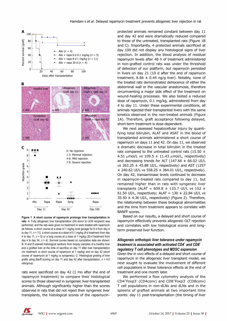
6965 October 14, 2017|Volume 23|Issue 38|WJG|www.wjgnet.com
protected animals remained constant between day 11 and day 42 and were dramatically reduced compared to those of the untreated, transplanted rats (Figure 1B and C). Importantly, 4 protected animals sacrificed at day 100 did not display any histological signs of liver rejection. In addition, the blood analysis of residual rapamycin levels after 48 h of treatment administered in non-grafted control rats was under the threshold of detection of our platform, but rapamycin persisted in livers on day 21 (10 d after the end of rapamycin treatment, 6.66 ± 0.44 ng/g liver). Notably, none of the treated rats demonstrated dehiscence of either the abdominal wall or the vascular anastomosis, therefore circumventing a major side effect of the treatment on wound-healing processes. We also tested a reduced dose of rapamycin, 0.1 mg/kg, administered from day 4 to day 11. Under these experimental conditions, all animals rejected their transplanted livers with the same kinetics observed in the non-treated animals (Figure 1A). Therefore, graft acceptance following delayed, short-term treatment is dose-dependent.
We next assessed hepatocellular injury by quanti-fying total bilirubin, ALAT and ASAT in the blood of transplanted animals administered a short course of rapamycin on days 11 and 42. On day 11, we observed a dramatic decrease in total bilirubin in the treated rats compared to the untreated control rats (15.50 ± 4.51 µmol/L vs 109.5 ± 11.43 µmol/L, respectively) and decreasing trends for ALT (147.66 ± 66.02 UI/L vs 263.25 ± 45.88 UI/L, respectively) and AST (1257 ± 240.62 UI/L vs 558.25 ± 364.01 UI/L, respectively). On day 42, transaminase levels continued to decrease in rapamycin-treated rats compared to day 11, but remained higher than in rats with syngeneic liver transplants (ALAT = 609.8 ± 133.7 UI/L vs 152 ± 61.50 UI/L, respectively; ALAT = 130 ± 22.84 UI/L vs 35.50 ± 4.36 UI/L, respectively) (Figure 2). Therefore, the relationship between these biological abnormalities and the time from treatment appears to correlate with BANFF scores.
Based on our results, a delayed and short course of rapamycin effectively prevents allogeneic OLT rejection and correlates with low histological scores and long-term preserved liver function.
Allogeneic orthotopic liver tolerance under rapamycin treatment is associated with activated CD4+ and CD8+ regulatory T cell phenotypes and MDSC expansionGiven the in vivo effects of a delayed and short course of rapamycin in the allogeneic liver transplant model, we next sought to evaluate the involvement of different cell populations in these tolerance effects at the end of treatment and one month later.
We performed a flow cytometry analysis of the CD4+Foxp3- (CD4conv) and CD8+Foxp3- (CD8conv) T cell populations in non-dLNs and dLNs and in the spleens of grafted animals at two important time points: day 11 post-transplantation (the timing of liver
rats were sacrificed on day 42 (1 mo after the end of rapamycin treatment) to compare their histological scores to those observed in syngeneic liver-transplanted animals. Although significantly higher than the scores observed in rats that did not reject their syngeneic liver transplants, the histological scores of the rapamycin-
Figure 1 A short course of rapamycin prolongs liver transplantation in rats. A: Fully allogeneic liver transplantation (DA donor to LEW recipient) was performed, and the rats were given no treatment or were treated with rapamycin as follows: a short course at a dose of 1 mg/kg (oral gavage for 8 d from day 4 to day 11, n = 11), a short course at a dose of 0.1 mg/kg (8-d treatment from day 4 to day 11, n = 5) or a long course at a dose of 1 mg/kg (30-d treatment from day 4 to day 34, n = 4). Survival curves based on cumulative data are shown; B: H and E-stained histological sections from biopsy samples of a healthy liver and a grafted liver at the time of sacrifice on day 11 after liver transplantation (no treatment vs short course of rapamycin at 1 mg/kg) and on day 42 (short course of rapamycin at 1 mg/kg vs syngeneic); C: Histological grading of liver grafts using Banff scoring on day 11 and day 42 after transplantation, n = 4-5 rats/group.
0: No rejection1-3: Minimal rejection4-6: Mild rejection7-9: Severe rejection
9
8
7
6
5
4
3
2
1
0
Reje
ctio
n gr
ade
Allo Rapa Rapa Syng Rapa
Day 11 Day 42 Day 100
C
Healthy liver AllogeneicGraft day 11
AllogeneicGraft + rapa day 11
SyngeneicGraft day 42
AllogeneicGraft + rapa day 42
× 400 × 400 × 400
× 400 × 400
B
Hamdani s et al . Delayed rapamycin treatment prevents allogeneic liver rejection in rat
100
80
60
40
20
0
Perc
ent
surv
ival
(gr
aft)
Allo (n = 4)Allo + rapa 8 d 0.1 mg/kg (n = 5)Allo + rapa 8 d 1 mg/kg (n = 11)Allo + rapa 30 d (n = 4)
20 40 60 80 100
Days after transplantation
A
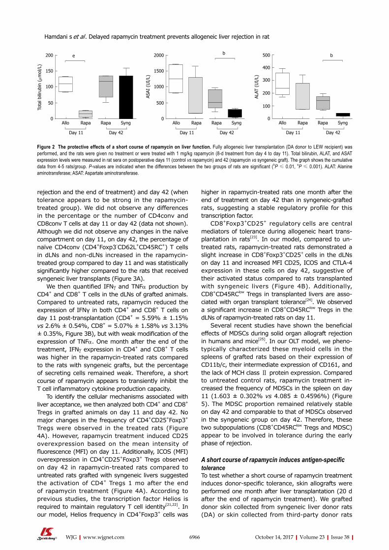
6966 October 14, 2017|Volume 23|Issue 38|WJG|www.wjgnet.com
rejection and the end of treatment) and day 42 (when tolerance appears to be strong in the rapamycin-treated group). We did not observe any differences in the percentage or the number of CD4conv and CD8conv T cells at day 11 or day 42 (data not shown). Although we did not observe any changes in the naïve compartment on day 11, on day 42, the percentage of naïve CD4conv (CD4+Foxp3-CD62L+CD45RC+) T cells in dLNs and non-dLNs increased in the rapamycin-treated group compared to day 11 and was statistically significantly higher compared to the rats that received syngeneic liver transplants (Figure 3A).
We then quantified IFNγ and TNFα production by CD4+ and CD8+ T cells in the dLNs of grafted animals. Compared to untreated rats, rapamycin reduced the expression of IFNγ in both CD4+ and CD8+ T cells on day 11 post-transplantation (CD4+ = 5.59% ± 1.15% vs 2.6% ± 0.54%, CD8+ = 5.07% ± 1.58% vs 3.13% ± 0.35%, Figure 3B), but with weak modification of the expression of TNFα. One month after the end of the treatment, IFNγ expression in CD4+ and CD8+ T cells was higher in the rapamycin-treated rats compared to the rats with syngeneic grafts, but the percentage of secreting cells remained weak. Therefore, a short course of rapamycin appears to transiently inhibit the T cell inflammatory cytokine production capacity.
To identify the cellular mechanisms associated with liver acceptance, we then analyzed both CD4+ and CD8+ Tregs in grafted animals on day 11 and day 42. No major changes in the frequency of CD4+CD25+Foxp3+ Tregs were observed in the treated rats (Figure 4A). However, rapamycin treatment induced CD25 overexpression based on the mean intensity of fluorescence (MFI) on day 11. Additionally, ICOS (MFI) overexpression in CD4+CD25+Foxp3+ Tregs observed on day 42 in rapamycin-treated rats compared to untreated rats grafted with syngeneic livers suggested the activation of CD4+ Tregs 1 mo after the end of rapamycin treatment (Figure 4A). According to previous studies, the transcription factor Helios is required to maintain regulatory T cell identity[21,22]. In our model, Helios frequency in CD4+Foxp3+ cells was
higher in rapamycin-treated rats one month after the end of treatment on day 42 than in syngeneic-grafted rats, suggesting a stable regulatory profile for this transcription factor.
CD8+Foxp3+CD25+ regulatory cells are central mediators of tolerance during allogeneic heart trans-plantation in rats[23]. In our model, compared to un-treated rats, rapamycin-treated rats demonstrated a slight increase in CD8+Foxp3+CD25+ cells in the dLNs on day 11 and increased MFI CD25, ICOS and CTLA-4 expression in these cells on day 42, suggestive of their activated status compared to rats transplanted with syngeneic livers (Figure 4B). Additionally, CD8+CD45RClow Tregs in transplanted livers are asso-ciated with organ transplant tolerance[24]. We observed a significant increase in CD8+CD45RClow Tregs in the dLNs of rapamycin-treated rats on day 11.
Several recent studies have shown the beneficial effects of MDSCs during solid organ allograft rejection in humans and mice[25]. In our OLT model, we pheno-typically characterized these myeloid cells in the spleens of grafted rats based on their expression of CD11b/c, their intermediate expression of CD161, and the lack of MCH class Ⅱ protein expression. Compared to untreated control rats, rapamycin treatment in-creased the frequency of MDSCs in the spleen on day 11 (1.603 ± 0.302% vs 4.085 ± 0.4596%) (Figure 5). The MDSC proportion remained relatively stable on day 42 and comparable to that of MDSCs observed in the syngeneic group on day 42. Therefore, these two subpopulations (CD8+CD45RClow Tregs and MDSC) appear to be involved in tolerance during the early phase of rejection.
A short course of rapamycin induces antigen-specific toleranceTo test whether a short course of rapamycin treatment induces donor-specific tolerance, skin allografts were performed one month after liver transplantation (20 d after the end of rapamycin treatment). We grafted donor skin collected from syngeneic liver donor rats (DA) or skin collected from third-party donor rats
Figure 2 The protective effects of a short course of rapamycin on liver function. Fully allogeneic liver transplantation (DA donor to LEW recipient) was performed, and the rats were given no treatment or were treated with 1 mg/kg rapamycin (8-d treatment from day 4 to day 11). Total bilirubin, ALAT, and ASAT expression levels were measured in rat sera on postoperative days 11 (control vs rapamycin) and 42 (rapamycin vs syngeneic graft). The graph shows the cumulative data from 4-5 rats/group. P-values are indicated when the differences between the two groups of rats are significant (bP ≤ 0.01, eP ≤ 0.001). ALAT: Alanine aminotransferase; ASAT: Aspartate aminotransferase.
500
400
300
200
100
0
ALAT
(U
I/L)
b
Allo Rapa Rapa Syng
Day 11 Day 42
2000
1500
1000
500
0
ASAI
(U
I/L)
b
Allo Rapa Rapa Syng
Day 11 Day 42
200
150
100
50
0
Tota
l bili
rubi
n ( µ
mol
/L)
e
Allo Rapa Rapa Syng
Day 11 Day 42
Hamdani s et al . Delayed rapamycin treatment prevents allogeneic liver rejection in rat

6967 October 14, 2017|Volume 23|Issue 38|WJG|www.wjgnet.com
(neither donor nor recipient, F344) onto rapamycin-treated rats. In control LEW rats grafted with both DA and F334 skin, we observed no signs of rejection on day 7. On day 18, all skin transplants were rejected. In rapamycin-treated rats that did not reject their DA liver transplants, we observed the first signs of F344 skin rejection starting on day 7 and all skin grafts were fully rejected on day 10. Conversely, no signs of rejection
were observed for DA-grafted skin on day 7 and black hairs from DA donor skin were detectable on protected animals until day 60 (Figure 6A and B).
DISCUSSIONSince the establishment of a small-animal liver trans-plant model[26], different preclinical studies have
Figure 3 The effects of a short course of rapamycin treatment on naïve and effector memory cells (A) seems to transiently inhibit the T cell function (B). A: The ndLNs, dLNs and spleens of grafted animals were collected on day 11 and day 42 and conventional T cells were analyzed by flow cytometry. Naïve T cells, defined as CD62L+CD45RC+, and effector/memory cells, defined as (CD62L-CD45RC-) + (CD62L+CD45RC-) + (CD62L-CD45RC+), were analyzed in CD4+Foxp3- and CD8+Foxp3- T cell populations. The results are expressed as the mean +/- SEM of the percentages of effector/memory and naïve T cells, respectively. P-values are indicated when the differences between the two groups of rats are significant (aP ≤ 0.05). The graph shows the cumulative data from 4-5 rats/group; B: Quantification of IFNγ and TNFα expression by CD4+ and CD8+ T cells in the dLNs of grafted animals after PMA/ionomycin stimulation for 4 h. The graph shows the cumulative data from 4-5 rats/group. The results are expressed as the mean +/- SEM. P-values are indicated when the differences between the two groups of rats are significant (aP ≤ 0.05).
100
80
60
40
20
0
% C
D62
L +
CD
45RC+
a
Allo Rapa Rapa Syng
Day 11 Day 42
ndLN
100
80
60
40
20
0
% C
D62
L +
CD
45RC+
a
Allo Rapa Rapa Syng
Day 11 Day 42
dLN
100
80
60
40
20
0
% C
D62
L +
CD
45RC+
Allo Rapa Rapa Syng
Day 11 Day 42
Spleen
100
80
60
40
20
0% E
ffect
or/m
emor
y su
bset
s
a
Allo Rapa Rapa Syng
Day 11 Day 42
100
80
60
40
20
0% E
ffect
or/m
emor
y su
bset
sa
Allo Rapa Rapa Syng
Day 11 Day 42
100
80
60
40
20
0% E
ffect
or/m
emor
y su
bset
s
Allo Rapa Rapa Syng
Day 11 Day 42
IFNγ
CD4
4.94
IFNγ
CD8
4.94
CD4
TNF α
2.48
CD8
TNF α
2.48
15
10
5
0
% I
FNγ
a
Allo Rapa Rapa Syng
Day 11 Day 42
% I
FNγ
15
10
5
0
a
Allo Rapa Rapa Syng
Day 11 Day 42
10
8
6
4
2
0
% T
NF α
Allo Rapa Rapa Syng
Day 11 Day 42
10
8
6
4
2
0
% T
NF α
Allo Rapa Rapa Syng
Day 11 Day 42
A
B
Hamdani s et al . Delayed rapamycin treatment prevents allogeneic liver rejection in rat

6968 October 14, 2017|Volume 23|Issue 38|WJG|www.wjgnet.com
Figure 4 The effects of a short course of rapamycin treatment on CD4+ and CD8+ regulatory T cell phenotypes. A: The dLNs of grafted animals were collected on day 11 and day 42 and regulatory CD4+ T cells were analyzed by flow cytometry. Different activation markers were studied (CD25+, ICOS+, Helios+) after gating on CD25+FOXP3+ among CD4+ cells. The results are expressed as the mean +/- SEM of the percentages of CD25+, ICOS+, and Helios+ or the MFI values for CD25, ICOS, and Helios; n = 4-5 rats/group. P-values are indicated when the differences between the two groups of rats are significant (aP ≤ 0.05, bP ≤ 0.01). MFI, mean fluorescence intensity; B: The ndLNs, dLNs and spleens of grafted animals were collected on day 11 and day 42 and the percentages of CD25+Foxp3+ regulatory CD8+ T cells were analyzed by flow cytometry. The MFI values of the activation markers (CD25, ICOS, and CTLA-4) are shown for the dLNs. The results are expressed as the mean +/- SEM. P-values are indicated when the differences between the two groups of rats are significant (aP ≤ 0.05, bP ≤ 0.01, cP ≤ 0.0001); C: The percentages of CD45RClow cells among CD8+ cells in the dLNs, ndLNs and spleens of grafted rats are shown. The results are expressed as the mean +/- SEM. P-values are indicated when the differences between the two groups of rats are significant (aP ≤ 0.05).
25
20
15
10
5
0
% C
D4+
FO
XP3+
CD
25+
Allo Rapa Rapa Syng
Day 11 Day 42
100
80
60
40
20
0
% I
COS
Allo Rapa Rapa Syng
Day 11 Day 42
100
80
60
40
% H
elio
s
Allo Rapa Rapa Syng
Day 11 Day 42
b
10000
8000
6000
4000
2000
0
CD25
(M
FI)
a
Allo Rapa Rapa Syng
Day 11 Day 42
8000
6000
4000
2000
0
ICO
S (M
FI)
b
Allo Rapa Rapa Syng
Day 11 Day 42
4000
3000
2000
1000
0
Hel
ios
(MFI
)
Allo Rapa Rapa Syng
Day 11 Day 42
2.0
1.5
1.0
0.5
0.0% C
D8+
FO
XP3+
CD
25+
Allo Rapa Rapa Syng
Day 11 Day 42
ndLN2.0
1.5
1.0
0.5
0.0% C
D8+
FO
XP3+
CD
25+
Allo Rapa Rapa Syng
Day 11 Day 42
dLN2.0
1.5
1.0
0.5
0.0% C
D8+
FO
XP3+
CD
25+
Allo Rapa Rapa Syng
Day 11 Day 42
Spleena
10000
8000
6000
4000
2000
0
CD25
(M
FI)
Allo Rapa Rapa Syng
Day 11 Day 42
dLN5000
4000
3000
2000
1000
0
ICO
S (M
FI)
b
Allo Rapa Rapa Syng
Day 11 Day 42
dLN4000
3000
2000
1000
0
CTLA
-4 (
MFI
)
Allo Rapa Rapa Syng
Day 11 Day 42
cdLN
60
40
20
0
% C
D8+
CD
45RClo
w
Allo Rapa Rapa Syng
Day 11 Day 42
ndLN60
40
20
0
% C
D8+
CD
45RClo
w
Allo Rapa Rapa Syng
Day 11 Day 42
dLNa
60
40
20
0
% C
D8+
CD
45RClo
w
Allo Rapa Rapa Syng
Day 11 Day 42
Spleen
A
B
C
Hamdani s et al . Delayed rapamycin treatment prevents allogeneic liver rejection in rat
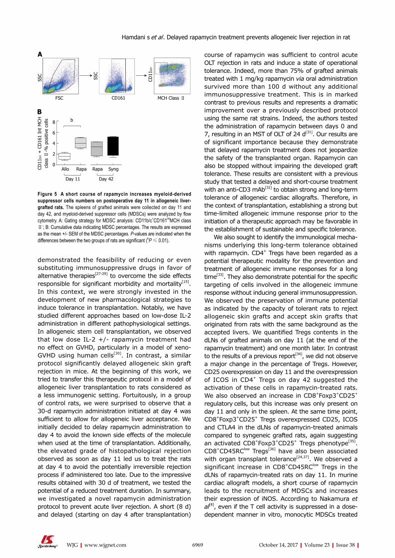
6969 October 14, 2017|Volume 23|Issue 38|WJG|www.wjgnet.com
demonstrated the feasibility of reducing or even substituting immunosuppressive drugs in favor of alternative therapies[27-29] to overcome the side effects responsible for significant morbidity and mortality[15]. In this context, we were strongly invested in the development of new pharmacological strategies to induce tolerance in transplantation. Notably, we have studied different approaches based on low-dose IL-2 administration in different pathophysiological settings. In allogeneic stem cell transplantation, we observed that low dose IL-2 +/- rapamycin treatment had no effect on GVHD, particularly in a model of xeno-GVHD using human cells[30]. In contrast, a similar protocol significantly delayed allogeneic skin graft rejection in mice. At the beginning of this work, we tried to transfer this therapeutic protocol in a model of allogeneic liver transplantation to rats considered as a less immunogenic setting. Fortuitously, in a group of control rats, we were surprised to observe that a 30-d rapamycin administration initiated at day 4 was sufficient to allow for allogeneic liver acceptance. We initially decided to delay rapamycin administration to day 4 to avoid the known side effects of the molecule when used at the time of transplantation. Additionally, the elevated grade of histopathological rejection observed as soon as day 11 led us to treat the rats at day 4 to avoid the potentially irreversible rejection process if administered too late. Due to the impressive results obtained with 30 d of treatment, we tested the potential of a reduced treatment duration. In summary, we investigated a novel rapamycin administration protocol to prevent acute liver rejection. A short (8 d) and delayed (starting on day 4 after transplantation)
course of rapamycin was sufficient to control acute OLT rejection in rats and induce a state of operational tolerance. Indeed, more than 75% of grafted animals treated with 1 mg/kg rapamycin via oral administration survived more than 100 d without any additional immunosuppressive treatment. This is in marked contrast to previous results and represents a dramatic improvement over a previously described protocol using the same rat strains. Indeed, the authors tested the administration of rapamycin between days 0 and 7, resulting in an MST of OLT of 24 d[31]. Our results are of significant importance because they demonstrate that delayed rapamycin treatment does not jeopardize the safety of the transplanted organ. Rapamycin can also be stopped without impairing the developed graft tolerance. These results are consistent with a previous study that tested a delayed and short-course treatment with an anti-CD3 mAb[32] to obtain strong and long-term tolerance of allogeneic cardiac allografts. Therefore, in the context of transplantation, establishing a strong but time-limited allogeneic immune response prior to the initiation of a therapeutic approach may be favorable in the establishment of sustainable and specific tolerance.
We also sought to identify the immunological mecha-nisms underlying this long-term tolerance obtained with rapamycin. CD4+ Tregs have been regarded as a potential therapeutic modality for the prevention and treatment of allogeneic immune responses for a long time[33]. They also demonstrate potential for the specific targeting of cells involved in the allogeneic immune response without inducing general immunosuppression. We observed the preservation of immune potential as indicated by the capacity of tolerant rats to reject allogeneic skin grafts and accept skin grafts that originated from rats with the same background as the accepted livers. We quantified Tregs contents in the dLNs of grafted animals on day 11 (at the end of the rapamycin treatment) and one month later. In contrast to the results of a previous report[34], we did not observe a major change in the percentage of Tregs. However, CD25 overexpression on day 11 and the overexpression of ICOS in CD4+ Tregs on day 42 suggested the activation of these cells in rapamycin-treated rats. We also observed an increase in CD8+Foxp3+CD25+ regulatory cells, but this increase was only present on day 11 and only in the spleen. At the same time point, CD8+Foxp3+CD25+ Tregs overexpressed CD25, ICOS and CTLA4 in the dLNs of rapamycin-treated animals compared to syngeneic grafted rats, again suggesting an activated CD8+Foxp3+CD25+ Tregs phenotype[35]. CD8+CD45RClow Tregs[36] have also been associated with organ transplant tolerance[24,37]. We observed a significant increase in CD8+CD45RClow Tregs in the dLNs of rapamycin-treated rats on day 11. In murine cardiac allograft models, a short course of rapamycin leads to the recruitment of MDSCs and increases their expression of iNOS. According to Nakamura et al[8], even if the T cell activity is suppressed in a dose-dependent manner in vitro, monocytic MDSCs treated
Figure 5 A short course of rapamycin increases myeloid-derived suppressor cells numbers on postoperative day 11 in allogeneic liver-grafted rats. The spleens of grafted animals were collected on day 11 and day 42, and myeloid-derived suppressor cells (MDSCs) were analyzed by flow cytometry. A: Gating strategy for MDSC analysis: CD11b/c+CD161intMCH class Ⅱ-; B: Cumulative data indicating MDSC percentages. The results are expressed as the mean +/- SEM of the MDSC percentages. P-values are indicated when the differences between the two groups of rats are significant (bP ≤ 0.01).
SSC
SSC
CD11
b/c
FSC CD161 MCH Class Ⅱ
b8
6
4
2
0
CD11
b/c +
CD
161
Int
MCH
cl
ass Ⅱ
-% p
ositi
ve c
ells
Allo Rapa Rapa Syng
Day 11 Day 42
A
B
Hamdani s et al . Delayed rapamycin treatment prevents allogeneic liver rejection in rat
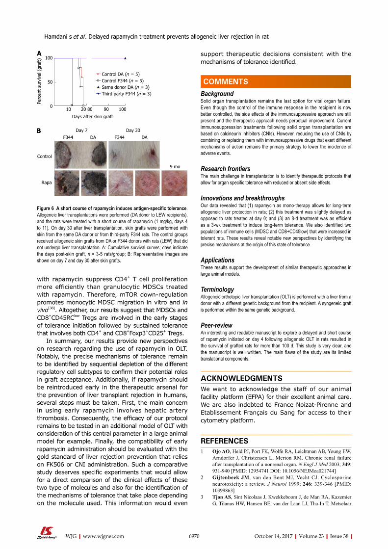
6970 October 14, 2017|Volume 23|Issue 38|WJG|www.wjgnet.com
with rapamycin suppress CD4+ T cell proliferation more efficiently than granulocytic MDSCs treated with rapamycin. Therefore, mTOR down-regulation promotes monocytic MDSC migration in vitro and in vivo[38]. Altogether, our results suggest that MDSCs and CD8+CD45RClow Tregs are involved in the early stages of tolerance initiation followed by sustained tolerance that involves both CD4+ and CD8+Foxp3+CD25+ Tregs.
In summary, our results provide new perspectives on research regarding the use of rapamycin in OLT. Notably, the precise mechanisms of tolerance remain to be identified by sequential depletion of the different regulatory cell subtypes to confirm their potential roles in graft acceptance. Additionally, if rapamycin should be reintroduced early in the therapeutic arsenal for the prevention of liver transplant rejection in humans, several steps must be taken. First, the main concern in using early rapamycin involves hepatic artery thrombosis. Consequently, the efficacy of our protocol remains to be tested in an additional model of OLT with consideration of this central parameter in a large animal model for example. Finally, the compatibility of early rapamycin administration should be evaluated with the gold standard of liver rejection prevention that relies on FK506 or CNI administration. Such a comparative study deserves specific experiments that would allow for a direct comparison of the clinical effects of these two type of molecules and also for the identification of the mechanisms of tolerance that take place depending on the molecule used. This information would even
support therapeutic decisions consistent with the mechanisms of tolerance identified.
COMMENTSBackgroundSolid organ transplantation remains the last option for vital organ failure. Even though the control of the immune response in the recipient is now better controlled, the side effects of the immunosuppressive approach are still present and the therapeutic approach needs perpetual improvement. Current immunosuppression treatments following solid organ transplantation are based on calcineurin inhibitors (CNIs). However, reducing the use of CNIs by combining or replacing them with immunosuppressive drugs that exert different mechanisms of action remains the primary strategy to lower the incidence of adverse events.
Research frontiersThe main challenge in transplantation is to identify therapeutic protocols that allow for organ specific tolerance with reduced or absent side effects.
Innovations and breakthroughsOur data revealed that (1) rapamycin as mono-therapy allows for long-term allogeneic liver protection in rats; (2) this treatment was slightly delayed as opposed to rats treated at day 0; and (3) an 8-d treatment was as efficient as a 3-wk treatment to induce long-term tolerance. We also identified two populations of immune cells (MDSC and CD8+CD45low) that were increased in tolerant rats. These results reveal notable new perspectives by identifying the precise mechanisms at the origin of this state of tolerance.
ApplicationsThese results support the development of similar therapeutic approaches in large animal models.
TerminologyAllogeneic orthotopic liver transplantation (OLT) is performed with a liver from a donor with a different genetic background from the recipient. A syngeneic graft is performed within the same genetic background.
Peer-reviewAn interesting and readable manuscript to explore a delayed and short course of rapamycin initiated on day 4 following allogeneic OLT in rats resulted in the survival of grafted rats for more than 100 d. This study is very clear, and the manuscript is well written. The main flaws of the study are its limited translational components.
ACKNOWLEDGMENTSWe want to acknowledge the staff of our animal facility platform (EFPA) for their excellent animal care. We are also indebted to France Noizat-Pirenne and Etablissement Français du Sang for access to their cytometry platform.
REFERENCES1 Ojo AO, Held PJ, Port FK, Wolfe RA, Leichtman AB, Young EW,
Arndorfer J, Christensen L, Merion RM. Chronic renal failure after transplantation of a nonrenal organ. N Engl J Med 2003; 349: 931-940 [PMID: 12954741 DOI: 10.1056/NEJMoa021744]
2 Gijtenbeek JM, van den Bent MJ, Vecht CJ. Cyclosporine neurotoxicity: a review. J Neurol 1999; 246: 339-346 [PMID: 10399863]
3 Tjon AS, Sint Nicolaas J, Kwekkeboom J, de Man RA, Kazemier G, Tilanus HW, Hansen BE, van der Laan LJ, Tha-In T, Metselaar
Figure 6 A short course of rapamycin induces antigen-specific tolerance. Allogeneic liver transplantations were performed (DA donor to LEW recipients), and the rats were treated with a short course of rapamycin (1 mg/kg, days 4 to 11). On day 30 after liver transplantation, skin grafts were performed with skin from the same DA donor or from third-party F344 rats. The control groups received allogeneic skin grafts from DA or F344 donors with rats (LEW) that did not undergo liver transplantation. A: Cumulative survival curves; days indicate the days post-skin graft, n = 3-5 rats/group; B: Representative images are shown on day 7 and day 30 after skin grafts.
100
50
0
Perc
ent
surv
ival
(gr
aft)
Control DA (n = 5)Control F344 (n = 5)Same donor DA (n = 3)Third party F344 (n = 3)
10 20 80 90 100
Days after skin graft
Day 7 Day 30F344 DA F344 DA
Control
Rapa
9 mo
A
B
COMMENTS
Hamdani s et al . Delayed rapamycin treatment prevents allogeneic liver rejection in rat

6971 October 14, 2017|Volume 23|Issue 38|WJG|www.wjgnet.com
HJ. Increased incidence of early de novo cancer in liver graft recipients treated with cyclosporine: an association with C2 monitoring and recipient age. Liver Transpl 2010; 16: 837-846 [PMID: 20583092 DOI: 10.1002/lt.22064]
4 Bianchi G, Marchesini G, Marzocchi R, Pinna AD, Zoli M. Metabolic syndrome in liver transplantation: relation to etiology and immunosuppression. Liver Transpl 2008; 14: 1648-1654 [PMID: 18975273 DOI: 10.1002/lt.21588]
5 Mondino A, Mueller DL. mTOR at the crossroads of T cell proliferation and tolerance. Semin Immunol 2007; 19: 162-172 [PMID: 17383196 DOI: 10.1016/j.smim.2007.02.008]
6 Steinman RM, Nussenzweig MC. Avoiding horror autotoxicus: the importance of dendritic cells in peripheral T cell tolerance. Proc Natl Acad Sci USA 2002; 99: 351-358 [PMID: 11773639 DOI: 10.1073/pnas.231606698]
7 Zeiser R, Nguyen VH, Beilhack A, Buess M, Schulz S, Baker J, Contag CH, Negrin RS. Inhibition of CD4+CD25+ regulatory T-cell function by calcineurin-dependent interleukin-2 production. Blood 2006; 108: 390-399 [PMID: 16522809 DOI: 10.1182/blood-2006-01-0329]
8 Nakamura T, Nakao T, Yoshimura N, Ashihara E. Rapamycin Prolongs Cardiac Allograft Survival in a Mouse Model by Inducing Myeloid-Derived Suppressor Cells. Am J Transplant 2015; 15: 2364-2377 [PMID: 25943210 DOI: 10.1111/ajt.13276]
9 Bronte V, Serafini P, Mazzoni A, Segal DM, Zanovello P. L-arginine metabolism in myeloid cells controls T-lymphocyte functions. Trends Immunol 2003; 24: 302-306 [PMID: 12810105]
10 MacDonald AS. A worldwide, phase III, randomized, controlled, safety and efficacy study of a sirolimus/cyclosporine regimen for prevention of acute rejection in recipients of primary mismatched renal allografts. Transplantation 2001; 71: 271-280 [PMID: 11213073]
11 Glover TE, Watson CJ, Gibbs P, Bradley JA, Ntzani EE, Kosmoliaptsis V. Conversion From Calcineurin to Mammalian Target of Rapamycin Inhibitors in Liver Transplantation: A Meta-Analysis of Randomized Controlled Trials. Transplantation 2016; 100 : 621-629 [PMID: 26636736 DOI: 10.1097/TP.0000000000001006]
12 Qiu Y, Wang X, Fan J, Rao Z, Lu Y, Lin T. Conversion From Calcineurin Inhibitors to Mammalian Target-of-Rapamycin Inhibitors in Heart Transplant Recipients: A Meta-Analysis of Randomized Controlled Trials. Transplant Proc 2015; 47: 2952-2956 [PMID: 26707320 DOI: 10.1016/j.transproceed.2015.09.059]
13 Mehrabi A, Fonouni H, Wente M, Sadeghi M, Eisenbach C, Encke J, Schmied BM, Libicher M, Zeier M, Weitz J, Büchler MW, Schmidt J. Wound complications following kidney and liver transplantation. Clin Transplant 2006; 20 Suppl 17: 97-110 [PMID: 17100709 DOI: 10.1111/j.1399-0012.2006.00608.x]
14 Moreno R, Berenguer M. Post-liver transplantation medical complications. Ann Hepatol 2006; 5: 77-85 [PMID: 16807513 ]
15 Romero FA, Razonable RR. Infections in liver transplant recipients. World J Hepatol 2011; 3: 83-92 [PMID: 21603030 DOI: 10.4254/wjh.v3.i4.83]
16 De Simone P, Nevens F, De Carlis L, Metselaar HJ, Beckebaum S, Saliba F, Jonas S, Sudan D, Fung J, Fischer L, Duvoux C, Chavin KD, Koneru B, Huang MA, Chapman WC, Foltys D, Witte S, Jiang H, Hexham JM, Junge G; H2304 Study Group. Everolimus with reduced tacrolimus improves renal function in de novo liver transplant recipients: a randomized controlled trial. Am J Transplant 2012; 12: 3008-3020 [PMID: 22882750 DOI: 10.1111/j.1600-6143.2012.04212.x]
17 Kamada N, Calne RY. Orthotopic liver transplantation in the rat. Technique using cuff for portal vein anastomosis and biliary drainage. Transplantation 1979; 28: 47-50 [PMID: 377595]
18 Stepkowski SM. Preclinical results of sirolimus treatment in transplant models. Transplant Proc 2003; 35: 219S-226S [PMID: 12742499]
19 Billingham RE, Medawar PB. The technique of free skin grafting in mammals. J Exp Biol 1951; 28: 385-402
20 Banff schema for grading liver allograft rejection: an international consensus document. Hepatology 1997; 25: 658-663 [PMID: 9049215 DOI: 10.1002/hep.510250328]
21 Kim HJ, Barnitz RA, Kreslavsky T, Brown FD, Moffett H, Lemieux ME, Kaygusuz Y, Meissner T, Holderried TA, Chan S, Kastner P, Haining WN, Cantor H. Stable inhibitory activity of regulatory T cells requires the transcription factor Helios. Science 2015; 350: 334-339 [PMID: 26472910 DOI: 10.1126/science.aad0616]
22 Nakagawa H, Sido JM, Reyes EE, Kiers V, Cantor H, Kim HJ. Instability of Helios-deficient Tregs is associated with conversion to a T-effector phenotype and enhanced antitumor immunity. Proc Natl Acad Sci USA 2016; 113: 6248-6253 [PMID: 27185917 DOI: 10.1073/pnas.1604765113]
23 Liu J, Liu Z, Witkowski P, Vlad G, Manavalan JS, Scotto L, Kim-Schulze S, Cortesini R, Hardy MA, Suciu-Foca N. Rat CD8+ FOXP3+ T suppressor cells mediate tolerance to allogeneic heart transplants, inducing PIR-B in APC and rendering the graft invulnerable to rejection. Transpl Immunol 2004; 13: 239-247 [PMID: 15589736]
24 Yamaguchi Y, Miyanari N, Ichiguchi O, Akizuki E, Matsumura F, Matsuda T, Okabe K, Liang J, Ohshiro H, Mori K, Ogawa M. Infiltrating CD45RC- T cells are associated with immunologic unresponsiveness induced by donor class I major histocompatibility complex antigens in rats. Hepatology 1998; 28: 450-458 [PMID: 9696011 DOI: 10.1002/hep.510280224]
25 Thomson AW, Turnquist HR. Regulators with potential: substantiating myeloid-derived suppressor cells in human organ transplantation. Am J Transplant 2013; 13: 3061-3062 [PMID: 24102710 DOI: 10.1111/ajt.12464]
26 Lee S, Charters AC, Chandler JG, Orloff MJ. A technique for orthotopic liver transplantation in the rat. Transplantation 1973; 16: 664-669 [PMID: 4585390]
27 Wang GY, Zhang Q, Yang Y, Chen WJ, Liu W, Jiang N, Chen GH. Rapamycin combined with allogenic immature dendritic cells selectively expands CD4+CD25+Foxp3+ regulatory T cells in rats. Hepatobiliary Pancreat Dis Int 2012; 11: 203-208 [PMID: 22484590]
28 Fujiki M, Esquivel CO, Martinez OM, Strober S, Uemoto S, Krams SM. Induced tolerance to rat liver allografts involves the apoptosis of intragraft T cells and the generation of CD4(+)CD25(+)FoxP3(+) T regulatory cells. Liver Transpl 2010; 16: 147-154 [PMID: 20104482 DOI: 10.1002/lt.21963]
29 Feng JF, Chen F, Liu H, Liu J. Induction of immune tolerance by pre-infusion of apoptotic lymphocytes derived from peripheral blood of donor rats before liver transplantation. Minerva Chir 2013; 68: 183-189 [PMID: 23612232]
30 Pérol L, Martin GH, Maury S, Cohen JL, Piaggio E. Potential limitations of IL-2 administration for the treatment of experimental acute graft-versus-host disease. Immunol Lett 2014; 162: 173-184 [PMID: 25445496 DOI: 10.1016/j.imlet.2014.10.027]
31 Yoshimura N, Ohsaka Y, Hamashima T, Yasui H, Yura H, Kobayashi Y, Shiho O, Oka T. Rapamycin in experimental liver transplantation in the rat. Transplant Proc 1996; 28: 1796-1797 [PMID: 8658887]
32 Goto R, You S, Zaitsu M, Chatenoud L, Wood KJ. Delayed anti-CD3 therapy results in depletion of alloreactive T cells and the dominance of Foxp3+ CD4+ graft infiltrating cells. Am J Transplant 2013; 13: 1655-1664 [PMID: 23750800 DOI: 10.1111/ajt.12272]
33 Ballarin R, Spaggiari M, Di Benedetto F, De Ruvo N, Cautero N, Montalti R, Guerrini GP, Longo C, Mimmo A, D’Amico G, Gerunda GE. [Is there an age limit for radical surgery in case of tumors infiltrating the duodenum?]. Minerva Chir 2010; 65: 1-9 [PMID: 20212411]
34 Tian L, Lu L, Yuan Z, Lamb JR, Tam PK. Acceleration of apoptosis in CD4+CD8+ thymocytes by rapamycin accompanied by increased CD4+CD25+ T cells in the periphery. Transplantation 2004; 77: 183-189 [PMID: 14742978 DOI: 10.1097/01.
Hamdani s et al . Delayed rapamycin treatment prevents allogeneic liver rejection in rat

6972 October 14, 2017|Volume 23|Issue 38|WJG|www.wjgnet.com
TP.0000101005.44661.3E]35 Wing K, Onishi Y, Prieto-Martin P, Yamaguchi T, Miyara M,
Fehervari Z, Nomura T, Sakaguchi S. CTLA-4 control over Foxp3+ regulatory T cell function. Science 2008; 322: 271-275 [PMID: 18845758 DOI: 10.1126/science.1160062]
36 Xystrakis E, Dejean AS, Bernard I, Druet P, Liblau R, Gonzalez-Dunia D, Saoudi A. Identification of a novel natural regulatory CD8 T-cell subset and analysis of its mechanism of regulation. Blood 2004; 104: 3294-3301 [PMID: 15271801 DOI: 10.1182/blood-2004-03-1214]
37 Guillonneau C, Hill M, Hubert FX, Chiffoleau E, Hervé C, Li
XL, Heslan M, Usal C, Tesson L, Ménoret S, Saoudi A, Le Mauff B, Josien R, Cuturi MC, Anegon I. CD40Ig treatment results in allograft acceptance mediated by CD8CD45RC T cells, IFN-gamma, and indoleamine 2,3-dioxygenase. J Clin Invest 2007; 117: 1096-1106 [PMID: 17404623 DOI: 10.1172/JCI28801]
38 Zhang Y, Bi Y, Yang H, Chen X, Liu H, Lu Y, Zhang Z, Liao J, Yang S, Chu Y, Yang R, Liu G. mTOR limits the recruitment of CD11b+Gr1+Ly6Chigh myeloid-derived suppressor cells in protecting against murine immunological hepatic injury. J Leukoc Biol 2014; 95: 961-970 [PMID: 24569105 DOI: 10.1189/jlb.0913473]
P- Reviewer: Chiu KW, Demirag A, Rodriguez-Peralvarez ML S- Editor: Gong ZM L- Editor: A E- Editor: Li D
Hamdani s et al . Delayed rapamycin treatment prevents allogeneic liver rejection in rat

Li-juan Xu, Zheng Chen, Qian-Qian Chen, Li Yan, Department of Gastroenterology, Institute of Geriatrics, Chinese PLA General Hospital, Beijing 100853, China
Shu-fang Wang, De-Qing Wang, Department of Blood Transfusion, Chinese PLA General Hospital, Beijing 100853, China
Lian-jun Ma, Department of Endoscopics, China-Japan Union Hospital of Jilin University, Changchun 130000, Jilin Province, China
jun Wang, Department of Gastroenterology, Institute of Surgery Research, Daping Hospital, Third Military Medical University, Chongqing 400042, China
ORCID number: Li-Juan Xu (0000-0002-2924-7213); Shu-fang Wang (0000-0001-5768-9687); De-qing Wang (0000-0002-5559-0541); Lian-Jun Ma (0000-0002-4512-1688); Zheng Chen (0000-0002-1253-6254); qian-qian Chen (0000-0001-8527-1018); Jun Wang (0000-0002-6757-0337); Li Yan (0000-0001-8881-1921).
Author contributions: Xu LJ, Wang Sf performed the research, analyzed the data, and wrote and revised the paper; Wang Dq provided the experimental site; Ma LJ, Chen Z, Chen qq performed the research; Wang J and Yan L designed the research and participated in the revision of the paper; Xu LJ, Wang Sf contributed equally to this work; all authors have read and approved the final manuscript.
Supported by the National Natural Science foundation of China, No. 30900669 and No. 81473271; Technology Nova Plan of Beijing City, No. 2011117; China Postdoctoral Science foundation, No. 2016T90994.
Institutional review board statement: The study was reviewed and approved by the Ethics Committee of Chinese PLA General Hospital, Beijing, China.
Institutional animal care and use committee statement: All experiments on animals were performed in accordance with the “Guide for the Care and Use of Laboratory Animals” (National Institutes of Health, 8th ed. 2011). All procedures involving animals were reviewed and approved by the Animal Care and Research Committee of Chinese PLA General Hospital, Beijing, China.
Conflict-of-interest statement: All the authors declare no potential conflict of interest related to this paper.
Data sharing statement: No additional data are available.
Open-Access: This article is an open-access article which was selected by an in-house editor and fully peer-reviewed by external reviewers. It is distributed in accordance with the Creative Commons Attribution Non Commercial (CC BY-NC 4.0) license, which permits others to distribute, remix, adapt, build upon this work non-commercially, and license their derivative works on different terms, provided the original work is properly cited and the use is non-commercial. See: http://creativecommons.org/licenses/by-nc/4.0/
Manuscript source: Unsolicited manuscript
Correspondence to: Li Yan, MD, PhD, Department of Ga-stroenterology, Institute of Geriatrics, Chinese PLA General Hospital, No. 28 Wanshou Road, Haidian District, Beijing 100853, China. [email protected] Telephone: +86-10-66876346fax: +86-10-66876346
Received: April 16, 2017Peer-review started: April 27, 2016first decision: June 22, 2017Revised: July 20, 2017Accepted: September 5, 2017 Article in press: September 5, 2017Published online: October 14, 2017
6973 October 14, 2017|Volume 23|Issue 38|WJG|www.wjgnet.com
ORIGINAL ARTICLE
Adipose-derived stromal cells resemble bone marrow stromal cells in hepatocyte differentiation potential in vitro and in vivo
Basic Study
Li-juan Xu, Shu-fang Wang, De-Qing Wang, Lian-jun Ma, Zheng Chen, Qian-Qian Chen, jun Wang, Li Yan
Submit a Manuscript: http://www.f6publishing.com
DOI: 10.3748/wjg.v23.i38.6973
World J Gastroenterol 2017 October 14; 23(38): 6973-6982
ISSN 1007-9327 (print) ISSN 2219-2840 (online)

AbstractAIMTo investigate whether mesenchymal stem cells (MSCs) from adipose-derived stromal cells (ADSCs) and bone marrow stromal cells (BMSCs) have similar hepatic differentiation potential.
METHODSMouse ADSCs and BMSCs were isolated and cultured. Their morphological and phenotypic characteristics, as well as their multiple differentiation capacity were compared. A new culture system was established to induce ADSCs and BMSCs into functional hepatocytes. Reverse transcription polymerase chain reaction, Western blot, and immunofluorescence analyses were performed to identify the induced hepatocyte-like cells. CM-Dil-labeled ADSCs and BMSCs were then transplanted into a mouse model of CCl4-induced acute liver failure. fluorescence microscopy was used to track the transplanted MSCs. Liver function was tested by an automatic biochemistry analyzer, and liver tissue histology was observed by hematoxylin and eosin (HE) staining.
RESULTSADSCs and BMSCs shared a similar morphology and multiple differentiation capacity, as well as a similar phenotype (with expression of CD29 and CD90 and no expression of CD11b or CD45). Morphologically, ADSCs and BMSCs became round and epithelioid following hepatic induction. These two cell types differentiated into hepatocyte-like cells with similar expression of albumin, cytokeratin 18, cytokeratin 19, alpha fetoprotein, and cytochrome P450. fluorescence microscopy revealed that both ADSCs and BMSCs were observed in the mouse liver at different time points. Compared to the control group, both the function of the injured livers and HE staining showed significant improvement in the ADSC- and BMSC-transplanted mice. There was no significant difference between the two MSC groups.
CONCLUSIONADSCs share a similar hepatic differentiation capacity and therapeutic effect with BMSCs in an acute liver failure model. ADSCs may represent an ideal seed cell type for cell transplantation or a bio-artificial liver support system.
Key words: Adipose-derived stromal cells; Bone marrow stromal cells; Cell differentiation; Hepatocyte differentiation
© The Author(s) 2017. Published by Baishideng Publishing Group Inc. All rights reserved.
Core tip: We investigated whether mesenchymal stem cells from adipose-derived stromal cells (ADSCs) and bone marrow stromal cells (BMSCs), have similar
hepatic differentiation potential. We found that adipose-derived stromal cells resemble bone marrow stromal cells in their hepatocyte differentiation potential in vitro and in vivo . Because ADSCs are obtained more easily and less invasive than BMSCs, ADSCs might be more suitable seed cells for cell transplant or liver tissue engineering. We also developed a new protocol of preparing mouse BMSCs and established a new hepatic induction system.
Xu LJ, Wang Sf, Wang Dq, Ma LJ, Chen Z, Chen qq, Wang J, Yan L. Adipose-derived stromal cells resemble bone marrow stromal cells in hepatocyte differentiation potential in vitro and in vivo. World J Gastroenterol 2017; 23(38): 6973-6982 Available from: URL: http://www.wjgnet.com/1007-9327/full/v23/i38/6973.htm DOI: http://dx.doi.org/10.3748/wjg.v23.i38.6973
INTRODUCTIONLiver transplantation remains the only effective treatment for liver failure. However, its clinical application is limited by donor organs and immune rejection. To this end, hepatocyte transplantation and a bio-artificial liver support system are two potential surrogate complementary therapies for patients with liver failure. Hepatocyte-like cells can be induced from mesenchymal stem cells (MSCs) for xenotransplantation and have been demonstrated to perform hepatocyte functions in preclinical animal studies[1,2]. However, the best type of MSC has not yet been investigated, and therefore it is important to screen for an ideal seed cell type for cell transplantation or a bio-artificial liver support system.
MSCs are non-hematopoietic multipotent stem cells that can be isolated from multiple tissues, such as bone marrow, adipose tissue, cord blood, and amniotic fluid[3-5]. Bone marrow stromal cells (BMSCs) are the most extensively studied and fertile stem cell source used in regenerative medicine and liver tissue engineering[6,7]. More recently, adipose-derived stromal cells (ADSCs) were identified as another promising and extensively studied stem cells for use, especially in liver tissue engineering[8]. Although both ADSCs and BMSCs can be induced into hepatocyte-like cells[9-12], there is no uniform culture system to evaluate their hepatic differentiation potential. Therefore, it is necessary to establish an ideal system to improve the efficiency of hepatic induction.
In this study, we developed a novel culture pro-tocol for mouse BMSCs and optimized the hepatic differentiation system. We compared the morphological and phenotypic characteristics, as well as the multi-differentiation capacity of BMSCs and ADSCs in vitro. We also compared therapeutic effect of BMSCs and ADSCs following transplantation into a mouse model of acute liver failure.
6974 October 14, 2017|Volume 23|Issue 38|WJG|www.wjgnet.com
Xu Lj et al . ADSCs and BMSCs have similar hepatic differentiation

MATERIALS AND METHODSExperimental animals Male BALB/c mice (3 d old and 6 wk old; Charles River, Beijing, China) were purchased from the Laboratory Animal Center of the Academy of Military Medical Sciences of China (Beijing). All studies were conducted after approved by the Ethics Committee of the Animal Facility of Chinese PLA General Hospital and complied with the guidelines for the care of laboratory animals. Mice were housed in cages in a controlled environment (25 ℃ and a 12 h light/dark cycle) and fed standard mouse chow and tap water, and were observed every day in our animal facility.
Isolation and purification of adipose-derived stromal cells and bone marrow stromal cellsThe isolation and purification of ADSCs were performed as previously described[13]. Fibrous tissue was excluded and adipose tissue was minced into pieces of < 1 mm3 and digested in 1 mg/mL collagenase I for 1 h at 37 ℃. The cell suspension was filtered through a 100 µmol/L cell strainer and centrifuged at 300 × g for 5 min. ADSCs were plated at a density of 5 × 10⁵/cm2 with alpha minimal essential medium (α-MEM) supplemented with 10% fetal bovine serum (FBS) and 1% penicillin/streptomycin, and cultured in a humidified incubator at 37 ℃ and 5% CO2. Cells were harvested after reaching a 90% confluence with 0.25% trypsin-EDTA (Gibco, American). Cells in passages 2-4 were used for subsequent experiments.
A new method was established to isolate mouse BMSCs as follows: 3-d-old male BALB/c mice were sacrificed by cervical dislocation and soaked in 75% alcohol for 5 min. The tibia and fibula were isolated under sterile conditions and washed twice with phosphate-buffered saline (PBS) containing 5% penicillin/streptomycin. Muscle and fibrous tissue were excluded. Tibias and fibulas were minced into pieces of < 1 mm3 and washed once with α-MEM, cultured directly by incubation with α-MEM supplemented with 10% FBS and 1% penicillin/streptomycin in a humidified incubator at 37 ℃ and 5% CO2. After 72 h, half of the medium was changed and the bone chips were kept. After reaching a 50% confluence, cells were harvested with 0.25% trypsin-EDTA and seeded as the first passage. At each passage, cells were diluted 1:3-4 every two days. BMSCs at passages 2-4 were used for subsequent experiments.
Measurement of adipose-derived stromal cells and bone marrow stromal cells proliferationFor the cell proliferation assay, 2 × 10³ viable ADSCs and BMSCs were seeded in triplicate onto a 96-well plate. Cell proliferation was measured using a Cell Counting Kit-8 (CCK-8; Beyotime, China). Plates were placed in a humidified incubator at 37 ℃ until the cells adhered to the plate. Next, 10 µL of the CCK-8 solution was added to each well and plates were incubated for
another 2 h at 37 ℃ prior to reading the absorbance at 450 nm on a microplate reader. The assay was repeated every day at the same time for 10 d.
Flow cytometryPassage 2 and 3 ADSCs and BMSCs were trypsinized and incubated with fluorescein isothiocyanate-conjugated CD45 and CD90, and phycoerythrin-conjugated CD11b and CD29 antibodies for 30 min at 4 ℃, followed by two washes with PBS. Fluorescent-labeled cells were analyzed on a flow cytometer.
Differentiation assays For adipogenic differentiation, cells were seeded at 1 × 10⁴/cm2 on 12-well plates. When cells adhered to the plate, the expansion medium (α-MEM supplemented with 10% FBS and 1% penicillin/streptomycin) was replaced with adipogenic induction medium containing 10−6 mmol/L dexamethasone (Dex), 0.5 µmol/L isobutylmethylxanthine, 200 µmol/L indomethacin, and 5 µg/mL (wt/v) insulin, and the cells were incubated for 8 d. Cells cultured in a base medium of α-MEM supplemented with 10% (v/v) FBS served as a negative control. Adipogenic differentiation was assessed by Oil-Red-O staining.
For osteogenic differentiation, cells were seeded at 5 × 10³/cm2 on 12-well plates. When cells adhered to the plate, the expansion medium was replaced with osteogenic induction medium containing 10−7
mmol/L Dex, 10 mmol/L β-glycerol phosphate, and 50 µmol/L ascorbate-2-phosphate. Cells cultured in a base medium of α-MEM supplemented with 10% FBS were used as a negative control. Cells were incubated for 3 wk and osteogenic differentiation was assessed by Alizarin Red staining.
Hepatic differentiation was achieved following a one-step procedure using mouse ADSCs and BMSCs. ADSCs and BMSCs (passage 3) were seeded at 5 × 10³/cm2 onto 24-well culture dishes in expansion medium. When cells adhered to the plate, the expansion medium was replaced with hepatocyte culture medium (HCM; DMEM containing 10% FBS) supplemented with 50 ng/mL hepatocyte growth factor (HGF), 25 ng/mL fibroblast growth factor 4 (FGF4), 30 ng/mL oncostatin M (OSM), 20 ng/mL epidermal growth factor (EGF), 25 ng/mL acidic fibroblast growth factor (aFGF), 10 ng/mL basic fibroblast growth factor (bFGF), 10-6 mmol/L Dex, 1 × insulin-transferrin-selenium (ITS), 2 mmol/L ascorbic acid (Vc), and 50 μmmol/L nicotinamide (Vpp). Differentiation medium (1 mL) was added to each 24-well culture dish and changed every 3 d. Afterwards, cells were cultured for 10 d in HCM. Undifferentiated cells served as negative controls and the HepG2 cell line served as a positive control.
Reverse transcription polymerase chain reaction On day 10 of hepatic differentiation, total RNA was isolated from ADSCs and BMSCs with Trizol reagent
6975 October 14, 2017|Volume 23|Issue 38|WJG|www.wjgnet.com
Xu Lj et al . ADSCs and BMSCs have similar hepatic differentiation
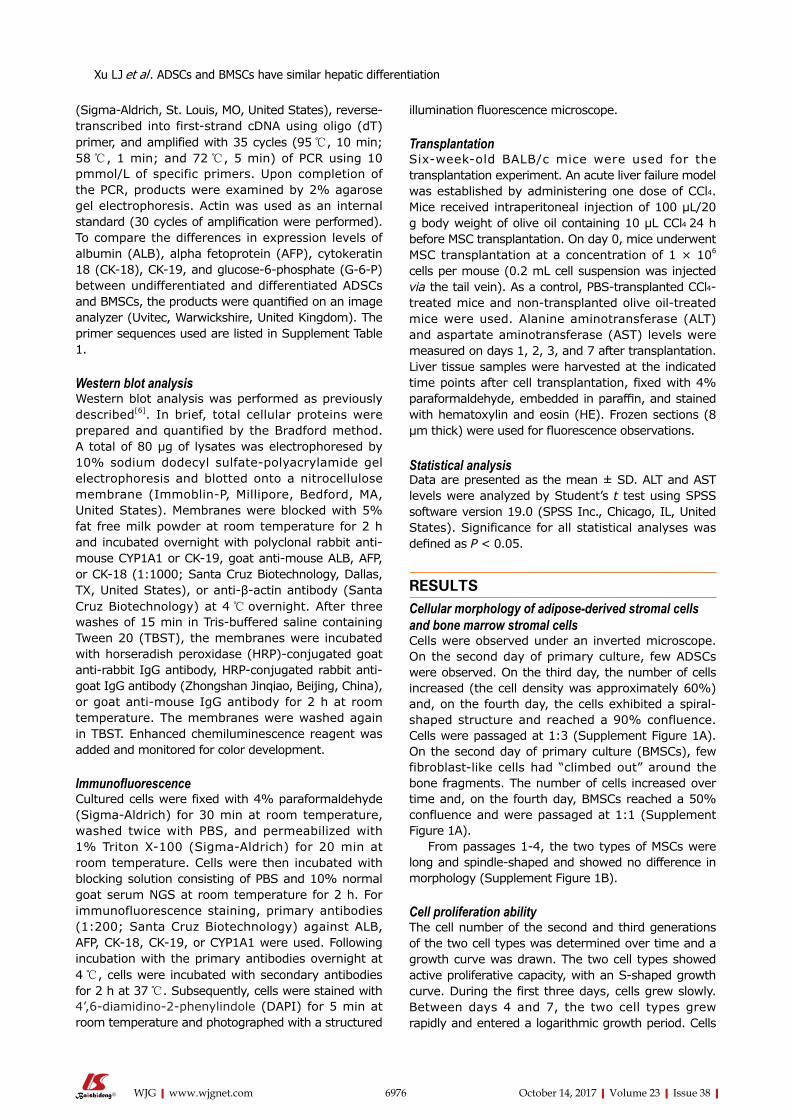
6976 October 14, 2017|Volume 23|Issue 38|WJG|www.wjgnet.com
illumination fluorescence microscope.
TransplantationSix-week-old BALB/c mice were used for the transplantation experiment. An acute liver failure model was established by administering one dose of CCl4. Mice received intraperitoneal injection of 100 μL/20 g body weight of olive oil containing 10 μL CCl4 24 h before MSC transplantation. On day 0, mice underwent MSC transplantation at a concentration of 1 × 10⁶ cells per mouse (0.2 mL cell suspension was injected via the tail vein). As a control, PBS-transplanted CCl4-treated mice and non-transplanted olive oil-treated mice were used. Alanine aminotransferase (ALT) and aspartate aminotransferase (AST) levels were measured on days 1, 2, 3, and 7 after transplantation. Liver tissue samples were harvested at the indicated time points after cell transplantation, fixed with 4% paraformaldehyde, embedded in paraffin, and stained with hematoxylin and eosin (HE). Frozen sections (8 μm thick) were used for fluorescence observations.
Statistical analysisData are presented as the mean ± SD. ALT and AST levels were analyzed by Student’s t test using SPSS software version 19.0 (SPSS Inc., Chicago, IL, United States). Significance for all statistical analyses was defined as P < 0.05.
RESULTSCellular morphology of adipose-derived stromal cells and bone marrow stromal cells Cells were observed under an inverted microscope. On the second day of primary culture, few ADSCs were observed. On the third day, the number of cells increased (the cell density was approximately 60%) and, on the fourth day, the cells exhibited a spiral-shaped structure and reached a 90% confluence. Cells were passaged at 1:3 (Supplement Figure 1A). On the second day of primary culture (BMSCs), few fibroblast-like cells had “climbed out” around the bone fragments. The number of cells increased over time and, on the fourth day, BMSCs reached a 50% confluence and were passaged at 1:1 (Supplement Figure 1A).
From passages 1-4, the two types of MSCs were long and spindle-shaped and showed no difference in morphology (Supplement Figure 1B).
Cell proliferation abilityThe cell number of the second and third generations of the two cell types was determined over time and a growth curve was drawn. The two cell types showed active proliferative capacity, with an S-shaped growth curve. During the first three days, cells grew slowly. Between days 4 and 7, the two cell types grew rapidly and entered a logarithmic growth period. Cells
(Sigma-Aldrich, St. Louis, MO, United States), reverse-transcribed into first-strand cDNA using oligo (dT) primer, and amplified with 35 cycles (95 ℃, 10 min; 58 ℃, 1 min; and 72 ℃, 5 min) of PCR using 10 pmmol/L of specific primers. Upon completion of the PCR, products were examined by 2% agarose gel electrophoresis. Actin was used as an internal standard (30 cycles of amplification were performed). To compare the differences in expression levels of albumin (ALB), alpha fetoprotein (AFP), cytokeratin 18 (CK-18), CK-19, and glucose-6-phosphate (G-6-P) between undifferentiated and differentiated ADSCs and BMSCs, the products were quantified on an image analyzer (Uvitec, Warwickshire, United Kingdom). The primer sequences used are listed in Supplement Table 1.
Western blot analysisWestern blot analysis was performed as previously described[6]. In brief, total cellular proteins were prepared and quantified by the Bradford method. A total of 80 µg of lysates was electrophoresed by 10% sodium dodecyl sulfate-polyacrylamide gel electrophoresis and blotted onto a nitrocellulose membrane (Immoblin-P, Millipore, Bedford, MA, United States). Membranes were blocked with 5% fat free milk powder at room temperature for 2 h and incubated overnight with polyclonal rabbit anti-mouse CYP1A1 or CK-19, goat anti-mouse ALB, AFP, or CK-18 (1:1000; Santa Cruz Biotechnology, Dallas, TX, United States), or anti-β-actin antibody (Santa Cruz Biotechnology) at 4 ℃ overnight. After three washes of 15 min in Tris-buffered saline containing Tween 20 (TBST), the membranes were incubated with horseradish peroxidase (HRP)-conjugated goat anti-rabbit IgG antibody, HRP-conjugated rabbit anti-goat IgG antibody (Zhongshan Jinqiao, Beijing, China), or goat anti-mouse IgG antibody for 2 h at room temperature. The membranes were washed again in TBST. Enhanced chemiluminescence reagent was added and monitored for color development.
ImmunofluorescenceCultured cells were fixed with 4% paraformaldehyde (Sigma-Aldrich) for 30 min at room temperature, washed twice with PBS, and permeabilized with 1% Triton X-100 (Sigma-Aldrich) for 20 min at room temperature. Cells were then incubated with blocking solution consisting of PBS and 10% normal goat serum NGS at room temperature for 2 h. For immunofluorescence staining, primary antibodies (1:200; Santa Cruz Biotechnology) against ALB, AFP, CK-18, CK-19, or CYP1A1 were used. Following incubation with the primary antibodies overnight at 4 ℃, cells were incubated with secondary antibodies for 2 h at 37 ℃. Subsequently, cells were stained with 4’,6-diamidino-2-phenylindole (DAPI) for 5 min at room temperature and photographed with a structured
Xu Lj et al . ADSCs and BMSCs have similar hepatic differentiation

6977 October 14, 2017|Volume 23|Issue 38|WJG|www.wjgnet.com
stopped growing at day 8 and entered the growth plateau phase (Supplement Figure 1C). There was no significant difference between the two types of adult stem cells in the same generation, and there was no significant difference between the same kinds of adult stem cells in the different generation (Supplement Table 1).
Phenotypic characterization of adipose-derived stromal cells and bone marrow stromal cellsADSC and BMSC cell surface markers were analyzed by flow cytometry. Both ADSCs and BMSCs expressed the stem cell-associated surface markers CD90 and CD29, but did not express CD11b or CD45 (Supplement Table 2). The two cell types had a similar proliferative ability and stem cell capacity, consistent with our previous study.
Multiple differentiation ability of adipose-derived stromal cells and bone marrow stromal cellsThe adipogenic and osteogenic differentiation of ADSCs and BMSCs was evaluated at passages 2 and 3. Small and round vacuoles began to appear in the cytoplasm on the third day of induction. On the 8th day, a large number of lipid droplets appeared in the majority of the induced cells and the cells became round, oval, or polygonal. Adipogenic differentiation was identified by Oil-Red-O staining, which stained the lipid vacuoles bright red.
After three weeks of induction, cells aggregated in some areas and formed a multilayered, nodular structure known as a bone nodule. Osteogenic differentiation was identified by Alizarin Red staining, which stained the bone nodules red. Oil-Red-O and Alizarin Red images of such cells were shown in our previous study.
Hepatic differentiation of adipose-derived stromal cells and bone marrow stromal cellsBefore differentiation, both ADSCs and BMSCs exhibited fibroblast morphology with spindle cell bodies. After differentiation, the cell morphology changed dramatically. On day 4 after hepatic diff-erentiation, cells became round in shape. On day 7, the number of oval and polygonal cells increased, and on day 10, the induced cells exhibited a clear polygonal shape (Figure 1). The expression of several hepatic genes was examined after hepatic differentiation by RT-PCR. Uninduced cells served as a negative control. On day 10 after hepatic differentiation, the expression of ALB, AFP, CK-18, CK-19, and G-6-P was significantly enhanced (Figure 2A).
The expression of ALB, AFP, CYP1A1, and CK-18 in hepatocyte-like cells was confirmed by Western blot analysis (Figure 2B). The expression of ALB, AFP, CYP1A1, and CK-18 increased continuously in the two types of hepatocyte-like cells compared with undifferentiated ADSCs and BMSCs, suggesting that these two types of MSCs were successfully induced into hepatocyte-like cells in vitro.
On day 10 after differentiation, immunofluorescence staining showed that the differentiated hepatocyte-like cells expressed the hepatocyte markers ALB, CK-19, CK-18, AFP, and CYP1A1 (Figure 2C), whereas the undifferentiated ADSCs and BMSCs (Figure 2C) did not express the hepatocyte-related markers.
Transplanted cells were found in damaged liversTo investigate whether the CM-Dil-labeled MSCs localized to the injured liver, animals were sacrificed on days 1, 2, 3 and 7 following treatment. In the ADSC and BMSC groups, red fluorescent cells were detected in the injured livers. In addition, the largest number of cells was detected on day 3 after transplantation and
Figure 1 Morphological changes of adipose-derived stromal cells and bone marrow stromal cells following hepatic differentiation. After hepatic differentiation, the cellular morphology changed gradually and, on day 10 after differentiation, the induced cells were polygonal in shape. ADSCs: Adipose-derived stromal cells; BMSCs: Bone marrow stromal cells.
A1 A2 A3 A4
B1 B2 B3 B4
BMSC
s A
DSC
s
Day 0 Day 3 Day 7 Day 10
Xu Lj et al . ADSCs and BMSCs have similar hepatic differentiation

6978 October 14, 2017|Volume 23|Issue 38|WJG|www.wjgnet.com
→68 kDa
→66 kDa
→56 kDa
→45 kDa
→42 kDa
AfP
ALB
CYP1A1
CK 18
β-actin
Hepati
c ind
uced
BMSC
sBM
SCs
ADSC
s
Hepati
c ind
uced
ADSC
s
3.0
2.0
1.0
0
RN
A re
latio
n ex
pres
sion
AfP G-6-P
n/an/a
3.0
2.0
1.0
0
RN
A re
latio
n ex
pres
sion
AfP G-6-P
n/an/a
ALB CK 18 CK 19
60
50
10
0
ADSCsHepatic induced ADSCs
RN
A re
latio
n ex
pres
sion
n/a
ADSCsA B
BMSC
s
ADSC
s
ALB CK-19 CK-18
AfP CYP1A1 Control
ALB CK-19 CK-18
AfP CYP1A1 Control
C
Xu Lj et al . ADSCs and BMSCs have similar hepatic differentiation
BMSCs110
108
106
2
0
RN
A re
latio
n ex
pres
sion
n/a
ALB CK 18 CK 19
BMSCsHepatic induced BMSCs
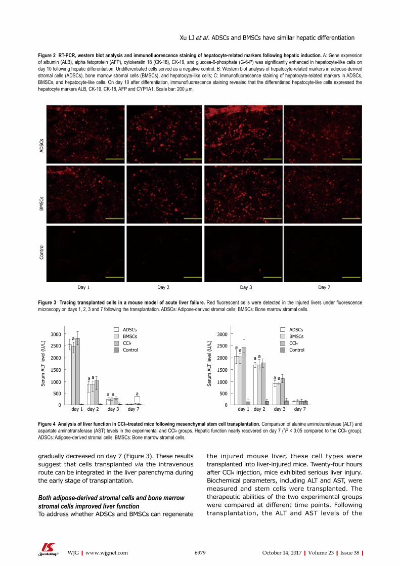
6979 October 14, 2017|Volume 23|Issue 38|WJG|www.wjgnet.com
gradually decreased on day 7 (Figure 3). These results suggest that cells transplanted via the intravenous route can be integrated in the liver parenchyma during the early stage of transplantation.
Both adipose-derived stromal cells and bone marrow stromal cells improved liver functionTo address whether ADSCs and BMSCs can regenerate
the injured mouse liver, these cell types were transplanted into liver-injured mice. Twenty-four hours after CCl4 injection, mice exhibited serious liver injury. Biochemical parameters, including ALT and AST, were measured and stem cells were transplanted. The therapeutic abilities of the two experimental groups were compared at different time points. Following transplantation, the ALT and AST levels of the
Day 1 Day 2 Day 3 Day 7
Cont
rol
B
MSC
s A
DSC
s
Figure 3 Tracing transplanted cells in a mouse model of acute liver failure. Red fluorescent cells were detected in the injured livers under fluorescence microscopy on days 1, 2, 3 and 7 following the transplantation. ADSCs: Adipose-derived stromal cells; BMSCs: Bone marrow stromal cells.
Figure 2 RT-PCR, western blot analysis and immunofluorescence staining of hepatocyte-related markers following hepatic induction. A: Gene expression of albumin (ALB), alpha fetoprotein (AFP), cytokeratin 18 (CK-18), CK-19, and glucose-6-phosphate (G-6-P) was significantly enhanced in hepatocyte-like cells on day 10 following hepatic differentiation. Undifferentiated cells served as a negative control; B: Western blot analysis of hepatocyte-related markers in adipose-derived stromal cells (ADSCs), bone marrow stromal cells (BMSCs), and hepatocyte-like cells; C: Immunofluorescence staining of hepatocyte-related markers in ADSCs, BMSCs, and hepatocyte-like cells. On day 10 after differentiation, immunofluorescence staining revealed that the differentiated hepatocyte-like cells expressed the hepatocyte markers ALB, CK-19, CK-18, AFP and CYP1A1. Scale bar: 200 μm.
Figure 4 Analysis of liver function in CCl4-treated mice following mesenchymal stem cell transplantation. Comparison of alanine aminotransferase (ALT) and aspartate aminotransferase (AST) levels in the experimental and CCl4 groups. Hepatic function nearly recovered on day 7 (aP < 0.05 compared to the CCl4 group). ADSCs: Adipose-derived stromal cells; BMSCs: Bone marrow stromal cells.
3000
2500
2000
1500
1000
500
0
Seru
m A
LT le
vel (
U/L
)
a
ADSCsBMSCsCCl4Control
a
a a a
day 1 day 2 day 3 day 7
a3000
2500
2000
1500
1000
500
0
Seru
m A
LT le
vel (
U/L
)
a
ADSCsBMSCsCCl4Control
a
a a
day 1 day 2 day 3 day 7
a
a
Xu Lj et al . ADSCs and BMSCs have similar hepatic differentiation

6980 October 14, 2017|Volume 23|Issue 38|WJG|www.wjgnet.com
experimental groups decreased significantly compared with the control group (Figure 4). HE staining revealed that the degree of injury was much weaker in the injured transplanted mice than in the injured, non-transplanted mice (Figure 5). On day 7, all of the experimental groups recovered histologically. However, damaged areas were still observed in the CCl4 group. These observations indicated that ADSCs and BMSCs possessed similar repairing abilities against CCl4-induced liver injury.
DISCUSSIONMSC transplantation is a promising alternative therapy for some liver diseases. Availability of a large number of functional hepatocytes is essential for cytotherapy. However, primary hepatocyte cultures have been hindered by their short life span and the rapid loss of hepatic function in vitro[14]. MSCs have become a promising type of seed cell for liver transplantation for their multiple differentiation potential, high proliferation capacity, and plastic adherence properties. However, it is difficult to determine the ideal seed cells from different
types of MSCs. In the present study, we established a novel isolation and culture protocol for mouse BMSCs that could provide a large number of BMSCs in a short period of time and share a similar multiple differentiation ability with ADSCs. We also established a new HCM that not only shortens the induction time but also produces a large number of hepatocyte-like cells. With the same induction system, ADSCs resembled BMSCs in term of hepatocyte differentiation potential both in vitro and in vivo.
A large quantity of ADSCs can be easily obtained by digesting adipose tissue derived from mice, rats, and humans with collagenase I[15]. However, it is difficult to obtain a large quantity of pure mouse BMSCs. We analyzed the conditioned medium method of Sun et al[16] and the compact bone method of Zhu et al[17], and developed a novel and reliable isolation and culture method that provides a large number of BMSCs in a short period of time. Compared to the method of Sun et al[16], the period required to obtain purified cells was shortened to 10-11 d. Compared to the method of Zhu et al[17], we omitted the digestion step of collagenase II. Additionally, the passage time was shortened to 2 d (compared to Zhu’s twice per week). Compared to the
Xu Lj et al . ADSCs and BMSCs have similar hepatic differentiation
ADSCs BMSCs CCl4 Control
Day
7
D
ay 3
D
ay 2
D
ay 1
Figure 5 Hematoxylin and eosin staining in CCl4-treated mice following mesenchymal stem cells transplantation. HE staining of liver tissue on different days following MSC transplantation (100 × magnification). The MSC groups showed smaller congested areas than the CCl4 group. ADSCs: Adipose-derived stromal cells; BMSCs: Bone marrow stromal cells; HE: Hematoxylin and eosin; MSC: Mesenchymal stem cells; Scale bar: 200 μm.

6981 October 14, 2017|Volume 23|Issue 38|WJG|www.wjgnet.com
method for the isolation and culture of mouse BMSCs from bone marrow[18], our protocol not only shows less contamination of hematopoietic lineage cells, but also obtains a large number of BMSCs in a short period of time. The number of BMSCs obtained from five mice can reach 107 in 10-11 d. We believe that the reason for the rapid proliferation of cells is that neonatal mice exhibit better cell viability. Therefore, we developed a novel and easy method to isolate mouse BMSCs from tibia and fibula bone fragments.
Among the various hepatic differentiation systems, a one-step induction method has been most extensively applied[19]. Various cytokines and chemicals can be used to induce MSCs into hepatocyte-like cells. Current studies have reported that HGF, FGF4, EGF, OSM, Dex, aFGF, bFGF, DMSO, Vpp, and Vc can be applied for the hepatic differentiation of ADSCs[10,20]. Additionally, HGF, FGF4, EGF, OSM, Dex, ITS, trichostatin A (TSA), and L-glutamine have been applied for the hepatic differentiation of BMSCs[21-24]. In the present study, we used multiple factors, including HGF, FGF4, OSM, aFGF, bFGF, Dex, Vpp, ITS, and Vc, to develop a new induction system to promote the differentiation of MSCs. This hepatic induction system induced ADSCs and BMSCs into hepatocyte-like cells in only 10 d. However, a previously reported one-step induction system usually took more than two weeks[25-26]. This hepatic induction system shortens the induction time compared to the previously reported one-step system. Therefore, we consider it as an optimized one-step hepatic induction system.
We also identified induced hepatocyte-like cells from ADSCs and BMSCs in terms of morphology, as well as gene and protein expression. Our results revealed a similar hepatic differentiation potential of these two cell types in vitro. In vivo, we established an animal model of acute liver failure and proved that ADSCs and BMSCs engrafted into the injured liver showed similar therapeutic effects in the improvement of liver function. The results in our study suggest that BMSCs and ADSCs may serve as ideal seed cells for treating liver injury.
In conclusion, we developed a new protocol for the preparation of mouse BMSCs and established a new hepatic induction system. Our findings proved that ADSCs resembled BMSCs in the hepatic differentiation potential in vitro and in vivo. ADSCs can be obtained more easily and are less invasive than BMSCs. Therefore, ADSCs may be a more suitable seed cell type for use in cell transplantion or liver tissue engineering. In future experiments, we will explore the roles of ADSCs in liver tissue engineering.
COMMENTSBackgroundLiver transplantation remains the only effective treatment for liver failure.
However, its clinical application is limited by donor organs and immune rejection. To this end, hepatocyte transplantation and a bio-artificial liver support system are two potential surrogate complementary therapies for patients with liver failure. Hepatocyte-like cells can be induced from mesenchymal stem cells (MSCs) for xenotransplantation and have been demonstrated to perform hepatocyte functions in preclinical animal studies. However, the best type of MSCs has not yet been investigated and therefore it is important to screen for an ideal seed cell type for use in cell transplantation or a bio-artificial liver support system.
Research frontiersMSCs are non-hematopoietic multipotent stem cells that can be isolated from multiple tissues, such as bone marrow, adipose tissue, cord blood, and amniotic fluid. Bone marrow stromal cells (BMSCs) are the most extensively studied and fertile stem cell source used in regenerative medicine and liver tissue engineering. More recently, adipose-derived stromal cells (ADSCs) were identified as another promising and extensively studied stem cells for use, especially in liver tissue engineering. Although both ADSCs and BMSCs can be induced into hepatocyte-like cells, there is no uniform culture system to evaluate their hepatic differentiation potential. Therefore, it is necessary to establish an ideal system to improve the efficiency of hepatic induction.
Innovations and breakthroughsA new protocol for the preparation of mouse BMSCs and a new hepatic induction system were established. This study proved that ADSCs resembled BMSCs in the hepatic differentiation potential in vitro and in vivo. ADSCs can be obtained more easily and are less invasive than BMSCs. Therefore, ADSCs may serve as a more suitable seed cell type for use in cell transplantion or liver tissue engineering.
ApplicationsADSCs are likely to serve as suitable seed cells for use in cell transplantion or liver tissue engineering.
TerminologyMSC transplant is a promising alternative therapy for some liver diseases. Availability of large number of functional hepatocytes is essential for cytotherapy. Both ADSCs and BMSCs can be induced into hepatocyte-like cells. Therefore, both ADSCs and BMSCs may be suitable seed cells for use in cell transplantion or liver tissue engineering.
Peer-review In this paper, the authors investigated a novel method of generating differentiated hepatocytes from two sources of mesenchymal stem cells and tested their ability to recover liver damage in an experimental mouse model. The paper thus adressed an important question as stem cell therapy could help overcome current limitations of treatment for metabolic liver diseases and liver injury.
REFERENCES1 Lee SY, Kim HJ, Choi D. Cell sources, liver support systems and
liver tissue engineering: alternatives to liver transplantation. Int J Stem Cells 2015; 8: 36-47 [PMID: 26019753 DOI: 10.15283/ijsc.2015.8.1.36]
2 Hu C, Li L. In vitro culture of isolated primary hepatocytes and stem cell-derived hepatocyte-like cells for liver regeneration. Protein Cell 2015; 6: 562-574 [PMID: 26088193 DOI: 10.1007/s13238-015-0180-2]
3 Kellner J, Sivajothi S, McNiece I. Differential properties of human stromal cells from bone marrow, adipose, liver and cardiac tissues. Cytotherapy 2015; 17: 1514-1523 [PMID: 26341480 DOI: 10.1016/j.jcyt.2015.07.009]
4 Park KM, Hussein KH, Ghim JH, Ahn C, Cha SH, Lee GS, Hong SH, Yang S, Woo HM. Hepatic differentiation of porcine
Xu Lj et al . ADSCs and BMSCs have similar hepatic differentiation
COMMENTS
6982 October 14, 2017|Volume 23|Issue 38|WJG|www.wjgnet.com
embryonic stem cells for translational research of hepatocyte transplantation. Transplant Proc 2015; 47: 775-779 [PMID: 25891729 DOI: 10.1016/j.transproceed.2015.01.020]
5 Yang L, Wang Y, Wang X, Liu Y. Effect of allogeneic umbilical cord mesenchymal stem cell transplantation in a rat model of hepatic cirrhosis. J Tradit Chin Med 2015; 35: 63-68 [PMID: 25842730 DOI: 10.1016/S0254-6272(15)30010-8]
6 Chen QQ, Yan L, Wang CZ, Wang WH, Shi H, Su BB, Zeng qH, Du HT, Wan J. Mesenchymal stem cells alleviate TNBS-induced colitis by modulating inflammatory and autoimmune responses. World J Gastroenterol 2013; 19: 4702-4717 [PMID: 23922467 DOI: 10.3748/wjg.v19.i29.4702]
7 Zheng Z, Li X, Li Z, Ma X. Artificial and bioartificial liver support systems for acute and acute-on-chronic hepatic failure: A meta-analysis and meta-regression. Exp Ther Med 2013; 6: 929-936 [PMID: 24137292 DOI: 10.3892/etm.2013.1241]
8 Yang D, Wang Zq, Deng Jq, Liao JY, Wang X, Xie J, Deng MM, Lü MH. Adipose-derived stem cells: A candidate for liver regeneration. J Dig Dis 2015; 16: 489-498 [PMID: 26121206 DOI: 10.1111/1751-2980.12268]
9 Yang T, Wang Y, Jiang S, Liu X, Yu Z. Hepatocyte growth factor-induced differentiation of bone mesenchymal stem cells toward hepatocyte-like cells occurs through nuclear factor-kappa B signaling in vitro. Cell Biol Int 2016; 40: 1017-1023 [PMID: 27249785 DOI: 10.1002/cbin.10630]
10 Banas A, Teratani T, Yamamoto Y, Tokuhara M, Takeshita f, Osaki M, Kato T, Okochi H, Ochiya T. Rapid hepatic fate specification of adipose-derived stem cells and their therapeutic potential for liver failure. J Gastroenterol Hepatol 2009; 24: 70-77 [PMID: 18624899 DOI: 10.1111/j.1440-1746.2008.05496.x]
11 Deng L, Kong X, Liu G, Li C, Chen H, Hong Z, Liu J, Xia J. Transplantation of Adipose-Derived Mesenchymal Stem Cells Efficiently Rescues Thioacetamide-Induced Acute Liver failure in Mice. Transplant Proc 2016; 48: 2208-2215 [PMID: 27569972 DOI: 10.1016/j.transproceed.2016.02.077]
12 Huang B, Cheng X, Wang H, Huang W, la Ga Hu Z, Wang D, Zhang K, Zhang H, Xue Z, Da Y, Zhang N, Hu Y, Yao Z, qiao L, Gao f, Zhang R. Mesenchymal stem cells and their secreted molecules predominantly ameliorate fulminant hepatic failure and chronic liver fibrosis in mice respectively. J Transl Med 2016; 14: 45 [PMID: 26861623 DOI: 10.1186/s12967-016-0792-1]
13 Xu L, Wang S, Sui X, Wang Y, Su Y, Huang L, Zhang Y, Chen Z, Chen q, Du H, Zhang Y, Yan L. Mesenchymal Stem Cell-Seeded Regenerated Silk fibroin Complex Matrices for Liver Regeneration in an Animal Model of Acute Liver failure. ACS Appl Mater Interfaces 2017; 9: 14716-14723 [PMID: 28409921 DOI: 10.1021/acsami.7b02805]
14 Kim BK, Chung KW, Sun HS, Suh JG, Min WS, Kang CS, Sim SI, Shin WS, Kim CC. Liver disease during the first post-transplant year in bone marrow transplantation recipients: retrospective study. Bone Marrow Transplant 2000; 26: 193-197 [PMID: 10918430 DOI: 10.1038/sj.bmt.1702453]
15 Feng Z, Li C, Jiao S, Hu B, Zhao L. In vitro differentiation
of rat bone marrow mesenchymal stem cells into hepatocytes. Hepatogastroenterology 2011; 58: 2081-2086 [PMID: 22024079 DOI: 10.5754/hge11220]
16 Sun S, Guo Z, Xiao X, Liu B, Liu X, Tang PH, Mao N. Isolation of mouse marrow mesenchymal progenitors by a novel and reliable method. Stem Cells 2003; 21: 527-535 [PMID: 12968107 DOI: 10.1634/stemcells.21-5-527]
17 Zhu H, Guo ZK, Jiang XX, Li H, Wang XY, Yao HY, Zhang Y, Mao N. A protocol for isolation and culture of mesenchymal stem cells from mouse compact bone. Nat Protoc 2010; 5: 550-560 [PMID: 20203670 DOI: 10.1038/nprot.2009.238]
18 Kitano Y, Radu A, Shaaban A, flake AW. Selection, enrichment, and culture expansion of murine mesenchymal progenitor cells by retroviral transduction of cycling adherent bone marrow cells. Exp Hematol 2000; 28: 1460-1469 [PMID: 11146168]
19 Seo MJ, Suh SY, Bae YC, Jung JS. Differentiation of human adipose stromal cells into hepatic lineage in vitro and in vivo. Biochem Biophys Res Commun 2005; 328: 258-264 [PMID: 15670778 DOI: 10.1016/j.bbrc.2004.12.158]
20 Zhang X, Dong J. Direct comparison of different coating matrix on the hepatic differentiation from adipose-derived stem cells. Biochem Biophys Res Commun 2015; 456: 938-944 [PMID: 25446084 DOI: 10.1016/j.bbrc.2014.11.004]
21 Xin J, Ding W, Hao S, Jiang L, Zhou q, Wu T, Shi D, Cao H, Li L, Li J. Human bone marrow mesenchymal stem cell-derived hepatocytes express tissue inhibitor of metalloproteinases 4 and follistatin. Liver Int 2015; 35: 2301-2310 [PMID: 25645195 DOI: 10.1111/liv.12797]
22 Lu T, Yang C, Sun H, Lv J, Zhang f, Dong XJ. fGf4 and HGf promote differentiation of mouse bone marrow mesenchymal stem cells into hepatocytes via the MAPK pathway. Genet Mol Res 2014; 13: 415-424 [PMID: 24535868 DOI: 10.4238/2014.January.21.9]
23 Ye D, Li T, Heraud P, Parnpai R. Effect of Chromatin-Remodeling Agents in Hepatic Differentiation of Rat Bone Marrow-Derived Mesenchymal Stem Cells In Vitro and In Vivo. Stem Cells Int 2016; 2016: 3038764 [PMID: 27242905 DOI: 10.1155/2016/3038764]
24 Piryaei A , Valojerdi MR, Shahsavani M, Baharvand H. Differentiation of bone marrow-derived mesenchymal stem cells into hepatocyte-like cells on nanofibers and their transplantation into a carbon tetrachloride-induced liver fibrosis model. Stem Cell Rev 2011; 7: 103-118 [PMID: 20182823 DOI: 10.1007/s12015-010-9126-5]
25 Stock P, Brückner S, Winkler S, Dollinger MM, Christ B. Human bone marrow mesenchymal stem cell-derived hepatocytes improve the mouse liver after acute acetaminophen intoxication by preventing progress of injury. Int J Mol Sci 2014; 15: 7004-7028 [PMID: 24758938 DOI: 10.3390/ijms15047004]
26 Zhang YN, Lie PC, Wei X. Differentiation of mesenchymal stromal cells derived from umbilical cord Wharton’s jelly into hepatocyte-like cells. Cytotherapy 2009; 11: 548-558 [PMID: 19657806 DOI: 10.1080/14653240903051533]
P- Reviewer: Ocker M S- Editor: Wei LJ L- Editor: Ma JY E- Editor: Ma YJ
Xu Lj et al . ADSCs and BMSCs have similar hepatic differentiation
Wei-Wei Wang, Yu Zhang, Xiao-Bing Huang, Nan You, Lu Zheng, Jing Li, Department of Hepatobiliary Surgery, the Second Affiliated Hospital of Third Military Medical University, Chongqing 400037, China
ORCID number: Wei-Wei Wang (0000-0002-8993-8411); Yu Zhang (0000-0001-8719-743X); Xiao-Bing Huang (0000-0002-4136-3639); Nan You (0000-0002-8107-4292); Lu Zheng (0000-0002-5672-330X); Jing Li (0000-0002-1594-9361).
Author contributions: Wang WW performed the research, analyzed the data, wrote and revised the paper; Zhang Y, Li J and You N performed the research; Zheng L participated in research design; Li J designed the research and participated in the revision of the paper; all authors have read and approved the final manuscript.
Institutional review board statement: The study was reviewed and approved by the Ethics Committee of the Second Affiliated Hospital of Third Military Medical University.
Institutional animal care and use committee statement: All experiments on animals were performed in accordance with the “Guide for the Care and Use of Laboratory Animals” (National Institutes of Health, 8th edition, 2011). All procedures involving animals were reviewed and approved by the Animal Care and Research Committee of the Second Affiliated Hospital of Third Military Medical University.
Conflict-of-interest statement: The authors declare no potential conflict of interest for this paper.
Data sharing statement: No additional data are available.
Open-Access: This article is an open-access article which was selected by an in-house editor and fully peer-reviewed by external reviewers. It is distributed in accordance with the Creative Commons Attribution Non Commercial (CC BY-NC 4.0) license, which permits others to distribute, remix, adapt, build upon this work non-commercially, and license their derivative works on different terms, provided the original work is properly cited and the use is non-commercial. See: http://creativecommons.org/licenses/by-nc/4.0/
Manuscript source: Unsolicited manuscript
Correspondence to: Jing Li, PhD, Department of Hepatobiliary Surgery, the Second Affiliated Hospital of Third Military Medical University, 183# Xinqiao zhengjie, Shapingba District, Chongqing 400037, China. [email protected]: +86-23-68774606Fax: +86-23-68755606
Received: July 3, 2017 Peer-review started: July 3, 2017First decision: July 27, 2017Revised: August 17, 2017 Accepted: September 6, 2017 Article in press: September 5, 2017Published online: October 14, 2017
AbstractAIMTo investigate whether fecal microbiota transplantation (FMT) prevents hepatic encephalopathy (HE) in rats with carbon tetrachloride (CCl4)-induced acute hepatic dysfunction.
METHODSA rat model of HE was established with CCl4. Rat behaviors and spatial learning capability were observed, and hepatic necrosis, intestinal mucosal barrier, serum ammonia levels and intestinal permeability were determined in HE rats receiving FMT treatment. Furthermore, the expression of tight junction proteins (Claudin-1, Claudin-6 and Occludin), Toll-like receptor (TLR) 4/TLR9, interleukin (IL)-1β, IL-6 and tumor necrosis factor (TNF)-α was examined.
RESULTSFMT improved rat behaviors, HE grade and spatial learning capability. Moreover, FMT prevented hepatic
6983 October 14, 2017|Volume 23|Issue 38|WJG|www.wjgnet.com
ORIGINAL ARTICLE
Fecal microbiota transplantation prevents hepatic encephalopathy in rats with carbon tetrachloride-induced acute hepatic dysfunction
Basic Study
Wei-Wei Wang, Yu Zhang, Xiao-Bing Huang, Nan You, Lu Zheng, Jing Li
Submit a Manuscript: http://www.f6publishing.com
DOI: 10.3748/wjg.v23.i38. 6983
World J Gastroenterol 2017 October 14; 23(38): 6983-6994
ISSN 1007-9327 (print) ISSN 2219-2840 (online)

necrosis and intestinal mucosal barrier damage, leading to hepatic clearance of serum ammonia levels and reduced intestinal permeability. The expression of TLR4 and TLR9, two potent mediators of inflammatory response, was significantly downregulated in the liver of rats treated with FMT. Consistently, circulating pro-inflammatory factors such as interleukin (IL)-1β, IL-6 and tumor necrosis factor-α were remarkably decreased, indicating that FMT is able to limit systemic inflammation by decreasing the expression of TLR4 and TLR9. Importantly, HE-induced loss of tight junction proteins (Claudin-1, Claudin-6 and Occludin) was restored in intestinal tissues of rats receiving FMT treatment.
CONCLUSIONFMT enables protective effects in HE rats, and it improves the cognitive function and reduces the liver function indexes. FMT may cure HE by altering the intestinal permeability and improving the TLR response of the liver.
Key words: Fecal microbiota transplantation; Hepatic encephalopathy; Cognitive function; Intestinal per-meability; Toll-like receptor
© The Author(s) 2017. Published by Baishideng Publishing Group Inc. All rights reserved.
Core tip: In this article, we first established a rat model of hepatic encephalopathy, and then carried on the fecal microbiota transplantation (FMT). Our results suggest that FMT can serve as a kind of new method for the treatment of hepatic encephalopathy, probably better than VSL#3.
Wang WW, Zhang Y, Huang XB, You N, Zheng L, Li J. Fecal microbiota transplantation prevents hepatic encephalopathy in rats with carbon tetrachloride-induced acute hepatic dysfunction. World J Gastroenterol 2017; 23(38): 6983-6994 Available from: URL: http://www.wjgnet.com/1007-9327/full/v23/i38/6983.htm DOI: http://dx.doi.org/10.3748/wjg.v23.i38.6983
INTRODUCTIONHepatic encephalopathy (HE) is a common and serious disorder with a wide spectrum of neuropsychiatric abnormalities due to chronic and acute liver dysfunction[1]. HE is associated with poor quality of life and increased mortality, thus representing a major healthcare burden in patients with liver cirrhosis[2,3]. The estimated one-year survival rate is 42% after the first episode of overt HE, and the three-year survival rate is 23%[1,4,5]. Therefore, HE with cirrhosis is considered to have a poor prognosis for subjects who do not undergo liver transplantation. The death rate is substantially lower for patients who receive a transplant. The survival rate is more than 70% in the
first 5 years after transplantation[1,6]. Although the pathogenesis of HE remains in-
completely understood, gut-derived neurotoxin ammonia that accumulates in the central nervous system (CNS) is the most frequent precipitating factor[7-11]. In cases of normal hepatic function, ammonia enters the portal circulation of the liver and is subsequently converted to urea through the urea cycle[1].
Gut microbiota has emerged as a critical factor in the development of HE. Dysbiosis or an altered gut microbiota population promotes a systemic pro-inflammatory milieu, resulting in neuro-inflammation and ultimately neuronal dysfunction including HE[12-17]. Lactulose and lactitol, which target gut microbiota, have been demonstrated to be effective as first-line therapies for both acute and chronic HE. However, lactulose and lactitol have significant gastrointestinal side effects[18,19]. Furthermore, approximately 20% of patients with chronic liver failure and HE have been found to be non-responsive to lactulose treatment. A systemic review demonstrated that lactulose and lactitol failed to impart any survival benefit to cirrhotic patients with HE[20].
Probiotics (e.g., VSL#3, a clinically tested probiotic formula consisting of Lactobacilli, Bifidobacteria, and Streptococci) contain living beneficial bacteria and have been shown to improve mental status and cognitive functioning. Therefore, probiotics may prevent HE recurrence by decreasing urease-producing bacteria populations and ammonia absorption while also increasing hepatic ammonia clearance[21]. Although probiotics are thought to have no adverse effects when used as long-term therapeutics, they are not widely utilized clinically due to the potential risk of introducing live bacteria into immunosuppressed patients. Therefore, the use of probiotics for cirrhotic patients with HE cannot be currently recommended, and rigorous clinical evaluation in randomized controlled trials is required[22-25].
Given the limitations of current HE therapy, research efforts are now aimed at improving existing treatments or developing novel therapies. Fecal microbiota transplantation (FMT) is rapidly being accepted as a viable, safe, and effective treatment for chronic gastrointestinal infections, recurrent inflammatory bowel diseases (IBD) and Clostridium difficile infection (CDI)[26-29]. It has also gained attention for its therapeutic potential for cardiometabolic, autoimmune, and other extra-intestinal conditions that were not previously considered to be associated with the intestinal microbiota[30-32]. However, whether FMT has a healing effect on HE has not been investigated.
In this study, we present in vivo evidence that FMT is effective for improving HE symptoms as evidenced by increased rat motor activity, spatial learning and memory following FMT. Moreover, FMT reduces systematic inflammation, leading to restored hepatic and intestinal dysfunction. Mechanically, FMT reduced liver expression of Toll-like receptor (TLR) 4 and
6984 October 14, 2017|Volume 23|Issue 38|WJG|www.wjgnet.com
Wang WW et al. Fecal microbiota transplantation prevents hepatic encephalopathy
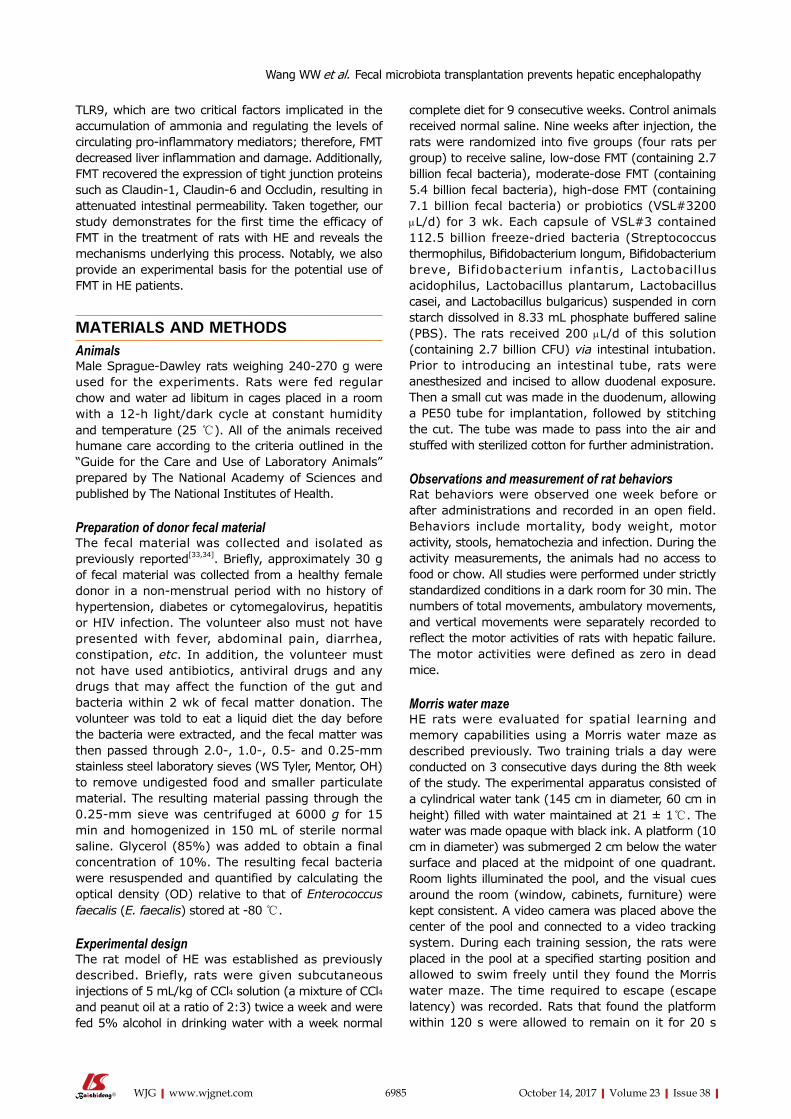
TLR9, which are two critical factors implicated in the accumulation of ammonia and regulating the levels of circulating pro-inflammatory mediators; therefore, FMT decreased liver inflammation and damage. Additionally, FMT recovered the expression of tight junction proteins such as Claudin-1, Claudin-6 and Occludin, resulting in attenuated intestinal permeability. Taken together, our study demonstrates for the first time the efficacy of FMT in the treatment of rats with HE and reveals the mechanisms underlying this process. Notably, we also provide an experimental basis for the potential use of FMT in HE patients.
MATERIALS AND METHODSAnimals Male Sprague-Dawley rats weighing 240-270 g were used for the experiments. Rats were fed regular chow and water ad libitum in cages placed in a room with a 12-h light/dark cycle at constant humidity and temperature (25 ℃). All of the animals received humane care according to the criteria outlined in the “Guide for the Care and Use of Laboratory Animals” prepared by The National Academy of Sciences and published by The National Institutes of Health.
Preparation of donor fecal materialThe fecal material was collected and isolated as previously reported[33,34]. Briefly, approximately 30 g of fecal material was collected from a healthy female donor in a non-menstrual period with no history of hypertension, diabetes or cytomegalovirus, hepatitis or HIV infection. The volunteer also must not have presented with fever, abdominal pain, diarrhea, constipation, etc. In addition, the volunteer must not have used antibiotics, antiviral drugs and any drugs that may affect the function of the gut and bacteria within 2 wk of fecal matter donation. The volunteer was told to eat a liquid diet the day before the bacteria were extracted, and the fecal matter was then passed through 2.0-, 1.0-, 0.5- and 0.25-mm stainless steel laboratory sieves (WS Tyler, Mentor, OH) to remove undigested food and smaller particulate material. The resulting material passing through the 0.25-mm sieve was centrifuged at 6000 g for 15 min and homogenized in 150 mL of sterile normal saline. Glycerol (85%) was added to obtain a final concentration of 10%. The resulting fecal bacteria were resuspended and quantified by calculating the optical density (OD) relative to that of Enterococcus faecalis (E. faecalis) stored at -80 ℃.
Experimental designThe rat model of HE was established as previously described. Briefly, rats were given subcutaneous injections of 5 mL/kg of CCl4 solution (a mixture of CCl4 and peanut oil at a ratio of 2:3) twice a week and were fed 5% alcohol in drinking water with a week normal
complete diet for 9 consecutive weeks. Control animals received normal saline. Nine weeks after injection, the rats were randomized into five groups (four rats per group) to receive saline, low-dose FMT (containing 2.7 billion fecal bacteria), moderate-dose FMT (containing 5.4 billion fecal bacteria), high-dose FMT (containing 7.1 billion fecal bacteria) or probiotics (VSL#3200 μL/d) for 3 wk. Each capsule of VSL#3 contained 112.5 billion freeze-dried bacteria (Streptococcus thermophilus, Bifidobacterium longum, Bifidobacterium breve, Bifidobacterium infantis, Lactobacillus acidophilus, Lactobacillus plantarum, Lactobacillus casei, and Lactobacillus bulgaricus) suspended in corn starch dissolved in 8.33 mL phosphate buffered saline (PBS). The rats received 200 μL/d of this solution (containing 2.7 billion CFU) via intestinal intubation. Prior to introducing an intestinal tube, rats were anesthesized and incised to allow duodenal exposure. Then a small cut was made in the duodenum, allowing a PE50 tube for implantation, followed by stitching the cut. The tube was made to pass into the air and stuffed with sterilized cotton for further administration.
Observations and measurement of rat behaviorsRat behaviors were observed one week before or after administrations and recorded in an open field. Behaviors include mortality, body weight, motor activity, stools, hematochezia and infection. During the activity measurements, the animals had no access to food or chow. All studies were performed under strictly standardized conditions in a dark room for 30 min. The numbers of total movements, ambulatory movements, and vertical movements were separately recorded to reflect the motor activities of rats with hepatic failure. The motor activities were defined as zero in dead mice.
Morris water mazeHE rats were evaluated for spatial learning and memory capabilities using a Morris water maze as described previously. Two training trials a day were conducted on 3 consecutive days during the 8th week of the study. The experimental apparatus consisted of a cylindrical water tank (145 cm in diameter, 60 cm in height) filled with water maintained at 21 ± 1 ℃. The water was made opaque with black ink. A platform (10 cm in diameter) was submerged 2 cm below the water surface and placed at the midpoint of one quadrant. Room lights illuminated the pool, and the visual cues around the room (window, cabinets, furniture) were kept consistent. A video camera was placed above the center of the pool and connected to a video tracking system. During each training session, the rats were placed in the pool at a specified starting position and allowed to swim freely until they found the Morris water maze. The time required to escape (escape latency) was recorded. Rats that found the platform within 120 s were allowed to remain on it for 20 s
6985 October 14, 2017|Volume 23|Issue 38|WJG|www.wjgnet.com
Wang WW et al. Fecal microbiota transplantation prevents hepatic encephalopathy

6986 October 14, 2017|Volume 23|Issue 38|WJG|www.wjgnet.com
Statistical analysisSPSS statistical software, version 19.0, was used for statistical analyses. The Pearson χ 2 test or Fisher’s exact test was used to compare qualitative variables and Student’s t-test was used for comparisons of quantitative variables. All statistical tests were two-sided. P < 0.05 was considered statistically significant
RESULTS FMT significantly alleviates behaviors of rats with chronic liver failure-induced HEChronic liver failure-induced HE was induced in rats by treatment with carbon tetrachloride-alcohol as previously described. All rats treated for 9 wk showed similar symptoms of HE, such as impaired spontaneous movement, cachexia and somnolence, when compared with saline-treated control mice. Histological examination verified the successful establishment of the rat HE model. The rats were then randomly divided into five groups including the model group, the probiotic-treated group (termed VSL) and the FMT treated groups across three doses (termed FMT-L, FMT-M and FMT-H for the low, mid and high FMT doses, respectively). Infections were observed during the experimental period; one rat each in the VSL, FMT-L, FMT-M and FMT-H groups died from infection. Data from these rats were excluded from the final statistical analysis. The behaviors of each group were observed clinically until the end of the experiment and were compared for mortality rate and the clinical grade by the scoring method represented in Table 1 and as previously described[35,36]. Significant weight loss and decreased appetite were observed in the control group relative to the other treated groups, whereas rats treated with either probiotics or any of the three FMT doses, but not the model group, started to increase food intake and gain weight during the first week. The feeding behavior of rats in the high-dose FMT treatment group normalized during the second week (Figures 1 and 2). Together, the data presented here demonstrate that FMT enables the healing of chronic liver failure-induced HE.
FMT markedly improves learning and memory functioning in the rat HE model The Morris water maze was used to evaluate the spatial learning of rats from different treatment groups. The Morris water maze test lasted six days. The first five days of acquisition training with an invisible platform were followed by 4 d of reversal training with an invisible platform. A probe trial was then carried out with no escape platform. Finally, four trials were conducted using the visible platform.
In contrast to the control group, the total travel distance was significantly decreased in the model group (P = 0.014), whereas the VSL or FMT treated
and were then returned to their home cages. If a rat did not reach the platform within 120 s, it was gently guided there by the experimenter, and allowed to stay on it for 20 s. The test was performed again at 12 wk to assess spatial cognitive function.
Histology examination of liver injury and intestinal damageFor histological examination, liver and distal ileum samples were fixed in 4% paraformaldehyde, dehydrated using graded ethanol, and then embedded in paraffin. The paraffin blocks were sectioned and stained with hematoxylin and eosin (HE) using standard histological techniques.
Measurement of intestinal permeabilityIntestinal permeability was determined by Evans blue staining as previously described. Rats were anesthetized and transcardiacally perfused with Evans Blue dye. Colonic tissue was removed and maintained in formamide solution at 60 ℃ for 24 h, followed by centrifugation at 5000 r/min for 20 min. Optical density at 632 nm was measured and calculated.
Liver functional tests and serum ammonia level detection Biochemical parameters were measured using standard clinical methods, including alanine aminotransferase (ALT), aspartate aminotransferase (AST), serum albumin (ALB), total bilirubin (TBIL) and direct bilirubin (DBIL). Blood samples (0.5 mL) were collected from the portal and tail veins and analyzed with an automatic biochemical analyzer.
Enzyme-linked immunosorbent assay (ELISA) analysis of circulating pro-inflammatory mediators and stool proteinsFor detection of interleukin (IL)-1β, IL-6 and tumor necrosis factor (TNF)-α, serum samples from each individual group were collected and separated from portal and tail veins, followed by analysis with ELISA kits according to the manufacturer’s instructions (Sangon, Shanghai). For Calprotectin and alpha-1-antitrypsin (A1AT) detection, equal weight stools from each group were collected and dissolved in PBS and then separated by centrifugation for ELISA testing according to the manufacturer’s instructions (Sangon, Shanghai).
Western blot analysis Protein expression was determined in lysates of liver and intestinal tissues using rabbit anti-TLR4, anti-TLR9, anti-Claudin-1, anti-Claudin-6 and anti-Occludin antibodies (Abcam) diluted in blocking buffer (5% milk in 0.2% Tween 20/TBS, 4 ℃, overnight). The results were normalized to GAPDH expression (mouse anti-GAPDH, Abcam).
Wang WW et al. Fecal microbiota transplantation prevents hepatic encephalopathy
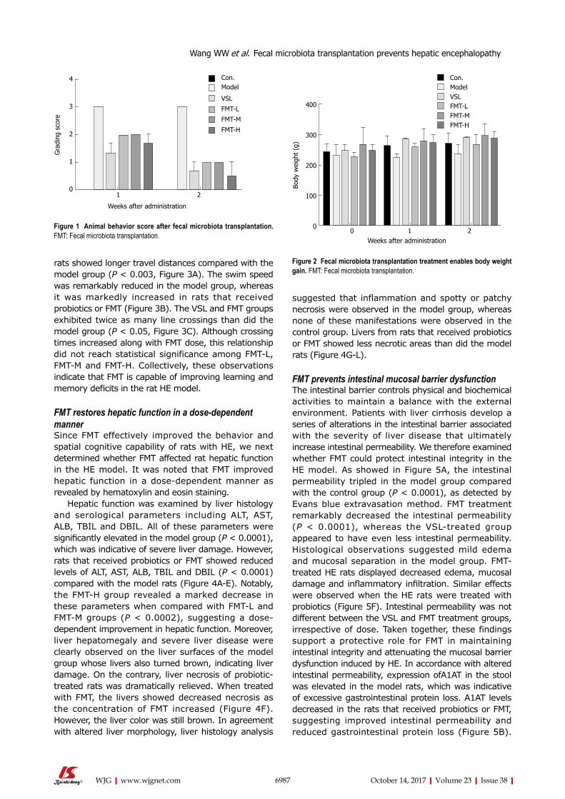
6987 October 14, 2017|Volume 23|Issue 38|WJG|www.wjgnet.com
rats showed longer travel distances compared with the model group (P < 0.003, Figure 3A). The swim speed was remarkably reduced in the model group, whereas it was markedly increased in rats that received probiotics or FMT (Figure 3B). The VSL and FMT groups exhibited twice as many line crossings than did the model group (P < 0.05, Figure 3C). Although crossing times increased along with FMT dose, this relationship did not reach statistical significance among FMT-L, FMT-M and FMT-H. Collectively, these observations indicate that FMT is capable of improving learning and memory deficits in the rat HE model.
FMT restores hepatic function in a dose-dependent mannerSince FMT effectively improved the behavior and spatial cognitive capability of rats with HE, we next determined whether FMT affected rat hepatic function in the HE model. It was noted that FMT improved hepatic function in a dose-dependent manner as revealed by hematoxylin and eosin staining.
Hepatic function was examined by liver histology and serological parameters including ALT, AST, ALB, TBIL and DBIL. All of these parameters were significantly elevated in the model group (P < 0.0001), which was indicative of severe liver damage. However, rats that received probiotics or FMT showed reduced levels of ALT, AST, ALB, TBIL and DBIL (P < 0.0001) compared with the model rats (Figure 4A-E). Notably, the FMT-H group revealed a marked decrease in these parameters when compared with FMT-L and FMT-M groups (P < 0.0002), suggesting a dose-dependent improvement in hepatic function. Moreover, liver hepatomegaly and severe liver disease were clearly observed on the liver surfaces of the model group whose livers also turned brown, indicating liver damage. On the contrary, liver necrosis of probiotic-treated rats was dramatically relieved. When treated with FMT, the livers showed decreased necrosis as the concentration of FMT increased (Figure 4F). However, the liver color was still brown. In agreement with altered liver morphology, liver histology analysis
suggested that inflammation and spotty or patchy necrosis were observed in the model group, whereas none of these manifestations were observed in the control group. Livers from rats that received probiotics or FMT showed less necrotic areas than did the model rats (Figure 4G-L).
FMT prevents intestinal mucosal barrier dysfunction The intestinal barrier controls physical and biochemical activities to maintain a balance with the external environment. Patients with liver cirrhosis develop a series of alterations in the intestinal barrier associated with the severity of liver disease that ultimately increase intestinal permeability. We therefore examined whether FMT could protect intestinal integrity in the HE model. As showed in Figure 5A, the intestinal permeability tripled in the model group compared with the control group (P < 0.0001), as detected by Evans blue extravasation method. FMT treatment remarkably decreased the intestinal permeability (P < 0.0001), whereas the VSL-treated group appeared to have even less intestinal permeability. Histological observations suggested mild edema and mucosal separation in the model group. FMT-treated HE rats displayed decreased edema, mucosal damage and inflammatory infiltration. Similar effects were observed when the HE rats were treated with probiotics (Figure 5F). Intestinal permeability was not different between the VSL and FMT treatment groups, irrespective of dose. Taken together, these findings support a protective role for FMT in maintaining intestinal integrity and attenuating the mucosal barrier dysfunction induced by HE. In accordance with altered intestinal permeability, expression ofA1AT in the stool was elevated in the model rats, which was indicative of excessive gastrointestinal protein loss. A1AT levels decreased in the rats that received probiotics or FMT, suggesting improved intestinal permeability and reduced gastrointestinal protein loss (Figure 5B).
Weeks after administration
4
3
2
1
0
Gra
ding
sco
re
1 2
Con.Model
VSL
FMT-L
FMT-M
FMT-H
Figure 1 Animal behavior score after fecal microbiota transplantation. FMT: Fecal microbiota transplantation.
400
300
200
100
0
Body
wei
ght
(g)
0 1 2
Con.ModelVSLFMT-LFMT-MFMT-H
Weeks after administration
Figure 2 Fecal microbiota transplantation treatment enables body weight gain. FMT: Fecal microbiota transplantation.
Wang WW et al. Fecal microbiota transplantation prevents hepatic encephalopathy

6988 October 14, 2017|Volume 23|Issue 38|WJG|www.wjgnet.com
Elevated Calprotectin levels in the stool, which are commonly used in the clinic to measure the intestinal inflammation index, were found to be reversed by probiotics and FMT, implying attenuated intestinal inflammation (Figure 5C). Overall, the data presented here indicate that FMT preserves intestinal mucosal barrier function.
FMT attenuates serum ammonia levels and the systemic proinflammatory response It is well known that ammonia serves as the most important precipitating factor leading to HE. In addition, accumulating evidence has demonstrated that inflammation, including systemic inflammation, neu-roinflammation and endotoxemia, acts concomitantly with ammonia to drive HE pathogenesis in cirrhotic patients. We next determined whether FMT affects serum ammonia levels and proinflammatory responses. Serum ammonia levels and proinflammatory cytokines including IL-1β, IL-6 and TNF-α were detected using Berthelot reaction and ELISA assay. The mean ammonia level from the portal vein was 12.37 μmol/L in the control group, which was increased to 36.28 μmol/L in HE rats. When the HE rats were given probiotics, the mean serum ammonia level decreased to 22 μmol/L. FMT therapy resulted in a notable decline in ammonia levels (P < 0.0001). In particular, high-dose FMT administration was almost as effective as probiotic treatment in decreasing ammonia levels. Moreover, similar effects were observed when the ammonia level was evaluated from the tail vein,
indicating that FMT enables effective clearance of serum ammonia and ultimately improves clinical symptoms (Figure 6A). Consistent with the decreased serum ammonia concentration, circulating levels of proinflammatory mediators such as IL-1β, IL-6 and TNF-α were elevated in the model group and reduced in groups that received probiotics or FMT treatment (P < 0.0001, Figure 6B-D). Significant differences in proinflammatory mediator levels were observed among the groups that received different FMT doses (P < 0.002). The data described here demonstrate a systematic relief of HE severity in rats, and it was clear that systemic inflammation, but not ammonia, was strongly correlated with increasing grades of HE.
FMT protects HE progression through distinct mechanisms Although FMT has been shown to be effective in a rat model of HE, leading to improved hepatic and intestinal function and impaired systemic inflammation, the underlying mechanisms of these processes have not been investigated. Liver TLRs are reported to be important determinants of HE severity and are intimately related to arterial ammonia concen-tration and levels of circulating pro-inflammatory mediators[37-39]. Additionally, tight junction proteins (Occludin and Claudins) are implicated in the regulation of intestinal permeability[40,41].
Therefore, we examined the protein expression levels of TLR4 and TLR9 in the liver tissues, and the expression levels of Claudin-1, Claudin-6 and Occludin were analyzed in the intestinal tissues (Figure 7). As expected, liver dysfunction stimulated the expression of TLR4 and TLR9 in the liver, which was downregulated in liver tissue from rats treated with probiotics and FMT, indicating that FMT can limit systemic inflammation by decreasing the expression of TLR4 and TLR9. TLR4 and TLR9 are potent mediators that promote the expression of pro-inflammatory factors such as IL-6 and TNF-α. Accordingly, the expression of Claudin-1, Claudin-6 and Occludin was lost in the intestinal tissues from the model rats,
60
40
20
0
Tota
l dis
tanc
e (m
)
Con. Model VSL FMT-L FMT-M FMT-H
50
40
30
20
10
0
Line
cro
ssin
g (t
imes
)
Con. Model VSL FMT-L FMT-M FMT-H
0.8
0.6
0.4
0.2
0.0
Swim
spe
ed (
m/s
) Con. Model VSL FMT-L FMT-M FMT-H
A B C
Figure 3 Cognitive function before and after VSL#3 or fecal microbiota transplantation treatment. A: FMT treatment increases rat total swim distance as revealed by the Morris water maze; B: Rats that received FMT showed improved swim speed; C: The number of line crossings in the FMT group was significantly increased relative to that of the model group. FMT: Fecal microbiota transplantation.
Table 1 Animal behavior scoring
Clinical score Definition0 Normal behavior1 Mild lethargy2 Decreased motor activity, poor gesture control,
diminished pain perception3 Severe ataxia, no spontaneous righting reflex4 No righting reflex, no reaction to pain stimuli5 Death
Wang WW et al. Fecal microbiota transplantation prevents hepatic encephalopathy

6989 October 14, 2017|Volume 23|Issue 38|WJG|www.wjgnet.com
1500
1000
500
0
Seru
m A
LT (
U/L
)
1 2
Weeks after administration
Con.ModelVSLFMT-LFMT-MFMT-H
A 150
100
50
0
AST/
GO
T (k
arU
)
1 2
Weeks after administration
Con.ModelVSLFMT-LFMT-MFMT-H
B
20
15
10
5
0
Seru
m T
BIL
( μm
ol/L
)
1 2
Weeks after administration
Con.ModelVSLFMT-LFMT-MFMT-H
D
30
20
10
0
Seru
m D
BIL
( μm
ol/L
)
1 2
Weeks after administration
Con.ModelVSLFMT-LFMT-MFMT-H
E F
A B C D E F
G H I J K L
Figure 4 The changes of hepatic function and hepatic pathology before and after treatment with VSL#3 or fecal microbiota transplantation. A-E: Serological parameter examinations suggested improved hepatic function. Blood samples were collected from the indicated groups at the described time points and analyzed using an automatic biochemistry analyzer; F: Rats treated with FMT showed attenuated liver damage (arrow); J-L: FMT administration decreased CCl4-induced liver necrosis in rats. I: Following VSL#3 treatment, liver dikaryocytes (arrow) were visible; K, L: Following FMT, liver cells with red nucleolus were visible (arrow). FMT: Fecal microbiota transplantation.
50
40
30
20
10
0
Seru
m A
LB (
g/L)
1 2
Weeks after administration
Con.ModelVSLFMT-LFMT-MFMT-H
C
Wang WW et al. Fecal microbiota transplantation prevents hepatic encephalopathy

6990 October 14, 2017|Volume 23|Issue 38|WJG|www.wjgnet.com
suggesting impaired tight junction function. Tight junction protein loss was reversed by probiotic as well as FMT treatment. High dose FMT appeared to induce more expression of Claudin-1, Claudin-6 and Occludin than did other doses of FMT (Figure 6A and B). In summary, these data demonstrate that FMT delays HE progression in rats by reducing the liver expression of TLR4 and TLR9 and triggering tight junction protein expression, resulting in attenuated systemic inflammation and decreased intestinal permeability.
DISCUSSION
HE is a potentially reversible spectral neuropsychiatric complication caused by acute and chronic liver diseases, significantly affecting the prognosis. Although decades of clinical practice have demonstrated that nonabsorbable disaccharides, such as lactulose or lactitol, are effective for approximately 80% of HE patients, little survival benefit in cirrhotic HE patients has been observed following treatment with these agents. Probiotics, thought to have a therapeutic effect with no adverse effects, are not widely used clinically
due to the potential risk of introducing live bacteria[42]. Acute liver organ shortage and increased morbidity have expanded research efforts aimed at improved treatment[28,43].
FMT is the introduction of a fecal suspension derived from a healthy donor into the gastrointestinal tract of a diseased individual. The clinical application of FMT has grown significantly within the last decade and is now gaining mainstream acceptance as a valuable, low cost procedure with apparently safe, and readily available materials, especially for the treatment of recurrent or refractory CDI[26,28,29]. Whether FMT is effective for HE patients has not yet been investigated. Here we provide experimental evidence that FMT has potent protective effects in improving motor activity in a rat model of HE and was comparable to probiotic administration. A significant negative correlation was found between the FMT dose and behavioral score. In addition, FMT has been shown to enhance spatial learning and memory as revealed by the Morris water maze assay. Since FMT effectively improves behavior and spatial cognitive capabilities, we next determined whether it affected hepatic function in
Figure 5 Expression of proteins related to intestinal permeability and intestinal wall stained with hematoxylin and eosin. A: FMT reduced intestinal permeability in HE rats as evidenced by Evans blue staining; B: Stool A1AT levels in rats were decreased following FMT treatment, which was indicative of improved intestinal permeability and reduced gastrointestinal protein loss. The stools in the indicated groups were collected and diluted in PBS and analyzed by ELISA; C: Stool Calprotectin levels in rats with HE were decreased following FMT treatment, which was indicative of reduced intestinal inflammation. Stools in the indicated groups were collected and diluted in PBS and analyzed by ELISA; D-I: Five-micron thick tissue sections were obtained and stained with hematoxylin and eosin for microscopic examination. The data shown are representative of hematoxylin and eosin staining in the indicated groups, but there were no differences among the tissues. FMT: Fecal microbiota transplantation.
D E F G H I
Wang WW et al. Fecal microbiota transplantation prevents hepatic encephalopathy
A 1.5
1.0
0.5
0.0
A1AT
(pg
/mL)
Con. Model VSL FMT-L FMT-M FMT-H
30
20
10
0
EB in
tens
ity (μg
/g)
Con. Model VSL FMT-L FMT-M FMT-H
B 1.5
1.0
0.5
0.0
Calp
rote
ctin
(pg
/mL)
Con. Model VSL FMT-L FMT-M FMT-H
C
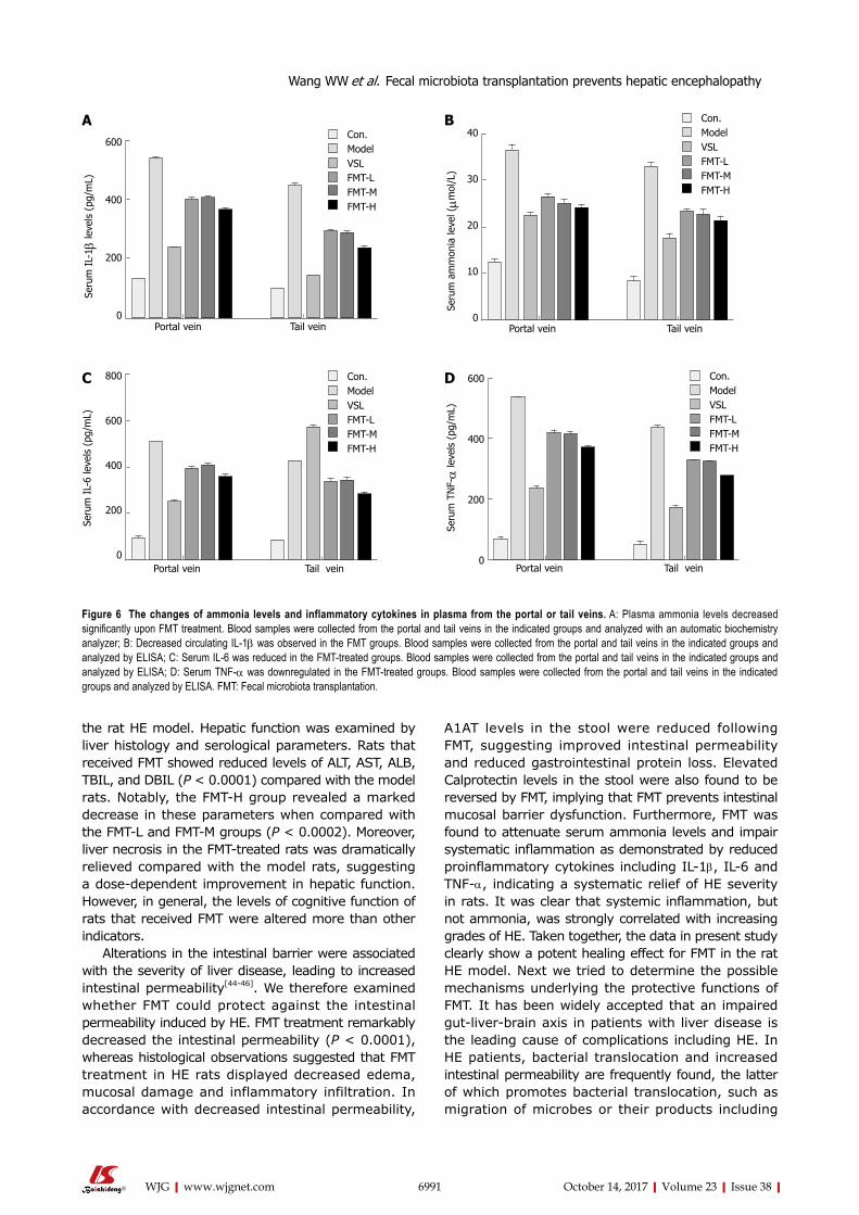
6991 October 14, 2017|Volume 23|Issue 38|WJG|www.wjgnet.com
the rat HE model. Hepatic function was examined by liver histology and serological parameters. Rats that received FMT showed reduced levels of ALT, AST, ALB, TBIL, and DBIL (P < 0.0001) compared with the model rats. Notably, the FMT-H group revealed a marked decrease in these parameters when compared with the FMT-L and FMT-M groups (P < 0.0002). Moreover, liver necrosis in the FMT-treated rats was dramatically relieved compared with the model rats, suggesting a dose-dependent improvement in hepatic function. However, in general, the levels of cognitive function of rats that received FMT were altered more than other indicators.
Alterations in the intestinal barrier were associated with the severity of liver disease, leading to increased intestinal permeability[44-46]. We therefore examined whether FMT could protect against the intestinal permeability induced by HE. FMT treatment remarkably decreased the intestinal permeability (P < 0.0001), whereas histological observations suggested that FMT treatment in HE rats displayed decreased edema, mucosal damage and inflammatory infiltration. In accordance with decreased intestinal permeability,
A1AT levels in the stool were reduced following FMT, suggesting improved intestinal permeability and reduced gastrointestinal protein loss. Elevated Calprotectin levels in the stool were also found to be reversed by FMT, implying that FMT prevents intestinal mucosal barrier dysfunction. Furthermore, FMT was found to attenuate serum ammonia levels and impair systematic inflammation as demonstrated by reduced proinflammatory cytokines including IL-1β, IL-6 and TNF-α, indicating a systematic relief of HE severity in rats. It was clear that systemic inflammation, but not ammonia, was strongly correlated with increasing grades of HE. Taken together, the data in present study clearly show a potent healing effect for FMT in the rat HE model. Next we tried to determine the possible mechanisms underlying the protective functions of FMT. It has been widely accepted that an impaired gut-liver-brain axis in patients with liver disease is the leading cause of complications including HE. In HE patients, bacterial translocation and increased intestinal permeability are frequently found, the latter of which promotes bacterial translocation, such as migration of microbes or their products including
A600
400
200
0
Seru
m I
L-1 β
leve
ls (
pg/m
L)
Portal vein Tail vein
Con.ModelVSLFMT-LFMT-MFMT-H
C 800
600
400
200
0
Seru
m I
L-6
leve
ls (
pg/m
L)
Portal vein Tail vein
Con.ModelVSLFMT-LFMT-MFMT-H
Figure 6 The changes of ammonia levels and inflammatory cytokines in plasma from the portal or tail veins. A: Plasma ammonia levels decreased significantly upon FMT treatment. Blood samples were collected from the portal and tail veins in the indicated groups and analyzed with an automatic biochemistry analyzer; B: Decreased circulating IL-1β was observed in the FMT groups. Blood samples were collected from the portal and tail veins in the indicated groups and analyzed by ELISA; C: Serum IL-6 was reduced in the FMT-treated groups. Blood samples were collected from the portal and tail veins in the indicated groups and analyzed by ELISA; D: Serum TNF-α was downregulated in the FMT-treated groups. Blood samples were collected from the portal and tail veins in the indicated groups and analyzed by ELISA. FMT: Fecal microbiota transplantation.
Wang WW et al. Fecal microbiota transplantation prevents hepatic encephalopathy
B
Seru
m a
mm
onia
leve
l (μm
ol/L
)
40
30
20
10
0Portal vein Tail vein
Con.ModelVSLFMT-LFMT-MFMT-H
600
400
200
0
Seru
m T
NF-α
leve
ls (
pg/m
L)
Portal vein Tail vein
Con.ModelVSLFMT-LFMT-MFMT-H
D

6992 October 14, 2017|Volume 23|Issue 38|WJG|www.wjgnet.com
pathogen-associated molecular patterns (PAMPs), the natural ligands for TLRs[40]. Liver expression of TLR4 and TLR9 is a critical determinant of HE severity, trig-gers the inflammation cascade and is intimately related to arterial ammonia concentration and circulating proinflammatory mediators such as IL-1β, IL-6 and TNF-α. We therefore evaluated the levels of liver TLR4 and TLR9 and circulating IL-1β, IL-6 and TNF-α in the HE model and FMT-treated rats.
Our data here suggest that VSL treatment efficiently reduced circulating IL-1β and TNF-α but not IL-6, a well-established independent risk factor for HE that was negatively associated with cognitive functions in HE patients[47-49]. Serum IL-6 was more evidently decreased in the FMT group than the VSL group in our study, which was consistent with more improved spatial learning and motor activity, suggesting that FMT is more effective than VSL, at least partially. Lata et al[50] also reported the limited effectiveness of VSL for HE.
Note that our study was the first to provide experimental data for potential use of FMT for HE, yet more accurate investigations are supposed to be done for optimized usage of FMT. Instead, VSL is commercially available with more detailed study.
FMT significantly reduced the liver expression levels of TLR4 and TLR9 and proinflammatory mediators, suggesting that FMT improves HE symptoms by
impairing liver inflammation and reducing systemic inflammation. Importantly, the expression of tight junction proteins Claudin-1, Claudin-6 and Occludin was increased in the intestinal tissues of rats that were given FMT, indicating improved intestinal mucosal barrier function. The limitation of this study was the similar infection rate observed between the probiotics group and the FMT group. Our experiment still cannot fully explain whether FMT is superior to VSL#3 or not, which requires more experimental or clinical evidence.
Collectively, our study provided experimental evidence for the first time that supports an FMT-enabled protective role in treating HE rats and revealed the mechanisms behind FMT function, thus providing a basis for potential clinical application of FMT in HE patients.
COMMENTSBackgroundHepatic encephalopathy (HE) is a disease of liver dysfunction, and there is currently no effective therapy for treatment of this condition. To all knowledge, gut microbiota has emerged as a critical factor in the development of HE. Fecal microbiota transplantation (FMT) has been confirmed to treat some diseases. However, the effect of FMT on HE has not been researched deeply.
Research frontiersPrevious clinical evidence has shown that many diseases of the digestive tract, including liver diseases, have intestinal flora abnormalities, while FMT
occludin
claudin-1
claudin-6
GAPDH
59 Kd
23 Kd
23 Kd
36 Kd
Figure 7 The expression of tight junction proteins in intestinal wall and Toll-like receptors. A: FMT induced the downregulation of liver TLR4 and TLR9 expression in a dose-dependent manner; B: FMT improved intestinal permeability by restoring the expression of Claudin-1, Claudin-6 and Occludin. FMT: Fecal microbiota transplantation.
TLR 4
TLR 9
gapdh
90 Kd
113 Kd
36 Kd
A B C D E F
A
COMMENTS
Wang WW et al. Fecal microbiota transplantation prevents hepatic encephalopathy

6993 October 14, 2017|Volume 23|Issue 38|WJG|www.wjgnet.com
can improve the composition and quantity of intestinal bacteria to achieve therapeutic purposes.
Innovations and breakthroughsThis is the first experimental study to confirm that FMT can be used to treat hepatic encephalopathy, and it also reflects the superiority of FMT relative to VSL#3.
ApplicationsAccording to the results of the article, FMT has the potential to become a new method for treating hepatic encephalopathy. At the same time, our results suggest that FMT has special advantages in some areas, providing more possibilities for further searching for new targets for treatment of hepatic encephalopathy.
TerminologyFMT is the introduction of a fecal suspension derived from a healthy donor into the gastrointestinal tract of a diseased individual. Initially as a treatment for Clostridium difficile infection, it has been shown to be effective in many diseases of the human body.
Peer-reviewThis is a very interesting paper about FMT for HE. The authors demonstrated that FMT has good effects in improving intestinal barrier function, by regulating the expression of tight junction proteins (Claudin-1, Claudin-6 and Occludin). Therefore, FMT has a protective role in treating HE.
REFERENCES1 Wijdicks EFM. Hepatic Encephalopathy. N Engl J Med 2017;
376: 186 [PMID: 28076712 DOI: 10.1056/NEJMc1614962]2 Ahluwalia V, Betrapally NS, Hylemon PB, White MB, Gillevet
PM, Unser AB, Fagan A, Daita K, Heuman DM, Zhou H, Sikaroodi M, Bajaj JS. Impaired Gut-Liver-Brain Axis in Patients with Cirrhosis. Sci Rep 2016; 6: 26800 [PMID: 27225869 DOI: 10.1038/srep26800]
3 Lauridsen MM, Bajaj JS. Hepatic encephalopathy treatment and its effect on driving abilities: A continental divide. J Hepatol 2015; 63: 287-288 [PMID: 25796480 DOI: 10.1016/j.jhep.2015.03.017]
4 Bustamante J, Rimola A, Ventura PJ, Navasa M, Cirera I, Reggiardo V, Rodés J. Prognostic significance of hepatic encephalopathy in patients with cirrhosis. J Hepatol 1999; 30: 890-895 [PMID: 10365817]
5 Dhiman RK, Kurmi R, Thumburu KK, Venkataramarao SH, Agarwal R, Duseja A, Chawla Y. Diagnosis and prognostic significance of minimal hepatic encephalopathy in patients with cirrhosis of liver. Dig Dis Sci 2010; 55: 2381-2390 [PMID: 20508990 DOI: 10.1007/s10620-010-1249-7]
6 Reuben A, Tillman H, Fontana RJ, Davern T, McGuire B, Stravitz RT, Durkalski V, Larson AM, Liou I, Fix O, Schilsky M, McCashland T, Hay JE, Murray N, Shaikh OS, Ganger D, Zaman A, Han SB, Chung RT, Smith A, Brown R, Crippin J, Harrison ME, Koch D, Munoz S, Reddy KR, Rossaro L, Satyanarayana R, Hassanein T, Hanje AJ, Olson J, Subramanian R, Karvellas C, Hameed B, Sherker AH, Robuck P, Lee WM. Outcomes in Adults With Acute Liver Failure Between 1998 and 2013: An Observational Cohort Study. Ann Intern Med 2016; 164: 724-732 [PMID: 27043883 DOI: 10.7326/M15-2211]
7 Gubbins GP, Moritz TE, Marsano LS, Talwalkar R, McClain CJ, Mendenhall CL. Helicobacter pylori is a risk factor for hepatic encephalopathy in acute alcoholic hepatitis: the ammonia hypothesis revisited. The Veterans Administration Cooperative Study Group No. 275. Am J Gastroenterol 1993; 88: 1906-1910 [PMID: 8237940]
8 Kramer L, Tribl B, Gendo A, Zauner C, Schneider B, Ferenci P, Madl C. Partial pressure of ammonia versus ammonia in hepatic encephalopathy. Hepatology 2000; 31: 30-34 [PMID: 10613724 DOI: 10.1002/hep.510310107]
9 Shawcross DL, Shabbir SS, Taylor NJ, Hughes RD. Ammonia and the neutrophil in the pathogenesis of hepatic encephalopathy in cirrhosis. Hepatology 2010; 51: 1062-1069 [PMID: 19890967 DOI: 10.1002/hep.23367]
10 Wright G, Jalan R. Ammonia and inflammation in the pathogenesis of hepatic encephalopathy: Pandora’s box? Hepatology 2007; 46: 291-294 [PMID: 17661413 DOI: 10.1002/hep.21843]
11 Ge PS, Runyon BA. Serum ammonia level for the evaluation of hepatic encephalopathy. JAMA 2014; 312: 643-644 [PMID: 25117134 DOI: 10.1001/jama.2014.2398]
12 Rai R, Saraswat VA, Dhiman RK. Gut microbiota: its role in hepatic encephalopathy. J Clin Exp Hepatol 2015; 5: S29-S36 [PMID: 26041954 DOI: 10.1016/j.jceh.2014.12.003]
13 Bajaj JS, Betrapally NS, Hylemon PB, Heuman DM, Daita K, White MB, Unser A, Thacker LR, Sanyal AJ, Kang DJ, Sikaroodi M, Gillevet PM. Salivary microbiota reflects changes in gut microbiota in cirrhosis with hepatic encephalopathy. Hepatology 2015; 62: 1260-1271 [PMID: 25820757 DOI: 10.1002/hep.27819]
14 Victor DW 3rd, Quigley EM. Hepatic encephalopathy involves interactions among the microbiota, gut, brain. Clin Gastroenterol Hepatol 2014; 12: 1009-1011 [PMID: 24462627 DOI: 10.1016/j.cgh.2014.01.022]
15 Dhiman RK. Gut microbiota and hepatic encephalopathy. Metab Brain Dis 2013; 28: 321-326 [PMID: 23463489 DOI: 10.1007/s11011-013-9388-0]
16 Garcovich M, Zocco MA, Roccarina D, Ponziani FR, Gasbarrini A. Prevention and treatment of hepatic encephalopathy: focusing on gut microbiota. World J Gastroenterol 2012; 18: 6693-6700 [PMID: 23239905 DOI: 10.3748/wjg.v18.i46.6693]
17 Dhiman RK. Gut microbiota, inflammation and hepatic encephalopathy: a puzzle with a solution in sight. J Clin Exp Hepatol 2012; 2: 207-210 [PMID: 25755435 DOI: 10.1016/j.jceh.2012.08.004]
18 Maharshi S, Sharma BC, Srivastava S, Jindal A. Randomised controlled trial of lactulose versus rifaximin for prophylaxis of hepatic encephalopathy in patients with acute variceal bleed. Gut 2015; 64: 1341-1342 [PMID: 25320105 DOI: 10.1136/gutjnl-2014-308521]
19 Kalaitzakis E, Björnsson E. Lactulose treatment for hepatic encephalopathy, gastrointestinal symptoms, and health-related quality of life. Hepatology 2007; 46: 949-950; author reply 951 [PMID: 17879365 DOI: 10.1002/hep.21760]
20 Als-Nielsen B, Gluud LL, Gluud C. Non-absorbable disaccharides for hepatic encephalopathy: systematic review of randomised trials. BMJ 2004; 328: 1046 [PMID: 15054035 DOI: 10.1136/bmj.38048.506134.EE]
21 Cesaro C, Tiso A, Del Prete A, Cariello R, Tuccillo C, Cotticelli G, Del Vecchio Blanco C, Loguercio C. Gut microbiota and probiotics in chronic liver diseases. Dig Liver Dis 2011; 43: 431-438 [PMID: 21163715 DOI: 10.1016/j.dld.2010.10.015]
22 Lunia MK, Sharma BC, Sharma P, Sachdeva S, Srivastava S. Probiotics prevent hepatic encephalopathy in patients with cirrhosis: a randomized controlled trial. Clin Gastroenterol Hepatol 2014; 12: 1003-1008.e1 [PMID: 24246768 DOI: 10.1016/j.cgh.2013.11.006]
23 McGee RG, Bakens A, Wiley K, Riordan SM, Webster AC. Probiotics for patients with hepatic encephalopathy. Cochrane Database Syst Rev 2011; 11: CD008716 [PMID: 22071855 DOI: 10.1002/14651858.CD008716.pub2]
24 Xu J, Ma R, Chen LF, Zhao LJ, Chen K, Zhang RB. Effects of probiotic therapy on hepatic encephalopathy in patients with liver cirrhosis: an updated meta-analysis of six randomized controlled trials. Hepatobiliary Pancreat Dis Int 2014; 13: 354-360 [PMID: 25100119]
25 Zhao LN, Yu T, Lan SY, Hou JT, Zhang ZZ, Wang SS, Liu FB. Probiotics can improve the clinical outcomes of hepatic encephalopathy: An update meta-analysis. Clin Res Hepatol Gastroenterol 2015; 39: 674-682 [PMID: 25956487 DOI: 10.1016/j.clinre.2015.03.008]
26 Lee CH, Steiner T, Petrof EO, Smieja M, Roscoe D, Nematallah
Wang WW et al. Fecal microbiota transplantation prevents hepatic encephalopathy
6994 October 14, 2017|Volume 23|Issue 38|WJG|www.wjgnet.com
A, Weese JS, Collins S, Moayyedi P, Crowther M, Ropeleski MJ, Jayaratne P, Higgins D, Li Y, Rau NV, Kim PT. Frozen vs Fresh Fecal Microbiota Transplantation and Clinical Resolution of Diarrhea in Patients With Recurrent Clostridium difficile Infection: A Randomized Clinical Trial. JAMA 2016; 315: 142-149 [PMID: 26757463 DOI: 10.1001/jama.2015.18098]
27 Ho N, Prasad V. Clostridium difficile diarrhea and fecal transplantation. J Clin Gastroenterol 2011; 45: 742-743 [PMID: 21555950 DOI: 10.1097/MCG.0b013e31821b1094]
28 Rohlke F, Stollman N. Fecal microbiota transplantation in relapsing Clostridium difficile infection. Therap Adv Gastroenterol 2012; 5: 403-420 [PMID: 23152734 DOI: 10.1177/1756283X12453637]
29 Youngster I, Russell GH, Pindar C, Ziv-Baran T, Sauk J, Hohmann EL. Oral, capsulized, frozen fecal microbiota transplantation for relapsing Clostridium difficile infection. JAMA 2014; 312: 1772-1778 [PMID: 25322359 DOI: 10.1001/jama.2014.13875]
30 Abdollahi-Roodsaz S, Abramson SB, Scher JU. The metabolic role of the gut microbiota in health and rheumatic disease: mechanisms and interventions. Nat Rev Rheumatol 2016; 12: 446-455 [PMID: 27256713 DOI: 10.1038/nrrheum.2016.68]
31 Groen AK, Nieuwdorp M. An evaluation of the therapeutic potential of fecal microbiota transplantation to treat infectious and metabolic diseases. EMBO Mol Med 2017; 9: 1-3 [PMID: 27861129 DOI: 10.15252/emmm.201607035]
32 Smits LP, Bouter KE, de Vos WM, Borody TJ, Nieuwdorp M. Therapeutic potential of fecal microbiota transplantation. Gastroenterology 2013; 145: 946-953 [PMID: 24018052 DOI: 10.1053/j.gastro.2013.08.058]
33 Hamilton MJ, Weingarden AR, Sadowsky MJ, Khoruts A. Standardized frozen preparation for transplantation of fecal microbiota for recurrent Clostridium difficile infection. Am J Gastroenterol 2012; 107: 761-767 [PMID: 22290405 DOI: 10.1038/ajg.2011.482]
34 Satokari R, Mattila E, Kainulainen V, Arkkila PE. Simple faecal preparation and efficacy of frozen inoculum in faecal microbiota transplantation for recurrent Clostridium difficile infection--an observational cohort study. Aliment Pharmacol Ther 2015; 41: 46-53 [PMID: 25355279 DOI: 10.1111/apt.13009]
35 Farjam M, Dehdab P, Abbassnia F, Mehrabani D, Tanideh N, Pakbaz S, Imanieh MH. Thioacetamide-induced acute hepatic encephalopathy in rat: behavioral, biochemical and histological changes. Iran Red Crescent Med J 2012; 14: 164-170 [PMID: 22737573]
36 Zimmermann C, Ferenci P, Pifl C, Yurdaydin C, Ebner J, Lassmann H, Roth E, Hörtnagl H. Hepatic encephalopathy in thioacetamide-induced acute liver failure in rats: characterization of an improved model and s tudy of amino acid-ergic neurotransmission. Hepatology 1989; 9: 594-601 [PMID: 2564368]
37 Tranah TH, Vijay GK, Ryan JM, Shawcross DL. Systemic inflammation and ammonia in hepatic encephalopathy. Metab Brain Dis 2013; 28: 1-5 [PMID: 23224356 DOI: 10.1007/s11011-012-9370-2]
38 , Chouhan M, Hughes RD. The role of infection and inflammation in the pathogenesis of hepatic encephalopathy and cerebral edema in acute liver failure. Nat Clin Pract Gastroenterol
Hepatol 2006; 3: 118-119 [PMID: 16511529 DOI: 10.1038/ncpgasthep0417]
39 Blei AT. Infection, inflammation and hepatic encephalopathy, synergism redefined. J Hepatol 2004; 40: 327-330 [PMID: 14739106]
40 Aguirre Valadez JM, Rivera-Espinosa L, Méndez-Guerrero O, Chávez-Pacheco JL, García Juárez I, Torre A. Intestinal permeability in a patient with liver cirrhosis. Ther Clin Risk Manag 2016; 12: 1729-1748 [PMID: 27920543 DOI: 10.2147/TCRM.S115902]
41 Benjamin J, Singla V, Arora I, Sood S, Joshi YK. Intestinal permeability and complications in liver cirrhosis: A prospective cohort study. Hepatol Res 2013; 43: 200-207 [PMID: 22726344 DOI: 10.1111/j.1872-034X.2012.01054.x]
42 Luo M, Guo JY, Cao WK. Inflammation: A novel target of current therapies for hepatic encephalopathy in liver cirrhosis. World J Gastroenterol 2015; 21: 11815-11824 [PMID: 26557005 DOI: 10.3748/wjg.v21.i41.11815]
43 Pathak R, Enuh HA, Patel A, Wickremesinghe P. Treatment of relapsing Clostridium difficile infection using fecal microbiota transplantation. Clin Exp Gastroenterol 2013; 7: 1-6 [PMID: 24421645 DOI: 10.2147/CEG.S53410]
44 Cariello R, Federico A, Sapone A, Tuccillo C, Scialdone VR, Tiso A, Miranda A, Portincasa P, Carbonara V, Palasciano G, Martorelli L, Esposito P, Cartenì M, Del Vecchio Blanco C, Loguercio C. Intestinal permeability in patients with chronic liver diseases: Its relationship with the aetiology and the entity of liver damage. Dig Liver Dis 2010; 42: 200-204 [PMID: 19502117 DOI: 10.1016/j.dld.2009.05.001]
45 Lambert JC, Zhou Z, Wang L, Song Z, McClain CJ, Kang YJ. Prevention of alterations in intestinal permeability is involved in zinc inhibition of acute ethanol-induced liver damage in mice. J Pharmacol Exp Ther 2003; 305: 880-886 [PMID: 12626662 DOI: 10.1124/jpet.102.047852]
46 Frazier TH, DiBaise JK, McClain CJ. Gut microbiota, intestinal permeability, obesity-induced inflammation, and liver injury. JPEN J Parenter Enteral Nutr 2011; 35: 14S-20S [PMID: 21807932 DOI: 10.1177/0148607111413772]
47 Luo M, Li L, Yang EN, Dai CY, Liang SR, Cao WK. Correlation between interleukin-6 and ammonia in patients with overt hepatic encephalopathy due to cirrhosis. Clin Res Hepatol Gastroenterol 2013; 37: 384-390 [PMID: 23084463 DOI: 10.1016/j.clinre.2012.08.007]
48 Li W , Li N, Wang R, Li Q, Wu H. Interferon gamma, interleukin-6, and -17a levels were correlated with minimal hepatic encephalopathy in HBV patients. Hepatol Int 2015; 9: 218-223 [PMID: 25794550 DOI: 10.1007/s12072-015-9610-8]
49 Tsai CF, Chu CJ, Huang YH, Wang YP, Liu PY, Lin HC, Lee FY, Lu CL. Detecting minimal hepatic encephalopathy in an endemic country for hepatitis B: the role of psychometrics and serum IL-6. PLoS One 2015; 10: e0128437 [PMID: 26039496 DOI: 10.1371/journal.pone.0128437]
50 Lata J, Jurankova J, Kopacova M, Vitek P. Probiotics in hepatology. World J Gastroenterol 2011; 17: 2890-2896 [PMID: 21734800 DOI: 10.3748/wjg.v17.i24.2890]
P- Reviewer: Hashimoto N, Kharbanda KK S- Editor: Ma YJ L- Editor: A E- Editor: Ma YJ
Wang WW et al. Fecal microbiota transplantation prevents hepatic encephalopathy

Ruo-Peng Liang, Tao Bai, Yu-Ling Sun, Department of Hepatobiliary and Pancreatic Surgery, The First Affiliated Hospital of Zhengzhou University, Zhengzhou 450052, Henan Province, China
Ruo-Peng Liang, Tao Bai, Yu-Ling Sun, Institute of Hepatobiliary and Pancreatic Diseases, Zhengzhou University, Zhengzhou 450052, Henan Province, China
Jun-Jun Jia, Jian-Hui Li, Ning He, Yan-Fei Zhou, Li Jiang, Hai-Yang Xie, Lin Zhou, Key Lab of Combined Multi-Organ Transplantation, Ministry of Public Health, Hangzhou 310003, Zhejiang Province, China
Jun-Jun Jia, Jian-Hui Li, Ning He, Yan-Fei Zhou, Li Jiang, Hai-Yang Xie, Lin Zhou, Division of Hepatobiliary and Pancreatic Surgery, Department of Surgery, First Affiliated Hospital, Zhejiang University School of Medicine, Hangzhou 310003, Zhejiang Province, China
Jun-Jun Jia, Jian-Hui Li, Ning He, Yan-Fei Zhou, Li Jiang, Hai-Yang Xie, Lin Zhou, Collaborative Innovation Center for Diagnosis and Treatment of Infectious Diseases, Hangzhou 310003, Zhejiang Province, China
ORCID number: Ruo-Peng Liang (0000-0002-3789-1666); J u n - J u n J i a ( 0 0 0 0 - 0 0 0 2 - 1 4 9 5 - 2 3 9 0 ) ; J i a n - H u i L i (0000-0002-9662-0583); Ning He (0000-0002-2768-0021); Ya n - F e i Z h o u ( 0 0 0 0 - 0 0 0 3 - 0 4 7 1 - 3 1 9 0 ) ; L i J i a n g (0000-0002-6461-3354); Tao Bai (0000-0002-3817-5602); H a i - Ya n g X i e ( 0 0 0 0 - 0 0 0 2 - 6 9 1 3 - 9 2 8 4 ) ; L i n Z h o u (0000-0001-5689-3512); Yu-Ling Sun (0000-0001-5289-4673).
Author contributions: Sun YL conceived the experiments; Liang RP, Jia JJ, He N, Bai T, Jiang L, Zhou YF conducted the experiments; Xie HY and Zhou L contributed reagents/materials/analysis tools; Liang RP, Bai T analyzed the results; Liang RP wrote the paper. All authors have reviewed the manuscript to be published.
Supported by Science and Technology Innovation Talents Support Plan, Department of Education, Henan Province, China, No. 17HASTIT044; China Postdoctoral Science Foundation, No. 2017M610374.
Institutional review board statement: This study was reviewed and approved by the Institutional Review Board of the First Affiliated Hospital of Zhengzhou University, Zhengzhou, China.
Institutional animal care and use committee statement: All procedures involving animals were reviewed and approved by the Institutional Animal Care and Use Committee of Zhengzhou University, Zhengzhou, China (Approval No. 8, 2017).
Conflict-of-interest statement: The authors declare that there is no conflict of interest related to this study.
Data sharing statement: Technical appendix, statistical code, and dataset are available from the corresponding author at [email protected].
Open-Access: This article is an open-access article which was selected by an in-house editor and fully peer-reviewed by external reviewers. It is distributed in accordance with the Creative Commons Attribution Non Commercial (CC BY-NC 4.0) license, which permits others to distribute, remix, adapt, build upon this work non-commercially, and license their derivative works on different terms, provided the original work is properly cited and the use is non-commercial. See: http://creativecommons.org/licenses/by-nc/4.0/
Manuscript source: Unsolicited manuscript
Correspondence to: Yu-Ling Sun, MD, PhD, Department of Hepatobiliary and Pancreatic Surgery, the First Affiliated Hospital, Zhengzhou University, 1st Jianshe East Road, Zhengzhou 450052, Henan Province, China. [email protected] Telephone: +86-371-67967126
6995 October 14, 2017|Volume 23|Issue 38|WJG|www.wjgnet.com
ORIGINAL ARTICLE
Mitofusin-2 mediated mitochondrial Ca2+ uptake 1/2 induced liver injury in rat remote ischemic perconditioning liver transplantation and alpha mouse liver-12 hypoxia cell line models
Basic Study
Ruo-Peng Liang, Jun-Jun Jia, Jian-Hui Li, Ning He, Yan-Fei Zhou, Li Jiang, Tao Bai, Hai-Yang Xie, Lin Zhou, Yu-Ling Sun
Submit a Manuscript: http://www.f6publishing.com
DOI: 10.3748/wjg.v23.i38.6995
World J Gastroenterol 2017 October 14; 23(38): 6995-7008
ISSN 1007-9327 (print) ISSN 2219-2840 (online)
Fax: +86-371-69767127
Received: July 28, 2017Peer-review started: August 2, 2017First decision: August 10, 2017Revised: August 19, 2017Accepted: September 6, 2017Article in press: September 5, 2017Published online: October 14, 2017
AbstractAIMTo investigate the protective mechanism of mitofusin-2 (Mfn2) in rat remote ischemic perconditioning (RIC) models and revalidate it in alpha mouse liver-12 (AML-12) hypoxia cell lines.
METHODSSprague-Dawley rats were divided into three groups (n = 6 each): sham, orthotopic liver transplantation and RIC. After operation, blood samples were collected to test alanine aminotransferase and aspartate aminotransferase. The liver lobes were harvested for histopathological examination, western blotting (WB) and quantitative real-time (qRT)-PCR. AML-12 cell lines were then subjected to normal culture, anoxic incubator tank culture (hypoxia) and anoxic incubator tank culture with Mfn2 knockdown (hypoxia + Si), and data of qRT-PCR, WB, mitochondrial membrane potential (ΔΨm), apoptosis, endoplasmic reticulum Ca2+ concentrations and mitochondrial Ca2+ concentrations were collected.
RESULTSBoth sham and normal culture groups showed no injury during the experiment. The RIC group showed amelioration of liver function compared with the orthotopic liver transplantation group (P < 0.05). qRT-PCR and WB confirmed that Mfn2-mitochondrial Ca2+ uptake 1/2 (MICUs) axis was changed (P < 0.005). In AML-12 cell lines, compared with the hypoxia group, the hypoxia + Si group attenuated the collapse of ΔΨm and apoptosis (P < 0.005). The endoplasmic reticulum Ca2+ decrease and mitochondrial Ca2+ overloading observed in the hypoxia group were also attenuated in the hypoxia + Si group (P < 0.005). Finally, qRT-PCR and WB confirmed the Mfn2-MICUs axis change in all the groups (P < 0.005).
CONCLUSIONMfn2 participates in liver injury in rat RIC models and AML-12 hypoxia cell lines by regulating the MICUs pathway.
Key words: Remote ischemic per-conditioning; Ischemia-reperfusion injury; Ca2+; Mitofusin-2; Mitochondrial Ca2+ uniporter
© The Author(s) 2017. Published by Baishideng Publishing
Group Inc. All rights reserved.
Core tip: Compared to the orthotopic liver tran-splantation, the remote ischemic perconditioning (RIC) model can significantly improve liver functions. But, knowledge of its mechanism remains largely unknown. This research is the first to prove the protective mechanism of the mitofusin-2-mitochondrial Ca2+ uptake 1/2 axis by affecting the metabolism of intracellular calcium in the RIC model of liver transplantation and to revalidate it in alpha mouse liver-12 hypoxia cell lines.
Liang RP, Jia JJ, Li JH, He N, Zhou YF, Jiang L, Bai T, Xie HY, Zhou L, Sun YL. Mitofusin-2 mediated mitochondrial Ca2+ uptake 1/2 induced liver injury in rat remote ischemic perconditioning liver transplantation and alpha mouse liver-12 hypoxia cell line models. World J Gastroenterol 2017; 23(38): 6995-7008 Available from: URL: http://www.wjgnet.com/1007-9327/full/v23/i38/6995.htm DOI: http://dx.doi.org/10.3748/wjg.v23.i38.6995
INTRODUCTIONLiver transplantation (LT) has become an important countermeasure for end-stage liver disease[1]. Ischemia-reperfusion injury (IRI) is one of the severe complications of LT, leading to graft dysfunction and thus increasing the morbidity and mortality[2-4]. Our previous studies confirmed that ischemia preconditioning (IPC) can effectively alleviate IRI by reducing hepatic enzymatic leakage, leukocyte infiltration and apoptosis formation[5,6]. However, because of its traumatic nature and ethical reasons, its application has failed to develop extensively[7]. Remote ischemic perconditioning (RIC) was first proposed by Przyklenk in 1993, and compared with IPC, it is a temporary ischemic treatment for distant organs and plays a protective role without damaging the target organs[8]. We have optimized the application of RIC and proved that the PI3K/Akt/eNOS/NO pathway could reduce the damage[9,10].
Mitofusin-2 (Mfn2), localized in the outer membrane of the mitochondria, maintains the network stability of mitochondria by participating in mitochondrial fusion and fission[11]. Ca2+ is an important intra-cellular signaling pathway, involved in important pathophysiological functions such as maintenance of biological potential, cell growth/proliferation regulation and apoptosis regulation[12-15]. Vasington et al[16] discovered mitochondrial Ca2+ uptake 1/2 (MICUs) in 1962. They used two Ca2+-binding EF hands to sense Ca2+ concentration, thereby maintaining mitochondrial Ca2+ stability, which were considered as “gatekeepers”for mitochondrial Ca2+ influx[17-20]. Furthermore, they are mediated by the mitochondrial Ca2+ uniporter (MCU) and form an MCU protein complex together with MCU, MICU-1 and MICU-2[21,22]. In our previous
6996 October 14, 2017|Volume 23|Issue 38|WJG|www.wjgnet.com
Liang RP et al. Mitofusin-2 mediated MICUs-induced liver injury
work, we also observed that Mfn2 induced apoptosis in HCC cells due to down-regulation of the MICUs gatekeepers[11].
There is little information about relationships between the protective effect of RIC and the Mfn2-MICUs axis during LT. Ca2+ plays an important role in apoptosis. In our previous RIC experiment, compared with the orthotopic liver transplantation (OLT) group, the RIC group showed decreased apoptosis. In the present study, we hypothesized and verified whether RIC model via Mfn2-MICUs axis has a protective effect for LT. As it is hard to perform gene knockout operations using primary cells, genetically engineered rats are expensive and the experiment is time-consuming, we designed the use of alpha mouse liver-12 (AML-12) hypoxia cell lines to simulate the RIC model and to revalidate it to prove our hypothesis.
MATERIALS AND METHODSRat sham, OLT and RIC modelsAll experimental protocols were conducted in accordance with the Animal Research: Reporting in vivo Experiments (ARRIVE) guidelines (http://www.nc3rs.org/ARRIVE). Adult male Sprague-Dawley rats weighing 250-300 g were chosen, placed in standard environment, and allowed ad libitum access to food and water. Thirty rats (including 12 donors) were randomly allocated to three groups (n = 6 in each group): sham, OLT and RIC. The following operations were performed: After fasting for 12 h, all rats were anesthetized for 3 h by intraperitoneal injection with 4% chloral hydrate (Shanghai Guchen Biological, China).
In the OLT group, the modified method of Kamada was adopted[23]. Donor liver was obtained, and cold physiological saline containing 25 U/mL heparin was used to perfuse the graft liver through portal vein and stored in 0-4 ℃ cold saline for about 45 min before the graft was transplanted into the recipient. After the suprahepatic inferior vena cava and portal vein were anastomosed by cuff method, the liver was reperfused along with the ligation of the hepatic artery. Then, the infra-hepatic inferior vena cava was connected, and common bile duct was reconstructed by tying the duct over a stent. Physiological saline (1 mL) was injected via the dorsal penile vein of the recipient after operation in order to prevent acid-base imbalance.
In the RIC group, the method used in our previous study was adopted. In the recipients who underwent OLT, hindlimb ischemia (using a standard tourniquet appending 1 kg weight to both legs) and reperfusion was performed for three 5-min cycles starting at the anhepatic phase[10] (Figure 1). After recovery from anesthesia, the rats allowed free access to sterile water and standard diet.
In the sham group, the abdomen was opened after 70 min, and then closed in order to acquire the mean time of the total ischemia in the OLT/RIC group (2-min
warm ischemia, 45-min cold ischemia, and 70-min total ischemia).
Collection of liver lobe and blood samples Sham group rats were euthanized after anesthesia. After 3 h of blood flow into the recipient’s portal vein, euthanasia was performed in OLT and RIC groups. Blood samples were drawn from the portal vein and then centrifuged for 15 min at 3000 × g, and serum alanine aminotransferase (ALT) and aspartate aminotransferase (AST) were measured to assess liver function (7600 automatic analyzer; Hitachi, Japan). Some of the liver lobes were kept in formalin solution for histopathological examination, and the other parts of the lobes were used for western blotting (WB) and PCR studies.
Quantitative real-time (qRT)-PCR qRT-PCR was used to detect the level of mRNA expression (Table 1). Total RNA was extracted from the previous phase of the experiment (liver lobes and cell lines) using Trizol reagent (Thermo Fisher Scientific, United States) and PrimeScript RT Reagent Kit with gDNA Eraser (Takara Bio, Japan) for reverse transcription to cDNA. Following the protocol, we performed qRT-PCR in a total volume of 10 μL with 7900Fast (Applied Biosystems Inc, United States) to test the efficiency of small interfering (si)RNA in the hypoxia + Si group. The mRNA levels of Mfn2, MCU, MICU-1 and MICU-2 in the three model groups of sham, OLT and RIC and three cell line groups of normal culture (NC), hypoxia and hypoxia + Si were compared.
WBWB was used to detect the level of protein expression of Mfn2, MCU, MICU-1, MICU-2 and β-actin. Total proteins were extracted from the previous phase of the experiment (liver lobes and cell lines). Cells and tissues (the liver tissues had first been ground in liquid nitrogen) were transfected after incubation in RIPA lysis buffer (Thermo Fisher Scientific) supplemented with protease inhibitor cocktail (Thermo Fisher Scientific) for 1 h on ice and then underwent ultrasonic oscillation crushing (Shanghai Experimental Instruments, China) for three 5-s cycles on ice. After centrifugation (14000 × g, 4 ℃, 15 min), the supernatant was collected for protein concentration measurement by Bicinchoninic Acid Protein Assay Kit (Pierce Biotechnology, United States).
The denatured protein samples (30 μg/10 μL) were separated by SDS-PAGE (Invitrogen, United States) and transferred to polyvinylidene fluoride membranes. Then, the membranes were blocked with 5% non-fat milk (BD Biosciences, United States) dissolved in 30 mL Tris-buffered saline with Tween (TBST) for 2 h at room temperature. Using primary antibodies against β-actin/Mfn2 (1:1000; Abcam, United States),
6997 October 14, 2017|Volume 23|Issue 38|WJG|www.wjgnet.com
Liang RP et al. Mitofusin-2 mediated MICUs-induced liver injury

6998 October 14, 2017|Volume 23|Issue 38|WJG|www.wjgnet.com
siRNA experiments (Table 2; Shanghai GenePharma Biochemical and Pharmaceutical, China) to knock down the Mfn2 expression. The same culturing was performed in the hypoxia group. Cells were collected for subsequent experiments.
Mitochondrial membrane potentialThe mitochondrial membrane potential (ΔΨm) was quantified using a JC-1 kit (Beijing Beyotime Institute of Biotechnology, China). JC-1 was used to cultivate the cells for 30 min, which was measured by flow cytometry (FCM) in the channels of FITC (green) and PE-A (red) of the FC500 flow cytometer (Cytomics, United States). Furthermore, cells were cultured for 30 min using JC-1 and for 15 min using Hoechst-33342 (Beijing Beyotime Institute of Biotechnology), and observed under confocal microscopy (FV1000; Olympus, Japan) to confirm the results obtained from FCM.
ApoptosisCell apoptosis was assayed using the Annexin V/Propidium Iodide Apoptosis (AV/PI) Detection Kit (BD Biosciences) and measured by FCM. Furthermore, cells were treated with 7.5 μmol/L of the CellEvent Caspase
MCU/MICU-1 (1:1000; Cell Signaling Technology, United States) and MICU-2 (1:1000; Santa Cruz Biotechnology, United States), the membranes were incubated overnight at 4 ℃.
After agitation washing of the membranes for three 5-min cycles with TBST, appropriate secondary horseradish peroxidase-linked antibodies (1:2000; all purchased from Abcam), including goat anti-mouse IgG for total Mfn2, goat anti-rabbit IgG for β-actin and MCU/MICU-1/MICU-2, were used to incubate the membranes for 1 h at room temperature. An ECL kit (Pierce Biotechnology) was used to acquire enhanced chemiluminescence.
Cell lines and culturingThe mouse liver hepatocyte cell line AML-12 [established from hepatocytes from a male mouse (CD1 strain, line MT42) non-tumorigenic], preserved by our laboratory, was cultured according to the ATCC guideline, with 10% fetal bovine serum (Gibco, United States) added in a 90% 1:1 mixture of Dulbecco’s modified Eagle’s medium and Ham’s F12 medium (DMEM/F12; Hangzhou Genom Biochemical and Pharmaceutical, China) with 0.005 mg/mL insulin, 0.005 mg/mL transferrin, 5 ng/mL selenium and 40 ng/mL dexamethasone (all purchased from Sigma-Aldrich). All cells were cultured in 5% CO2 at 37 ℃ in a moderately moist environment.
Cells were assigned to three groups (NC, hypoxia and hypoxia + Si) in the logarithmic phase, and less than 70% had good conditions. The NC group cells were cultured using the normal protocol. The hypoxia group cells were first cultured for 3 h in a humidified atmosphere containing 2% O2, 5% CO2 and 93% N2 at 37 ℃ in a noxic incubator tank (Thermo Fisher Scientific), and then cultured for another 3 h in normal atmosphere as NC group cells to simulate the IRI process. The hypoxia + Si cells were first used in
Figure 1 Remote ischemic perconditioning models. A: Tourniquet was used for ligature of each hindlimb with 1 kg additional weight; B: P - Perfusion phase, I - Ischemic phase, R - Reperfusion phase. The remote ischemic perconditioning group underwent orthotopic liver transplantation with hindlimb ischemia and reperfusion for three 5-min cycles starting at the beginning of the anhepatic phase.
1 kg
P I R
B
1 kg
A
5 min × 3Hindlimb
Table 1 Primer sequences for qRT-PCR
Target gene Primer sequence
Rat-β-Actin Forward: 5'-ACGGTCAGGTCATCACTATCG-3'Reverse: 5'-GAGGTCTTTACGGATGTCAACG-3'
Rat-Mfn2 Forward: 5'-GAAGAAGAGTGTCAAGACCGTG-3'Reverse: 5'-CAGGCAAAACTTATCAATCCAG-3'
Rat-Mcu Forward: 5'-GACCCTGAACGATGTGAAGAC-3'Reverse: 5'-TTCTCCGCTTTCCTGCTAAT-3'
Rat-MICU-1 Forward: 5'-GACTAAGCGGAGACTGATGTTG-3'Reverse: 5'-GAGATTCTGCGTGAGCCTTC-3'
Rat-MICU-2 Forward: 5'-ACAAAGCCTCACTCTGGGT-3'Reverse: 5'-TCACTGTTGGTTCCTGGTATT-3'
Mus-β-Actin Forward: 5'-CATTGCTGACAGGATGCAGAAGG-3'Reverse: 5'-TGCTGGAAGGTGGACAGTGAGG-3'
Mus-Mfn2 Forward: 5'-CAAGTGTCCGCTCCTGAAG-3'Reverse: 5'-CCACCAGCACAAACACATC-3'
Mus-Mcu Forward: 5'-GAGCCGCATATTGCAGTACGGT-3'Reverse: 5'-AAACACGCCGACTGAGTCAGAG-3'
Mus-MICU-1 Forward: 5'-GAAGTGTCCAGCCGTGAAGGAA-3'Reverse: 5'-TGGTGTGGAGTAGGCTCGGATT-3'
Mus-MICU-2 Forward: 5'-GGAAGACTTTGCTATCGCCATGC-3'Reverse: 5'-GGTGTCCAGAAGATTGTCCGAG-3'
Mfn2: Mitofusin-2; Mcu: Mitochondrial Ca2+ uniporter; MICU: Mito-chondrial Ca2+ uptake.
Table 2 Primer sequences for siRNA
Target gene Primer sequence
Mus-Mfn2 Sense strand:5′-CUGCGAAUUAAGCAGAUUATTdTdT-3′;
Anti-sense strand:5′-UAAUCUGCUUAAUUCGCAGTTdTdT-3′
Mfn2: Mitofusin-2.
Liang RP et al. Mitofusin-2 mediated MICUs-induced liver injury

6999 October 14, 2017|Volume 23|Issue 38|WJG|www.wjgnet.com
3/7 Green Detection Reagent (Invitrogen, United States) and 10 nmol/L tetramethylrhodamine methyl ester perchlorate (Invitrogen), and observed under confocal microscopy to confirm the results obtained from FCM.
Measurement of mitochondrial Ca2+ concentrationsRhod-2 acetoxymethyl ester (Rhod-2, AM; GeneCopoeia, United States) was used to measure the mitochondrial Ca2+ concentrations. According to our previous experience, the fluorescence value of Rhod-2 AM approximately represented the fluorescence value of mitochondrial Ca2+[11,24]. The day before the experiment, 1 x 10⁵ cells were plated in 10-mm confocal dishes (Nest Biotechnology, China). After overnight incubation, the growth medium was removed from the cell plates, and added with 500 μL
medium with 4 μM Rhod-2 AM and 0.1% Pluronic® F-127 (GeneCopoeia) and allowed to incubate at 37 ℃ in humidified air (containing 5% CO2) for 2 h. Next, D-Hanks balanced salt solution (Hangzhou Genom Biochemical and Pharmaceutical, China) was used to wash the cells three times, followed by addition of 500 μL Hoechst-33342 and 200 nmol/L MitoTracker Green (Beijing Beyotime Institute of Biotechnology), and allowed to incubate in the same environment for 30 min of cultivation. Confocal microscopy confirmed that the vast majority of Rhod-2 AM fluorescence was associated with the mitochondria. Furthermore, cells were incubated in 96-well plates with Rhod-2 AM and F-127 in the same process and observed by VarioskanFlash (Thermo Fisher Scientific) with the illumination at excitation 552 nm and emission 581 nm to confirm the results obtained from confocal
1000
800
600
400
200
0
a
AST
(U/L
)
c
Sham OLT RIC
800
600
400
200
0
b
ALT
(U/L
)
c
Sham OLT RIC
Sham OLT RIC
3
2
1
0
c
Deg
ree
of c
onge
stat
ion
c
Sham OLT RIC
4
3
2
1
0
c
Deg
ree
of n
ecro
sis
c
Sham OLT RIC
Figure 2 Remote ischemic perconditioning models model improves the liver graft function. A: Results of ALT and AST in the three model groups; B: Liver histopathological presentations (hematoxylin and eosin stain, original magnification × 200); C: Results of hepatocyte necrosis and congestion of the sinusoids and central vein in histopathologic examination. Data represent mean ± SEM for 6 rats per group. ALT: Alanine aminotransferase; AST: aspartate aminotransferase; OLT: Orthotopic liver transplantation; RIC: Remote ischemic perconditioning. aP < 0.05; bP < 0.01; cP < 0.001.
Liang RP et al. Mitofusin-2 mediated MICUs-induced liver injury
7000 October 14, 2017|Volume 23|Issue 38|WJG|www.wjgnet.com
microscopy.
Quantification of endoplasmic reticulum (ER) Ca2+ concentrationsWe used the FLUOFORTE® Calcium Assay (Enzo Life Sciences, United States) to detect ER Ca2+ concentrations. The day before the experiment, 5 × 10⁴ cells were plated per well in 96-well assay plates (Corning, United States). After overnight incubation, the growth medium was removed from the cell plates and 100 μL FLUOFORTE® Dye-Loading Solution (prepared by Enzo’s protocol, containing 0.5 mmol/L EGTA to eliminate Ca2+ concentrations in the medium) was added to each well, and the cells were incubated at room temperature for 45 min. The illumination at excitation 490 nm and emission 525 nm was observed by VarioskanFlash for quantification of cytosolic Ca2+ concentrations at baseline. Then, 5 μM thapsigargin (Thermo Fisher Scientific) was added to each measurement well to detect the total values, including cytosolic baseline and Ca2+ released from the ER, by VarioskanFlash with the same parameters. SkanIt software (Thermo Fisher Scientific) was used to measure the ER Ca2+ concentrations.
Statistical analysisThe results were expressed as mean ± SEM. One-way analysis of variance was used for comparisons among three groups and the t-test was used for comparison between the two groups. All statistical analyses and related statistical diagramming were performed using the GraphPad Prism software (ver. 5.0 for Windows; GraphPad Software Inc., United States). P < 0.05 was considered statistically significant.
RESULTSRIC model improves liver graft functionAccording to the literature and our previous research, RIC does not cause muscle and remote organ function damage, so this experiment focused on liver function[9,10]. In the present study, the sham group showed no injury during the experiment. Compared with the OLT group, ALT and AST levels were lower in the RIC group (ALT: P = 0.0029, AST: P = 0.0121; Figure 2A). Liver histopathological examinations showed that the RIC group had fewer sinusoids, central vein congestion and hepatocyte necrosis than the OLT group (P < 0.001; Figure 2B and C).
RIC model lowers expression of Mfn2 and alters MICUs expression Expression of Mfn2 and MICUs was measured by WB (Figure 3A) and qRT-PCR (Figure 3B). Taking the OLT group as the reference, both the sham and RIC groups showed lower expression of Mfn2 (WB: sham vs RIC: P < 0.001; qRT-PCR: sham P < 0.001, RIC P = 0.0348). The MICUs expression was also altered. Compared
with the OLT group, both the sham and RIC groups showed higher expression of MICU-1 (WB: sham vs RIC, P < 0.001; qRT-PCR: sham P = 0.0005, RIC P = 0.0030) and similar expression of MICU-2 (WB: sham P = 0.0011, RIC P = 0.0033; qRT-PCR: sham P = 0.0255, RIC: P = 0.0137). However, we did not observe any significant changes in the expression of MCU (WB: sham P = 0.1044, RIC P = 0.9243; qRT-PCR: sham P = 0.0005, RIC P = 0.1507).
Mfn2 knockdown alters MICUs expression in AML-12 hypoxia cell linesThe NC group was cultured in normal environment. The hypoxia group followed our protocol to simulate the IRI process. In the hypoxia + Si group, siRNA was used to knock down Mfn2 expression (WB and qRT-PCR; Figure 4A) and culturing was performed by the same protocol as in the hypoxia group. Expression of MICUs was measured by WB and qRT-PCR. Taking the hypoxia group as the reference, both the NC and hypoxia + Si groups showed higher expression of MICU-1 (WB: NC P = 0.0001, hypoxia + Si P = 0.0010; qRT-PCR: NC P = 0.0026, hypoxia + Si P = 0.0430) and similar expression of MICU-2 (WB: sham P < 0.0001, hypoxia + Si P = 0.0020; qRT-PCR: sham P = 0.0004, RIC P = 0.0137). We observed the change in expression of MCU by WB (sham: P = 0.0158, RIC: P = 0.0040), but did not obtain significant findings by qRT-PCR (sham: P = 0.2597, RIC: P = 0.2420) (Figure 4B and C).
Mfn2 knockdown attenuates the collapse of ΔΨm in AML-12 hypoxia cell lines We first used the JC-1 kit to measure the ΔΨm in AML-12 hypoxia cell lines cultured by FCM with our protocol. JC-1 fluorescence shifted from red (JC-1 aggregates bound to mitochondria) to green (JC-1 monomers bound to cytoplasm), indicating a decline of ΔΨm. Taking the hypoxia group as the reference, the green to red fluorescence ratio was lower in the NC and hypoxia + Si groups (FCM: NC P = 0.0329, hypoxia + Si P = 0.0391; Figure 5A and C). We obtained the same result by confocal microscopy as that obtained from FCM using the JC-1 kit and Hoechst-33342 (Figure 5B).
Mfn2 knockdown attenuates apoptosis in AML-12 hypoxia cell linesThe Annexin V/Propidium Iodide Apoptosis Detection Kit was used to detect cell apoptosis by FCM. Annexin V fluorescence reflects early cell apoptosis while propidium iodide fluorescence reflects late apoptosis and dying cells. Taking the hypoxia group as the reference, the ratio of apoptosis cells was lower in the NC and hypoxia + Si groups (FCM: NC P = 0.0092, hypoxia + Si P = 0.0369; Figure 6A and C). We obtained the same result by confocal microscopy as that obtained from FCM using the caspase 3/7
Liang RP et al. Mitofusin-2 mediated MICUs-induced liver injury
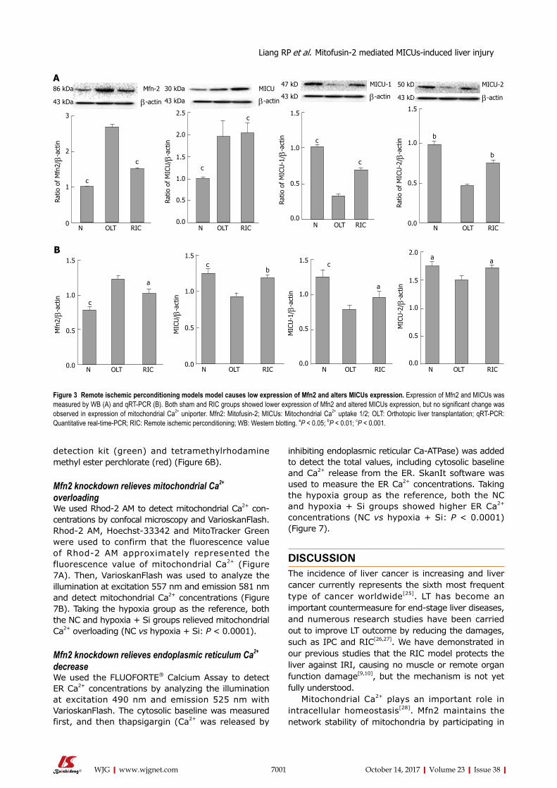
7001 October 14, 2017|Volume 23|Issue 38|WJG|www.wjgnet.com
detection kit (green) and tetramethylrhodamine methyl ester perchlorate (red) (Figure 6B).
Mfn2 knockdown relieves mitochondrial Ca2+ overloading We used Rhod-2 AM to detect mitochondrial Ca2+ con-centrations by confocal microscopy and VarioskanFlash. Rhod-2 AM, Hoechst-33342 and MitoTracker Green were used to confirm that the fluorescence value of Rhod-2 AM approximately represented the fluorescence value of mitochondrial Ca2+ (Figure 7A). Then, VarioskanFlash was used to analyze the illumination at excitation 557 nm and emission 581 nm and detect mitochondrial Ca2+ concentrations (Figure 7B). Taking the hypoxia group as the reference, both the NC and hypoxia + Si groups relieved mitochondrial Ca2+ overloading (NC vs hypoxia + Si: P < 0.0001).
Mfn2 knockdown relieves endoplasmic reticulum Ca2+ decrease We used the FLUOFORTE® Calcium Assay to detect ER Ca2+ concentrations by analyzing the illumination at excitation 490 nm and emission 525 nm with VarioskanFlash. The cytosolic baseline was measured first, and then thapsigargin (Ca2+ was released by
inhibiting endoplasmic reticular Ca-ATPase) was added to detect the total values, including cytosolic baseline and Ca2+ release from the ER. SkanIt software was used to measure the ER Ca2+ concentrations. Taking the hypoxia group as the reference, both the NC and hypoxia + Si groups showed higher ER Ca2+ concentrations (NC vs hypoxia + Si: P < 0.0001) (Figure 7).
DISCUSSIONThe incidence of liver cancer is increasing and liver cancer currently represents the sixth most frequent type of cancer worldwide[25]. LT has become an important countermeasure for end-stage liver diseases, and numerous research studies have been carried out to improve LT outcome by reducing the damages, such as IPC and RIC[26,27]. We have demonstrated in our previous studies that the RIC model protects the liver against IRI, causing no muscle or remote organ function damage[9,10], but the mechanism is not yet fully understood.
Mitochondrial Ca2+ plays an important role in intracellular homeostasis[28]. Mfn2 maintains the network stability of mitochondria by participating in
1.5
1.0
0.5
0.0
a M
ICU
-1/ β
-act
in
c
N OLT RIC N OLT RIC
2.0
1.5
1.0
0.5
0.0
a
MIC
U-2
/ β-a
ctin
a
3
2
1
0
c
Ratio
of
Mfn
2/β-
actin
c
N OLT RIC
2.5
2.0
1.5
1.0
0.5
0.0
c
Ratio
of
MIC
U/ β
-act
inc
N OLT RIC
86 kDa Mfn-2
43 kDa β-actin
A
1.5
1.0
0.5
0.0
c
Ratio
of
MIC
U-1
/ β-a
ctin c
N OLT RIC
47 kD MICU-1
43 kD β-actin
N OLT RIC
1.5
1.0
0.5
0.0
b
Ratio
of
MIC
U-2
/ β-a
ctin
b
50 kD MICU-2
43 kD β-actin
1.5
1.0
0.5
0.0
b
MIC
U/ β
-act
in
c
N OLT RIC
Figure 3 Remote ischemic perconditioning models model causes low expression of Mfn2 and alters MICUs expression. Expression of Mfn2 and MICUs was measured by WB (A) and qRT-PCR (B). Both sham and RIC groups showed lower expression of Mfn2 and altered MICUs expression, but no significant change was observed in expression of mitochondrial Ca2+ uniporter. Mfn2: Mitofusin-2; MICUs: Mitochondrial Ca2+ uptake 1/2; OLT: Orthotopic liver transplantation; qRT-PCR: Quantitative real-time-PCR; RIC: Remote ischemic perconditioning; WB: Western blotting. aP < 0.05; bP < 0.01; cP < 0.001.
1.5
1.0
0.5
0.0
a
Mfn
2/β-
actin c
N OLT RIC
B
30 kDa MICU
43 kDa β-actin
Liang RP et al. Mitofusin-2 mediated MICUs-induced liver injury

7002 October 14, 2017|Volume 23|Issue 38|WJG|www.wjgnet.com
mitochondrial fusion and fission[29]. MCU, MICU-1 and MICU-2 form the mitochondrial calcium uniporter complex. Along with the increase in intracellular Ca2+, Ca2+ combined with EF-hand of MICUs promotes the opening of MCU pores and leads to an increase in the mitochondrial Ca2+[30,31]. Furthermore, excessive intake of Ca2+ can increase mitochondrial permeability transition, thereby inducing pathophysiological phenomena such as apoptosis[32]. Previous studies have shown that Mfn2 provides a convenient pathway for Ca2+ transport
from mitochondria to ER in intracellular high Ca2+ environment[33-37]. However, there is little information about relationships between the protective effect of RIC and the Mfn2-MICUs axis during LT.
We, therefore, designed and performed the present RIC experiment (Figure 1) and found that, compared with the sham group, LT (including RIC) could cause liver damage and change the Mfn2-MICUs axis at the same time. While the RIC model reduced liver damage, Mfn2 expression was lower and MICUs
1.5
1.0
0.5
0.0
c
Ratio
of
Mfn
2/β-
actin
c
NC SI-1 SI-2 SI-2
c
A NC SI-1 SI-2 SI-3
86 kDa Mfn2-siRNA
43 kD β-actin
30 kDa MICU
43 kDa β-actin
47 kDa MICU-1
43 kDa β-actin
50 kDa MICU-2
43 kDa β-actin
1.5
1.0
0.5
0.0
Ratio
of
MIC
U/G
APD
H
N Hypoxia Hypoxia + si
a
b1.5
1.0
0.5
0.0
Ratio
of
MIC
U-1
/GAP
DH
N Hypoxia Hypoxia + si
c
b
Ratio
of
MIC
U-2
/GAP
DH
1.5
1.0
0.5
0.0N Hypoxia Hypoxia + si
c
b
1.5
1.0
0.5
0.0
MIC
U/ β
-act
in
NC Hypoxia Hypoxia+si
aa
MICU 1.5
1.0
0.5
0.0
MIC
U-1
/ β-a
ctin
NC Hypoxia Hypoxia + si
a
a
MICU-1 1.5
1.0
0.5
0.0
MIC
U-2
/ β-a
ctin
NC Hypoxia Hypoxia + si
c
a
MICU-2
Figure 4 Mfn2 knockdown alters MICUs expression in AML-12 hypoxia cell lines. A: Mfn2 knockdown was tested by WB and qRT-PCR. Expression of MICUs in AML-12 hypoxia cell lines was measured by WB (B) and qRT-PCR (C). Taking the hypoxia group as the reference, both the NC and hypoxia + Si groups showed higher expression of MICU-1 and similar expression of MICU-2; MCU altered expression in WB, but no significant change in qRT-PCR. AML: Alpha mouse liver; MCU: Mitochondrial Ca2+ uniporter; Mfn2: Mitofusin-2; MICU: Mitochondrial Ca2+ uptake; NC: Normal culture; qRT-PCR: Quantitative real-time-PCR; WB: Western blotting. aP < 0.05; bP < 0.01.S cP < 0.001.
B
C
Liang RP et al. Mitofusin-2 mediated MICUs-induced liver injury

7003 October 14, 2017|Volume 23|Issue 38|WJG|www.wjgnet.com
expression was higher than in LT (Figures 2 and 3). These results demonstrated that the RIC model might exert its protective effect for LT via the Mfn2-MICUs axis.
To further verify our findings, we decided to knock down the Mfn2 expression by siRNA. Since genetically engineered mice are expensive, the experiment is time-consuming, and it is hard to perform gene knockout operations using primary cells, we designed the use of AML-12 hypoxia cell lines to simulate a RIC model. After determining the Mfn2 knockdown
efficiency (Figure 4A), we proved that the hypoxia + Si group alleviated the hypoxia damage and showed higher expression of MICU-1 and MICU-2 compared with the hypoxia group (Figure 4B and C). Revalidating the same result from the RIC experiment provides the basis for our future mechanism studies.
Depolarization of mitochondrial membrane potential is usually associated with early apoptosis. According to the JC-1 kit instructions, in the absence of apoptotic cells, JC-1 aggregates are bound to mitochondria and red fluorescence occurs at 590 nm.
Hypoxia + Si
Q11.59
Q45.98
Q318.2
Q274.2
Com
p-PE
-A
105
104
103
102
101
100
103 0 103 104 105
Comp-FITC-A
NC Hypoxia
Q10.71
Q257.6
Q334.9
Q46.84
103 0 103 104 105
105
104
103
102
101
100
Com
p-PE
-AComp-FITC-A
Q10.84
Q287.1
Q39.51
Q42.56
Com
p-PE
-A
105
104
103
102
101
100
103 0 103 104 105
Comp-FITC-A
A
B
Figure 5 Mfn2 knockdown attenuates the collapse of ΔΨm in AML-12 hypoxia cell lines. JC-1 kit and Hoechst-33342 were used to measure the ΔΨm by flow cytometry (A and C) and confocal microscopy (B). Collapse of ΔΨm in the hypoxia group was attenuated both in the NC and hypoxia + Si groups. Mfn2: Mitofusin-2; NC: Normal culture; ΔΨm: Mitochondrial membrane potential. aP < 0.05.
0.6
0.4
0.2
0.0
Ratio
of
gree
n to
red
fluo
resc
ence
NC Hypoxia Hypoxia + si
a
a
C
Liang RP et al. Mitofusin-2 mediated MICUs-induced liver injury
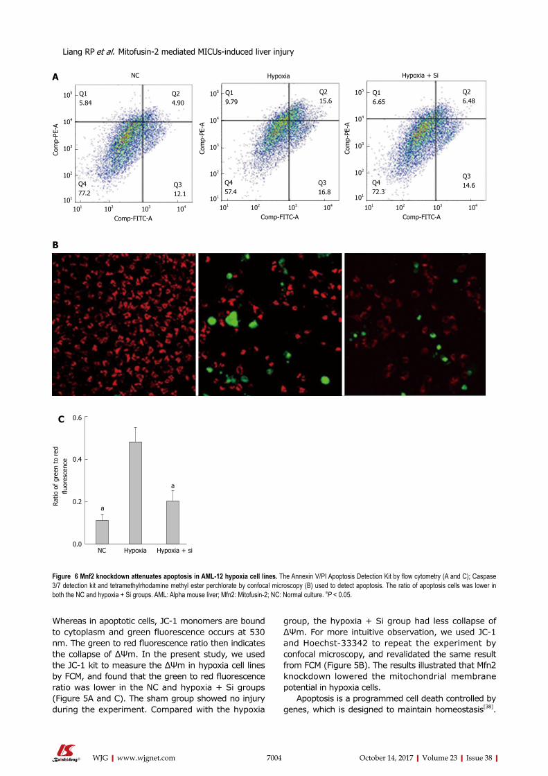
7004 October 14, 2017|Volume 23|Issue 38|WJG|www.wjgnet.com
Whereas in apoptotic cells, JC-1 monomers are bound to cytoplasm and green fluorescence occurs at 530 nm. The green to red fluorescence ratio then indicates the collapse of ΔΨm. In the present study, we used the JC-1 kit to measure the ΔΨm in hypoxia cell lines by FCM, and found that the green to red fluorescence ratio was lower in the NC and hypoxia + Si groups (Figure 5A and C). The sham group showed no injury during the experiment. Compared with the hypoxia
group, the hypoxia + Si group had less collapse of ΔΨm. For more intuitive observation, we used JC-1 and Hoechst-33342 to repeat the experiment by confocal microscopy, and revalidated the same result from FCM (Figure 5B). The results illustrated that Mfn2 knockdown lowered the mitochondrial membrane potential in hypoxia cells.
Apoptosis is a programmed cell death controlled by genes, which is designed to maintain homeostasis[38].
Liang RP et al. Mitofusin-2 mediated MICUs-induced liver injury
0.6
0.4
0.2
0.0
Ratio
of
gree
n to
red
flu
ores
cenc
e
a
NC Hypoxia Hypoxia + si
a
C
A
Q15.84
Q24.90
Q312.1
Q477.2
NC
Com
p-PE
-A
105
104
103
102
101
101 102 103 104 Comp-FITC-A
Q19.79
Q215.6
Q316.8
Q457.4
Hypoxia
Com
p-PE
-A
105
104
103
102
101
101 102 103 104 Comp-FITC-A
Figure 6 Mnf2 knockdown attenuates apoptosis in AML-12 hypoxia cell lines. The Annexin V/PI Apoptosis Detection Kit by flow cytometry (A and C); Caspase 3/7 detection kit and tetramethylrhodamine methyl ester perchlorate by confocal microscopy (B) used to detect apoptosis. The ratio of apoptosis cells was lower in both the NC and hypoxia + Si groups. AML: Alpha mouse liver; Mfn2: Mitofusin-2; NC: Normal culture. aP < 0.05.
B
Hypoxia + Si
105
104
103
102
101
101 102 103 104
Q26.48
Q16.65
Comp-FITC-A
Q472.3
Q314.6
Com
p-PE
-A

7005 October 14, 2017|Volume 23|Issue 38|WJG|www.wjgnet.com
In the early stage of apoptosis, phosphotidylserine can be translocated from the inside of the cell membrane to the outside of the cell membrane and combined with annexin V (FITC). In late apoptotic and dead cells, the membranes are permeable to propidium iodide (PE-A). According to the staining status of the cells, we can distinguish the apoptosis status (annexin V/propidium iodide positive or negative). In the present study, we found that the ratio of cell apoptosis was lower in the
NC and hypoxia + Si groups (Figure 6A and C). The sham group showed no injury during the experiment, indicating that compared with the hypoxia group, the hypoxia + Si group had fewer cells undergoing apoptosis. We used the caspase 3/7 detection reagent (green) and tetramethylrhodamine methyl ester perchlorate (red) to repeat the experiment by confocal microscopy and revalidate the same result from FCM (Figure 6B). The results demonstrated that Mfn2
Hoechst 33342 Mito-tracker green Rhod-2, AM
H + M H + R H + M + R
Figure 7 Mfn2 knockdown relieves mitochondrial Ca2+ overloading. Rhod-2 AM was used to detect mitochondrial Ca2+ concentrations by confocal microscopy and VarioskanFlash. A: Confocal microscopy confirmed that the vast majority of Rhod-2 AM fluorescence was associated with the mitochondria; B: VarioskanFlash was used to detect Rhod-2 AM to determine the fluorescence of mitochondrial Ca2+ concentrations. Taking the hypoxia group as the reference, both the NC and hypoxia + Si groups relieved mitochondrial Ca2+ overloading. Mfn2: Mitofusin-2; NC: Normal culture; Rhod-2 AM: Rhod-2 acetoxymethyl ester. cP < 0.001.
A
2.5
2.0
1.5
1.0
0.5
0.0
Fold
of
NC
c
c
NC Hypoxia Hypoxia + si
Liang RP et al. Mitofusin-2 mediated MICUs-induced liver injury
Time
0 200 400 600 800 1000
150
100
50
Valu
e
NCHypoxia Hypoxia + si
B

7006 October 14, 2017|Volume 23|Issue 38|WJG|www.wjgnet.com
knockdown reduced the apoptosis in hypoxia cells. As mentioned earlier, whether IRI in LT or hypoxia
in cells leads to injury should be verified. Mfn2 provides a convenient pathway for Ca2+ transport from mitochondria to ER in intracellular high Ca2+ environment. Excessive uptake of Ca2+ caused mitochondrial pathogenesis and affected ‘gatekeeper’ expression, thus leading to a higher Ca2+ concentration. In the present study, after confirming that the Mfn2-MICUs were altered in both rat models and cell models, we continued to validate Ca2+ changes at the mitochondrial level. We used Rhod-2 AM to analyze the mitochondrial Ca2+ concentration (Figure 7A), and VarioskanFlash to analyze the illumination of Rhod-2 AM to get the fluorescence of mitochondrial Ca2+ concentrations. We found that the hypoxia + Si group relieved mitochondrial Ca2+ overloading compared with the hypoxia group (Figure 7B). This indicated that Mfn2 knockdown reduced the mitochondrial Ca2+ overload by Mfn2-MICUs axis in cell models.
The ER is an important organelle of Ca2+ storage, the stored Ca2+ in ER is mainly released through the inositol 1, 4, 5-trisphosphate (InsP3) receptor and ryanodine receptor pathways[13]. In the present study, we investigated the relationships between ER Ca2+ concentrations and mitochondrial Ca2+ overloading. After the cytosolic Ca2+ concentration at baseline was quantified by VarioskanFlash using the FLUOFORTE® Calcium Assay, we added thapsigargin to release the ER Ca2+ and performed repeat measurement. The hypoxia + Si group showed relief of the ER Ca2+ decrease compared with the hypoxia group (Figure 8). This result proved that Mfn2 knockdown relieved the ER Ca2+ decrease by affecting the Mfn2-MICUs axis, thus altering the mitochondrial Ca2+ status in cell models.
The MCU protein complex is a highly selective calcium channel consisting of three components: MCU, MICU-1 and MICU-2. The complex can sense Ca2+
level and determine whether it passes through the mitochondrial membrane[19]. In our experiment, we failed to observe the consistency of MCU expression changes in rat and cell models. According to the literature, ER was mediated by the InsP3 receptor, Mfn2 showed no effect on expression of InsP3R, but when specific inhibitors of InsP3 receptor were used, the decline of ER Ca2+ induced by Mfn2 no longer occurred, suggesting that InsP3R Ca2+ release pathways affected the ER Ca2+ efflux[11]. The specific mechanism in InsP3R and IRI/cell hypoxia injury is not clear. Nevertheless, our research provides a clear clue that Mfn2 down-regulated expression of MICUs can affect the state of Ca2+ in organelles, thus playing a protective role in the two models. We will continue to apply gene-editing mouse models and specific inhibitors to conduct an in-depth research in the future.
In conclusion, we used an RIC model and confirmed that IRI was prevented by altered organelles’ Ca2+ status via the Mfn2-MICUs axis, and revalidated the effect in AML-12 hypoxia cell models.
COMMENTSBackgroundRemote ischemic perconditioning (RIC) alleviates the damage of ischemia-reperfusion injury (IRI). The Mfn2-MICUs axis is involved in the regulation of intracellular calcium, thereby affecting the process of apoptosis. But there is little information about the role of the mitofusin-2-mitochondrial Ca2+ uptake 1/2 (Mfn2-MICUs) axis in the protective effect of RIC during liver transplantation (LT).
Research frontiersMost previous studies only confirmed the protective effect of RIC, but few showed the detailed mechanism. In the protective role of RIC, the mechanism of apoptosis related to the Mfn2-MICUs axis is worthy of being explored.
Innovations and breakthroughsThis research is the first to prove the protective mechanism of the Mfn2-MICUs axis as affecting the metabolism of intracellular calcium to reduce apoptosis in RIC of LT and revalidate it in AML-12 hypoxia cell lines.
Figure 8 Mfn2 knockdown relieves endoplasmic reticulum Ca2+ decrease. FLUOFORTE® Calcium Assay and thapsigargin were used to detect ER Ca2+ concentrations by VarioskanFlash. Taking the hypoxia group as the reference, both the NC and hypoxia + Si groups showed higher ER Ca2+ concentrations. ER: Endoplasmic reticulum; Mfn2: Mitofusin-2; NC: Normal culture. cP < 0.001.
1.0
0.5
0.0
Fold
of
NC
c
c
NC Hypoxia Hypoxia + Si
seconds
400 600 800 1000
25
20
15
10
5
0
Valu
e
ControlNCHypoxia Hypoxia + si
Thapsigargin
COMMENTS
Liang RP et al. Mitofusin-2 mediated MICUs-induced liver injury

7007 October 14, 2017|Volume 23|Issue 38|WJG|www.wjgnet.com
ApplicationsThis study showed the relationship between the protective effect of RIC and cell hypoxia injury, and that the Mfn2-MICUs axis and apoptosis are affected by the intracellular calcium homeostasis, thus laying a basic foundation for future studies.
TerminologyRIC was first proposed by Przyklenk in 1993, and it alleviates the damage of IRI in a temporary ischemic treatment for distant organs without damaging the target organs. RIC operations include remote-ischemic-preconditioning (RIPreC), remote-ischemic-postconditioning and RIPerC.
Peer-reviewThis is an interesting paper looking at the role of Mnf2 in liver IRI.
ACKNOWLEDGMENTSWe are grateful to the Collaborative Innovation Center for Diagnosis and Treatment of Infectious Diseases of the First Affiliated Hospital of Zhejiang University for providing the experimental sites and reagents for this study. We also wish to thank Wen-Feng Song for technical assistance.
REFERENCES1 Eshraghian A, Imanieh MH, Dehghani SM, Nikeghbalian S,
Shamsaeefar A, Barshans F, Kazemi K, Geramizadeh B, Malek-Hosseini SA. Post-transplant lymphoproliferative disorder after liver transplantation: Incidence, long-term survival and impact of serum tacrolimus level. World J Gastroenterol 2017; 23: 1224-1232 [PMID: 28275302 DOI: 10.3748/wjg.v23.i7.1224]
2 Eggenhofer E, Sabet-Rashedi M, Lantow M, Renner P, Rovira J, Koehl GE, Schlitt HJ, Geissler EK, Kroemer A. RORγt(+) IL-22-producing NKp46(+) cells protect from hepatic ischemia reperfusion injury in mice. J Hepatol 2016; 64: 128-134 [PMID: 26341825 DOI: 10.1016/j.jhep.2015.08.023]
3 Peralta C, Jiménez-Castro MB, Gracia-Sancho J. Hepatic ischemia and reperfusion injury: effects on the liver sinusoidal milieu. J Hepatol 2013; 59: 1094-1106 [PMID: 23811302 DOI: 10.1016/j.jhep.2013.06.017]
4 Zhai Y, Petrowsky H, Hong JC, Busuttil RW, Kupiec-Weglinski JW. Ischaemia-reperfusion injury in liver transplantation--from bench to bedside. Nat Rev Gastroenterol Hepatol 2013; 10: 79-89 [PMID: 23229329 DOI: 10.1038/nrgastro.2012.225]
5 Jin LM, Liu YX, Zhou L, Xie HY, Feng XW, Li H, Zheng SS. Ischemic preconditioning attenuates morphological and biochemical changes in hepatic ischemia/reperfusion in rats. Pathobiology 2010; 77: 136-146 [PMID: 20516729 DOI: 10.1159/000292647]
6 Yan S, Jin LM, Liu YX, Zhou L, Xie HY, Zheng SS. Outcomes and mechanisms of ischemic preconditioning in liver transplantation. Hepatobiliary Pancreat Dis Int 2010; 9: 346-354 [PMID: 20688596]
7 Kanoria S, Jalan R, Davies NA, Seifalian AM, Williams R, Davidson BR. Remote ischaemic preconditioning of the hind limb reduces experimental liver warm ischaemia-reperfusion injury. Br J Surg 2006; 93: 762-768 [PMID: 16609953 DOI: 10.1002/bjs.5331]
8 Heusch G, Bøtker HE, Przyklenk K, Redington A, Yellon D. Remote ischemic conditioning. J Am Coll Cardiol 2015; 65: 177-195 [PMID: 25593060 DOI: 10.1016/j.jacc.2014.10.031]
9 Jia J, Li J, Jiang L, Zhang J, Chen S, Wang L, Zhou Y, Xie H, Zhou L, Zheng S. Protective effect of remote limb ischemic perconditioning on the liver grafts of rats with a novel model. PLoS One 2015; 10: e0121972 [PMID: 25785455 DOI: 10.1371/journal.pone.0121972]
10 He N, Jia JJ, Li JH, Zhou YF, Lin BY, Peng YF, Chen JJ, Chen TC, Tong RL, Jiang L, Xie HY, Zhou L, Zheng SS. Remote ischemic perconditioning prevents liver transplantation-induced ischemia/reperfusion injury in rats: Role of ROS/RNS and eNOS. World J Gastroenterol 2017; 23: 830-841 [PMID: 28223727 DOI: 10.3748/wjg.v23.i5.830]
11 Wang W, Xie Q, Zhou X, Yao J, Zhu X, Huang P, Zhang L, Wei J, Xie H, Zhou L, Zheng S. Mitofusin-2 triggers mitochondria Ca2+ influx from the endoplasmic reticulum to induce apoptosis in hepatocellular carcinoma cells. Cancer Lett 2015; 358: 47-58 [PMID: 25541060 DOI: 10.1016/j.canlet.2014.12.025]
12 Jouaville LS, Pinton P, Bastianutto C, Rutter GA, Rizzuto R. Regulation of mitochondrial ATP synthesis by calcium: evidence for a long-term metabolic priming. Proc Natl Acad Sci USA 1999; 96: 13807-13812 [PMID: 10570154 DOI: 10.1073/pnas.96.24.13807]
13 Berridge MJ, Bootman MD, Roderick HL. Calcium signalling: dynamics, homeostasis and remodelling. Nat Rev Mol Cell Biol 2003; 4: 517-529 [PMID: 12838335 DOI: 10.1038/nrm1155]
14 Berridge MJ, Lipp P, Bootman MD. The versatility and universality of calcium signalling. Nat Rev Mol Cell Biol 2000; 1: 11-21 [PMID: 11413485 DOI: 10.1038/35036035]
15 Carafoli E, Santella L, Branca D, Brini M. Generation, control, and processing of cellular calcium signals. Crit Rev Biochem Mol Biol 2001; 36: 107-260 [PMID: 11370791 DOI: 10.1080/20014091074183]
16 Vasington FD, Murphy JV. Ca ion uptake by rat kidney mitochondria and its dependence on respiration and ph-osphorylation. J Biol Chem 1962; 237: 2670-2677 [PMID: 13925019]
17 Baughman JM, Perocchi F, Girgis HS, Plovanich M, Belcher-Timme CA, Sancak Y, Bao XR, Strittmatter L, Goldberger O, Bogorad RL, Koteliansky V, Mootha VK. Integrative genomics identifies MCU as an essential component of the mitochondrial calcium uniporter. Nature 2011; 476: 341-345 [PMID: 21685886 DOI: 10.1038/nature10234]
18 De Stefani D, Raffaello A, Teardo E, Szabò I, Rizzuto R. A forty-kilodalton protein of the inner membrane is the mitochondrial calcium uniporter. Nature 2011; 476: 336-340 [PMID: 21685888 DOI: 10.1038/nature10230]
19 Kirichok Y, Krapivinsky G, Clapham DE. The mitochondrial calcium uniporter is a highly selective ion channel. Nature 2004; 427: 360-364 [PMID: 14737170 DOI: 10.1038/nature02246]
20 Csordás G, Golenár T, Seifert EL, Kamer KJ, Sancak Y, Perocchi F, Moffat C, Weaver D, de la Fuente Perez S, Bogorad R, Koteliansky V, Adijanto J, Mootha VK, Hajnóczky G. MICU1 controls both the threshold and cooperative activation of the mitochondrial Ca²⁺ uniporter. Cell Metab 2013; 17: 976-987 [PMID: 23747253 DOI: 10.1016/j.cmet.2013.04.020]
21 Mallilankaraman K, Doonan P, Cárdenas C, Chandramoorthy HC, Müller M, Miller R, Hoffman NE, Gandhirajan RK, Molgó J, Birnbaum MJ, Rothberg BS, Mak DO, Foskett JK, Madesh M. MICU1 is an essential gatekeeper for MCU-mediated mitochondrial Ca(2+) uptake that regulates cell survival. Cell 2012; 151: 630-644 [PMID: 23101630 DOI: 10.1016/j.cell.2012.10.011]
22 Ahuja M, Muallem S. The gatekeepers of mitochondrial calcium influx: MICU1 and MICU2. EMBO Rep 2014; 15: 205-206 [PMID: 24531720 DOI: 10.1002/embr.201438446]
23 Shen K, Zheng SS, Park O, Wang H, Sun Z, Gao B. Activation of innate immunity (NK/IFN-gamma) in rat allogeneic liver transplantation: contribution to liver injury and suppression of hepatocyte proliferation. Am J Physiol Gastrointest Liver Physiol 2008; 294: G1070-G1077 [PMID: 18292182 DOI: 10.1152/ajpgi.00554.2007]
24 Uzhachenko R, Ivanov SV, Yarbrough WG, Shanker A, Medzhitov R, Ivanova AV. Fus1/Tusc2 is a novel regulator of mitochondrial calcium handling, Ca2+-coupled mitochondrial processes, and Ca2+-dependent NFAT and NF-κB pathways in CD4+ T cells. Antioxid Redox Signal 2014; 20: 1533-1547 [PMID: 24328503
Liang RP et al. Mitofusin-2 mediated MICUs-induced liver injury
7008 October 14, 2017|Volume 23|Issue 38|WJG|www.wjgnet.com
DOI: 10.1089/ars.2013.5437]25 Della Corte CM, Viscardi G, Papaccio F, Esposito G, Martini G,
Ciardiello D, Martinelli E, Ciardiello F, Morgillo F. Implication of the Hedgehog pathway in hepatocellular carcinoma. World J Gastroenterol 2017; 23: 4330-4340 [PMID: 28706416 DOI: 10.3748/wjg.v23.i24.4330]
26 Wang Y, Shen J, Xiong X, Xu Y, Zhang H, Huang C, Tian Y, Jiao C, Wang X, Li X. Remote ischemic preconditioning protects against liver ischemia-reperfusion injury via heme oxygenase-1-induced autophagy. PLoS One 2014; 9: e98834 [PMID: 24914543 DOI: 10.1371/journal.pone.0098834]
27 Björnsson B, Winbladh A, Bojmar L, Sundqvist T, Gullstrand P, Sandström P. Conventional, but not remote ischemic preconditioning, reduces iNOS transcription in liver ischemia/reperfusion. World J Gastroenterol 2014; 20: 9506-9512 [PMID: 25071345 DOI: 10.3748/wjg.v20.i28.9506]
28 Balaban RS. The role of Ca(2+) signaling in the coordination of mitochondrial ATP production with cardiac work. Biochim Biophys Acta 2009; 1787: 1334-1341 [PMID: 19481532 DOI: 10.1016/j.bbabio.2009.05.011]
29 Peng C, Rao W, Zhang L, Wang K, Hui H, Wang L, Su N, Luo P, Hao YL, Tu Y, Zhang S, Fei Z. Mitofusin 2 ameliorates hypoxia-induced apoptosis via mitochondrial function and signaling pathways. Int J Biochem Cell Biol 2015; 69: 29-40 [PMID: 26434502 DOI: 10.1016/j.biocel.2015.09.011]
30 Motloch LJ, Larbig R, Gebing T, Reda S, Schwaiger A, Leitner J, Wolny M, Eckardt L, Hoppe UC. By Regulating Mitochondrial Ca2+-Uptake UCP2 Modulates Intracellular Ca2+. PLoS One 2016; 11: e0148359 [PMID: 26849136 DOI: 10.1371/journal.pone.0148359]
31 Perocchi F, Gohil VM, Girgis HS, Bao XR, McCombs JE, Palmer AE, Mootha VK. MICU1 encodes a mitochondrial EF hand protein required for Ca(2+) uptake. Nature 2010; 467: 291-296 [PMID:
20693986 DOI: 10.1038/nature09358]32 Patron M, Checchetto V, Raffaello A, Teardo E, Vecellio Reane
D, Mantoan M, Granatiero V, Szabò I, De Stefani D, Rizzuto R. MICU1 and MICU2 finely tune the mitochondrial Ca2+ uniporter by exerting opposite effects on MCU activity. Mol Cell 2014; 53: 726-737 [PMID: 24560927 DOI: 10.1016/j.molcel.2014.01.013]
33 Bernardi P, Rasola A. Calcium and cell death: the mitochondrial connection. Subcell Biochem 2007; 45: 481-506 [PMID: 18193649 DOI: 10.1007/978-1-4020-6191-2_18]
34 Rizzuto R, Pinton P, Carrington W, Fay FS, Fogarty KE, Lifshitz LM, Tuft RA, Pozzan T. Close contacts with the endoplasmic reticulum as determinants of mitochondrial Ca2+ responses. Science 1998; 280: 1763-1766 [PMID: 9624056 DOI: 10.1126/science.280.5370.1763]
35 Csordás G, Várnai P, Golenár T, Roy S, Purkins G, Schneider TG, Balla T, Hajnóczky G. Imaging interorganelle contacts and local calcium dynamics at the ER-mitochondrial interface. Mol Cell 2010; 39: 121-132 [PMID: 20603080 DOI: 10.1016/j.molcel.2010.06.029]
36 Giacomello M, Drago I, Bortolozzi M, Scorzeto M, Gianelle A, Pizzo P, Pozzan T. Ca2+ hot spots on the mitochondrial surface are generated by Ca2+ mobilization from stores, but not by activation of store-operated Ca2+ channels. Mol Cell 2010; 38: 280-290 [PMID: 20417605 DOI: 10.1016/j.molcel.2010.04.003]
37 de Brito OM, Scorrano L. Mitofusin 2 tethers endoplasmic reticulum to mitochondria. Nature 2008; 456: 605-610 [PMID: 19052620 DOI: 10.1038/nature07534]
38 Agha Amiri S, Shahhosseini S, Zarei N, Khorasanizadeh D, Aminollahi E, Rezaie F, Zargari M, Azizi M, Khalaj V. A novel anti-CD22 scFv-apoptin fusion protein induces apoptosis in malignant B-cells. AMB Express 2017; 7: 112 [PMID: 28582973 DOI: 10.1186/s13568-017-0410-5]
P- Reviewer: Gunay Y, Hori T, Tsoulfas G S- Editor: Wei LJ L- Editor: Filipodia E- Editor: Ma YJ
Liang RP et al. Mitofusin-2 mediated MICUs-induced liver injury

Feng Han, Huang-Yong Lv, Chuan Nie, Ling Lin, Department of Gastroenterology, the PLA No. 59 Hospital, Kaiyuan 661600, Yunnan Province, China
Feng Han, Shikha Shrestha, Hua Huang, Ming-Liang Lu, Department of Gastroenterology, The Second Affiliated Hospital, Kunming Medical University, Kunming 650101, Yunnan Province, China
ORCID number: Feng Han (0000-0003-1810-5452); S h i k h a S h r e s t h a ( 0 0 0 0 - 0 0 0 3 - 1 5 5 5 - 1 0 8 6 ) ; H u a H u a n g ( 0 0 0 0 - 0 0 0 3 - 1 6 3 4 - 2 6 7 8 ) ; H u a n g - Yo n g L v (0000-0003-1636-2690); Chuan Nie (0000-0002-9700-7844); L ing L in (0000-0003-1089-3095) ; Ming-Liang Lu (0000-0003-4208-6773).
Author contributions: Lu ML and Huang H designed the research, and controlled the structure and quality of the paper; Han F and Shrestha S proposed the study and collected and analyzed the data, and wrote the first draft; Lv Hy, Nie C and Lin L enriched and improved the discussion section.
Institutional review board statement: The medical records and research data of the subjects are kept confidential. When the results are published, Information related to the subject will not be disclosed. The research program meets the medical ethics. The research topic is ethical.
Conflict-of-interest statement: All the authors have no conflicts of interests to declare.
Data sharing statement: No additional unpublished data are available.
Open-Access: This article is an open-access article which was selected by an in-house editor and fully peer-reviewed by external reviewers. It is distributed in accordance with the Creative Commons Attribution Non Commercial (CC BY-NC 4.0) license, which permits others to distribute, remix, adapt, build upon this work non-commercially, and license their derivative works on different terms, provided the original work is properly cited and the use is non-commercial. See: http://creativecommons.org/licenses/by-nc/4.0/
Manuscript source: Unsolicited manuscript
Correspondence to: Ming-liang Lu, MD, Associate Professor, Department of Gastroenterology, the Second Affiliated Hospital, Kunming Medical University, Kunming 650101, Yunnan Province, China. [email protected]: +86-13187818116Fax: +86-871-65361017
Received: May 12, 2017Peer-review started: May 16, 2017First decision: June 22, 2017Revised: July 30, 2017Accepted: August 25, 2017Article in press: August 25, 2017Published online: October 14, 2017
AbstractAIMTo investigate the expression of annexin II in gastric carcinoma and its role in the metastasis of gastric cancer.
METHODSThe expression of annexin II in 51 cases of gastric carcinoma and 24 cases of adjacent tissues was detected by immunohistochemistry. The relationship between annexin II and clinical features of gastric cancer was analyzed. Annexin II specific siRNA was used to inhibit the expression of annexin II in gastric cancer HGC-27 cells, and the effects of annexin II on the migration and secretion of matrix metalloproteinases (MMPs) were observed.
RESULTSThe positive rate of annexin II protein was 82.4% in gastric cancer tissues and 37.5% in adjacent tissues. There was significant difference between the two groups (P < 0.01); and the positive expression of annexin II was not related to the sex and age of the patients (P > 0.05). The expression of annexin II
7009 October 14, 2017|Volume 23|Issue 38|WJG|www.wjgnet.com
ORIGINAL ARTICLE
Expression of annexin II in gastric carcinoma and its role in gastric cancer metastasis
Basic Study
Feng Han, Shikha Shrestha, Hua Huang, Huang-Yong Lv, Chuan Nie, Ling Lin, Ming-Liang Lu
Submit a Manuscript: http://www.f6publishing.com
DOI: 10.3748/wjg.v23.i38.7009
World J Gastroenterol 2017 October 14; 23(38): 7009-7015
ISSN 1007-9327 (print) ISSN 2219-2840 (online)

protein was correlated with tumor size, histological differentiation, TNM stage, Lymph node metastasis and other clinical features were significantly correlated, the difference was statistically significant (P < 0.05). Inhibition of annexin II expression, gastric cancer HGC-27 cells migration and secretion of MMPs were significantly decreased, the difference was statistically significant (P < 0.05).
CONCLUSIONAnnexin II is highly expressed in gastric cancer tissues, annexin II protein expression is related to tumor size, histological differentiation, TNM staging, lymph node metastasis and other clinical features were significantly correlated. Annexin II high expression can promote the invasion and metastasis of gastric cancer.
Key words: Gastric cancer; Annexin II; Metastasis; Real-time quantitative PCR; Human gastric cancer; HGC-27 cells; Matrix metalloproteinases
© The Author(s) 2017. Published by Baishideng Publishing Group Inc. All rights reserved.
Core tip: In this study, we retrospectively analyzed the expression of annexin II in 51 cases of gastric carcinoma and 24 cases of adjacent tissue to investigate the relationship between the positive expression of annexin II and the invasion and metastasis of gastric cancer.
Han F, Shrestha S, Huang H, Lv HY, Nie C, Lin L, Lu ML. Expression of annexin II in gastric carcinoma and its role in gastric cancer metastasis. World J Gastroenterol 2017; 23(38): 7009-7015 Available from: URL: http://www.wjgnet.com/1007-9327/full/v23/i38/7009.htm DOI: http://dx.doi.org/10.3748/wjg.v23.i38.7009
INTRODUCTIONGastric cancer is one of the most common malignancies in my country, the first in the digestive tract malignant tumor, ranking the fourth in the world[1], Because of its symptoms hidden, the disease developed rapidly, when diagnosed in the late, the treatment effect is often poor, a serious threat to human health, but the early treatment of gastric cancer is good, 5 years after radical resection rate of up to 90% or more, So in-depth study of the occurrence and development of gastric cancer mechanism, For the prognosis of gastric cancer and the treatment of gastric cancer has a very important clinical significance.
Annexin Ⅱ is a calcium-dependent phospholipid-binding protein, Mainly in the form of monomer, dimer and tetramer, the study shows[2-6]: Annexin II are highly expressed in liver cancer, breast cancer, colon cancer, lung cancer and other malignant tumors, And
play an important role in the occurence, development and infiltration of tumor. In this study, the expression of annexin II in 51 cases of gastric carcinoma and 24 cases of adjacent tissues was detected by immunohistochemistry, to investigate the relationship between the positive expression of annexin II and the clinical characteristics of gastric cancer. And annexin II specific siRNA was used to inhibit the expression of annexin II in gastric cancer HGC-27 cells, Detection of cell adhesion, invasion, migration, matrix metalloproteinases (MMPs) secretion and other capacity changes.
MATERIALS AND METHODSMaterialsFifty-one cases of surgical specimens of gastric cancer were collected during the period from January 2012 to June 2014, among them, 30 males and 21 females; aged 32-78 years, mean age 58.6 ± 3.2 years; the above cases were not treated before radiotherapy, chemotherapy and other anti-tumor therapy. From the above 51 cases of gastric cancer, twenty-four patients were randomly selected from the normal tissue of the stomach. All specimens were fixed with formaldehyde, Paraffin embedded, sliced. Gastric cancer HGC-27 cells were purchased from the Shanghai Institute of Biochemistry and Cell Biology. Study has been reviewed by the appropriate ethics committee and has therefore been performed in accordance with the ethical standards laid down in an appropriate version of the Declaration of Helsinki. All persons gave their informed consent prior to their inclusion in the study.
MethodsImmunohistochemical staining: Annexin II antibody was purchased from Abcam Reagent Company; Immunohistochemistry kit, DAB kit was purchased from Pikiton Reagent Company. Sliced was kept in high temperature and high pressure tissue antigen repair. The dyeing step is carried out according to the kit instructions. Positive plates were used as positive control, PBS instead of primary antibody as a negative control. The results showed that annexin II positive expression of brown matter in the cytoplasm appears brown matter. Each slice 5 representative of the high power field, Each field count 100 cells, The percentage of positive cells and the intensity of staining were scored separately. Specific criteria are as follows: (1) according to the degree of staining score: no staining for 0 points, Dyeing strength is 1 point; Medium dyeing intensity of 2 points; Strong staining intensity of 3 points. (2) Score according to the percentage of colored cells: The percentage of cells in the stained cells was < 5% for 0 points, 5%-25% for 1 points, 26%-50% for 2 points, 51%-75% for 3 points, > 75% for 4 points. (1) + (2) for the total points, The total score of 0-7 points. 0 is negative (-),
7010 October 14, 2017|Volume 23|Issue 38|WJG|www.wjgnet.com
Han F et al . Gastric carcinoma and annexin II
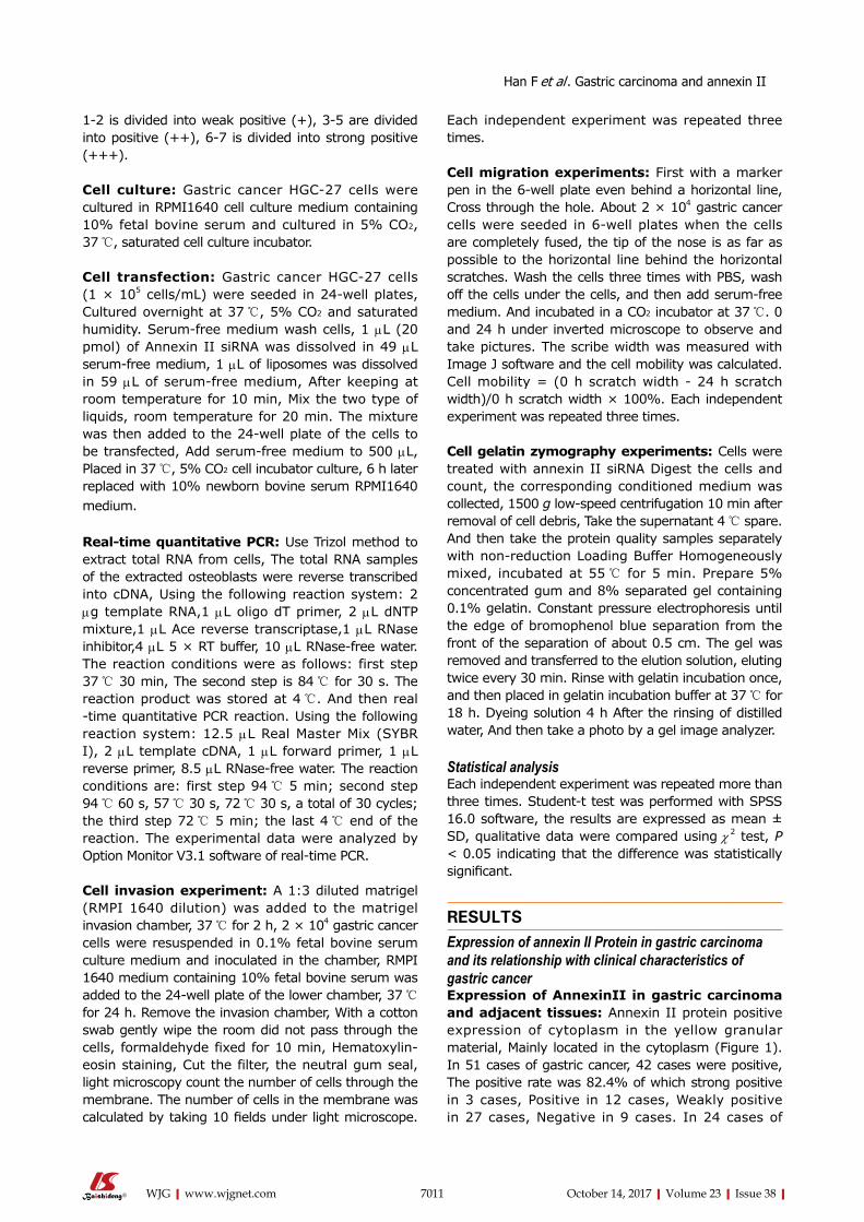
1-2 is divided into weak positive (+), 3-5 are divided into positive (++), 6-7 is divided into strong positive (+++).
Cell culture: Gastric cancer HGC-27 cells were cultured in RPMI1640 cell culture medium containing 10% fetal bovine serum and cultured in 5% CO2, 37 ℃, saturated cell culture incubator.
Cell transfection: Gastric cancer HGC-27 cells (1 × 10⁵ cells/mL) were seeded in 24-well plates, Cultured overnight at 37 ℃, 5% CO2 and saturated humidity. Serum-free medium wash cells, 1 μL (20 pmol) of Annexin II siRNA was dissolved in 49 μL serum-free medium, 1 μL of liposomes was dissolved in 59 μL of serum-free medium, After keeping at room temperature for 10 min, Mix the two type of liquids, room temperature for 20 min. The mixture was then added to the 24-well plate of the cells to be transfected, Add serum-free medium to 500 μL, Placed in 37 ℃, 5% CO2 cell incubator culture, 6 h later replaced with 10% newborn bovine serum RPMI1640 medium.
Real-time quantitative PCR: Use Trizol method to extract total RNA from cells, The total RNA samples of the extracted osteoblasts were reverse transcribed into cDNA, Using the following reaction system: 2 μg template RNA,1 μL oligo dT primer, 2 μL dNTP mixture,1 μL Ace reverse transcriptase,1 μL RNase inhibitor,4 μL 5 × RT buffer, 10 μL RNase-free water. The reaction conditions were as follows: first step 37 ℃ 30 min, The second step is 84 ℃ for 30 s. The reaction product was stored at 4 ℃. And then real -time quantitative PCR reaction. Using the following reaction system: 12.5 μL Real Master Mix (SYBR I), 2 μL template cDNA, 1 μL forward primer, 1 μL reverse primer, 8.5 μL RNase-free water. The reaction conditions are: first step 94 ℃ 5 min; second step 94 ℃ 60 s, 57 ℃ 30 s, 72 ℃ 30 s, a total of 30 cycles; the third step 72 ℃ 5 min; the last 4 ℃ end of the reaction. The experimental data were analyzed by Option Monitor V3.1 software of real-time PCR.
Cell invasion experiment: A 1:3 diluted matrigel (RMPI 1640 dilution) was added to the matrigel invasion chamber, 37 ℃ for 2 h, 2 × 10⁴ gastric cancer cells were resuspended in 0.1% fetal bovine serum culture medium and inoculated in the chamber, RMPI 1640 medium containing 10% fetal bovine serum was added to the 24-well plate of the lower chamber, 37 ℃ for 24 h. Remove the invasion chamber, With a cotton swab gently wipe the room did not pass through the cells, formaldehyde fixed for 10 min, Hematoxylin-eosin staining, Cut the filter, the neutral gum seal, light microscopy count the number of cells through the membrane. The number of cells in the membrane was calculated by taking 10 fields under light microscope.
Each independent experiment was repeated three times.
Cell migration experiments: First with a marker pen in the 6-well plate even behind a horizontal line, Cross through the hole. About 2 × 10⁴ gastric cancer cells were seeded in 6-well plates when the cells are completely fused, the tip of the nose is as far as possible to the horizontal line behind the horizontal scratches. Wash the cells three times with PBS, wash off the cells under the cells, and then add serum-free medium. And incubated in a CO2 incubator at 37 ℃. 0 and 24 h under inverted microscope to observe and take pictures. The scribe width was measured with Image J software and the cell mobility was calculated. Cell mobility = (0 h scratch width - 24 h scratch width)/0 h scratch width × 100%. Each independent experiment was repeated three times.
Cell gelatin zymography experiments: Cells were treated with annexin II siRNA Digest the cells and count, the corresponding conditioned medium was collected, 1500 g low-speed centrifugation 10 min after removal of cell debris, Take the supernatant 4 ℃ spare. And then take the protein quality samples separately with non-reduction Loading Buffer Homogeneously mixed, incubated at 55 ℃ for 5 min. Prepare 5% concentrated gum and 8% separated gel containing 0.1% gelatin. Constant pressure electrophoresis until the edge of bromophenol blue separation from the front of the separation of about 0.5 cm. The gel was removed and transferred to the elution solution, eluting twice every 30 min. Rinse with gelatin incubation once, and then placed in gelatin incubation buffer at 37 ℃ for 18 h. Dyeing solution 4 h After the rinsing of distilled water, And then take a photo by a gel image analyzer.
Statistical analysisEach independent experiment was repeated more than three times. Student-t test was performed with SPSS 16.0 software, the results are expressed as mean ± SD, qualitative data were compared using χ 2 test, P < 0.05 indicating that the difference was statistically significant.
RESULTSExpression of annexin II Protein in gastric carcinoma and its relationship with clinical characteristics of gastric cancer Expression of AnnexinII in gastric carcinoma and adjacent tissues: Annexin II protein positive expression of cytoplasm in the yellow granular material, Mainly located in the cytoplasm (Figure 1). In 51 cases of gastric cancer, 42 cases were positive, The positive rate was 82.4% of which strong positive in 3 cases, Positive in 12 cases, Weakly positive in 27 cases, Negative in 9 cases. In 24 cases of
7011 October 14, 2017|Volume 23|Issue 38|WJG|www.wjgnet.com
Han F et al . Gastric carcinoma and annexin II
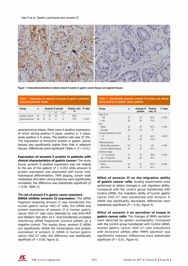
7012 October 14, 2017|Volume 23|Issue 38|WJG|www.wjgnet.com
Effect of annexin II on the migration ability of gastric cancer cells: Scaling experiments were performed to detect changes in cell migration ability. Compared with the control group transfected with Control siRNA, the migration ability of human gastric cancer HGC-27 cells transfected with Annexin II siRNA was significantly decreased, differences were statistically significant (P < 0.01; figure 3).
Effect of annexin ii on secretion of mmps in gastric cancer cells: The changes of MMPs secretion were detected by gelatin zymography. Compared with the control group transfected with Control siRNA, Human gastric cancer HGC-27 cells transfected with AnnexinII siRNAs after MMPs secretion was significantly reduced, Differences were statistically significant (P < 0.01; figure 4).
paracancerous tissue, there were 9 positive expression, of which strong positive 0 cases, positive in 3 cases, weak positive in 6 cases, The positive rate was 37.5%. The expression of AnnexinII protein in gastric cancer tissues was significantly higher than that in adjacent tissues, Differences were significant (Table 1, P < 0.01).
Expression of annexin ii protein in patients with clinical characteristics of gastric cancer: The study found, annexin II positive expression was not related to the sex of the patient (P > 0.05) while annexin II protein expression was associated with tumor size, histological differentiation, TNM staging, lymph node metastasis and other clinical features were significantly correlated, the difference was statistically significant (P < 0.05, Table 2).
The role of annexin II in gastric cancer metastasisSiRNA inhibits annexin II expression: The siRNA fragment targeting annexin II was transfected into human gastric cancer HGC-27 cells, the mRNA and protein expression of annexin II in human gastric cancer HGC-27 cells were detected by real time-PCR and Western blot after 24 h. And transfected unrelated interfering siRNA fragments (control siRNA) as a negative control. The results show, annexin II siRNA can significantly inhibit the transcription and protein expression of annexin II mRNA in human gastric cancer HGC-27 cells, the difference was statistically significant (P < 0.05; figure 2).
Figure 1 Immunohistochemistry to detect annexin II protein in gastric cancer tissues and adjacent tissues.
Table 1 Expression of annexin II protein in gastric carcinoma and paracancerous tissues
Group n Annexin II protein Positive rate, %
P value
- + ++ +++Gastric cancer 51 9 27 12 3 82.4Paracancerous 24 15 6 3 0 37.5 < 0.01
Table 2 Relationship between annexin II protein and clinical characteristics of gastric cancer patients
Group n Annexin II positive
Positive rate, %
P value
Age in yr ≤ 60 27 23 85.2 > 0.05 > 60 24 19 79.2Sex Male 31 27 87.1 > 0.05 Female 20 15 75Tumor diameter in cm ≤ 5 19 12 63.2 < 0.05 > 5 32 30 93.8Differentiation Well differentiated 15 9 60 < 0.05 Poorly differentiated 36 33 91.7Clinical stage Ⅰ+ Ⅱ period 17 10 58.8 < 0.05 Ⅲ + IV period 34 32 94.1Lymph node metastasis Have 33 31 93.9 < 0.05 Not have 18 11 61.1
Han F et al . Gastric carcinoma and annexin II
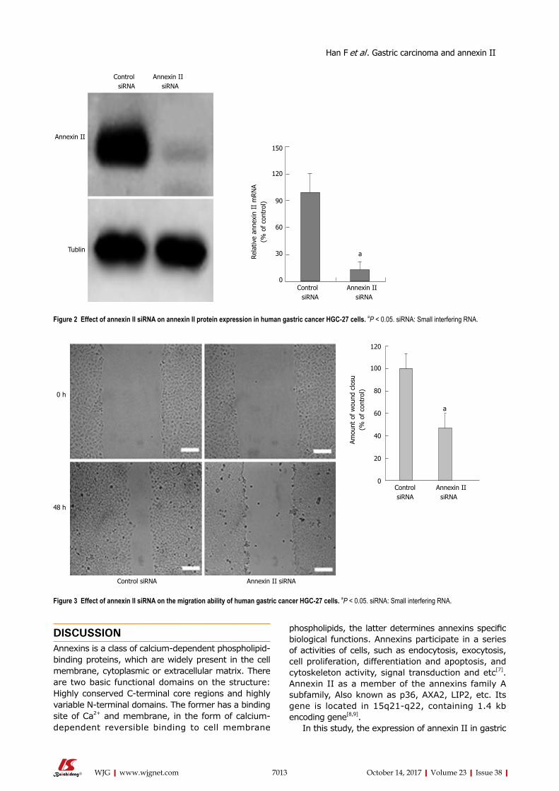
7013 October 14, 2017|Volume 23|Issue 38|WJG|www.wjgnet.com
DISCUSSIONAnnexins is a class of calcium-dependent phospholipid-binding proteins, which are widely present in the cell membrane, cytoplasmic or extracellular matrix. There are two basic functional domains on the structure: Highly conserved C-terminal core regions and highly variable N-terminal domains. The former has a binding site of Ca2+ and membrane, in the form of calcium-dependent reversible binding to cell membrane
phospholipids, the latter determines annexins specific biological functions. Annexins participate in a series of activities of cells, such as endocytosis, exocytosis, cell proliferation, differentiation and apoptosis, and cytoskeleton activity, signal transduction and etc[7]. Annexin II as a member of the annexins family A subfamily, Also known as p36, AXA2, LIP2, etc. Its gene is located in 15q21-q22, containing 1.4 kb encoding gene[8,9].
In this study, the expression of annexin II in gastric
Control Annexin IIsiRNA siRNA
Annexin II
Tublin
150
120
90
60
30
0
Rela
tive
anne
xin
II m
RN
A(%
of
cont
rol)
a
Control Annexin IIsiRNA siRNA
Figure 2 Effect of annexin II siRNA on annexin II protein expression in human gastric cancer HGC-27 cells. aP < 0.05. siRNA: Small interfering RNA.
120
100
80
60
40
20
0
Amou
nt o
f w
ound
clo
su(%
of
cont
rol)
a
Control Annexin II siRNA siRNA
0 h
48 h
Control siRNA Annexin II siRNA
Figure 3 Effect of annexin II siRNA on the migration ability of human gastric cancer HGC-27 cells. aP < 0.05. siRNA: Small interfering RNA.
Han F et al . Gastric carcinoma and annexin II

7014 October 14, 2017|Volume 23|Issue 38|WJG|www.wjgnet.com
cancer tissues and adjacent tissues was analyzed. The results showed that the expression of annexin II protein in gastric cancer tissues was significantly higher than that in adjacent tissues. To analyze the correlation between the expression of annexin II protein and the clinical characteristics of gastric cancer patients. The positive expression of annexin II was not related to the sex and age of the patients, while annexin II protein was expressed with tumor size, histological differentiation, TNM stage, lymph node metastasis and other clinical features were significantly correlated, the difference was statistically significant. The results suggest that annexin II protein is closely related to the malignant degree of gastric cancer.
Tumor metastasis is the most important malignant phenotype of the tumor and is the direct cause of this high mortality. Tumor metastasis is required in tumor cells themselves, host cells and extracellular matrix between the interaction can be completed, Involves a series of complex processes and steps, Including tumor cells from the primary tumor lesions, through the secretion of MMPs degradation of the extracellular matrix into the blood vessels or lymphatic vessels migrate to the distant, and then attack to the secondary organization or organ in the formation of metastases[10-14]. Therefore, the study of tumor metastasis for clinical diagnosis and treatment is important. To clarify whether annexin II in the process of gastric cancer metastasis play a role, In this study, annexin II-specific siRNA was used to inhibit annexin II expression in gastric cancer HGC-27 cells, after the expression of annexin II was reduced, gastric cancer HGC-27 cells migration, MMPs secretion and other capacity changes. The study found: In gastric cancer HGC-27 cells using annexin II- specific siRNA inhibited annexin II expression, gastric cancer cell migration, MMPs secretion and other capacity were reduced, suggesting that annexin II in the process of gastric cancer metastasis plays an important role.
In summary, annexin II in gastric cancer showed high expression, and the degree of differentiation of gastric cancer, TNM stage, lymph node metastasis and
other clinical features are closely related, application of annexin II specific siRNA in gastric cancer HGC-27 cells inhibited annexin II expression, gastric cancer cell migration, MMPs secretion and other capacity are reduced, suggesting that annexin II plays an important role in the process of gastric cancer metastasis. The development and progression of gastric cancer is a complex multi-factor comprehensive results. At present, the etiology and pathogenesis of gastric cancer are more studied, but it is not clear. This study confirmed that annexin II was highly expressed in gastric cancer tissues and had the important role in gastric cancer metastasis. With the deepening of related research, annexin II gene expression, biological function and the role of progress in gastric cancer will be gradually clear, is expected to be used in the diagnosis and treatment of gastric cancer, predict the prognosis of markers and therapeutic targets.
COMMENTSBackgroundGastric cancer is one of the most common malignancies in my country, the first in the digestive tract malignant tumor, ranking the fourth in the world, Because of its symptoms hidden, the disease developed rapidly, when diagnosed in the late, the treatment effect is often poor, a serious threat to human health, but the early treatment of gastric cancer is good, 5 years after radical resection rate of up to 90% or more.
Research frontiersAnnexin II is a calcium-dependent phospholipid-binding protein, mainly in the form of monomer, dimer and tetramer, the study shows: Annexin II are highly expressed in liver cancer, breast cancer, colon cancer, lung cancer and other malignant tumors, and play an important role in the occurence, development and infiltration of tumor.
Innovations and breakthroughsThe authors restropectively analysed the expression of annexin II in 51 cases of gastric carcinoma and 24 cases of adjacent tissues, to investigate the relationship between the positive expression of annexin II and the invasion and metastasis of gastric cancer.
ApplicationsAnnexin II in gastric cancer showed high expression, And the degree of
Control Annexin IIsiRNA siRNA
MMP-9
MMP-2
150
120
90
60
30
0
Den
sity
of
MM
Ps(%
of
cont
rol)
a
Control Annexin IIsiRNA siRNA
Figure 4 Effect of annexin II siRNA on secretion of matrix metalloproteinases in human gastric cancer HGC-27 cells. aP < 0.05. MMPs: Matrix metalloproteinases; siRNA: Small interfering RNA.
COMMENTS
Han F et al . Gastric carcinoma and annexin II

7015 October 14, 2017|Volume 23|Issue 38|WJG|www.wjgnet.com
differentiation of gastric cancer, TNM stage, lymph node metastasis and other clinical features are closely related, suggesting that annexin II plays an important role in the process of gastric cancer metastasis.
TerminologyAnnexins is a class of calcium-dependent phospholipid-binding proteins, they are widely present in the cell membrane, in the form of calcium-dependent reversible binding to ceembrane phospholipids, the latter determines Annexins specific biological functions.
Peer-reviewThis is an interesting study with great promise. AnnexinII was highly expressed in gastric cancer tissues and had the important role in the effect of promoting gastric cancer metastasis.
REFERENCES1 Digklia A, Wagner AD. Advanced gastric cancer: Current treatment
landscape and future perspectives. World J Gastroenterol 2016; 22: 2403-2414 [PMID: 26937129 DOI: 10.3748/wjg.v22.i8.2403]
2 El-Abd N, Fawzy A, Elbaz T, Hamdy S. Evaluation of annexin A2 and as potential biomarkers for hepatocellular carcinoma. Tumour Biol 2016; 37: 211-216 [PMID: 26189841 DOI: 10.1007/s13277-015-3524-x]
3 Jeon YR, Kim SY, Lee EJ, Kim YN, Noh DY, Park SY, Moon A. Identification of annexin II as a novel secretory biomarker for breast cancer. Proteomics 2013; 13: 3145-3156 [PMID: 24019232 DOI: 10.1002/pmic.201300127]
4 Sharma MR, Koltowski L, Ownbey RT, Tuszynski GP, Sharma MC. Angiogenesis-associated protein annexin II in breast cancer: selective expression in invasive breast cancer and contribution to tumor invasion and progression. Exp Mol Pathol 2006; 81: 146-156 [PMID: 16643892 DOI: 10.1016/j.yexmp.2006.03.003]
5 Tristante E, Martínez CM, Jiménez S, Mora L, Carballo
F, Martínez-Lacaci I, de Torre-Minguela C. Association of a characteristic membrane pattern of annexin A2 with high invasiveness and nodal status in colon adenocarcinoma. Transl Res 2015; 166: 196-206 [PMID: 25795236 DOI: 10.1016/j.trsl.2015.02.006]
6 Jia JW, Li KL, Wu JX, Guo SL. Clinical significance of annexin II expression in human non-small cell lung cancer. Tumour Biol 2013; 34: 1767-1771 [PMID: 23494179 DOI: 10.1007/s13277-013-0715-1]
7 Mirsaeidi M, Gidfar S, Vu A, Schraufnagel D. Annexins family: insights into their functions and potential role in pathogenesis of sarcoidosis. J Transl Med 2016; 14: 89 [PMID: 27071553 DOI: 10.1186/s12967-016-0843-7]
8 Monastyrskaya K, Babiychuk EB, Draeger A. The annexins: spatial and temporal coordination of signaling events during cellular stress. Cell Mol Life Sci 2009; 66: 2623-2642 [PMID: 19381436 DOI: 10.1007/s00018-009-0027-1]
9 Laohavisit A, Davies JM. Annexins. New Phytol 2011; 189: 40-53 [PMID: 21083562 DOI: 10.1111/j.1469-8137.2010.03533.x]
10 Bhowmick NA, Moses HL. Tumor-stroma interactions. Curr Opin Genet Dev 2005; 15: 97-101 [PMID: 15661539 DOI: 10.1016/j.gde.2004.12.003]
11 Steeg PS. Tumor metastasis: mechanistic insights and clinical challenges. Nat Med 2006; 12: 895-904 [PMID: 16892035 DOI: 10.1038/nm1469]
12 Liotta LA, Kohn EC. The microenvironment of the tumour-host interface. Nature 2001; 411: 375-379 [PMID: 11357145 DOI: 10.1038/35077241]
13 Friedl P, Wolf K. Tumour-cell invasion and migration: diversity and escape mechanisms. Nat Rev Cancer 2003; 3: 362-374 [PMID: 12724734 DOI: 10.1038/nrc1075]
14 Porporato PE, Payen VL, Baselet B, Sonveaux P. Metabolic changes associated with tumor metastasis, part 2: Mitochondria, lipid and amino acid metabolism. Cell Mol Life Sci 2016; 73: 1349-1363 [PMID: 26646069 DOI: 10.1007/s00018-015-2100-2]
P- Reviewer:Eleftheriadis NP, Garcia-Olmo D, Goral V, Mura G S- Editor: Gou SX L- Editor: A E- Editor: Ma YJ
Han F et al . Gastric carcinoma and annexin II

Kwan Mo Yang, Chang Sik Yu, Jong Lyul Lee, Chan Wook Kim, Yong Sik Yoon, In Ja Park, Seok-Byung Lim, Jin Cheon Kim, Division of Colon and Rectal Surgery, Department of Surgery, University of Ulsan College of Medicine, Asan Medical Center, Seoul 05505, South Korea
Sang Hyoung Park, Byong Duk Ye, Suk-Kyun Yang, Department of Gastroenterology, University of Ulsan College of Medicine, Asan Medical Center, Seoul 05505, South Korea
ORCID number: Kwan Mo Yang (0000000191903296); Chang S ik Yu (0000000194019981) ; Jong Lyu l L e e ( 0 0 0 0 0 0 0 2 5 8 7 8 8 0 0 0 ) ; C h a n Wo o k K i m (0000000223820939); Yong Sik Yoon (0000000231968423); In Ja Park (0000000153553969); SeokByung Lim (0000000188244808); Sang Hyoung Park; Byong Duk Ye; SukKyun Yang ; Jin Cheon Kim (0000000348238619).
Author contributions: Yang KM drafted the manuscript and analyzed data; Lee JL, Kim CW, Yoon YS, Park IJ, Lim SB, Park SH, Ye BD, Yang SK, Kim JC generated the data; Yu CS had the original idea for the paper, gave critical comments, and revised the manuscript; all authors approved the final version of the manuscript.
Institutional review board statement: The study was reviewed and approved by the Institutional Review Board of Asan Medical Center (IRB No. 20161139).
Informed consent statement: Informed consent was waived due to the retrospective nature of this study.
Conflict-of-interest statement: All authors declare no conflict of interest related to this study or its publication. Data sharing statement: No additional data are available. Open-Access: This article is an openaccess article which was selected by an inhouse editor and fully peerreviewed by external reviewers. It is distributed in accordance with the Creative Commons Attribution Non Commercial (CC BYNC 4.0) license, which permits others to distribute, remix, adapt, build upon this work noncommercially, and license their derivative works on
different terms, provided the original work is properly cited and the use is noncommercial. See: http://creativecommons.org/licenses/bync/4.0/ Manuscript source: Unsolicited manuscript
Correspondence to: Chang Sik Yu, MD, PhD, Professor, The Division of Colon and Rectal Surgery, Department of Surgery, University of Ulsan College of Medicine, Asan Medical Center, 88, Olympicro 43gil, Songpagu, Seoul 05505, South Korea. [email protected]: +82230103494Fax: +8224746027
Received: July 23, 2017Peer-review started: July 27, 2017First decision: August 10, 2017Revised: August 24, 2017Accepted: September 13, 2017 Article in press: September 13, 2017Published online: October 14, 2017
AbstractAIMTo evaluate the risk factors for postoperative recurrence after primary bowel resection in a cohort of Korean Crohn’s disease (CD) patients.
METHODSThis study included 260 patients with no history of previous bowel surgery who underwent primary surgery for CD between January 2000 and December 2010 at Asan Medical Center (Seoul, South Korea). The median follow-up period was 101 mo.
RESULTSDuring the follow-up period, 66 patients (25.4%) underwent a second operation for disease recurrence.
7016 October 14, 2017|Volume 23|Issue 38|WJG|www.wjgnet.com
ORIGINAL ARTICLE
Risk factors for postoperative recurrence after primary bowel resection in patients with Crohn’s disease
Retrospective Study
Kwan Mo Yang, Chang Sik Yu, Jong Lyul Lee, Chan Wook Kim, Yong Sik Yoon, In Ja Park, Seok-Byung Lim, Sang Hyoung Park, Byong Duk Ye, Suk-Kyun Yang, Jin Cheon Kim
Submit a Manuscript: http://www.f6publishing.com
DOI: 10.3748/wjg.v23.i38.7016
World J Gastroenterol 2017 October 14; 23(38): 7016-7024
ISSN 1007-9327 (print) ISSN 2219-2840 (online)
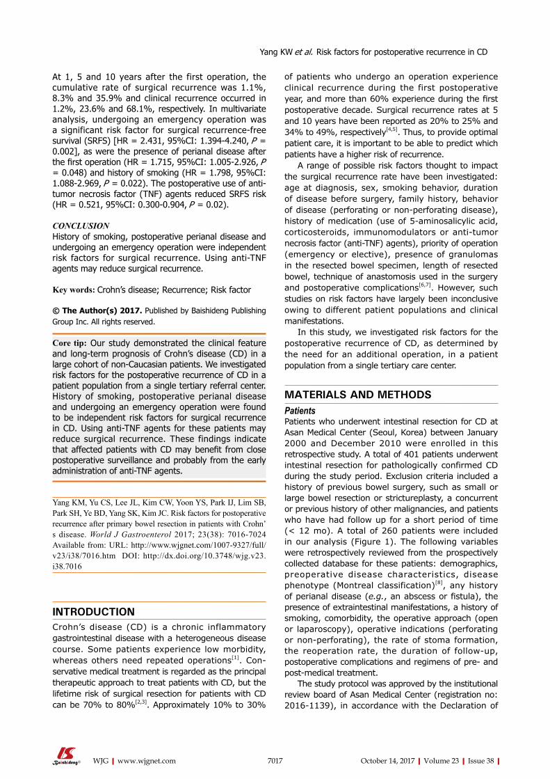
At 1, 5 and 10 years after the first operation, the cumulative rate of surgical recurrence was 1.1%, 8.3% and 35.9% and clinical recurrence occurred in 1.2%, 23.6% and 68.1%, respectively. In multivariate analysis, undergoing an emergency operation was a significant risk factor for surgical recurrence-free survival (SRFS) [HR = 2.431, 95%CI: 1.394-4.240, P = 0.002], as were the presence of perianal disease after the first operation (HR = 1.715, 95%CI: 1.005-2.926, P = 0.048) and history of smoking (HR = 1.798, 95%CI: 1.088-2.969, P = 0.022). The postoperative use of anti-tumor necrosis factor (TNF) agents reduced SRFS risk (HR = 0.521, 95%CI: 0.300-0.904, P = 0.02).
CONCLUSIONHistory of smoking, postoperative perianal disease and undergoing an emergency operation were independent risk factors for surgical recurrence. Using anti-TNF agents may reduce surgical recurrence.
Key words: Crohn’s disease; Recurrence; Risk factor
© The Author(s) 2017. Published by Baishideng Publishing Group Inc. All rights reserved.
Core tip: Our study demonstrated the clinical feature and long-term prognosis of Crohn’s disease (CD) in a large cohort of non-Caucasian patients. We investigated risk factors for the postoperative recurrence of CD in a patient population from a single tertiary referral center. History of smoking, postoperative perianal disease and undergoing an emergency operation were found to be independent risk factors for surgical recurrence in CD. Using anti-TNF agents for these patients may reduce surgical recurrence. These findings indicate that affected patients with CD may benefit from close postoperative surveillance and probably from the early administration of anti-TNF agents.
Yang KM, Yu CS, Lee JL, Kim CW, Yoon YS, Park IJ, Lim SB, Park SH, Ye BD, Yang SK, Kim JC. Risk factors for postoperative recurrence after primary bowel resection in patients with Crohn’s disease. World J Gastroenterol 2017; 23(38): 70167024 Available from: URL: http://www.wjgnet.com/10079327/full/v23/i38/7016.htm DOI: http://dx.doi.org/10.3748/wjg.v23.i38.7016
INTRODUCTIONCrohn’s disease (CD) is a chronic inflammatory gastrointestinal disease with a heterogeneous disease course. Some patients experience low morbidity, whereas others need repeated operations[1]. Conservative medical treatment is regarded as the principal therapeutic approach to treat patients with CD, but the lifetime risk of surgical resection for patients with CD can be 70% to 80%[2,3]. Approximately 10% to 30%
of patients who undergo an operation experience clinical recurrence during the first postoperative year, and more than 60% experience during the first postoperative decade. Surgical recurrence rates at 5 and 10 years have been reported as 20% to 25% and 34% to 49%, respectively[4,5]. Thus, to provide optimal patient care, it is important to be able to predict which patients have a higher risk of recurrence.
A range of possible risk factors thought to impact the surgical recurrence rate have been investigated: age at diagnosis, sex, smoking behavior, duration of disease before surgery, family history, behavior of disease (perforating or nonperforating disease), history of medication (use of 5aminosalicylic acid, corticosteroids, immunomodulators or antitumor necrosis factor (antiTNF) agents), priority of operation (emergency or elective), presence of granulomas in the resected bowel specimen, length of resected bowel, technique of anastomosis used in the surgery and postoperative complications[6,7]. However, such studies on risk factors have largely been inconclusive owing to different patient populations and clinical manifestations.
In this study, we investigated risk factors for the postoperative recurrence of CD, as determined by the need for an additional operation, in a patient population from a single tertiary care center.
MATERIALS AND METHODSPatientsPatients who underwent intestinal resection for CD at Asan Medical Center (Seoul, Korea) between January 2000 and December 2010 were enrolled in this retrospective study. A total of 401 patients underwent intestinal resection for pathologically confirmed CD during the study period. Exclusion criteria included a history of previous bowel surgery, such as small or large bowel resection or strictureplasty, a concurrent or previous history of other malignancies, and patients who have had follow up for a short period of time (< 12 mo). A total of 260 patients were included in our analysis (Figure 1). The following variables were retrospectively reviewed from the prospectively collected database for these patients: demographics, preoperative disease characteristics, disease phenotype (Montreal classification)[8], any history of perianal disease (e.g., an abscess or fistula), the presence of extraintestinal manifestations, a history of smoking, comorbidity, the operative approach (open or laparoscopy), operative indications (perforating or nonperforating), the rate of stoma formation, the reoperation rate, the duration of followup, postoperative complications and regimens of pre and postmedical treatment.
The study protocol was approved by the institutional review board of Asan Medical Center (registration no: 20161139), in accordance with the Declaration of
7017 October 14, 2017|Volume 23|Issue 38|WJG|www.wjgnet.com
Yang KW et al. Risk factors for postoperative recurrence in CD

Helsinki.Assessment and follow-upCD distribution was assessed based on pathological and intraoperative findings, radiologic imaging using a small bowel series (SBS), abdomen and pelvis computed tomography (CT), CT enterography, magnetic resonance imaging (MRI) enterography and colonofiberscopy (CFS). Perianal disease included stricture, fistula or an abscess in the perianal area, anal canal or perirectal space at any time during the course of CD. After primary intestinal resection, reassessment was performed by CT enterography, MRI enterography or CFS after 6 to 12 mo. Surgical recurrence was defined as a repeated operation for pathologically confirmed CD on any part of the bowel or a repeated operation for pathologically confirmed anastomotic disease, including instances where the small bowel was diseased at the anastomosis or the stoma site. Clinical recurrence was defined as the recurrence of CDrelated symptoms, confirmed by objective signs of radiology or endoscopy findings. As this was a retrospective study the Crohn’s disease Activity Index (CDAI) and Rutgeerts’ score were not available.
Medical treatment of CDOur medical treatment policy for treating CD was based on a stepup approach, with more potent therapies added if patients became unresponsive to first-line or less toxic agents, as described in a previous report from our institution[9]. The use of antiTNF agents in this study was less frequent than in Western studies because of the strict Korean government health insurance reimbursement policy during our study period. During this period, two antiTNF agents (infliximab and adalimumab) were available in Korea.
Statistical analysisSurvival curves were plotted using the KaplanMeier method and compared using the logrank test. Multivariate analyses for survival that included the variables that showed statistical significance in the logrank test were performed using Cox proportional hazards model with a 95%CI to assess the risk factors associated with surgical recurrencefree survival (SRFS) and clinical recurrencefree survival (CRFS). Statistical significance was defined as P < 0.05; all analyses were performed using SPSS software version 21 (IBM Corp., Armonk, NY).
RESULTSPatient characteristicsThe clinicopathological characteristics of the patients are summarised in Tables 1 and 2. The median followup time for the 260 patients was 101 mo (range, 12193 mo). The interval between the first intestinal resection and clinical recurrence was 64 mo (range, 12148 mo)
and the interval between the first intestinal resection and surgical recurrence was 77.5 mo (range, 15149 mo).
Elective operations were performed for 210 (80.8%) patients; the remaining 50 (19.2%) patients underwent emergency operations because of generalised peritonitis or severe obstruction. Among 260 patients, 122 (46.9%) patients had perianal disease before first surgery. Of these, 42 (34.4%) patients had perianal disease after first operation. Because of the strict Korean government health insurance reimbursement policy, only 27 (10.4%) patients treated with antiTNF agents before first surgery, and 17 (63%) patients continued antiTNF agents after first operation. Antibiotics such as ciprofloxacin or metronidazole were administered to 163 patients (62.7%) in case of infectious complications such as intraabdominal abscess, perianal abscess or fistula before first surgery. All the patients who underwent first surgery used antibiotics for a certain period of time. During the followup period, three patients (1.5%) experienced colorectal cancer, one patient experienced lung cancer (0.4%).
Montreal classification of the patientsThe majority (215/260, 82.7%) of the patients
were diagnosed between the ages of 17 and 40 years, with 9.6% experiencing childhood onset of CD (≤ 16 years) and 7.7% diagnosed as older adults (> 40 years). Montreal classifications were distributed as follows: 43.5% exhibited disease in the terminal ileum region (L1), 41.2% in the ileocolon (L3) and 14.6% in the colon region (L2); 67% had penetrating disease (B3, fistula, abscess or perforation), 31% had stricturing disease (B2) and 2% had inflammatory disease (B1, nonstricturing and nonpenetrating). Because these patients were treated with surgical intervention, proportion of disease behavior was different from the whole patients with CD in Korea. Disease behavior of whole CD patients at diagnosis was inflammatory (B1) in 1591 patients (77.1%), stricturing (B2) in 174 (10.3%), and penetrating (B3) in 211 (12.6%)[9].
Clinical and surgical recurrenceClinical recurrence occurred in 1.9%, 24.2% and 68.3% of the patients at 1, 5 and 10 years, respectively, after the first intestinal resection (Figure 2). In multivariate analysis (Table 3), history of smoking, presence of perianal disease after the first operation and undergoing an emergency operation were associated with CRFS.
During the followup period, 66 patients (25.4%) underwent a second intestinal resection for disease recurrence. Among these, 13 (20%) patients underwent a third intestinal resection, four patients (31%) a fourth and one patient (25%) a fifth. The cumulative rate of surgical recurrence was 1.1%, 9.2%
7018 October 14, 2017|Volume 23|Issue 38|WJG|www.wjgnet.com
Yang KW et al. Risk factors for postoperative recurrence in CD
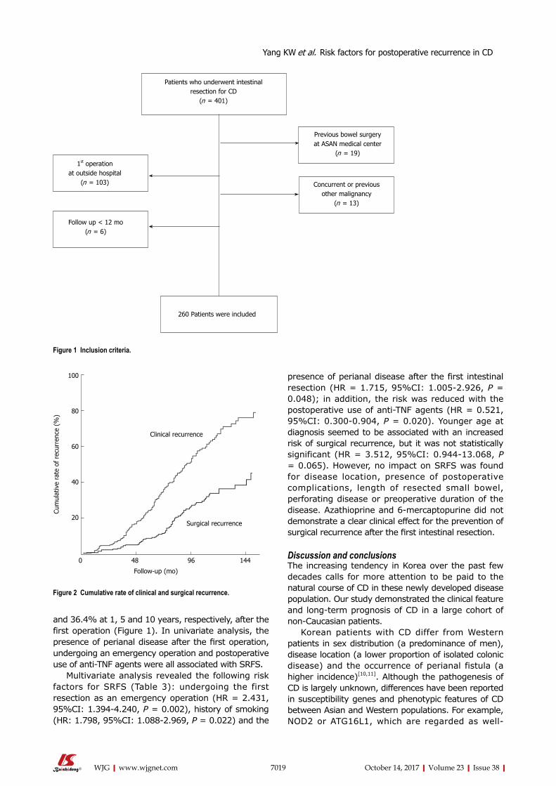
7019 October 14, 2017|Volume 23|Issue 38|WJG|www.wjgnet.com
presence of perianal disease after the first intestinal resection (HR = 1.715, 95%CI: 1.0052.926, P = 0.048); in addition, the risk was reduced with the postoperative use of antiTNF agents (HR = 0.521, 95%CI: 0.3000.904, P = 0.020). Younger age at diagnosis seemed to be associated with an increased risk of surgical recurrence, but it was not statistically significant (HR = 3.512, 95%CI: 0.94413.068, P = 0.065). However, no impact on SRFS was found for disease location, presence of postoperative complications, length of resected small bowel, perforating disease or preoperative duration of the disease. Azathioprine and 6mercaptopurine did not demonstrate a clear clinical effect for the prevention of surgical recurrence after the first intestinal resection.
Discussion and conclusionsThe increasing tendency in Korea over the past few decades calls for more attention to be paid to the natural course of CD in these newly developed disease population. Our study demonstrated the clinical feature and longterm prognosis of CD in a large cohort of nonCaucasian patients.
Korean patients with CD differ from Western patients in sex distribution (a predominance of men), disease location (a lower proportion of isolated colonic disease) and the occurrence of perianal fistula (a higher incidence)[10,11]. Although the pathogenesis of CD is largely unknown, differences have been reported in susceptibility genes and phenotypic features of CD between Asian and Western populations. For example, NOD2 or ATG16L1, which are regarded as well
and 36.4% at 1, 5 and 10 years, respectively, after the first operation (Figure 1). In univariate analysis, the presence of perianal disease after the first operation, undergoing an emergency operation and postoperative use of antiTNF agents were all associated with SRFS.
Multivariate analysis revealed the following risk factors for SRFS (Table 3): undergoing the first resection as an emergency operation (HR = 2.431, 95%CI: 1.3944.240, P = 0.002), history of smoking (HR: 1.798, 95%CI: 1.0882.969, P = 0.022) and the
Patients who underwent intestinalresection for CD
(n = 401)
1st operationat outside hospital
(n = 103)
Previous bowel surgeryat ASAN medical center
(n = 19)
Concurrent or previousother malignancy
(n = 13)
Follow up < 12 mo(n = 6)
260 Patients were included
Figure 1 Inclusion criteria.
Yang KW et al. Risk factors for postoperative recurrence in CD
Figure 2 Cumulative rate of clinical and surgical recurrence.
Clinical recurrence
Surgical recurrence
Follow-up (mo)
Cum
ulat
ive
rate
of
recu
rren
ce (
%)
100
80
60
40
20
0 48 96 144

7020 October 14, 2017|Volume 23|Issue 38|WJG|www.wjgnet.com
established CD susceptibility genes among Caucasians, are not replicated in Asians, whereas the association of TNFSF15 with CD is stronger in Koreans and Japanese than in Caucasians[12,13].
The surgical recurrence rate was 25% to 45% 10 years after the first intestinal resection[14,15], although postoperative clinical recurrence occurred earlier during the disease course. Our study found that cumulative surgical and clinical recurrence rates were 36.4% and 68.4%, respectively, at 10 years after the first intestinal resection.
Our study demonstrated that patients with CD who had a higher risk of surgical recurrence after primary bowel surgery were those who underwent the first resection as an emergency operation, those with history of smoking and those with postoperative perianal disease. The use of postoperative antiTNF
agents seemed to reduce surgical recurrence.Korean patients with CD show higher incidence
of perianal disease than Western patients[9]. Previous study in our center[11], perianal fistulas occurred in 46.8% of patients. Also, the cumulative frequency of perianal fistula was 54.3% after 15 years. These results are in contrast to those of Western studies in which the cumulative frequency of perianal fistula in patients with CD attending referral centers was reported to be 13%38%[16]. Furthermore, the presence of perianal disease was one of the predictors of postoperative recurrence, as has been suggested by previous studies in Asian population[1719]. Gao et al[17] also assessed risk factors for surgical recurrence and advocated perianal CD correlated with a higher clinical recurrence rate. Korean CD patients with perianal disease had a higher risk of surgical recurrence[18,20]. The present study showed that patients with CD who developed perianal disease after the first resection had a high risk of surgical and clinical recurrence, indicating that these patients had a poor disease
Variables Number of patients
(n = 260)SEX Male 187 (71.9) Female 73 (28.1)Follow-up period, month 101 (12-193)Age at diagnosis 23 (12-74)Age at 1st operation 28 (15-74)Age at 2nd operation 34 (22-59)Montreal classificationAge at diagnosis, yr A1 ( ≤ 16) 25 (9.6) A2 (17-40) 215 (82.7) A3 ( > 40) 20 (7.7)Location L1 (Terminal ileum) 113 (43.5) L2 (Colon) 38 (14.6) L3 (Ileocolon) 107 (41.2) L4 (Isolated upper disease) 2 (0.8)Behavior B1 (Inflammatory) 6 (2.3) B2 (Stricturing) 80 (30.8) B3 (Penetrating) 174 (66.9)Family history (Crohn's disease) 7 (2.7)Presence of extraintestinal manifestation 20 (7.7)Perianal disease at diagnosis 122 (46.9) after 1st operation 62 (23.8)History of smoking 70 (26.9) Current smoker 12 (4.6) Ex-smoker 58 (22.3) Non-smoker 190 (73.1)Preoperative use of medication Anti-TNF 27 (10.4) 5-ASA 223 (85.8) Steroid 147 (56.5) Immunomodulator1 128 (49.2) Antibiotics2 128 (49.2)Malignancy 163 (62.7) Colorectal cancer 3 (1.2) Lung cancer 1 (0.4)
Table 1 Demographic and clinical characteristics of the patients undergoing intestinal resection n (%)
1Use of azathioprine/6-mercaptopurine or methotrexate; 2Ciprofloxacin or metronidazole. EIM: Extraintestinal manifestation; 5-ASA: Aminosalicylates; TNF: Tumor necrosis factor.
Table 2 Operative details and outcomes n (%)
Variables Number of patients(n = 266)
Operative approach Open 215 (82.7) Laparoscopy 45 (17.3)Type of operation Elective 210 (80.8) Emergency 50 (19.2)Indication for 1st operation Perforating 167 (64.2) Non-perforating 93 (35.8)OP name ICR 85 (32.7) RHC 72 (27.7) SB R&A 49 (18.8) TC/STC 32 (12.3) TPC/APR 19 (7.3) Others 3 (1.2)Type of anastomosis Handsewn 5 (1.9) Stapled 225 (86.5) Permanent stomy 28 (10.8)Postoperative use of medication Anti-TNF 95 (36.5) 5-ASA 244 (93.8) Steroid 82 (31.5) Immunomodulator 220 (84.6)Presence of complication 58 (22.3) Wound infection 17 (6.5) Anastomosis leakage 12 (4.6) Intra-abdominal abscess 11 (4.2) Entero-cutaneous fistula 7 (2.7) Bleeding 4 (1.5) Ileus 3 (1.2) Others 4 (1.5)Time to clinical recurrence, mo 64 ± 29.8Time to surgical recurrence, mo 77.5 ± 30.8
ICR: Ileocecal resection; RHC: Right hemicolectomy; SB R&A: Small bowel resection and anastomosis; TC: Total colectomy; STC: Subtotal colectomy; TPC: Total proctocolectomy; Others: Segmental resection of colon.
Yang KW et al. Risk factors for postoperative recurrence in CD

7021 October 14, 2017|Volume 23|Issue 38|WJG|www.wjgnet.com
outcome after the primary operation. Similarly, despite a wide variation on reported risk of recurrence, the presence of perianal disease is thus considered to be a risk factor for postoperative recurrence in Caucasian population[3,21,22]. The presence of perianal disease is a sign of an increased inflammatory process and this may also influence intestinal CD.
Most patients with CD can be operated on electively, with only few patients requiring acute surgical intervention. In the present study, an indication for an emergency first intestinal resection was significantly associated with surgical recurrence. Riss et al[23] reported that an urgent indication for surgery was found to be significantly associated with the rate of surgical recurrence. Another retrospective study of 116 consecutive patients undergoing their first ileocolectomy at a large Austrian referral centre between 1997 and 2006 demonstrated that urgent index ileocolectomy increased the risk of repeat surgery approximately sixfold[24]. An emergency presentation could be a sign of a significant lack of an immune response to the ongoing inflammation, making an emergency operation necessary. Furthermore, preoperative work up and intraoperative evaluation were limited in emergency situation and an emergency operation was usually performed by less experienced surgeons. As a consequence, these patients may be prone to developing early recurrence after the surgical treatment.
Smoking is the most widely recognized environmental risk factor for the initiation and recurrence of Crohn’s disease[25]. Sutherland et al[26], reported that the 5- and 10-year recurrence rates were significantly higher in smokers (36% and 70%) than in nonsmokers (20% and 41%; OR = 2.1, P = 0.007). Indeed, smoking is a significant predictive risk factor
for all types of recurrences: endoscopic, clinical and surgical[6,27]. Cottone et al[25], conducted multivariate analyses to examine predictive factors for three types of recurrence (clinical, endoscopic and surgical recurrences) after primary surgery. They found that smoking was an independent significant factor for clinical, endoscopic and surgical recurrence. In the present study, history of smoking was significant risk factor for clinical and surgical recurrence.
Another important finding was that postoperative exposure to antiTNF agents significantly reduced the likelihood of surgical recurrence. The introduction of antiTNF agents has significantly changed the treatment strategy for CD. Recent studies have shown that antiTNF agents reduced postoperative recurrence[2830]. In the present study, postoperative use of antiTNF agents seemed to reduce the risk of reoperation, indicating that medical treatment is one of the important factors affecting surgical recurrence[31]. A randomised controlled trial was able to demonstrate clearly an advantage of antiTNF agents in maintaining clinical remission in patients with CD[32].
Immunomodulators such as azathioprine or 6MP has been widely used, and several studies[33,34] showed that immunomodulators could reduce rate of surgery. Similarly, previous study [9] in our center revealed that early use of immunomodulators was independently related to the decreased surgery rate (P = 0.01). However, because immunomodulators were generally used after first surgery, it is difficult to determine their impact on decreasing the postoperative recurrence. In present study, 85% of patients used azathioprine or 6-MP after first surgery and use of immunomodulator showed no significant difference in postoperative recurrence. More recently, these drugs are used in combination with antiTNF drugs to decrease
Yang KW et al. Risk factors for postoperative recurrence in CD
Table 3 Univariate and multivariate analysis of factors associated with surgical and clinical recurrence free survival after 1st intestinal resection
Univariate MultivariateP value HR 95%CI P value
Clinical recurrence free survivalType of operation 0.001a 0.002a
Elective 1 Emergency 1.856 1.246-2.763Perianal disease after the 1st operation 0.077 1.446 1.005-2.081 0.047a
History of smoking 0.001a 1.727 1.220-2.444 0.002a
Surgical recurrence free survivalAge at diagnosis, yr 0.097 0.065 A1 ( ≤ 16) 3.512 0.944-13.068 A2 ( 17-40) 1.547 0.475-5.036 A1 ( > 40)Type of operation 0.001a 0.002a
Elective 1 Emergency 2.431 1.394-4.240Perianal disease after the 1st operation 0.056 1.715 1.005-2.926 0.048a
History of smoking 0.047a 1.798 1.088-2.969 0.022a
Postoperative use of anti-TNF 0.016a 0.521 0.300-0.904 0.02a
aP < 0.05; HR: Hazard ratio.
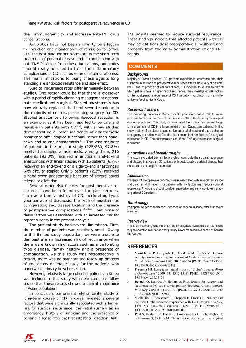
7022 October 14, 2017|Volume 23|Issue 38|WJG|www.wjgnet.com
their immunogenicity and increase antiTNF drug concentrations.
Antibiotics have not been shown to be effective for induction and maintenance of remission for active CD. The best data for antibiotics are in the shortterm treatment of perianal disease and in combination with antiTNF[35]. Aside from these indications, antibiotics should really be used to treat the inflammatory complications of CD such as enteric fistula or abscess. The main limitations to using these agents long standing are antibiotic resistance and side effect.
Surgical recurrence rates differ immensely between studies. One reason could be that there is crossover with a period of rapidly changing management options, both medical and surgical. Stapled anastomosis has now virtually replaced the handsewn technique in the majority of centres performing surgery for CD. Stapled anastomosis following ileocecal resection is an example, as it has been reported to be safe and feasible in patients with CD[36], with a few studies demonstrating a lower incidence of anastomotic recurrence after stapled functional rather than handsewn endtoend anastomosis[37]. The vast majority of patients in the present study (225/230, 97.8%) received a stapled anastomosis. Among them, 210 patients (93.3%) received a functional endtoend anastomosis with linear stapler, with 15 patients (6.7%) receiving an endtoend or a sidetoend anastomosis with circular stapler. Only 5 patients (2.2%) received a handsewn anastomosis because of severe bowel edema or dilatation.
Several other risk factors for postoperative recurrence have been found over the past decades, such as a family history of CD, perforating type, younger age at diagnosis, the type of anastomotic configuration, sex, disease location, and the presence of postoperative complications[15,38,39], but none of these factors was associated with an increased risk for repeat surgery in the present analysis.
The present study had several limitations. First, the number of patients was relatively small. Owing to this limited study population, we were unable to demonstrate an increased risk of recurrence when there were known risk factors such as a perforating type disease, family history and a presence of complication. As this study was retrospective in design, there was no standardised followup protocol of endoscopy or image study for the patients who underwent primary bowel resection.
However, relatively large cohort of patients in Korea was included in this study with near complete follow up, so that these results showed a clinical importance in Asian population.
In conclusion, our present referral center study of longterm course of CD in Korea revealed a several factors that were significantly associated with a higher risk for surgical recurrence: the initial surgery as an emergency, history of smoking and the presence of perianal disease after the first intestinal resection. Anti-
TNF agents seemed to reduce surgical recurrence. These findings indicate that affected patients with CD may benefit from close postoperative surveillance and probably from the early administration of antiTNF agents.
COMMENTSBackgroundMajority of Crohn’s disease (CD) patients experienced recurrence after their first bowel resection and postoperative recurrence affects the quality of patients’ lives. Thus, to provide optimal patient care, it is important to be able to predict which patients have a higher risk of recurrence. They investigated risk factors for the postoperative recurrence of CD in a patient population from a single tertiary referral center in Korea.
Research frontiersThe increasing tendency in Korea over the past few decades calls for more attention to be paid to the natural course of CD in these newly developed disease population. This study demonstrated the clinical feature and long-term prognosis of CD in a large cohort of non-Caucasian patients. In this study, history of smoking, postoperative perianal disease and undergoing an emergency operation were found to be independent risk factors for surgical recurrence in CD. The postoperative use of anti-TNF agents reduced surgical recurrence.
Innovations and breakthroughsThis study evaluated the risk factors which contribute the surgical recurrence and showed that Korean CD patients with postoperative perianal disease had increased risk of surgical recurrence.
ApplicationsPresence of postoperative perianal disease associated with surgical recurrence and using anti-TNF agents for patients with risk factors may reduce surgical recurrence. Physicians should consider aggressive and early top-down therapy for perianal CD patients.
TerminologyPostoperative perianal disease: Presence of perianal disease after first bowel resection.
Peer-reviewThis is an interesting study in which the investigators evaluated the risk factors for postoperative recurrence after primary bowel resection in a cohort of Korean CD patients.
REFERENCES1 Munkholm P, Langholz E, Davidsen M, Binder V. Disease
activity courses in a regional cohort of Crohn’s disease patients. Scand J Gastroenterol 1995; 30: 699706 [PMID: 7481535 DOI: 10.3109/00365529509096316]
2 Freeman HJ. Longterm natural history of Crohn’s disease. World J Gastroenterol 2009; 15: 13151318 [PMID: 19294760 DOI: 10.3748/wjg.15.1315]
3 Bernell O, Lapidus A, Hellers G. Risk factors for surgery and recurrence in 907 patients with primary ileocaecal Crohn’s disease. Br J Surg 2000; 87: 16971701 [PMID: 11122187 DOI: 10.1046/j.13652168.2000.01589.x]
4 Michelassi F, Balestracci T, Chappell R, Block GE. Primary and recurrent Crohn’s disease. Experience with 1379 patients. Ann Surg 1991; 214: 230238; discussion 238240 [PMID: 1929605 DOI: 10.1097/0000065819910900000006]
5 Post S, Herfarth C, Böhm E, Timmermanns G, Schumacher H, Schürmann G, Golling M. The impact of disease pattern, surgical
COMMENTS
Yang KW et al. Risk factors for postoperative recurrence in CD
7023 October 14, 2017|Volume 23|Issue 38|WJG|www.wjgnet.com
management, and individual surgeons on the risk for relaparotomy for recurrent Crohn’s disease. Ann Surg 1996; 223: 253260 [PMID: 8604905]
6 Reese GE, Nanidis T, Borysiewicz C, Yamamoto T, Orchard T, Tekkis PP. The effect of smoking after surgery for Crohn’s disease: a metaanalysis of observational studies. Int J Colorectal Dis 2008; 23: 12131221 [PMID: 18762954 DOI: 10.1007/s0038400805429]
7 Simillis C, Yamamoto T, Reese GE, Umegae S, Matsumoto K, Darzi AW, Tekkis PP. A metaanalysis comparing incidence of recurrence and indication for reoperation after surgery for perforating versus nonperforating Crohn’s disease. Am J Gastroenterol 2008; 103: 196205 [PMID: 17900320 DOI: 10.1111/j.15720241.2007.01548.x]
8 Satsangi J, Silverberg MS, Vermeire S, Colombel JF. The Montreal classification of inflammatory bowel disease: controversies, consensus, and implications. Gut 2006; 55: 749753 [PMID: 16698746 DOI: 10.1136/gut.2005.082909]
9 Park SH, Yang SK, Park SK, Kim JW, Yang DH, Jung KW, Kim KJ, Ye BD, Byeon JS, Myung SJ, Yoon YS, Yu CS, Kim JH. Longterm prognosis of crohn’s disease and its temporal change between 1981 and 2012: a hospitalbased cohort study from Korea. Inflamm Bowel Dis 2014; 20: 488494 [PMID: 24412992 DOI: 10.1097/01.mib.0000441203.56196.46]
10 Yang SK, Yun S, Kim JH, Park JY, Kim HY, Kim YH, Chang DK, Kim JS, Song IS, Park JB, Park ER, Kim KJ, Moon G, Yang SH. Epidemiology of inflammatory bowel disease in the Songpa-Kangdong district, Seoul, Korea, 19862005: a KASID study. Inflamm Bowel Dis 2008; 14: 542549 [PMID: 17941073 DOI: 10.1002/ibd.20310]
11 Ye BD, Yang SK, Cho YK, Park SH, Yang DH, Yoon SM, Kim KJ, Byeon JS, Myung SJ, Yu CS, Kim JH. Clinical features and longterm prognosis of Crohn’s disease in Korea. Scand J Gastroenterol 2010; 45: 11781185 [PMID: 20560811 DOI: 10.3109/00365521.2010.497936]
12 Yang SK, Park M, Lim J, Park SH, Ye BD, Lee I, Song K. Contribution of IL23R but not ATG16L1 to Crohn’s disease susceptibility in Koreans. Inflamm Bowel Dis 2009; 15: 13851390 [PMID: 19334001 DOI: 10.1002/ibd.20921]
13 Yang SK, Lim J, Chang HS, Lee I, Li Y, Liu J, Song K. Association of TNFSF15 with Crohn’s disease in Koreans. Am J Gastroenterol 2008; 103: 14371442 [PMID: 18422820 DOI: 10.1111/j.15720241.2007.01752.x]
14 Heimann TM, Greenstein AJ, Lewis B, Kaufman D, Heimann DM, Aufses AH Jr. Comparison of primary and reoperative surgery in patients with Crohns disease. Ann Surg 1998; 227: 492495 [PMID: 9563535 DOI: 10.1097/0000065819980400000007]
15 Bernell O, Lapidus A, Hellers G. Risk factors for surgery and postoperative recurrence in Crohn’s disease. Ann Surg 2000; 231: 3845 [PMID: 10636100 DOI: 10.1097/0000065820000100000006]
16 American Gastroenterological Association Clinical Practice Committee.. American Gastroenterological Association medical position statement: perianal Crohn’s disease. Gastroenterology 2003; 125: 15031507 [PMID: 14598267]
17 Gao X, Yang RP, Chen MH, Xiao YL, He Y, Chen BL, Hu PJ. Risk factors for surgery and postoperative recurrence: analysis of a south China cohort with Crohn’s disease. Scand J Gastroenterol 2012; 47: 11811191 [PMID: 22845663 DOI: 10.3109/00365521.2012.668931]
18 Han YM, Kim JW, Koh SJ, Kim BG, Lee KL, Im JP, Kim JS, Jung HC. Patients with perianal Crohn’s disease have poor disease outcomes after primary bowel resection. J Gastroenterol Hepatol 2016; 31: 14361442 [PMID: 26850090 DOI: 10.1111/jgh.13310]
19 Yang CH, Ding J, Gao Y, Chen X, Yang ZB, Xiao SD. Risk factors that predict the requirement of aggressive therapy among Chinese patients with Crohn’s disease. J Dig Dis 2011; 12: 99104 [PMID: 21401894 DOI: 10.1111/j.17512980.2011.00484.x]
20 Lee JL, Yu CS, Lim SB, Park IJ, Yoon YS, Kim CW, Yang SK, Kim JC. Surgical Treatment of Crohn Colitis Involving More
Than 2 Colonic Segments: LongTerm Outcomes From a Single Institution. Medicine (Baltimore) 2016; 95: e3793 [PMID: 27258512 DOI: 10.1097/md.0000000000003793]
21 Parente F, Sampietro GM, Molteni M, Greco S, Anderloni A, Sposito C, Danelli PG, Taschieri AM, Gallus S, Bianchi Porro G. Behaviour of the bowel wall during the first year after surgery is a strong predictor of symptomatic recurrence of Crohn’s disease: a prospective study. Aliment Pharmacol Ther 2004; 20: 959968 [PMID: 15521843 DOI: 10.1111/j.13652036.2004.02245.x]
22 Van Assche G, Dignass A, Reinisch W, van der Woude CJ, Sturm A, De Vos M, Guslandi M, Oldenburg B, Dotan I, Marteau P, Ardizzone A, Baumgart DC, D’Haens G, Gionchetti P, Portela F, Vucelic B, Söderholm J, Escher J, Koletzko S, Kolho KL, Lukas M, Mottet C, Tilg H, Vermeire S, Carbonnel F, Cole A, Novacek G, Reinshagen M, Tsianos E, Herrlinger K, Oldenburg B, Bouhnik Y, Kiesslich R, Stange E, Travis S, Lindsay J; European Crohn’s and Colitis Organisation (ECCO). The second European evidencebased Consensus on the diagnosis and management of Crohn’s disease: Special situations. J Crohns Colitis 2010; 4: 63101 [PMID: 21122490 DOI: 10.1016/j.crohns.2009.09.009]
23 Riss S, Schuster I, Papay P, Mittlböck M, Stift A. Repeat intestinal resections increase the risk of recurrence of Crohn’s disease. Dis Colon Rectum 2013; 56: 881887 [PMID: 23739195 DOI: 10.1097/DCR.0b013e31828cb80c]
24 Riss S, Schuster I, Papay P, Herbst F, Mittlböck M, Chitsabesan P, Stift A. Surgical recurrence after primary ileocolic resection for Crohn’s disease. Tech Coloproctol 2014; 18: 365371 [PMID: 23982768 DOI: 10.1007/s1015101310614]
25 Cottone M, Rosselli M, Orlando A, Oliva L, Puleo A, Cappello M, Traina M, Tonelli F, Pagliaro L. Smoking habits and recurrence in Crohn’s disease. Gastroenterology 1994; 106: 643648 [PMID: 8119535 DOI: 10.1016/00165085(94)906971]
26 Sutherland LR, Ramcharan S, Bryant H, Fick G. Effect of cigarette smoking on recurrence of Crohn’s disease. Gastroenterology 1990; 98: 11231128 [PMID: 2323505 DOI: 10.1016/00165085(90)90324T]
27 Connelly TM, Messaris E. Predictors of recurrence of Crohn’s disease after ileocolectomy: a review. World J Gastroenterol 2014; 20: 1439314406 [PMID: 25339826 DOI: 10.3748/wjg.v20.i39.14393]
28 Sorrentino D, Terrosu G, Avellini C, Maiero S. Infliximab with lowdose methotrexate for prevention of postsurgical recurrence of ileocolonic Crohn disease. Arch Intern Med 2007; 167: 18041807 [PMID: 17846401 DOI: 10.1001/archinte.167.16.1804]
29 Regueiro M, Schraut W, Baidoo L, Kip KE, Sepulveda AR, Pesci M, Harrison J, Plevy SE. Infliximab prevents Crohn’s disease recurrence after ileal resection. Gastroenterology 2009; 136: 441450.e1; quiz 716 [PMID: 19109962 DOI: 10.1053/j.gastro.2008.10.051]
30 Yoshida K, Fukunaga K, Ikeuchi H, Kamikozuru K, Hida N, Ohda Y, Yokoyama Y, Iimuro M, Takeda N, Kato K, Kikuyama R, Nagase K, Hori K, Nakamura S, Miwa H, Matsumoto T. Scheduled infliximab monotherapy to prevent recurrence of Crohn’s disease following ileocolic or ileal resection: a 3year prospective randomized open trial. Inflamm Bowel Dis 2012; 18: 16171623 [PMID: 22081474 DOI: 10.1002/ibd.21928]
31 Bouguen G, Siproudhis L, Gizard E, Wallenhorst T, Billioud V, Bretagne JF, Bigard MA, PeyrinBiroulet L. Longterm outcome of perianal fistulizing Crohn’s disease treated with infliximab. Clin Gastroenterol Hepatol 2013; 11: 975981.e1e4 [PMID: 23376316 DOI: 10.1016/j.cgh.2012.12.042]
32 Sandborn WJ, Hanauer SB, Rutgeerts P, Fedorak RN, Lukas M, MacIntosh DG, Panaccione R, Wolf D, Kent JD, Bittle B, Li J, Pollack PF. Adalimumab for maintenance treatment of Crohn’s disease: results of the CLASSIC II trial. Gut 2007; 56: 12321239 [PMID: 17299059 DOI: 10.1136/gut.2006.106781]
33 Lakatos PL, Golovics PA, David G, Pandur T, Erdelyi Z, Horvath A, Mester G, Balogh M, Szipocs I, Molnar C, Komaromi E, Veres G, Lovasz BD, Szathmari M, Kiss LS, Lakatos L. Has there been a change in the natural history of Crohn’s disease? Surgical rates and
Yang KW et al. Risk factors for postoperative recurrence in CD
7024 October 14, 2017|Volume 23|Issue 38|WJG|www.wjgnet.com
medical management in a populationbased inception cohort from Western Hungary between 19772009. Am J Gastroenterol 2012; 107: 579588 [PMID: 22233693 DOI: 10.1038/ajg.2011.448]
34 Peyrin-Biroulet L, Oussalah A, Williet N, Pillot C, Bresler L, Bigard MA. Impact of azathioprine and tumour necrosis factor antagonists on the need for surgery in newly diagnosed Crohn’s disease. Gut 2011; 60: 930936 [PMID: 21228429 DOI: 10.1136/gut.2010.227884]
35 Schwartz DA, Loftus EV Jr, Tremaine WJ, Panaccione R, Harmsen WS, Zinsmeister AR, Sandborn WJ. The natural history of fistulizing Crohn’s disease in Olmsted County, Minnesota. Gastroenterology 2002; 122: 875880 [PMID: 11910338 DOI: 10.1053/gast.2002.32362]
36 Riss S, Bittermann C, Zandl S, Kristo I, Stift A, Papay P, Vogelsang H, Mittlböck M, Herbst F. Shortterm complications of wide
lumen stapled anastomosis after ileocolic resection for Crohn’s disease: who is at risk? Colorectal Dis 2010; 12: e298e303 [PMID: 20041915 DOI: 10.1111/j.14631318.2009.02180.x]
37 Scarpa M, Angriman I, Barollo M, Polese L, Ruffolo C, Bertin M, D’Amico DF. Role of stapled and handsewn anastomoses in recurrence of Crohn’s disease. Hepatogastroenterology 2004; 51: 10531057 [PMID: 15239245]
38 Unkart JT, Anderson L, Li E, Miller C, Yan Y, Gu CC, Chen J, Stone CD, Hunt S, Dietz DW. Risk factors for surgical recurrence after ileocolic resection of Crohn’s disease. Dis Colon Rectum 2008; 51: 12111216 [PMID: 18536967 DOI: 10.1007/s1035000893487]
39 Yamamoto T. Factors affecting recurrence after surgery for Crohn’s disease. World J Gastroenterol 2005; 11: 39713979 [PMID: 15996018 DOI: 10.3748/wjg.v11.i26.3971]
P- Reviewer: Chowdhury P, FernandezRodriguez CM, Lihaug Hoff DA, S- Editor: Qi Y L- Editor: A E- Editor: Ma YJ
Yang KW et al. Risk factors for postoperative recurrence in CD

Ayman El Nakeeb, Waleed Askar, Ehab Atef, Ehab El Hanafy, Ahmad M Sultan, Tarek Salah, Ahmed shehta, Mohamed El Sorogy, Emad Hamdy, Mohamed El Hemly, Ahmed A El-Geidi, Tharwat Kandil, Mohamed El Shobari, Talaat Abd Allah, Amgad Fouad, Mostafa Abu Zeid, Ahmed Abu El Eneen, Nabil Gad El-Hak, Gamal El Ebidy, Omar Fathy, Ahmed Sultan, Mohamed Abdel Wahab, Gastroenterology surgical center, Mansoura University, Mansoura 35516, Egypt.
Author contributions: El Nakeeb A designed the research; El Nakeeb A, Askar W, Atef E, El Hanafy E, Sultan AM, Salah T, Shehta A, El Sorogy M, Hamdy E, El Hemly M, El-Geidi AA, Kandil T, El Shobari M, Allah TA, Fouad A, Zeid MA, El Eneen AA, El-Hak NG, El Ebidy G, Fathy O, Sultan A and Wahab MA performed the research; El Nakeeb A analyzed the data; El Nakeeb A, Shehta A and El Sorogy M wrote the paper.
Institutional review board statement: This study was approved by the Institutional Review Board of Mansoura University.
Informed consent statement: Informed consent was obtained from all patients to undergo pancreaticoduodenectomy after a careful explanation of the nature of the disease and possible complications.
Conflict-of-interest statement: All authors declare no conflict of interest.
Open-Access: This article is an open-access article which was selected by an in-house editor and fully peer-reviewed by external reviewers. It is distributed in accordance with the Creative Commons Attribution Non Commercial (CC BY-NC 4.0) license, which permits others to distribute, remix, adapt, build upon this work non-commercially, and license their derivative works on different terms, provided the original work is properly cited and the use is non-commercial. See: http://creativecommons.org/
licenses/by-nc/4.0/
Manuscript source: Unsolicited manuscript
Correspondence to: Ayman El Nakeeb, Professor, Gas-troenterology Surgical Center, Mansoura University, Mansoura 35516, Egypt. [email protected]: +2-0100-6752021
Received: May16, 2017Peer-review started: May 18, 2017First decision: June22, 2017Revised: June 28, 2017Accepted: August 2, 2017Article in press: August 2, 2017Published online: October 14, 2017
AbstractAIMTo evaluate the evolution, trends in surgical approaches and reconstruction techniques, and important lessons learned from performing 1000 consecutive pancreaticoduodenectomies (PDs) for periampullary tumors.
METHODSThis is a retrospective review of the data of all patients who underwent PD for periampullary tumor during the period from January 1993 to April 2017. The data were categorized into three periods, including early period (1993-2002), middle period (2003-2012), and late period (2013-2017).
7025 October 14, 2017|Volume 23|Issue 38|WJG|www.wjgnet.com
ORIGINAL ARTICLE
Trends and outcomes of pancreaticoduodenectomy for periampullary tumors: A 25-year single-center study of 1000 consecutive cases
Retrospective Study
Ayman El Nakeeb, Waleed Askar, Ehab Atef, Ehab El Hanafy, Ahmad M Sultan, Tarek Salah, Ahmed shehta, Mohamed El Sorogy, Emad Hamdy, Mohamed El Hemly, Ahmed A El-Geidi, Tharwat Kandil, Mohamed El Shobari, Talaat Abd Allah, Amgad Fouad, Mostafa Abu Zeid, Ahmed Abu El Eneen, Nabil Gad El-Hak, Gamal El Ebidy, Omar Fathy, Ahmed Sultan, Mohamed Abdel Wahab
Submit a Manuscript: http://www.f6publishing.com
DOI: 10.3748/wjg.v23.i38.7025
World J Gastroenterol 2017 October 14; 23(38): 7025-7036
ISSN 1007-9327 (print) ISSN 2219-2840 (online)
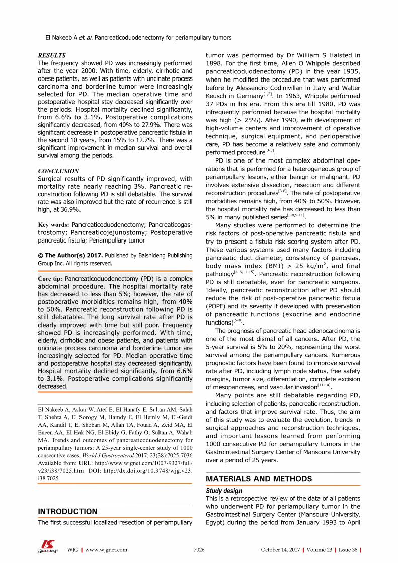
RESULTSThe frequency showed PD was increasingly performed after the year 2000. With time, elderly, cirrhotic and obese patients, as well as patients with uncinate process carcinoma and borderline tumor were increasingly selected for PD. The median operative time and postoperative hospital stay decreased significantly over the periods. Hospital mortality declined significantly, from 6.6% to 3.1%. Postoperative complications significantly decreased, from 40% to 27.9%. There was significant decrease in postoperative pancreatic fistula in the second 10 years, from 15% to 12.7%. There was a significant improvement in median survival and overall survival among the periods.
CONCLUSIONSurgical results of PD significantly improved, with mortality rate nearly reaching 3%. Pancreatic re-construction following PD is still debatable. The survival rate was also improved but the rate of recurrence is still high, at 36.9%.
Key words: Pancreaticoduodenectomy; Pancreaticogas-trostomy; Pancreaticojejunostomy; Postoperative pancreatic fistula; Periampullary tumor
© The Author(s) 2017. Published by Baishideng Publishing Group Inc. All rights reserved.
Core tip: Pancreaticoduodenectomy (PD) is a complex abdominal procedure. The hospital mortality rate has decreased to less than 5%; however, the rate of postoperative morbidities remains high, from 40% to 50%. Pancreatic reconstruction following PD is still debatable. The long survival rate after PD is clearly improved with time but still poor. Frequency showed PD is increasingly performed. With time, elderly, cirrhotic and obese patients, and patients with uncinate process carcinoma and borderline tumor are increasingly selected for PD. Median operative time and postoperative hospital stay decreased significantly. Hospital mortality declined significantly, from 6.6% to 3.1%. Postoperative complications significantly decreased.
El Nakeeb A, Askar W, Atef E, EI Hanafy E, Sultan AM, Salah T, Shehta A, El Sorogy M, Hamdy E, El Hemly M, El-Geidi AA, Kandil T, El Shobari M, Allah TA, Fouad A, Zeid MA, El Eneen AA, EI-Hak NG, El Ebidy G, Fathy O, Sultan A, Wahab MA. Trends and outcomes of pancreaticoduodenectomy for periampullary tumors: A 25-year single-center study of 1000 consecutive cases. World J Gastroenterol 2017; 23(38):7025-7036 Available from: URL: http://www.wjgnet.com/1007-9327/full/v23/i38/7025.htm DOI: http://dx.doi.org/10.3748/wjg.v23.i38.7025
INTRODUCTIONThe first successful localized resection of periampullary
tumor was performed by Dr William S Halsted in 1898. For the first time, Allen O Whipple described pancreaticoduodenectomy (PD) in the year 1935, when he modified the procedure that was performed before by Alessendro Codinivillan in Italy and Walter Keusch in Germany[1,2]. In 1963, Whipple performed 37 PDs in his era. From this era till 1980, PD was infrequently performed because the hospital mortality was high (> 25%). After 1990, with development of high-volume centers and improvement of operative technique, surgical equipment, and perioperative care, PD has become a relatively safe and commonly performed procedure[3-5].
PD is one of the most complex abdominal ope-rations that is performed for a heterogeneous group of periampullary lesions, either benign or malignant. PD involves extensive dissection, resection and different reconstruction procedures[3-8]. The rate of postoperative morbidities remains high, from 40% to 50%. However, the hospital mortality rate has decreased to less than 5% in many published series[5-8,9-11].
Many studies were performed to determine the risk factors of post-operative pancreatic fistula and try to present a fistula risk scoring system after PD. These various systems used many factors including pancreatic duct diameter, consistency of pancreas, body mass index (BMI) > 25 kg/m2, and final pathology[4-6,11-15]. Pancreatic reconstruction following PD is still debatable, even for pancreatic surgeons. Ideally, pancreatic reconstruction after PD should reduce the risk of post-operative pancreatic fistula (POPF) and its severity if developed with preservation of pancreatic functions (exocrine and endocrine functions)[5-8].
The prognosis of pancreatic head adenocarcinoma is one of the most dismal of all cancers. After PD, the 5-year survival is 5% to 20%, representing the worst survival among the periampullary cancers. Numerous prognostic factors have been found to improve survival rate after PD, including lymph node status, free safety margins, tumor size, differentiation, complete excision of mesopancreas, and vascular invasion[11-14].
Many points are still debatable regarding PD, including selection of patients, pancreatic reconstruction, and factors that improve survival rate. Thus, the aim of this study was to evaluate the evolution, trends in surgical approaches and reconstruction techniques, and important lessons learned from performing 1000 consecutive PD for periampullary tumors in the Gastrointestinal Surgery Center of Mansoura University over a period of 25 years.
MATERIALS AND METHODSStudy designThis is a retrospective review of the data of all patients who underwent PD for periampullary tumor in the Gastrointestinal Surgery Center (Mansoura University, Egypt) during the period from January 1993 to April
7026 October 14, 2017|Volume 23|Issue 38|WJG|www.wjgnet.com
El Nakeeb A e t al. Pancreaticoduodenectomy for periampullary tumors

2017. Patient data were recorded in a prospectively maintained database for all patients undergoing PD since the year 2000; for patients from before 2000, the data were obtained from the archived files. An informed consent for the surgical procedures was obtained from each patient. The Gastrointestinal Surgery Center of Mansoura University is a high-volume center of pancreatic surgery that was established in 1992. The first PD was performed in 1993, and was regularly performed afterwards in our center over a period of 25 years.
Inclusion criteriaThis study included 1000 patients who underwent PD for different periampullary tumors (benign and malignant lesions) at our Center in Mansoura University, Egypt during the period from January 1993 to April 2017. Over the 25-year period, 1000 consecutive PD were performed by 20 surgeons. For this study, the data were categorized into three periods, early period (1993-2002), middle period (2003-2012), and late period (2013-2017). This study was approved by the local institutional review board (IRB).
Exclusion criteriaPatients with periampullary lesions who were explored during the same period and failed to complete the PD procedure due to the presence of locally advanced or distant metastatic disease that was not detected in preoperative radiological workup.
Preoperative assessmentPreoperative diagnostic workup included clinical assessment, detailed laboratory investigations, including tumor markers, and radiological investigations (abdominal ultrasound, abdominal triphasic computed tomography (CT), CT angiography, magnetic resonance cholangiopancreatography, chest X-ray and bone survey). Preoperative biliary drainage was performed by endoscopic retrograde cholangiopancreatography in selected patients[16].
Surgical proceduresOver the study period and with accumulating experience, evolution of the surgical approach and techniques occurred.
Dissection techniqueIn the early period, the anterior approach was utilized in surgical dissection. Afterwards, we shifted to utilize the posterior approach (one of the artery first approaches), aiming to identify vascular invasion at an early stage of the dissection, and to allow more radical excision of the mesopancreas[17]. Standard regional lymphadenectomy was performed, which included resection of nodes within the outlines of the hepatoduodenal ligament, right side of the superior
mesenteric vessels, and inferior vena cava. In the early period, diathermy dissection and ligatures were used during the resection stage. Afterwards, a shift to use modern energy devices occurred, such as for Ligasure and Harmonic scalpel.
ApproachIn most of our study period, we utilized an open surgical approach through extended right subcostal or inverted J incisions. In the late period, we started to utilize laparoscopic approach, with laparoscopy-assisted approach used in the beginning. This included complete dissection by the laparoscopy followed by reconstruction carried out through a small upper midline or transverse incision. In the last year, we performed a total of 10 laparoscopic PDs. This included completing the whole approach (dissection and reconstruction) by laparoscopy.
Meso-pancreatectomyA complete removal of all lympho-vascular tissues between the uncinate process and superior mesenteric artery was mandatory in PD. These tissues are the most important site for local recurrence after PD. This concept had evolved in the recent years and became a standard step in the radical resection of periampullary tumors. We adopted this concept in the recent years of our study.
Division of the pancreatic neckInitially, we divided the pancreatic neck sharply by surgical scalpel and then carried out the hemostasis after division. Recently, we started to divide the pancreatic neck by diathermy and Harmonic scalpel.
ReconstructionIn the beginning of our series, we performed simple loop pancreaticojejunostomy (PJ) for the reconstruction of the pancreatic stump. However, a high rate of pancreatic fistula was noticed. A shift of the reconstruction plan occurred to pancreaticogastrostomy (PG). Short-term outcomes were improved and lower rate of pancreatic fistula was noticed, but the long-term outcomes regarding the digestive and nutritional conditions were not appropriate.
With accumulating experience and refinement of the surgical technique, a re-shift to PJ (simple loop or isolated loop) occurred, which improved long-term outcomes[18,19]. Recently, we adopted a tailored approach for pancreatic stump management. In patients at high risk of pancreatic fistula (presence of two or more risk factors) PG is preferred. In low- and moderate-risk patients (absence of risk factors or presence of one risk factor) PJ is preferred.
Postoperative managementAll patients were transferred to the intensive care unit (ICU) postoperatively. Antibiotics and analgesic were
7027 October 14, 2017|Volume 23|Issue 38|WJG|www.wjgnet.com
El Nakeeb A et al . Pancreaticoduodenectomy for periampullary tumors

7028 October 14, 2017|Volume 23|Issue 38|WJG|www.wjgnet.com
variables were described using frequency distributions. The χ 2 test was used to compare categorical variables and one-way ANOVA for continuous variables. The predictive factors for postoperative complications were evaluated by binary logistic regression method. Survival outcomes were calculated by the Kaplan-Meier method. The predictive factors for survival were evaluated by the Cox regression method. P values < 0.05 were considered to be significant.
RESULTSOne thousand patients underwent PD for resection of periampullary tumors from January 1993 to April 2017. Of the 1000 patients who underwent PD, 556 involved pancreatic head mass, 312 were ampullary tumors, 61 were duodenal tumors, 41 were cholangiocarcinoma and 30 were uncinate process mass. The median age of patients was 54-years-old. The data were categorized into three periods. For the first 10 years (1993-2002), a total of 300 patients underwent PD (30 cases/year). In the next 10 years (2003-2012), the total number was 442 patients who underwent PD (44.2 cases/year). In the last 5 years (2013-2017), the total number was 258 patients who underwent PD (51.6 cases/year) (Figure 1).
Preoperative dataElderly patients were increasingly selected for PD, as the median age was 53 in the first 10 years and 55 in the last 5 years. Obese patients were increasingly selected in the last five years. There were no significant changes for selection of patients for PBD in the period of the study. PBD was indicated for patients with high serum bilirubin (> 10 mg%) with high liver enzymes, renal impairment or associated cholangitis (Table 1).
Intraoperative dataPatients with periampullary tumors and well-compensated chronic liver disease were increasingly selected for PD (Table 2). In the early period, we performed simple loop PJ (21.7%) for the reconstruction of the pancreatic stump, then shifted to PG (78.3%). In the second 10 years, 94.3% of cases underwent PG. In the last 5 years, there was a re-shift to PJ (simple loop or isolated loop) (46.1%). Complete meso-pancreatecomy was achieved in all cases in the last 5 years Operative time was significantly reduced, from 6 h in the first 10 years to 5 h in last 5 years. The median intraoperative blood loss decreased from 500 cc in the first 10 years to 300 cc in last 5 years.
Postoperative dataThe overall morbidity of all 1000 patients was 32.3%. The postoperative complications decreased markedly in the recent years, from 40% to 27.9%. There was a significant decrease in POPF in the second 10
given to all patients. Octreotide analogue was given to some patients postoperatively. Abdominal drains and nasogastric tubes outputs were recorded daily. Patients started oral feeding once bowel sounds restarted and they were able to tolerate nutrition by a fluid diet, then a regular diet.
Abdominal ultrasound was done routinely in all patients postoperatively. Serum amylase and liver function tests were measured on postoperative day (POD) 1 and POD 5. Ultrasound-guided tubal drainage was carried out in patients who had an abdominal collection.
Follow-up occurred at 1 wk, 3 mo and 6 mo po-stoperatively, and then at 1 year. Patients were also seen at outpatient clinics if symptoms developed between follow-up visits.
DefinitionsComplications were defined as adverse events resulting in deviation from the normal postoperative course within 30 d after operation. Severity of complications was assessed using the Clavien classification system, from 1 to 5. Major complications represent those requiring endoscopic, radiologic or surgical intervention, and were defined as class 3 or higher[20].
Postoperative pancreatic fistula was defined by International Study Group of Pancreatic Fistula as any measurable volume of fluid on or after POD 3 with amylase content greater than three times the serum amylase activity, and classified into three grades: A, B or C[21-23].
Delayed gastric emptying (DGE) was defined as output from a nasogastric tube of greater than 500 mL per day that persisted beyond POD 10, the failure to maintain oral intake by POD 14, or reinsertion of a nasogastric tube[21-24].
Outcomes of the studyThe aim of this study was to evaluate the milestones, trends in surgical approaches, reconstruction techniques and important lessons learned from performing 1000 consecutive PD for periampullary tumors in the Gastrointestinal Surgery Center of Mansoura University over a period of 25 years. The main outcome of the study was the rates of postoperative morbidity, according to Clavien-Dindo classifications, and of mortality after PD. Special concern was focused on POPF, biliary complications, DGE and the predictive factors of each. In addition, we evaluated the survival outcomes of the PD patients, including recurrence, overall survival (OS) and the different predictive factors of each.
Statistical analysisStatistical analysis of the data in this study was performed using SPSS software for windows, version 20. For continuous variables, descriptive statistics were calculated and reported as median. Categorical
El Nakeeb A et al . Pancreaticoduodenectomy for periampullary tumors

7029 October 14, 2017|Volume 23|Issue 38|WJG|www.wjgnet.com
years, from 15% to 12.7% with a decrease in the severity. But the incidence of POPF increased again in last 5 years to 14.7%. DGE was the most common complication (18%). It was secondary to other postoperative complications in 15.2%. Primary DGE presented in 2.8% of cases (Table 3).
The median hospital stay and the day of drain removal were significantly shortest in the late period, decreasing from 9 days to 8 days. The overall hospital mortality of all 1000 patients was 4.3% (43 patients). The hospital mortality declined significantly, from 6.6% to 3.1%. The causes of death were sepsis secondary to POPF in 17 patients, with 6 cases due to cardiac arrest, 6 cases due to liver cell failure, 5 cases due to pulmonary embolism, 3 cases due to pancreatitis, 3 cases due to respiratory failure secondary to severe chest infection, 2 cases due to secondary hemorrhage, and 1 case due to PG–related uncontrolled bleeding.
Seventy patients developed intra-abdominal collection and were managed by ultrasound-guided tubal drainage. Seventy-four patients (7.4%) required re-explorations due to internal hemorrhage (26 patients, with 7/26 due to erosion of gastroduodenal artery), bleeding GJ (17 patients), bleeding PG (15 patients), peritonitis (12 patients) or debridement and drainage (4 patients). Completion spleno-pancreatectomy was needed in 2 cases due to POPF that eroded the gastroduodenal artery and were complicated by secondary internal hemorrhage
The overall recurrence rate in 870 patients who had malignant pathology after PD was 36.9.2%. This rate decreased from 50.4% to 28.7%
Univariate analysis of risk factors for development of POPF found that six variables were significantly associated with POPF (BMI > 25 kg/m2, liver cirrhosis, soft pancreas, main pancreatic duct < 3 mm, pancreatic duct close to posterior edge < 3 mm, and period of the study). These six risk factors of POPF
identified in univariate analysis were further analyzed in multivariate analysis. Soft pancreas, main pancreatic duct < 3 mm pancreatic duct close to posterior edge < 3 mm, BMI > 25 kg/m2 and period of the study were found to be independent risk factors (Table 4).
Postoperative pathologyThere was significant difference among groups regarding site of periampullary tumor, type of pathology, number of dissected lymph nodes, number of infiltrated lymph nodes, lymph node ratio, safety margin, perivascular infiltration and perineural invasion (Table 5).
Survival rateThe 1-, 3- and 5-year OS rates for all cases were 90%, 33% and 19% respectively, with a median survival of 26 mo. There was a significant difference among the groups regarding the median survival and the OS rates at 1-, 3- and 5-years (Tables 3 and 6, Figure 2). The survival analysis in this study revealed that female sex, patients who did not develop major complications, presence of ampullary tumor, type of pathology, negative safety margin, negative lymph nodes, chemoradiotherapy and period of the study were all favorable prognostic variables in univariate and multivariate analyses. The improvement of survival with recent years may be due to complete excision of mesopancreas, greater use of postoperative chemoradiotherapy, improvement of surgical techniques, and strict follow up of most of cases (Table 6).
DISCUSSIONPD is a complex procedure including extensive dissection, resection and multiple reconstruction. Allen O Whipple described PD in the year 1935. From Whipple’s era till 1980, PD was performed infrequently
Figure 1 In the first 10 years (1993-2002), the total number of patients who underwent PD was 300 (30 cases/yr), in the next 10 years (2003-2012) 442 patients underwent PD (44.2 cases/yr), and in the last 4 years (2013-2017) 258 cases underwent PD (51.6 cases/yr).
7065605550454035302520151050
Freq
uenc
y
2016.002015.002014.002013.002012.002011.002010.002009.002008.002007.002006.002005.002004.002003.002002.002001.002000.001999.001998.001997.001996.001995.001994.001993.00
T/yr
Mo
Cum
sur
viva
l
1.0
0.8
0.6
0.4
0.2
0.0
30028827626425224022821620419218016815614413212010896847260483624120
Period1993-20022003-20122013-2017
Figure 2 Overall survival curves of patients according to the 3 periods of study. The overall survival rate significantly improved and 5-yr survival rate for the early period was 11%, followed by 21% for the middle period and 64% for the late period (P = 0.0001)
El Nakeeb A et al . Pancreaticoduodenectomy for periampullary tumors
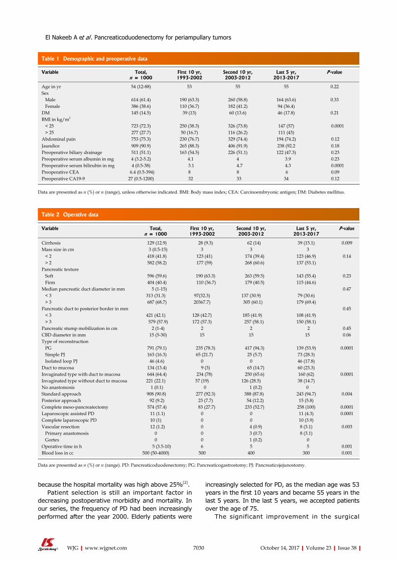
Variable Total, n = 1000
First 10 yr,1993-2002
Second 10 yr,2003-2012
Last 5 yr,2013-2017
P-value
Cirrhosis 129 (12.9) 28 (9.3) 62 (14) 39 (15.1) 0.009Mass size in cm 3 (0.5-15) 3 3 3 < 2 418 (41.8) 123 (41) 174 (39.4) 123 (46.9) 0.14 > 2 582 (58.2) 177 (59) 268 (60.6) 137 (53.1)Pancreatic texture Soft 596 (59.6) 190 (63.3) 263 (59.5) 143 (55.4) 0.23 Firm 404 (40.4) 110 (36.7) 179 (40.5) 115 (44.6)Median pancreatic duct diameter in mm 5 (1-15) 0.47 < 3 313 (31.3) 97(32.3) 137 (30.9) 79 (30.6) > 3 687 (68.7) 20367.7) 305 (60.1) 179 (69.4)Pancreatic duct to posterior border in mm 0.45 < 3 421 (42.1) 128 (42.7) 185 (41.9) 108 (41.9) > 3 579 (57.9) 172 (57.3) 257 (58.1) 150 (58.1)Pancreatic stump mobilization in cm 2 (1-4) 2 2 2 0.45CBD diameter in mm 15 (5-30) 15 15 15 0.06Type of reconstruction PG 791 (79.1) 235 (78.3) 417 (94.3) 139 (53.9) 0.0001 Simple PJ 163 (16.3) 65 (21.7) 25 (5.7) 73 (28.3) Isolated loop PJ 46 (4.6) 0 0 46 (17.8)Duct to mucosa 134 (13.4) 9 (3) 65 (14.7) 60 (23.3)Invaginated type with duct to mucosa 644 (64.4) 234 (78) 250 (65.6) 160 (62) 0.0001Invaginated type without duct to mucosa 221 (22.1) 57 (19) 126 (28.5) 38 (14.7)No anastomosis 1 (0.1) 0 1 (0.2) 0Standard approach 908 (90.8) 277 (92.3) 388 (87.8) 243 (94.7) 0.004Posterior approach 92 (9.2) 23 (7.7) 54 (12.2) 15 (5.8)Complete meso-pancreatectomy 574 (57.4) 83 (27.7) 233 (52.7) 258 (100) 0.0001Laparoscopic assisted PD 11 (1.1) 0 0 11 (4.3) 0.0001Complete laparoscopic PD 10 (1) 0 0 10 (3.9)Vascular resection 12 (1.2) 0 4 (0.9) 8 (3.1) 0.003 Primary anastomosis 0 0 3 (0.7) 8 (3.1) Gortex 0 0 1 (0.2) 0Operative time in h 5 (3.5-10) 6 5 5 0.001Blood loss in cc 500 (50-4000) 500 400 300 0.001
Data are presented as n (%) or n (range). PD: Pancreaticoduodenectomy; PG: Pancreaticogastrostomy; PJ: Pancreaticojejunostomy.
7030 October 14, 2017|Volume 23|Issue 38|WJG|www.wjgnet.com
because the hospital mortality was high above 25%[2]. Patient selection is still an important factor in
decreasing postoperative morbidity and mortality. In our series, the frequency of PD had been increasingly performed after the year 2000. Elderly patients were
increasingly selected for PD, as the median age was 53 years in the first 10 years and became 55 years in the last 5 years. In the last 5 years, we accepted patients over the age of 75.
The significant improvement in the surgical
Table 1 Demographic and preoperative data
Variable Total,n = 1000
First 10 yr,1993-2002
Second 10 yr,2003-2012
Last 5 yr,2013-2017
P-value
Age in yr 54 (12-88) 53 55 55 0.22Sex Male 614 (61.4) 190 (63.3) 260 (58.8) 164 (63.6) 0.33 Female 386 (38.6) 110 (36.7) 182 (41.2) 94 (36.4)DM 145 (14.5) 39 (13) 60 (13.6) 46 (17.8) 0.21BMI in kg/m2
< 25 723 (72.3) 250 (38.3) 326 (73.8) 147 (57) 0.0001 > 25 277 (27.7) 50 (16.7) 116 (26.2) 111 (43)Abdominal pain 753 (75.3) 230 (76.7) 329 (74.4) 194 (74.2) 0.12Jaundice 909 (90.9) 265 (88.3) 406 (91.9) 238 (92.2 0.18Preoperative biliary drainage 511 (51.1) 163 (54.5) 226 (51.1) 122 (47.3) 0.23Preoperative serum albumin in mg 4 (3.2-5.2) 4.1 4 3.9 0.23Preoperative serum bilirubin in mg 4 (0.5-38) 3.1 4.7 4.3 0.0001Preoperative CEA 6.4 (0.5-394) 8 8 6 0.09Preoperative CA19-9 27 (0.5-1200) 32 33 34 0.12
Data are presented as n (%) or n (range), unless otherwise indicated. BMI: Body mass index; CEA: Carcinoembryonic antigen; DM: Diabetes mellitus.
Table 2 Operative data
El Nakeeb A et al . Pancreaticoduodenectomy for periampullary tumors

7031 October 14, 2017|Volume 23|Issue 38|WJG|www.wjgnet.com
outcome of PD has encouraged surgeons to approach periampullary tumors as aggressively in elderly patients[7,12]. Patients with periampullary tumors and well-compensated chronic liver disease are increasingly selected for PD with accepted surgical outcomes. PD is only recommended in patients with Child A cirrhosis without portal hypertension[13]. PD is associated with an increased risk of postoperative morbidity in obese patients. With time, obesity has lost its status as a limitation for PD and such patients have been increasingly selected[25-27].
Patients with uncinate process carcinoma are increasingly selected for PD as well. However, the loco-regional recurrence rate was found to be common and the OS rate to be lower than other periampullary tumors[28]. The role of postoperative chemoradiotherapy may improve the results. Borderline resectable pancreatic cancer should be included as an indication of PD with advancements in chemoradiotherapy and techniques of vascular resection[29].
The impact of PBD on postoperative outcomes remains controversial. PBD before PD was associated
with major postoperative morbidities and stent-related morbidities, including infection, pancreatitis or adhesions. There are no significant changes for selection of patients for PBD in the period of the study. PBD is indicated for patients with high serum bilirubin (> 10 mg%) with high liver enzymes, renal impairment, or associated cholangitis[16,30,31].
In our Center, laparoscopic pancreaticoduo-denectomy (LPD) has been introduced as a feasible alternative to open PD since 2013, involving dissection in some cases, then complete LPD was performed at the end of 2013; unfortunately, however, the patients died on POD 7. We restarted again in January 2016 to perform 10 complete LPDs; in all cases, the pancreatic reconstruction was PG. The median operative time was 8 h, so the procedure was performed by two teams (one team for complete dissection and the other one to perform all reconstruction). Only one hospital mortality occurred due to severe pancreatitis, and all cases passed smoothly without any complications. The median hospital stay was 5 days. The most important point to performing a safe LPD is to perform the procedure under skilled hands in selected patients and
Table 3 Postoperative data
Variable Total, n = 1000
First 10 yr,1993-2002
Second 10 yr,2003-2012
Last 5 yr,2013-2017
P-value
Hospital stay in d 8 (5-71) 9 8 8 0.0001Time to oral intake in d 5 (4-56) 6 5 5 0.33Total amount of drainage in mL 700 (40-35000) 1200 600 600 0.0001Drain removal in d 8 (4-71) 8 8 8 0.03Total postoperative complications 323 (32.3) 120 (40) 131 (29.6) 72 (27.9) 0.02Dindo grade I 114 (11.4) 24 47 43 II 97 (9.7) 40 45 12 0.11 III 69 (6.9) 36 24 9 IV and V 43 (4.3) 20 15 8Severe complications, ≥ III Minor 211 (21.1) 84 92 55 0.23 Major 109 (10.9) 56 69 17Pancreatic fistula 139 (13.9) 45 (15) 56 (12.7) 38 (14.7) 0.01 Grade A 67 (6.7) 14 33 20 0.04 Grade B 48 (4.8) 20 17 11 Grade C 24 (2.4) 11 6 7DGE 180 (18) 76 (25.3) 67 (15.2) 37 (14.3) 0.06Types of DGE Secondary DGE 152 (15.2) 70 (23.3) 54 (12.2) 27 (10.5) 0.03 Primary DGE 28 (2.8) 5 (1.7) 13 (2.9) 10 (3.9)Pulmonary complications 46 (4.6) 20 (6.7) 21 (4.8) 5 (1.6) 0.01Bile leak 73 (7.3) 39 (13) 19 (4.3) 15 (5.8) 0.001Postoperative bleeding 25 (2.5) 13 (4.3) 7 (1.6) 5 (1.9) 0.49Pancreatitis 20 (2) 12 (4.3) 7 (1.6) 1 (0.4) 0.004Bleeding PG 15 (1.5) 8 (2.7) 5 (1.2) 2 (0.8) 0.14Wound infection 50 (5) 13 (4.6) 25 (5.7) 12 (4.7) 0.77Re-operation 74 (7.4) 25 (8.3) 33 (7.5) 16 (6.2) 0.21Recurrence 321 (36.9) 130 (50.4) 125 (32.6) 66 (28.7) 0.0001Hospital mortality 43 (4.3) 20 (6.6%) 15 (3.4) 8 (3.1) 0.02Postoperative chemoradiotherapy 275 (27.5) 0 132 (29.9) 143 (55.4) 0.0001Overall median survival in mo 26 (1-300) 21 30 37 0.0001 1 yr 90% 87% 93% 87% 3 yr 33% 19% 37% 64% 5 yr 19% 11% 21%
Data are presented as n (%) or n (range), unless otherwise indicated. DGE: Delayed gastric emptying; PG: Pancreaticogastrostomy.
El Nakeeb A et al . Pancreaticoduodenectomy for periampullary tumors

7032 October 14, 2017|Volume 23|Issue 38|WJG|www.wjgnet.com
using suitable surgical techniques[32,33].The ideal and safe pancreatic reconstruction
following PD is still debatable. We performed a comparative randomized study between PG and isolated loop PJ which revealed no significant difference between both methods in regards to POPF, but the
pancreatic function was preserved with isolated loop PJ[18,19]. Recently, with accumulating experience and refinement of the surgical technique, we adopted a tailored approach for the management of pancreatic stump management. Another prospective randomized study comparing duct to mucosa and invagination PJ
Table 4 Univariate and multivariate analyses of risk factors of development of post-operative pancreatic fistula
Univariate Multivariate Exp(B) 95%CI for Exp(B)
P -value P -value Lower UpperAge grouping > 60 yr 0.2Sex 0.99DM 0.58BMI >25 kg/m2 0.0001 0.0001 6.468 4.193 9.977Preoperative serum bilirubin > 10 mg% 0.62Preoperative ERCP 0.52Liver cirrhosis 0.05 0.328 0.699 0.341 1.434Size of the tumor > 2 cm 0.91Soft pancreas 0.0001 0.0001 0.218 0.14 0.341Pancreatic duct diameter > 3 mm 0.0001 0.0001 0.182 0.118 0.279Pancreatic duct closely related to posterior border of pancreas > 3 mm 0.0001 0.0001 0.372 0.243 0.57Blood loss > 1000 mL 0.67Blood transfusion 0.94Type of pancreatic reconstruction 0.62Duration of operation 0.75Site of the tumor 0.34Period of the study 0.001 0.001 0.615 0.461 0.82
BMI: Body mass index; DM: Diabetes mellitus; ERCP: Endoscopic retrograde cholangiopancreatography.
Table 5 Postoperative pathology n (%)
Variable Total, n = 1000
First 10 yr,1993-2002
Second 10 yr,2003-2012
Last 5 yr,2013-2017
Site of the tumor Ampullary tumor 312 (31.2) 92 (30.7) 145 (32.8) 75 (29) Pancreatic head mass 556 (55.6) 171 (57) 257 (58.1) 128 (49.6) 0.0001 CBD duct tumor 41 (4.1) 16 (5.3) 13 (2.9) 12 (4.7) Duodenal tumor 61 (6.1) 20 (6.7) 27 (6.1) 14 (5.4)Uncinate process mass 30 (3) 1 (0.3) 0 29 (11.3)Pathological diagnosis Adenocarcinoma 836 (83.6) 250 (83.3) 357 (80.7) 229 (88.8) Undifferentiated carcinoma 20 (2) 1 (1.7) 19 (4.3) 0 Papillary cystadenocarcinoma 6 (0.6) 4 (1.3) 2 (0.5) 0 0.0001 Lymphoma 4 (0.4) 2 (0.6) 2 (0.5) 0 Neuroendocrine tumor 28 (2.8) 12 (4) 10 (2.3) 6 (2.3) Solid pseudopapillary tumor 20 (2) 5 (1.7) 8 (1.8) 7 (2.7) Chronic pancreatitis 24 (2.4) 6 (2) 14 (3.2) 4 (1.6) Benign cyst 12 (1.2) 3 (1) 8 (1.8) 1 (0.4) Adenoma with dysplasia 41 (4.1) 15 (5) 19 (4.3) 7 (2.7) Gastrointestinal stromal tumor 2 (0.2) 0 1 (0.2) 1 (0.4) Glomus 1 (0.1) 1 (0.3) 0 0 Adenosquamous 2 (0.2) 1 (0.3) 1 (0.2) 0 Pleomorphic adenoma 1 (0.1) 0 1(0.2) 0 Adenomyoma 3 (0.3) 0 0 3 (1.2)Malignant 870 (87) 258 (86) 382 (86.4) 230 (89.1) 0.38Borderline 48 (4.8) 17(5.7) 18 (4.1) 13 (5.1)benign 82 (8.2) 25 (8.3) 42 (9.5) 15 (5.8)Number of dissected lymph node 6 (0-40) 5 (0-18) 6 (0-40) 6 (0-40) 0.59Number of lymph node infiltration 0 (0-14) 0 (0-3) 0 (0-14) 0 (0-14) 0.07Perineural infiltration 187 (18.7) 62 (20.7) 75 (17) 50 (19.9) 0.09Perivascular infiltration 134 (134) 40 (13.3) 66 (14.9) 28 (10.9) 0.13Pancreatic safety margin R1 91 (9.1) 40 (13.3) 41 (9.3) 10 (3.8) 0.01 R2 15 (1.5) 7 (2.3) 6 (1.4) 2 (0.7)
El Nakeeb A et al . Pancreaticoduodenectomy for periampullary tumors

7033 October 14, 2017|Volume 23|Issue 38|WJG|www.wjgnet.com
El Nakeeb A et al . Pancreaticoduodenectomy for periampullary tumors
Table 6 Univariate and multivariate analyses of factors affecting the survival
Median survival
1 yr survival
3 yr survival
5 yr survival
Univariate P -value
Multivariate P -value
Exp(B) 95%CI for Exp(B)
Lower Upper> 60 yr 26 91 35 20 0.07< 60 yr 24 87 31 18Male 24 89 30 16 0.001 0.003 0.77 0.651 0.914Female 30 91 39 25BMI < 25 kg/m2 27 92 32 19 0.99BMI > 25 kg/m2 25 84 37 20Preoperative serum bilirubin < 10 mg% 26 90 34 21 0.6Preoperative serum bilirubin > 10 mg% 25 90 33 18Preoperative CA19-9 < 37 26 90 35 24 0.17Preoperative CA 19-9 > 37 25 89 31 14Preoperative CEA < 5 26 90 34 21 0.69Preoperative CEA > 5 25 91 33 18History of preoperative ERCP 25 88 33 15 0.19No history of ERCP 26 92 33 25Normal liver 32 90 37 24 0.11Liver cirrhosis 25 90 33 19Size of the tumor < 2 cm 28 91 37 20 0.16Size of the tumor > 2 cm 25 89 31 19Type of pancreatic reconstructionPG 25 90 31 17 0.11Simple loop PJ 32 90 44 28Isolated loop PJ 32 80 48 48Without POPF 26 90 34 19 0.79With POPF 25 89 28 23Without major postoperative complications 27 93 35 20 0.03 0.005 0.784 0.661 0.929With major postoperative complications 24 82 31 18Site of the tumor Ampullary tumor 33 90 42 22 Pancreatic head mass 23 81 28 11 0.02 0.3 1.049 0.958 1.148 CBD duct tumor 32 90 30 20 Duodenal tumor 31 90 42 21 Uncinate process mass 12 79 18 0Type of pathology Adenocarcinoma 26 90 27 8 Neuroendocrine tumor 65 85 71 60 Solid pseudopapillary tumor 150 100 94 94 0.0001 0.005 0.97 0.951 0.991 Chronic pancreatitis 160 96 96 84 Benign cyst 130 100 90 70 Adenoma with dysplasia 85 91 67 48Lymph node negative 29 92 38 27 0.0001 0.0001 0.559 0.413 0.757Lymph node infiltrated 25 87 27 8Lymph node ratio 0 28 91 38 27 < 0.2 25 89 31 18 0.0001 0.15 0.908 0.797 1.034 0.2-0.4 25 86 28 11 > 0.4 24 88 21 4Without perineural infiltration 25 89 35 21 0.06With perineural infiltration 24 91 28 12Without perivascular infiltration 29 91 44 24 0.29With perivascular infiltration 25 86 35 14Pancreatic safety margin R0 26 90 35 20 R1 22 92 27 19 0.0001 0.0001 1.695 1.345 2.136 R2 12 70 19 0With postoperative chemoradiotherapy 39 92 53 34 0.0001 0.0001 0.567 0.435 0.741Without postoperative chemoradiotherapy 24 89 28 15Period of the study 1993-2002 21 87 19 11 2003-2012 30 93 37 21 0.001 0.0001 0.64 0.555 0.738 2013-2017 37 87 63
BMI: Body mass index; CBD: Common bile duct; CEA: Carcinoembryonic antigen; ERCP: Endoscopic retrograde cholangiopancreatography; PG: Pancreaticogastrostomy; PJ: Pancreaticojejunostomy; POPF: Postoperative pancreatic fistula.
7034 October 14, 2017|Volume 23|Issue 38|WJG|www.wjgnet.com
carried out in our center concluded that invagination PJ is preferred in small pancreatic duct and provides less incidence of postoperative steatorrhea and was associated with less severity of POPF, if developed, than duct to mucosa[19].
In our series, 32.3% of patients undergoing PD had complications. The majority of complications were minor and not life threatening. The postoperative complications decreased markedly in the recent years, from 40% to 27.9%. There was significant decrease in POPF in the second 10 years, from 15% to 12.7% with decrease in severity due to shift of pancreatic reconstruction from PJ to PG. But, the incidence of POPF increased again in last 5 years to 14.7%, due to re-shift again to PJ in order to achieve better long-term outcomes regarding function and morphology of pancreas[18,19]. In this study, the development of major complications had a negative impact on OS.
The median hospital stay was significantly shortest in the last 10 years. In many high-volume centers, there has been significant decrease in postoperative stay after PD as a result of the increase in the frequency of PD, the decrease in incidence of complications, especially of DGE, the decrease in use of pylorus-preserving PD, which is complicated by high incidence of DGE, and the improvements in postoperative care and management of postoperative complications[6,7,34-36].
In this study, the overall hospital mortality was 4.3%. Of the patients who died, 17/43 (39.5%) of the deaths were due to sepsis secondary to POPF. The decrease in hospital mortality following PD over the time is the most prominent achievement in PD. In this study, the hospital mortality rate decreased significantly from 6.6% to 3.1%, which is comparable to the reported mortality rates in other high-volume centers[6-8].
Long-term survival after PD for periampullary tumor adenocarcinoma is still poor. However, the survival after PD has clearly improved with time, due to improvement of surgical techniques, complete excision of mesopancreas, greater use of neoadjuvant and/or adjuvant chemoradiotherapy, and strict follow up of most of cases. There are many high-volume centers in which patients with pancreatic head carcinoma treated by PD have a 5-year survival rate of around 20%[6,7,35,36].
In conclusion, the frequency of PD has increased and become a relatively safe procedure in our Center. With time, elderly, cirrhotic, obese patients, patients with uncinate process carcinoma and borderline tumors have been increasingly selected for PD. Surgical results of PD, including operative time, hospital stay and postoperative complications, have significantly improved, with the mortality rate reaching nearly 3%. Pancreatic reconstruction following PD is still debatable.
PG provides better short-term outcomes including POPF, but the long-term outcomes regarding the pancreatic function and nutrition were not appropriate. However, PJ provides better long-term outcomes. The survival rate also improved due to complete meso-pancreatectomy and utilization of adjuvant chemoradiotherapy, but the rate of recurrence is still high at 36.9%. LPD has been introduced as a feasible alternative to open PD.
REFERENCES1 Kausch W. Das carcinoma der papilla duodeni und seni-
neradikaleentfeinung. Beitr 2 ClincChir 1912; 78: 439e4862 Whipple AO. Pancreaticoduodenectomy for Islet Carcinoma :
A Five-Year Follow-Up. Ann Surg 1945; 121: 847-852 [PMID: 17858621 DOI: 10.1097/00000658-194506000-00008]
3 Takagi K, Yagi T, Yoshida R, Shinoura S, Umeda Y, Nobuoka D, Kuise T, Watanabe N, Sui K, Fujii T, Fujiwara T. Surgical Outcome of Patients Undergoing Pancreaticoduodenectomy: Analysis of a 17‒Year Experience at a Single Center. Acta Med Okayama 2016; 70: 197-203 [PMID: 27339209]
4 Kimura W, Miyata H, Gotoh M, Hirai I, Kenjo A, Kitagawa Y, Shimada M, Baba H, Tomita N, Nakagoe T, Sugihara K, Mori M. A pancreaticoduodenectomy risk model derived from 8575 cases from a national single-race population (Japanese) using a web-based data entry system: the 30-day and in-hospital mortality rates for pancreaticoduodenectomy. Ann Surg 2014; 259: 773-780 [PMID: 24253151 DOI: 10.1097/SLA.0000000000000263]
5 El Nakeeb A, Salah T, Sultan A, El Hemaly M, Askr W, Ezzat H, Hamdy E, Atef E, El Hanafy E, El-Geidie A, Abdel Wahab M, Abdallah T. Pancreatic anastomotic leakage after pancreaticoduodenectomy. Risk factors, clinical predictors, and management (single center experience). World J Surg 2013; 37: 1405-1418 [PMID: 23494109 DOI: 10.1007/s00268-013-1998-5]
6 Cameron JL, Riall TS, Coleman J, Belcher KA. One thousand consecutive pancreaticoduodenectomies. Ann Surg 2006; 244: 10-15 [PMID: 16794383 DOI: 10.1097/01.sla.0000217673.04165.ea]
7 Cameron JL, He J. Two thousand consecutive pancrea-ticoduodenectomies. J Am Coll Surg 2015; 220: 530-536 [PMID: 25724606 DOI: 10.1016/j.jamcollsurg.2014.12.031]
8 He J, Ahuja N, Makary MA, Cameron JL, Eckhauser FE, Choti MA, Hruban RH, Pawlik TM, Wolfgang CL. 2564 resected periampullary adenocarcinomas at a single institution: trends over three decades. HPB (Oxford) 2014; 16: 83-90 [PMID: 23472829 DOI: 10.1111/hpb.12078]
9 Yoshioka R, Yasunaga H, Hasegawa K, Horiguchi H, Fushimi K, Aoki T, Sakamoto Y, Sugawara Y, Kokudo N. Impact of hospital volume on hospital mortality, length of stay and total costs after pancreaticoduodenectomy. Br J Surg 2014; 101: 523-529 [PMID: 24615349 DOI: 10.1002/bjs.9420]
10 Balzano G, Zerbi A, Capretti G, Rocchetti S, Capitanio V, Di Carlo V. Effect of hospital volume on outcome of pancrea-ticoduodenectomy in Italy. Br J Surg 2008; 95: 357-362 [PMID: 17933001 DOI: 10.1002/bjs.5982]
11 de Wilde RF, Besselink MG, van der Tweel I, de Hingh IH, van Eijck CH, Dejong CH, Porte RJ, Gouma DJ, Busch OR, Molenaar IQ; Dutch Pancreatic Cancer Group. Impact of nationwide centralization of pancreaticoduodenectomy on hospital mortality. Br J Surg 2012; 99: 404-410 [PMID: 22237731 DOI: 10.1002/bjs.8664]
12 El Nakeeb A , Atef E , El Hanafy E , Sa lem A, Askar W, Ezzat H, Shehta A, Abdel Wahab M. Outcomes of pancreaticoduodenectomy in elderly patients. Hepatobiliary
El Nakeeb A et al . Pancreaticoduodenectomy for periampullary tumors

7035 October 14, 2017|Volume 23|Issue 38|WJG|www.wjgnet.com
Pancreat Dis Int 2016; 15: 419-427 [PMID: 27498583 DOI: 10.1016/S1499-3872(16)60105-4]
13 El Nakeeb A, Sultan AM, Salah T, El Hemaly M, Hamdy E, Salem A, Moneer A, Said R, AbuEleneen A, Abu Zeid M, Abdallah T, Abdel Wahab M. Impact of cirrhosis on surgical outcome after pancreaticoduodenectomy. World J Gastroenterol 2013; 19: 7129-7137 [PMID: 24222957 DOI: 10.3748/wjg.v19.i41.7129]
14 Winter JM, Cameron JL, Campbell KA, Arnold MA, Chang DC, Coleman J, Hodgin MB, Sauter PK, Hruban RH, Riall TS, Schulick RD, Choti MA, Lillemoe KD, Yeo CJ. 1423 pancreaticoduodenectomies for pancreatic cancer: A single-institution experience. J Gastrointest Surg 2006; 10: 1199-1210; discussion 1210-1211 [PMID: 17114007 DOI: 10.1016/j.gassur.2006.08.018]
15 Vallance AE , Young AL, Macutkiewicz C, Roberts KJ, Smith AM. Calculating the risk of a pancreatic fistula after a pancreaticoduodenectomy: a systematic review. HPB (Oxford) 2015; 17: 1040-1048 [PMID: 26456948 DOI: 10.1111/hpb.12503]
16 El Nakeeb A, Salem A, Mahdy Y, El Dosoky M, Said R, Ellatif MA, Ezzat H, Elsabbagh AM, Hamed H, Alah TA, El Ebidy G. Value of preoperative biliary drainage on postoperative outcome after pancreaticoduodenectomy: A case-control study. Asian J Surg 2016; [PMID: 27955973 DOI: 10.1016/j.asjsur.2016.10.004]
17 Abdel-Wahab M, Sultan A, elGwalby N, Fathy O, AboElenen A, Zied MA, Fouad A, Allah TA, el-Ebiedy G, Gad-ElHak N, Elfiky A, Ezzat F. Modified pancreaticoduodenectomy: experience with 81 cases, Wahab modification. Hepatogastroenterology 2001; 48: 1572-1576 [PMID: 11813575]
18 El Nakeeb A, Hamdy E, Sultan AM, Salah T, Askr W, Ezzat H, Said M, Zeied MA, Abdallah T. Isolated Roux loop pancreaticojejunostomy versus pancreaticogastrostomy after pancreaticoduodenectomy: a prospective randomized study. HPB (Oxford) 2014; 16: 713-722 [PMID: 24467711 DOI: 10.1111/hpb.12210]
19 El Nakeeb A, El Hemaly M, Askr W, AbdEllatif M, Hamed H, Elghawalby A, Attia M, Abdallah T, AbdElWahab M. Comparative study between duct to mucosa and invagination pancreaticojejunostomy after pancreaticoduodenectomy: a prospective randomized study. Int J Surg 2015; 16: 1-6 [PMID: 25682724 DOI: 10.1016/j.ijsu.2015.02.002]
20 Dindo D, Demartines N, Clavien PA. Classification of surgical complications: a new proposal with evaluation in a cohort of 6336 patients and results of a survey. Ann Surg 2004; 240: 205-213 [PMID: 15273542 DOI: 10.1097/01.sla.0000133083.54934.ae]
21 Bassi C, Dervenis C, Butturini G, Fingerhut A, Yeo C, Izbicki J, Neoptolemos J, Sarr M, Traverso W, Buchler M; International Study Group on Pancreatic Fistula Definition. Postoperative pancreatic fistula: an international study group (ISGPF) definition. Surgery 2005; 138: 8-13 [PMID: 16003309 DOI: 10.1016/j.surg.2005.05.001]
22 Bassi C, Marchegiani G, Dervenis C, Sarr M, Abu Hilal M, Adham M, Allen P, Andersson R, Asbun HJ, Besselink MG, Conlon K, Del Chiaro M, Falconi M, Fernandez-Cruz L, Fernandez-Del Castillo C, Fingerhut A, Friess H, Gouma DJ, Hackert T, Izbicki J, Lillemoe KD, Neoptolemos JP, Olah A, Schulick R, Shrikhande SV, Takada T, Takaori K, Traverso W, Vollmer CR, Wolfgang CL, Yeo CJ, Salvia R, Buchler M; International Study Group on Pancreatic Surgery (ISGPS). The 2016 update of the International Study Group (ISGPS) definition and grading of postoperative pancreatic fistula: 11 Years After. Surgery 2017; 161: 584-591 [PMID: 28040257 DOI: 10.1016/j.surg.2016.11.014]
23 DeOliveira ML, Winter JM, Schafer M, Cunningham SC, Cameron JL, Yeo CJ, Clavien PA. Assessment of complications after pancreatic surgery: A novel grading system applied to 633 patients undergoing pancreaticoduodenectomy. Ann Surg 2006;
244: 931-937; discussion 937-939 [PMID: 17122618 DOI: 10.1097/01.sla.0000246856.03918.9a]
24 McMillan MT , Malleo G, Bassi C, Sprys MH, Ecker BL, Drebin JA, Vollmer CM Jr. Pancreatic fistula risk for pancreatoduodenectomy: an international survey of surgeon perception. HPB (Oxford) 2017; 19: 515-524 [PMID: 28202218 DOI: 10.1016/j.hpb.2017.01.022]
25 Williams TK, Rosato EL, Kennedy EP, Chojnacki KA, Andrel J, Hyslop T, Doria C, Sauter PK, Bloom J, Yeo CJ, Berger AC. Impact of obesity on perioperative morbidity and mortality after pancreaticoduodenectomy. J Am Coll Surg 2009; 208: 210-217 [PMID: 19228532 DOI: 10.1016/j.jamcollsurg.2008.10.019]
26 El Nakeeb A, Hamed H, Shehta A, Askr W, El Dosoky M, Said R, Abdallah T. Impact of obesity on surgical outcomes post-pancreaticoduodenectomy: a case-control study. Int J Surg 2014; 12: 488-493 [PMID: 24486933 DOI: 10.1016/j.ijsu.2014.01.017]
27 Tsai S, Choti MA, Assumpcao L, Cameron JL, Gleisner AL, Herman JM, Eckhauser F, Edil BH, Schulick RD, Wolfgang CL, Pawlik TM. Impact of obesity on perioperative outcomes and survival following pancreaticoduodenectomy for pancreatic cancer: a large single-institution study. J Gastrointest Surg 2010; 14: 1143-1150 [PMID: 20431978 DOI: 10.1007/s11605-010-1201-3]
28 El Nakeeb A, Roshdy S, Ask W, Sonbl A, Ali M, Abdelwahab K, Shams N, Abdelwahab M. Comparative study between uncinate process carcinoma and pancreatic head carcinoma after pancreaticodudenectomy (clincopathological features and surgical outcomes). Hepatogastroenterology 2014; 61: 1748-1755 [PMID: 25436374]
29 Chen KT, Devarajan K, Milestone BN, Cooper HS, Denlinger C, Cohen SJ, Meyer JE, Hoffman JP. Neoadjuvant chemoradiation and duration of chemotherapy before surgical resection for pancreatic cancer: does time interval between radiotherapy and surgery matter? Ann Surg Oncol 2014; 21: 662-669 [PMID: 24276638 DOI: 10.1245/s10434-013-3396-5]
30 Addeo P, Rosso E, Fuchshuber P, Oussoultzoglou E, De Blasi V, Simone G, Belletier C, Dufour P, Bachellier P. Resection of Borderline Resectable and Locally Advanced Pancreatic Adenocarcinomas after Neoadjuvant Chemotherapy. Oncology 2015; 89: 37-46 [PMID: 25766660 DOI: 10.1159/000371745]
31 Arkadopoulos N, Kyriazi MA, Papanikolaou IS, Vasiliou P, Theodoraki K, Lappas C, Oikonomopoulos N, Smyrniotis V. Preoperative biliary drainage of severely jaundiced patients increases morbidity of pancreaticoduodenectomy: results of a case-control study. World J Surg 2014; 38: 2967-2972 [PMID: 24952079 DOI: 10.1007/s00268-014-2669-x]
32 Stauffer JA, Coppola A, Villacreses D, Mody K, Johnson E, Li Z, Asbun HJ. Laparoscopic versus open pancreaticoduodenectomy for pancreatic adenocarcinoma: long-term results at a single institution. Surg Endosc 2017; 31: 2233-2241 [PMID: 27604369 DOI: 10.1007/s00464-016-5222-1]
33 Jin WW, Xu XW, Mou YP, Zhou YC, Zhang RC, Yan JF, Zhou JY, Huang CJ, Lu C. [Laparoscopic pancreaticoduodenectomy: a report of 233 cases by a single team]. Zhonghua WeiKe ZaZhi 2017; 55: 354-358 [PMID: 28464575]
34 El Nakeeb A, Askr W, Mahdy Y, Elgawalby A, El Sorogy M, Abu Zeied M, Abdallah T, AbdElwahab M. Delayed gastric emptying after pancreaticoduodenectomy. Risk factors, predictors of severity and outcome. A single center experience of 588 cases. J Gastrointest Surg 2015; 19: 1093-1100 [PMID: 25759078 DOI: 10.1007/s11605-015-2795-2]
35 Balcom JH 4th, Rattner DW, Warshaw AL, Chang Y, Fernandez-del Castillo C. Ten-year experience with 733 pancreatic resections: changing indications, older patients, and decreasing length of hospitalization. Arch Surg 2001; 136: 391-398 [PMID: 11296108
El Nakeeb A et al . Pancreaticoduodenectomy for periampullary tumors

7036 October 14, 2017|Volume 23|Issue 38|WJG|www.wjgnet.com
DOI: 10.1001/archsurg.136.4.391]36 Schmidt CM, Powell ES, Yiannoutsos CT, Howard TJ, Wiebke
EA, Wiesenauer CA, Baumgardner JA, Cummings OW, Jacobson LE, Broadie TA, Canal DF, Goulet RJ Jr, Curie EA,
Cardenes H, Watkins JM, Loehrer PJ, Lillemoe KD, Madura JA. Pancreaticoduodenectomy: a 20-year experience in 516 patients. Arch Surg 2004; 139: 718-725; discussion 725-727 [PMID: 15249403 DOI: 10.1001/archsurg.139.7.718]
P- Reviewer: Elpek GO, Katuchova J, Zhang J S- Editor: Ma YJ L- Editor: Filipodia E- Editor: Ma YJ
El Nakeeb A et al . Pancreaticoduodenectomy for periampullary tumors

Anders Boyd, Julie Bottero, Fabrice Carrat, Pierre-Marie Girard, Karine Lacombe, Institute Pierre Louis d’épidémiologie et de Santé Publique (IPLESP UMRS 1136), INSERM, Sorbonne Universités, F-75012 Paris, France
Julie Bottero,Pierre-Marie Girard, Karine Lacombe, Hayette Rougier, Service de maladies infectieuses, Hôpital Saint-Antoine, AP-HP, F-75012 Paris, France
Fabrice Carrat, Département de santé publique, Hôpital Saint-Antoine, AP-HP, F-75012 Paris, France
Joël Gozlan, Laboratoire de Virologie, Hôpital Saint-Antoine, AP-HP, F-75012 Paris, France
Joël Gozlan, INSERM, UMR_S1135 CIMI, Paris F-75013, France
ORCID number: Anders Boyd (0000-0001-9512-8928); Julie Bottero (0000-0002-0294-5628); Fabrice Carrat (0000-0002-8672-7918); Joël Gozlan (0000-0002-4073-1866); Haye t t e Roug ie r (0000-0001-5861-576X) ; P i e r r e -Marie Girard (0000-0002-9287-7277); Karine Lacombe (0000-0001-8772-9029).
Author contributions: Bottero J, Boyd A, Carrat F and Lacombe K conceived and designed the study; Bottero J and Rougier H conducted the study and ensured data quality; Gozlan J validated serological and virological results; Boyd A analyzed the data; Boyd A and Bottero J drafted the initial version of the paper; Carrat F, Gozlan J, Rougier H, Girard PM and Lacombe K gave critical revisions to the manuscript. All authors approved the final version of the manuscript.
Supported by the ANRS (Agence Nationale de Recherche contre le Sida et les Hépatites) and Mairie de Paris, No. 2010-334. Other unrestricted grants were received by Gilead Sciences and Roche. A post-doctoral fellowship from the ANRS and SIDACTION was awarded to A.B. for some of the work presented in this manuscript.
Institutional review board statement: The study was approved by the Hôtel-Dieu Hospital Ethics Committee (Paris, France) in
accordance with the Helsinki Declaration.
Clinical trial registration statement: clinicaltrials.gov, no. NCT01767597.
Informed consent statement: Signed written informed consent was obtained for all eligible participants.
Conflict-of-interest statement: The authors report no conflicts of interest relevant to the manuscript.
Data sharing statement: The authors confirm that, for approved reasons, some access restrictions apply to the data underlying the findings. The data cannot be publicly available due to legal and ethical restrictions from the French Authority (Commission nationale de l’informatique et des libertés). Requests for data use can be made to the Scientific Manager of the study (Anders Boyd) at anders.boyd@ upmc.fr.
Open-Access: This article is an open-access article which was selected by an in-house editor and fully peer-reviewed by external reviewers. It is distributed in accordance with the Creative Commons Attribution Non Commercial (CC BY-NC 4.0) license, which permits others to distribute, remix, adapt, build upon this work non-commercially, and license their derivative works on different terms, provided the original work is properly cited and the use is non-commercial. See: http://creativecommons.org/licenses/by-nc/4.0/
Manuscript source: Invited manuscript
Correspondence to: Julie Bottero, MD, PhD, INSERM, UMR_S1136, Institut Pierre Louis d’Epidémiologie et de Santé Publique, 184 rue du faubourg St Antoine, F-75012 Paris, France. [email protected]: +33-1-71970424Fax: +33-1-49282595
Received: August 7, 2017Peer-review started: August 8, 2017First decision: August 31, 2017Revised: September 11, 2017Accepted: September 19, 2017
7037 October 14, 2017|Volume 23|Issue 38|WJG|www.wjgnet.com
ORIGINAL ARTICLE
Testing for hepatitis B virus alone does not increase vaccine coverage in non-immunized persons
Prospective Study
Anders Boyd, Julie Bottero, Fabrice Carrat, Joël Gozlan, Hayette Rougier, Pierre-Marie Girard, Karine Lacombe
Submit a Manuscript: http://www.f6publishing.com
DOI: 10.3748/wjg.v23.i38.7037
World J Gastroenterol 2017 October 14; 23(38): 7037-7046
ISSN 1007-9327 (print) ISSN 2219-2840 (online)
Article in press: September 19, 2017Published online: October 14, 2017
AbstractAIMTo determine whether hepatitis B virus (HBV)-testing could serve as a gateway to vaccinate non-immunized individuals in a low-prevalent country.
METHODSNon-immunized subjects participating in a multi-center, HBV-testing campaign in Paris, France were identified and contacted via telephone 3-9 mo after testing in order to determine vaccination status. Vaccination coverage was evaluated in per-protocol (for all respondents) and intent-to-treat analysis (assuming all non-responders did not vaccinate).
RESULTSIn total, 1215/4924 (24.7%) enrolled subjects with complete HBV serology were identified as non-immunized and eligible for analysis. There were 99/902 successfully contacted subjects who had initiated HBV vaccination after screening: per-protocol, 11.0% (95%CI: 9.0-13.2); intent-to-treat, 8.2% (95%CI: 6.7-9.8). In multivariable analysis, vaccination was more likely to be initiated in individuals originating from moderate or high HBV-endemic countries (P < 0.001), patients with limited healthcare coverage (P = 0.01) and men who have sex with men (P = 0.02). When asked about the reasons for not initiating HBV vaccination, the most frequent response was “will be vaccinated later” (33.4%), followed by “did not want to vaccinate” (29.8%), and “vaccination was not proposed by the physician” (21.5%). Sub-group analysis indicated a stark contrast in vaccination coverage across centers, ranging from 0%-56%.
CONCLUSIONHBV-vaccination after HBV screening was very low in this study, which appeared largely attributed to physician-patient motivation towards vaccination. Increased vaccination coverage might be achieved by emphasizing its need at the organizational level.
Key words: Health service organization; Hepatitis B virus vaccination; Public health; Testing intervention; Vaccine coverage
© The Author(s) 2017. Published by Baishideng Publishing Group Inc. All rights reserved.
Core tip: Testing for hepatitis B virus (HBV) not only serves as a means to identify HBV-infected individuals, but also those who are non-immunized and could further benefit from HBV vaccination. In this mass
HBV-screening study within the Paris metropolitan region, vaccine uptake was achieved in 11% of non-immunized patients and was deemed poor. Strategies to increase vaccination after testing need to be considered.
Boyd A, Bottero J, Carrat F, Gozlan J, Rougier H, Girard PM, Lacombe K. Testing for hepatitis B virus alone does not increase vaccine coverage in non-immunized persons. World J Gastroenterol 2017; 23(38): 7037-7046 Available from: URL: http://www.wjgnet.com/1007-9327/full/v23/i38/7037.htm DOI: http://dx.doi.org/10.3748/wjg.v23.i38.7037
INTRODUCTIONHepatitis B virus (HBV) is one of the most frequent chronic infectious diseases in Western countries[1], with a prevalence of roughly one million in the United States[2] and 14 million in Europe[3]. The effectiveness of HBV vaccination in reducing infection incidence has been demonstrated in several countries with varying HBV prevalence[4-6], making it the single most effective tool for prevention. Nevertheless, coverage rates remain insufficient despite the wide availability of vaccines[3,7] and are particularly low in certain at-risk infection groups[8,9].
In France, the proportion of vaccinated individuals is surprisingly modest compared to other industrialized countries[10], even though vaccination is reimbursed in part by the national healthcare system. Recent epidemiological studies have estimated vaccine coverage at 50%[6], compared to 83% worldwide and higher than 90% in the Western Pacific region[1]. Prevalence of individuals with hepatitis B surface antigen (HBsAg) positive serology has remained exceedingly low over the past decade (0.7%)[11]. Nevertheless, 2500 persons are acutely infected each year, with the majority belonging to high-risk groups[12]. Vaccination campaigns targeted at these risk groups could help curb incident infections.
Accordingly, in current national guidelines, vaccination of non-immunized individuals who fall into these high-risk categories is strongly recommended[13]. Considering that HBV testing is necessary in determining those eligible for vaccination and HBV tests are given frequently in France (at almost 3.4 million tests per year), HBV screening could serve as a wide-reaching gateway to vaccination. Unfortunately, vaccination practices, specifically following HBV-testing, are poorly understood. The objective of this study was then to describe rates of vaccination among non-immunized individuals during a mass-screening program. We also intended to evaluate the reasons for
7038 October 14, 2017|Volume 23|Issue 38|WJG|www.wjgnet.com
Boyd A et al. Immunization practices after HBV screening

not initiating HBV vaccination after testing.
MATERIALS AND METHODSStudy design and participantsParticipants were selected from the multi-center OPTISCREEN-B study, which took place in the Paris, France metropolitan region from 2011-2012[14,15]. Briefly, the study was conducted in two phases. The first phase was a cross-sectional study aimed at evaluating several rapid test candidates that could detect serological markers commonly used in HBV screening. The second phase was a parallel-group, randomized trial aimed at comparing the use of standard HBV serological tests versus rapid tests (NCT01767597). Participant recruitment occurred at 10 primary healthcare centers, with various objectives related to HBV screening, vaccination, and care - three were sexually transmitted disease (STD) clinics, three were clinics with predominately general practitioners, three focused on immigrant health, and one on incarcerated individuals. The study procedures of both phases have been described in detail[14,15]. The study was approved by the Hôtel-Dieu Hospital Ethics Committee (Paris, France) in accordance with the Helsinki Declaration.
Volunteers were asked to participate during their regular consultations if ≥ 18 years old and could be available for further contact and medical follow-up at a single university teaching hospital. For both studies, only individuals eligible for HBV screening were invited to participate. Individuals were not included for the following reasons - for both phases: unwilling to participate; for Phase II only: already participated in the OPTISCREEN-B Phase I validation study or were not covered under the national healthcare system. Signed written informed consent was obtained for all eligible participants.
In this sub-study, we restricted our sample po-pulation to participants who were identified as non-immunized by complete HBV serological testing [i.e. HBsAg, anti-hepatitis B core (HBc) antibody, and anti-hepatitis B surface (HBs) antibody negative serology].
Hepatitis B virus serological testingDuring the study visit, roughly 10 mL of blood was drawn and then tested for HBsAg, anti-HBs antibody, and anti-HBc antibody. Serostatus was determined using a commercially-available enzyme-linked immunoassay assay (MONOLISA AgHBs Ultra, anti-HBs plus, anti-hepatitis B core antibody-anti-HBc-plus; Bio-Rad, Hercules, California, United States of Americ). Results were provided 7-14 d after testing and, depending on the study center, were either mailed to the participants or left at the study center for collection.
Hepatitis B virus risk-factor questionnaireQuestionnaires were administered by a trained clinical
research assistant during a face-to-face interview with the participant. Questions were asked in lay terms on a variety of sociodemographic characteristics, healthcare coverage, HBV transmission risk-factors, as well as other potential risk-factors. HBV-endemicity of birth country was established according to World Health Organization classification (prevalence of HBsAg-positive individuals): high (8%), intermediate (2%-8%), and low (2%).
Assessing post-test hepatitis B virus vaccinationNon-immunized individuals were identified by serology (HBsAg, anti-HBs antibody and anti-HBc antibody negative) and then contacted via telephone roughly 6-9 and 3-6 mo after testing in Phase I and II, respectively. If a participant could not be contacted after the third attempt, they were considered as having “no reply”. Subjects were asked whether or not they initiated immunization against HBV and at which dates their injection(s) were administered. As we conducted follow-up during a telephone interview, physical verification of the participants’ vaccination card was not performed. If they indicated no vaccination, they were asked to choose one or several reason(s) from a list for not initiating HBV immunization.
Statistical analysisThe primary outcome in per-protocol analysis was defined as the proportion of non-immunized par-ticipants initiating vaccination during follow-up who were able to be contacted. In intent-to-treat analysis, the outcome was defined as the proportion of non-immunized participants initiating vaccination assuming that those who were unable to be contacted did not initiate or complete vaccination. Clopper-Pearson 95% confidence intervals (CI) were calculated for these proportions.
In order to evaluate the determinants of HBV va-ccination during follow-up (defined by per-protocol analysis), we used random-effects logistic regression to estimate univariate odds ratios (OR) of various risk-factors and their 95% CIs while accounting for within-center correlation. A multivariable model was then constructed in a backwards-stepwise fashion. A full model containing risk-factors with a P-value ≤ 0.10 in univariable analysis was initially selected and covariates with a P-value > 0.10, as tested using a likelihood ratio test, were sequentially excluded from the model.
All statistical analysis was performed using STATA (v13.1, College Station, TX, United States of America) and reviewed by two biomedical statisticians. Significance was determined using a P-value < 0.05.
RESULTSStudy participantsThe flow of study participants is represented in Figure
7039 October 14, 2017|Volume 23|Issue 38|WJG|www.wjgnet.com
Boyd A et al. Immunization practices after HBV screening

7040 October 14, 2017|Volume 23|Issue 38|WJG|www.wjgnet.com
countries. A smaller proportion was men who had sex with men (MSM) or partook in nasal or intravenous drug-use. Characteristics of study participants were compered between enrollment phases in Supplementary Table 1.
Post-test hepatitis B virus vaccination ratesAmong the 1215 participants included in analysis, 99 (8.2%) claimed to have initiated HBV vaccination, while the first injection was given a median 31 d (IQR = 12-55) after testing; 803 (66.1%) did not initiate vaccination; and 313 (25.7%) did not respond after the third contact attempt. Participants who did not respond were significantly younger (P = 0.02), more likely to come from an intermediate HBV-endemic country (P < 0.001), did not have any healthcare coverage (P < 0.001), and had a lower number of lifetime sexual partners (P = 0.001) (Table 1).
In per-protocol analysis (Table 2), immunization coverage was 11.0% (95%CI 9.0-13.2) during follow-up. This proportion varied substantially across centers (0%-55.6%), with the lowest vaccine coverage observed in STD clinics (7.5%) and centers with mainly general practitioners (6.3%) and the highest
1. A total of 4924 participants were included in the OPTISCREEN-B study, 1876 (38.1%) of whom were non-immunized and considered for the present sub-study. Of them, 661 were not included because they sought care at a center where, at the time of Phase I, did not offer vaccination services (n = 478) or declined participation in the follow-up questionnaire (n = 183). In total, 1215 participants were included in analysis.
Description of the study populationTable 1 provides a summary of the study population. The majority of individuals was male, coming from a country of low HBV-prevalence, and covered under the French national healthcare system. Included participants were born in the following geographical regions: West/East Europe, n = 710 (58.4%); Mediterranean, n = 183 (15.1%); Asia, n = 90 (7.4%); Sub-Saharan Africa, n = 135 (11.1%); South America, n = 43 (3.5%); other, n = 54 (4.4%). A total of 1043 (85.8%) of participants were indicated for vaccination according to French recommendations, the most frequent reasons for which were multiple lifetime sexual partners, originating from a moderate/high HBV-endemic country and traveling to high endemic
Figure 1 Study flow chart. 1Only applies to certain participants in PhaseⅠ.
4924 included in initial study Phase Ⅰ, n = 3929Phase Ⅱ, n = 995
1876 non immunized Phase Ⅰ, n = 1475Phase Ⅱ, n = 401
1215 included in intent-to-treat analysisPhase Ⅰ, n = 814Phase Ⅱ, n = 401
902 included per-protocol analysisPhase Ⅰ, n = 564Phase Ⅱ, n = 338
Non-InclusionRecruited in center without vaccinaticn services, n = 4781
Declined participation in the follow-up questionnaire, n = 183
Excluded from analysis:Did not reply, n = 313
Boyd A et al. Immunization practices after HBV screening

7041 October 14, 2017|Volume 23|Issue 38|WJG|www.wjgnet.com
rates in immigrant-health clinics (46.8%). Assuming those with no reply did not initiate HBV vaccination
(intent-to-treat analysis), immunization coverage was 8.2% (95%CI 6.7-9.8), also with high between-center
Table 1 Description of the study population n (%)
Total (n = 1215) Follow-up contact P valueReply No reply
(n = 902) (n = 313)Male 702 (57.8) 511 (56.7) 191 (61.0) 0.18Age (yr), mean ± SD 36 ± 15 37 ± 15 34 ± 14 0.02HBV prevalence of birth country < 0.001 Low (< 2.0%) 670 (55.1) 527 (58.4) 143 (45.7) Intermediate (2.0%-8.0%) 331 (27.2) 222 (24.6) 109 (34.8) High (> 8.0%) 214 (17.6) 153 (17.0) 61 (19.5)Parents from high endemic region 276 (22.7) 205 (22.7) 71 (22.7) 0.9Traveled to high endemic region1 284 (23.4) 204 (22.6) 80 (25.6) 0.3Healthcare in high endemic region 177 (14.6) 124 (13.8) 53 (16.9) 0.17Health insurance plan < 0.001 Social security/CMU2 913 (75.2) 719 (79.7) 194 (62.0) CMUc3 61 (5.0) 47 (5.2) 14 (4.5) AME4/Other/ None 241 (19.8) 136 (15.1) 105 (33.6)Received transfusion 48 (4.0) 33 (3.7) 15 (4.8) 0.4Received tattoos 151 (12.4) 100 (11.1) 51 (16.3) 0.02Received piercing 510 (42.0) 387 (42.9) 123 (39.3) 0.3Close contact with HBV+ individual 75 (6.2) 57 (6.4) 18 (5.8) 0.7Number of life-time sexual partners 0.001 0-1 181 (14.9) 118 (13.1) 63 (20.1) 2-9 560 (46.1) 409 (45.3) 151 (48.2) ≥ 10 474 (39.0) 375 (41.6) 99 (31.6) > 1 sexual partner within 12 mo 593 (48.8) 447 (49.6) 146 (46.7) 0.4Men who have sex with men 93 (8.9) 73 (8.1) 20 (6.4) 0.3Nasal drug-use 145 (11.9) 108 (12.0) 37 (11.8) 0.9Intravenous drug-use 5 (0.4) 5 (0.6) 0 0.3Long-term stay at a medical center 50 (4.1) 38 (4.2) 12 (3.8) 0.8Previously incarcerated 87 (7.2) 63 (7.0) 24 (7.7) 0.7
Participants with reply and no reply were compared using Kruskal-Wallis test for continuous variables and Pearson χ ² test or Fisher's exact test for categorical variables. 1Period of stay was longer than 3 mo; 2Couverture médicale universelle, minimum health insurance coverage that is given to persons living in precarious situations (i.e. unemployed, poverty, etc.); 3Couverture médicale universelle complémentaire, additional health insurance coverage that is given to stable residents in France living with limited financial resources; 4Aide médicale d’état, health insurace generally given to immigrants without proper documentation.
Table 2 Hepatitis B virus vaccination and no reply rates across study centers
No reply Immunization coverage Vaccinated
n 3Per protocol1
(%)ITT2
(%)n n (%)Total 1215 313 (25.8) 99 11 8.2General practitioner 418 85 (20.3) 21 6.3 5Policlinique Saint Antoine 165 47 (28.5) 10 8.6 6.1Travel clinic Saint Antoine 31 5 (16.1) 3 11.5 10CPAM 222 33 (14.9) 8 4.2 3.6STD clinics 635 166 (26.1) 35 7.5 5.5CDAG Saint Antoine 51 10 (19.6) 0 0 0CDAG Figuier 275 78 (28.4) 22 11.2 8CDAG Belleville 309 78 (25.2) 13 5.6 4.2Immigrant-health clinics 140 61 (43.6) 37 46.8 26.4Croix-Rouge Moulin Joly 9 0 (0) 5 55.6 55.6CRAMIF 18 0 (0) 9 50 50Médecin du Monde 113 61 (54.0) 23 44.2 20.4Prison (UCSA Paris) 22 0 (0) 6 27.3 27.3
1Per-protocol end-point defined as the proportion of non-immunized participants initiating vaccination who were contacted; 2Intent-to-treat (ITT) end-point defined as the proportion of non-immunized participants initiating vaccination assuming that those who were unable to be contacted did not initiate or complete vaccination; 3Participants initiating HBV vaccination sequence after HBV testing in the OPTISCREEN-B study. CDAG: Centre de dépistage anonyme et gratuit; CPAM: Caisse primaire d’assurance maladie; CRAMIF: Caisse régionale d'assurance maladie d'Ile-de-France; UCSA: Unité de consultations et de soins ambulatoire; STD: transmitted disease.
Boyd A et al. Immunization practices after HBV screening

7042 October 14, 2017|Volume 23|Issue 38|WJG|www.wjgnet.com
variability (Table 2).Table 3 describes the per-protocol analysis of risk-
factors associated with HBV vaccination. Of note, there was no association between vaccination and study phase (Phase II versus I, OR = 0.66, 95%CI 0.27-1.62, P = 0.4). In multivariable analysis, vaccination was more likely to be initiated in MSM (P = 0.02), participants born in intermediate/high endemic zones (P < 0.001) and those without national healthcare coverage (P = 0.006). When replacing gender with MSM in the multivariable model, there was no significant difference between males and females (adjusted - OR = 0.66, 95%CI 0.40-1.10).
Reasons for not initiating vaccinationIn the 803 non-vaccinees who were able to be con-tacted, Table 4 summarizes their reasons for not vaccinating. Roughly one-third stated that they would vaccinate at a later time. Other reasons included no desire to vaccinate (29.8%), vaccination was not suggested by their physician (21.5%), or the participant did not return to obtain their serological results confirming non-immunized status (14.0%). Participants who preferred not to vaccinate were significantly more likely to be male (P = 0.002), older (P < 0.001), come from a low endemic country (P < 0.001), be covered under the French national
healthcare system (P < 0.001), and have higher number of lifetime sexual partners (P = 0.004) (Supplementary Table 2).
DISCUSSIONHBV testing has been proposed as a useful gateway to identify non-immunized individuals and encourage them to initiate vaccination[16], yet data regarding the extent of how successful this strategy would be, if applied, are sparse. Previous studies in France and California have shown that vaccine coverage ranges from 8.6%-11.2% after testing for HBV in the context of STD clinics[17,18]; however, these results were obtained from mostly younger, sexually-active individuals or more specifically, among adults diagnosed with chlamydia, gonorrhoae and/or syphilis who were still susceptible to HBV infection. In the OPTISCREEN-B studies, serving as a testing campaign against HBV infection in a population with diverse at-risk characteristics, we similarly observed a low vaccination rate of 11.0% in per-protocol and 8.2% in intent-to-treat analysis.
The reasons for low vaccine coverage are somewhat difficult to pinpoint. While examining individual characteristics, it was interesting to find that those originating from regions of moderate or high HBV
Table 3 Determinants for initiating hepatitis B virus vaccination sequence
Risk-factor Univariate Multivariable5
OR (95%CI) P value OR (95%CI) P valueFemale vs male 0.61 (0.37-0.99) 0.04Age (per yr) 1.00 (0.98-1.02) 0.8Parents from high endemic region 1.52 (0.89-2.58) 0.12Traveled to high endemic region1 1.43 (0.83-2.45) 0.19Care in high endemic region 1.45 (0.79-2.67) 0.2Transfusion 0.28 (0.03-2.28) 0.2Tattoos 1.26 (0.63-2.55) 0.5Piercing 1.05 (0.66-1.67) 0.8Close contact with HBV+ individual 1.14 (0.48-2.70) 0.8Men who have sex with men 2.05 (0.97-4.34) 0.06 2.53 (1.14-5.60) 0.02Nasal drug-use 0.37 (0.13-1.07) 0.07 0.41 (0.14-1.21) 0.1Intravenous drug-use 3.42 (0.41-28.40) 0.3Long-term stay at a medical center 1.02 (0.33-3.14) 0.9Previously incarcerated 0.71 (0.24-2.06) 0.5HBV-prevalence of birth region Low (< 2.0%) 1 1 Intermediate (2.0%-8.0%) 4.39 (2.35-8.19) < 0.001 3.58 (1.85-6.95) < 0.001 High (> 8.0%) 4.43 (2.24-8.75) < 0.001 3.44 (1.65-7.15) 0.001Health insurance plan Social security or CMU2 1 1 CMUc3 1.13 (0.35-3.62) 0.8 0.76 (0.23-2.58) 0.7 AME4, Other, or none 4.54 (2.21-9.30) < 0.001 2.60 (1.23-5.52) 0.01Number of life-time sexual partners 0-1 1 2-9 0.93 (0.47-1.87) 0.8 ≥ 10 0.75 (0.35-1.60) 0.5
Per-protocol analysis on the 902 non-immunized participants who were able to be contacted. 1Period of stay was longer than 3 mo; 2Couverture médicale universelle, health insurance coverage that is given to persons living in precarious situations (i.e. unemployed, poverty, etc; 3Couverture médicale universelle complémentaire, additional health insurance coverage that is given to stable residents in France living with limited financial resources; 4Aide médicale d’état, health insurace generally given to immigrants without proper documentation; 5When constructing the multivariable model, gender was not included due to collinearity with men who have sex with men.
Boyd A et al. Immunization practices after HBV screening
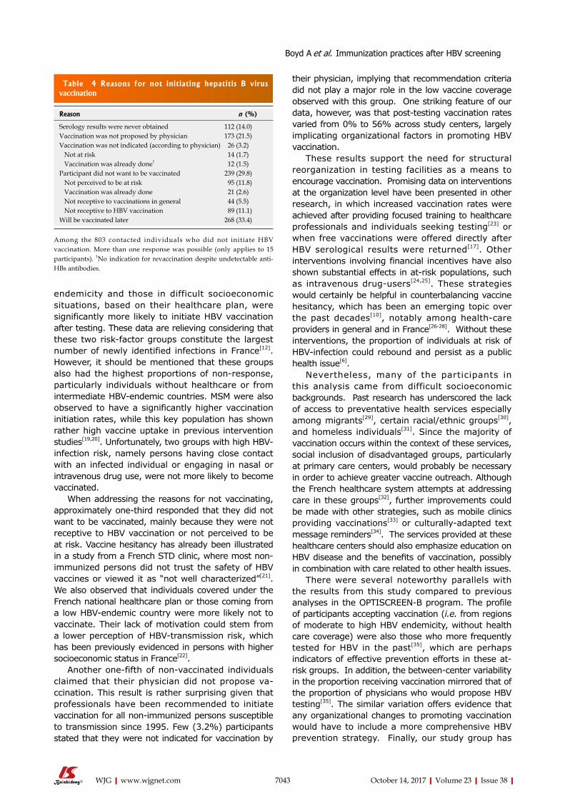
7043 October 14, 2017|Volume 23|Issue 38|WJG|www.wjgnet.com
endemicity and those in difficult socioeconomic situations, based on their healthcare plan, were significantly more likely to initiate HBV vaccination after testing. These data are relieving considering that these two risk-factor groups constitute the largest number of newly identified infections in France[12]. However, it should be mentioned that these groups also had the highest proportions of non-response, particularly individuals without healthcare or from intermediate HBV-endemic countries. MSM were also observed to have a significantly higher vaccination initiation rates, while this key population has shown rather high vaccine uptake in previous intervention studies[19,20]. Unfortunately, two groups with high HBV-infection risk, namely persons having close contact with an infected individual or engaging in nasal or intravenous drug use, were not more likely to become vaccinated.
When addressing the reasons for not vaccinating, approximately one-third responded that they did not want to be vaccinated, mainly because they were not receptive to HBV vaccination or not perceived to be at risk. Vaccine hesitancy has already been illustrated in a study from a French STD clinic, where most non-immunized persons did not trust the safety of HBV vaccines or viewed it as “not well characterized”[21]. We also observed that individuals covered under the French national healthcare plan or those coming from a low HBV-endemic country were more likely not to vaccinate. Their lack of motivation could stem from a lower perception of HBV-transmission risk, which has been previously evidenced in persons with higher socioeconomic status in France[22].
Another one-fifth of non-vaccinated individuals claimed that their physician did not propose va-ccination. This result is rather surprising given that professionals have been recommended to initiate vaccination for all non-immunized persons susceptible to transmission since 1995. Few (3.2%) participants stated that they were not indicated for vaccination by
their physician, implying that recommendation criteria did not play a major role in the low vaccine coverage observed with this group. One striking feature of our data, however, was that post-testing vaccination rates varied from 0% to 56% across study centers, largely implicating organizational factors in promoting HBV vaccination.
These results support the need for structural reorganization in testing facilities as a means to encourage vaccination. Promising data on interventions at the organization level have been presented in other research, in which increased vaccination rates were achieved after providing focused training to healthcare professionals and individuals seeking testing[23] or when free vaccinations were offered directly after HBV serological results were returned[17]. Other interventions involving financial incentives have also shown substantial effects in at-risk populations, such as intravenous drug-users[24,25]. These strategies would certainly be helpful in counterbalancing vaccine hesitancy, which has been an emerging topic over the past decades[10], notably among health-care providers in general and in France[26-28]. Without these interventions, the proportion of individuals at risk of HBV-infection could rebound and persist as a public health issue[6].
Nevertheless, many of the participants in this analysis came from difficult socioeconomic backgrounds. Past research has underscored the lack of access to preventative health services especially among migrants[29], certain racial/ethnic groups[30], and homeless individuals[31]. Since the majority of vaccination occurs within the context of these services, social inclusion of disadvantaged groups, particularly at primary care centers, would probably be necessary in order to achieve greater vaccine outreach. Although the French healthcare system attempts at addressing care in these groups[32], further improvements could be made with other strategies, such as mobile clinics providing vaccinations[33] or culturally-adapted text message reminders[34]. The services provided at these healthcare centers should also emphasize education on HBV disease and the benefits of vaccination, possibly in combination with care related to other health issues.
There were several noteworthy parallels with the results from this study compared to previous analyses in the OPTISCREEN-B program. The profile of participants accepting vaccination (i.e. from regions of moderate to high HBV endemicity, without health care coverage) were also those who more frequently tested for HBV in the past[35], which are perhaps indicators of effective prevention efforts in these at-risk groups. In addition, the between-center variability in the proportion receiving vaccination mirrored that of the proportion of physicians who would propose HBV testing[35]. The similar variation offers evidence that any organizational changes to promoting vaccination would have to include a more comprehensive HBV prevention strategy. Finally, our study group has
Table 4 Reasons for not initiating hepatitis B virus vaccination
Reason n (%)
Serology results were never obtained 112 (14.0)Vaccination was not proposed by physician 173 (21.5)Vaccination was not indicated (according to physician) 26 (3.2) Not at risk 14 (1.7) Vaccination was already done1 12 (1.5)Participant did not want to be vaccinated 239 (29.8) Not perceived to be at risk 95 (11.8) Vaccination was already done 21 (2.6) Not receptive to vaccinations in general 44 (5.5) Not receptive to HBV vaccination 89 (11.1)Will be vaccinated later 268 (33.4)
Among the 803 contacted individuals who did not initiate HBV vaccination. More than one response was possible (only applies to 15 participants). 1No indication for revaccination despite undetectable anti-HBs antibodies.
Boyd A et al. Immunization practices after HBV screening
7044 October 14, 2017|Volume 23|Issue 38|WJG|www.wjgnet.com
observed in a past evaluation that roughly 20%-50% participants newly diagnosed with HBV returned for further consultation at a referent center[15], reflecting the potential for inadequate transition to “appropriate care”[36]. The increased need to foster individuals into appropriate care appears to be shared among non-immunized patients.
Several methodological limitations need to be noted when interpreting the results of our study. First, we used a convenience sample combining participants from two studies. Certain risk-factor groups, such as intravenous drug-users and sex-workers, might not be adequately represented and could have different vaccine coverage than those reported herein. In addition, the lack in numbers may have resulted in inadequate power in order to establish whether they were less likely to complete vaccination. Second, the time-frame of follow-up was relatively short and perhaps some participants received vaccination 6-9 mo or more after their interview. Non-immunized patients did vaccinate a median 30 d, suggesting that few individuals would have initiated vaccination after the end of follow-up and vaccine coverage would have been similar if follow-up were prolonged. Nevertheless, roughly one-third of unvaccinated individuals stated that they would do so in the future. Third, the short time-frame also made it impossible to ascertain whether participants completed HBV vaccination, as the date of last vaccination would have fallen outside the follow-up period. Lastly, the proportion with no reply, which was comparable to other studies[17], could have biased results.
Furthermore, we should highlight that individuals with low levels of anti-HBs antibodies (> 0-10 mIU/mL), who could possibly benefit from booster HBV vaccination, were not included in this analysis. The results from our study might not be generalizable to these individuals.
In conclusion, HBV vaccination after screening was very low in this study, implying that HBV testing alone cannot encourage non-immunized individuals to vaccinate. The fact that large discrepancies in vaccine coverage were observed across centers and that both physician and participant motivation towards vaccination was rather limited provides evidence that involvement at the health structure level is crucial in increasing vaccination coverage. Future interventions and campaigns should work at increasing local awareness of low vaccine coverage rates, especially in groups with poor access to health services, and the need to vaccinate non-immunized individuals as soon as they are identified.
ARTICLE HIGHLIGHTSResearch backgroundTesting campaigns for hepatitis B virus (HBV) have often focused on identifying infected patients [i.e.
those with hepatitis B surface antigen (HBsAg)-positive serology] and increasing disease-status awareness. Few evaluations have been performed on how testing campaigns can impact vaccination uptake after receiving non-immunized status.
Research motivationSince France has a low proportion of vaccinated individuals compared to other Western countries, interventions to increase HBV vaccination rates need to be evaluated in this population. This analysis addressed vaccination initiation among non-immunized participants in a large-scale screening campaign within the Paris metropolitan region. These individuals reflect a wide range of groups at-risk of HBV infection, allowing potential identification of subgroups with low vaccination coverage.
Research objectivesWe aimed to describe rates of vaccination among non-immunized individuals during a mass-screening program. We also intended to evaluate the reasons for not initiating HBV vaccination after testing. This allows a first-hand account of why individuals do not vaccinate and helps tailor the needs for future intervention campaigns.
Research methodsParticipants were recruited from two large phases of a multi-center, HBV-testing campaign in Paris, France. Non-immunized subjects were identified and contacted via telephone 3-9 mo after testing in order to determine whether they initiated vaccination. We considered vaccination coverage of all respondents (in a per-protocol analysis) and the overall non-immunized study population while assuming no vaccination in non-responders (in an intent-to-treat analysis).
Research resultsOverall vaccination uptake was low with 11% of respondents declaring HBV vaccination initiation within 3-6 mo of testing. Few risk-factors for increased vaccination initiation were identified: from moderate or high HBV-endemic countries, with limited healthcare coverage, and men who have sex with men. Compelling differences were observed between centers with vaccination coverage ranging from 0%-56%.
Research conclusionsGiven the low vaccine uptake in individuals at potential risk of HBV-infection, there is a major concern in the cascade of care among non-immunized individuals. The contrasting vaccination rates between centers indicate that the challenge to increase vaccination initiation lies within center-specific practices. At the individual level, increasing motivation to vaccinate among physicians and non-immunized persons alike
Boyd A et al. Immunization practices after HBV screening
ARTICLE HIGHLIGHTS

7045 October 14, 2017|Volume 23|Issue 38|WJG|www.wjgnet.com
should be stressed.
ACKNOWLEDGMENTSWe wish to thank all study participants as well as all medical and paramedical centers participating in the study, Tabassome Simon and nurses from the CRC-Est, and the data management center, especially Frederic Chau, Frederic Fotre, Sylvain Bitschine, Isabelle Goderel and Gregory Pannetier. We also acknowledge Samia Hicham, Judith Leblanc, Manuela Sébire-Le Cam, Farid Djoumad, Julie Lamarque, and Christelle Pauleau for their extraordinary effort in data collection.
REFERENCES1 World Health Organization. Global health sector strategy on
viral hepatitis 2016-2021. Towards ending viral hepatitis[J]. 20162 Weinbaum CM, Williams I, Mast EE, Wang SA, Finelli L,
Wasley A, Neitzel SM, Ward JW; Centers for Disease Control and Prevention (CDC). Recommendations for identification and public health management of persons with chronic hepatitis B virus infection. MMWR Recomm Rep 2008; 57: 1-20 [PMID: 18802412]
3 Hatzakis A, Wait S, Bruix J, Buti M, Carballo M, Cavaleri M, Colombo M, Delarocque-Astagneau E, Dusheiko G, Esmat G, Esteban R, Goldberg D, Gore C, Lok AS, Manns M, Marcellin P, Papatheodoridis G, Peterle A, Prati D, Piorkowsky N, Rizzetto M, Roudot-Thoraval F, Soriano V, Thomas HC, Thursz M, Valla D, van Damme P, Veldhuijzen IK, Wedemeyer H, Wiessing L, Zanetti AR, Janssen HL. The state of hepatitis B and C in Europe: report from the hepatitis B and C summit conference*. J Viral Hepat 2011; 18 Suppl 1: 1-16 [PMID: 21824223 DOI: 10.1111/j.1365-2893.2011.01499.x]
4 Ni YH, Huang LM, Chang MH, Yen CJ, Lu CY, You SL, Kao JH, Lin YC, Chen HL, Hsu HY, Chen DS. Two decades of universal hepatitis B vaccination in taiwan: impact and implication for future strategies. Gastroenterology 2007; 132: 1287-1293 [PMID: 17433322 DOI: 10.1053/j.gastro.2007.02.055]
5 Mast EE, Margolis HS, Fiore AE, Brink EW, Goldstein ST, Wang SA, Moyer LA, Bell BP, Alter MJ; Advisory Committee on Immunization Practices (ACIP). A comprehensive immunization strategy to eliminate transmission of hepatitis B virus infection in the United States: recommendations of the Advisory Committee on Immunization Practices (ACIP) part 1: immunization of infants, children, and adolescents. MMWR Recomm Rep 2005; 54: 1-31 [PMID: 16371945]
6 Ramière C, Roche L, Scholtès C, Iwaz J, Saison J, Ecochard R, André P. Evolution of the incidence of hepatitis B virus infection and immunization rates in a large French cohort born between 1960 and 1994. Clin Microbiol Infect 2016; 22: 889.e1-889.e7 [PMID: 27451939 DOI: 10.1016/j.cmi.2016.07.014]
7 Fonteneau L, Urcun JM, Guthmann JP, Collet M, Neulat N, Bristol-Gauzy P, Guignon N, Lévy-Bruhl D, Herbet JB. [Vaccination coverage in 6-year-old preschool children, France, 2005-2006]. Arch Pediatr 2013; 20: 241-247 [PMID: 23375080 DOI: 10.1016/j.arcped.2012.12.019]
8 Beck CR, Cloke R, O’Moore É, Puleston R. Hepatitis B vaccination coverage and uptake in prisons across England and Wales 2003-2010: a retrospective ecological study. Vaccine 2012; 30 : 1965-1971 [PMID: 22265944 DOI: 10.1016/j.vaccine.2012.01.020]
9 Hoover KW, Butler M, Workowski KA, Follansbee S, Gratzer B, Hare CB, Johnston B, Theodore JL, Tao G, Smith BD, Chorba T, Kent CK; Evaluation Group for Adherence to STD and Hepatitis Screening. Low rates of hepatitis screening and vaccination of HIV-infected MSM in HIV clinics. Sex Transm Dis 2012; 39: 349-353
[PMID: 22504597 DOI: 10.1097/OLQ.0b013e318244a923]10 Larson HJ, Jarrett C, Eckersberger E, Smith DM, Paterson P.
Understanding vaccine hesitancy around vaccines and vaccination from a global perspective: a systematic review of published literature, 2007-2012. Vaccine 2014; 32: 2150-2159 [PMID: 24598724 DOI: 10.1016/j.vaccine.2014.01.081]
11 Caisse nationale de l’assurance maladie des travailleurs salariés (France), Meffre C, Centre technique d’appui et de formation des centres d’examens de santé (France), Institut de veille sanitaire (France). Prévalence des hépatites B et C en France en 2004. Saint-Maurice: Institut de veille sanitaire; 2007
12 Antona D, Letort M-J, Lévy-Bruhl D. Estimation du nombre annuel de nouvelles infections par le virus de l’hépatite B en France, 2004-2007. Bull Epidemiol Hebd 2009; 20-21: 196-199
13 Institut de veille sanitaire. Calendrier des vaccinations et recommandations vaccinales 2016. Bull Epidemiol Hebd 2016; Hors-série
14 Bottero J, Boyd A, Gozlan J, Lemoine M, Carrat F, Collignon A, Boo N, Dhotte P, Varsat B, Muller G, Cha O, Picard O, Nau J, Campa P, Silbermann B, Bary M, Girard PM, Lacombe K. Performance of rapid tests for detection of HBsAg and anti-HBsAb in a large cohort, France. J Hepatol 2013; 58: 473-478 [PMID: 23183527 DOI: 10.1016/j.jhep.2012.11.016]
15 Bottero J, Boyd A, Gozlan J, Carrat F, Lemoine M, Rougier H, Varsat B, Boo N, Charlois-Ou C, Collignon A, Cha O, Campa P, Dhotte P, Girard PM, Lacombe K. Effectiveness of hepatitis B rapid tests toward linkage-to-care: results of a randomized, multicenter study. Eur J Gastroenterol Hepatol 2016; 28: 633-639 [PMID: 26954517 DOI: 10.1097/MEG.0000000000000620]
16 Bottero J, Brouard C, Roudot-Thoraval F, Deuffic-Burban S, Hofliger P, Abergel A, Volant J, Dhumeaux D, Yazdanpanah Y; Viral Hepatitis Testing Experts group. 2014 French guidelines for hepatitis B and C screening: a combined targeted and mass testing strategy of chronic viruses namely HBV, HCV and HIV. Liver Int 2016; 36: 1442-1449 [PMID: 27043826 DOI: 10.1111/liv.13135]
17 Launay O, Le Strat Y, Tosini W, Kara L, Quelet S, Lévy S, Danan J, Réveillon J, Houdayer J, Bouvet E, Lévy-Bruhl D; ANRS-FORMVAC Study Group. Impact of free on-site vaccine and/or healthcare workers training on hepatitis B vaccination acceptability in high-risk subjects: a pre-post cluster randomized study. Clin Microbiol Infect 2014; 20: 1033-1039 [PMID: 24850059 DOI: 10.1111/1469-0691.12689]
18 Hechter RC, Jacobsen SJ, Luo Y, Nomura JH, Towner WJ, Tartof SY, Tseng HF. Hepatitis B testing and vaccination among adults with sexually transmitted infections in a large managed care organization. Clin Infect Dis 2014; 58: 1739-1745 [PMID: 24571863 DOI: 10.1093/cid/ciu103]
19 van Rijckevorsel G, Whelan J, Kretzschmar M, Siedenburg E, Sonder G, Geskus R, Coutinho R, van den Hoek A. Targeted vaccination programme successful in reducing acute hepatitis B in men having sex with men in Amsterdam, the Netherlands. J Hepatol 2013; 59: 1177-1183 [PMID: 23954670 DOI: 10.1016/j.jhep.2013.08.002]
20 Chonwattana W, Raengsakulrach B, Holtz TH, Wasinrapee P, Tongtoyai J, Chaikummao S, Pattanasin S, McNicholl JM, van Griensven F, Curlin ME. Hepatitis B vaccination uptake and correlates of serologic response among HIV-infected and uninfected men who have sex with men (MSM) in Bangkok, Thailand. Vaccine 2016; 34: 2044-2050 [PMID: 26685092 DOI: 10.1016/j.vaccine.2015.11.071]
21 Moyroud L, Hustache S, Goirand L, Hauzanneau M, Epaulard O. Negative perceptions of hepatitis B vaccination among attendees of an urban free testing center for sexually transmitted infections in France. Hum Vaccin Immunother 2017; 13: 998-1004 [PMID: 27937074 DOI: 10.1080/21645515.2016.1264549]
22 Brouard C, Gautier A, Saboni L, Jestin C, Semaille C, Beltzer N; KABP France group. Hepatitis B knowledge, perceptions and practices in the French general population: the room for improvement. BMC Public Health 2013; 13: 576 [PMID:
Boyd A et al. Immunization practices after HBV screening
7046 October 14, 2017|Volume 23|Issue 38|WJG|www.wjgnet.com
23764171 DOI: 10.1186/1471-2458-13-576]23 Zacharias T, Wang W, Dao D, Wojciechowski H, Lee WM, Do
S, Singal AG. HBV Outreach Programs Significantly Increase Knowledge and Vaccination Rates Among Asian Pacific Islanders. J Community Health 2015; 40: 619-624 [PMID: 25476035 DOI: 10.1007/s10900-014-9975-y]
24 Weaver T, Metrebian N, Hellier J, Pilling S, Charles V, Little N, Poovendran D, Mitcheson L, Ryan F, Bowden-Jones O, Dunn J, Glasper A, Finch E, Strang J. Use of contingency management incentives to improve completion of hepatitis B vaccination in people undergoing treatment for heroin dependence: a cluster randomised trial. Lancet 2014; 384: 153-163 [PMID: 24725468 DOI: 10.1016/S0140-6736(14)60196-3]
25 Topp L, Day CA, Wand H, Deacon RM, van Beek I, Haber PS, Shanahan M, Rodgers C, Maher L; Hepatitis Acceptability and Vaccine Incentives Trial (HAVIT) Study Group. A randomised controlled trial of financial incentives to increase hepatitis B vaccination completion among people who inject drugs in Australia. Prev Med 2013; 57: 297-303 [PMID: 23639625 DOI: 10.1016/j.ypmed.2013.04.013]
26 Paterson P, Meurice F, Stanberry LR, Glismann S, Rosenthal SL, Larson HJ. Vaccine hesitancy and healthcare providers. Vaccine 2016; 34: 6700-6706 [PMID: 27810314 DOI: 10.1016/j.vaccine.2016.10.042]
27 Larson HJ, de Figueiredo A, Xiahong Z, Schulz WS, Verger P, Johnston IG, Cook AR, Jones NS. The State of Vaccine Confidence 2016: Global Insights Through a 67-Country Survey. EBioMedicine 2016; 12: 295-301 [PMID: 27658738 DOI: 10.1016/j.ebiom.2016.08.042]
28 Verger P, Collange F, Fressard L, Bocquier A, Gautier A, Pulcini C, Raude J, Peretti-Watel P. Prevalence and correlates of vaccine hesitancy among general practitioners: a cross-sectional telephone survey in France, April to July 2014. Euro Surveill 2016; 21 [PMID: 27918262 DOI: 10.2807/1560-7917.ES.2016.21.47.30406]
29 Rosano A, Dauvrin M, Buttigieg SC, Ronda E, Tafforeau J, Dias S. Migrant’s access to preventive health services in five EU countries.
BMC Health Serv Res 2017; 17: 588 [PMID: 28830423 DOI: 10.1186/s12913-017-2549-9]
30 Kim HS, Rotundo L, Yang JD, Kim D, Kothari N, Feurdean M, Ruhl C, Unalp-Arida A. Racial/ethnic disparities in the prevalence and awareness of Hepatitis B virus infection and immunity in the United States. J Viral Hepat 2017 [PMID: 28581638 DOI: 10.1111/jvh.12735]
31 van Laere IR, de Wit MA, Klazinga NS. Pathways into homelessness: recently homeless adults problems and service use before and after becoming homeless in Amsterdam. BMC Public Health 2009; 9: 3 [PMID: 19128448 DOI: 10.1186/1471-2458-9-3]
32 Nay O, Béjean S, Benamouzig D, Bergeron H, Castel P, Ventelou B. Achieving universal health coverage in France: policy reforms and the challenge of inequalities. Lancet 2016; 387: 2236-2249 [PMID: 27145707 DOI: 10.1016/S0140-6736(16)00580-8]
33 Whelan C, Chambers C, Chan M, Thomas S, Ramos G, Hwang SW. Why do homeless people use a mobile health unit in a country with universal health care? J Prim Care Community Health 2010; 1: 78-82 [PMID: 23804366 DOI: 10.1177/2150131910372233]
34 Phillips AL, Kumar D, Patel S, Arya M. Using text messages to improve patient-doctor communication among racial and ethnic minority adults: an innovative solution to increase influenza vaccinations. Prev Med 2014; 69: 117-119 [PMID: 25241643 DOI: 10.1016/j.ypmed.2014.09.009]
35 Bottero J, Boyd A, Lemoine M, Carrat F, Gozlan J, Collignon A, Boo N, Dhotte P, Varsat B, Muller G, Cha O, Valin N, Nau J, Campa P, Silbermann B, Bary M, Girard PM, Lacombe K. Current state of and needs for hepatitis B screening: results of a large screening study in a low-prevalent, metropolitan region. PLoS One 2014; 9: e92266 [PMID: 24663387 DOI: 10.1371/journal.pone.0092266]
36 Bottero J, Boyd A, Gozlan J, Carrat F, Nau J, Pauti MD, Rougier H, Girard PM, Lacombe K. Simultaneous Human Immunodeficiency Virus-Hepatitis B-Hepatitis C Point-of-Care Tests Improve Outcomes in Linkage-to-Care: Results of a Randomized Control Trial in Persons Without Healthcare Coverage. Open Forum Infect Dis 2015; 2: ofv162 [PMID: 26668814 DOI: 10.1093/ofid/ofv162]
P- Reviewer: Chen XX, Guo YM, Watson DI S- Editor: Chen K L- Editor: A E- Editor: Ma YJ
Boyd A et al. Immunization practices after HBV screening
Gastric adenocarcinoma of fundic gland type spreading to heterotopic gastric glands
Shigeo Manabe, Ken-ichi Mukaisho, Takayuki Yasuoka, Fumitaka Usui, Tatsuzo Matsuyama, Ikuhiro Hirata, Yoshio Boku, Shuji Takahashi
Shigeo Manabe, Takayuki Yasuoka, Fumitaka Usui, Tatsuzo Matsuyama, Ikuhiro Hirata, Shuji Takahashi, Department of Gastroenterology, Kouseikai Takeda Hospital, Kyoto 600-8558, Japan
Ken-ichi Mukaisho, Department of Pathology, Shiga University of Medical Science, Seta, Tsukinwa-cho, Otsu, Shiga 520-2192, Japan
Yoshio Boku, Fujita Clinic, 67, Gokiya-cho, Oomiya-dori Shichijo-kudaru, Shimogyo-ku, Kyoto 600-8267, Japan
ORCID number: Shigeo Manabe (0000-0002-0654-0599); Ken-ichi Mukaisho (0000-0002-0488-0100); Takayuki Yasuoka (0000-0002-1537-6882); Fumitaka Usui (0000-0002-7468-8691); Tatsuzo Matsuyama (0000-0003-2567-0126); Ikuhiro Hirata (0000-0001-6926-0589); Yoshio Boku (0000-0002-1291-4745); Shuji Takahashi (0000-0001-6533-1618).
Author contributions: Manabe S was responsible for the patient, performed the endoscopic examination and treatment, and wrote the initial draft of the manuscript; Mukaisho K contributed to the histopathological diagnosis; all other authors contributed to the retrieval and interpretation of the results and critically reviewed the manuscript; all authors approved the final version of the manuscript.
Institutional review board statement: This case report was exempted from the standards of the Institutional Review Board at the Kouseikai Takeda Hospital.
Informed consent statement: The patient provided an oral informed consent prior to the conduction of the study.
Conflict-of-interest statement: We declare that there is no conflict of interest.
Open-Access: This article is an open-access article which was selected by an in-house editor and fully peer-reviewed by external reviewers. It is distributed in accordance with the Creative Commons Attribution Non Commercial (CC BY-NC 4.0) license, which permits others to distribute, remix, adapt, build upon this work non-commercially, and license their derivative works on different terms, provided the original work is properly cited and
the use is non-commercial. See: http://creativecommons.org/licenses/by-nc/4.0/
Manuscript source: Unsolicited manuscript
Correspondence to: Shigeo Manabe, MD, Department of Gastroenterology, Kouseikai Takeda Hospital, 841-5, Higashi Shiokoji-cho, Shiokoji-dori Nishinotoin-higashiiru, Shimogyo-ku, Kyoto 600-8558, Japan. [email protected] Telephone: +81-75-3611351Fax: +81-75-3617602
Received: June 6, 2017Peer-review started: June 14, 2017First decision: July 17, 2017Revised: August 18, 2017Accepted: September 13, 2017Article in press: September 13, 2017Published online: October 14, 2017
AbstractHerein, we present a case of gastric adenocarcinoma of fundic gland type (GA-FG) spreading to heterotopic gastric glands (HGG) in the submucosa. A 58-year-old man with epigastric pain was referred to our hospital and underwent an esophagogastroduodenoscopy. A Borrmann type II gastric cancer at the antrum and a 10 mm submucosal tumor-like lesion in the lesser curvature of the upper third of the stomach were detected. Histological examination of the biopsy specimens obtained from the submucosal tumor-like lesion suggested a GA-FG. Therefore, endoscopic submucosal dissection was performed as excisional biopsy, and histopathological examination of the resected specimen confirmed a GA-FG and HGG proximal to the GA-FG. Although the GA-FG invaded the submucosal layer slightly, the submucosal lesion of the GA-FG had a poor stromal reaction and was located just above the HGG in the submucosa. Therefore, we
CASE REPORT
7047 October 14, 2017|Volume 23|Issue 38|WJG|www.wjgnet.com
Submit a Manuscript: http://www.f6publishing.com
DOI: 10.3748/wjg.v23.i38.7047
World J Gastroenterol 2017 October 14; 23(38): 7047-7053
ISSN 1007-9327 (print) ISSN 2219-2840 (online)

finally diagnosed the lesion as a GA-FG invading the submucosal layer by spreading to HGG.
Key words: Gastric adenocarcinoma of fundic gland type; Heterotopic gastric glands; Endoscopic submucosal dissection; Paracancerous lesion; Pepsinogen-I; H/K-ATPase
© The Author(s) 2017. Published by Baishideng Publishing Group Inc. All rights reserved.
Core tip: A 58-year-old man had a 10mm submucosal tumor-like lesion in the lesser curvature of the upper third of the stomach. Histological examination of the biopsy specimens suggested a gastric adenocarcinoma of fundic gland type (GA-FG), therefore, endoscopic submucosal dissection was performed as excisional biopsy. Histopathological examination of the resected specimen confirmed a GA-FG invading the submucosal layer slightly, and heterotopic gastric glands (HGG) proximal to the GA-FG. A GA-FG spreading to HGG in the submucosa was diagnosed because the submucosal lesion of the GA-FG had a poor stromal reaction and was located just above the HGG.
Manabe S, Mukaisho K, Yasuoka T, Usui F, Matsuyama T, Hirata I, Boku Y, Takahashi S. Gastric adenocarcinoma of fundic gland type spreading to heterotopic gastric glands. World J Gastroenterol 2017; 23(38): 7047-7053 Available from: URL: http://www.wjgnet.com/1007-9327/full/v23/i38/7047.htm DOI: http://dx.doi.org/10.3748/wjg.v23.i38.7047
INTRODUCTIONGastric adenocarcinoma of fundic gland type (GA-FG) is a new histological type of gastric cancer proposed by Ueyama et al[1] in 2010. As the concept of GA-FG has spread since then, new findings have been reported[2]. GA-FG tends to invade the submucosal layer while it is still small because it arises from the deep layer of the lamina propria mucosae[1]. On the other hand, heterotopic gastric glands (HGG) are regarded as paracancerous lesions[3,4], and gastric cancers associated with HGG have been reported[5,6]. However, HGG are not considered to contribute to the development of GA-FG because the pathogenesis of GA-FG is different from that of the regular histological type of gastric cancer. We encountered a case of GA-FG suggested to have invaded the submucosal layer by spreading to HGG, which coincidentally existed proximal to the GA-FG. In addition to a GA-FG with a very rare mode of submucosal invasion, this case demonstrated the possibility of GA-FG arising from the gastric mucosa with atrophic change, provided there are remnant gastric fundic glands. Herein, we report this case with a brief review of the literature.
CASE REPORTA 58-year-old man with epigastric pain was referred to our hospital. Esophagogastroduodenoscopy (EGD) revealed a Borrmann type II gastric cancer at the antrum (Figure 1A) and a 10 mm submucosal tumor-like lesion in the lesser curvature of the upper third of the stomach (Figure 1B and C). Atrophy of the gastric mucosa was classified as O-1 (according to the Kimura and Takemoto classification[7]), although the patient had undergone Helicobacter pylori (H. pylori) eradication therapy 4 years ago, and has not taken any proton pump inhibitors since then. Histological examination of the biopsy specimens obtained from the submucosal tumor-like lesion showed that the superficial area retained the normal foveolar epithelium. However, atypical cells mimicking fundic gland cells with mildly enlarged nuclei were observed in the deep layer of the lamina propria mucosae. These findings suggested a GA-FG. Narrow-band imaging with magnifying endoscopy (NBI-ME) performed later showed a regular microvascular pattern and a regular microsurface pattern according to the vessel plus surface classification system[8]
with a concomitant depressed area at the center of the submucosal tumor-like lesion expected to be a biopsy scar (Figure 2). Endoscopic ultrasonography (EUS) findings revealed the tumor slightly invading the third layer and a hypoechoic mass located in the third layer near the tumor (Figure 3). Blood test findings revealed carcinoembryonic antigen level of 2.2 ng/mL and carbohydrate antigen 19-9 level of 14.3 U/mL, indicating that the tumor markers were within the normal range. Although gastric wall thickening due to the advanced gastric cancer at the antrum was seen on the abdominal CT scan, nodal or distant metastasis was absent. Based on the above findings, a partial gastrectomy was required for gastric cancer at the antrum. The submucosal tumor-like lesion in the lesser curvature of the upper third of the stomach was suspected to be a GA-FG; hence, we decided to perform endoscopic submucosal dissection (ESD) as excisional biopsy. Since histopathological findings of the ESD-resected specimen may indicate the need for an additional surgery, ESD was performed before surgery for the gastric cancer at the antrum, and en bloc resection was achieved. The ESD-resected specimen was 30 mm in diameter, whilst a 7 mm × 4 mm slightly elevated lesion was identified (Figure 4). Similar to the histological findings of the biopsy specimens, atypical cells mimicking fundic gland cells, mainly chief cells and partially parietal cells, with mildly enlarged nuclei were seen mainly in the deep layer of the lamina propria mucosae. The mucosal surface was covered completely with non-neoplastic foveolar epithelium; thus, the tumor was not exposed to the mucosal surface (Figure 5A).
7048 October 14, 2017|Volume 23|Issue 38|WJG|www.wjgnet.com
Manabe S et al. GA-FG spreading to HGG
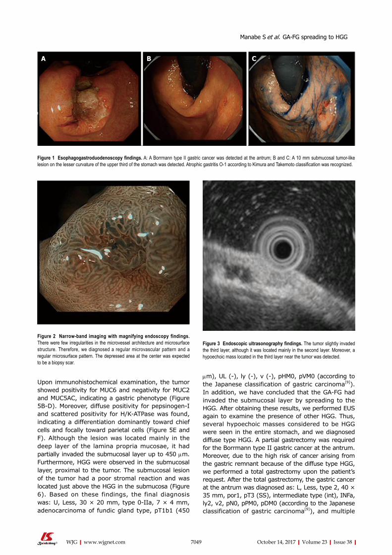
Upon immunohistochemical examination, the tumor showed positivity for MUC6 and negativity for MUC2 and MUC5AC, indicating a gastric phenotype (Figure 5B-D). Moreover, diffuse positivity for pepsinogen-I and scattered positivity for H/K-ATPase was found, indicating a differentiation dominantly toward chief cells and focally toward parietal cells (Figure 5E and F). Although the lesion was located mainly in the deep layer of the lamina propria mucosae, it had partially invaded the submucosal layer up to 450 μm. Furthermore, HGG were observed in the submucosal layer, proximal to the tumor. The submucosal lesion of the tumor had a poor stromal reaction and was located just above the HGG in the submucosa (Figure 6). Based on these findings, the final diagnosis was: U, Less, 30 × 20 mm, type 0-IIa, 7 × 4 mm, adenocarcinoma of fundic gland type, pT1b1 (450
μm), UL (-), ly (-), v (-), pHM0, pVM0 (according to the Japanese classification of gastric carcinoma[9]). In addition, we have concluded that the GA-FG had invaded the submucosal layer by spreading to the HGG. After obtaining these results, we performed EUS again to examine the presence of other HGG. Thus, several hypoechoic masses considered to be HGG were seen in the entire stomach, and we diagnosed diffuse type HGG. A partial gastrectomy was required for the Borrmann type II gastric cancer at the antrum. Moreover, due to the high risk of cancer arising from the gastric remnant because of the diffuse type HGG, we performed a total gastrectomy upon the patient’s request. After the total gastrectomy, the gastric cancer at the antrum was diagnosed as: L, Less, type 2, 40 × 35 mm, por1, pT3 (SS), intermediate type (int), INFa, ly2, v2, pN0, pPM0, pDM0 (according to the Japanese classification of gastric carcinoma[9]), and multiple
7049 October 14, 2017|Volume 23|Issue 38|WJG|www.wjgnet.com
A B C
Figure 1 Esophagogastroduodenoscopy findings. A: A Borrmann type II gastric cancer was detected at the antrum; B and C: A 10 mm submucosal tumor-like lesion on the lesser curvature of the upper third of the stomach was detected. Atrophic gastritis O-1 according to Kimura and Takemoto classification was recognized.
Figure 2 Narrow-band imaging with magnifying endoscopy findings. There were few irregularities in the microvessel architecture and microsurface structure. Therefore, we diagnosed a regular microvascular pattern and a regular microsurface pattern. The depressed area at the center was expected to be a biopsy scar.
Figure 3 Endoscopic ultrasonography findings. The tumor slightly invaded the third layer, although it was located mainly in the second layer. Moreover, a hypoechoic mass located in the third layer near the tumor was detected.
Manabe S et al. GA-FG spreading to HGG
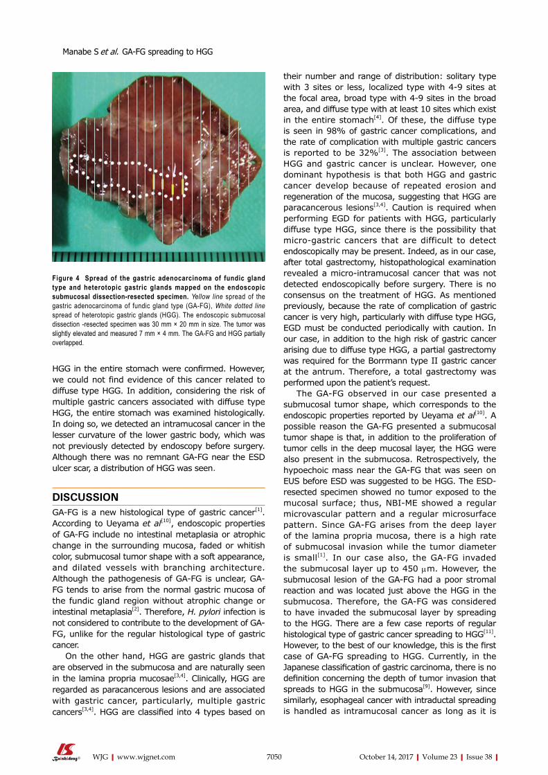
7050 October 14, 2017|Volume 23|Issue 38|WJG|www.wjgnet.com
their number and range of distribution: solitary type with 3 sites or less, localized type with 4-9 sites at the focal area, broad type with 4-9 sites in the broad area, and diffuse type with at least 10 sites which exist in the entire stomach[4]. Of these, the diffuse type is seen in 98% of gastric cancer complications, and the rate of complication with multiple gastric cancers is reported to be 32%[3]. The association between HGG and gastric cancer is unclear. However, one dominant hypothesis is that both HGG and gastric cancer develop because of repeated erosion and regeneration of the mucosa, suggesting that HGG are paracancerous lesions[3,4]. Caution is required when performing EGD for patients with HGG, particularly diffuse type HGG, since there is the possibility that micro-gastric cancers that are difficult to detect endoscopically may be present. Indeed, as in our case, after total gastrectomy, histopathological examination revealed a micro-intramucosal cancer that was not detected endoscopically before surgery. There is no consensus on the treatment of HGG. As mentioned previously, because the rate of complication of gastric cancer is very high, particularly with diffuse type HGG, EGD must be conducted periodically with caution. In our case, in addition to the high risk of gastric cancer arising due to diffuse type HGG, a partial gastrectomy was required for the Borrmann type II gastric cancer at the antrum. Therefore, a total gastrectomy was performed upon the patient’s request.
The GA-FG observed in our case presented a submucosal tumor shape, which corresponds to the endoscopic properties reported by Ueyama et al[10]. A possible reason the GA-FG presented a submucosal tumor shape is that, in addition to the proliferation of tumor cells in the deep mucosal layer, the HGG were also present in the submucosa. Retrospectively, the hypoechoic mass near the GA-FG that was seen on EUS before ESD was suggested to be HGG. The ESD-resected specimen showed no tumor exposed to the mucosal surface; thus, NBI-ME showed a regular microvascular pattern and a regular microsurface pattern. Since GA-FG arises from the deep layer of the lamina propria mucosa, there is a high rate of submucosal invasion while the tumor diameter is small[1]. In our case also, the GA-FG invaded the submucosal layer up to 450 μm. However, the submucosal lesion of the GA-FG had a poor stromal reaction and was located just above the HGG in the submucosa. Therefore, the GA-FG was considered to have invaded the submucosal layer by spreading to the HGG. There are a few case reports of regular histological type of gastric cancer spreading to HGG[11]. However, to the best of our knowledge, this is the first case of GA-FG spreading to HGG. Currently, in the Japanese classification of gastric carcinoma, there is no definition concerning the depth of tumor invasion that spreads to HGG in the submucosa[9]. However, since similarly, esophageal cancer with intraductal spreading is handled as intramucosal cancer as long as it is
HGG in the entire stomach were confirmed. However, we could not find evidence of this cancer related to diffuse type HGG. In addition, considering the risk of multiple gastric cancers associated with diffuse type HGG, the entire stomach was examined histologically. In doing so, we detected an intramucosal cancer in the lesser curvature of the lower gastric body, which was not previously detected by endoscopy before surgery. Although there was no remnant GA-FG near the ESD ulcer scar, a distribution of HGG was seen.
DISCUSSIONGA-FG is a new histological type of gastric cancer[1]. According to Ueyama et al[10], endoscopic properties of GA-FG include no intestinal metaplasia or atrophic change in the surrounding mucosa, faded or whitish color, submucosal tumor shape with a soft appearance, and dilated vessels with branching architecture. Although the pathogenesis of GA-FG is unclear, GA-FG tends to arise from the normal gastric mucosa of the fundic gland region without atrophic change or intestinal metaplasia[2]. Therefore, H. pylori infection is not considered to contribute to the development of GA-FG, unlike for the regular histological type of gastric cancer.
On the other hand, HGG are gastric glands that are observed in the submucosa and are naturally seen in the lamina propria mucosae[3,4]. Clinically, HGG are regarded as paracancerous lesions and are associated with gastric cancer, particularly, multiple gastric cancers[3,4]. HGG are classified into 4 types based on
Figure 4 Spread of the gastric adenocarcinoma of fundic gland type and heterotopic gastric glands mapped on the endoscopic submucosal dissection-resected specimen. Yellow line spread of the gastric adenocarcinoma of fundic gland type (GA-FG), White dotted line spread of heterotopic gastric glands (HGG). The endoscopic submucosal dissection -resected specimen was 30 mm × 20 mm in size. The tumor was slightly elevated and measured 7 mm × 4 mm. The GA-FG and HGG partially overlapped.
Manabe S et al. GA-FG spreading to HGG
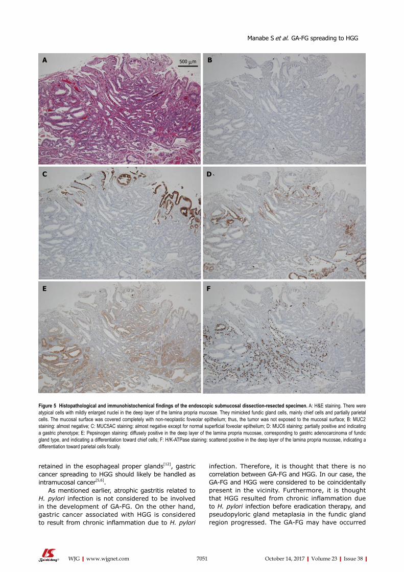
7051 October 14, 2017|Volume 23|Issue 38|WJG|www.wjgnet.com
retained in the esophageal proper glands[12], gastric cancer spreading to HGG should likely be handled as intramucosal cancer[5,6].
As mentioned earlier, atrophic gastritis related to H. pylori infection is not considered to be involved in the development of GA-FG. On the other hand, gastric cancer associated with HGG is considered to result from chronic inflammation due to H. pylori
infection. Therefore, it is thought that there is no correlation between GA-FG and HGG. In our case, the GA-FG and HGG were considered to be coincidentally present in the vicinity. Furthermore, it is thought that HGG resulted from chronic inflammation due to H. pylori infection before eradication therapy, and pseudopyloric gland metaplasia in the fundic gland region progressed. The GA-FG may have occurred
A B
C D
E F
500 μm
Figure 5 Histopathological and immunohistochemical findings of the endoscopic submucosal dissection-resected specimen. A: H&E staining. There were atypical cells with mildly enlarged nuclei in the deep layer of the lamina propria mucosae. They mimicked fundic gland cells, mainly chief cells and partially parietal cells. The mucosal surface was covered completely with non-neoplastic foveolar epithelium; thus, the tumor was not exposed to the mucosal surface; B: MUC2 staining: almost negative; C: MUC5AC staining: almost negative except for normal superficial foveolar epithelium; D: MUC6 staining: partially positive and indicating a gastric phenotype; E: Pepsinogen staining: diffusely positive in the deep layer of the lamina propria mucosae, corresponding to gastric adenocarcinoma of fundic gland type, and indicating a differentiation toward chief cells; F: H/K-ATPase staining: scattered positive in the deep layer of the lamina propria mucosae, indicating a differentiation toward parietal cells focally.
Manabe S et al. GA-FG spreading to HGG

7052 October 14, 2017|Volume 23|Issue 38|WJG|www.wjgnet.com
from the remnant gastric fundic glands despite the progression of pseudopyloric gland metaplasia and spread continuous to the proximally located HGG.
Until recently, GA-FG has been recognized as cancer arising from gastric mucosa without atrophic change[2]. However, cases of GA-FG arising from gastric mucosa with atrophic change have now been reported[13]. Together with these cases, our case suggests that GA-FG could occur even from the gastric mucosa with atrophic change, where HGG could be present, provided the remnant gastric fundic glands exist. This is an important case demonstrating that when performing endoscopy, one needs to keep in mind the possibility of GA-FG presenting on the gastric mucosa with atrophic change, chronic gastritis, or intestinal metaplasia.
COMMENTSCase characteristicsA 58-year-old man with epigastric pain but no other symptoms.
Clinical diagnosis A gastric adenocarcinoma of fundic gland type (GA-FG) was suspected based on the histological examination of the biopsy specimens obtained from the submucosal tumor-like lesion that revealed atypical cells mimicking fundic gland cells with mildly enlarged nuclei.
Differential diagnosisGastric submucosal tumors, including leiomyoma, leiomyosarcoma, gastro-intestinal stromal tumor, and lipoma.
Laboratory diagnosis Initial laboratory findings were within the normal limits.
Imaging diagnosisEsophagogastroduodenoscopy revealed a 10 mm submucosal tumor-like lesion in the lesser curvature of the upper third of the stomach, which had a regular microvascular pattern and a regular microsurface pattern based on narrow- band imaging with magnifying endoscopy.
Pathological diagnosisAn endoscopic submucosal dissection (ESD)-resected specimen revealed GA-FG spreading to heterotopic gastric glands (HGG) in the submucosa because the submucosal lesion of the GA-FG had a poor stromal reaction and was located just above the HGG.
TreatmentESD as excisional biopsy and total gastrectomy.
Related reportsAlthough there are a few case reports of regular histological type of gastric cancer spreading to HGG, this is the first case of GA-FG spreading to HGG.
Term explanationGA-FG, which differentiates into chief cells, is a new histological type of gastric cancer proposed by Ueyama et al in 2010. HGG, which are observed in the submucosa, are considered to be paracancerous lesions.
Experiences and lessonsWe need to keep in mind the possibility of GA-FG presenting on the gastric mucosa with atrophic change, chronic gastritis, or intestinal metaplasia.
Peer-reviewDear Editor, this is well done case report with literature review. Authors described the case extensively and compare it against the literature. Also they provided excellent documentation and provided all necessary explanations.
REFERENCES1 Ueyama H, Yao T, Nakashima Y, Hirakawa K, Oshiro Y, Hirahashi
COMMENTS
Heterotopic gastric glandHeterotopic gastric gland
Figure 6 Histopathological findings of the endoscopic submucosal dissection-resected specimen. Yellow line the gastric adenocarcinoma of fundic gland type (GA-FG), Black line muscularis mucosae. The GA-FG partially invaded the submucosa layer up to 450 μm, although it was located mainly in the deep layer of the lamina propria mucosae. Enlarged ducts of the heterotopic gastric glands were concomitantly observed just under the submucosal lesion of the GA-FG. In addition, the submucosal lesion had a poor stromal reaction.
1000mm1000 μm
Manabe S et al. GA-FG spreading to HGG

7053 October 14, 2017|Volume 23|Issue 38|WJG|www.wjgnet.com
M, Iwashita A, Watanabe S. Gastric adenocarcinoma of fundic gland type (chief cell predominant type): proposal for a new entity of gastric adenocarcinoma. Am J Surg Pathol 2010; 34: 609-619 [PMID: 20410811 DOI: 10.1097/PAS.0b013e3181d94d53]
2 Miyazawa M, Matsuda M, Yano M, Hara Y, Arihara F, Horita Y, Matsuda K, Sakai A, Noda Y. Gastric adenocarcinoma of the fundic gland (chief cell-predominant type): A review of endoscopic and clinicopathological features. World J Gastroenterol 2016; 22: 10523-10531 [PMID: 28082804 DOI: 10.3748/wjg.v22.i48.10523]
3 Iwanaga T, Koyama H, Takahashi Y, Taniguchi H, Wada A. Diffuse submucosal cysts and carcinoma of the stomach. Cancer 1975; 36: 606-614 [PMID: 1157023 DOI: 10.1002/1097-0142(197508)36:2<606::AID-CNCR2820360243>3.0.CO;2-V]
4 Iwanaga T, Furukawa Y, Ishiguro S. Histological observation of 102 cases with submucosal diffuse heteropathia of the stomach. Saishinigaku 1986; 41: 2418-2426
5 Kosugi S, Kanda T, Hatakeyama K. Adenocarcinoma arising from heterotopic gastric mucosa in the stomach. J Gastroenterol Hepatol 2006; 21: 483-484 [PMID: 16509885 DOI: 10.1111/j.1440-1746.2006.04108.x]
6 Kim DH, Kim KM, Oh SJ, Oh JA, Choi MG, Noh JH, Sohn TS, Bae JM, Kim S. Early gastric cancer arising from heterotopic gastric mucosa in the gastric submucosa. J Korean Surg Soc 2011; 80 Suppl 1: S6-S11 [PMID: 22066087 DOI: 10.4174/jkss.2011.80.Suppl1.S6]
7 Kimura K, Takemoto T. An endoscopic recognition of the atrophic border and its significance in chronic gastritis. Endoscopy 1969; 1:
87-978 Yao K, Takaki Y, Matsui T, Iwashita A, Anagnostopoulos GK,
Kaye P, Ragunath K. Clinical application of magnification endoscopy and narrow-band imaging in the upper gastrointestinal tract: new imaging techniques for detecting and characterizing gastrointestinal neoplasia. Gastrointest Endosc Clin N Am 2008; 18: 415-433, vii-viii [PMID: 18674694 DOI: 10.1016/j.giec.2008.05.011]
9 Japanese Gastric Cancer Association. Japanese classification of gastric carcinoma: 3rd English edition. Gastric Cancer 2011; 14: 101-112 [PMID: 21573743 DOI: 10.1007/s10120-011-0041-5]
10 Ueyama H, Matsumoto K, Nagahara A, Hayashi T, Yao T, Watanabe S. Gastric adenocarcinoma of the fundic gland type (chief cell predominant type). Endoscopy 2014; 46: 153-157 [PMID: 24338239 DOI: 10.1055/s-0033-1359042]
11 Tanaka H, Baba Y, Isono Y, Matsusaki S, Sase T, Okano H, Saito T, Mukai K, Ito T, Watanabe G. [Gastric intramucosal cancer spreading to submucosal heterotopic gastric glands]. Nihon Shokakibyo Gakkai Zasshi 2015; 112: 1657-1663 [PMID: 26346356 DOI: 10.11405/nisshoshi.112.1657]
12 Japan Esophageal Society. Japanese Classification of Esophageal Cancer, 11th Edition: part II and III. Esophagus 2017; 14: 37-65 [PMID: 28111536 DOI: 10.1007/s10388-016-0556-2]
13 Isono Y, Baba Y, Tanaka H, Mukai K, Sase T, Saito T, Okano H, Matsusaki S, Kumazawa H, Watanabe G. Long-term follow-up of a gastric adenocarcinoma of the fundic gland type. Gastroenterol Endosc 2015; 57: 2639-2644 [DOI:10.11280/gee.57.2639]
P- Reviewer: Amedei A, Hauser G, Barone M S- Editor: Qi Y L- Editor: A E- Editor: Ma YJ
Manabe S et al. GA-FG spreading to HGG
High-grade myofibroblastic sarcoma in the liver: A case report
Jun Wen, Wei Zhao, Chuan Li, Jun-Yi Shen, Tian-Fu Wen
Jun Wen, Chuan Li, Jun-Yi Shen, Tian-Fu Wen, Department of Liver Surgery and Liver Transplantation Center, West China Hospital, Sichuan University, Chengdu 610041, Sichuan Province, China
Jun Wen, Department of General Surgery, The Third People’s Hospital of Chengdu, Chengdu 610031, Sichuan Province, China
Wei Zhao, Department of Pathology, West China Hospital, Sichuan University, Chengdu 610041, Sichuan Province, China
Author contributions: Wen J collected the clinical data, searched the relevant literature, and wrote the paper; Li C, Shen JY ,Wen TF performed the surgical operation; Zhao W performed the pathological diagnosis; Wen TF approved the final manuscript version to be submitted.
Institutional review board statement: This study was approved by West China Hospital Ethics Committee, Chengdu, China.
Informed consent statement: The patient in this study provided written informed consent prior to the manuscript preparation.
Conflict-of-interest statement: The authors declare no conflict of interest.
Open-Access: This article is an open-access article which was selected by an in-house editor and fully peer-reviewed by external reviewers. It is distributed in accordance with the Creative Commons Attribution Non Commercial (CC BY-NC 4.0) license, which permits others to distribute, remix, adapt, build upon this work non-commercially, and license their derivative works on different terms, provided the original work is properly cited and the use is non-commercial. See: http://creativecommons.org/licenses/by-nc/4.0/
Manuscript source: Unsolicited manuscript
Correspondence to: Tian-Fu Wen, MD, Director, Department of Liver Surgery and Liver Transplantation Center, West China Hospital, Sichuan University, No. 37, Guoxue Alley, Chengdu 610041, Sichuan Province, China. [email protected]: +86-28-85422867
Fax:+86-28-85422867
Received: April 23, 2017Peer-review started: April 25, 2017First decision: May 12, 2017Revised: June 18, 2017Accepted: August 15, 2017Article in press: August 15, 2017Published online: October 14, 2017
AbstractOnly two cases of myofibroblastic sarcoma in the liver have been reported in the literature. Here, we report the case of a male patient with high-grade myofibroblastic sarcoma mimicking echinococcosis in the liver. The 25-year-old male patient complained of right upper quadrant swelling pain for one week and was initially diagnosed with echinococcosis. He was then scheduled for an exploratory laparotomy. During the operation, a huge mass exceeding 16 cm in diameter was found to occupy nearly the entire right trisegment of the liver, with a clear boundary and a round shape, and the mass was resected by right hepatic trisegmentectomy. Immunohistochemical staining revealed that the tumor tissue was positive for desmin, α-smooth muscle actin, CD56, and vimentin and negative for ALK-1, myogenin, calponin, β-catenin, S100, and glypican-3, with a Ki-67 (MIB-1) index of approximately 20%. Based on the histological manifestations and immunohistochemical staining, a diagnosis of myofibroblastic sarcoma was established. The postoperative recovery was uneventful. There was no evidence of recurrence or metastasis through the last follow-up, 6 mo after surgery, despite a lack of postoperative chemotherapy or radiotherapy. To the best of our knowledge, the present case is the first reported case of high-grade myofibroblastic sarcoma in the liver, and it is also the first reported case in a male patient.
CASE REPORT
7054 October 14, 2017|Volume 23|Issue 38|WJG|www.wjgnet.com
Submit a Manuscript: http://www.f6publishing.com
DOI: 10.3748/wjg.v23.i38.7054
World J Gastroenterol 2017 October 14; 23(38): 7054-7058
ISSN 1007-9327 (print) ISSN 2219-2840 (online)
Key words: Myofibroblastic sarcoma; Liver; High-grade; Male; Immunohistochemical staining
© The Author(s) 2017. Published by Baishideng Publishing Group Inc. All rights reserved.
Core tip: The development of myofibroblastic sarcoma in the liver is exceedingly rare. We present a case of high-grade myofibroblastic sarcoma in a male patient. The 25-year-old male patient complained of right upper quadrant swelling pain for one week and was diagnosed with high-grade myofibroblastic sarcoma after right hepatic trisegmentectomy based on the histological manifestations and immunohistochemical staining. There was no evidence of recurrence or metastasis through the last follow-up, 6 mo after surgery, despite a lack of postoperative chemotherapy or radiotherapy. The case presented is the first reported case of high-grade myofibroblastic sarcoma in the liver.
Wen J, Zhao W, Li C, Shen JY, Wen TF. High-grade myofibroblastic sarcoma in the liver: A case report. World J Gastroenterol 2017; 23(38): 7054-7058 Available from: URL: http://www.wjgnet.com/1007-9327/full/v23/i38/7054.htm DOI: http://dx.doi.org/10.3748/wjg.v23.i38.7054
INTRODUCTIONMyofibroblastic sarcoma is a rare disease that was initially identified by Mentzel et al[1] in 1998, and all its reports are case reports or case series. It is widely accepted that myofibroblastic sarcoma predominantly develops in the head and neck region, followed by the extremities and then the trunk, with the abdominal cavity being rarely involved. The development of myofibroblastic sarcoma in the liver is exceedingly rare, and there have been only two relevant case reports to date[2,3]. Although there is generally a slight male predominance[1,4], both of the case reports described female patients. In the present report, we present a case of high-grade myofibroblastic sarcoma in a male patient. The clinical manifestations, blood test results, imaging findings, histological and immunohistochemical findings, and treatment are discussed here.
CASE REPORTA 25-year-old male patient was referred to the liver surgery department because of right upper quadrant swelling pain for one week without any concomitant symptoms. His medical history was unremarkable, with no hepatitis or surgical history. No family or genetic history was claimed either, and a general physical examination showed no abnormal signs.
Hematological and blood biochemical tests revealed no abnormalities. Serum tumor markers were within
the normal limits: α-fetoprotein (AFP), 4.14 ng/ml; carcinoembryonic antigen (CEA), 1.30 ng/ml; and carbohydrate antigen (CA19-9), 6.06 U/mA. Among all of the hepatitis B markers, only surface antibody was positive.
Abdominal color ultrasound revealed a heteroechoic mass in the right trisegment of the liver, measuring 18 cm × 15 cm × 11 cm, with a clear boundary but with no remarkable blood signal. Abdominal contrast-enhanced color ultrasound revealed a heteroechoic mass showing peripheral hyper-enhancement in the arterial phase and peripheral hypo-enhancement in the portal phase, whereas the central portion showed no enhancement throughout the two phases. Dynamic contrast-enhanced computed tomography (CT) imaging revealed a cyst-like lesion with an obvious capsule located in the right trisegment of the liver; this lesion contained a stripe-like, slightly hyperdense area and patchy hypodense areas that were hyper-enhanced in the arterial phase and hypo-enhanced in the portal phase, whereas the central portion remained unenhanced throughout the arterial and portal phases (Figure 1).
Based on the aforementioned findings, liver echinococcosis was suspected. For treatment, a right hepatic trisegmentectomy was scheduled.
Upon laparotomy, a huge solid mass exceeding 16 cm in diameter with a clear boundary was found to occupy nearly the entire right trisegment of the liver. The remnant liver had a normal appearance, showing no cirrhosis. There was also no ascites or peritoneal dissemination, and enlarged lymph nodes were not found in the peritoneal cavity. Intraoperative ultrasound showed that the right hepatic vein was occluded, whereas the middle hepatic vein was pushed away by the mass. In addition, no disseminated lesions were found to be located in the remnant liver. The resected tumor measured 19 cm × 16 cm × 11 cm and weighed 2400 g. Sections through the tumor disclosed grayish-yellow, solid tissue that showed relatively homogenous internal structures (Figure 1).
Histologically, the tumor cells were spindle shaped and located in a loose myxoid background and contained small-to medium-sized nucleoli with readily identified mitotic figures (Figures 2 and 3). Histological images revealed high mitotic activity, with more than 19 mitoses per 10 high-power fields (HPFs), along with certain small necrotic areas (< 50%). Immunohistochemical staining showed that the tumor tissue was positive for desmin (Figure 4), α-smooth muscle actin (α-SMA), CD56, and vimentin and negative for ALK-1, myogenin, calponin, β-catenin, CD34, CD10, S100, glypican-3, CDK4, PCK, and CD117, with a Ki-67 (MIB-1) index of approximately 20%. For differential diagnosis, the tumor tissue was negative for CD31 (Figure 5), Factor VIII (to deny vascular tumor, such as angiosarcoma or epithelioid hemangioendothelioma), Melan A or HMB45 (to deny angiomyolipoma). Based on the histological
7055 October 14, 2017|Volume 23|Issue 38|WJG|www.wjgnet.com
Wen J et al. High-grade myofibroblastic sarcoma in the liver
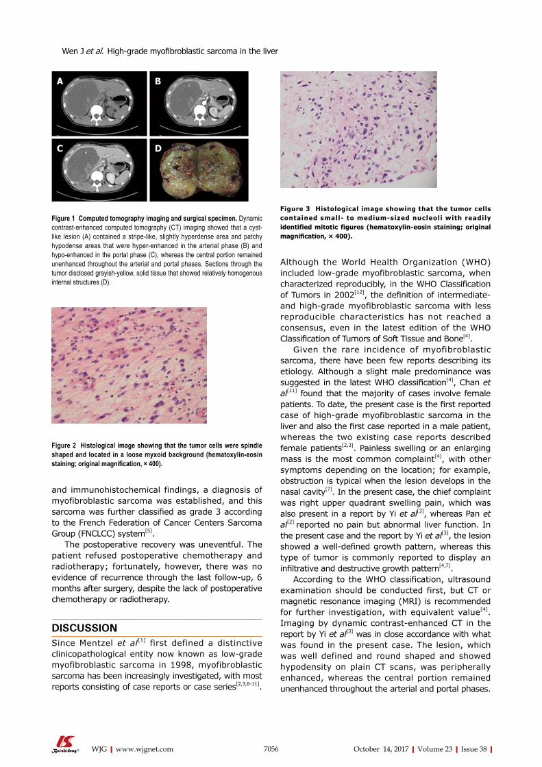
and immunohistochemical findings, a diagnosis of myofibroblastic sarcoma was established, and this sarcoma was further classified as grade 3 according to the French Federation of Cancer Centers Sarcoma Group (FNClCC) system[5].
The postoperative recovery was uneventful. The patient refused postoperative chemotherapy and radiotherapy; fortunately, however, there was no evidence of recurrence through the last follow-up, 6 months after surgery, despite the lack of postoperative chemotherapy or radiotherapy.
DISCUSSIONSince Mentzel et al[1] first defined a distinctive clinicopathological entity now known as low-grade myofibroblastic sarcoma in 1998, myofibroblastic sarcoma has been increasingly investigated, with most reports consisting of case reports or case series[2,3,6-11].
Although the World Health Organization (WHO) included low-grade myofibroblastic sarcoma, when characterized reproducibly, in the WHO Classification of Tumors in 2002[12], the definition of intermediate- and high-grade myofibroblastic sarcoma with less reproducible characteristics has not reached a consensus, even in the latest edition of the WHO Classification of Tumors of Soft Tissue and Bone[4].
Given the rare incidence of myofibroblastic sarcoma, there have been few reports describing its etiology. Although a slight male predominance was suggested in the latest WHO classification[4], Chan et al[11] found that the majority of cases involve female patients. To date, the present case is the first reported case of high-grade myofibroblastic sarcoma in the liver and also the first case reported in a male patient, whereas the two existing case reports described female patients[2,3]. Painless swelling or an enlarging mass is the most common complaint[4], with other symptoms depending on the location; for example, obstruction is typical when the lesion develops in the nasal cavity[7]. In the present case, the chief complaint was right upper quadrant swelling pain, which was also present in a report by Yi et al[3], whereas Pan et al[2] reported no pain but abnormal liver function. In the present case and the report by Yi et al[3], the lesion showed a well-defined growth pattern, whereas this type of tumor is commonly reported to display an infiltrative and destructive growth pattern[4,7].
According to the WHO classification, ultrasound examination should be conducted first, but CT or magnetic resonance imaging (MRI) is recommended for further investigation, with equivalent value[4]. Imaging by dynamic contrast-enhanced CT in the report by Yi et al[3] was in close accordance with what was found in the present case. The lesion, which was well defined and round shaped and showed hypodensity on plain CT scans, was peripherally enhanced, whereas the central portion remained unenhanced throughout the arterial and portal phases.
7056 October 14, 2017|Volume 23|Issue 38|WJG|www.wjgnet.com
A B
C D
Figure 1 Computed tomography imaging and surgical specimen. Dynamic contrast-enhanced computed tomography (CT) imaging showed that a cyst-like lesion (A) contained a stripe-like, slightly hyperdense area and patchy hypodense areas that were hyper-enhanced in the arterial phase (B) and hypo-enhanced in the portal phase (C), whereas the central portion remained unenhanced throughout the arterial and portal phases. Sections through the tumor disclosed grayish-yellow, solid tissue that showed relatively homogenous internal structures (D).
Figure 2 Histological image showing that the tumor cells were spindle shaped and located in a loose myxoid background (hematoxylin-eosin staining; original magnification, × 400).
Figure 3 Histological image showing that the tumor cells contained small- to medium-sized nucleoli with readily identified mitotic figures (hematoxylin-eosin staining; original magnification, × 400).
Wen J et al. High-grade myofibroblastic sarcoma in the liver
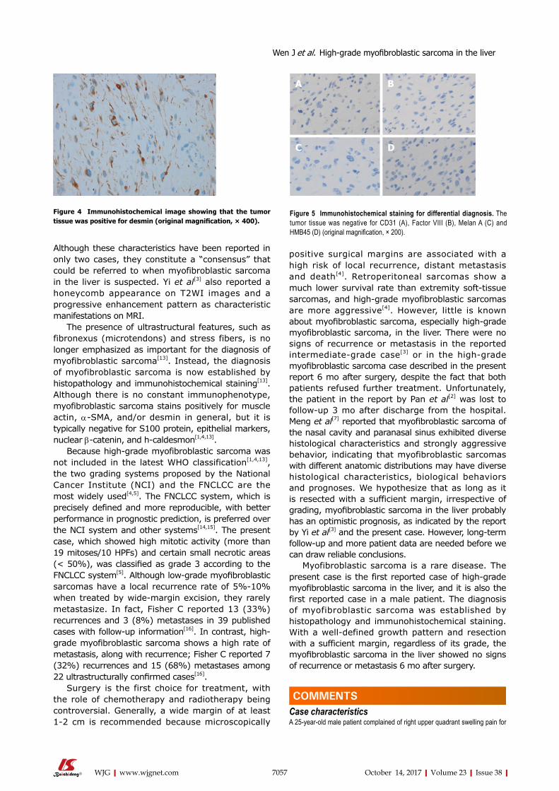
7057 October 14, 2017|Volume 23|Issue 38|WJG|www.wjgnet.com
positive surgical margins are associated with a high risk of local recurrence, distant metastasis and death[4]. Retroperitoneal sarcomas show a much lower survival rate than extremity soft-tissue sarcomas, and high-grade myofibroblastic sarcomas are more aggressive[4]. However, little is known about myofibroblastic sarcoma, especially high-grade myofibroblastic sarcoma, in the liver. There were no signs of recurrence or metastasis in the reported intermediate-grade case[3] or in the high-grade myofibroblastic sarcoma case described in the present report 6 mo after surgery, despite the fact that both patients refused further treatment. Unfortunately, the patient in the report by Pan et al[2] was lost to follow-up 3 mo after discharge from the hospital. Meng et al[7] reported that myofibroblastic sarcoma of the nasal cavity and paranasal sinus exhibited diverse histological characteristics and strongly aggressive behavior, indicating that myofibroblastic sarcomas with different anatomic distributions may have diverse histological characteristics, biological behaviors and prognoses. We hypothesize that as long as it is resected with a sufficient margin, irrespective of grading, myofibroblastic sarcoma in the liver probably has an optimistic prognosis, as indicated by the report by Yi et al[3] and the present case. However, long-term follow-up and more patient data are needed before we can draw reliable conclusions.
Myofibroblastic sarcoma is a rare disease. The present case is the first reported case of high-grade myofibroblastic sarcoma in the liver, and it is also the first reported case in a male patient. The diagnosis of myofibroblastic sarcoma was established by histopathology and immunohistochemical staining. With a well-defined growth pattern and resection with a sufficient margin, regardless of its grade, the myofibroblastic sarcoma in the liver showed no signs of recurrence or metastasis 6 mo after surgery.
COMMENTSCase characteristicsA 25-year-old male patient complained of right upper quadrant swelling pain for
Although these characteristics have been reported in only two cases, they constitute a “consensus” that could be referred to when myofibroblastic sarcoma in the liver is suspected. Yi et al[3] also reported a honeycomb appearance on T2WI images and a progressive enhancement pattern as characteristic manifestations on MRI.
The presence of ultrastructural features, such as fibronexus (microtendons) and stress fibers, is no longer emphasized as important for the diagnosis of myofibroblastic sarcoma[13]. Instead, the diagnosis of myofibroblastic sarcoma is now established by histopathology and immunohistochemical staining[13]. Although there is no constant immunophenotype, myofibroblastic sarcoma stains positively for muscle actin, α-SMA, and/or desmin in general, but it is typically negative for S100 protein, epithelial markers, nuclear β-catenin, and h-caldesmon[1,4,13].
Because high-grade myofibroblastic sarcoma was not included in the latest WHO classification[1,4,13], the two grading systems proposed by the National Cancer Institute (NCI) and the FNClCC are the most widely used[4,5]. The FNClCC system, which is precisely defined and more reproducible, with better performance in prognostic prediction, is preferred over the NCI system and other systems[14,15]. The present case, which showed high mitotic activity (more than 19 mitoses/10 HPFs) and certain small necrotic areas (< 50%), was classified as grade 3 according to the FNClCC system[5]. Although low-grade myofibroblastic sarcomas have a local recurrence rate of 5%-10% when treated by wide-margin excision, they rarely metastasize. In fact, Fisher C reported 13 (33%) recurrences and 3 (8%) metastases in 39 published cases with follow-up information[16]. In contrast, high-grade myofibroblastic sarcoma shows a high rate of metastasis, along with recurrence; Fisher C reported 7 (32%) recurrences and 15 (68%) metastases among 22 ultrastructurally confirmed cases[16].
Surgery is the first choice for treatment, with the role of chemotherapy and radiotherapy being controversial. Generally, a wide margin of at least 1-2 cm is recommended because microscopically
Figure 4 Immunohistochemical image showing that the tumor tissue was positive for desmin (original magnification, × 400).
A B
C D
Figure 5 Immunohistochemical staining for differential diagnosis. The tumor tissue was negative for CD31 (A), Factor VIII (B), Melan A (C) and HMB45 (D) (original magnification, × 200).
Wen J et al. High-grade myofibroblastic sarcoma in the liver
COMMENTS

7058 October 14, 2017|Volume 23|Issue 38|WJG|www.wjgnet.com
1 week without any concomitant symptoms.
Clinical diagnosisLiver echinococcosis was suspected.
Differential diagnosisLiver echinococcosis, gastrointestinal stromal tumor, vascular tumor, such as angiosarcoma or epithelioid hemangioendothelioma, angiomyolipoma.
Laboratory diagnosisAll laboratory tests were within normal limits.
Imaging diagnosisCT imaging found a cyst-like lesion with an obvious capsule located in the right trisegment of the liver.
Pathological diagnosisThe diagnosis of high-grade myofibroblastic sarcoma was established by histopathology and immunohistochemical staining.
TreatmentThe patient successfully underwent a right hepatic trisegmentectomy.
Related reportsThere have been no case reports on high-grade myofibroblastic sarcoma in the liver. Only two reports were published reporting intermediate-grade myofbroblastic sarcoma or myofibroblastic sarcoma. The diagnosis was established on the basis of morphology and immunohistochemistry features. Surgical resection was the treatment of choice with gratifying results.
Experiences and lessonsThe diagnosis of myofibroblastic sarcoma is established by histopathology and immunohistochemical staining. Surgical resection is the treatment of choice with gratifying results.
Peer-reviewThe authors report a case of myofibroblastic sarcoma of the liver. The manuscript is well-written and the case is pathologically interesting.
REFERENCES1 Mentzel T, Dry S, Katenkamp D, Fletcher CD. Low-grade
myofibroblastic sarcoma: analysis of 18 cases in the spectrum of myofibroblastic tumors. Am J Surg Pathol 1998; 22: 1228-1238 [PMID: 9777985]
2 Pan Y, Wu X, Liu J, Muheremu A. Abnormal liver function induced by myofibroblastic sarcoma infiltrating the liver: A case report. Oncol Lett 2015; 9: 798-800 [PMID: 25621053 DOI: 10.3892/ol.2014.2740]
3 Yi X, Xiao D, Long X. Myofibroblastic sarcoma in liver: a
case report. Int J Clin Exp Pathol 2015; 8: 1073-1076 [PMID: 25755822]
4 Fletcher CD, Bridge J, Hogendoorn P, Mertens F. WHO Classification of Tumours of Soft Tissue and Bone. 4th ed. Lyon: IARC Press, 2013: 15-18
5 Neuville A, Chibon F, Coindre JM. Grading of soft tissue sarcomas: from histological to molecular assessment. Pathology 2014; 46: 113-120 [PMID: 24378389 DOI: 10.1097/PAT.0000000000000048]
6 Kolli S, Ziaee A, Lee R, Lim MJ, Levy B, Labovitz AJ. Myofibroblastic sarcoma of mitral valve: a case report. J Am Soc Echocardiogr 2005; 18: 285-286 [PMID: 15746724 DOI: 10.1016/j.echo.2004.12.011]
7 Meng GZ, Zhang HY, Bu H, Yang GH, Zhang XL, Yang G. Myofibroblastic sarcoma of the nasal cavity and paranasal sinus: a clinicopathologic study of 6 cases and review of the literature. Oral Surg Oral Med Oral Pathol Oral Radiol Endod 2007; 104: 530-539 [PMID: 17142072 DOI: 10.1016/j.tripleo.2006.08.021]
8 Morii T, Mochizuki K, Sano H, Fujino T, Harasawa A, Satomi K. Occult myofibroblastic sarcoma detected on FDG-PET performed for cancer screening. Ann Nucl Med 2008; 22: 811-815 [PMID: 19039560 DOI: 10.1007/s12149-008-0194-4]
9 Takácsi-Nagy Z, Muraközy G, Pogány P, Fodor J, Orosz Z. Myofibroblastic sarcoma of the base of tongue. Case report and review of the literature. Strahlenther Onkol 2009; 185: 198-201 [PMID: 19330298 DOI: 10.1007/s00066-009-1925-8]
10 Miyazawa M, Naritaka Y, Miyaki A, Asaka S, Isohata N, Yamaguchi K, Murayama M, Shimakawa T, Katsube T, Ogawa K, Fujibayashi M. A low-grade myofibroblastic sarcoma in the abdominal cavity. Anticancer Res 2011; 31: 2989-2994 [PMID: 21868549]
11 Chan JY, Gooi Z, Wong EW, Ng SK, Tong MC, Vlantis AC. Low-grade myofibroblastic sarcoma: A population-based study. Laryngoscope 2017; 127: 116-121 [PMID: 27377169 DOI: 10.1002/lary.26146]
12 Fletcher CD, Unni KK, Mertens F. World Health Organization Classification of Tumours. Pathology and Genetics of Tumours of Soft Tissue and Bone. 3rd ed. Lyon: IARC Press, 2002: 94-96
13 Mentzel T. Myofibroblastic sarcomas: a brief review of sarcomas showing a myofibroblastic line of differentiation and discussion of the differential diagnosis. Current Diagnostic Pathology 2001; 7: 17-24 [DOI: 10.1054/cdip.2000.0057]
14 Guillou L, Coindre JM, Bonichon F, Nguyen BB, Terrier P, Collin F, Vilain MO, Mandard AM, Le Doussal V, Leroux A, Jacquemier J, Duplay H, Sastre-Garau X, Costa J. Comparative study of the National Cancer Institute and French Federation of Cancer Centers Sarcoma Group grading systems in a population of 410 adult patients with soft tissue sarcoma. J Clin Oncol 1997; 15: 350-362 [PMID: 8996162 DOI: 10.1200/JCO.1997.15.1.350]
15 Deyrup AT, Weiss SW. Grading of soft tissue sarcomas: the challenge of providing precise information in an imprecise world. Histopathology 2006; 48: 42-50 [PMID: 16359536 DOI: 10.1111/j.1365-2559.2005.02288.x]
16 Fisher C. Myofibroblastic malignancies. Adv Anat Pathol 2004; 11: 190-201 [PMID: 15220822]
P- Reviewer: Kai K S- Editor: Gong ZM L- Editor: Wang TQ E- Editor: Ma YJ
Wen J et al. High-grade myofibroblastic sarcoma in the liver

© 2017 Baishideng Publishing Group Inc. All rights reserved.
Published by Baishideng Publishing Group Inc7901 Stoneridge Drive, Suite 501, Pleasanton, CA 94588, USA
Telephone: +1-925-223-8242Fax: +1-925-223-8243
E-mail: [email protected] Desk: http://www.f6publishing.com/helpdesk
http://www.wjgnet.com
I S S N 1 0 0 7 - 9 3 2 7
9 7 7 1 0 07 9 3 2 0 45
3 8


