27
World Journal of Biological Chemistry ISSN 1949-8454 (online) World J Biol Chem 2020 November 27; 11(3): 76-118 Published by Baishideng Publishing Group Inc

World Journal ofBiological Chemistry
ISSN 1949-8454 (online)
World J Biol Chem 2020 November 27; 11(3): 76-118
Published by Baishideng Publishing Group Inc

WJBC https://www.wjgnet.com I November 27, 2020 Volume 11 Issue 3
World Journal of
Biological ChemistryW J B CContents Irregular Volume 11 Number 3 November 27, 2020
REVIEW
Current understanding of glucose transporter 4 expression and functional mechanisms76
Wang T, Wang J, Hu X, Huang XJ, Chen GX
MINIREVIEWS
Deciphering the modifiers for phenotypic variability of X-linked adrenoleukodystrophy99
Palakuzhiyil SV, Christopher R, Chandra SR
ORIGINAL ARTICLE
Observational Study
Prevalence, serotyping and drug susceptibility patterns of Escherichia coli isolates from kidney transplanted patients with urinary tract infections
112
Najafi Khah A, Hakemi-Vala M, Samavat S, Nasiri MJ
WJBC https://www.wjgnet.com II November 27, 2020 Volume 11 Issue 3
World Journal of Biological ChemistryContents
Irregular Volume 11 Number 3 November 27, 2020
ABOUT COVER
Editorial board member of World Journal of Biological Chemistry, Dr. Jian-Xun Ding is a Professor at Changchun Institute of Applied Chemistry (CIAC), Chinese Academy of Sciences (CAS). Dr. Ding received his Bachelor of Science degree from the University of Science and Technology of China in 2007 and obtained his PhD degree from CIAC, CAS in 2013. His research focuses on the synthesis of functional biodegradable polymers, the development of smart polymer platforms for controlled drug delivery, and the exploitation of polymer-based adjuvants for immunotherapy. Heretofore, he has published more than 100 articles, which have amassed over 7000 citations. Moreover, he has applied for over 70 patents in China and has won more than 10 awards for his career accomplishments. He was selected for the Young Talents Promotion Project of Jilin Province and joined the CAS Young Innovation Promotion Association in 2019. (L-Editor: Filipodia)
AIMS AND SCOPE
The primary aim of the World Journal of Biological Chemistry(WJBC, World J Biol Chem) is to provide scholars and readers from various fields of biological chemistry a platform to publish high-quality basic and clinical research articles and communicate their research findings online. WJBC mainly publishes articles reporting research results and findings obtained in the field of biological chemistry and covering a wide range of topics including bioenergetics, cell biology, chromosomes, developmental biology, DNA, enzymology, extracellular matrices, gene regulation, genomics, glycobiology, immunology, lipids, membrane biology, metabolism, molecular bases of disease, molecular biophysics, neurobiology, plant biology, protein structure and folding, protein synthesis and degradation, proteomics, and signal transduction.
INDEXING/ABSTRACTING
The WJBC is now abstracted and indexed in PubMed, PubMed Central, China National Knowledge Infrastructure (CNKI), China Science and Technology Journal Database (CSTJ), and Superstar Journals Database.
RESPONSIBLE EDITORS FOR THIS ISSUE
Production Editor: Yu-Jie Ma; Production Department Director: Yun-Xiaojian Wu; Editorial Office Director: Jia-Ping Yan.
NAME OF JOURNAL INSTRUCTIONS TO AUTHORS
World Journal of Biological Chemistry https://www.wjgnet.com/bpg/gerinfo/204
ISSN GUIDELINES FOR ETHICS DOCUMENTS
ISSN 1949-8454 (online) https://www.wjgnet.com/bpg/GerInfo/287
LAUNCH DATE GUIDELINES FOR NON-NATIVE SPEAKERS OF ENGLISH
July 26, 2010 https://www.wjgnet.com/bpg/gerinfo/240
FREQUENCY PUBLICATION ETHICS
Irregular https://www.wjgnet.com/bpg/GerInfo/288
EDITORS-IN-CHIEF PUBLICATION MISCONDUCT
Vsevolod Gurevich, Chun-Peng Wan https://www.wjgnet.com/bpg/gerinfo/208
EDITORIAL BOARD MEMBERS ARTICLE PROCESSING CHARGE
https://www.wjgnet.com/1949-8454/editorialboard.htm https://www.wjgnet.com/bpg/gerinfo/242
PUBLICATION DATE STEPS FOR SUBMITTING MANUSCRIPTS
November 27, 2020 https://www.wjgnet.com/bpg/GerInfo/239
COPYRIGHT ONLINE SUBMISSION
© 2020 Baishideng Publishing Group Inc https://www.f6publishing.com
© 2020 Baishideng Publishing Group Inc. All rights reserved. 7041 Koll Center Parkway, Suite 160, Pleasanton, CA 94566, USA
E-mail: [email protected] https://www.wjgnet.com

WJBC https://www.wjgnet.com 76 November 27, 2020 Volume 11 Issue 3
World Journal of
Biological ChemistryW J B CSubmit a Manuscript: https://www.f6publishing.com World J Biol Chem 2020 November 27; 11(3): 76-98
DOI: 10.4331/wjbc.v11.i3.76 ISSN 1949-8454 (online)
REVIEW
Current understanding of glucose transporter 4 expression and functional mechanisms
Tiannan Wang, Jing Wang, Xinge Hu, Xian-Ju Huang, Guo-Xun Chen
ORCID number: Tiannan Wang 0000-0001-5584-3628; Jing Wang 0000-0003-3663-8332; Xinge Hu 0000-0002-3253-7537; Xian-Ju Huang 0000-0001-8130-1961; Guo-Xun Chen 0000-0001-6226-4050.
Author contributions: Wang T, Wang J and Hu X conducted the PubMed Search; Wang T, Wang J, Hu X, Huang XJ and Chen GX outlined and wrote the manuscript.
Conflict-of-interest statement: All authors declare that there is no conflict of interest to report.
Open-Access: This article is an open-access article that was selected by an in-house editor and fully peer-reviewed by external reviewers. It is distributed in accordance with the Creative Commons Attribution NonCommercial (CC BY-NC 4.0) license, which permits others to distribute, remix, adapt, build upon this work non-commercially, and license their derivative works on different terms, provided the original work is properly cited and the use is non-commercial. See: http://creativecommons.org/Licenses/by-nc/4.0/
Manuscript source: Invited manuscript
Specialty type: Biochemistry and molecular biology
Tiannan Wang, Xinge Hu, Guo-Xun Chen, Department of Nutrition, The University of Tennessee, Knoxville, TN 37996, United States
Jing Wang, Xian-Ju Huang, College of Pharmacy, South-Central University for Nationalities, Wuhan 430074, Hubei Province, China
Corresponding author: Guo-Xun Chen, PhD, Associate Professor, Department of Nutrition, The University of Tennessee, Room 229 Jessie Harris Building, 1215 West Cumberland Avenue Knoxville, TN 37996, United States. [email protected]
AbstractGlucose is used aerobically and anaerobically to generate energy for cells. Glucose transporters (GLUTs) are transmembrane proteins that transport glucose across the cell membrane. Insulin promotes glucose utilization in part through promoting glucose entry into the skeletal and adipose tissues. This has been thought to be achieved through insulin-induced GLUT4 translocation from intracellular compartments to the cell membrane, which increases the overall rate of glucose flux into a cell. The insulin-induced GLUT4 translocation has been investigated extensively. Recently, significant progress has been made in our understanding of GLUT4 expression and translocation. Here, we summarized the methods and reagents used to determine the expression levels of Slc2a4 mRNA and GLUT4 protein, and GLUT4 translocation in the skeletal muscle, adipose tissues, heart and brain. Overall, a variety of methods such real-time polymerase chain reaction, immunohistochemistry, fluorescence microscopy, fusion proteins, stable cell line and transgenic animals have been used to answer particular questions related to GLUT4 system and insulin action. It seems that insulin-induced GLUT4 translocation can be observed in the heart and brain in addition to the skeletal muscle and adipocytes. Hormones other than insulin can induce GLUT4 translocation. Clearly, more studies of GLUT4 are warranted in the future to advance of our understanding of glucose homeostasis.
Key Words: Glucose transporter 4; Insulin; Skeletal muscle; Adipocytes; Brain; Heart; Antibodies
©The Author(s) 2020. Published by Baishideng Publishing Group Inc. All rights reserved.

Wang T et al. Current status of GLUT4 studies
WJBC https://www.wjgnet.com 77 November 27, 2020 Volume 11 Issue 3
Country/Territory of origin: United States
Peer-review report’s scientific quality classificationGrade A (Excellent): 0 Grade B (Very good): B Grade C (Good): C, C Grade D (Fair): D Grade E (Poor): 0
Received: June 26, 2020 Peer-review started: June 26, 2020 First decision: August 9, 2020 Revised: August 22, 2020 Accepted: September 22, 2020 Article in press: September 22, 2020 Published online: November 27, 2020
P-Reviewer: Merigo F, Salceda R, Soriano-Ursúa MA S-Editor: Ma YJ L-Editor: A P-Editor: Ma YJ
Core Tip: Glucose transporter 4 (GLUT4) can be detected not only in the skeletal muscle and adipocytes, but also in the brain and heart. In addition to the translocation from vesicles in the cytosol to the cell membrane by insulin, the expression levels of Slc2a4 mRNA and GLUT4 proteins are also regulated by many factors. A variety of methods and antibodies from various sources have been used to evaluate GLUT4 expression and translocation.
Citation: Wang T, Wang J, Hu X, Huang XJ, Chen GX. Current understanding of glucose transporter 4 expression and functional mechanisms. World J Biol Chem 2020; 11(3): 76-98URL: https://www.wjgnet.com/1949-8454/full/v11/i3/76.htmDOI: https://dx.doi.org/10.4331/wjbc.v11.i3.76
INTRODUCTIONCurrently, diabetes is a problem of public health[1]. Based on the American Diabetes Association definition, diabetes is a serious chronic health condition of your body that causes blood glucose levels to rise higher than normal, which will lead to multiple complications if hyperglycemia is left untreated or mismanaged[2]. Diabetes occurs when your body cannot make insulin or cannot effectively respond to insulin to regulate blood glucose level. There are two type of diabetes, insulin-dependent type 1 diabetes mellitus (T1DM) and -independent type 2 diabetes mellitus (T2DM). T2DM accounts for about 90% to 95% of all diagnosed cases of diabetes, and is due to the lack of responses to insulin in the body[3]. Insulin resistance is a characteristic of T2DM. For a person with diabetes, a major challenge is to control or manage blood glucose level. Glucose is a common molecule used for production of energy or other metabolites in cells. As a quick energy source, glucose can be metabolized aerobically or anaerobically depending on the availability of oxygen or cell characteristics[4]. Glucose is a hydrophilic molecule, and cannot diffuse into or out of a cell freely. It needs transporters to cross the cell membrane. Glucose transporters (GLUTs) are proteins that serve this purpose.
GLUTs are members of the major facilitator superfamily (MFS) transporters, which are responsible for the transfer of a large array of small molecules such as nutrients, metabolites and toxins across the cell membrane[5]. Multiple members have been identified in each family of MFS transporters, and changes of their functions have been associated with a number of diseases[5]. Members of MFS transporters have 12 transmembrane helices, and transport their substrates as uniporters, symporters or antiporters[5]. Upon binding of the substrates on side of the membrane, a conformation change occurs, which is achieved through coordinative interactions of those helices through a “clamp-and-switch” mechanism. Structural studies have shown that the substrate specificity is achieved through the conserved amino acid residues within each family[5]. Thus, it is important to understand GLUT functions, expressions and regulations for the control of blood glucose homeostasis.
Insulin and GLUTsDietary starch is first digested into glucose before being absorbed into the body and utilized[4]. The first transporter identified is GLUT1, which is expressed universally in all cells, and responsible for basal glucose transport[6]. Insulin stimulates glucose utilization in the body. This is in part through the insulin-induced glucose uptake in the muscle and adipose tissues. In addition to insulin stimulation, physical activity can also increase glucose entering into the skeletal muscle cells[7]. The observation that insulin promotes the redistribution of GLUTs from intracellular locations to the plasma membrane in adipocytes began in the 1980s[8-10]. Few years later, the insulin-induced glucose transport was also found in muscle cells[11,12]. To understand the underlying mechanism of insulin-stimulated glucose uptake, antibodies against membrane glucose transport proteins were created[13]. Subcellular fractionation, cytokinin B (glucose-sensitive ligand), and glucose absorption into isolated vesicles were used to study the phenomenon. It was proposed that these GLUTs are moved from intracellular components to the plasma membrane of adipocytes and muscle cells upon insulin stimulation[6]. In 1988, a specific antibody against a GLUT sample preparation was created, which eventually led to the identification of a molecular

Wang T et al. Current status of GLUT4 studies
WJBC https://www.wjgnet.com 78 November 27, 2020 Volume 11 Issue 3
clone that encodes an insulin-induced GLUT from mouse adipocytes[6]. It was named GLUT4. Since the 1990s, fluorescent-labeled fusion proteins, GLUT4-specific antibodies, photoaffinity labeling reagents, immunofluorescence microscopy, and high-resolution electron microscope have been used to confirm the insulin-induced translocation and underlying mechanisms[6]
It has been widely accepted that insulin mainly stimulates transfer of GLUT4 from intracellular storage vesicles to the plasma membrane. Insulin stimulation accelerates the movement rate of GLUT4 containing vesicles to the cell membrane[14]. When more GLUT4 is on the plasma membrane, more glucose enters the cells without any change of the GLUT4 specific activity. During insulin stimulation, GLUT4 is not statically maintained in the plasma membrane but continuously recycled[6]. After insulin is removed, the amount of GLUT4 on the plasma membrane drops and the rate of movement returns to basal level.
Since identification and cloning of GLUT1, 13 additional GLUTs have been cloned using recombinant DNA techniques[15]. Based on their phylogeny or genetic and structural similarities, GLUTs are classified into three classes. Class I includes GLUTs 1-4, and GLUT14 which are responsible for glucose transfer. Class II consists of GLTUs 5, 7, 9 and GLUT 11 which are considered as fructose transporters. Class III contains GLTUs 6, 8, 10, 12 and GLUT 13[16]. All GLUTs have nearly 500 amino acid residues that form 12 transmembrane helices[15].
Each GLUT has its own unique affinity and specificity for its substrate, tissue distribution, intracellular location, regulatory mechanisms and physiological functions[17]. The most well studied and known members are GLUTs 1-6. GLUT1 is found evenly distributed in the fetal tissues. In human adults, GLUT1 Level is high in erythrocytes and endothelial cells. It is responsible for basal glucose uptake[18]. GLUT2 is expressed in the liver and pancreas, and contributes to glucose sensing and homeostasis[17]. In enterocytes, GLUT2 is responsible to transport the absorbed glucose, fructose and galactose out of the basolateral membrane to enter into the blood circulation through the portal vein[19]. GLUT3 just like GLUT1 is expressed in almost all mammalian cells and is responsible for the basal uptake of glucose. GLUT3 is considered as the main GLUT isoform expressed in neurons and the placenta, but has also been detected in the testis, placenta, and skeletal muscle[20-22]. GLUT5 is specific for uptake of fructose in a passive diffusion manner, and is expressed in the small intestine, testes and kidney[17]. GLUT6 is expressed in the spleen, brain, and leukocytes as well as in muscle and adipose tissue[15,23]. GLUT6 has been shown to move from the intracellular locations and plasma membrane of rat adipocytes in a dynamin-dependent manner[23]. Table 1[24-57] summarizes names, numbers of amino acids, Kms, expression profiles and potential functions of those GLUTs.
GLUT4 gene, its tissue distribution, and physiological functionsHuman GLUT4 has 509 amino acid residues and is encoded by SLC2A4 gene in the human genome. It is mainly expressed in adipocytes and skeletal muscle. The unique N-terminal and COOH terminal sequences are responsible for GLUT4's response to insulin signaling and membrane transport[58]. The Km of GLUT4 is about 5 mmol/L. In response to insulin stimulation, intracellular vesicles containing GLUT4 are moved from cytosol to the cell membrane. As shown in Figure 1, insulin receptor is a tetramer with two alpha-subunits and two beta-subunits linked by disulfide bonds[59]. When insulin binds to its receptor on the cell membrane, insulin receptor beta subunits that contain tyrosine kinase domain autophosphorylate each other. The phosphorylated β-subunits recruit insulin receptor substrates (IRS) and phosphorylate them. Then phosphorylated IRSs bind to and activate phosphatidylinositol 3-kinase (PI3K) which is recruited to the plasma membrane and converts PIP2 to PIP3. On the plasma membrane, PI3K activates PIP3 dependent protein kinase, which phosphorylates and activates AKT (also referred to as protein kinase B, PKB). Akt activation triggers vesicle fusion, which results in the translocation of GLUT4 containing vesicles from intracellular compartments to the plasma membrane. The elevation of GLUT4 on the membrane leads to increase of glucose entry into the cell.
Upon refeeding, elevated glucose levels in the blood stimulates insulin secretion from pancreatic beta cells. Insulin stimulates GLUT4 translocation to the cell membrane, which increases glucose uptake in cells, and achieves glucose homeostasis[60,61]. After the insulin stimulation disappears, GLUT4 is transferred back into the cytosol from the plasma membrane. More than 90% of GLUT4 is located in the intracellular body, trans-Golgi network, and heterogeneous tube-like vesicle structure, etc., which constitute the GLUT4 storage vesicle (GSV). In an unstimulated state, most GLUT4 is in the intracellular vesicles of muscle and adipocytes[62].
The amount of GLUT4 on the cell membrane is determined by the rate of the

Wang T et al. Current status of GLUT4 studies
WJBC https://www.wjgnet.com 79 November 27, 2020 Volume 11 Issue 3
Table 1 Summary of glucose transporter family members
Protein (gene) Amino acids Km
(mm) Expression sites Function/substrates Ref.
GLUT1 (SLC2A1)
492 3-7 Ubiquitous distribution in tissues and culture cells
Basal glucose uptake; glucose, galactose, glucosamine, mannose
[24-30]
GLUT2 (SLC2A2)
524 17 Liver, pancreas, brain, kidney, small intestine
High-capacity low-affinity transport; glucose, galactose, fructose, glucosamine, mannose
[25-27,29-34]
GLUT3 (SLC2A3)
496 1.4 Brain and nerves cells Neuronal transport; glucose, galactose, mannose[25-27,29,30,33-35]
GLUT4 (SLC2A4)
509 5 Muscle, fat, heart, hippocampal neurons
Insulin-regulated transport in muscle and fat; glucose, glucosamine
[25-27,29,31,36,37]
GLUT5 (SLC2A5)
501 6 Intestine, kidney, testis, brain Fructose[25-27,29,30,34,38-42]
GLUT6 (SLC2A6)
507 5 Spleen, leukocytes, brain Glucose[25-27,29,30,43]
GLUT7 (SLC2A7)
524 0.3 Small intestine, colon, testis, liver Fructose and glucose[25-27,29,30,38]
GLUT8 (SLC2A8)
477 2 Testis, blastocyst, brain, muscle, adipocytes
Insulin-responsive transport in blastocyst; glucose, fructose, galactose
[25-27,29,30,44,45]
GLUT9 (SLC2A9)
Major 540, Minor 512
0.9 Liver, kidney Glucose, fructose[25-27,29,30, 46-48]
GLUT10 (SLC2A10)
541 0.3 Heart, lung, brain, skeletal muscle, placenta, liver, pancreas
Glucose and galactose[25-27,29,30,48,49]
GLUT11 (SLC2A11)
496 0.2 Heart, muscle, adipose tissue, pancreas
Muscle-specific; fructose and glucose transporter[25-27,29,30,50-54]
GLUT12 (SLC2A12)
617 4-5 Heart, prostate, skeletal muscle, fat, mammary gland
Glucose[25-27,29,30,53,55]
GLUT13 (SLC2A13)
Rat 618, human 629
0.1 Brain (neurons intracellular vesicles) H+-myo-inositol transporter[25-27,29,30,56]
GLUT14 (SLC2A14)
Short 497, Long 520
unknown Testis Glucose transport[25-27,29,30,57]
movement from intracellular GSV to the cell membrane. In adipocytes and skeletal muscle cells, insulin increases the rate of GLUT4 translocation from GSVs to membrane and decreases the rate of GLUT4 movement from membrane back to the vesicles, which lead to elevation of GLUT4 content on the cell membrane by 2-3 times[63]. Moreover, in adipocytes, insulin increases the GLUT4 recirculation to maintain a stable and releasable vesicle[64].
So far, insulin-induced GLUT4 translocation has been studied extensively. However, questions still remain. Methods and reagents used to determine the expression levels of GLUT4 and its translocation mechanism deserve to be summarized and analyzed. Therefore, we searched the relevant articles in PubMed and investigated the methods and reagents used in the studies. “Glucose transporter 4” and “GLUT4” as the protein and “SLC2A4” as the gene name were used as keywords in the search. In order to have a more clearly overview, we further divided and focused on the search into three parts, GLUT4 in the skeletal muscle, GLUT4 in adipose tissues, and GLUT4 in heart and brain.
GLUT4 IN THE SKELETAL MUSCLEThe term "muscle" covers a variety of cell types. Mammals have four main types of muscle cells: skeletal, heart, smooth, and myoepithelial cells. They are different in function, structure and development[65]. The skeletal muscle mass accounts for 40% of the total body mass, and the regulation of skeletal muscle glucose metabolism will significantly affect the body's glucose homeostasis[66,67]. Skeletal muscle is composed of many muscle fibers connected by collagen and reticular fibers. Each skeletal muscle fiber is a syncytium that derives from the fusion of many myoblasts. Myoblasts proliferate in large quantities, but once fused, they no longer divide. The fusion

Wang T et al. Current status of GLUT4 studies
WJBC https://www.wjgnet.com 80 November 27, 2020 Volume 11 Issue 3
Figure 1 Schematic of insulin-induced translocation of glucose transporter 4 from cytosol to the cell membrane. The binding of insulin to its receptors initiates a signal transduction cascade, which results in the activation of Akt. Akt acts on the glucose transporter 4 (GLUT4) containing vesicles in the cytosol to facilitate their fusion with the cell membrane. When more GLUT4 molecules are present in the membrane, the rate of glucose uptake is elevated. GLUT4: Glucose transporter 4.
usually follows the onset of myoblast differentiation[65]. Different fiber types have distinct contractile and metabolic properties[68]. The skeletal muscle maintains skeletal structure and essential daily activities[69]. Also, it is a source of proteins that can be broken down into amino acids for the body to use.
Insulin stimulates glucose uptake and utilization in the skeletal muscle. GLUT4 plays a key role in the uptake process. Glucose can be stored as glycogen, which is used as a quick source of energy in physical activity[70]. In the skeletal muscle, exercise helps increase insulin sensitivity and stimulates SLC2A4 gene transcription[60]. Physiological factors such as the type of muscle fibers can also affect the GLUT4 Level. An increase in physical activity will induce the GLUT4 Levels, whereas a decrease in activity level will reduce GLUT4[68]. The skeletal muscle not only maintains the activities, but also regulates the glucose homeostasis in the body, which plays a key role in the development of metabolic diseases[69]. Obesity and T2DM have a negative impact on skeletal muscle glucose metabolism[71].
To review the methods and reagents of GLUT4 studies in skeletal muscle, "GLUT4, skeletal muscle" and "SLC2A4, skeletal muscle" as keywords were used to search the PubMed database to retrieve relevant articles. The skeletal muscle is a highly specialized tissue made of well-organized muscle fibers. The unique structural characteristic of muscle inherits difficulties to be lysed for biochemical studies. Therefore, we want to focus on the sample preparation of skeletal muscle in GLUT4 studies. The retrieved articles were screened mainly according to the research methods and reagents used for skeletal muscle preparation, experimental groups included, Slc2a4 mRNA and GLUT4 protein measurements and the source of GLUT4 antibodies obtained. In the end, 10 representative articles were selected for analysis and summary as shown in Table 2[72-81].
Overall, the current research methods of GLUT 4 studies in skeletal muscle are listed below: (1) Samples were homogenized to prepare membrane fractions for analysis of GLUT 4 in western blot using monoclonal or polyclonal antibodies; (2) Real-time polymerase chain reaction (PCR) was used to determine the mRNA abundance of Slc2a4; (3) Immunocytochemical staining was used to detect GLUT4 in situ. The fibers were labeled for GLUT4 by a preembedding technique and observed as whole mounts by immunofluorescence microscopy or after sectioning, by immunogold electron microscopy. Preembedding is a technique to label GLUT4 immediately after tissues or cells are collected, which allows that the antibody interacts with the antigen before denaturation; (4) Muscle cell lines stably expressing tagged GLUT4 were established to study the translocation; and (5) Radiolabeled 2-deoxyglucose was used to determine the glucose uptake in muscle tissue slices.
The antibodies used in these articles were from Santa Cruz, Millipore, East Acres, Biogenesis and other sources not specified. Only two of the publications have a positive control group of GLUT4 expression using overexpression of a fusion protein and tissue preparation as a standard for determination. Positive controls are important

Wang T et al. Current status of GLUT4 studies
WJBC https://www.wjgnet.com 81 November 27, 2020 Volume 11 Issue 3
Table 2 Recent studies of glucose transporter 4 expression and translocation in the skeletal muscle
Methods Materials Comparisons Observations/conclusions Ref.
Western blot. Cell fractions of rat L6 myotubes, 3T3-L1, and mouse muscle and adipose tissues. Anti-GLUT4 from Santa Cruz Biotechnology (1:1000).
Cell: Total cell lysate vs membrane fractions. Mouse tissues: Control vs high-fat diets.
Insulin treatments increases GLUT4 levels in membrane fractions without any change in the total cell lysate. GLUT4 levels in adipose tissue and muscle of mice fed a high-fat diet are lower in all fractions than that fed the control diet.
[72]
Western blot. Whole cell and cell fractions from rat L6 and mouse C2C12 muscle cells, and soleus muscle of hind limb from mice. Anti-GLUT4 from Santa Cruz. Biotechnology (1:1000).
Whole cell lysate vs membrane fractions. Treatments without or with insulin or AICAR.
GLUT4 translocation occurs in L6 myotubes and 3T3-L1 adipocytes stimulated by insulin and AICAR. GLUT4 translocation occurs in muscle at 15 to 30 minutes and in adipose tissue at 15 minutes after glucose treatment.
[73]
Western blot. Giant sarcolemmal vesicles from soleus muscles of Sprague-Dawley rats. Anti-GLUT4 from Millipore (1:4000).
Tissue samples without or with insulin released in the presence of glucose as a stimulant and lipid as a control.
A glucose-dependent insulinotropic polypeptide increases glucose transport and plasma membrane GLUT4 protein content.
[74]
Real-time PCR for Slc2a4 mRNA levels. Total RNA of the skeletal muscle from male C57BL/6J and ICR mice fed different diets.
mRNA levels in muscle samples from mice fed the control or CLA supplement diet
Dietary CLA does not affect Slc2a4 mRNA levels in the mouse skeletal muscle [75]
Western blot. Preparations of sarcolemmal membrane fractions and crude lysates from male Muscovy ducklings. Anti-GLUT4 from East Acres (1:500).
GLUT4 from a unique crude membrane fraction of rat skeletal muscle was used as an arbitrary unit and from erythrocyte ghost as a negative control
Polyclonal antibodies detect a protein of similar size (approximately 45 kDa) of GLUT4 in the crude membrane preparations from rat (positive control) and duckling skeletal muscle. No signal was obtained for rat erythrocyte ghost membrane preparation.
[76]
ATB-BMPA-labelling of glucose transporters, Immunoprecipitation, liquid-scintillation counting, Western blot.
Tissue samples of isolated and perfused EDL or soleus muscle from GLUT1 transgenic C57BL’KsJ-Leprdbj and control mice. Anti-GLUT4 (R1184; C-terminal) from an unknown source.
Non-transgenic mice vs transgenic mice.
Basal levels of cell-surface GLUT4 in isolated or perfused EDL are similar in transgenic and non- transgenic mice. Insulin induces cell-surface GLUT4 by 2-fold in isolated EDL and by 6-fold in perfused EDL of both transgenic and non-transgenic mice. Western blot results were not shown.
[77]
Preembedding technique (immune reaction occurs prior to resin embedding to label GLUT4), and observations of whole mounts by immunofluorescence microscopy, or after sectioning by immunogold electron microscopy.
Muscle samples from male Wistar rats. Anti-GLUT4 (C-terminal, 1:1000), and anti-GLUT4 (13 N-terminal, 1:500) from unknown species.
Rats were divided in four groups: Control, contraction received saline, insulin and insulin plus contraction groups. They received glucose followed by insulin injection.
Two populations of intracellular GLUT4 vesicles are differentially recruited by insulin and muscle contractions. The increase in glucose transport by insulin and contractions in the skeletal muscle is due to an additive translocation to both the plasma membrane and T tubules. Unmasking of GLUT4 COOH-terminal epitopes and changes in T tubule diameters does not contribute to the increase in glucose transport.
[78]
Immunoprecipitation, and Western blot. Membrane fractions from skeletal muscle of male Wistar rats treated without or with insulin. Anti-GLUT-4 from Genzyme, Anti-GLUT-4 from Santa Cruz Biotechnology.
Crude membrane preparations and cytosolic fractions in samples of rats treated without or with insulin.
In vitro activation of PLD in crude membranes results in movement of GLUT4 to vesicles/microsomes. This GLUT4 translocation is blocked by the PLD inhibitor, neomycin, which also reduces insulin-stimulated glucose transport in rat soleus muscle.
[79]
Western blot for GLUT4 protein in homogenates of epitrochlearis muscles. Tissue slices labeled with 2-[1,2-3H]-deoxy-d-glucose and counted in a gamma counter.
Muscle homogenate and slices from male Sprague-Dawley rats. Anti- GLUT4 from Dr. Osamu Ezaki.
Sedentary control vs a 5-day swimming training group.
The change of insulin responsiveness after detraining is directly related to muscle GLUT-4 protein content. The greater the increase in GLUT-4 protein content induced by training, the longer an effect on insulin responsiveness lasts after training.
[80]
Immunofluorescence for membrane preparations, and 2-Deoxyglucose uptake in isolated skeletal
Membrane preparations from L6 cells over-expressing GLUT4myc. Isolated
L6 cells over-expressing GLUT4myc treated without or with Indinavir.
HIV-1 protease inhibitor indinavir at 100 µmol/l inhibits 80% of basal and insulin-stimulated 2-deoxyglucose uptake in L6 myotubes with stable expression of GLUT4myc.
[81]

Wang T et al. Current status of GLUT4 studies
WJBC https://www.wjgnet.com 82 November 27, 2020 Volume 11 Issue 3
muscles. skeletal muscle samples from mice. Anti-GLUT4 from Biogenesis.
AICAR: 5-aminoimidazole-4- carboxamide ribonucleotide; CLA: Conjugated linoleic acid; EDL: Extensor digitorum longus; HIV: Human immunodeficiency virus; ICR: Institute of Cancer Research; PLD: Phospholipase D; GLUT4: Glucose transporter 4.
when Western blot and fusion protein immunofluorescence methods are used to determine the GLUT4 protein levels.
According to studies summarized in Table 2, the following key points can be obtained. Insulin and muscle contraction increase glucose uptake in the skeletal muscle, which is associated with increases of GLUT4 content and its translocation. Neomycin, a phospholipase D inhibitor, blocks GLUT4 translocation. In the skeletal muscle isolated from GLUT1 transgenic mice, insulin-induced GLUT4 translocation response is lost, which is not due to down-regulation of GLUT4 expression. Conjugated linoleic acid in the diet does not affect the Slc2a4 mRNA expression in the skeletal muscle. Indinavir, an HIV-1 protease inhibitor, can block the glucose uptake mediated by GLUT4 in normal skeletal muscle and adipocytes without or with insulin stimulation. More studies are anticipated to elucidate how insulin resistance and T2DM affect the functions of GLUT4 system and whether overnutrition plays a role in it.
GLUT4 IN ADIPOSE TISSUESThe ability for an organism to store excessive amount of energy in the form of fat is helpful for it to navigate a condition of an uncertain supply of food[82]. Adipocytes, the main type of cells in adipose tissue, are not only a place for fat storage, but also endocrine cells to secrete cytokines for the regulation of whole body energy homeostasis[82]. Based on the mitochondrial content and physiological functions, adipocytes are divided into white, beige and brown fat cells. Structurally, 90% of the cell volume of a white adipocyte is occupied by lipid droplets. In normal-weight adults, white adipose tissue accounts for 15% to 20% of body weight[83,84]. Excessive accumulation of body fat results in the development of obesity, which can lead to the development of T2DM if not managed[85,86]. In addition, adipose tissues secrete cytokines such as leptin and adiponectin with abilities to regulate food intake and insulin sensitivity[87]. GLUT4 is expressed in adipocytes, where insulin stimulates its translocation from intracellular locations to the cell membrane, which leads to increase of glucose uptake[88]. High expression levels of GLUT4 in adipose tissue can enhance insulin sensitivity and glucose tolerance[89].
Insulin-induced GLUT4 translocation in adipose tissue and skeletal muscle has been studied extensively. Overall, in recent years, great progress has been made in the

Wang T et al. Current status of GLUT4 studies
WJBC https://www.wjgnet.com 83 November 27, 2020 Volume 11 Issue 3
understanding of GLUT4 vesicles movement, the fusion of the vesicles with the cell membrane, and the translocation mechanism in response to insulin. As shown in Figure 2, in adipocytes of an adipose depot, GLUT4 vesicles move from the specialized intracellular compartment to the cell periphery (near cell membrane), which is followed by tethering and docking. Tethering is the interaction between GLUT4 vesicles and the plasma membrane. Docking is the assembly of the soluble N-ethylmaleimide-sensitive factor-attachment protein (SNAP) receptor (SNARE) complex. Fusion occurs when the lipid bilayers of the vesicles with GLUT4 and the cell membrane merge[90]. The actin cytoskeleton system plays an important role in retaining GLUT4 vesicles in adipocytes. After insulin stimulation, remodeling of cortical actin causes the release GLUT4 vesicles to the plasma membrane[90-92]. β-catenin plays an important role in regulating the transport of synaptic vesicles. The amount of GLUT4 within the insulin sensitive pool is determined by the β-catenin levels in adipocytes, which allows GLUT4 translocate to the cell membrane in response to insulin stimulation[93,94].
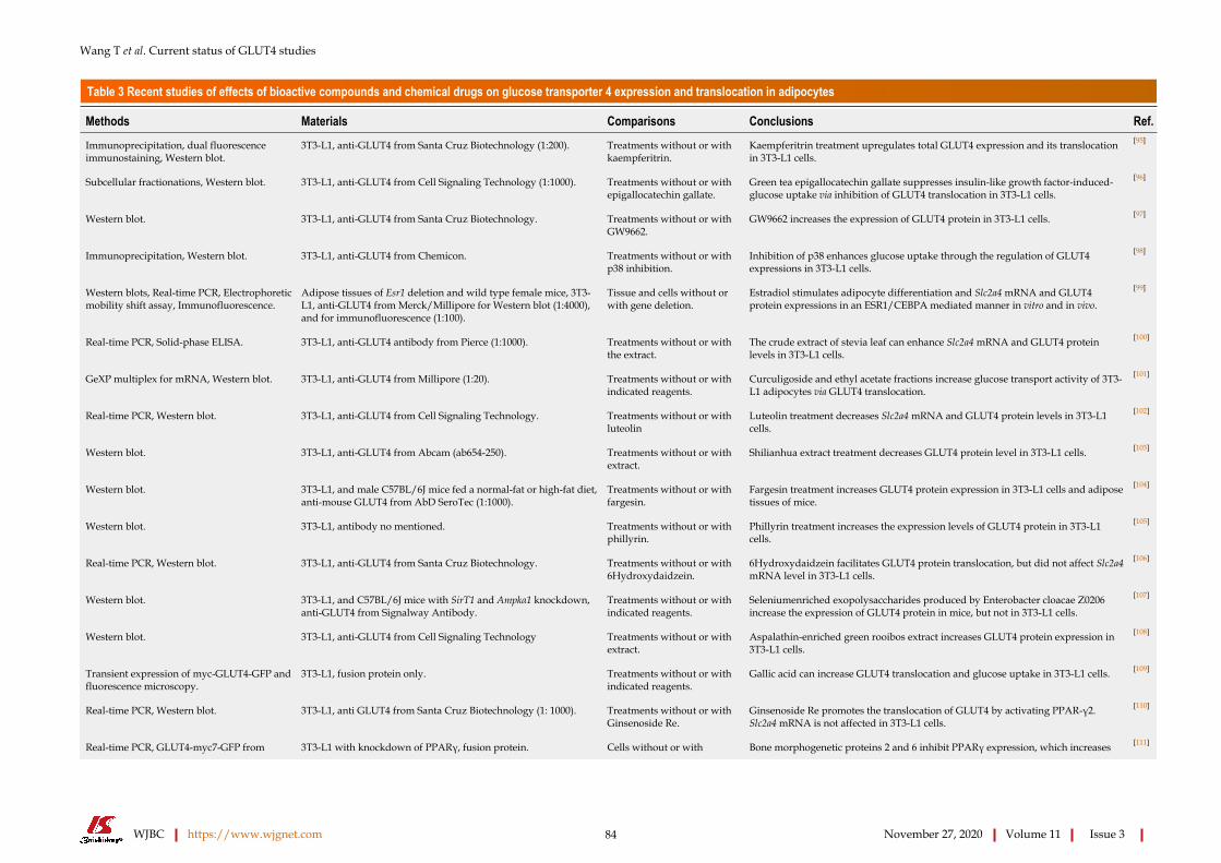
To summarize the current methods and reagents used for GLUT4 analysis in adipose tissues and adipocytes, “GLUT4, 3T3-L1”, and “GLUT4, adipocytes” were used to search the literature published in the past 15 years. We ignored those studies that only measured Slc4a2 mRNA, lacked the focus on adipocytes, and did not have full text versions. The resulted 30 articles were analyzed and summarized in Tables 3[95-112] and 4[113-124]. Table 3 contains 18 articles and summarizes the effects of drugs or bioactive compounds on Slc2a4 mRNA and GLUT4 protein expressions, and GLUT4 translocation. Table 4 contains 12 articles and summarizes studies of the regulatory mechanisms of GLUT4 system.
There are 11 and 2 articles respectively using real-time PCR and Northern blot to evaluate Slc2a4 mRNA levels. Western blot and ELISA are used in 24 articles to detect GLUT4 protein content. The antibodies were from Santa Cruz (6), Millipore (3), Cell Signaling Technology (4), Chemicon (4), Abcam (3), Pierce (1), Oxford (1) and Signalway (1). Two articles did not indicate the antibody sources. One article used antibodies from two companies. Nine articles indicated the dilutions of antibodies, and only 3 articles included the production catalog number. Immunofluorescence was used to detect the content and translocation of GLUT4 protein in 9 articles. Three articles used flow cytometry to detect GLUT4 protein. Twenty four of these 30 articles directly assessed levels of Slc2a4 mRNA or GLUT4 protein. The remaining 6 of the 30 articles measured GLUT4 protein using fusion proteins. No article has a positive control group that uses overexpression of GLUT4 via a recombinant construct or purified recombinant GLUT4 protein.
As shown in Table 3, GLUT4 translocation, and Slc2a4 mRNA and GLUT4 expression levels in 3T3-L1 cells can be regulated by bioactive compounds, crude extract of herbs, agonists of nuclear receptors, proteins and chemical drugs. Sl2a4 mRNA or/and GLUT4 expressions in 3T3-L1 cells or adipose tissues can be increased by kaempferitrin, GW9662, inhibitor of p38 kinase, estradiol, crude extract of stevia leaf, fargesin, phillyrin, selenium-enriched exopolysaccharide, aspalathin-enriched green rooibos extract, bone morphogenetic proteins 2 and 6, and glucose pulse. On the other hand, Slc2a4 mRNA and GLUT4 protein levels can be reduced by luteolin, and shilianhua extract in 3T3-L1 cells. GLUT4 translocation can be enhanced by kaempferitrin, curculigoside and ethyl acetate fractions, gallic acid, 6-hydroxydaidzein and ginsenoside Re, and reduced by green tea epigallocatechin gallate.
As shown in Table 4, a variety of methods have been used to study the regulatory mechanisms of GLUT4 system. 3T3-L1 cells have been the major model in those studies. In addition to the insulin, pathways involved in the Slc2a4 gene expression, GLUT4 protein expression and its translocation include cannabinoid receptor 1 (CB1), ADP-ribosylation factor-related protein 1, MiR-29 family, proteasome system, estrogen pathway, oxidative stress via CCAAT/enhancer-binding protein alpha (C/EBPα), obesity development, differentially expressed in normal and neoplastic cells domain-containing protein 4C, nuclear factor-κB, Akt and Akt substrate of 160 kDa, phosphatidylinositol-3,4,5-trisphosphate dependent Rac exchange factor 1, secreted protein acidic and rich in cysteine (SPARC), sterol regulatory element-binding protein 1 (SREBP-1), and AMP-activated protein kinase (AMPK) pathway. CB1 receptor antagonists increase Slc2a4 mRNA and GLUT4 protein expressions through NF-kB and SREBP-1 pathways. Akt pathway regulates the rate of vesicle tethering/fusion by controlling the concentration of primed, and fusion-competent GSVs with the plasma membrane. Inhibition of the SPARC expression reduces Slc2a4 mRNA and GLUT4 expressions. The expressions of C/EBPα and δ alter the C/EBP-dimer formation at the Slc2a4 gene promoter, which regulates its transcription. Inhibition of differentially expressed in normal and neoplastic cells domain-containing
Wang T et al. Current status of GLUT4 studies
WJBC https://www.wjgnet.com 84 November 27, 2020 Volume 11 Issue 3
Table 3 Recent studies of effects of bioactive compounds and chemical drugs on glucose transporter 4 expression and translocation in adipocytes
Methods Materials Comparisons Conclusions Ref.
Immunoprecipitation, dual fluorescence immunostaining, Western blot.
3T3-L1, anti-GLUT4 from Santa Cruz Biotechnology (1:200). Treatments without or with kaempferitrin.
Kaempferitrin treatment upregulates total GLUT4 expression and its translocation in 3T3-L1 cells.
[95]
Subcellular fractionations, Western blot. 3T3-L1, anti-GLUT4 from Cell Signaling Technology (1:1000). Treatments without or with epigallocatechin gallate.
Green tea epigallocatechin gallate suppresses insulin-like growth factor-induced-glucose uptake via inhibition of GLUT4 translocation in 3T3-L1 cells.
[96]
Western blot. 3T3-L1, anti-GLUT4 from Santa Cruz Biotechnology. Treatments without or with GW9662.
GW9662 increases the expression of GLUT4 protein in 3T3-L1 cells. [97]
Immunoprecipitation, Western blot. 3T3-L1, anti-GLUT4 from Chemicon. Treatments without or with p38 inhibition.
Inhibition of p38 enhances glucose uptake through the regulation of GLUT4 expressions in 3T3-L1 cells.
[98]
Western blots, Real-time PCR, Electrophoretic mobility shift assay, Immunofluorescence.
Adipose tissues of Esr1 deletion and wild type female mice, 3T3-L1, anti-GLUT4 from Merck/Millipore for Western blot (1:4000), and for immunofluorescence (1:100).
Tissue and cells without or with gene deletion.
Estradiol stimulates adipocyte differentiation and Slc2a4 mRNA and GLUT4 protein expressions in an ESR1/CEBPA mediated manner in vitro and in vivo.
[99]
Real-time PCR, Solid-phase ELISA. 3T3-L1, anti-GLUT4 antibody from Pierce (1:1000). Treatments without or with the extract.
The crude extract of stevia leaf can enhance Slc2a4 mRNA and GLUT4 protein levels in 3T3-L1 cells.
[100]
GeXP multiplex for mRNA, Western blot. 3T3-L1, anti-GLUT4 from Millipore (1:20). Treatments without or with indicated reagents.
Curculigoside and ethyl acetate fractions increase glucose transport activity of 3T3-L1 adipocytes via GLUT4 translocation.
[101]
Real-time PCR, Western blot. 3T3-L1, anti-GLUT4 from Cell Signaling Technology. Treatments without or with luteolin
Luteolin treatment decreases Slc2a4 mRNA and GLUT4 protein levels in 3T3-L1 cells.
[102]
Western blot. 3T3-L1, anti-GLUT4 from Abcam (ab654-250). Treatments without or with extract.
Shilianhua extract treatment decreases GLUT4 protein level in 3T3-L1 cells. [103]
Western blot. 3T3-L1, and male C57BL/6J mice fed a normal-fat or high-fat diet, anti-mouse GLUT4 from AbD SeroTec (1:1000).
Treatments without or with fargesin.
Fargesin treatment increases GLUT4 protein expression in 3T3-L1 cells and adipose tissues of mice.
[104]
Western blot. 3T3-L1, antibody no mentioned. Treatments without or with phillyrin.
Phillyrin treatment increases the expression levels of GLUT4 protein in 3T3-L1 cells.
[105]
Real-time PCR, Western blot. 3T3-L1, anti-GLUT4 from Santa Cruz Biotechnology. Treatments without or with 6Hydroxydaidzein.
6Hydroxydaidzein facilitates GLUT4 protein translocation, but did not affect Slc2a4 mRNA level in 3T3-L1 cells.
[106]
Western blot. 3T3-L1, and C57BL/6J mice with SirT1 and Ampkα1 knockdown, anti-GLUT4 from Signalway Antibody.
Treatments without or with indicated reagents.
Seleniumenriched exopolysaccharides produced by Enterobacter cloacae Z0206 increase the expression of GLUT4 protein in mice, but not in 3T3-L1 cells.
[107]
Western blot. 3T3-L1, anti-GLUT4 from Cell Signaling Technology Treatments without or with extract.
Aspalathin-enriched green rooibos extract increases GLUT4 protein expression in 3T3-L1 cells.
[108]
Transient expression of myc-GLUT4-GFP and fluorescence microscopy.
3T3-L1, fusion protein only. Treatments without or with indicated reagents.
Gallic acid can increase GLUT4 translocation and glucose uptake in 3T3-L1 cells. [109]
Real-time PCR, Western blot. 3T3-L1, anti GLUT4 from Santa Cruz Biotechnology (1: 1000). Treatments without or with Ginsenoside Re.
Ginsenoside Re promotes the translocation of GLUT4 by activating PPAR-γ2. Slc2a4 mRNA is not affected in 3T3-L1 cells.
[110]
Real-time PCR, GLUT4-myc7-GFP from Cells without or with Bone morphogenetic proteins 2 and 6 inhibit PPARγ expression, which increases 3T3-L1 with knockdown of PPARγ, fusion protein. [111]

Wang T et al. Current status of GLUT4 studies
WJBC https://www.wjgnet.com 85 November 27, 2020 Volume 11 Issue 3
retroviral vector, flow cytometry, fluorescence microscopy.
knockdown. total GLUT4 levels, but not GLUT4 translocation3T3-L1 cells.
Western blot, real-time PCR. 3T3-L1, anti-GLUT4 antibody from Santa Cruz Biotechnology (sc-1608).
Treatments without or with pulse or manipulations.
Glucose pulse (25 mM) increases GLUT4 expression. GLUT4 level is partially restored by increasing intracellular NAD/P levels. A liver X receptor element on Slc2a4 promoter is responsible for glucose-dependent transcription.
[112]
GLUT4: Glucose transporter 4; AMPK: AMP-activated protein kinase; CEBPA and C/EBP: CCAAT/enhancer-binding protein alpha; ESR1: Estrogen receptor 1.
protein 4C can block GLUT4 translocation. Rac exchange factor 1 activation seems to promote GLUT4 translocation via arrangement of actin cytoskeleton. The mechanism of AMPK-mediated GLUT4 translocation in 3T3-L1 adipocytes seems to be distinct from that of insulin-induced one. Future studies are needed to integrate the roles of all these players in the regulation of GLUT4 system in adipocytes.
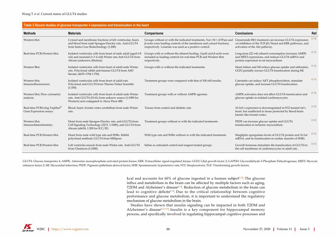
GLUT4 IN THE HEART The heart works constantly to support the blood circulation throughout the lifespan. Cardiomyocytes constantly contract to pump blood, oxygen, metabolic substrates, and hormones to other parts of the body. This requires continuous ATP production for energy supply. The primary fuel for the heart is fatty acids, whereas glucose and lactate contribute to 30% of energy for ATP production[125]. In addition, glucose plays an important role in circumstances like ischemia, increased workload, and pressure overload hypertrophy.
Glucose is transported into cardiac myocytes through GLUTs. GLUT4 represents around 70% of the total glucose transport activities[15]. GLUT4 protein expression can be found as early as 21 days of gestation in rats[126]. The expression level of GLUT4 in the heart may increase or decrease depending on the different models. For example, GLUT4 protein content decreases along with aging in male Fischer rats, but increases 4-5 times in C57 Bl6 mice[127,128]. In basal state, GLUT4 is found mainly in intracellular membrane compartments, and can be stimulated by insulin and ischemia to translocate to the cell membrane[129]. The binding of myocyte enhancer factor-2 (MEF2) and thyroid hormone receptor alpha 1 is needed for transcription of Slc2a4 gene in cardiac and skeletal muscle in rats[130]. In addition, Slc2a4 gene expression can also be regulated by other transcription factors. For example, overexpression of peroxisome proliferator-activated receptor gamma coactivator 1 works with MEF2-C to induce Slc2a4 mRNA expression in L6 muscle cells[131]. Moreover, GLUT4 expression level can be affected by cardiovascular diseases, and myocardial sarcolemma, which reduce the expression and translocation of GLUT4[132]. The development of T1DM decreases GLUT4 expression level and its translocation in the heart of mice[132]. T2DM development also reduces GLUT4 content and translocation due to insulin resistance

Wang T et al. Current status of GLUT4 studies
WJBC https://www.wjgnet.com 86 November 27, 2020 Volume 11 Issue 3
Table 4 Recent studies of mechanisms of glucose transporter 4 expression and translocation in adipocytes
Methods Materials Comparisons Observations/conclusions Ref.
Western blot, real-time PCR, Electrophoretic mobility shift assay.
3T3-L1 pre and differentiated adipocytes. Anti-GLUT4 antibody from Chemicon (1:4,000).
Treatment groups without or with the antagonist.
CB1 receptor antagonist markedly increases Slc2a4 mRNA and GLUT4 protein levels in 3T3-L1 cells via NF-kB and SREBP-1 pathways.
[113]
Immunohistochemistry, Western blot, real-time PCR.
Brown adipose tissue of Arfrp1 flox/flox and Arfrp1 ad-/-mouse embryos (ED 18.5) and 3T3-L1 cells with knockdown of Arfrp1. Anti-GLUT4 without specifying the vendor (1:1000).
Mice without or with deletion, and 3T3-L1 cells without or with knockdown.
In Arfrp1 ad-/- adipocytes, GLUT4 protein accumulates on the cell membrane rather than staying intracellularly without any change of Slc2a4 mRNA. siRNA-mediated knockdown of Arfrp1 in 3T3-L1 adipocytes has a similar result and increases basal glucose uptake.
[114]
Real-time PCR, Western blot. 3T3-L1 transfected with Mmu-miR-29a/b/c. Anti-GLUT4 from Santa Cruz Biotechnology (SC-7938).
Cells with or without transfection. Transfection of miR-29 family members inhibits Slc2a4 mRNA and GLUT4 protein levels in 3T3-L1 cells by inhibiting SPARC expression.
[115]
Northern blot, Western blot, Nuclear run-on assay for the rate of GLUT4 gene transcription.
3T3-L1 pre and differentiated adipocytes. Rabbit polyclonal GLUT4 antibody form Chemicon.
Treatment groups without or with inhibitors.
Inhibitions of proteasome using Lactacystin and MG132 reduce Slc2a4 mRNA and GLUT4 protein levels in 3T3-L1 cells.
[116]
AFFX miRNA expression chips for mRNA, Western blot.
Human Omental adipose tissue, 3T3-L1 pre and differentiated adipocytes with miR-222 silenced by antisense oligonucleotides. Anti-GLUT4 from Abcam.
Groups without or with transfection. High levels of estrogen reduce the expression and translocation of GLUT4 protein. miR -222 silencing dramatically increases the GLUT4 expression and the insulin-stimulated translocation of GLUT4 in 3T3-L1 adipocytes.
[117]
Northern blot for mRNA, Western blot. 3T3-L1 pre and differentiated adipocytes. Anti-GLUT4 from Chemicon.
Treatment groups without or with oxidative stress.
Oxidative stress mediated by hydrogen peroxide induces expressions of C/EBPα and δ, resulting in altered C/EBP-dimer composition on the GLUT4 promoter, which reduces GLUT4 mRNA and protein levels.
[118]
Real-time PCR, Western blot. Human Subcutaneous pre and differentiated adipocytes from control and obese subjects, 3T3-L1 pre and differentiated adipocytes transfected with miR-155. Anti-GLUT4 from Abcam.
Primary pre and differentiated adipocytes from normal and obese subjects, and cells without or with transfection.
The level of SLC2A4 is reduced in obese people, and the expression of GLUT4 protein is reduced in 3T3-L1 cells and differentiated human mesenchymal stem cells transfected with miR-155.
[119]
HA-GLUT4-GFP from transfected lentiviral plasmid and analyzed by flow cytometry, and fluorescence microscopy.
3T3-L1 pre and differentiated adipocytes with knockdown of Dennd4C. Fusion protein.
Groups without or with knockdown. Knockdown of Dennd4C inhibits GLUT4 translocation, and over- expression of DENND4C slightly stimulates it. DENND4C is found in isolated GLUT4 vesicles.
[120]
HA-Glut4-GFP from transfected plasmid, and analyzed by flow cytometry, fluorescence microscopy
3T3-L1 pre and differentiated adipocytes with AS160 knockdown.Fusion protein.
Groups without or with knockdown. Akt regulates the rate of vesicle tethering/fusion by regulating the concentration of primed, and fusion-competent GSVs with the plasma membrane, but not changing the intrinsic rate constant for tethering/fusion.
[121]
HA-tagged GLUT4 by fluorescence microscopy, Western blots, Immune pulldown.
3T3-L1 pre and differentiated adipocytes without or with GST-ClipR-59 transfection. Rabbit anti-GLUTlut4 from Millipore; Mouse monoclonal anti-GLUT4 from Cell Signaling Technology.
Pull down antibodies. By interacting with AS160 and enhancing the association of AS160 with Akt, ClipR-59 promotes phosphorylation of AS160 and GLUT4 membrane translocation.
[122]
Transfection of GFP-GLUT4 and indirect immunofluorescence.
3T3-L1 pre and differentiated adipocytes with siRNA knockdown of P-Rex1. Fusion protein.
Without or with knockdown. P-Rex1 activates Rac1 in adipocytes, which leads to actin rearrangement, GLUT4 trafficking, increase of glucose uptake.
[123]
Transfection of GLUT4-eGFP plasmid and analyzed by fluorescence microscopy.
3T3-L1 pre and differentiated adipocytes. Fusion protein. Treatment groups without or with activators.
AMPK-activated GLUT4 translocation in 3T3-L1 adipocytes is mediated through the insulin-signaling pathway distal to the site of activated phosphatidylinositol 3-kinase or through a signaling system distinct from that activated by insulin.
[124]

Wang T et al. Current status of GLUT4 studies
WJBC https://www.wjgnet.com 87 November 27, 2020 Volume 11 Issue 3
GLUT4: Glucose transporter 4; ARFRP1: ADP-ribosylation factor-related protein 1; AMPK: AMP-activated protein kinase; AS160: Akt substrate of 160 kDa; CB1: Cannabinoid receptor 1; CEBPA and C/EBP: CCAAT/enhancer-binding protein alpha; CLIPR-59: Cytoplasmic linker protein R-59; DENND4C: Differentially expressed in normal and neoplastic cells domain-containing protein 4C; GSV: GLUT4 storage vesicle; NF-Κb: Nuclear factor-κB; PREX1: Phosphatidylinositol-3,4,5-trisphosphate dependent Rac exchange factor 1; SPARC: Secreted protein acidic and rich in cysteine; SREBP-1: Sterol regulatory element-binding protein 1.
and impairments of insulin signaling pathway in human cardiomyocytes[133].To investigate the methods and reagents used for GLUT4 studies in the heart,
“GLUT4, heart”, and “cardiomyocytes, GLUT4 expression” were used as key words to search PubMed for articles published after 2000. We went through all papers with cardiomyocytes and GLUT4 in titles or short descriptions and selected 9 of them that are mainly focused on GLUT4 expression and translocation in the heart as shown in Table 5[134-142].
Rats are used in all 9 studies. Various methods and reagents are used to analyze Slc2a4 mRNA and GLUT4 protein levels in the heart and cardiomyocytes. Real-time PCR was used in 4 of them to determine Slc2a4 mRNA levels. Antibodies and Western blot were used to assess GLUT 4 protein in 8 of them. Immunohistochemistry was used in 1 of them. Two of them used immunofluorescence to track down GLUT4 translocation.
To determine the content of GLUT4 protein in the heart, Western blot and fusion protein immunofluorescence methods were used. As shown in Table 5, these studies do not include overexpressed GLUT4 or cell samples with Slc2a4 deletion as controls. Several of them did not mention sources of anti GLUT4 antibodies used in Western blots. Some used polyclonal antibodies, which may need a positive control to indicate the correct size and location of GLUT4 protein.
From the papers listed above, GLUT4 expression and translocation in the heart and cardiomyocytes can be affected through activations of ERK and Akt pathways. Proteins like growth hormone, catestatin or pigment epithelium-derived factor can stimulate GLUT4 translocation and glucose uptake. Chemicals like sitagliptin and ethanol can up- and down-regulate Slc2a4’s mRNA expression levels, respectively. However, the underlying mechanisms responsible for these regulations of GLUT4 translocation and Slc2a4 mRNA expression remain to be revealed. In addition, the research results summarized here are from tissue and cells of rats. It will be interesting to see whether same results will be observed when tissues and cells from other animal models are used.
GLUT4 IN THE BRAINThe brain is a complex organ in the body and controls a variety of functions from emotions to metabolism. It consists of cerebrum, the brainstem, and the cerebellum[143]. Brain cells utilize glucose constantly to produce energy in normal physiological conditions. The brain can consume about 120 g of glucose per day, which is about 420
Wang T et al. Current status of GLUT4 studies
WJBC https://www.wjgnet.com 88 November 27, 2020 Volume 11 Issue 3
Table 5 Recent studies of glucose transporter 4 expression and translocation in the heart
Methods Materials Comparisons Conclusions Ref.
Western blot. Cytosol and membrane fractions of left ventricular, heart, and blood from male Sprague-Dawley rats. Anti-GLUT4 from Santa Cruz Biotechnology (1:200).
Groups without or with the indicated treatments. Na+/K+-ATPase and β-actin were loading controls of the membrane and cytosol fractions, respectively. Losartan was used as a positive control.
Ginsenoside Rb1 treatment can increase GLUT4 expression via inhibition of the TGF-β1/Smad and ERK pathways, and activation of the Akt pathway.
[134]
Real-time PCR,Western blot. Isolated ventricular cells from heart of male adult (aged 6-8 wk) and neonatal (1-3 d old) Wistar rats.Anti GLUT4 from Abcam (unknown dilution).
Groups with or without the ethanol feeding. Gapdh and β-actin were included as loading controls for real-time PCR and Western blot, respectively.
Long-term (22 wk) ethanol consumption increases AMPK and MEF2 expressions, and reduces GLUT4 mRNA and protein expression in rat myocardium
[135]
Western Blot. Isolated ventricular cells from heart of adult male Wistar rats. Polyclonal rabbit anti-human GLUT4 from AbD Serotec (4670–1704 1:750)
Groups with or without the indicated treatments. Heart failure and MI reduce glucose uptake and utilization. GGF2 partially rescues GLUT4 translocation during MI.
[136]
Western blot, Immunofluorescence.
Isolated ventricular cells from heart of adult rats. Polyclonal anti-GLUT4 from Thermo Fisher Scientific (1:100).
Treatment groups were compared with that of 100 nM insulin. Catestatin can induce AKT phosphorylation, stimulate glucose uptake, and increase GLUT4 translocation.
[137]
Western blot, Flow cytometric analysis.
Isolated ventricular cells from heart of adult male Wistar rats. Anti-GLUT4 (H-61) from unknow source (1:1000 for Western) and conjugated to Alexa Fluor 488.
Treatment groups with or without AMPK agonists. AMPK activation does not affect GLUT4 translocation and glucose uptake in isolated cardiomyocytes.
[138]
Real-time PCRUsing TaqMan® Gene Expression assays.
Blood, heart, frontal cortex cerebellum from male Wistar rats.
Tissues from control and diabetic rats. Slc2a4’s expression is downregulated in STZ-treated rat’s heart, but unaffected in tissue protected by blood-brain barrier like frontal cortex.
[139]
Western blot, Immunohistochemistry.
Heart from male Sprague-Dawley rats, anti-GLUT4 from Cell Signaling Technology (2213, 1:1000), anti GLUT4 from Abcam (ab654, 1:200 for ICC/IF)
Treatment groups without or with the indicated treatments. PEDF can increase glucose uptake and GLUT4 translocation in ischemic myocardium.
[140]
Real-time PCR,Western blot. Heart from male wild type rats and SHRs. Rabbit polyclonal antibody GLUT4 from Millipore
Wild type rats and SHRs without or with the indicated treatments. Sitagliptin upregulates levels of GLUT4 protein and Slc2a4 mRNA, and its translocation in cardiac muscles of SHRs.
[141]
Real-time PCR,Western blot. Left ventricles muscle from male Wistar rats. Anti-GLUT4 from Chemicon (1:1000)
Saline as untreated control and reagent treated groups. Growth hormone stimulates the translocation of GLUT4 to the cell membrane of cardiomyocytes in adult rats.
[142]
GLUT4: Glucose transporter 4; AMPK: Adenosine monophosphate-activated protein kinase; ERK: Extracellular signal-regulated kinase; GGF2: Glial growth factor 2; GAPDH: Glyceraldehyde-3-Phosphate Dehydrogenase; MEF2: Myocyte enhancer factor-2; MI: Myocardial infarction; PEDF: Pigment epithelium-derived factor; SHR: Spontaneously hypertensive rats; STZ: Streptozotocin; TGF: Transforming growth factors.
kcal and accounts for 60% of glucose ingested in a human subject[125]. The glucose influx and metabolism in the brain can be affected by multiple factors such as aging, T2DM and Alzheimer’s disease[144]. Reduction of glucose metabolism in the brain can lead to cognitive deficits[145]. Due to the critical relationship between cognitive performance and glucose metabolism, it is important to understand the regulatory mechanism of glucose metabolism in the brain.
Studies have shown that insulin signaling can be impacted in both T2DM and Alzheimer’s disease[146,147]. Insulin is a key component for hippocampal memory process, and specifically involved in regulating hippocampal cognitive processes and

Wang T et al. Current status of GLUT4 studies
WJBC https://www.wjgnet.com 89 November 27, 2020 Volume 11 Issue 3
Figure 2 The movement of glucose transporter 4 in adipocytes. Adipose tissue is made of adipocytes. In adipocytes, glucose transporter 4 (GLUT4) can be found in the cell membrane and in the cytosol. The translocation of GLUT4 from cytosolic vesicles to the cell membrane leads to elevated glucose uptake, whereas endocytosis brings GLUT4 back to the cytosol. (1): In unstimulated cells, GLUT4 containing membrane portions are internalized in an endocytosis manner to generate vesicles containing GLUT4. GLUT4 vesicles are internalized into early (or sorted) endosomes. They can enter the recovery endoplasmic body, and follow the retrograde pathway to the trans-Golgi network and endoplasmic reticulum-Golgi intermediate compartment or other donor membrane compartments. (2): GLUT4 vesicles derived from the donor membrane structures are secured by tether containing a UBX domain for GLUT4 (TUG) protein. (3): During insulin signal stimulation, GLUT4 vesicles are released and loaded onto the microtubule motor to be transferred to the plasma membrane. The continuous presence of insulin leads to the direct movement of these vesicles to the plasma membrane. (4): GLUT4 vesicles are tethered to motor protein kinesin and other proteins. A stable ternary SNARE complex forms when this occurs. (5): The stable ternary SNARE complex is docked on the target membrane. (6): The docked vesicles rely on SNARE to move to and fuse with the target membrane[60,90,94]. GLUT4: Glucose transporter 4.
metabolism[148]. Insulin-modulated glucose metabolism depends on regions in the brain. The cortex and hippocampus are the most sensitive areas in the brain[149]. Hippocampus located deeply in temporal lobe plays an important role in learning and memory, and relates to diseases like Alzheimer’s disease, short term memory loss and disorientation[150]. Hippocampal cognitive and metabolic impairments are relatively common in T2DM, which may be caused by diet-induced obesity and systemic insulin resistance[151]. On the other hand, insulin stimulation can enhance memory and cognitive function[152]. This enhancement may require the brain GLUT4 translocation as shown in rats[153]. It is very important to determine the expression profile of GLUT4 in the brain, and factors that impact GLUT4 expression and translocation.
To summarize methods and reagents used in brain GLUT4 studies, “brain, GLUT4 expression” were used as key words to search PubMed for articles that have brain and GLUT4 in their titles or short descriptions. Ten research articles published after 2000 were identified as representative ones, which are focused on GLUT4 translocations and content in the brain of rats and mice. We summarized the methods and reagents for GLUT4 analysis, and conclusions as shown in Table 6[154-163].
In these 10 papers, two of them used real-time PCR to determine the Slc2a4 mRNA in the brain. Eight of them used anti-GLUT4 antibodies and Western blot to detect GLUT 4 protein. Five papers included β-actin as loading control in Western blot. Four used immunohistochemistry. One paper used electrophysiological technique, and one paper used fluorescent microscopy to identify GLUT 4 in neurons. One study used brain specific Slc2a4 knockout and wild type mice to study the functions of GLUT4 in the brain.
In conclusion, results of Western blot and real-time PCR demonstrate that GLUT4 protein and Slc2a4 mRNA can be detected in rat’s brain and central nervous system. Deletion of Slc2a4 in the brain causes insulin resistance, glucose intolerance, and impaired glucose sensing in the ventromedial hypothalamus. GLUT4 mediates the effects of insulin, or insulin-like growth factor on regulations of cognition, memory, behavior, motor activity and seizures. GLUT4 positive neurons are responsible for glucose sensing. Physical activity improves GLUT4 translocation in neurons, a process that needs Rab10 phosphorylation. Interestingly, 27-OH cholesterol treatment seems to decrease GLUT4 expression in the brain. Studies of Slc2a4 mRNA and GLUT4 protein in the brain and central nervous system have begun to demonstrate the potential roles of GLUT4 expression and its translocation in the regulation of glucose metabolism in

Wang T et al. Current status of GLUT4 studies
WJBC https://www.wjgnet.com 90 November 27, 2020 Volume 11 Issue 3
Table 6 Recent studies of glucose transporter 4 expression and translocation in the brain
Methods Materials Comparisons Conclusions Ref.
Western blot. Brain, skeletal muscle, heart, and whiteadipose tissue from mice. Anti-GLUT4 from Chemicon (1:1000).
Samples from wild type and knockout mice. Deletion of Slc2a4 in the brain leads to insulin resistance, glucose intolerance, and impaired glucose sensing in the VMH.
[154]
Western blot,Real time-PCR,Immunofluorescence.
Cortex, hypothalamus, cerebella samples from CD-1 mice. Monoclonal anti Glut4 (1F8) from Dr. Paul Pilch, Polyclonal anti Glut4 MC2A from Dr. Giulia Baldini, Polyclonal anti Glut4 αG4 from Dr. Samuel Cushman, Polyclonal anti Glut4 (C-20) from Santa Cruz Biotechnology.
Expression profile in the mouse and rat brain samples.
Slc2a4 mRNA is expressed in cultured neurons. GLUT4 protein is highly expressed in the granular layer of the mouse cerebellum. GLUT4 translocation to the plasma membrane can be stimulated by physical activity.
[155]
Western blot. Brian tissue from STZ-induced diabetic male Sprague-Dawley rats.Anti-GLUT4 from Millipore (1:1000).
Comparing treatment samples using β-actin and NA/K ATPase as loading controls in Western blot.
Chronic infusion of insulin into the VMH in poorly controlled diabetes is sufficient to normalize the sympathoadrenal response to hypoglycemia via restoration of GLUT4 expression.
[156]
Immunocytochemistry. Cerebellum and hippocampus from male Sprague-Dawley rats.Rabbit anti-GLUT4 antibody from Alomone Labs (AGT-024, RRID: AB_2631197).
Identifying expression profile and translocation. GLUT4 is expressed in neurons including nerve terminals.Exercising axons rely on translocation of GLUT4 to the cell membrane for metabolic homeostasis.
[157]
Real-time PCR, Immunocytochemistry.
Cerebral cortex, hippocampus, thalamus, cerebellum, medulla oblongata, cervical spinal cord, biceps muscles from male Wistar rats. Unknown source of antibody.
Identifying expression profile using β -actin as loading control in real-time PCR.
Slc2a4 mRNA is detected in many neurons located in brain and spinal cord. GLUT4 protein is detected in different regions of the CNS including certain allocortical regions, temporal lobe, hippocampus, and substantia nigra.
[158]
Immunocytochemistry, Western blot.
Brain, spleen, kidney from Lrrk2 knockout mice.Anti GLUT4 Avivasysbio (ARP43785_P050, 1:100), andanti GLUT4 from R&D Systems (MAB1262, 1:1000).
Samples from wild type and knockout mice, andanti-β-Tubulin as loading control.
Phosphorylation of Rab10 by LRRK2 is necessary for GLUT4 translocation.Lrrk2 deficiency increases GLUT4 expression on the cell surface in “aged” cells.
[159]
Western blot, Immunofluorescence, real-time PCR.
Brain from Cyp27Tg mice. Anti GLUT4 from Cell Signaling Technology (#2213,1:1000 dilution).
Mice with different treatments. A reduction of GLUT4 protein expression in brain occurs after 27-OH cholesterol treatment.
[160]
Immunohistochemistry. Brain, hypothalamus, and other tissues from Sprague–Dawley rats.Anti-GLUT4 antibody from Santa Cruz Biochemicals (1:200), Anti GLUT4 from S. Cushman (1:1000).
Identifying the expression profiles. Soleus muscle as GLUT4 positive control. Antibodies after pre-absorption with the corresponding synthetic peptide were used as negative control. for GLUT4 antibody.
GLUT4 is localized to the micro vessels comprising the blood brain barrier of the rat VMH.GLUT4 is co-expressed with both GLUT1 and zonula occludens-1 on the endothelial cells of these capillaries.
[161]
Electrophysiological analyses, fluorescent microscope.
Brain from GLUT4-EYFP transgenic mice. Fusion protein. Comparing samples from treatments. A scrambled RNA expressed by AAV acted as a negative control.
GLUT4 neurons are responsible for glucose sensing. [162]
Western blot,Immunohistochemistry.
Brain samples from 7, 11, 15, 21 and 60 d old Balb/c mice. Rabbit anti-rat GLUT4 from an unknow source (1:2500 dilution for Western and 1:2000 for immunohistochemistry).
Determine the expression profiles. Vinculin is used as the loading control in Western blot.
GLUT4 is expressed in neurons of the postnatal mouse brain. GLUT4 and GLUT8 may mediate the effects of insulin, or insulin-like growth factor on regulations of cognition, memory, behavior, motor activity and seizures.
[163]
AAV: Adeno-associated virus; CNS: Central nervous system; CRR: Counterregulatory response; EYFP: Enhanced Yellow Fluorescent Protein; LRRK2: Leucine-rich repeat kinase 2; STZ: Streptozotocin; VMH: Ventromedial hypothalamus.
the brain and central nervous system. More studies of GLUT4 expression and translocation in the control of functions and metabolism in various region in the brain

Wang T et al. Current status of GLUT4 studies
WJBC https://www.wjgnet.com 91 November 27, 2020 Volume 11 Issue 3
and central nervous system are expected in the future.
CONCLUSIONGLUT4 is generally thought to contribute to insulin-stimulated glucose uptake in adipocytes and skeletal muscle. Studies summarized here seem to show that GLUT4 is also expressed in the brain, neurons, and heart. GLUT4 is expressed concurrently with other GLUTs in multiple tissues in a temporal and spatial specific manner such as during brain development[163]. Hormones and cytokines other than insulin can also regulate the expression levels and translocation of GLUT4 in different tissues[141,142,163]. In adipocytes alone, many bioactive compounds or chemical reagents have shown to affect GLUT4 pathways as shown in Table 3. All these seem to indicate that the regulatory mechanism of the GLUT4 pathway is complicated than we originally proposed.
So far, various methods from gene knockout to immunohistochemistry have been used to study the mechanisms of Slc2a4 mRNA and GLUT4 expressions, and its translocation in different cells. Every technique has its pros and cons. Based on the studies summarized here, anti-GLUT4 antibodies from a variety of sources have been used to study GLUT4 expression and translocation. The conclusions of these studies are based on the experimental results derived from the use of those antibodies. A positive control derived from a cell or tissue with unique overexpression or silencing of GLUT4 is critical to confirm the antibody’s specificity to pick up a right signaling in the study system. This is especially true for Western blot. It appears that some of the studies did not include control groups like this. Another challenge facing biochemical study of GLUT4 translocation in the muscle may be the sample processing. This probably explains why fusion proteins and stable cell lines are developed to enhance signals and specificities for detection. Confirmation of the antibody specificity in a particular system probably should be done first.
As glucose homeostasis is a complicate process involved in many players. It is anticipated to see that many proteins seem to play a role in the regulation of GLUT4 system. It will be interesting to see how GLUT4 in different regions of the brain contributes to the regulation of glucose metabolism, and what the roles of insulin-induced GLUT4 translocation in those areas are. In addition, other GLUTs are also expressed in the same cells that GLUT4 are expressed. How GLUT4 works with other GLUTs to regulate metabolism also deserves to be investigated. Last, as glucose usage in the skeletal muscle is altered in insulin resistance and T2DM, how GLUT4 system contributes to progressions and interventions of these diseases still remains to be the focus in the future. Nevertheless, further understanding GLUT4 system will be very helpful for us to combat the development of T2DM.
ACKNOWLEDGEMENTSWe would like to thank the Department of Nutrition at the University of Tennessee at Knoxville for financial support to T.W. G.C. would like to thank Yantai Zestern Biotechnique Co. LTD for the research funding support.
REFERENCESAl-Lawati JA. Diabetes Mellitus: A Local and Global Public Health Emergency! Oman Med J 2017; 32: 177-179 [PMID: 28584596 DOI: 10.5001/omj.2017.34]
1
La Flamme KE, Mor G, Gong D, La Tempa T, Fusaro VA, Grimes CA, Desai TA. Nanoporous alumina capsules for cellular macroencapsulation: transport and biocompatibility. Diabetes Technol Ther 2005; 7: 684-694 [PMID: 16241869 DOI: 10.1089/dia.2005.7.684]
2
Salt IP, Johnson G, Ashcroft SJ, Hardie DG. AMP-activated protein kinase is activated by low glucose in cell lines derived from pancreatic beta cells, and may regulate insulin release. Biochem J 1998; 335 (Pt 3): 533-539 [PMID: 9794792 DOI: 10.1042/bj3350533]
3
Stipanuk MH, Caudill MA. Biochemical, Physiological, and Molecular Aspects of Human Nutrition. 3rd ed. St. Louis, MO: Elsevier, 2012: 968
4
Quistgaard EM, Löw C, Guettou F, Nordlund P. Understanding transport by the major facilitator superfamily (MFS): structures pave the way. Nat Rev Mol Cell Biol 2016; 17: 123-132 [PMID: 26758938 DOI: 10.1038/nrm.2015.25]
5
Klip A, McGraw TE, James DE. Thirty sweet years of GLUT4. J Biol Chem 2019; 294: 11369-11381 [PMID: 31175156 DOI: 10.1074/jbc.REV119.008351]
6

Wang T et al. Current status of GLUT4 studies
WJBC https://www.wjgnet.com 92 November 27, 2020 Volume 11 Issue 3
Richter EA, Derave W, Wojtaszewski JF. Glucose, exercise and insulin: emerging concepts. J Physiol 2001; 535: 313-322 [PMID: 11533125 DOI: 10.1111/j.1469-7793.2001.t01-2-00313.x]
7
Cushman SW, Wardzala LJ. Potential mechanism of insulin action on glucose transport in the isolated rat adipose cell. Apparent translocation of intracellular transport systems to the plasma membrane. J Biol Chem 1980; 255: 4758-4762 [PMID: 6989818 DOI: 10.1093/aje/kwm394]
8
Suzuki K, Kono T. Evidence that insulin causes translocation of glucose transport activity to the plasma membrane from an intracellular storage site. Proc Natl Acad Sci 77: 2542-2545 [PMID: 6771756 DOI: 10.1073/pnas.77.5.2542]
9
Wardzala LJ, Cushman SW, Salans LB. Mechanism of insulin action on glucose transport in the isolated rat adipose cell. Enhancement of the number of functional transport systems. J Biol Chem 1978; 253: 8002-8005 [PMID: 711732]
10
Klip A, Ramlal T, Young DA, Holloszy JO. Insulin-induced translocation of glucose transporters in rat hindlimb muscles. FEBS Lett 1987; 224: 224-230 [PMID: 2960560 DOI: 10.1016/0014-5793(87)80452-0]
11
Wardzala LJ, Jeanrenaud B. Potential mechanism of insulin action on glucose transport in the isolated rat diaphragm. Apparent translocation of intracellular transport units to the plasma membrane. J Biol Chem 1981; 256: 7090-7093 [PMID: 6265437]
12
James DE, Brown R, Navarro J, Pilch PF. Insulin-regulatable tissues express a unique insulin-sensitive glucose transport protein. Nature 1988; 333: 183-185 [PMID: 3285221 DOI: 10.1038/333183a0]
13
Olson AL. Regulation of GLUT4 and Insulin-Dependent Glucose Flux. ISRN Mol Biol 2012; 2012: 856987 [PMID: 27335671 DOI: 10.5402/2012/856987]
14
Mueckler M, Thorens B. The SLC2 (GLUT) family of membrane transporters. Mol Aspects Med 2013; 34: 121-138 [PMID: 23506862 DOI: 10.1016/j.mam.2012.07.001]
15
Chen L, Tuo B, Dong H. Regulation of Intestinal Glucose Absorption by Ion Channels and Transporters. Nutrients 2016; 8 [PMID: 26784222 DOI: 10.3390/nu8010043]
16
Navale AM, Paranjape AN. Glucose transporters: physiological and pathological roles. Biophys Rev 2016; 8: 5-9 [PMID: 28510148 DOI: 10.1007/s12551-015-0186-2]
17
Grefner NM, Gromova LV, Gruzdkov AA, Komissarchik YY. Interaction of glucose transporters SGLT1 and GLUT2 with cytoskeleton in enterocytes and Caco2 cells during hexose absorption. Cell and Tissue Biol 2015; 9: 45-52 [DOI: 10.1134/S1990519X15010034]
18
Gropper S, Smith J, Carr T. Advanced Nutrition and Human Metabolism. 7th ed. Publisher: Wadsworth Publishing, 2016: 640
19
Bell GI, Kayano T, Buse JB, Burant CF, Takeda J, Lin D, Fukumoto H, Seino S. Molecular biology of mammalian glucose transporters. Diabetes Care 1990; 13: 198-208 [PMID: 2407475 DOI: 10.2337/diacare.13.3.198]
20
Haber RS, Weinstein SP, O'Boyle E, Morgello S. Tissue distribution of the human GLUT3 glucose transporter. Endocrinology 1993; 132: 2538-2543 [PMID: 8504756 DOI: 10.1210/endo.132.6.8504756]
21
Stuart CA, Wen G, Gustafson WC, Thompson EA. Comparison of GLUT1, GLUT3, and GLUT4 mRNA and the subcellular distribution of their proteins in normal human muscle. Metabolism 2000; 49: 1604-1609 [PMID: 11145124 DOI: 10.1053/meta.2000.18559]
22
Lisinski I, Schürmann A, Joost HG, Cushman SW, Al-Hasani H. Targeting of GLUT6 (formerly GLUT9) and GLUT8 in rat adipose cells. Biochem J 2001; 358: 517-522 [PMID: 11513753 DOI: 10.1042/0264-6021:3580517]
23
Takata K, Kasahara T, Kasahara M, Ezaki O, Hirano H. Erythrocyte/HepG2-type glucose transporter is concentrated in cells of blood-tissue barriers. Biochem Biophys Res Commun 1990; 173: 67-73 [PMID: 2256938 DOI: 10.1016/S0006-291X(05)81022-8]
24
Joost HG, Thorens B. The extended GLUT-family of sugar/polyol transport facilitators: nomenclature, sequence characteristics, and potential function of its novel members (review). Mol Membr Biol 2001; 18: 247-256 [PMID: 11780753 DOI: 10.1080/09687680110090456]
25
Zhao FQ, Keating AF. Functional properties and genomics of glucose transporters. Curr Genomics 2007; 8: 113-128 [PMID: 18660845 DOI: 10.2174/138920207780368187]
26
27 Szablewski L. Glucose Homeostasis and Insulin Resistance. Bentham Science Publishers, 2011: 21127 Hruz PW, Mueckler MM. Structural analysis of the GLUT1 facilitative glucose transporter (review). Mol Membr Biol 2001; 18: 183-193 [PMID: 11681785 DOI: 10.1080/09687680110072140]
28
Cheeseman C, Long WL. Structure of, and functional insight into the GLUT family of membrane transporters. Cell Health Cytoskeleton 2015; 7: 167 [DOI: 10.2147/chc.s60484]
29
Olson AL, Pessin JE. Structure, function, and regulation of the mammalian facilitative glucose transporter gene family. Annu Rev Nutr 1996; 16: 235-256 [PMID: 8839927 DOI: 10.1146/annurev.nu.16.070196.001315]
30
Uldry M, Ibberson M, Hosokawa M, Thorens B. GLUT2 is a high affinity glucosamine transporter. FEBS Lett 2002; 524: 199-203 [PMID: 12135767 DOI: 10.1016/s0014-5793(02)03058-2]
31
Johnson JH, Newgard CB, Milburn JL, Lodish HF, Thorens B. The high Km glucose transporter of islets of Langerhans is functionally similar to the low affinity transporter of liver and has an identical primary sequence. J Biol Chem 1990; 265: 6548-6551 [PMID: 2182619 DOI: 10.1016/0008-6215(90)84177-V]
32
Colville CA, Seatter MJ, Jess TJ, Gould GW, Thomas HM. Kinetic analysis of the liver-type (GLUT2) and brain-type (GLUT3) glucose transporters in Xenopus oocytes: substrate specificities and effects of transport inhibitors. Biochem J 1993; 290 (Pt 3): 701-706 [PMID: 8457197 DOI: 10.1042/bj2900701]
33
Longo N, Elsas LJ. Human glucose transporters. Adv Pediatr 1998; 45: 293-313 [PMID: 9742306]34 Kayano T, Fukumoto H, Eddy RL, Fan YS, Byers MG, Shows TB, Bell GI. Evidence for a family of human glucose transporter-like proteins. Sequence and gene localization of a protein expressed in fetal skeletal muscle and other tissues. J Biol Chem 1988; 263: 15245-15248 [PMID: 3170580]
35
Kasahara T, Kasahara M. Characterization of rat Glut4 glucose transporter expressed in the yeast Saccharomyces cerevisiae: comparison with Glut1 glucose transporter. Biochim Biophys Acta 1997; 1324: 111-119 [PMID: 9059504 DOI: 10.1016/S0005-2736(96)00217-9]
36

Wang T et al. Current status of GLUT4 studies
WJBC https://www.wjgnet.com 93 November 27, 2020 Volume 11 Issue 3
Fukumoto H, Kayano T, Buse JB, Edwards Y, Pilch PF, Bell GI, Seino S. Cloning and characterization of the major insulin-responsive glucose transporter expressed in human skeletal muscle and other insulin-responsive tissues. J Biol Chem 1989; 264: 7776-7779 [PMID: 2656669 DOI: 10.1111/j.1432-1033.1989.tb14792.x]
37
Li Q, Manolescu A, Ritzel M, Yao S, Slugoski M, Young JD, Chen XZ, Cheeseman CI. Cloning and functional characterization of the human GLUT7 isoform SLC2A7 from the small intestine. Am J Physiol Gastrointest Liver Physiol 2004; 287: G236-G242 [PMID: 15033637 DOI: 10.1152/ajpgi.00396.2003]
38
Burant CF, Takeda J, Brot-Laroche E, Bell GI, Davidson NO. Fructose transporter in human spermatozoa and small intestine is GLUT5. J Biol Chem 1992; 267: 14523-14526 [PMID: 1634504]
39
Drozdowski LA, Thomson AB. Intestinal sugar transport. World J Gastroenterol 2006; 12: 1657-1670 [PMID: 16586532 DOI: 10.3748/wjg.v12.i11.1657]
40
Douard V, Ferraris RP. Regulation of the fructose transporter GLUT5 in health and disease. Am J Physiol Endocrinol Metab 2008; 295: E227-E237 [PMID: 18398011 DOI: 10.1152/ajpendo.90245.2008]
41
Rand EB, Depaoli AM, Davidson NO, Bell GI, Burant CF. Sequence, tissue distribution, and functional characterization of the rat fructose transporter GLUT5. Am J Physiol 1993; 264: G1169-G1176 [PMID: 8333543 DOI: 10.1152/ajpgi.1993.264.6.G1169]
42
Doege H, Bocianski A, Joost HG, Schürmann A. Activity and genomic organization of human glucose transporter 9 (GLUT9), a novel member of the family of sugar-transport facilitators predominantly expressed in brain and leucocytes. Biochem J 2000; 350 Pt 3: 771-776 [PMID: 10970791 DOI: 10.1042/0264-6021:3500771]
43
Schmidt S, Joost HG, Schürmann A. GLUT8, the enigmatic intracellular hexose transporter. Am J Physiol Endocrinol Metab 2009; 296: E614-E618 [PMID: 19176349 DOI: 10.1152/ajpendo.91019.2008]
44
Ibberson M, Uldry M, Thorens B. GLUTX1, a novel mammalian glucose transporter expressed in the central nervous system and insulin-sensitive tissues. J Biol Chem 2000; 275: 4607-4612 [PMID: 10671487 DOI: 10.1074/jbc.275.7.4607]
45
Phay JE, Hussain HB, Moley JF. Cloning and expression analysis of a novel member of the facilitative glucose transporter family, SLC2A9 (GLUT9). Genomics 2000; 66: 217-220 [PMID: 10860667 DOI: 10.1006/geno.2000.6195]
46
Augustin R, Carayannopoulos MO, Dowd LO, Phay JE, Moley JF, Moley KH. Identification and characterization of human glucose transporter-like protein-9 (GLUT9): alternative splicing alters trafficking. J Biol Chem 2004; 279: 16229-16236 [PMID: 14739288 DOI: 10.1074/jbc.M312226200]
47
Dawson PA, Mychaleckyj JC, Fossey SC, Mihic SJ, Craddock AL, Bowden DW. Sequence and functional analysis of GLUT10: a glucose transporter in the Type 2 diabetes-linked region of chromosome 20q12-13.1. Mol Genet Metab 2001; 74: 186-199 [PMID: 11592815 DOI: 10.1006/mgme.2001.3212]
48
McVie-Wylie AJ, Lamson DR, Chen YT. Molecular cloning of a novel member of the GLUT family of transporters, SLC2a10 (GLUT10), localized on chromosome 20q13.1: a candidate gene for NIDDM susceptibility. Genomics 2001; 72: 113-117 [PMID: 11247674 DOI: 10.1006/geno.2000.6457]
49
Doege H, Bocianski A, Scheepers A, Axer H, Eckel J, Joost HG, Schürmann A. Characterization of human glucose transporter (GLUT) 11 (encoded by SLC2A11), a novel sugar-transport facilitator specifically expressed in heart and skeletal muscle. Biochem J 2001; 359: 443-449 [PMID: 11583593 DOI: 10.1042/0264-6021:3590443]
50
Scheepers A, Schmidt S, Manolescu A, Cheeseman CI, Bell A, Zahn C, Joost HG, Schürmann A. Characterization of the human SLC2A11 (GLUT11) gene: alternative promoter usage, function, expression, and subcellular distribution of three isoforms, and lack of mouse orthologue. Mol Membr Biol 2005; 22: 339-351 [PMID: 16154905 DOI: 10.1080/09687860500166143]
51
Wu X, Li W, Sharma V, Godzik A, Freeze HH. Cloning and characterization of glucose transporter 11, a novel sugar transporter that is alternatively spliced in various tissues. Mol Genet Metab 2002; 76: 37-45 [PMID: 12175779 DOI: 10.1016/s1096-7192(02)00018-5]
52
Manolescu AR, Witkowska K, Kinnaird A, Cessford T, Cheeseman C. Facilitated hexose transporters: new perspectives on form and function. Physiology (Bethesda) 2007; 22: 234-240 [PMID: 17699876 DOI: 10.1152/physiol.00011.2007]
53
Sasaki T, Minoshima S, Shiohama A, Shintani A, Shimizu A, Asakawa S, Kawasaki K, Shimizu N. Molecular cloning of a member of the facilitative glucose transporter gene family GLUT11 (SLC2A11) and identification of transcription variants. Biochem Biophys Res Commun 2001; 289: 1218-1224 [PMID: 11741323 DOI: 10.1006/bbrc.2001.6101]
54
Rogers S, Macheda ML, Docherty SE, Carty MD, Henderson MA, Soeller WC, Gibbs EM, James DE, Best JD. Identification of a novel glucose transporter-like protein-GLUT-12. Am J Physiol Endocrinol Metab 2002; 282: E733-E738 [PMID: 11832379 DOI: 10.1152/ajpendo.2002.282.3.E733]
55
Uldry M, Ibberson M, Horisberger JD, Chatton JY, Riederer BM, Thorens B. Identification of a mammalian H(+)-myo-inositol symporter expressed predominantly in the brain. EMBO J 2001; 20: 4467-4477 [PMID: 11500374 DOI: 10.1093/emboj/20.16.4467]
56
Wu X, Freeze HH. GLUT14, a duplicon of GLUT3, is specifically expressed in testis as alternative splice forms. Genomics 2002; 80: 553-557 [PMID: 12504846 DOI: 10.1006/geno.2002.7010]
57
Vargas E, Podder V, Carrillo Sepulveda MA. Physiology, Glucose Transporter Type 4 (GLUT4) 2020 [PMID: 30726007]
58
Grunberger G, ZickY. Insulin Signaling: From Cultured Cells to Animal Models. Taylor & Francis, 2003: 448
59
Bryant NJ, Govers R, James DE. Regulated transport of the glucose transporter GLUT4. Nat Rev Mol Cell Biol 2002; 3: 267-277 [PMID: 11994746 DOI: 10.1038/NRM782]
60
Watson RT, Pessin JE. GLUT4 translocation: the last 200 nanometers. Cell Signal 2007; 19: 2209-2217 [PMID: 17629673 DOI: 10.1016/j.cellsig.2007.06.003]
61
Stöckli J, Fazakerley DJ, James DE. GLUT4 exocytosis. J Cell Sci 2011; 124: 4147-4159 [PMID: 22247191 DOI: 10.1242/jcs.097063]
62

Wang T et al. Current status of GLUT4 studies
WJBC https://www.wjgnet.com 94 November 27, 2020 Volume 11 Issue 3
Blot V, McGraw TE. GLUT4 is internalized by a cholesterol-dependent nystatin-sensitive mechanism inhibited by insulin. EMBO J 2006; 25: 5648-5658 [PMID: 17139247 DOI: 10.1038/sj.emboj.7601462]
63
Govers R, Coster AC, James DE. Insulin increases cell surface GLUT4 levels by dose dependently discharging GLUT4 into a cell surface recycling pathway. Mol Cell Biol 2004; 24: 6456-6466 [PMID: 15226445 DOI: 10.1128/MCB.24.14.6456-6466.2004]
64
Alberts B, Johnson A, Lewis J, Raff M, Roberts K, Walter P. Molecular Biology of the Cell. 4th ed. New York: Garland Science, 2002: 45-47
65
Sinacore DR, Gulve EA. The role of skeletal muscle in glucose transport, glucose homeostasis, and insulin resistance: implications for physical therapy. Phys Ther 1993; 73: 878-891 [PMID: 8248296 DOI: 10.1093/ptj/73.12.878]
66
Kim KM, Jang HC, Lim S. Differences among skeletal muscle mass indices derived from height-, weight-, and body mass index-adjusted models in assessing sarcopenia. Korean J Intern Med 2016; 31: 643-650 [PMID: 27334763 DOI: 10.3904/kjim.2016.015]
67
Daugaard JR, Richter EA. Relationship between muscle fibre composition, glucose transporter protein 4 and exercise training: possible consequences in non-insulin-dependent diabetes mellitus. Acta Physiol Scand 2001; 171: 267-276 [PMID: 11412139 DOI: 10.1046/j.1365-201x.2001.00829.x]
68
Brook MS, Wilkinson DJ, Phillips BE, Perez-Schindler J, Philp A, Smith K, Atherton PJ. Skeletal muscle homeostasis and plasticity in youth and ageing: impact of nutrition and exercise. Acta Physiol (Oxf) 2016; 216: 15-41 [PMID: 26010896 DOI: 10.1111/apha.12532]
69
Nedachi T, Kanzaki M. Regulation of glucose transporters by insulin and extracellular glucose in C2C12 myotubes. Am J Physiol Endocrinol Metab 2006; 291: E817-E828 [PMID: 16735448 DOI: 10.1152/ajpendo.00194.2006]
70
Stanford KI, Goodyear LJ. Exercise and type 2 diabetes: molecular mechanisms regulating glucose uptake in skeletal muscle. Adv Physiol Educ 2014; 38: 308-314 [PMID: 25434013 DOI: 10.1152/advan.00080.2014]
71
Nishiumi S, Ashida H. Rapid preparation of a plasma membrane fraction from adipocytes and muscle cells: application to detection of translocated glucose transporter 4 on the plasma membrane. Biosci Biotechnol Biochem 2007; 71: 2343-2346 [PMID: 17827673 DOI: 10.1271/bbb.70342]
72
Yamamoto N, Yamashita Y, Yoshioka Y, Nishiumi S, Ashida H. Rapid Preparation of a Plasma Membrane Fraction: Western Blot Detection of Translocated Glucose Transporter 4 from Plasma Membrane of Muscle and Adipose Cells and Tissues. Curr Protoc Protein Sci 2016; 85: 29.18.1-29.18. 12 [PMID: 27479506 DOI: 10.1002/cpps.13]
73
Snook LA, Nelson EM, Dyck DJ, Wright DC, Holloway GP. Glucose-dependent insulinotropic polypeptide directly induces glucose transport in rat skeletal muscle. Am J Physiol Regul Integr Comp Physiol 2015; 309: R295-R303 [PMID: 26041107 DOI: 10.1152/ajpregu.00003.2015]
74
Takahashi Y, Kushiro M, Shinohara K, Ide T. Dietary conjugated linoleic acid reduces body fat mass and affects gene expression of proteins regulating energy metabolism in mice. Comp Biochem Physiol B Biochem Mol Biol 2002; 133: 395-404 [PMID: 12431407 DOI: 10.1016/s1096-4959(02)00164-1]
75
Thomas-Delloye V, Marmonier F, Duchamp C, Pichon-Georges B, Lachuer J, Barré H, Crouzoulon G. Biochemical and functional evidences for a GLUT-4 homologous protein in avian skeletal muscle. Am J Physiol 1999; 277: R1733-R1740 [PMID: 10600921 DOI: 10.1152/ajpregu.1999.277.6.R1733]
76
Etgen GJ Jr, Zavadoski WJ, Holman GD, Gibbs EM. Insulin-sensitive regulation of glucose transport and GLUT4 translocation in skeletal muscle of GLUT1 transgenic mice. Biochem J 1999; 337 ( Pt 1): 51-57 [PMID: 9854024 DOI: 10.1042/0264-6021:3370051]
77
Ploug T, van Deurs B, Ai H, Cushman SW, Ralston E. Analysis of GLUT4 distribution in whole skeletal muscle fibers: identification of distinct storage compartments that are recruited by insulin and muscle contractions. J Cell Biol 1998; 142: 1429-1446 [PMID: 9744875 DOI: 10.1083/jcb.142.6.1429]
78
Kristiansen S, Nielsen JN, Bourgoin S, Klip A, Franco M, Richter EA. GLUT-4 translocation in skeletal muscle studied with a cell-free assay: involvement of phospholipase D. Am J Physiol Endocrinol Metab 2001; 281: E608-E618 [PMID: 11500317 DOI: 10.1152/ajpendo.2001.281.3.E608]
79
Kawanaka K, Tabata I, Katsuta S, Higuchi M. Changes in insulin-stimulated glucose transport and GLUT-4 protein in rat skeletal muscle after training. J Appl Physiol (1985) 1997; 83: 2043-2047 [PMID: 9390979 DOI: 10.1152/jappl.1997.83.6.2043]
80
Rudich A, Konrad D, Török D, Ben-Romano R, Huang C, Niu W, Garg RR, Wijesekara N, Germinario RJ, Bilan PJ, Klip A. Indinavir uncovers different contributions of GLUT4 and GLUT1 towards glucose uptake in muscle and fat cells and tissues. Diabetologia 2003; 46: 649-658 [PMID: 12712244 DOI: 10.1007/s00125-003-1080-1]
81
Wozniak SE, Gee LL, Wachtel MS. Adipose tissue: the new endocrine organ? Xiaohua Jibing Yu Kexue 2009; 54: 1847-56 [DOI: 10.1007/s10620-008-0585-3]
82
Kershaw EE, Flier JS. Adipose tissue as an endocrine organ. J Clin Endocrinol Metab 2004; 89: 2548-2556 [PMID: 15181022 DOI: 10.1210/jc.2004-0395]
83
Li XW, Huang M, Lo K, Chen WL, He YY, Xu Y, Zheng H, Hu H, Wang J. Anti-Diabetic Effect of a Shihunine-Rich Extract of Dendrobium loddigesii on 3T3-L1 Cells and db/db Mice by Up-Regulating AMPK-GLUT4-PPARα. Molecules 2019; 24 [PMID: 31340585 DOI: 10.3390/molecules24142673]
84
Lopez JA, Burchfield JG, Blair DH, Mele K, Ng Y, Vallotton P, James DE, Hughes WE. Identification of a distal GLUT4 trafficking event controlled by actin polymerization. Mol Biol Cell 2009; 20: 3918-3929 [PMID: 19605560 DOI: 10.1091/mbc.e09-03-0187]
85
Nishida M, Funahashi T, Shimomura I. Pathophysiological significance of adiponectin. Med Mol Morphol 2007; 40: 55-67 [PMID: 17572841 DOI: 10.1007/s00795-007-0366-7]
86
Pond CM, Mattacks CA. The anatomy of adipose tissue in captive Macaca monkeys and its implications for human biology. Folia Primatol (Basel) 1987; 48: 164-185 [PMID: 2965088 DOI: 10.1159/000156293]
87
Giordano A, Smorlesi A, Frontini A, Barbatelli G, Cinti S. White, brown and pink adipocytes: the extraordinary plasticity of the adipose organ. Eur J Endocrinol 2014; 170: R159-R171 [PMID: 24468979
88

Wang T et al. Current status of GLUT4 studies
WJBC https://www.wjgnet.com 95 November 27, 2020 Volume 11 Issue 3
DOI: 10.1530/EJE-13-0945]Boguslavsky S, Chiu T, Foley KP, Osorio-Fuentealba C, Antonescu CN, Bayer KU, Bilan PJ, Klip A. Myo1c binding to submembrane actin mediates insulin-induced tethering of GLUT4 vesicles. Mol Biol Cell 2012; 23: 4065-4078 [PMID: 22918957 DOI: 10.1091/mbc.E12-04-0263]
89
Kawaguchi T, Tamori Y, Yoshikawa M, Kanda H, Kasuga M. Insulin-stimulated fusion of GLUT4 vesicles to plasma membrane is dependent on wortmannin-sensitive insulin signaling pathway in 3T3-L1 adipocytes. Kobe J Med Sci 2008; 54: E209-E216 [PMID: 19258741]
90
Hwang JT, Kim SH, Lee MS, Kim SH, Yang HJ, Kim MJ, Kim HS, Ha J, Kim MS, Kwon DY. Anti-obesity effects of ginsenoside Rh2 are associated with the activation of AMPK signaling pathway in 3T3-L1 adipocyte. Biochem Biophys Res Commun 2007; 364: 1002-1008 [PMID: 17971295 DOI: 10.1016/j.bbrc.2007.10.125]
91
Kanzaki M, Pessin JE. Insulin-stimulated GLUT4 translocation in adipocytes is dependent upon cortical actin remodeling. J Biol Chem 2001; 276: 42436-42444 [PMID: 11546823 DOI: 10.1074/jbc.M108297200]
92
Dissanayake WC, Sorrenson B, Cognard E, Hughes WE, Shepherd PR. β-catenin is important for the development of an insulin responsive pool of GLUT4 glucose transporters in 3T3-L1 adipocytes. Exp Cell Res 2018; 366: 49-54 [PMID: 29540328 DOI: 10.1016/j.yexcr.2018.03.011]
93
Li DT, Habtemichael EN, Julca O, Sales CI, Westergaard XO, DeVries SG, Ruiz D, Sayal B, Bogan JS. Focus: Organelles: GLUT4 Storage Vesicles: Specialized Organelles for Regulated Trafficking. The Yale J Biol Med 2019; 92: 453
94
Tzeng YM, Chen K, Rao YK, Lee MJ. Kaempferitrin activates the insulin signaling pathway and stimulates secretion of adiponectin in 3T3-L1 adipocytes. Eur J Pharmacol 2009; 607: 27-34 [PMID: 19326566 DOI: 10.1016/j.ejphar.2009.01.023]
95
Ku HC, Tsuei YW, Kao CC, Weng JT, Shih LJ, Chang HH, Liu CW, Tsai SW, Kuo YC, Kao YH. Green tea (-)-epigallocatechin gallate suppresses IGF-I and IGF-II stimulation of 3T3-L1 adipocyte glucose uptake via the glucose transporter 4, but not glucose transporter 1 pathway. Gen Comp Endocrinol 2014; 199: 46-55 [PMID: 24486085 DOI: 10.1016/j.ygcen.2014.01.008]
96
Park SY, Lee JH, Kim KY, Kim EK, Yun SJ, Kim CD, Lee WS, Hong KW. Cilostazol increases 3T3-L1 preadipocyte differentiation with improved glucose uptake associated with activation of peroxisome proliferator-activated receptor-gamma transcription. Atherosclerosis 2008; 201: 258-265 [PMID: 18355828 DOI: 10.1016/j.atherosclerosis.2008.02.006]
97
Carlson CJ, Rondinone CM. Pharmacological inhibition of p38 MAP kinase results in improved glucose uptake in insulin-resistant 3T3-L1 adipocytes. Metabolism 2005; 54: 895-901 [PMID: 15988698 DOI: 10.1016/j.metabol.2005.02.003]
98
Fatima LA, Campello RS, Barreto-Andrade JN, Passarelli M, Santos RS, Clegg DJ, Machado UF. Estradiol stimulates adipogenesis and Slc2a4/GLUT4 expression via ESR1-mediated activation of CEBPA. Mol Cell Endocrinol 2019; 498: 110447 [PMID: 31100494 DOI: 10.1016/j.mce.2019.05.006]
99
Bhasker S, Madhav H, Chinnamma M. Molecular evidence of insulinomimetic property exhibited by steviol and stevioside in diabetes induced L6 and 3T3L1 cells. Phytomedicine 2015; 22: 1037-1044 [PMID: 26407946 DOI: 10.1016/j.phymed.2015.07.007]
100
Ooi J, Azmi NH, Imam MU, Alitheen NB, Ismail M. Curculigoside and polyphenol-rich ethyl acetate fraction of Molineria latifolia rhizome improved glucose uptake via potential mTOR/AKT activated GLUT4 translocation. J Food Drug Anal 2018; 26: 1253-1264 [PMID: 30249324 DOI: 10.1016/j.jfda.2018.03.003]
101
Nishina A, Ukiya M, Fukatsu M, Koketsu M, Ninomiya M, Sato D, Yamamoto J, Kobayashi-Hattori K, Okubo T, Tokuoka H, Kimura H. Effects of Various 5,7-Dihydroxyflavone Analogs on Adipogenesis in 3T3-L1 Cells. Biol Pharm Bull 2015; 38: 1794-1800 [PMID: 26521830 DOI: 10.1248/bpb.b15-00489]
102
Yin J, Zuberi A, Gao Z, Liu D, Liu Z, Ye J. Shilianhua extract inhibits GSK-3beta and promotes glucose metabolism. Am J Physiol Endocrinol Metab 2009; 296: E1275-E1280 [PMID: 19351808 DOI: 10.1152/ajpendo.00092.2009]
103
Lee YS, Cha BY, Choi SS, Harada Y, Choi BK, Yonezawa T, Teruya T, Nagai K, Woo JT. Fargesin improves lipid and glucose metabolism in 3T3-L1 adipocytes and high-fat diet-induced obese mice. Biofactors 2012; 38: 300-308 [PMID: 22674784 DOI: 10.1002/biof.1022]
104
Xu X, Saadeldeen FSA, Xu L, Zhao Y, Wei J, Wang HD, Liu Z, Kang W. The Mechanism of Phillyrin from the Leaves of Forsythia suspensa for Improving Insulin Resistance. Biomed Res Int 2019; 2019: 3176483 [PMID: 31355254 DOI: 10.1155/2019/3176483]
105
Chen L, Li QY, Shi XJ, Mao SL, Du YL. 6-Hydroxydaidzein enhances adipocyte differentiation and glucose uptake in 3T3-L1 cells. J Agric Food Chem 2013; 61: 10714-10719 [PMID: 24180341 DOI: 10.1021/jf402694m]
106
Zhou X, Wang F, Yang H, Chen J, Ren Y, Yuan Z, Wang X, Wang Y. Seleniumenriched exopolysaccharides produced by Enterobacter cloacae Z0206 alleviate adipose inflammation in diabetic KKAy mice through the AMPK/SirT1 pathway. Mol Med Rep 2014; 9: 683-688 [PMID: 24337047 DOI: 10.3892/mmr.2013.1859]
107
Mazibuko SE, Joubert E, Johnson R, Louw J, Opoku AR, Muller CJ. Aspalathin improves glucose and lipid metabolism in 3T3-L1 adipocytes exposed to palmitate. Mol Nutr Food Res 2015; 59: 2199-2208 [PMID: 26310822 DOI: 10.1002/mnfr.201500258]
108
Prasad CN, Anjana T, Banerji A, Gopalakrishnapillai A. Gallic acid induces GLUT4 translocation and glucose uptake activity in 3T3-L1 cells. FEBS Lett 2010; 584: 531-536 [PMID: 19962377 DOI: 10.1016/j.febslet.2009.11.092]
109
Gao Y, Yang MF, Su YP, Jiang HM, You XJ, Yang YJ, Zhang HL. Ginsenoside Re reduces insulin resistance through activation of PPAR-γ pathway and inhibition of TNF-α production. J Ethnopharmacol 2013; 147: 509-516 [PMID: 23545455 DOI: 10.1016/j.jep.2013.03.057]
110
Schreiber I, Dörpholz G, Ott CE, Kragesteen B, Schanze N, Lee CT, Köhrle J, Mundlos S, Ruschke K, Knaus P. BMPs as new insulin sensitizers: enhanced glucose uptake in mature 3T3-L1 adipocytes via PPARγ and GLUT4 upregulation. Sci Rep 2017; 7: 17192 [PMID: 29222456 DOI: 10.1038/s41598-017-17595-5]
111

Wang T et al. Current status of GLUT4 studies
WJBC https://www.wjgnet.com 96 November 27, 2020 Volume 11 Issue 3
Jackson RM, Griesel BA, Gurley JM, Szweda LI, Olson AL. Glucose availability controls adipogenesis in mouse 3T3-L1 adipocytes via up-regulation of nicotinamide metabolism. J Biol Chem 2017; 292: 18556-18564 [PMID: 28916720 DOI: 10.1074/jbc.M117.791970]
112
Furuya DT, Poletto AC, Freitas HS, Machado UF. Inhibition of cannabinoid CB1 receptor upregulates Slc2a4 expression via nuclear factor-κB and sterol regulatory element-binding protein-1 in adipocytes. J Mol Endocrinol 2012; 49: 97-106 [PMID: 22735681 DOI: 10.1530/JME-12-0037]
113
Hesse D, Hommel A, Jaschke A, Moser M, Bernhardt U, Zahn C, Kluge R, Wittschen P, Gruber AD, Al-Hasani H, Joost HG, Schürmann A. Altered GLUT4 trafficking in adipocytes in the absence of the GTPase Arfrp1. Biochem Biophys Res Commun 2010; 394: 896-903 [PMID: 20230794 DOI: 10.1016/j.bbrc.2010.03.059]
114
Song H, Ding L, Zhang S, Wang W. MiR-29 family members interact with SPARC to regulate glucose metabolism. Biochem Biophys Res Commun 2018; 497: 667-674 [PMID: 29462611 DOI: 10.1016/j.bbrc.2018.02.129]
115
Cooke DW, Patel YM. GLUT4 expression in 3T3-L1 adipocytes is repressed by proteasome inhibition, but not by inhibition of calpains. Mol Cell Endocrinol 2005; 232: 37-45 [PMID: 15737467 DOI: 10.1016/j.mce.2004.12.008]
116
Shi Z, Zhao C, Guo X, Ding H, Cui Y, Shen R, Liu J. Differential expression of microRNAs in omental adipose tissue from gestational diabetes mellitus subjects reveals miR-222 as a regulator of ERα expression in estrogen-induced insulin resistance. Endocrinology 2014; 155: 1982-1990 [PMID: 24601884 DOI: 10.1210/en.2013-2046]
117
Pessler-Cohen D, Pekala PH, Kovsan J, Bloch-Damti A, Rudich A, Bashan N. GLUT4 repression in response to oxidative stress is associated with reciprocal alterations in C/EBP alpha and delta isoforms in 3T3-L1 adipocytes. Arch Physiol Biochem 2006; 112: 3-12 [PMID: 16754198 DOI: 10.1080/13813450500500399]
118
Tryggestad JB, Teague AM, Sparling DP, Jiang S, Chernausek SD. Macrophage-Derived microRNA-155 Increases in Obesity and Influences Adipocyte Metabolism by Targeting Peroxisome Proliferator-Activated Receptor Gamma. Obesity (Silver Spring) 2019; 27: 1856-1864 [PMID: 31531958 DOI: 10.1002/oby.22616]
119
Sano H, Peck GR, Kettenbach AN, Gerber SA, Lienhard GE. Insulin-stimulated GLUT4 protein translocation in adipocytes requires the Rab10 guanine nucleotide exchange factor Dennd4C. J Biol Chem 2011; 286: 16541-16545 [PMID: 21454697 DOI: 10.1074/jbc.C111.228908]
120
Brewer PD, Romenskaia I, Kanow MA, Mastick CC. Loss of AS160 Akt substrate causes Glut4 protein to accumulate in compartments that are primed for fusion in basal adipocytes. J Biol Chem 2011; 286: 26287-26297 [PMID: 21613213 DOI: 10.1074/jbc.M111.253880]
121
Ren W, Cheema S, Du K. The association of ClipR-59 protein with AS160 modulates AS160 protein phosphorylation and adipocyte Glut4 protein membrane translocation. J Biol Chem 2012; 287: 26890-26900 [PMID: 22689584 DOI: 10.1074/jbc.M112.357699]
122
Balamatsias D, Kong AM, Waters JE, Sriratana A, Gurung R, Bailey CG, Rasko JE, Tiganis T, Macaulay SL, Mitchell CA. Identification of P-Rex1 as a novel Rac1-guanine nucleotide exchange factor (GEF) that promotes actin remodeling and GLUT4 protein trafficking in adipocytes. J Biol Chem 2011; 286: 43229-43240 [PMID: 22002247 DOI: 10.1074/jbc.M111.306621]
123
Yamaguchi S, Katahira H, Ozawa S, Nakamichi Y, Tanaka T, Shimoyama T, Takahashi K, Yoshimoto K, Imaizumi MO, Nagamatsu S, Ishida H. Activators of AMP-activated protein kinase enhance GLUT4 translocation and its glucose transport activity in 3T3-L1 adipocytes. Am J Physiol Endocrinol Metab 2005; 289: E643-E649 [PMID: 15928020 DOI: 10.1152/ajpendo.00456.2004]
124
Berg J, Tymoczko J, Berg JM TJ, Stryer L. Biochemistry. 5th ed. New York: WH freeman, section. Section 30.2 Each Organ Has a Unique Metabolic Profile, 2002: 18
125
Santalucía T, Camps M, Castelló A, Muñoz P, Nuel A, Testar X, Palacin M, Zorzano A. Developmental regulation of GLUT-1 (erythroid/Hep G2) and GLUT-4 (muscle/fat) glucose transporter expression in rat heart, skeletal muscle, and brown adipose tissue. Endocrinology 1992; 130: 837-846 [PMID: 1370797 DOI: 10.1210/endo.130.2.1370797]
126
Mooradian AD, Chehade JM, Kim J. Age-related changes in thyroid hormone effects on glucose transporter isoforms of rat heart. Life Sci 1999; 65: 981-989 [PMID: 10499866 DOI: 10.1016/s0024-3205(99)00342-2]
127
Martineau LC, Chadan SG, Parkhouse WS. Age-associated alterations in cardiac and skeletal muscle glucose transporters, insulin and IGF-1 receptors, and PI3-kinase protein contents in the C57BL/6 mouse. Mech Ageing Dev 1999; 106: 217-232 [PMID: 10100151 DOI: 10.1016/s0047-6374(98)00106-7]
128
Wang F, Liang GY, Liu DX, Tang Q, Zhang J, Cai QY, Zhang DS, Han X. Effect of Si-RNA-silenced HIF-1α gene on myocardial ischemia-reperfusion-induced insulin resistance. Int J Clin Exp Med 2015; 8: 15514-15520 [PMID: 26629042]
129
Santalucía T, Moreno H, Palacín M, Yacoub MH, Brand NJ, Zorzano A. A novel functional co-operation between MyoD, MEF2 and TRalpha1 is sufficient for the induction of GLUT4 gene transcription. J Mol Biol 2001; 314: 195-204 [PMID: 11718554 DOI: 10.1006/jmbi.2001.5091]
130
Michael LF, Wu Z, Cheatham RB, Puigserver P, Adelmant G, Lehman JJ, Kelly DP, Spiegelman BM. Restoration of insulin-sensitive glucose transporter (GLUT4) gene expression in muscle cells by the transcriptional coactivator PGC-1. Proc Natl Acad Sci USA 2001; 98: 3820-3825 [PMID: 11274399 DOI: 10.1073/pnas.061035098]
131
Maria Z, Campolo AR, Lacombe VA. Diabetes Alters the Expression and Translocation of the Insulin-Sensitive Glucose Transporters 4 and 8 in the Atria. PLoS One 2015; 10: e0146033 [PMID: 26720696 DOI: 10.1371/journal.pone.0146033]
132
Dutka DP, Pitt M, Pagano D, Mongillo M, Gathercole D, Bonser RS, Camici PG. Myocardial glucose transport and utilization in patients with type 2 diabetes mellitus, left ventricular dysfunction, and coronary artery disease. J Am Coll Cardiol 2006; 48: 2225-2231 [PMID: 17161251 DOI: 10.1016/j.jacc.2006.06.078]
133
Zheng X, Wang S, Zou X, Jing Y, Yang R, Li S, Wang F. Ginsenoside Rb1 improves cardiac function and 134
Wang T et al. Current status of GLUT4 studies
WJBC https://www.wjgnet.com 97 November 27, 2020 Volume 11 Issue 3
remodeling in heart failure. Exp Anim 2017; 66: 217-228 [PMID: 28367863 DOI: 10.1538/expanim.16-0121]Chen L, Wang F, Sun X, Zhou J, Gao L, Jiao Y, Hou X, Qin CY, Zhao J. Chronic ethanol feeding impairs AMPK and MEF2 expression and is associated with GLUT4 decrease in rat myocardium. Exp Mol Med 2010; 42: 205-215 [PMID: 20164678 DOI: 10.3858/emm.2010.42.3.021]
135
Shoop S, Maria Z, Campolo A, Rashdan N, Martin D, Lovern P, Lacombe VA. Glial Growth Factor 2 Regulates Glucose Transport in Healthy Cardiac Myocytes and During Myocardial Infarction via an Akt-Dependent Pathway. Front Physiol 2019; 10: 189 [PMID: 30971932 DOI: 10.3389/fphys.2019.00189]
136
Gallo MP, Femminò S, Antoniotti S, Querio G, Alloatti G, Levi R. Catestatin Induces Glucose Uptake and GLUT4 Trafficking in Adult Rat Cardiomyocytes. Biomed Res Int 2018; 2018: 2086109 [PMID: 30370303 DOI: 10.1155/2018/2086109]
137
Webster I, Friedrich SO, Lochner A, Huisamen B. AMP kinase activation and glut4 translocation in isolated cardiomyocytes. Cardiovasc J Afr 2010; 21: 72-78 [PMID: 20532430]
138
Soltésová D, Veselá A, Mravec B, Herichová I. Daily profile of glut1 and glut4 expression in tissues inside and outside the blood-brain barrier in control and streptozotocin-treated rats. Physiol Res 2013; 62 Suppl 1: S115-S124 [PMID: 24329691]
139
Yuan Y, Liu X, Miao H, Huang B, Liu Z, Chen J, Quan X, Zhu L, Dong H, Zhang Z. PEDF increases GLUT4-mediated glucose uptake in rat ischemic myocardium via PI3K/AKT pathway in a PEDFR-dependent manner. Int J Cardiol 2019; 283: 136-143 [PMID: 30819588 DOI: 10.1016/j.ijcard.2019.02.035]
140
Giannocco G, Oliveira KC, Crajoinas RO, Venturini G, Salles TA, Fonseca-Alaniz MH, Maciel RM, Girardi AC. Dipeptidyl peptidase IV inhibition upregulates GLUT4 translocation and expression in heart and skeletal muscle of spontaneously hypertensive rats. Eur J Pharmacol 2013; 698: 74-86 [PMID: 23051671 DOI: 10.1016/j.ejphar.2012.09.043]
141
Sertié RA, Sertié AL, Giannocco G, Poyares LL, Nunes MT. Acute growth hormone administration increases myoglobin expression and Glut4 translocation in rat cardiac muscle cells. Metabolism 2014; 63: 1499-1502 [PMID: 25306099 DOI: 10.1016/j.metabol.2014.08.012]
142
143 Standring S. Gray's anatomy: the anatomical basis of clinical practice. Standring S, Gray H, editors. Churchill Livingstone Elsevier, 2015
143
McNay EC, Williamson A, McCrimmon RJ, Sherwin RS. Cognitive and neural hippocampal effects of long-term moderate recurrent hypoglycemia. Diabetes 2006; 55: 1088-1095 [PMID: 16567533 DOI: 10.2337/diabetes.55.04.06.db05-1314]
144
Pearson-Leary J, McNay EC. Intrahippocampal administration of amyloid-β(1-42) oligomers acutely impairs spatial working memory, insulin signaling, and hippocampal metabolism. J Alzheimers Dis 2012; 30: 413-422 [PMID: 22430529 DOI: 10.3233/jad-2012-112192]
145
Weinger K, Jacobson AM, Draelos MT, Finkelstein DM, Simonson DC. Blood glucose estimation and symptoms during hyperglycemia and hypoglycemia in patients with insulin-dependent diabetes mellitus. Am J Med 1995; 98: 22-31 [PMID: 7825615 DOI: 10.1016/s0002-9343(99)80077-1]
146
Shin J, Tsui W, Li Y, Lee SY, Kim SJ, Cho SJ, Kim YB, de Leon MJ. Resting-state glucose metabolism level is associated with the regional pattern of amyloid pathology in Alzheimer's disease. Int J Alzheimers Dis 2011; 2011: 759780 [PMID: 21461406 DOI: 10.4061/2011/759780]
147
Biessels GJ, Reagan LP. Hippocampal insulin resistance and cognitive dysfunction. Nat Rev Neurosci 2015; 16: 660-671 [PMID: 26462756 DOI: 10.1038/nrn4019]
148
Hirvonen J, Virtanen KA, Nummenmaa L, Hannukainen JC, Honka MJ, Bucci M, Nesterov SV, Parkkola R, Rinne J, Iozzo P, Nuutila P. Effects of insulin on brain glucose metabolism in impaired glucose tolerance. Diabetes 2011; 60: 443-447 [PMID: 21270256 DOI: 10.2337/db10-0940]
149
Anand KS, Dhikav V. Hippocampus in health and disease: An overview. Ann Indian Acad Neurol 2012; 15: 239-246 [PMID: 23349586 DOI: 10.4103/0972-2327.104323]
150
McNay EC, Ong CT, McCrimmon RJ, Cresswell J, Bogan JS, Sherwin RS. Hippocampal memory processes are modulated by insulin and high-fat-induced insulin resistance. Neurobiol Learn Mem 2010; 93: 546-553 [PMID: 20176121 DOI: 10.1016/j.nlm.2010.02.002]
151
Kern W, Peters A, Fruehwald-Schultes B, Deininger E, Born J, Fehm HL. Improving influence of insulin on cognitive functions in humans. Neuroendocrinology 2001; 74: 270-280 [PMID: 11598383 DOI: 10.1159/000054694]
152
Craft S, Newcomer J, Kanne S, Dagogo-Jack S, Cryer P, Sheline Y, Luby J, Dagogo-Jack A, Alderson A. Memory improvement following induced hyperinsulinemia in Alzheimer's disease. Neurobiol Aging 1996; 17: 123-130 [PMID: 8786794 DOI: 10.1016/0197-4580(95)02002-0]
153
Reno CM, Puente EC, Sheng Z, Daphna-Iken D, Bree AJ, Routh VH, Kahn BB, Fisher SJ. Brain GLUT4 Knockout Mice Have Impaired Glucose Tolerance, Decreased Insulin Sensitivity, and Impaired Hypoglycemic Counterregulation. Diabetes 2017; 66: 587-597 [PMID: 27797912 DOI: 10.2337/db16-0917]
154
Bakirtzi K, Belfort G, Lopez-Coviella I, Kuruppu D, Cao L, Abel ED, Brownell AL, Kandror KV. Cerebellar neurons possess a vesicular compartment structurally and functionally similar to Glut4-storage vesicles from peripheral insulin-sensitive tissues. J Neurosci 2009; 29: 5193-5201 [PMID: 19386915 DOI: 10.1523/jneurosci.0858-09.2009]
155
Agrawal R, Vieira-de-Abreu A, Durupt G, Taylor C, Chan O, Fisher SJ. Insulin regulates GLUT4 in the ventromedial hypothalamus to restore the sympathoadrenal response to hypoglycemia in diabetic rats. Am J Physiol Endocrinol Metab 2018; 315: E1286-E1295 [PMID: 30226996 DOI: 10.1152/ajpendo.00324.2018]
156
Ashrafi G, Wu Z, Farrell RJ, Ryan TA. GLUT4 Mobilization Supports Energetic Demands of Active Synapses. Neuron 2017; 93: 606-615. e3 [PMID: 28111082 DOI: 10.1016/j.neuron.2016.12.020]
157
El Messari S, Aït-Ikhlef A, Ambroise DH, Penicaud L, Arluison M. Expression of insulin-responsive glucose transporter GLUT4 mRNA in the rat brain and spinal cord: an in situ hybridization study. J Chem Neuroanat 2002; 24: 225-242 [PMID: 12406499 DOI: 10.1016/s0891-0618(02)00058-3]
158
Funk N, Munz M, Ott T, Brockmann K, Wenninger-Weinzierl A, Kühn R, Vogt-Weisenhorn D, Giesert F, 159

Wang T et al. Current status of GLUT4 studies
WJBC https://www.wjgnet.com 98 November 27, 2020 Volume 11 Issue 3
Wurst W, Gasser T, Biskup S. The Parkinson's disease-linked Leucine-rich repeat kinase 2 (LRRK2) is required for insulin-stimulated translocation of GLUT4. Sci Rep 2019; 9: 4515 [PMID: 30872638 DOI: 10.1038/s41598-019-40808-y]Ismail MA, Mateos L, Maioli S, Merino-Serrais P, Ali Z, Lodeiro M, Westman E, Leitersdorf E, Gulyás B, Olof-Wahlund L, Winblad B, Savitcheva I, Björkhem I, Cedazo-Mínguez A. 27-Hydroxycholesterol impairs neuronal glucose uptake through an IRAP/GLUT4 system dysregulation. J Exp Med 2017; 214: 699-717 [PMID: 28213512 DOI: 10.1084/jem.20160534]
160
Ngarmukos C, Baur EL, Kumagai AK. Co-localization of GLUT1 and GLUT4 in the blood-brain barrier of the rat ventromedial hypothalamus. Brain Res 2001; 900: 1-8 [PMID: 11325341 DOI: 10.1016/s0006-8993(01)02184-9]
161
Ren H, Vieira-de-Abreu A, Yan S, Reilly AM, Chan O, Accili D. Altered Central Nutrient Sensing in Male Mice Lacking Insulin Receptors in Glut4-Expressing Neurons. Endocrinology 2019; 160: 2038-2048 [PMID: 31199472 DOI: 10.1210/en.2019-00341]
162
Sankar R, Thamotharan S, Shin D, Moley KH, Devaskar SU. Insulin-responsive glucose transporters-GLUT8 and GLUT4 are expressed in the developing mammalian brain. Brain Res Mol Brain Res 2002; 107: 157-165 [PMID: 12425944 DOI: 10.1016/s0169-328x(02)00487-4]
163

Published by Baishideng Publishing Group Inc
7041 Koll Center Parkway, Suite 160, Pleasanton, CA 94566, USA
Telephone: +1-925-3991568
E-mail: [email protected]
Help Desk: https://www.f6publishing.com/helpdesk
https://www.wjgnet.com
© 2020 Baishideng Publishing Group Inc. All rights reserved.


