POSITION STATEMENT Italian Association of Clinical Endocrinologists (AME) position statement: a stepwise clinical approach to the diagnosis of gastroenteropancreatic neuroendocrine neoplasms Franco Grimaldi • Nicola Fazio • Roberto Attanasio • Andrea Frasoldati • Enrico Papini • Francesco Angelini • Roberto Baldelli • Debora Berretti • Sara Bianchetti • Giancarlo Bizzarri • Marco Caputo • Roberto Castello • Nadia Cremonini • Anna Crescenzi • Maria Vittoria Davı ` • Angela Valentina D’Elia • Antongiulio Faggiano • Stefano Pizzolitto • Annibale Versari • Michele Zini • Guido Rindi • Kjell O ¨ berg Received: 10 February 2014 / Accepted: 29 March 2014 / Published online: 20 July 2014 Ó The Author(s) 2014. This article is published with open access at Springerlink.com Keywords Neuroendocrine tumors Diagnostic work- up Markers Imaging Incidental findings Non- functioning tumors Carcinoid syndrome Gastrinoma Insulinoma NET NEC NEN Abbreviations 5-HIAA 5-Hydroxy-indolacetic acid ACE Angiotensin-converting enzyme ACTH Adrenocorticotropin AJCC American Joint Committee on Cancer AKT A protein-serine-threonine kinase that is activated by phosphorylation in response to growth factors or insulin CD117 Antigen specific for the proto-oncogene c-kit CD56 Antigen expressed by all lymphocytes CD99 Cluster of differentiation CDX-2 Transcription factor expressed specifically in gut epithelium CEACAM1 Cell adhesion molecule CEUS Contrast-enhanced US CgA Chromogranin A CK19 Cytokeratin 19 CS Carcinoid syndrome CT Computerized tomography DBE Double balloon enteroscopy On behalf of AME. Other members of AME oncologic endocrinology group are listed in the conclusions. Franco Grimaldi and Nicola Fazio contributed equally as first authors. F. Grimaldi (&) Endocrinology and Metabolic Disease Unit, Azienda Ospedaliero-Universitaria ‘‘S. Maria della Misericordia’’, P.le S.M. della Misericordia, 15-33100, Udine, Italy e-mail: [email protected]N. Fazio Unit of Gastrointestinal and Neuroendocrine Tumors, European Institute of Oncology, Milan, Italy e-mail: [email protected]R. Attanasio Endocrinology Service, Galeazzi Institute IRCCS, Milan, Italy e-mail: [email protected]A. Frasoldati M. Zini Endocrinology Unit, Arcispedale S. Maria Nuova IRCCS, Reggio Emilia, Italy e-mail: [email protected]M. Zini e-mail: [email protected]E. Papini Endocrinology Unit, Regina Apostolorum Hospital, Albano Laziale, Rome, Italy e-mail: [email protected]F. Angelini S. Bianchetti Oncology and Hematology Unit, Regina Apostolorum Hospital, Albano Laziale, Rome, Italy e-mail: [email protected]S. Bianchetti e-mail: [email protected]R. Baldelli Endocrinology Section, Regina Elena National Cancer Institute, Rome, Italy e-mail: [email protected]D. Berretti Gastroenterology Unit, Azienda Ospedaliero-Universitaria ‘‘S. Maria della Misericordia’’, Udine, Italy e-mail: [email protected]123 J Endocrinol Invest (2014) 37:875–909 DOI 10.1007/s40618-014-0119-0
Transcript
POSITION STATEMENT
Italian Association of Clinical Endocrinologists (AME) positionstatement: a stepwise clinical approach to the diagnosisof gastroenteropancreatic neuroendocrine neoplasms
Franco Grimaldi • Nicola Fazio • Roberto Attanasio • Andrea Frasoldati • Enrico Papini • Francesco Angelini •
Roberto Baldelli • Debora Berretti • Sara Bianchetti • Giancarlo Bizzarri • Marco Caputo • Roberto Castello •
Nadia Cremonini • Anna Crescenzi • Maria Vittoria Davı • Angela Valentina D’Elia • Antongiulio Faggiano •
(appendix, ileum, cecum, ascending colon) and hindgut
(distal colon and rectum) will be avoided.
1.4 Classification
In the last 10 years WHO has repeatedly revised the
pathologic classification of GEP-NENs (Table 1) [16].
According to the 2010 classification, NET G1 includes
the ‘‘carcinoids’’ or ‘‘well-differentiated tumors’’ of the
Table 1 WHO classifications of GEP-NENs
WHO 1980 WHO 2000 WHO 2010
I. Carcinoid Well-differentiated
endocrine tumor
Well-differentiated
endocrine carcinoma
Poorly differentiated
endocrine carcinoma/
small-cell carcinoma
Neuroendocrine
tumors
NET G1 (Grade 1)
NET G2 (Grade 2)
Neuroendocrine
carcinoma
NEC G3 (Grade 3):
Large-cell NEC
small-cell NEC
II. Mucocarcinoid
III. Mixed
carcinoid-
adenocarcinoma
forms
Mixed exocrine–
endocrine carcinoma
Mixed adeno-
neuroendocrine
carcinoma
(MANEC)
IV. Pseudotumor
lesions
Tumor-like lesions Hyperplastic and
preneoplastic
lesions
878 J Endocrinol Invest (2014) 37:875–909
123
1980 and 2000 WHO classifications. These tumors are
usually indolent, but can occasionally behave as malignant.
NET G2 may be considered a ‘‘grey zone’’, with het-
erogeneous behavior, and requires a tailored management.
NEC (G3) is a malignant neoplasm with an aggressive
clinical course.
MANEC has a malignant phenotype with features of
both adenocarcinoma and NET. This definition requires the
presence of at least 25 % of each component. Neuroen-
docrine cells are usually interspersed and the two popula-
tions may be identified only by immunohistochemistry
(IHC). Less frequently, neuroendocrine cells may be
grouped in distinct regions that are recognized by light
microscopy.
The WHO 2010 classification strongly relies on tumor
grading. Grading relates to the biological aggressiveness of
the neoplasm, whereas differentiation indicates its simi-
larity to the tissue of origin [15]. The clinical behavior of
NENs may be basically predicted by their grading, staging,
and evidence of hormonal syndromes. All these data should
be collected and weighted to establish the prognosis and
management of the patient.
1.4.1 Grading assessment
The grade of a tumor is the primary predictor of its clinical
outcome. Grading is based on the proliferation rate of the
tumor, as assessed by the Ki-67 cell labeling and by the
mitotic count (number of mitosis 9 10 high power fields—
HPF) (Table 2) [15–23].
Visual estimates are currently used as the standard
technique for evaluating both Ki-67 and the mitotic count
[24, 25]. Several areas should be assessed within the tumor
to reduce the risk of evaluation bias due to intratumoral
heterogeneity. Densely stained regions (‘‘hot spots’’)
should be preferentially evaluated. Results from these areas
should be reported as a single percentage reflecting the
highest identified count [16, 21, 22].
Potential pitfalls and limitations are:
a. technical problems (e.g., tissue processing, differences
in Ki-67 antibodies, etc.);
b. intratumoral heterogeneity and sampling limitations
(e.g., a single biopsy sample may not be representative
of the tumor grade within the whole neoplastic mass)
[24, 26];
c. discordances:
I. between the proliferative rate and the degree of
differentiation (e.g., a morphologically well-dif-
ferentiated NEN may exhibit a high proliferative
rate);
II. between the predictive value for prognosis and
that for treatment response (e.g., Ki-67 is a
reliable predictor of disease progression and
overall survival (OS), but seems a less efficient
predictor of response to medical treatment) [27].
1.4.2 Pathologic staging
GEP-NENs are staged according to tumor size, site of
origin, and locoregional or distant spreading [21–23]. The
staging information is integrated with the 2010 WHO
classification to stratify the prognostic risk and optimize
the therapeutic and follow-up strategies (Fig. 1).
We recommend the use of the 2010 WHOclassification.We recommend for staging the use of the AJCC-TNM 2009, and just for pancreas and appendixAJCC-TNM 2009 and ENETS-TNM 2006/07. Theselected system should be specified in the pathologicreport.We recommend NEN classification and clinicalactions be based on the less favorable data in case ofconflicting findings.
2 Diagnostic tools
2.1 Histology, cytology, immunohistochemistry,
and molecular biology
2.1.1 Morphologic criteria
Pathologic assessment is required for the diagnosis, clas-
sification and staging of NENs.
GEP-NENs present a broad architectural spectrum [28].
Well-differentiated tumors show an organoid pattern that
ranges from solid nests to micro–macrotrabecular/gyriform
pattern. A rich sinusoidal vascularity is usually observed.
Stromal fibrosis, amyloid deposition, and calcification may
be present. Necrosis can be present either as large infarct-
Table 2 Grading system for GEP-NENs (adapted from 19)
Ki-67 index (%)a Mitotic count/10 HPFb
NET G1 B2 \2
NET G2 3–20 2–20
NEC G3 [20 [20
a Assessed by MIB-1 labeling in at least 2,000 tumor cells in high
nuclear density (‘‘hot spot’’) areasb 10 HPF = 2 mm2, at least 50 optical fields in high-density mitotic
areas
J Endocrinol Invest (2014) 37:875–909 879
123
like areas or as punctate foci in the center of neoplastic
nests. Regardless of their growth pattern, NEN cells have a
similar cytological appearance: small- to medium-size cells
with round to oval shape and eosinophilic, lightly granular,
cytoplasm. The nuclei are usually centrally placed, fairly
uniform, with a finely dispersed (‘‘salt and pepper’’)
chromatin pattern. Rarely, the neoplastic cells have a
‘‘plasmocytoid appearance’’ due to peripherally located
nuclei. Nucleoli are usually inconspicuous or absent. In-
tracytoplasmatic hyaline globules or nuclear pseudoinclu-
sions may be seen.
High-grade NENs are composed of small or large-to-
intermediate cells with high-grade features (marked
nuclear atypia, multifocal necrosis, high mitotic index) and
diffuse growth, sometimes with organoid feature resem-
bling NEN.
The subgroup of GEP-NENs with Ki67 [20 % (and
therefore G3 according to WHO 2010), but with a mor-
phology of well-/moderately differentiated tumor should be
considered low/intermediate rather than high-grade NENs
[29].
Cytological specimens, which may be the only source of
diagnostic material, pose some problems for clinical
at the cell membrane level), for planning the treatment
with somatostatin analogs (SA);
• Akt/mTOR pathway molecules (PIK3, PTEN, TSC2),
for treatment with everolimus;
• thymidylate synthase, for treatment with antifolates;
• ERCC-1, for treatment with platinum;
• topoisomerase Iia, for treatment with etoposide;
• epigenetic events, as methylation of MGMT promoter,
for treatment with alkylating agents.
We recommend routine IHC assessment of synap-tophysin and CgA.We suggest IHC assessment of peptide hormones orbioamines as optional in selected cases.We recommend against routine use of other IHCmarkers in clinical practice.
2.1.3 Working with the pathologist and his pathologic
report
The modality and timing of sampling techniques should be
planned by a multidisciplinary team.
The pathologist should be provided with accurate clin-
ical information including signs and symptoms, laboratory
findings and imaging studies [36].
The ideal pathologic report should include:
• description of the macroscopic specimen;
• tumor size (three dimensions);
• description of cell features and histologic architecture;
• differentiation (well or poorly differentiated);
• IHC findings (CgA and synaptophysin routinely,
SSTR2A when appropriate (e.g., when functional
imaging for SSTR2 is negative);
• Ki-67 and mitotic count;
• completeness of resection, distance of the surgical
margins from the tumoral edge, depth of invasion;
• signs of malignancy (angiolymphatic and/or perineural
invasion, necrosis, infiltration of the capsule and/or of
lymph node metastases; presence of micrometastases;
diameter of largest metastasis;
• presence of distant metastases, if demonstrated;
• functional activity (if appropriate).
The report should be concluded with the WHO diag-
nosis and classification of the lesion (NET G1–G2 or NEC
G3) based on proliferative index (Ki-67 and/or mitotic
count), and with the tumor stage (the staging system should
be specified).
The minimum pathology data set for resected specimens
(both primary and metastatic) should include [37]:
• site;
• diagnosis (e.g., pure neuroendocrine neoplasm);
• differentiation (i.e., well or poor);
• proliferation (i.e., G1 or G2 or G3).
We recommend histology as the diagnostic standard,cytology if histology is not available.We recommend classification according to WHO2010.We recommend grading according to Ki-67 indexand/or mitotic count.We recommend staging according to AJCC/UICCTNM and ENETS.
2.1.4 Genetic assessment
Approximately 5–10 % of GEP-NENs have a hereditary
background as part of tumor susceptibility syndromes:
multiple endocrine neoplasia type 1 (MEN-1), von Hippel-
Lindau disease (VHL), neurofibromatosis type 1 (von
Recklinghausen disease, NF1) and the tuberous sclerosis
complex (TSC). All are inherited autosomal dominant
disorders [38].
MEN-1 GEP-NENs are the second most common mani-
festation of MEN-1, reported in 30–70 % of cases in dif-
ferent series [mostly non-functioning (NF)] [39, 40]. A
germ-line MEN-1 mutation is identifiable in about 80–90 %
of familial cases [41] and in about 42 % of sporadic cases
[42]. Germline mutations arise de novo without any family
history in approximately 10 % of patients [43]. MEN-1
J Endocrinol Invest (2014) 37:875–909 881
123
mutation testing should be offered to index cases and to their
first-degree relatives, even if asymptomatic [40]. Genetic
counseling is recommended [40]. The family members who
carry the MEN-1 mutation require routine surveillance for
early detection of endocrine tumors, whereas those who do
not carry the mutation can be reassured. When molecular
genetic testing is not available locally, patients highly sus-
pected for MEN-1 should be addressed to a referral centers.
No genotype/phenotype correlations have been demon-
strated in MEN-1 syndrome [44, 45].
VHL Endocrine pancreatic NF tumors occur in 11–17 %
of patients with VHL disease [46]. The penetrance of VHL
mutations is almost complete by age 65 years [47]. Genetic
testing detects mutations in virtually all affected individ-
uals [48] and should be offered to all individuals with
clinical evidence of VHL and to first-degree relatives. As
ophthalmologic screening for those at risk for VHL disease
begins before age five, molecular genetic testing is sug-
gested also in young asymptomatic children [49, 50].
NF1 GEP-NENs occur in 1 % of the NF1 patients [51].
Half of affected individuals have NF1 as the result of a de
novo mutation. The offspring of an affected individual is at
a 50 % risk of inheriting the altered NF1 gene, and the
disease manifestations are extremely variable, even within
the same family [52]. Molecular testing for NF1 is not
usually recommended in the clinical practice: screening for
NF1 mutations is useful only in individuals who do not
completely fulfill the NIH diagnostic criteria.
TSC A few cases of pancreatic (p)NENs have been
described in patients with TSC [53–55]. The diagnosis of
TSC is usually based on clinical findings and mutations can
be identified in approximately 85 % of individuals who
meet the diagnostic criteria [56]. Two-thirds of affected
individuals have TSC as the result of a de novo mutation.
We recommend germ-line DNA testing only inpresence of a family history or clinical findingssuggestive of MEN-1 or VHL. Genetic testing shouldinclude mutational screening and sequencing. Apreliminary genetic counseling is needed.We suggest the routine determination of serum cal-cium and PTH levels in patients with duodeno-pan-creatic NEN as a first-line screening for MEN-1.We recommend against routine somatic (tumor tis-sue) DNA testing.
2.2 Laboratory assessment
The determination of GEP-NENs serum markers should
not be used as a first-line diagnostic tool whereas it is
appropriate for monitoring the response to treatment and
for long-term follow-up [57, 58].
Serum markers should be determined after:
1. an established diagnosis or strong clinical suspicion of
GEP-NEN;
2. exclusion of physiologic and pathologic confounding
conditions.
NEN markers may be regarded as ‘‘unspecific’’ or
‘‘disease-specific’’.
2.2.1 ‘‘Unspecific markers’’
2.2.1.1 Chromogranin A Chromogranin A is a widely
employed serum marker for GEP-NENs, but its use pre-
sents limitations [59]. CgA circulates under different
antigenic forms and no universal calibration standard is
available [60]. IRMA and RIA results may be considered
roughly equivalent [61], but the reference intervals are
variable and results obtained with different assays cannot
be compared.
Chromogranin A level may be increased in a number of
pathologic conditions (Table 3), and in healthy subjects
after eating or physical exercise. Accordingly, CgA levels
are highly variable in the general population [62], and may
partially overlap between GEP-NEN patients and controls.
Hence, CgA has a poor first-line diagnostic value [5, 60,
62–66].
Proton pump inhibitors (PPIs) increase (up to sevenfold)
CgA levels. The effects of PPIs persist for several days
after drug discontinuation. Therefore, CgA testing should
be performed after an at least 2-week PPIs withdrawal [62,
67]. The effects of H2-receptor antagonists (H2RAs) on
and flushing. The diagnosis is established by high-volume
secretory diarrhea associated with VIP levels higher than
75 pg/mL (to be confirmed by a second RIA determina-
tion) [92, 93]. VIP blood concentration is, in fact, extre-
mely low in healthy subjects. Commercial kits are
available, but their use is usually limited to tertiary referral
centers.
Table 5 Main drugs and foods that may interfere in gastrin assay
False negative results
Acetylsalicylic acid
LevoDOPA
False positive results
Hypochlorhydria/achlorhydria due to chronic use of PPIs and
H2RAs or chronic atrophic gastritis (often associated with
pernicious anemia)
Helicobacter pylori infection
Gastric outlet obstruction
Renal failure
Antral G-cell syndromes
Short-bowel syndrome
Retained antrum
884 J Endocrinol Invest (2014) 37:875–909
123
We recommend against the use of biochemicalmarkers as the initial diagnostic step for potentialGEP-NEN patients.We recommend the determination of the appropriatebiochemical marker only after the diagnosis or strongclinical suspicion of GEP-NEN. The panel of markersshould take into account the clinical picture and localavailability/expertise.We recommend considering all possible clinical andanalytical interfering factors in presence of elevatedserum or urinary levels of GEP-NEN markers. Thedetermination should be repeated, if possible, aftertheir timely withdrawal.We suggest PPIs discontinuation at least 2 weeksbefore CgA and gastrin measurements.We recommend, after the finding of an elevatedgastrin level, its repeated determination together withthe assessment of gastric pH.We recommend for the follow-up of the markersexpressed by GEP-NENs a serial measurement withthe same laboratory assay.
2.3 Imaging procedures
2.3.1 Radiologic procedures
2.3.1.1 Ultrasonography Transabdominal US is an
inexpensive, safe, rapid and non-invasive tool. US accu-
racy is, however, operator dependent and its sensitivity is
generally low (13–27 %), when compared with MultiDe-
tector CT (MDCT) and magnetic resonance imaging (MRI)
[94]. In case of pNEN, a mean 39 % US detection rate has
been reported [95, 96].
Contrast-enhanced US (CEUS) enables identification of
hypervascular lesions, even in case of fast-flow tumor
circulation, as in NF pNENs. Therefore, CEUS is signifi-
cantly superior to B-mode US both in the detection of NF
pNENs and in the diagnosis of liver metastases, visualized
as hyperenhancing non-homogeneous lesions [96–98], with
a reported sensitivity of 82 % [99, 100]. US may help in
defining complications of advanced disease (i.e., biliary
stricture) and/or guide diagnostic or therapeutic procedures
[101].
Endoscopic ultrasonography and EUS-guided FNA, a
fundamental procedure for the diagnosis of pNENs [96,
102, 103], will be treated in ‘‘Pancreatic NENs’’.
2.3.1.2 Multislice triple phase CT Multidetector CT is
considered the first choice imaging modality for detection,
staging and follow-up of GEP-NENs. When compared to
conventional CT, MDCT allows a markedly higher spatial
and temporal resolution. MDCT sensitivity and specificity
are increased due to multiphase scanning. Images should be
acquired in precontrast, arterial, portal and equilibrium
phases.
Non-functioning pNENs and NEN liver metastases
typically appear as hypervascular lesions. In the evaluation
of NF pNENs, the combination of arterial dominant-phase
and portal venous-phase CT improves the detection of
primary tumors and hepatic metastases [96].
Reported mean sensitivity and specificity of MDCT are
73 % (63–82 %) and 96 % (83–100 %) for pNENs, and
82 % (78–100 %) and 92 % (83–100 %) for liver metas-
tases, respectively [104–106].
When a small ileum lesion is suspected, MDCT enter-
ography can be performed by distending the small bowel
with a large volume of neutral or low-attenuating oral
contrast medium [107–109]. The reported sensitivity and
specificity of MDCT enterography are variable, ranging
from 50 to 85 % and from 25 to 97 %, respectively.
Due to radiation exposure, MDCT examination should
be tailored, particularly in young people, to reduce the
scanned volume and the number of phases.
2.3.1.3 MRI Like MDCT, MRI offers a high spatial and
time resolution with the possibility of multiplanar acqui-
sition and reconstruction and multiphase examination after
contrast injection. Along with the absence of ionizing
radiations, an advantage of MRI over MDCT is the
intrinsic signal difference (contrast) between the neoplasm
and the healthy parenchyma. This characteristic is
increased with imaging sequences based on proton diffu-
sion. If compared with MDCT, the major drawbacks of
MRI are the higher cost, lower accessibility and longer
scanning time. Furthermore, MRI is more dependent on
patient cooperation. At MRI, GEP-NENs show the same
enhancement characteristic described for MDCT. As for
contrast medium, Gadolinium-based (Gd-EOB DTPA)
agents (Primovist for MRI) should not be used in patients
with advanced renal function impairment.
Magnetic resonance imaging demonstrates a particular
sensitivity for liver, bone, soft-tissue, and central nervous
system metastases [87, 95]. Multiphase CT scan and MRI
have similar effectiveness in the detection of islet cell
tumors if fat-saturated T1-weighted and delayed enhanced
T1-weighted sequences are included.
In clinical practice, MRI should be used when MDCT
does not offer clear-cut results or when contrast medium is
contraindicated [95]. Due to the absence of radiation
exposure, MRI is used, in association with US, either as a
screening image modality in young patients or in long-term
surveillance [110].
J Endocrinol Invest (2014) 37:875–909 885
123
We recommend chest-abdomen MDCT as the rou-tine morphologic imaging modality for the detectionand staging of GEP-NENs.We recommend MRI when the evaluation of boneand CNS is required. In all the other cases MRIshould be used as a second-line imaging study, whenMDCT is not conclusive or contraindicated.We suggest CEUS or MRI for a better character-ization of liver involvement.
2.3.2 Nuclear medicine procedures
2.3.2.1 SSTR functional imaging Up to 80 % of GEP-
NENs express primarily SSTR2 and SSTR5: this feature
enables imaging with SA compounds, labeled with radio-
active tracers.
The most common radiopharmaceutical SA is 111In-
pentetreotide (commercially available as Octreoscan�)
used for scintigraphy, SPECT and SPECT/CT [111, 112].
Modern hybrid acquisition systems as SPECT/CT allow a
coregistration of functional and morphologic imaging,
which improves the localization of lesions [113].
Due to its high affinity to SSTR2, Octreoscan� shows a
higher detection rate of NEN lesions as compared to con-
ventional imaging, with a sensitivity ranging from 67 to
near 100 % [114–119].
Among other radiolabeled SA, 68Ga-DOTA-D-Phe1-
Tyr3-octreotide (DOTATOC) binds SSTR2 and SSTR5
with higher affinity than Octreoscan� [120]. In light of
higher spatial resolution (3–5 mm) and better quantifica-
tion of tracer uptake offered by PET in comparison with
scintigraphy, PET and PET/CT scan with 68Ga-DOTATOC
have significant advantages over SRS imaging, particularly
in organs with high physiologic uptake (e.g., liver) and in
case of small lesions (\1.5 cm) [121–123]. Furthermore,68Ga-DOTATOC has proven to be superior to CT and bone
scintigraphy in the detection of bone metastases from GEP-
NENs [124].
Similar results have been obtained with PET imaging
using other 68Ga-labeled peptides (e.g., 68Ga-DOTATATE
and 68Ga-DOTANOC) [125–130]. PET/CT with 68Ga-
labeled SA is quite effective, both in terms of diagnostic
accuracy and impact on clinical management [131–134].
Accordingly, this imaging procedure is recommended for
routine use [73]. PET/CT with 68Ga-labeled SA is presently
available at a limited number of institutions, but will
hopefully become diffusely adopted worldwide in the next
future (Table 6).
Clinical indications for nuclear imaging based on radi-
olabeled SA are [135]:
• primary tumor localization and staging;
• restaging (detection of residual, recurrent or progres-
sive disease);
• SSTR status evaluation (patients with high positivity
are more likely to respond to octreotide therapy);
• response to therapy monitoring;
• selection of patients eligible for peptide receptor
radionuclide therapy.
As octreotide therapy can theoretically interfere with111In-pentetreotide uptake, a brief (1–2 months) with-
drawal of long-acting SA or a transient switch to short-
acting SA should be considered [135].
2.3.2.2 PET with other tracers 18F-FDG-PET/CT has
been traditionally thought to play a minor role in GEP-
NENs imaging due to the expected low FDG uptake of
low-grade GEP-NENs [136]. As FDG uptake is greater in
high-grade tumors, 18F-FDG-PET/CT has been proposed in
patients with advanced, metastatic GEP-NENs with
promising results [137, 138]. In addition, combined func-
tional imaging with both 68Ga-DOTATATE and 18F-FDG
may be useful for a more comprehensive tumor assessment
in intermediate and high-grade tumors [125]. Two recent
studies confirm that FDG-PET is a sensitive technique for
staging GEP-NENs with high (C10–15 %) Ki-67 [139,
140]. As for other tumors, it has been suggested that FDG
positivity points to a worse prognosis [141–143].18F- and 11C-labeled amine precursors L-dihydroxy-
phenylalanine (DOPA) [144–148] and 5-hydroxy-L-tryp-
tophan [146, 149, 150] have been utilized for PET imaging
of GEP-NENs in a limited number of studies with prom-
ising results. A still investigational tool is 18F-fluorot-
hymidine PET that seems to provide non-invasive
assessment of cell proliferation. Finally, there is the pos-
sibility of utilizing glucagon-like peptide-1 receptor
imaging for the localization of insulinomas [151]. Clinical
application of these radiopharmaceuticals is not for routine
use and needs confirmation.
Table 6 Comparison between
Octreoscan and Ga-DOTA-
peptides
Availability Duration Accuracy NPV PPV
111In-pentetreotide (Octreoscan�) Widespread 2 days ?? ?? ???68Ga-DOTA-conjugate peptides Low 2 h ??? ??? ???
886 J Endocrinol Invest (2014) 37:875–909
123
We recommend the use of SSTR functional imagingfor localization and staging of G1-G2 GEP-NENs.We recommend PET/CT with 68Ga-labeled SA asthe procedure of choice. When not available, 111In-pentetreotide (Octreoscan ) scintigraphy may beused.We recommend against the routine use of 18F-FDGPET/CT.We suggest 18F-FDG PET/CT for staging high grade(G3) and selected G2 GEP-NENs.
2.3.3 Endoscopic procedures
2.3.3.1 Upper and lower gastrointestinal NENs Upper
gastrointestinal endoscopy (EGDS) with gastric biopsy is
required for the detection of gastric NENs.
Esophago-gastro-duodenoscopy is the only recom-
mended imaging procedure in small (\1 cm) enterochro-
maffin-like cell tumors (ECLomas). Type 1 and 2 gastric
NENs generally present (in 65–77 % of cases) as small
(\2 cm) multifocal polypoid mucosal protrusions in the
body and/or fundus of the stomach. Type 3 tumors are
usually solitary, ulcerated and larger than 2 cm. In addition
to biopsies of the largest polyps, samples should be taken
from the antrum (two biopsies) and body/fundus (four
biopsies) [152, 153]. Regardless of the type of gastric
NEN, EUS may help to determine the presence of tumor
invasion of the gastric wall and it is recommended before
the resection of polyps [1–2 cm in diameter. EUS is useful
for the assessment of the regional lymph node involvement
and for cyto-histologic confirmation by FNA [154].
Duodenal NENs are approached in the same manner,
namely EGDS with biopsies and EUS [155, 156].
The majority of rectal NENs are diagnosed endoscopi-
cally. Most lesions present as polyps, which are completely
removed by snare polypectomy, but their diagnosis may be
established only after histologic evaluation. Full colono-
scopic assessment is required to exclude concomitant
colonic disease as part of staging, and the possibility of
synchronous carcinoma must be excluded. EUS is very
useful in assessing rectal NENs extension preoperatively
and it accurately assesses tumor size, depth of invasion and
perirectal lymph node metastases. Hence, EUS provides
information critical for the choice of final treatment
(endoscopic vs. surgical) [157, 158].
2.3.3.2 Small-bowel NENs Direct visualization of small-
bowel NENs may be obtained by standard colonoscopy if
the tumor is prolapsed through the ileocecal valve into the
colon, or if intubation of the ileum via the ileocecal valve is
performed. Newer modalities to investigate the proximal
parts of the ileum or the jejunum include video-capsule
endoscopy (VCE) and enteroscopy. Small-scale studies
have reported successful detection of occult small-bowel
NENs by VCE where other techniques have failed. It is
advisable to use a dissolvable ‘‘patency’’ capsule to avoid
capsule ‘‘retention’’ within strictures. Major VCE limita-
tions are as follows: (a) precise localization of the tumor is
not usually possible; (b) in case of predominantly extra-
luminal GEP-NEN, the evaluation of the tumor cannot be
accurate; and (c) cost and operating time. VCE revealed a
sensitivity of 60 % and a specificity of 100 % as compared
to CT enteroclysis [107, 159].
In selected cases, double balloon enteroscopy (DBE)
seems to be a valuable method. It allows histologic con-
firmation by luminal biopsy and accurate preoperative
localization by tumor marking with ink injection. A 33 %
diagnostic yield of DBE for primary tumor detection in
patients with metastatic or suspected GEP-NEN has been
reported [160].
2.3.3.3 Pancreatic NENs Endoscopic ultrasonography is
an effective tool to identify pNENs, which typically appear
as well-defined hypoechoic, hypervascular masses. Cystic
change, calcifications, and necrosis are common in large
tumors. EUS-guided FNA (or biopsy, FNAB) is useful to
confirm the diagnosis of pNEN. EUS sensitivity is quite
high (79–100 %) with a PPV close to 100 % [161–164].
The accuracy decreases in case of lesions located in the
pancreatic tail [165]. While EUS shows a higher sensitivity
than cross sectional imaging in the diagnosis of small,
multiple pNENs in MEN-1 or VHL syndromes, its accu-
racy in the detection of small duodenal tumor is contro-
versial. The combination of dual-phase thin-section
multidetector CT and EUS has been reported as the most
accurate procedure to detect insulinomas [166]. EUS plus
FNA is highly cost-effective when used early in the pre-
operative work-up, reducing the need for additional inva-
sive tests [167, 168]; complication rate is quite low (\1 %)
[168]. A close correlation between aspiration cytology and
the final histology after resection has been demonstrated
[169]. EUS is thus useful in the preoperative setting as it
provides information that significantly influences the ther-
apeutic planning [170].
J Endocrinol Invest (2014) 37:875–909 887
123
We recommend endoscopy with biopsy in gastro-duodenal and colorectal NENs. If an ileal involve-ment is suspected, colonoscopy should possibly beextended to terminal ileum.We recommend EUS to locally stage the diseasebefore resection of gastric, duodenal and rectalpolypoid lesions.We suggest VCE and/or DBE as second-line tools forthe diagnosis of small bowel NENs.We recommend EUS plus FNA for the diagnosis ofsuspected pNENs.
3 A step-by-step multidisciplinary approach to clinical
diagnosis
The suspicion of GEP-NEN can be raised in four different
scenarios: (1) incidental finding either in a totally asymp-
tomatic patient or in a patient with symptoms unrelated to
GEP-NEN; (2) symptomatic patient with GEP-NEN-rela-
ted local effects, (3) syndromes, and (4) metastases from
unknown primary GEP-NEN. The first two scenarios are
typical of NF GEP-NENs.
3.1 Incidental finding
GEP-NENs are often suspected following incidental
imaging (e.g., US, CT, MRI) or endoscopic findings, in
patients without signs or symptoms related to GEP-NEN
[1, 3, 6].
The patient should be checked for minor GI complains
may be useful to categorize a gastric NEN (Fig. 2).
3.1.2 GEP-NEN suspected at morphological (US/CT/MR)
imaging
This incidental finding is usually related to primary pan-
creatic tumor or liver metastases from a GEP-NEN.
A pNEN might be suspected in case of hypoechoic,
hypervascular, and/or well-defined lesions at US/CEUS
and of enhancing hypervascular lesions at CT scan or MRI.
Cystic changes, calcifications, and necrosis are frequently
observed in large lesions [171].
Endoscopy ± biopsy
68Ga-PET-CT or SRS (G1-G2)18F-FDG-PET-CT if G3 or high G2
small bowel NEN
CT, MRI ± biopsy
gastric or duodenal or rectal NEN
EUS ± FNA
Dia
gnos
isSt
agin
g
Fig. 2 Diagnostic flow-chart
for GEP-NEN suspected at
endoscopy
888 J Endocrinol Invest (2014) 37:875–909
123
False positives, especially in case of US imaging, like
hemangiomas, hepatocellular and pancreatic carcinomas,
intraductal pancreatic mucinous tumors, adenomas and
metastasis from other tumors [94, 95, 97–101] should be
ruled out by the multidisciplinary team.
A histologic/cytological specimen should possibly be
obtained [96, 172].
Once the diagnosis of GEP-NEN is pathologically
confirmed, proceed to morphologic and functional staging
(see below, ‘‘When and how to stage a previously diag-
nosed GEP-NEN’’). If biopsy is unfeasible or inconclusive,
a second imaging technique (e.g., EUS, CEUS, liver-spe-
cific contrast-enhanced MRI, etc.) should be performed
according to local expertise and availability [6].
Metastatic lesion(s) from occult primary may require a
specific work-up (see below, ‘‘Work-up in the patient with
metastatic disease and unknown primary tumor’’).
No lab tests are recommended in the diagnostic work-
up. Nevertheless, elevated 5-HIAA urinary excretion is
highly specific of GEP-NEN liver metastases and may,
therefore, be a strong diagnostic clue in case of a non-
diagnostic biopsy. In patients with pNENs, the occurrence
of subclinical, vague functional signs/symptoms possibly
indicating a functional syndrome should always be care-
fully checked. Accordingly, specific hormonal assays may
be required in selected cases (Fig. 3).
We recommend biopsy as the first diagnostic step inall lesions suspected for GEP-NEN.We recommend diagnostic work-up to be routinelydiscussed within a NEN multidisciplinary team.We recommend against the use of laboratory assaysor functional imaging as a first-line diagnosticprocedure.
3.1.3 GEP-NEN suspected after elevated serum CgA levels
Chromogranin A must never be considered a first-line
diagnostic test. Nevertheless, NEN suspicion may occa-
sionally be driven by the finding of elevated serum CgA
levels, measured on the basis of unspecific symptoms or
signs.
Before proceeding to imaging/endoscopic studies, all
factors affecting CgA levels must thoroughly be ruled out
(see ‘‘Table 3’’). A second CgA determination is always
required for confirmation. In patients on PPI treatment,
serum CgA should be repeated after a two-week PPI
withdrawal.
If CgA levels are confirmed elevated in absence of
confounding factors, transabdominal US should be per-
formed. A further diagnostic work-up should be discussed
by a multidisciplinary team or a referral center should be
involved (Fig. 4).
High CgA
Clinical history/Pharmacological wash-out
Transabdominal US
High CgA
Multisciplinary team discussion or patient’s referral
Normal CgA
Stop
Fig. 4 Diagnostic flow-chart for NEN suspected after high CgA
CT/MRI
US-guided biopsy
68Ga-PET-CT or SRS (G1-G2)18F-FDG-PET-CT if G3 or high G2
We recommend careful exclusion of all potentiallyinterfering factors in patients with elevated serumCgA levels and no previous NEN diagnosis.We suggest transabdominal US as first step in case ofconfirmed CgA increase.We recommend discussion of further work-up in amultidisciplinary team, involving a referral centerwhen required.
3.2 Symptomatic patient with symptoms due to GEP-
NEN-related local effects
3.2.1 When to suspect a GEP-NEN
Non-functioning GEP-NENs (Box 1) may become symp-
tomatic when they compress or invade adjacent structures or
when they metastasize. The suspicion of GEP-NEN might be
raised by suggestive imaging findings (see above) and/or by
the apparently slow progression of the disease [73]. Lab
findings (e.g., frankly elevated CgA levels in absence of
confounding factors) may reinforce the suspicion. As previ-
ously stated, only pathology (cytological or histologic char-
acterization), however, will establish the diagnosis [6].
3.2.2 Work-up in the patient with local compressive
symptoms
A detailed history and complete physical examination are
required.
Abdominal pain is the most common presenting symp-
tom of NF GEP-NENs and may be related to the primary
tumor or metastatic lesions [1, 3]. Pain localization and
characteristics should be carefully examined. Four different
scenarios can be distinguished.
3.2.2.1 Isolated abdominal pain A persistent and
oppressive upper-abdominal pain may signal a pancreatic
or retroperitoneal mass (pattern 1a) [96, 172], while a
discontinuous cramping pain usually refers to an intestinal
origin (pattern 1b) [73, 173]. In the former case, a radio-
logical imaging should be performed first, followed by
endoscopy/EUS as second step for pancreatic and duodenal
lesions. In the latter case, endoscopy is recommended [73,
173]. A cytologic/histologic sampling should be obtained
whenever possible (Fig. 5) [96, 172].
An ill-defined and diffuse abdominal pain (pattern 1c)
can also be related to liver or nodal metastases. Abdominal
US followed by a whole-body CT scan and a US-guided
biopsy should be performed (Fig. 5).
3.2.2.2 Subocclusive picture It may be due to a large,
often metastatic, ileal NEN and/or peritoneal carcinoma-
tosis. Depending on the severity of the clinical picture, a
direct abdomen-X-ray and/or an endoscopy could be per-
formed [73, 173]. If an extrinsic obstruction is suspected,
then an abdomen CT scan should be performed. If a peri-
toneal carcinomatosis is suspected, a transit evaluation
water-soluble contrast medium X-ray could be useful
(Fig. 6). If possible, histological specimens should be
obtained through endoscopy. If not, a US/CT-guided biopsy
Box 1
Non-functioning GEP-NENs
Definition: NF GEP-NENs are tumors that do not show
symptoms related to hormonal hypersecretion. Intracellular
hormones or peptides may be demonstrated by IHC, but they
are either not secreted, or secreted in quantities unable to
elicit a clinical syndrome and/or in an inactive form [3].
Clinical presentation of NF GEP-NENs depends upon the
site of origin and metastases. They can be incidentally
discovered when asymptomatic due to the widespread use of
diagnostic imaging [1, 3]. Clinical presentations according
to the site of origin are listed below.
Pancreas: Up to 60% of pNENs is NF. Most NF pNENs
are well differentiated. Annual incidence is 1.8 and 2.6
per million in females and males, respectively [3]. NF
pNEN were traditionally diagnosed late in the course of
the disease, with metastases in 46 to 73% of cases, but
presently the number of incidentally found small lesions
is steeply increasing. Presenting symptoms and signs are
abdominal pain (35–78%), weight loss (20–35%),
anorexia and nausea (45%), intra-abdominal hemor-
rhage (4–20%), jaundice (17–50%), and a palpable
mass (7–40%) [96, 172]. NF pNEN may occur in
familiar syndromes such as MEN-1, VHL, and TSC.
Gastrointestinal: NENs are frequently detected during a
screening program or an imaging exam performed to
search the primary tumor in an asymptomatic but meta-
static patient [1, 3]. Alternatively, a common clinical
presentation is abdominal pain that may be caused by
gastro-intestinal dysmotility or obstruction (associated or
not to nausea, vomiting or constipation), or by bacterial
overgrowth. Less common symptoms and signs are jaun-
dice, weight loss, fatigue, fever and bleeding (massive or
dripping). Clinical presentation of appendiceal NEN may
mimic acute appendicitis [1, 3]. Obstructive symptoms are
typical of small bowel, whereas minor bleeding is frequent
in rectal disease [6, 73, 173].
890 J Endocrinol Invest (2014) 37:875–909
123
of the liver or other site lesions or laparoscopy-guided
biopsy should be discussed in a multidisciplinary team.
3.2.2.3 Jaundice This clinical presentation points to the
involvement of the liver, biliary tract or pancreas. Liver
function and structure should be assessed by blood tests
and US, to rule out the obstruction of the biliary tract.
Compressive effects of lymphadenopathies or a pancreatic
mass may cause an extra-hepatic tract dilatation, whereas
liver metastases are more likely related to an intra-hepatic
tract dilatation [96, 172]. In case of obstructive jaundice, a
cholangio-MRI and endoscopic-retrograde-cholangio-pan-
creatography (ERCP) can be considered. Cytology by
means of brushing or histology can be obtained through
ERCP. Whole-body CT scan and endoscopy should be
used to define the primary site of the tumor and for staging
purpose (Fig. 7).
3.2.2.4 Gastrointestinal bleeding It can be related to the
compressive and infiltrating effects of a tumor mass.
Bleeding can be massive (hematemesis, melena and rectal
bleeding) or dripping and occult. Blood tests, iron assess-
ment and endoscopy must be performed. Massive bleeding
always requires hospitalization and may require angiogra-
phy [73, 173]. In case of lesions located in the stomach-
duodenum or in terminal ileum-colon tract, a histologic
diagnosis may be obtained through biopsy during EGDS or
ileo-colonoscopy. If upper and lower endoscopy is nega-
tive, enteroscopy, enteroCT/MRI, VCE should be dis-
cussed in the multidisciplinary team according to the local
Persistent, oppressiveor vague and diffuse
Endoscopy/EUS ± biopsyCT, MRI, US ± biopsy
68Ga-PET-CT or SRS18F-FDG PET-CT if G3 or high G2
IF PRIMARY NOT FOUND
Dia
gnos
isSt
agin
g
Discontinuous cramping
CT, MRI, US ± biopsy
Abdominal painFig. 5 Diagnostic flow-chart
for GEP-NEN suspected after
pattern 1a and 1b
Obstructive symptoms
Endoscopy, EUS ± FNA/B
CT, MRI, US ± FNA/B
68Ga-PET-CT or SRS18F-FDG PET-CT if G3 or high G2
Dia
gnos
isSt
agin
g
Abdomen X-ray: obstruction?
Surgery
YES
NO
EXTRINSIC OBSTRUCTION
SUSPECTEDPERITONEAL CARCINOMATOSIS
SUSPECTED
Transit evaluation X-Ray
Fig. 6 Diagnostic flow-chart
for GEP-NEN suspected after
subocclusive picture
J Endocrinol Invest (2014) 37:875–909 891
123
availability and expertise (Fig. 8). For lesions located in
the small bowel, a surgical diagnostic/therapeutic approach
should be considered
We recommend to consider GEP-NEN as possiblecauses of isolated abdominal pain, subocclusivesymptoms, jaundice or gastrointestinal bleeding.We recommend to obtain a histologic or cytologicaldiagnosis.We recommend a specific diagnostic work-up,according to clinical presentation.
3.3 Symptomatic patient with syndromes
3.3.1 Diarrhea and flushing
3.3.1.1 Clinical approach: when to suspect a GEP-NEN
The patient with diarrhea and flushing should raise the
suspicion of CS (Box 2).
Carcinoid syndrome diagnosis may be difficult. A
detailed history and complete physical examination are
must. Symptoms may be under-reported by patients or be
attributed to other, more common GI disorders. Differential
diagnoses include irritable/inflammatory bowel diseases,
overgrowth, celiac disease, hypersecretory states (i.e.,
gastrinoma, see ‘‘Resistant/relapsing ulcer disease’’),
chronic pancreatitis, other neoplastic (i.e., colon carci-
noma, lymphoma) and non-neoplastic conditions (asthma,
anxiety, alcoholism) [6].
Diarrhea in patients with CS is chronic, predomi-
nantly secretory, does not change with fasting, and is
associated with fluid and electrolyte imbalance. A
detailed history of the diarrhea and specific questioning
about other possible manifestations of CS (i.e., facial
flushing) are required. The stools are usually watery and
result from intestinal hypermotility and hypersecretion.
Nocturnal diarrhea is generally considered as character-
istic of CS. The incomplete response to antidiarrhoic
treatment should raise the suspicion of possible CS
[174].
Flushing is the most common symptom in CS. Eating,
emotion, alcohol, and exercise may worsen flushing. The
face, neck and upper trunk usually turn pink to red in color
and the skin is characteristically dry. Flushing may also be
associated with transient hypotension and bronchocon-
striction. Other causes of flushing/sweating disorders to be
considered are [175]:
• pheochromocytoma, menopause, ZES, and medullary
thyroid carcinoma (intermittent flushing);
• alcoholism, polycythemia, mitral stenosis, and Cush-
ing’s syndrome (constant flushing).
Jaundice
ERCP ± brushing-biopsy
68Ga-PET-CT or SRS18F-FDG PET-CT if G3 or high G2
Liver function, US, CEUS, CT, MRI, EUS ± FNA/B
Dia
gnos
isSt
agin
g
Fig. 7 Diagnostic flow-chart for GEP-NEN suspected after jaundice
Upper or lower endoscopy ± biopsy
CT, MRI, 68Ga-PET-CT or SRS 18F-FDG PET-CT if G3 or high G2
IF NOT DIAGNOSTIC
VCE/enteroCT/MRI, enteroscopy
IF MASSIVE
Angiography
Bleeding
Dia
gnos
is
Stag
ing
Fig. 8 Diagnostic flow-chart
for GEP-NEN suspected after
GI bleeding
892 J Endocrinol Invest (2014) 37:875–909
123
We recommend considering CS in patients withchronic diarrhea and/or flushing. The additional pre-sence of abdominal pain and wheezing strengthensthe suspicion.
3.3.1.2 Work-up in the patient with suspected carcinoid
syndrome Before proceeding to the work-up, other causes
of flushing with or without diarrhea must be excluded
(Table 7) [187]. To this aim it could be useful a 2 to 4-week
detailed self-recording of the flushing and diarrhea episodes.
Since symptoms associated with CS can be triggered by
alcohol intake and serotonin-rich foods [188–190], the
patient should follow an exclusion diet for at least 3 days
before starting urinary collection for 5-HIAA and should
avoid for at least 24 h (or according to half-life) drugs that
affect this test (see Table 4).
Biochemical testing: Urinary excretion of 5-HIAA is the
most useful test in patients with typical CS due to jejuno-
ileal NENs. Atypical CS is induced by gastroduodenal and
bronchial NENs that only rarely secrete serotonin because
they lack DOPA-decarboxylase, the enzyme that converts
5-hydroxytryptophan into serotonin [191]. These tumors
may thus produce 5-hydroxytryptophan and histamine
instead of serotonin, but no assay for urinary 5-hydroxy-
tryptophan is commercially available, whereas histamine
assays are limited to very few centers.
5-HIAA testing is highly sensitive (up to 90 %) and
specific (85–90 %) for the diagnosis of CS. In patients with
CS 5-HIAA levels are usually at least twice as high as the
upper normal limit. They may reflect the tumor burden and
are rarely normal in patients with CS [57, 73, 76, 77, 192–
194]. Attention must always be paid to factors causing
falsely high or low levels (see Table 4).
Serum serotonin determination is not recommended
because it may vary considerably according to activity and
stress levels [73]. CgA is poorly specific whereas NSE has
no diagnostic role [77, 193, 195].
Imaging procedures: Carcinoid syndrome is most fre-
quently due to a NEN in the small bowel associated with
bronchial wheezing, and central nervous system dys-
function [184].
Carcinoid heart disease affects 10–20 % of the patients
at presentation. CS causes a thickening of the heart
valves, impairing their proper function, resulting in
insufficiency. Heart failure typically involves the right-
side valves. Signs and symptoms include fatigue and
shortness of breath during physical activity and
peripheral edema in 1 out of 5 patients. Up to 50 % of
deaths in CS are due to heart failure [185, 186].
Table 7 Differential diagnosis of flushing
Drugs All vasodilators, calcium channel blockers,
morphine and other opiates, etc.
Menopause Associated with sweating
Mastocytosis Flushing lasting longer than CS, may be
accompanied by headache, dyspnea,
palpitations, abdominal pain and diarrhea
Medullary thyroid
carcinoma
Associated with diarrhea in patients with
advanced disease
Pheochromocytoma Rare, but it may occur after a paroxysm of
hypertension, tachycardia and palpitations
and is preceded by pallor
J Endocrinol Invest (2014) 37:875–909 893
123
primary tumor and small metastases, and as a predictive
factor for somatostatin receptor driven therapies. Combi-
nation of SRS or PET with CT increases the sensitivity
[117].
In case of persistently negative results of morphological
and functional studies, the primary tumor may be located
by intraoperative palpation [73].
Transthoracic echocardiography should be performed at
diagnosis of CS and then annually to detect any right-sided
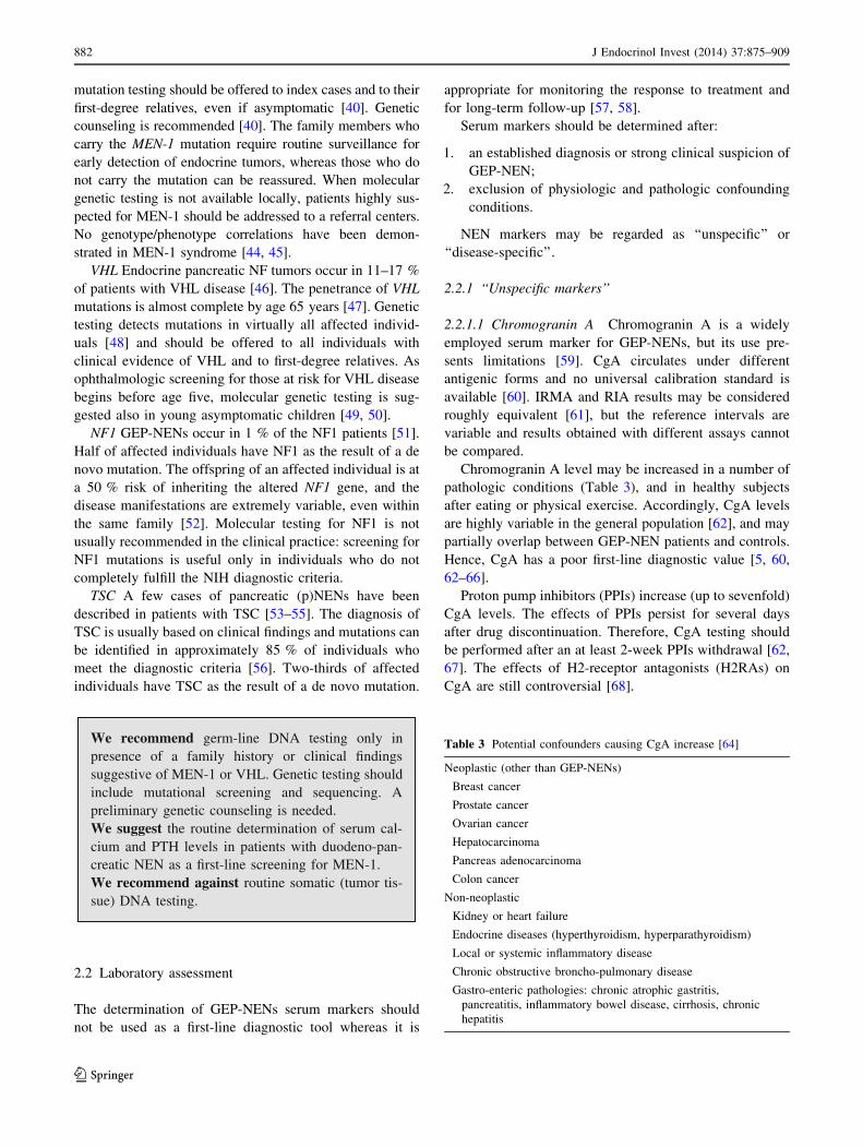
fibrosis involving tricuspid and pulmonary valves [201] (Fig. 9).
We recommend to rule out other causes of flushingand diarrhea (history and physical examination)before proceeding with work-up of CS.We recommend to perform urinary 5-HIAA andserum CgA plus abdomen US as first step if CS isclinically suspected.We recommend against routine use of other circu-lating biomarkers.We recommend contrast-enhanced abdominal CTscan or MRI plus SSTR-related imaging when firststep studies are positive.If first step studies are negative, we suggest con-sidering other diagnoses, atypical CS or very rarenon-metastatic NENs in a multidisciplinary team,involving a referral center when required.We recommend echocardiography for each patientwith CS.
3.3.2 Resistant/relapsing ulcer disease
3.3.2.1 Clinical approach: when to suspect a GEP-NEN
ZES (Box 3) is characterized by gastric acid hypersecretion
resulting in severe peptic disease and gastroesophageal
reflux disease (GERD) [202–205].
The majority of ZES patients presents with a single
duodenal ulcer, peptic symptoms, GERD symptoms or
ulcer complications and diarrhea. Multiple ulcers or
ulcers in unusual locations are a less frequent presenting
feature than in the past [8, 84, 85, 87, 202, 204–208].
With the widespread use of gastric antisecretory drugs,
particularly PPIs and H2RAs, symptoms may be masked.
The diagnosis is most often suggested by a long history
of peptic ulcer disease or GERD symptoms or their
recurrence after treatment [83–85, 206, 208]. This delay
may postpone the diagnosis of gastrinoma to a higher
stage of the disease.
Clinical suspicion: diarrhea, and/or flushing
Rule out other causes(clinical history and physical examination)
5-HIAA ± CgA + abdominal US
CT/MRI enterography, endoscopy
Liver US/CEUS, SRS, echocardiography
Dia
gnos
isSt
agin
g
PATHOLOGIC NEGATIVE
Reconsider diagnosis or rule out atypical CS
Fig. 9 Diagnostic flow-chart
for suspected carcinoid
syndrome
894 J Endocrinol Invest (2014) 37:875–909
123
3.3.2.2 Work-up in the patient with suspected gastrin-
oma History and clinical examination are the first steps in
the diagnosis of ZES. The use of acetylsalicylic acid and
other non-steroidal anti-inflammatory drugs, which can
mimic a ZES picture, should be ruled out [215].
Multiple endocrine neoplasms should be considered in
all patients with ZES, especially in case of familial or
personal history of endocrine disease, kidney stones, other
NENs [88, 207]. Due to high penetrance of primary
hyperparathyroidism in MEN-1 [40], serum calcium and
PTH are the first step to rule out the diagnosis.
Biochemical testing: Fasting serum gastrin is an excel-
lent screening test ([98 % sensitivity). False positive
conditions should always be excluded (Table 5). The
diagnosis of ZES requires inappropriately elevated FSG
levels in association with a [15 mEq/h ([5 mEq/h in
gastrectomized patients) basal acid output or in association
with a gastric pH \2.0. Under these conditions,
FSG [1,000 pg/mL means a certain diagnosis of ZES. On
the contrary, a gastric pH [2.0 virtually excludes ZES
[84]. In subjects under chronic therapy with PPIs these
drugs have to be withdrawn for at least 1 week [84, 216],
although the optimal wash-out time for PPIs should be
longer (4 weeks). H2RAs exert a less pronounced sup-
pression of gastric acid output than PPIs [217, 218]. In case
of subjects on PPIs who are at risk of bleeding ulcer,
diarrhea with dehydration or hypokalemia, these drugs may
be replaced with H2RAs for at least 1 week under medical
supervision [219, 220].
Secretin test (2 U/kg rapid infusion), a gastrin provoc-
ative test, may be performed in controversial cases [77,
221]. Withdrawal of antacid and anticholinergic drugs
(12 h), and of PPIs (1 week) is recommended [222]. The
secretin test is positive when a [120 pg/mL increase of
FSG over the basal value is found (sensitivity 94 %,
specificity 100 %) [88, 223]. Calcium stimulation test
(5 mg/kg body weight per hour, infused over 3 h, increa-
se [395 pg/mL over the basal FSG as cut-off) may alter-
natively be used. However, it is hampered by lower
sensitivity, specificity and higher side effects [88]. Gastric
acid secretion stimuli are no longer performed [203].
Imaging: After biochemical diagnosis, EGDS is
required. In ZES, peptic ulcer disease is found distally to
the duodenal bulb within the descending part of the duo-
denum or even further distally within the jejunum. Peptic
ulcers frequently occur in groups indicating some sub-
stantial acid hypersecretion [84].
The following imaging procedures may be used to
localize the primary tumor, determine the extent of the
disease, evaluate indication to surgery, and assess response
We recommend considering ZES (marker of gastrinoma) in case of:• Recurrent, severe or familial peptic ulcer disease;• Or peptic ulcer disease:
without Helicobacter pylori or other risk factors;associated with severe GERD;resistant to treatment or associated with complications (perforation, penetration, bleeding);associated with endocrinopathies or diarrhea (which promptly resolved under PPIs);with prominent gastric folds at endoscopy;
• MEN-1.We recommend MEN-1 be suspected in patients with refractory peptic ulcer disease or a confirmed ZES.
Box 3Gastrinoma
Gastrinoma is a functioning GEP-NEN, usually located
in the duodenum or pancreas that secretes gastrin and
causes a clinical syndrome known as ZES.
The incidence of gastrinomas is 0.5–2/million population/
year. Gastrinoma is one of the most common functioning
GEP-NEN in the general population [8] and occurs in
25–40 % of subjects with MEN-1 [207, 209]. ZES occurs at
an earlier age (mean 32–35 years] in patients with MEN-1
than in those with sporadic disease [204, 207, 209].
Pancreatic gastrinomas may occur in any portion of the
pancreas, while duodenal gastrinomas are predomi-
nantly found in the first part of the duodenum including
the bulb [210, 211]. At surgery, 70–85 % of gastrinomas
are found in the right upper quadrant (duodenal and
pancreatic head area), the so-called ‘‘gastrinoma tri-
angle’’ [210, 211, 212].
The main symptoms classically associated to ZES are due to
gastric acid hypersecretion and are represented by
abdominal pain (75–98 % of the cases), diarrhea (30–73 %),
Accurate localization of the tumor can result in com-
plete surgical resection, decreased rate of developing
lymph node metastases, and increasing survival [88, 222,
224–226] (Fig. 10).
We recommend fasting serum gastrin as the first stepin patients with clinical suspicion of ZES.We recommend exclusion of all other causes ofhypergastrinemia before proceeding with the diag-nostic work-up.We suggest secretin test when the diagnosis of ZESis unclear/controversial.We recommend morphological and functional ima-ging in all patients with biochemically establishedZES.
3.3.3 Spontaneous hypoglycemia
3.3.3.1 Clinical approach: when to suspect a GEP-
NEN Hypoglycemia (plasma glucose \60 mg/dL on a
venous blood sample) is an uncommon clinical problem in
non-diabetic adults. The presence of symptoms reinforces
the clinical relevance of this finding because some normal
subjects may have an asymptomatic low glucose level after
prolonged fasting. Symptoms may be due to sympathoad-
altered concentration, confusion, blurred vision and, in
extreme cases, coma and death) [227–229]. Symptoms
may present at a variable glucose level (generally as low
as \55–60 mg/dL) [227, 228, 230, 231].
Hypoglycemia may be due to several conditions beyond
insulin-secreting tumors [232] (Table 8).
Insulinoma (Box 4) should be strongly suspected in
presence of the Whipple triad, which occurs in about 75 %
of patients and combines (1) symptoms of hypoglycemia,
(2) low blood sugar concurrent with symptoms, and (3)
reversal of symptoms after glucose administration [233].
Neuroglycopenic symptoms usually dominate the clinical
picture so that insulinoma may be misdiagnosed with
cognitive impairment, psychiatric illnesses or seizure dis-
orders. Frequently, the occurrence of bizarre behavior or
confusion states is more precisely described by concerned
relatives or friends than by the patient himself. Adrenergic
and neuroglycopenic symptoms may coexist, especially in
the early phase of the disease. A detailed description of
pure adrenergic symptoms, however, is more specific of a
‘‘functional hypoglycemia’’.
Hypoglycemic symptoms occur most frequently at
night and/or early morning and, anyway, in a protracted
fasting state. Yet, the occurrence of post-prandial hypo-
glycemia does not exclude an insulinoma [234, 235].
Symptoms can be worsened by exercise, alcohol, hy-
pocaloric diet, and by concomitant clinical conditions or
use of drugs (see above) [236, 237]. Weight gain occurs
in 20–40 % of patients that may develop overweight
because of hyperinsulinism.
Clinical suspicion
Withdraw interfering drugs (if possible)
Fasting gastrin ± gastric pH
EGDS, CT/MRI, EUS
Liver US/CEUS, SRS, PET
Dia
gnos
isSt
agin
g
Secretin test± Ca stimulation
intra-arterial Ca angiography
Fig. 10 Diagnostic flow-chart
for suspected gastrinoma
896 J Endocrinol Invest (2014) 37:875–909
123
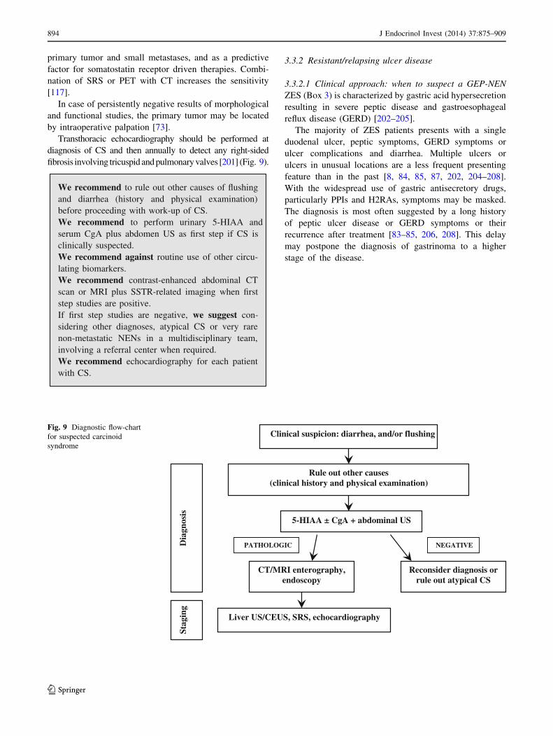
We recommend the immediate determination ofblood glucose and insulin in any subject with acutechange in mental status, especially if recurrent.We recommend the exclusion of all alternativecauses of hypoglycemia before starting a work-up forinsulinoma.We suggest considering insulinoma as a probablecause in patients with predominant neuroglycopenicsymptoms, recurrent fasting hypoglycemia, andweight gain.
3.3.3.2 Work-up in the patient with suspected insuli-
els B2.7 mmol/L may confirm the diagnosis, demonstrating
the suppressive effect of insulin on ketogenesis even during a
protracted fasting [231].
At the end of the 72-h fasting test, in the absence of
hypoglycemia, the use of stimulation tests was proposed
[231]. Stimulation tests, e.g., tolbutamide, glucagon or cal-
cium, are not recommended because they may induce a
prolonged and refractory hypoglycemic condition, but long-
term fasting can be finished after 72 h with bicycle test.
Imaging: In all patients with a confirmed biochemical
diagnosis, imaging is indicated to localize the tumor [241].
Since 80 % of insulinomas are \2 cm in size, they are
frequently missed by high-resolution transabdominal US
(50 % sensitivity), while EUS is more sensitive (77 %) and
should be preferred [242]. Helical or multislice CT and
MRI offer a comparable (82–94 %), but incomplete, sen-
sitivity [243, 244]. Selective arteriography has an 82 %
sensitivity and a 95 % specificity.
Due to small size and/or lack of SSTR2 expression in
50 % of insulinoma [151], SSTR-related imaging plays a
minor role than morphological imaging. DOPA-PET has
been proposed as an alternative tool [245]. Radiolabelling
with 111In-labeled glucagon-like peptide-1 receptors ago-
nist (111In-DOTA-exendin-4) is a promising technique, still
not routinely used [246].
Arteriography combined with selective calcium stimu-
lation: Calcium is able to stimulate insulin release from
neoplastic tissue, but not from normal islets. Hence, the
catheterization of the arterial branches of the celiac system
and the measurement of insulin in the blood sampled from
hepatic veins during selective intra-arterial calcium injec-
tion localize the pancreatic area nesting the tumors in
88–100 % of cases [34, 247, 248]. This test is cumbersome,
expensive and poorly available. Accordingly, it should be
reserved only to selected, biochemically proved cases with
negative imaging studies.
In spite of all the above reported diagnostic techniques,
only 60–70 % of patients have a successful preoperative
localization. In patients with less threatening symptoms
that are fairly controlled by medical treatment a close
surveillance may be advisable. In severely symptomatic
cases, the use of intraoperative US and the pancreatic
exploration conducted by an experienced surgeon identifies
more than 90 % of the insulin-secreting tumors [242, 249]
(Fig. 11).
Liver NEN metastases
IHC for site of origin + lab tests
MRI, EUS, enteroCT/MRI, 68Ga-DOTA-PET, VCE, DBE
Dia
gnos
is
Unknown primary at conventional imaging
Low gradeHigh grade
18F-FDG-PET
Fig. 12 Diagnostic flow-chart
in the patient with metastatic
disease and unknown primary
tumor
898 J Endocrinol Invest (2014) 37:875–909
123
We recommend the simultaneous evaluation ofblood glucose, insulin and C-peptide to detect endo-genous hyperinsulinemia in all patients with sponta-neous hypoglycemia.We recommend a prolonged fasting test (up to 72 h)in patients referred for a previous hypoglycemicepisode who are free of symptoms at the moment ofmedical examination.We recommend against the use of stimulation testsfor the diagnosis of insulinoma.We recommend the use of localization tests (CT/CEUS and EUS) only after the biochemical diagnosisof insulinoma is established.
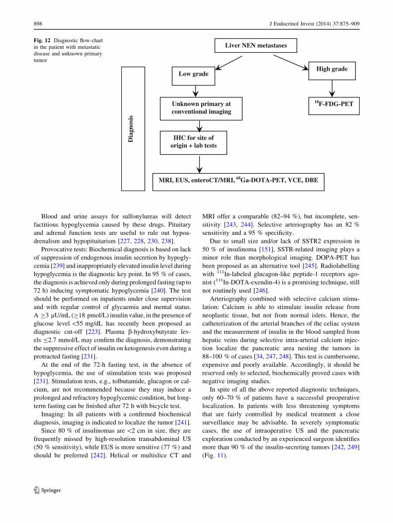
3.4 Work-up in the patient with metastatic disease
and unknown primary tumor
Unknown primary NEN (UPN) is a condition of metastatic
histologic or cytological confirmed NEN without evidence of a
primary site after a first diagnostic work-up, including chest-
abdomen CT scan, SRS, and upper and lower endoscopy.
The frequency of well-differentiated UPNs ranges from
9 to 19 % [250, 251]. The presence of liver metastases
largely influences prognosis in all types of NENs and is
dependent on primary tumor site, tumor extent (T-stage),
and histologic differentiation (NET vs. NEC). Reported
survival rate at 5 years of G1–G2 small intestinal and
pancreatic NENs in the SEER database is 54 and 27 %,
respectively [252]. Furthermore, survival is reportedly
worse in UPN patients as compared to patients with liver
metastases whose primary NEN is known [253]. In liver
metastatic patients survival rate is influenced by the pre-
sence of obstructive symptoms or symptoms related to
peptide secretion.
The evaluation of a patient with UPN should encompass
a detailed clinical history, including family history to
identify affected relatives and a patient’s increased risk for
endocrine tumors (i.e., MEN type 1 or 2), laboratory and
radiographic studies [254].
Histologic preparations should be reevaluated by IHC to
guide the search for the primary tumor: TTF-1 (pulmonary
or medullary thyroid carcinoma), CDX-2 (intestinal), PAX-
gastrin (occult gastrinoma), and PP/glucagon (pancreatic)
[38, 255]. Biochemical work-up may include 5-HIAA,
gastrin, and other locally available tumor markers [256].
It has been recently reported that most UPNs are derived
from pancreas and small bowel [257]. Accordingly, further
investigations for localizing the primary site in well-dif-
ferentiated NENs might include abdomen MRI, EUS,
enteroCT/MR, 68Ga-PET, VCE, DBE to be shared within a
multidisciplinary team according to clinics, local avail-
ability and expertise [124, 258, 259]. In NECs 18F-FDG-
PET may be useful (Fig. 12)
We recommend biopsy at the metastatic site withhistologic and IHC examination as a first step toconfirm NEN diagnosis.We recommend a detailed clinical history to elicitsigns or symptoms that could point to the primary site(carcinoid syndrome, asthma, diarrhea, etc.), as wellas a complete family history.In all cases of unknown primary of low grade meta-static liver NEN after a conventional imaging,including chest-abdomen CT scan, upper and lowerendoscopy and SRS, we recommend an IHC for siteof origin, and imaging according to results.
3.5 When and how to stage a previously diagnosed
GEP-NEN
Evaluation of disease extension has a pivotal role in
treatment planning.
Pre-treatment staging should include morphologic and
functional imaging. Morphological imaging is required for
all GEP-NENs, irrespectively of their grade. SSTR-based
functional imaging (SRS or 68Ga-DOTA-peptide-PET)
should be used for low-/intermediate-grade GEP-NENs
(WHO 2010 G1-G2), whereas 18F-FDG-PET should be
preferentially used in G3 GEP-NENs and in some G2
cases.
For morphologic staging, a chest-abdomen-pelvis mul-
tidetector CT or a chest basal CT plus abdomen-pelvis MRI
should be used [87]. For functional staging, SRS using111In-pentetreotide (Octreoscan�) is presently regarded as
the gold standard. However, if available, 68Ga-DOTA-
peptide-PET with simultaneous CT should be preferred to
SRS. In facts, PET lacks the anatomic details required for
therapeutic stratification (surgical planning or dose calcu-
lation for radioembolization with radiolabeled micro-
spheres). Recently, MRI with liver-specific contrast
combined with 68Ga-DOTA-peptides-PET has been
reported to be more accurate than PET-CT to detect GEP-
NEN hepatic metastases [260].18F-DOPA-PET-CT and 11C-5HTP-PET-CT are prom-
ising tools. Their use might be considered if results of SRS
or 68Ga-DOTA-peptides-PET are negative [261].
Gastric NENs In small (\1 cm) type 1 and type 2
tumors, EGDS is usually the only recommended imaging
J Endocrinol Invest (2014) 37:875–909 899
123
procedure [153]. Tumor invasiveness through the gastric
wall must be evaluated with EUS study: it is recommended
before resection for polyps [1 cm in diameter. EUS is also
useful in assessing regional lymph nodes involvement, and
allows histological confirmation by FNA. Type 1 tumors
do not require either abdomen multislice CT or MRI, or
SRS/68Ga-DOTA-peptides-PET; these imaging studies
should be performed for type 2 and type 3 neoplasm
staging.
Duodenal NENs EUS is useful before resection of pol-
ypoid lesions; multislice abdomen CT or MRI should be
performed to assess local and distant disease extension. In
patients with local advanced neoplasm and/or liver
metastases, bone scan and MRI of spine and pelvis should
be performed [153].
Jejuno-ileum NENs Chest-abdomen-pelvis CT scan or
chest basal CT scan and abdomen-pelvis MRI, SRS or68Ga-DOTA-peptides-PET should be performed looking
for distant metastases [73]. Liver CT scan should be per-
formed by multislice and multiphase technique. Colonos-
copy should be performed to rule out synchronous
colorectal carcinoma.
Colorectal NENs Chest-abdomen-pelvis multislice CT
should be carried out. Endoanal/rectal US is very useful for
assessing preoperatively the depth of tumor invasion in the
rectal wall and regional lymph node involvement [173].
NF pNENs For morphologic staging a multislice/multi-
phase CT or fat-saturated T1-weighted and delayed
enhanced T1-weighted MRI can be performed and EUS
with biopsy [262]. Afterwards, SRS or 68Ga-DOTA-pep-
tides-PET should be performed.
We recommend contrast enhanced chest-abdomen-pelvis multidetector CT scan or basal chest CT scanplus abdomen-pelvis MRI in pre-treatment staging ofGEP-NENs.We recommend 68Ga-DOTA-peptides-PET-CT forfunctional staging of well-differentiated GEP-NENs,or, if not available, 111In-pentetreotide (Octreoscan®)scintigraphy.If SSTR-related imaging is negative, we suggestfurther functional staging with alternativeradiotracers.We suggest 18F-FDG-PET-CT for staging of selec-ted G3 and high G2 GEP-NENs.We recommend EUS study for local staging of 1cm gastric, duodenal, and rectal polypoid NENlesions.
4 Conclusions
The management of patients with GEP-NENs poses a
significant challenge to clinicians from the very start of the
diagnostic work-up. The wide heterogeneity of disease
presentation, with a majority of asymptomatic patients and
poorly specific clinical pictures may account for a delay in
definite diagnosis and appropriate treatment. The present
document has, therefore, been drawn with the purpose of
offering a practical guide to physicians facing the suspicion
of GEP-NENs, in light of the available clinical evidence
and experience. Of course, many questions are still to be
fully answered and many others still to be addressed in the
near future, as we move forward to new promising tech-
niques and diagnostic tools. For these reasons, in spite of
its goal as a state-of-the-art update, our document has not
been conceived as the repository of the ‘‘ultimate truth’’ in
the field of GEP-NENs diagnosis. Instead, much attention
has been devoted to the logical framework, which should
back up the clinical reasoning. Furthermore, the diagnosis
of GEP-NENs is heavily based on the contribution of a
wide range of know-how and skills provided by different
specialists. The core team may include a varying combi-
nation of different specialists, according to the local
expertise and facilities; nevertheless, the pathologist plays
a key role in the diagnosis and classification of GEP-NENs,
because his/her information is critical to guide the prog-
nosis and treatment planning. Hence, a multidisciplinary
team model is recommended as the best opportunity to
reach an accurate, safe and cost-effective diagnosis, likely
to improve the outcome of patients with GEP-NENs.
In conclusion, the Italian Association of Clinical
Endocrinologists (AME) hopes the present Position State-
ment will constitute an effective tool in helping the clinical
management of patients with GEP-NENs. Further imple-
mentations and updates of this document will follow as
new evidence and progress in the field emerge.
Other members of AME oncologic endocrinology group
Giorgio Borretta, Cuneo; Renato Cozzi, Milan; Giuseppe
Francia, Verona; Rinaldo Guglielmi, Albano Laziale; Ga-