Volume 8 • Issue 3 • 10001088J Clin Case Rep, an open access journalISSN: 2165-7920
Open AccessCase Report
Iuliano et al., J Clin Case Rep 2018, 8:3DOI: 10.4172/2165-7920.10001088
Journal of Clinical Case ReportsJour
nal o
f Clinical Case Reports
ISSN: 2165-7920
*Corresponding author: Donato Iuliano, Department of Internal Medicine, Hospital Dr. A. Guerriero, Italy, Tel: 3391268451; E-mail: [email protected]
Received February 05, 2018; Accepted March 16, 2018; Published March 20, 2018
Citation: Iuliano D, Mauro M, Migliore N, Dattolo P, Migliore G, et al. (2018) An Unusual Acute Pancreatitis due to a Migrated Foreign Body. J Clin Case Rep 8: 1088. doi: 10.4172/2165-7920.10001088
An Unusual Acute Pancreatitis due to a Migrated Foreign BodyDonato Iuliano*, Maria Mauro, Nicola Migliore, Pellegrino Dattolo, Giuseppe Migliore, Biagio Mauro, Maria Erminia Bottiglieri, Angelo Tiso, Caterina Russo and Carmine NapolitanoDepartment of Internal Medicine, Hospital Dr. A. Guerriero, Italy
AbstractWe present a peculiar case of acute pancreatitis. A 23-year-old man was admitted to our Gastroenterology Unit
with a diagnosis of acute pancreatitis based on clinical symptoms and on biochemical data. After the necessary diagnostic work-up, a diagnosis of acute traumatic pancreatitis was made, due to a migrated foreign body (needle).
IntroductionWe describe a usual case of acute pancreatitis, due to foreign body.
A 23-year-old man was admitted with a diagnosis of acute pancreatitis based on clinical symptoms and on biochemical data. In this study it was showed that, in patients with acute abdominal pain admitted at Emergency Room, an abdominal plain radiograph did not bring any added value as diagnostic accuracy after the clinic and laboratory evaluation.
Case ReportA 23-year-old man, without co morbidities or voluptuary habits, was
admitted to the Emergency Room of our Hospital with severe epigastric pain, without irradiation. The on-set of the pain dated 15 days before the admission and had become progressively more intense. Following the advice of his general practitioner, the patient began a therapy with proton-pump inhibitors, without improvements. At clinical examination in Emergency Room, the patient presented moderate abdominal pain without tenderness; Murphy’s and Blumberg’ signs were negative. There was not detected any abnormality of canalization. Negative history for previous abdominal surgery.
The laboratory data showed hyperamylasemia (956 UI/L; normal values (n.v.): 9-100 UI/L). Other biochemical values were normal. PCR=0.06 mg/dl (n.v. 0,00-0,50). WBC=10000/mm3. AST, ALT, total and fractionated bilirubin, alkaline phosphatase and Gamma-Glutamyl-Transpeptidase, as well as renal and metabolic function tests were normal. The patient denied alcohol abuse and CDT (Carbohydrate Deficient Transferrin) was normal.
Therefore, he was admitted to the Gastroenterology ward with a diagnosis of mild acute pancreatitis (Classification of Acute Pancreatitis- International Consensus-GUT 2013; 62: 102-111).
Echo-fast showed normal liver without biliary tree dilatation. No gallbladder stones. Pancreas showed a small subcentimetric hypoechogenic area in the head-body portion of uncertain meaning. The Duct of Wirsung was slightly dilated (4 mm calibre) and interrupted at the above-mentioned formation from which a hyperechogenic stripe was projected, reaching the gastric antrum. Spleen normal. Kidneys normal (Figure 1).
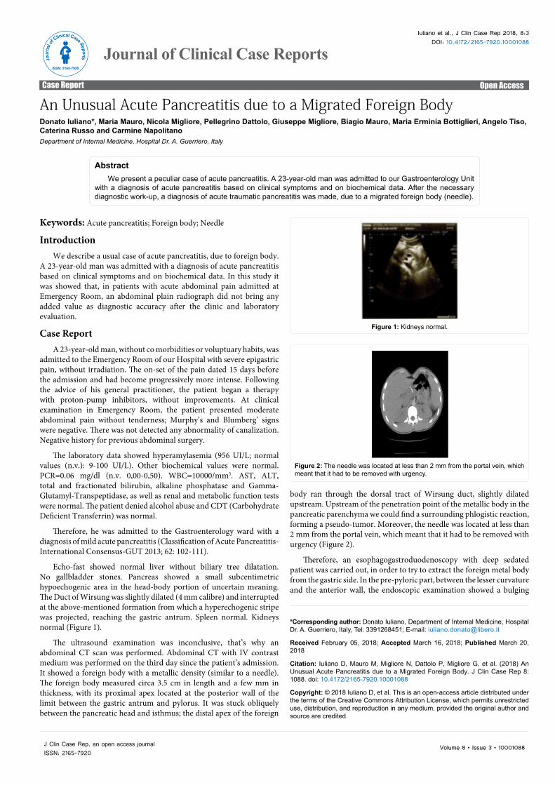
The ultrasound examination was inconclusive, that’s why an abdominal CT scan was performed. Abdominal CT with IV contrast medium was performed on the third day since the patient’s admission. It showed a foreign body with a metallic density (similar to a needle). The foreign body measured circa 3.5 cm in length and a few mm in thickness, with its proximal apex located at the posterior wall of the limit between the gastric antrum and pylorus. It was stuck obliquely between the pancreatic head and isthmus; the distal apex of the foreign
Figure 1: Kidneys normal.
Figure 2: The needle was located at less than 2 mm from the portal vein, which meant that it had to be removed with urgency.
body ran through the dorsal tract of Wirsung duct, slightly dilated upstream. Upstream of the penetration point of the metallic body in the pancreatic parenchyma we could find a surrounding phlogistic reaction, forming a pseudo-tumor. Moreover, the needle was located at less than 2 mm from the portal vein, which meant that it had to be removed with urgency (Figure 2).
Therefore, an esophagogastroduodenoscopy with deep sedated patient was carried out, in order to try to extract the foreign metal body from the gastric side. In the pre-pyloric part, between the lesser curvature and the anterior wall, the endoscopic examination showed a bulging
Citation: Iuliano D, Mauro M, Migliore N, Dattolo P, Migliore G, et al. (2018) An Unusual Acute Pancreatitis due to a Migrated Foreign Body. J Clin Case Rep 8: 1088. doi: 10.4172/2165-7920.10001088
Page 2 of 3
Volume 8 • Issue 3 • 10001088J Clin Case Rep, an open access journalISSN: 2165-7920
there was no visible portion of the needle. We closed the incision by metallic clips (Figure 7).
In this urgency setting, we decided to send the patient to the surgeon. Surgery time: after having carried out a midline xipho-umbilical laparotomy and a section of the gastro colic ligament to access the lesser sac, in the gastric antrum, phlogistic adhesions were found between the gastric posterior wall and the pancreatic parenchyma, in proximity to the common hepatic artery. After having passed under the adherences and carried out hemostatic ligatures at the two ends, the needle became visible and could be extracted (Figures 8-10).
DiscussionThe common causes of Acute Pancreatitis (AP) are gallstones
disease (40-70%), alcohol excess (20-35%). Approximately 10-20% of AP occur without a detectable cause (“idiopathic” AP). Among the known causes of AP, trauma is rare. An AP due to a metallic migrate foreign body, after ingestion, is extremely rare.
The peculiarity of this case consists in its rare occurrence. Moreover, compared to the other cases encountered in previous reports [1-4], what is also peculiar in this case is the extraluminal location from the stomach of the needle, which was stuck in the parenchyma of the pancreatic head. Since it was not possible to remove it by endoscopy through
area with an eroded surface, suggestive for reactive granulomatous-inflammatory process (Figure 3).
Compared to the fluoroscopy, the proximal end of the metallic body corresponded to the lesion of the gastric antrum showed by the endoscopy, with a distance of circa 1 cm and the with tip of the endoscopic probe arguably located at the point of penetration of the needle-shaped foreign body (Figure 4).
However, there was no visible portion of the afore-mentioned foreign body. Therefore, a small incision of the mucosal wall was made by a pre-cut needle, in order to expose the end of the metallic part and then extract it (Figures 5 and 6). Unfortunately, without success, since
Figure 3: Endoscopic examination showed a bulging area with an eroded surface, suggestive for reactive granulomatous-inflammatory process.
Figure 4: Penetration of the needle-shaped foreign body.
Figure 5: A small incision of the mucosal.
Figure 6: A small incision of the mucosal.
Figure 7: closed the incision by metallic clips.
Figure 8: After having passed under the adherences and carried out hemostatic ligatures at the two ends, the needle became visible and could be extracted.
Figure 9: After having passed under the adherences and carried out hemostatic ligatures at the two ends, the needle became visible and could be extracted.
Citation: Iuliano D, Mauro M, Migliore N, Dattolo P, Migliore G, et al. (2018) An Unusual Acute Pancreatitis due to a Migrated Foreign Body. J Clin Case Rep 8: 1088. doi: 10.4172/2165-7920.10001088
Page 3 of 3
Volume 8 • Issue 3 • 10001088J Clin Case Rep, an open access journalISSN: 2165-7920
the stomach, the patient had to necessarily undergo a surgery, with a positive result. We invited the patient to undergo a CT after 3 months, in order to check the outcomes and the possible complications [5].
ConclusionWe wanted, however, to share another consideration about the case:
Acute abdominal pain accounts for 5-10% of visit to Emergency
Room. The evaluation of a patient with an acute abdominal pain represents a challenge in the differential diagnosis and rapid and accurate detection of urgent conditions isssss crucial for managing patients and to decide whether the patient needs an emergency procedure.
The imaging strategies for detection of urgent condition in patients with acute abdominal pain have been evaluated in a study of Laméris et al. (BMJ 2009; 338:b2431). In this study it was showed that, in patients with acute abdominal pain admitted at Emergency Room, an abdominal plain radiograph did not bring any added value as diagnostic accuracy after the clinic and laboratory evaluation.
The Guidelines of the American Pancreatic Association, the American College of Gastroenterology and the American College of Radiology for imaging management in AP recommends an initial CT scan or Magnetic Resonance Imaging (MRI) assessment when diagnosis is uncertain. Optimal timing is at last 72-96 hours after onset of symptoms.
In the case we described, an abdominal X-Ray plain (carried out during the abdominal CT scan), already showed an epigastric metallic foreign body (Figure 11). The abdominal X-Ray plain (or, better, an abdominal CT scan), instead, if carried out at admission in Emergency Room after Echo-Fast, could have led to a faster diagnosis and modified our diagnostic and therapeutic strategy. Luckily, a MRI wasn’t performed!
References
1. Bargiggia LR, Vailati C, Tagliabue F, Airoldi A, Parente F (2015) Pancreatic pseudotumor caused by toothpick ingestion.s. Gastroint End 82: 179-180.
2. Ishikura H, Koyama T, Komuro K, Ito T, Oshima T, et al. (1986) Foreign bodies of unknown origin found in a serosal hematoma between the stomach and the pancreas of the patient. Hokkaido Igaku Zasshi 61: 108-119.
3. Deboever G, Sels F (1990) An exceptional case of foreign body-induced pancreatitis. Am J Gastroenterol 85: 632-633.
4. Pezzilli R, Barakat B, Bertaccini P (2000) Foreign body of the pancreas. Digest Liver Dis 32: 179.
5. Chamoun S, Testart J, Metayer P (1983) Pancreatite chronique segmentaire par corps Stranger du canal de Wirsung. J Chir (Paris) 120: 191-193.
Figure 10: After having passed under the adherences and carried out hemostatic ligatures at the two ends, the needle became visible and could be extracted.
Figure 11: An abdominal X-Ray plain (carried out during the abdominal CT scan), already showed an epigastric metallic foreign body.