23
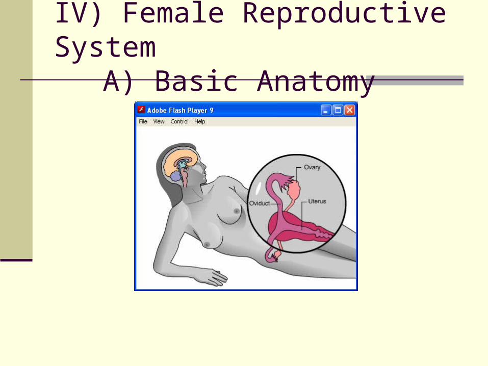
IV) Female Reproductive System A) Basic Anatomy
| Date post: | 02-Jan-2016 |
| Category: |
Documents |
| Upload: | lucinda-fowler |
| View: | 228 times |
| Download: | 1 times |
IV) Female Reproductive System
A) Basic Anatomy
IV) Female Reproductive SystemA) Basic Anatomy
IV) Female Reproductive SystemA) Basic Anatomy
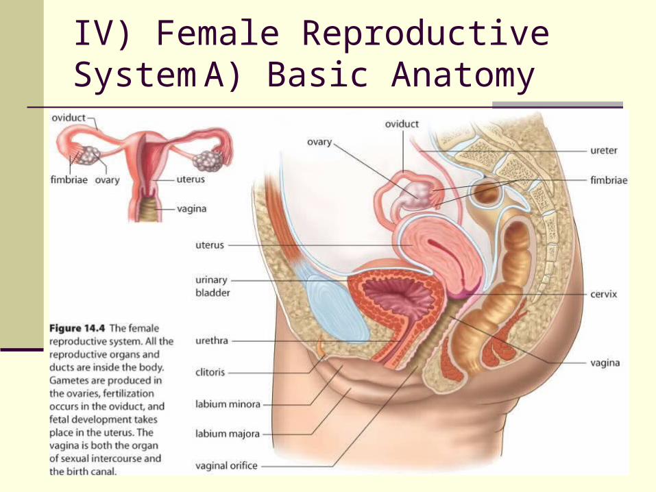
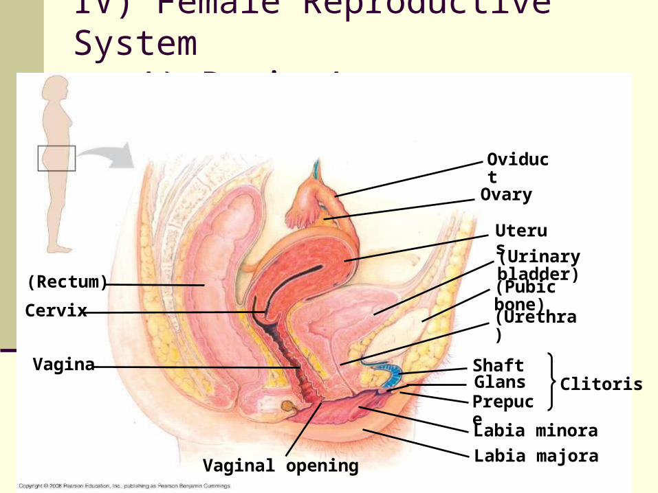
(Rectum)
Cervix
Vagina
Vaginal opening
Oviduct
Ovary
Uterus
(Urinary bladder)
(Pubic bone)
(Urethra)
ClitorisShaftGlansPrepuce
Labia minora
Labia majora

OvariesOviduct
FolliclesCorpus luteum
Uterine wallUterus
Cervix
Endometrium
Vagina
IV) Female Reproductive SystemA) Basic Anatomy
in comparison: males produce sperm cells at a constant rate starting from
sexual maturity females are born with a set number of viable eggs and one is
matured every 28 days after puberty. the excretory system of the females does not cross the
reproductive system like in the males (sperm out the urethra)
females follow a complicated sexual cycle where one ovum matures approximately every 28 days.
during fetal development in females the ovaries form in same abdominal region as the testis in the male.
the ovaries also descend but only as far as the pelvic region. at birth the oocytes (immature ova) are already present within
the ovaries.
IV) Female Reproductive SystemA) Basic Anatomy
IV) Female Reproductive SystemA) Basic Anatomy
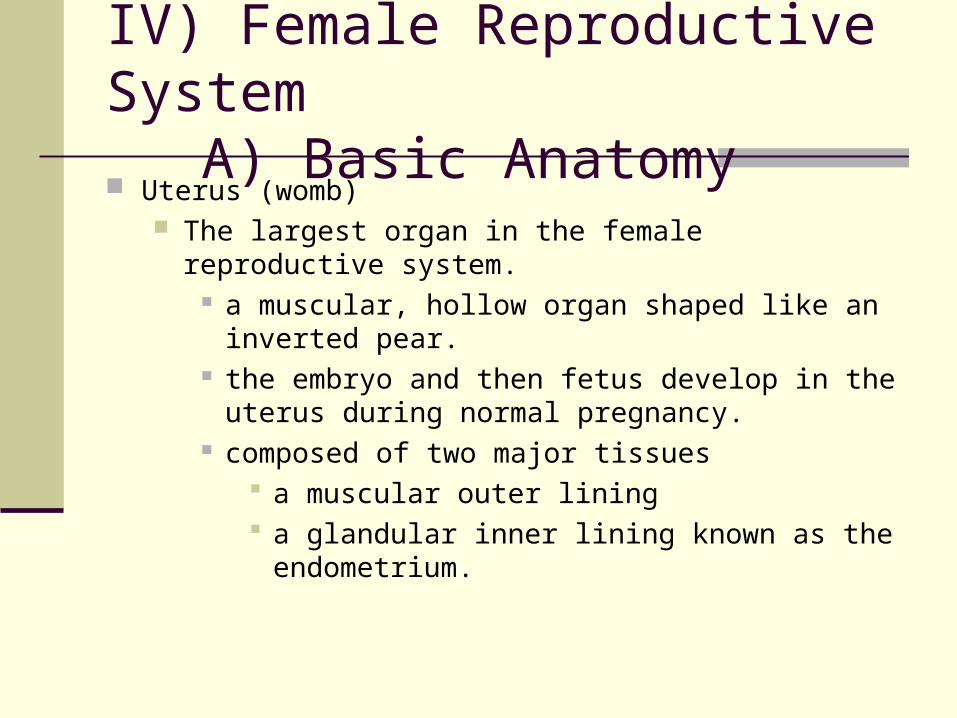
Uterus (womb) The largest organ in the female reproductive system.
a muscular, hollow organ shaped like an inverted pear. the embryo and then fetus develop in the uterus during
normal pregnancy. composed of two major tissues
a muscular outer lining a glandular inner lining known as the endometrium.
IV) Female Reproductive SystemA) Basic Anatomy
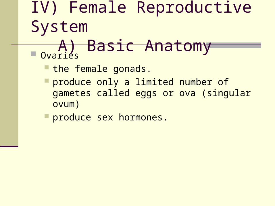
Ovaries the female gonads. produce only a limited number of gametes called
eggs or ova (singular ovum) produce sex hormones.
IV) Female Reproductive SystemA) Basic Anatomy
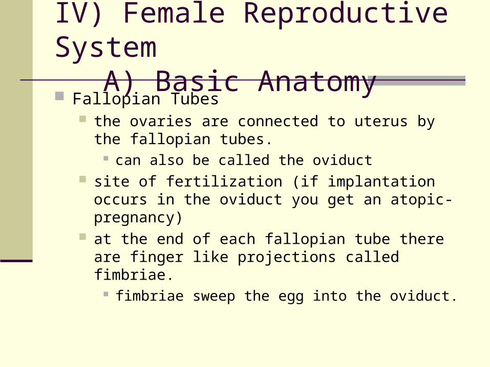
Fallopian Tubes the ovaries are connected to uterus by the fallopian
tubes. can also be called the oviduct
site of fertilization (if implantation occurs in the oviduct you get an atopic-pregnancy)
at the end of each fallopian tube there are finger like projections called fimbriae.
fimbriae sweep the egg into the oviduct.
IV) Female Reproductive SystemA) Basic Anatomy
III) Reproductive System A) Basic Anatomy
Vagina connects the uterus with the outside environment. site of sexual intercourse and the birth canal. is acidic, creating a hostile environment for microbes
and other pathogens. is connected to the uterus by the cervix.
the cervix is a muscular band
IV) Female Reproductive System
B) Oogenesis
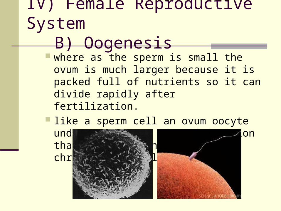
where as the sperm is small the ovum is much larger because it is packed full of nutrients so it can divide rapidly after fertilization.
like a sperm cell an ovum oocyte undergoes a type of cell division that halves the number of chromosomes (called meiosis).
IV) Female Reproductive SystemB) Oogenesis
IV) Female Reproductive SystemB) Oogenesis
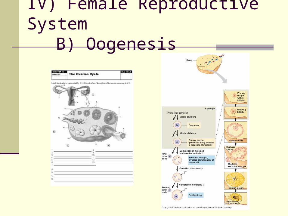
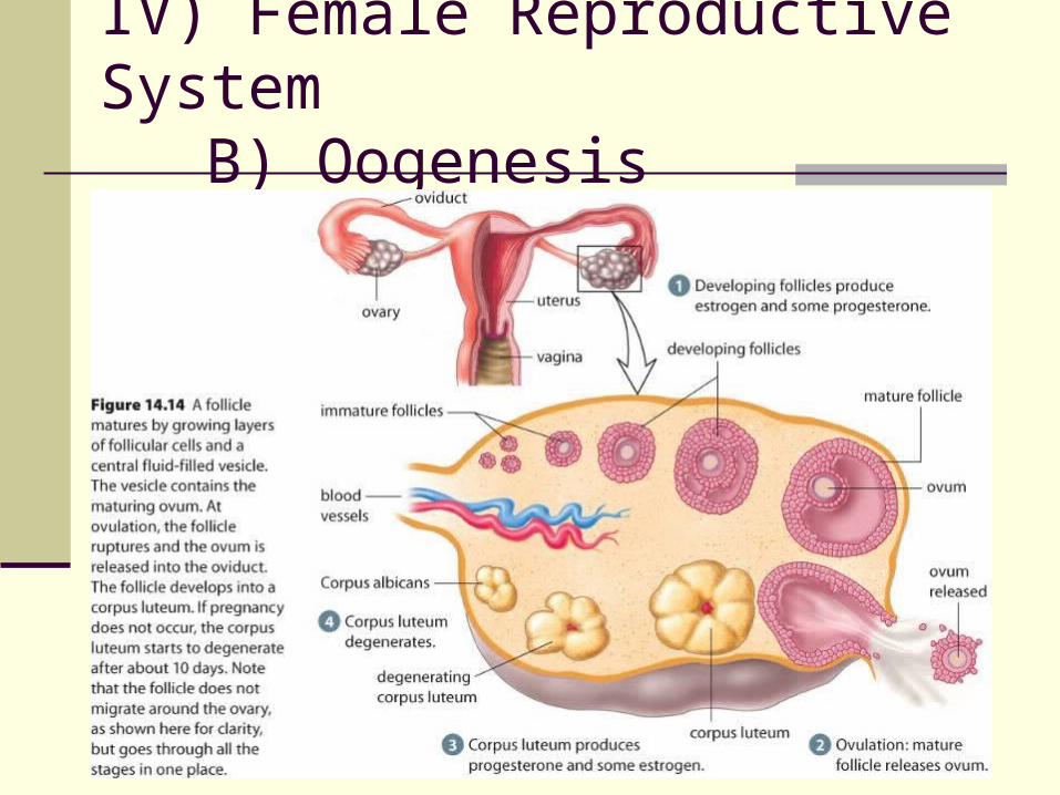
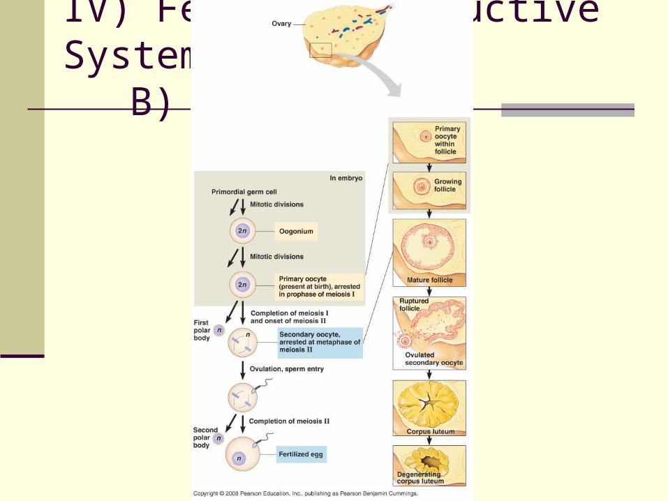
Oogenesis is the formation of an ovum occurs in specialized cells in the ovaries called
follicles. a follicle contains two types of cells
a primary oocyte cells of the granulosa
~ the granulosa is the layer of cells that form the follicle wall. They provide nutrients for the developing oocytes.
IV) Female Reproductive SystemB) Oogenesis
begins when the nutrient follicle cells which ARE surrounding the primary oocyte begin to divide.
the primary oocyte undergoes cell division and while this occurs the majority of cytoplasmic nutrients move to one of the poles and form a secondary oocyte.
the secondary oocyte contains 23 chromosomes. the remaining cell, called the first polar body, receives little
cytoplasm and dies.
IV) Female Reproductive SystemB) Oogenesis
as the follicle cells surrounding the secondary oocyte start to proliferate a fluid filled cavity forms.
the dominant follicle begins to push outwards trying to escape the ovary.
constriction of blood vessels weaken the ovarian wall.
enzymes weaken the follicle wall from the inside. the seconday oocyte (with the first polar body) is
released, this is called ovulation.
IV) Female Reproductive SystemB) Oogenesis
IV) Female Reproductive SystemB) Oogenesis

the follicle cells that were surrounding the secondary oocyte stay in the ovary and transform into the corpus luteum.
the corpus luteum secretes hormones essential for pregnancy.
if no pregnancy occurs, the corpus luteum degenerates.
after its release from the ovary the secondary oocyte is swept into the end of the Fallopian tube by the fimbriae.
the secondary ooctye is moved along the Fallopian tube by cilia
if healthy sperm is present fertilization will occur.
IV) Female Reproductive SystemB) Oogenesis
IV) Female Reproductive SystemB) Oogenesis
IV) Female Reproductive SystemB) Oogenesis
IV) Female Reproductive SystemB) Oogenesis
after fertilization in the Fallopian tube the secondary oocyte undergoes an uneven division
again. the cells that retain most of the cytoplasm
(nutrients) becomes the ovum. the other cell becomes the second polar body,
which deteriorates. if no fertilization occurs, the secondary oocyte will
deteriorate within 24 hours and die. the women will then undergo a menstrual cycle.
IV) Female Reproductive SystemB) Oogenesis
IV) Female Reproductive System
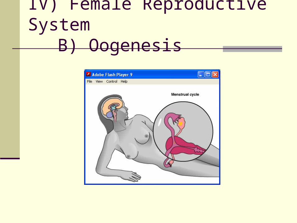
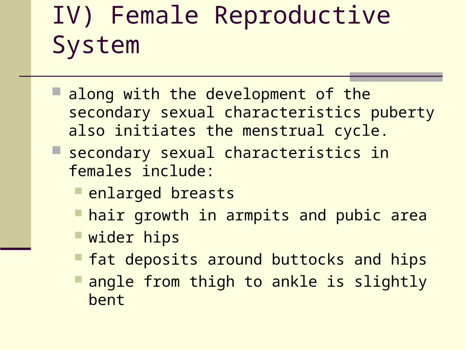
along with the development of the secondary sexual characteristics puberty also initiates the menstrual cycle.
secondary sexual characteristics in females include: enlarged breasts hair growth in armpits and pubic area wider hips fat deposits around buttocks and hips angle from thigh to ankle is slightly bent


