Brit. J. Ophthal. (I974) 58, 293 Electrophysiology of the retraction syndromes A. HUBER Ziirich, Switzerland With the aid of modern electromyography, numerous authors (Pabst and Esslen, I960; Sato, ig60; Orlowski and W6jtowicz, I962; Burger, I963; Blodi, van Allen, and Yar- brough, I964; Huber and Esslen, I969) have recently come to the (surprisingly) unanimous conclusion that a paradoxical innervation of the external rectus muscle of the affected eye represents the pathogenetic principle of all the retraction syndromes (Stilling-Turk-Duane) (Duane, I905) and that the explanations given by the majority of earlier authors based on mechanical concepts (birth injury, congenital or acquired musculo-facial anomaly, etc.) are either insufficient or are at variance with the facts (Kriiger, I969). In this connection it must be emphasized that to accept a disorder of the antagonistic interaction of the extraocular muscles as the primary cause of the retraction syndromes does not exclude anatomical alterations of the muscular tissues, the more so as primary innervational disturbances are known to induce pathological changes in the muscles. An analysis of published cases (Lyle and Bridgman, I959; Malbran, 1953) and of our own series, has enabled us to distinguish three types of Duane's retraction syndrome: Duane I (corresponding to Lyle's Type B or Malbramn's Type I) Marked limitation or complete absence of abduction, normal or only slightly defective adduction, narrowing of the palpebral fissure and retraction of the affected eyeball on adduction, widening of the palpebral fissure on attempted abduction (Fig. I). Duane II (corresponding to Lyle's Type C or Malbran's Type II) Instead of an impairment of abduction there is a limitation or complete defect of adduction with exotropia of the affected eye. Abduction appears to be normal or only slightly limited. There is further distinct narrowing of the palpebral fissure and retraction of the globe on attempted adduction (Fig. 2). Duane III (corresponding to Lyle's Type A or C) Combination of limitation or absence of both abduction and adduction of the affected eye (If abduc- tion and adduction are defective in the same degree, the affected eye is in the parallel position; if adduction is more defective than abduction, the affected eye diverges). There is further characteristic retraction of the globe and narrowing of the palpebral fissure on attempted adduction (Fig. 3). It is well known that the retraction syndromes of Type I, II, and III, apart from the disorders of motility in the horizontal plane, frequently produce additional vertical motor anomalies. These are mostly evident as sursum- or deorsumductions on adduction (sometimes also on attempted abduction) of the affected eye. Some retraction syndromes, especially Duane I, are charac- terized by changes in the ocular axes on looking up or down, which cause an A-pattern, a Address: Prof. A. Huber, Stadelhoferstrasse 42, 8ooI Zurich, Switzerland. copyright. on January 31, 2022 by guest. Protected by http://bjo.bmj.com/ Br J Ophthalmol: first published as 10.1136/bjo.58.3.293 on 1 March 1974. Downloaded from
Transcript
Brit. J. Ophthal. (I974) 58, 293
Electrophysiology of the retractionsyndromes
A. HUBER
Ziirich, Switzerland
With the aid of modern electromyography, numerous authors (Pabst and Esslen, I960;Sato, ig60; Orlowski and W6jtowicz, I962; Burger, I963; Blodi, van Allen, and Yar-brough, I964; Huber and Esslen, I969) have recently come to the (surprisingly) unanimousconclusion that a paradoxical innervation of the external rectus muscle of the affected eye represents thepathogenetic principle of all the retraction syndromes (Stilling-Turk-Duane) (Duane, I905) andthat the explanations given by the majority of earlier authors based on mechanical concepts(birth injury, congenital or acquired musculo-facial anomaly, etc.) are either insufficientor are at variance with the facts (Kriiger, I969). In this connection it must be emphasizedthat to accept a disorder of the antagonistic interaction of the extraocular muscles as theprimary cause of the retraction syndromes does not exclude anatomical alterations of themuscular tissues, the more so as primary innervational disturbances are known to inducepathological changes in the muscles. An analysis of published cases (Lyle and Bridgman,I959; Malbran, 1953) and of our own series, has enabled us to distinguish three types ofDuane's retraction syndrome:
Duane I (corresponding to Lyle's Type B or Malbramn's Type I)Marked limitation or complete absence of abduction, normal or only slightly defective adduction,narrowing of the palpebral fissure and retraction of the affected eyeball on adduction, widening ofthe palpebral fissure on attempted abduction (Fig. I).
Duane II (corresponding to Lyle's Type C or Malbran's Type II)
Instead of an impairment of abduction there is a limitation or complete defect of adduction withexotropia of the affected eye. Abduction appears to be normal or only slightly limited. There isfurther distinct narrowing of the palpebral fissure and retraction of the globe on attempted adduction(Fig. 2).Duane III (corresponding to Lyle's Type A or C)Combination of limitation or absence of both abduction and adduction of the affected eye (If abduc-tion and adduction are defective in the same degree, the affected eye is in the parallel position; ifadduction is more defective than abduction, the affected eye diverges). There is further characteristicretraction of the globe and narrowing of the palpebral fissure on attempted adduction (Fig. 3).
It is well known that the retraction syndromes of Type I, II, and III, apartfrom the disorders ofmotility in the horizontal plane, frequently produce additional vertical motor anomalies. These aremostly evident as sursum- or deorsumductions on adduction (sometimes also on attemptedabduction) of the affected eye. Some retraction syndromes, especially Duane I, are charac-terized by changes in the ocular axes on looking up or down, which cause an A-pattern, a
Address: Prof. A. Huber, Stadelhoferstrasse 42, 8ooI Zurich, Switzerland.
copyright. on January 31, 2022 by guest. P
rotected byhttp://bjo.bm
j.com/
Br J O
phthalmol: first published as 10.1136/bjo.58.3.293 on 1 M
V-pattern, or an X-pattern. With convergent strabismus in the primary position, there isalways compensatory head tilting either backwards or forwards. Electromyographic analysisof these specialforms of the retraction syndrome shows that the paradoxical innervation of the lateralrectus muscle comprises variable synergistic innervations not only with the medial rectus, but also withthe superior or inferior rectus or both.The retraction syndromes always show the phenomenon ofparadoxical synergistic innerva-
tion of the eye muscles which are innervated by different nerves, as is demonstrated by the followingelectrophysiological observations.
Duane I
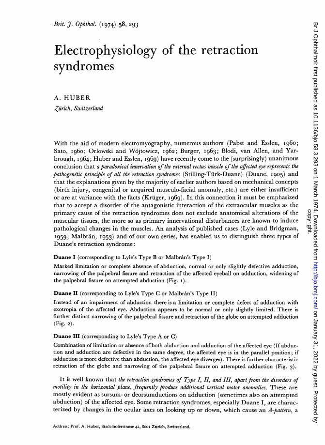
On attempted abduction of the affected eye, an insufficient activation of motor units inthe lateral rectus muscle leads to a progressive diminution of electrical discharge, sometimesinterrupted by short nystagmoid innervational outbursts of I0-20 msec. duration. Onadduction, however, the lateral rectus, which under normal conditions should be inhibited,manifests a distinct paradoxical electric activity progressing to a nearly normal interferencepattern. The lateral rectus muscle of the affected eye has its peak of innervation on adduction and itsminimum on attempted abduction. The medial rectus behaves normally, with a peak of inner-vation on adduction and inhibition on abduction. The clinically observed limitation ofabduction is explained by the insufficient or absent innervation of the lateral rectus onattempted abduction; the co-contraction produces the retraction of the globe (Fig. i).
r > / W f, > 0 0 ,<:3
:M~~~~~~~~~~~~~~~~I
FIG. i Duane syndrome LLimitation of abduction of left eye, normal adduction. Narrowingof globe on adduction.Electromyogram:Simultaneous recording of lateral rectus (upper curve) and medi:Paradoxical innervation of lateral rectus shows peak innervatiornervation on attempted abduction.Normal electrical behaviour of medial rectus: maximum innervatition on abduction.
Duane II
seu visec
of palpebral fissure and retraction
al rectus (lower curve) of left eye.n on adduction and defective in-
ion on adduction, complete inhibi-
In spite of impaired or absent adduction, we find a normal innervational pattern of the
A TT Icopyright.
on January 31, 2022 by guest. Protected by
http://bjo.bmj.com
/B
r J Ophthalm
ol: first published as 10.1136/bjo.58.3.293 on 1 March 1974. D
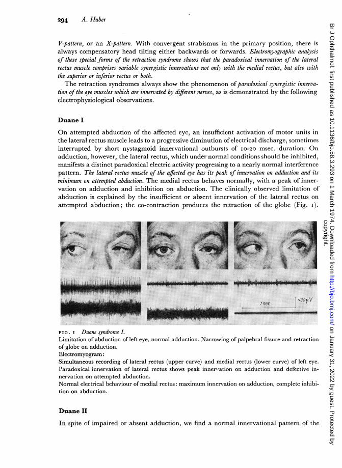
medial rectus of the affected eye, with full electrical activity on attempted adduction andnormal inhibition on abduction. The lateral rectus, however, has two peaks of innervation: oneon attempted adduction and the other on abduction. The limitation of adduction is not causedby a paresis of the medial rectus; its contraction is ineffective because it is counterbalancedby an equally strong activity of the lateral rectus on adduction. The resulting co-contractionof antagonistic muscles again leads to retraction of the globe. Duane II shows no limitationor absence of abduction (as is the case in Duane I), because on attempted abduction thelateral rectus of the affected eye activates a normal number of motor units (Fig. 2).
i'
I-};;'d. - ':":01 Fh :,
LUW PU*wW!WPSM~~~mq
isec
F I G. 2 Duane syndrome IH.Complete absence of adduction of left eye, normal abduction. Narrowing of palpebral fissure andretraction of globe on attempted adduction.Electromyogram:Simultaneous recording of lateral rectus (upper curve) and medial rectus (lower curve) of left eye.Lateral rectus shows peak innervation on abduction and a second paradoxical peak on attemptedadduction.Normal electrical behaviour of medial rectus: maximum innervation on attempted adduction andcomplete inhibition on abduction.
Duane III
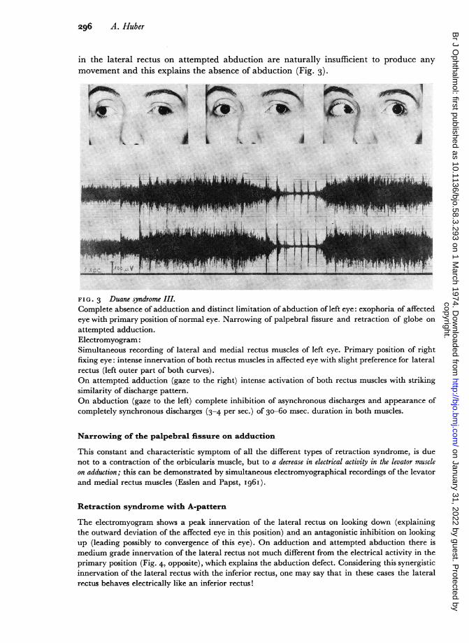
The phenomenon of paradoxical synergistic innervation of the lateral and medial rectusmuscles here reaches its climax. The usual agonist-antagonist relationship between the twomuscles is completely abolished, so that they behave like parts of one and the same muscle.In the primary position both manifest equal intense innervation.On attempted adduction, there is intense simultaneous activity of both muscles with a
striking similarity of the discharge pattern. We see again the phenomenon of paradoxicalco-contraction which produces retraction of the globe and makes adduction impossible.On attempted abduction, both muscles show complete inhibition of the asynchronous
discharges; instead they manifest completely synchronous discharges (3-4 per sec.) of30-60 msec. duration. These synchronous discharges in both the medial and lateral rectusmuscles appear so strikingly similar in every respect that the recording seems to come fromtwo electrodes placed close together on the same muscle. The short nystagmoid outbursts
Wl'IF. rwq
295
t ttIc
copyright. on January 31, 2022 by guest. P
rotected byhttp://bjo.bm
j.com/
Br J O
phthalmol: first published as 10.1136/bjo.58.3.293 on 1 M
in the lateral rectus on attempted abduction are naturally insufficient to produce anymovement and this explains the absence of abduction (Fig. 3).
_zK 1.1 L
a-M~ 4kao~~~~~~~~~~~~~~ =
FIG. 3 Duane syndrome III.Complete absence of adduction and distinct limitation of abduction of left eye: exophoria of affectedeye with primary position ofnormal eye. Narrowing of palpebral fissure and retraction of globe onattempted adduction.Electromyogram:Simultaneous recording of lateral and medial rectus muscles of left eye. Primary position of rightfixing eye: intense innervation of both rectus muscles in affected eye with slight preference for lateralrectus (left outer part of both curves).On attempted adduction (gaze to the right) intense activation of both rectus muscles with strikingsimilarity of discharge pattern.On abduction (gaze to the left) complete inhibition of asynchronous discharges and appearance ofcompletely synchronous discharges (3-4 per sec.) of 30-6o msec. duration in both muscles.
Narrowing of the palpebral fissure on adduction
This constant and characteristic symptom of all the different types of retraction syndrome, is duenot to a contraction of the orbicularis muscle, but to a decrease in electrical activity in the levator muscleon adduction; this can be demonstrated by simultaneous electromyographical recordings of the levatorand medial rectus muscles (Esslen and Papst, I96I).
Retraction syndrome with A-pattern
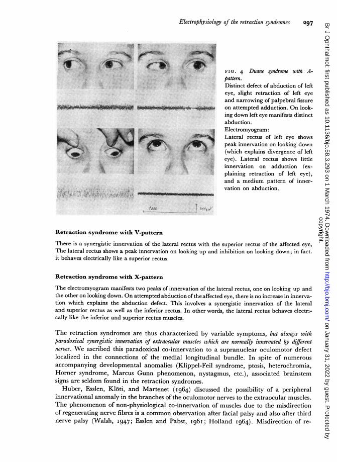
The electromyogram shows a peak innervation of the lateral rectus on looking down (explainingthe outward deviation of the affected eye in this position) and an antagonistic inhibition on lookingup (leading possibly to convergence of this eye). On adduction and attempted abduction there ismedium grade innervation of the lateral rectus not much different from the electrical activity in theprimary position (Fig. 4, opposite), which explains the abduction defect. Considering this synergisticinnervation of the lateral rectus with the inferior rectus, one may say that in these cases the lateralrectus behaves electrically like an inferior rectus!
copyright. on January 31, 2022 by guest. P
rotected byhttp://bjo.bm
j.com/
Br J O
phthalmol: first published as 10.1136/bjo.58.3.293 on 1 M
F IG . 4 Duane syndrome with A-pattern.Distinct defect of abduction of lefteye, slight retraction of left eyeand narrowing of palpebral fissureon attempted adduction. On look-ing down left eye manifests distinctabduction.Electromyogram:Lateral rectus of left eye showspeak innervation on looking down(which explains divergence of lefteye). Lateral rectus shows littleinnervation on adduction (ex-plaining retraction of left eye),and a medium pattern of inner-vation on abduction.
T p*
Retraction syndrome with V-pattern
There is a synergistic innervation of the lateral rectus with the superior rectus of the affected eye,The lateral rectus shows a peak innervation on looking up and inhibition on looking down; in fact.it behaves electrically like a superior rectus.
Retraction syndrome with X-pattern
The electromyogram manifests two peaks of innervation of the lateral rectus, one on looking up andthe other on looking down. On attempted abduction of the affected eye, there is no increase in innerva-tion which explains the abduction defect. This involves a synergistic innervation of the lateraland superior rectus as well as the inferior rectus. In other words, the lateral rectus behaves electri-cally like the inferior and superior rectus muscles.
The retraction syndromes are thus characterized by variable symptoms, but always withparadoxical synergistic innervation of extraocular muscles which are normally innervated by differentnerves. We ascribed this paradoxical co-innervation to a supranuclear oculomotor defectlocalized in the connections of the medial longitudinal bundle. In spite of numerousaccompanying developmental anomalies (Klippel-Feil syndrome, ptosis, heterochromia,Horner syndrome, Marcus Gunn phenomenon, nystagmus, etc.), associated brainstemsigns are seldom found in the retraction syndromes.
Huber, Esslen, Kloti, and Martenet (I964) discussed the possibility of a peripheralinnervational anomaly in the branches of the oculomotor nerves to the extraocular muscles.The phenomenon of non-physiological co-innervation of muscles due to the misdirectionof regenerating nerve fibres is a common observation after facial palsy and also after thirdnerve palsy (Walsh, I947; Esslen and Pabst, i96i; Holland I964). Misdirection of re-
-= z 8-W s :Ls us~~~~O40p owoo-
A4
copyright. on January 31, 2022 by guest. P
rotected byhttp://bjo.bm
j.com/
Br J O
phthalmol: first published as 10.1136/bjo.58.3.293 on 1 M
generating nerve fibres only occurs with a simultaneous lesion of the endoneurium: theregenerating axons cannot grow into the original endoneural tube, but reach foreignendoneural tubes which guide them to the wrong muscles. It is obvious that this type ofacquired misdirection is limited to fibres of the same nerve and is clinically significant onlyif it concerns nerves that operate a set of muscles with different and even antagonisticfunctions (as in the case of the oculomotor nerve). Moreover, an exchange of regeneratingfibres between two different nerves (in the case of the retraction syndrome the abducensand oculomotor nerve) has not been observed, and it is difficult to imagine how this couldhappen. Finally, the clinical picture of acquired misdirection of regenerating fibres inthe oculomotor nerve is very complex, variable, and widely different from the retractionsyndrome, although some retraction on upward or downward gaze has been observed.Simple acquired misdirection of regenerating nerve fibres cannot therefore explain theclinical signs and electrophysiological data in the retraction syndrome.We therefore began to consider the possibility of a misdirection of nerve branches in
embryo. The extrinsic muscles of the eye are developed by a condensation of the mesodermround the eye. At first (length of embryo 7 mm.) they form one mass which is supplied bythe third nerve only. Later (length of embryo 9 mm.), when the fourth and the sixth nervegrow towards the eye, this mass divides into separate muscles innervated by the differentoculomotor nerves. It is conceivable that, through disturbing influences of unknown origin,branches of the third nerve remain or come into contact with that part of the muscle masswhich is later to become the lateral rectus. Such an abnormal contact of third nervebranches with the lateral rectus might occur in the presence of a normal abducens nerveor in compensation for an aplastic or absent abducens nerve. When first considering sucha misdirection of third nerve branches due to embryological maldevelopment, we wereunaware of any neuroanatomical proof of our hypothesis. Hoyt and Nachtigaller (i 965a, b),going through the widely scattered literature of the anatomy of the orbit, succeededin finding about half a dozen observations of anomalous branches of the third nerveentering the lateral rectus, both when the abducens nerve was normal and when it wasaplastic or absent (Generali, I842; Fasebeck, i842; Tillack and Winer, i962; Heubner,I900; Bremer, 192I). Other anatomical studies also mention anastomoses between theoculomotor and abducens nerves within the cavernous sinus or the orbit. These werecasual observations in the anatomy theatre, unrelated to the clinical notion of a retractionsyndrome, but they show that the postulated congenital misdirection of branches of thethird nerve does exist.On the basis of these anatomical observations, the clinical and electromyographical
symptomatology of Duane III is the easiest to explain. The similar discharge patterns ofthe lateral and medial rectus muscles can be explained by the assumption that an anoma-lous branch of that part of the third nerve which innervates the medial rectus also suppliesthe lateral rectus, and that the latter receives no innervation from the abducens nervebecause it is weak or absent. The "substitute" innervation of the lateral rectus muscleby a branch of the third nerve when the abducens nerve was absent or aplastic has beenobserved in two cadavers (Generali, I842; Tillack and Winer, I962).
It is more difficult to apply this theory to Duane II, in which the lateral rectus has adual character (on adduction it behaves like the medial rectus innervated by elements ofthe oculomotor nerve, and on abduction like a lateral rectus innervated by the abducensnerve). Here one must assume a dual nerve supply in the sense that the single musclefibres have two end-plates, one innervated by an end-twig of the third nerve and theother by an end-twig of the sixth nerve. For this double innervation there exist well-
copyright. on January 31, 2022 by guest. P
rotected byhttp://bjo.bm
j.com/
Br J O
phthalmol: first published as 10.1136/bjo.58.3.293 on 1 M
documented anatomical findings of an anomalous branch from the third nerve to thelateral rectus with a normal abducens nerve (Fasebeck, I842; Henle, I879).
Duane I may be explained by a partial double innervation of the lateral rectus; onlysome of the fibres have a dual supply, and the quantitative relation between those suppliedby the sixth nerve only and those supplied by both the sixth and the third nerve can varyfrom case to case. This explains not only the varying degree of abduction defect, but alsothe occasional occurrence of an adduction defect.
In the retraction syndromes with A, V, and X patterns, one must assume that the additionalnerve supply to the lateral rectus, apart from the abducens nerve, derives either from thesuperior rectus division of the oculomotor nerve (V-pattern), from the inferior rectusdivision (A-pattern), or from both (X-pattern). Anomalous branches of the third nerveof this type have also been reported in anatomical specimens (Fasebeck, I842; Henle,I 879).
Differential diagnosisOther anomalies which have to be considered, especially for Duane I, are congenital absenceof the sixth nerve (Generali, I842; White, I935) and congenital agenesis of the lateral rectusmuscle.
In both instances the electromyogram is unable to register any electrical activity withinthe area of the lateral rectus muscle, whereas in the retraction syndrome one sees pro-nounced electrical activity in the lateral rectus on adduction.
Long-standing peripheral-neurogenic abducens nerve palsies with partial or total denervationand secondary muscle fibre degeneration within the lateral rectus may sometimes simulatea retraction syndrome. However, there is no narrowing of the palpebral fissure on adductionand only a slight degree of retraction of the globe (due to structural alterations ofthe pareticlateral rectus). The electromyogram reveals no paradoxical innervation of the lateralrectus on adduction, but there are typical signs of loss of motor units (with loss of theinterference pattern on abduction) and symptoms of denervation (fibrillation potentials).
ConclusionThe main pathogenetic principle of the retraction syndromes is a paradoxical anomalousinnervation of the lateral rectus muscle of the affected eye, a synergistic innervation ofthe extraocular muscles which are normally innervated by different nerves. In correlatingthe electromyographic data with anatomical reports, we have been able to provide alogical interpretation of the different retraction syndromes, based on the concept of a mis-direction of peripheral branches of the third nerve to the lateral rectus muscle in embryo.Conclusive proof ofsuch misdirection by necropsy studies of cases of the retraction syndromeshas still to be obtained. Two patients with Duane's syndrome (Phillips, Dirion, and Graves,I932; Matteucci, I946) showed aplasia or absence of the abducens nerve, but the peri-pheral branches of the oculomotor nerves in these cases were not examined.The causes of these defects of-embryogenesis are not yet known, but the incidence of
the retraction syndromes in children whose mothers were exposed to thalidomide poisoningduring the first months of pregnancy is significant.
It is also of interest that, in cases of the Marcus Gunn jaw-winking phenomenon, electro-myograms from the levator and masseter muscles also show simultaneous electrical activity(Pabst and Rossmann, I966). The levator thus appears to be innervated from both thethird and the fifth nerve, but where this abnormal connection between the two nervesis located or how the misdirection of the fifth nerve occurs, is still a matter of speculation.
299copyright.
on January 31, 2022 by guest. Protected by
http://bjo.bmj.com
/B
r J Ophthalm
ol: first published as 10.1136/bjo.58.3.293 on 1 March 1974. D
As in the retraction syndromes, electromyography furnishes important data for the hypo-thesis of misdirection of nerves during embryogenesis.
Summary
Three types of retraction syndromes (Duane, I905) can be distinguished: Duane I withdefective abduction, Duane II with defective adduction, and Duane III with defectiveadduction and abduction. All show more or less pronounced narrowing of the palpebralfissure and retraction of the globe on adduction. Some patients also manifest A, V, orX patterns.
Electromyography reveals that a paradoxical anomalous innervation of the lateral rectusmuscle is the underlying cause. This paradoxical innervation may be due to an anomalouscontact of the lateral rectus with branches of the third nerve if the abducens nerve is absentor defective, or to a double innervation by the abducens nerve and anomalous branches ofthe oculomotor nerve. The electromyographic findings correspond with anatomical-bservations of abnormal innervations of this kind.
Discussion
HART How is it that the Marcus Gunn phenomenon improves with time?
HUBER It is known that the Marcus Gunn phenomenon can disappear with time. There is nodoubt that in our case we have been able to show a direct connection of co-contraction between thelevator and masseter muscles. Exactly where the connection between these two is taking place isnot certain.LYLE Have any electrophysiological investigations been carried out on cases of Brown's ('superioroblique tendon sheath') syndrome?HUBER Paradoxical innervation occurs in Brown's syndrome and this has been proved electro-myographically.STRACHAN If the muscle was primarily innervated through the spindle, and if the muscle wasprimarily fibrotic, then the electromyographical changes could be explained in this manner. I donot really agree with Dr. Huber's concept. I have found frequency changes in the electromyographpointing to a dispersion of fibres in motor units, perhaps by fibrous tissue. My idea of the situation israther that the muscle changes occur first and that all the electrophysiological changes are secondary.HUBER Retraction changes occur in internuclear ophthalmoplegia. About a year ago we foundthat some co-contraction of the lateral rectus occurs but not to the same extent or with the sameintensity as in the retraction syndromes. I am almost certain that the intense co-contraction of thelateral rectus has a great deal to do with the retraction. It is also the reason why the eye cannot movein adduction in Duane Type II. I agree that certain things are not explained, but I am still certainthat this is an innervational disorder.MAURER In view of the significance of Hering and Sherrington's Laws, and since some authoritiesclaim that the anomalous innervation is confined to the affected eye, has anyone done any studiesin Duane's syndrome, fixing the sound eye and studying the affected eye simultaneously? If so, whatwere the results?HUBER If there is movement there is innervation, and if there is no movement there is no in-nervation. Duane's syndrome is therefore not a disturbance of outflow from the higher centres, buta disturbance of the motor neurones of the extraocular muscles.ABRAHAMS and WATSON Has Dr. Huber found any cases in which there were no electromyo-graphical changes in Duane's syndrome?HUBER These changes are found universally, and without exception.
copyright. on January 31, 2022 by guest. P
rotected byhttp://bjo.bm
j.com/
Br J O
phthalmol: first published as 10.1136/bjo.58.3.293 on 1 M