NEW FOSSIL PORTUNIDS FROM WASHINGTON, USA, AND ARGENTINA, AND A RE-EVALUATION OF GENERIC AND FAMILY RELATIONSHIPS
WITHIN THE PORTUNOIDEA RAFINESQUE, 1815 (DECAPODA: BRACHYURA)
CARRIE E. SCHWEITZER AND RODNEY M. FELDMANN Department of Geology, Kent State University, Kent, Ohio 44242, <[email protected]>, <[email protected]>
ABSTRACT—New portunoid fossils from southern Argentina and from the west coast of North America permit the reevaluation of the generic and family relationships within the Portunoidea Rafinesque, 1815. It has previously been suggested that the Portunidae and the Geryonidae Colosi, 1923, are closely related families (Manning and Holthuis, 1989). The new fossils suggest that the Geryonidae may in fact be derived from a portunid progenitor, Proterocarcinus Feldmann, Casadio, Chirino-Galvez, and Aguirre Urreta, 1995, through a process of peramorphosis in which juveniles of the geryonid species Chaceon peruvianas (d'Orbigny, 1842) resemble adults of Proterocarcinus latus (Glaessner, 1933). Examination of several genera within the portunid subfamily Polybiinae Ortmann, 1893, including Imaizumila Karasawa, 1993; Megokkos new genus; Minohellenus Karasawa, 1990; Pororaria Glaessner, 1980; Portunites Bell, 1858; and Proterocarcinus, suggests that the subfamily had an amphitropical distribution early in its history. New taxa reported here include Megokkos new genus and Portunites nodosus new species. New combinations include Chaceon peruvianus (d'Orbigny, 1842), Imaizumila araucana (Philippi, 1887), Megokkos alaskensis (Rathbun, 1926), Megokkos hexagonalis (Nagao, 1932), Megokkos macrospinus (Schweitzer, Feldmann, Tucker, and Berglund, 2000), Minohellenus triangulum (Rathbun, 1926), and Proterocarcinus latus (Glaessner, 1933).
INTRODUCTION
NEW PORTUNOID fossils from the west coast of North America and southern Argentina permit the re-evaluation of generic
and family relationships within the Portunoidea Rafinesque, embracing the families Portunidae Rafinesque, 1815, Geryonidae Colosi, 1923, and Carcineretidae Beurlen, 1930. Based upon larval studies, Rice (1980) suggested that the Polybiinae Ortmann, 1893 was most likely the ancestral subfamily within the Portunidae. The earliest known authentic portunid is in fact a member of the Polybiinae and is known from Danian rocks of Argentina; Proterocarcinus lophos Feldmann, Casadio, Chirino-Galvez, and Aguirre Urreta, 1995 was described from the Danian Roca Formation (Feldmann et al., 1995). Two species of this genus are now known with the referral herein of Archaeogeryon latus Glaessner, 1933, to that genus, suggesting that the Portunidae, subfamily Polybiinae, arose in the southern hemisphere as early as the early Paleocene. Feldmann et al. (1995) did not assign Proterocarcinus to a subfamily; however, the new specimens of P. latus possess paddle-like propodi and dactyli of the fifth pe-reiopods (Fig. 8.2), which were not known either in the types of that species or in P. lophos. This makes it possible to assign the genus to the Polybiinae. The oldest known member of the Geryonidae, Chaceon peruvianus (d'Orbigny, 1842), also occurs in southern Argentina, in middle Eocene rocks of the Centinela Formation. This species was previously thought to be Miocene in age, but recent work indicates a middle Eocene age for these rocks (Casadio, Feldmann and Poland, in press). Therefore, it seems probable that the Geryonidae also evolved in the southern hemisphere.
Manning and Holthuis (1989) demonstrated that the Geryonidae and Portunidae were closely related based upon external morphology, and their position is supported by serological and larval studies (Leone, 1949, 1951; Ingle, 1979; Rice, 1980). Recently collected fossils from southern Argentina indicate that the Geryonidae, specifically Chaceon Manning and Holthuis, 1989, may in fact be derived from the Portunidae and that the possible progenitor is Proterocarcinus within the Portunidae. Compelling
evidence for evolution via peramorphosis within the genera Proterocarcinus and Chaceon is provided by a suite of newly collected, well-preserved specimens of Proterocarcinus latus and C. peruvianus from southern Argentina, in which juvenile individuals of C. peruvianus exhibit characters of adult Proterocarcinus latus found at the same localities.
Furthermore, there is strong morphological similarity among Proterocarcinus, known only from Argentina, and four portunid genera, Imaizumila Karasawa, 1993, Megokkos n. gen., Minohellenus, Karasawa, 1990, and Portunites Bell, 1858, known from middle to high northern and southern latitudes. All of these taxa are referrable to the Polybiinae Ortmann, which suggests that the subfamily, and thus the family, had an amphitropical distribution early in its history.
Re-examination of all species assigned to the portunid genus Portunites indicates that the genus may be rearranged into three genera, Portunites sensu stricto, Megokkos n. gen., and Minohellenus. The generic diagnosis of Portunites s.s. is therefore restricted accordingly. The two genera, Megokkos n. gen. and Minohellenus, both exhibit a North Pacific distribution pattern, while Portunites is recognized from both the North Pacific and North Atlantic basins. The new genus, Megokkos, and a new species, Portunites nodosus, resulted from the reevaluation of fossil material previously assigned to Portunites and examination of new material from Washington, USA. The extinct genus Imaizumila Karasawa now appears to have had an amphitropical distribution based upon the reassignment of Portunites araucana (Philippi, 1887) from Chile to that genus; Imaizumila was previously known only from Japan (Karasawa, 1993).
The Carcineretidae Beurlen, 1930, is restricted to the Cretaceous and is distributed in localities that were likely tropical to subtropical (Fraaye, 1996; Vega et al., 1997). The new fossils suggest a southern hemisphere origin for both the Portunidae and the Geryonidae. An amphitropical distribution of the Portunidae is documented early in its history. The occurrence of the Carcineretidae in primarily tropical latitudes suggests that the Portunoidea may have arisen in the tropics and subsequently dispersed from those locales to temperate regions in the southern hemisphere.
Discussion.—Members of the Polybiinae are diagnosed by possession of a narrow carapace, three to five anterolateral spines, possession of some walking legs as long as the first pe-reiopods, and paddle-like fifth pereiopods (Glaessner, 1969). The four genera Portunites, Imaizumila, Megokkos n. gen., and Mi-nohellenus are placed within the Polybiinae. Placement of Portunites sensu stricto within a subfamily is difficult, because none of the included species has preserved fifth pereiopods. Glaessner (1969) and Tucker and Feldmann (1990) placed Portunites within the Carcininae Macleay, 1838, based upon carapace shape and the number of anterolateral spines. Schweitzer and Feldmann (1999) and Schweitzer et al. (2000) placed Portunites within the Polybiinae based on their judgement that Portunites alaskensis Rathbun, 1926, which exhibits paddle-like dactyls on the fifth pereiopods, was an authentic member of the genus. Portunites alaskensis is removed to Megokkos n. gen. to be discussed below and another species of Megokkos, Megokkos ma-crospinus (Schweitzer, Feldmann, Tucker, and Berglund, 2000) also exhibits paddle-like dactyls of the fifth pereiopod. Minoh-ellenus, another portunid genus to be discussed below, exhibits paddle-like dactyls on the fifth pereiopods. Imaizumila, with the referral of Portunites araucanus to that genus, is also placed within the Polybiinae based upon possession of a paddle-like fifth pereiopod. Philippi (1887) reported that the fifth pereiopod of this species was lanceolate in shape; however, examination of photographs of specimen SGO.PI-012.860, housed at the Mu-seo Nacional de Historia Natural, Seccion Paleontologia, Santiago, Chile, indicates that the fifth pereiopod has a paddle-like dactyl, permitting placement within the Polybiinae. The morphological similarity between Portunites, Megokkos n. gen., Mi-nohellenus, and Imaizumila makes it likely that these four genera are especially closely related. Therefore, it is possible to refer Portunites to the Polybiinae, at least until evidence to the contrary is discovered. Proterocarcinus is here referred to the Polybiinae because it possesses paddle-like propodi and dactyli on the fifth pereiopods.
Genus PORTUNITES sensu stricto Bell, 1858 Type species.—Portunites incerta Bell, 1858, by original des
ignation.
Included species.—Portunites eocenica Ldrenthey in L6ren-they and Beurlen, 1929 (type is lost, Quayle and Collins, 1981); P. incerta Bell, 1858; P. insculpta Rathbun, 1926; P. kattachien-sis Karasawa, 1992; P. nodosus new species; P. stintoni Quayle, 1984; P. sylviae Quayle and Collins, 1981.
Diagnosis.—Carapace wider than long, LAV about 0.61-0.76, usually widest at position of last anterolateral spine; ovate-hexagonal in shape; carapace regions inflated, well-defined, delimited by broad grooves. Front not projected beyond orbits, with six lobes or small, blunt spines including the inner orbital spines. Orbits circular, two supra-orbital fissures, fronto-orbital width ranging from 50 to 71 percent maximum carapace width. Anterolateral margin convex, five spines including outer-orbital spine, second spine smallest and sometimes missing, last spine usually longest, outer-orbital spine directed forward, other anterolateral spines directed anterolaterally. Epibranchial region arcuate, extending axially from fifth anterolateral spine, terminating in broadly swollen tubercle. Branchial region with longitudinal ridge parallel to, and on either side of, the longitudinal axis of the cardiac region, ridge usually with tubercles at either end.
Discussion.—Numerous taxa have been assigned to Portunites sensu lato since the genus was erected by Bell (1858) to accommodate Eocene fossils from Britain. Taxa assigned to Portunites have typically possessed an arcuate epibranchial ridge and four to five spines on the anterolateral margin. The genus is herein restricted to those species possessing four blunt frontal spines, a fronto-orbital width to total width ratio of 0.50-0.71; four to five anterolateral spines, arcuate epigastric ridges, longitudinal ridges on the branchial regions, and well-defined carapace regions. Schweitzer and Feldmann (1999) provided an overview of the history of the genus and suggested that P. alaskensis Rathbun and P. hexagonalis Nagao might be better placed in a separate genus. Those two species along with P. macros-pinus Schweitzer et al. (2000) have been referred to Megokkos (as discussed below). Subsequently, Schweitzer et al. (2000) suggested that P. triangulum be removed from the genus, and that species is herein placed within Minohellenus.
Schweitzer and Feldmann (1999) also removed P. subovata Quayle and Collins, 1981 from Portunites based upon its lack of a diagnostic epibranchial ridge. Examination of the holotype of P. subovata (In.61715) suggests that it is most likely a member of the Cheiragonidae Ortmann, 1893 and might possibly be referrable to the genus Montezumella Rathbun, 1930b. The species has a granular carapace that is almost as long as wide, and the overall shape of the carapace regions is similar to Montezumella spp. In fact, Quayle and Collins (1981) reported a species of Montezumella, M. scabra Quayle and Collins, 1981, from the same locality as P. subovata, suggesting that the two species may be synonymous.
The diagnostic characters for Portunites are based upon the seven species herein assigned to the genus; all display these characters where preserved. Portunites insculpta and P. kattachiensis are especially similar to one another and differ from the other species of Portunites in several regards. The anterolateral spines of these two species are larger and better developed than are those of other species of Portunites. The epibranchial ridge is less distinct on P. insculpta and P. kattachiensis than on the other species, and the fronto-orbital width to total width ratio for those two species averages about 50 percent. In the other
638 JOURNAL OF PALEONTOLOGY, V. 74, NO. 4, 2000
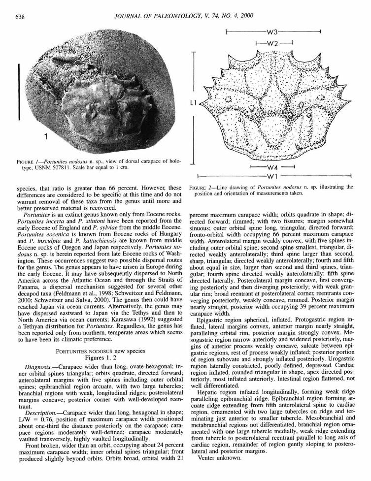
FIGURE 1—Portunites nodosus n. sp., view of dorsal carapace of holo-type, USNM 507811. Scale bar equal to 1 cm.
species, that ratio is greater than 66 percent. However, these differences are considered to be specific at this time and do not warrant removal of these taxa from the genus until more and better preserved material is recovered.
Portunites is an extinct genus known only from Eocene rocks. Portunites incerta and P. stintoni have been reported from the early Eocene of England and P. sylviae from the middle Eocene. Portunites eocenica is known from Eocene rocks of Hungary and P. insculpta and P. kattachiensis are known from middle Eocene rocks of Oregon and Japan respectively. Portunites nodosus n. sp. is herein reported from late Eocene rocks of Washington. These occurrences suggest two possible dispersal routes for the genus. The genus appears to have arisen in Europe during the early Eocene. It may have subsequently dispersed to North America across the Atlantic Ocean and through the Straits of Panama, a dispersal mechanism suggested for several other decapod taxa (Feldmann et al., 1998; Schweitzer and Feldmann, 2000; Schweitzer and Salva, 2000). The genus then could have reached Japan via ocean currents. Alternatively, the genus may have dispersed eastward to Japan via the Tethys and then to North America via ocean currents; Karasawa (1992) suggested a Tethyan distribution for Portunites. Regardless, the genus has been reported only from northern, temperate areas which seems to have been its climatic preference.
PORTUNITES NODOSUS new species Figures 1, 2
Diagnosis.—Carapace wider than long, ovate-hexagonal; inner orbital spines triangular; orbits quadrate, directed forward; anterolateral margins with five spines including outer orbital spines; epibranchial region arcuate, with two large tubercles; branchial regions with weak, longitudinal ridges; posterolateral margins concave; posterior comer with well-developed reentrant.
Description.—Carapace wider than long, hexagonal in shape; LAV = 0.76, position of maximum carapace width positioned about one-third the distance posteriorly on the carapace; carapace regions moderately well-defined; carapace moderately vaulted transversely, highly vaulted longitudinally.
Front broken, wider than an orbit, occupying about 24 percent maximum carapace width; inner orbital spines triangular; front produced slightly beyond orbits. Orbits broad, orbital width 21
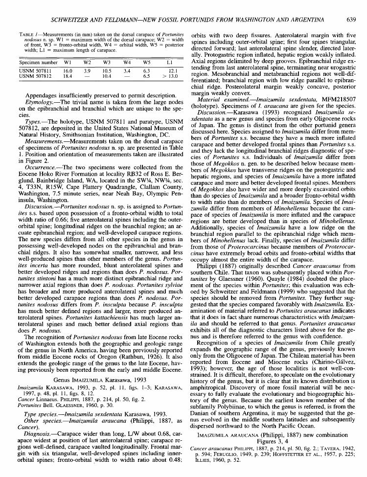
FIGURE 2—Line drawing of Portunites nodosus n. sp. illustrating the position and orientation of measurements taken.
percent maximum carapace width; orbits quadrate in shape; directed forward; rimmed; with two fissures; margin somewhat sinuous; outer orbital spine long, triangular, directed forward; fronto-orbital width occupying 66 percent maximum carapace width. Anterolateral margin weakly convex; with five spines including outer orbital spine; second spine smallest, triangular, directed weakly anterolaterally; third spine larger than second, sharp, triangular, directed weakly anterolaterally; fourth and fifth about equal in size, larger than second and third spines, triangular; fourth spine directed weakly anterolaterally; fifth spine directed laterally. Posterolateral margin concave, first converging posteriorly and then diverging posteriorly; with weak granular rim; broad reentrant at posterolateral comer, reentrants converging posteriorly, weakly concave, rinmied. Posterior margin nearly straight, posterior width occupying 39 percent maximum carapace width.
Epigastric region spherical, inflated. Protogastric region inflated, lateral margins convex, anterior margin nearly straight, paralleling orbital rim, posterior margin strongly convex. Me-sogastric region narrow anteriorly and widened posteriorly, margins of anterior process weakly concave, sulcate between epigastric regions, rest of process weakly inflated; posterior portion of region subovate and strongly inflated posteriorly. Urogastric region laterally constricted, poorly defined, depressed. Cardiac region inflated, rounded triangular in shape, apex directed posteriorly, most inflated anteriorly. Intestinal region flattened, not well differentiated.
Hepatic region inflated longitudinally, forming weak ridge paralleling epibranchial ridge. Epibranchial region forming arcuate ridge extending from fifth anterolateral spine to cardiac region, ornamented with two large tubercles on ridge and terminating just anterior to smaller tubercle. Mesobranchial and metabranchial regions not differentiated, branchial region ornamented with one large tubercle medially, weak ridge extending from tubercle to posterolateral reentrant parallel to long axis of cardiac region, remainder of region gently sloping to posterolateral and posterior margins.
Venter unknown.
SCHWEITZER AND FELDMANN—NEW FOSSIL PORTUNIDS FROM WASHINGTON AND ARGENTINA 639
TABLE 1—Measurements (in mm) taken on the dorsal carapace of Portunites nodosus n. sp. Wl = maximum width of the dorsal carapace; W2 = width of front, W3 = fronto-orbital width, W4 = orbital width, W5 = posterior width; LI = maximum length of carapace.
Specimen number Wl W2 W3 W4 W5 LI
USNM 507811 USNM 507812
16.0 18.4
3.9 10.5 10.4
3.4 6.3 6.5
12.1 > 13.0
Appendages insufficiently preserved to permit description. Etymology.—The trivial name is taken from the large nodes
on the epibranchial and branchial which are unique to the species.
Types.—The holotype, USNM 507811 and paratype, USNM 507812, are deposited in the United States National Museum of Natural History, Smithsonian Institution, Washington, DC.
Measurements.—Measurements taken on the dorsal carapace of specimens of Portunites nodosus n. sp. are presented in Table 1. Position and orientation of measurements taken are illustrated in Figure 2.
Occurrence.—The two specimens were collected from the Eocene Hoko River Formation at locality RB32 of Ross E. Ber-glund, Bainbridge Island, WA, located in the SW^^, NW /̂4, sec. 4, T33N, R15W, Cape Flattery Quadrangle, Clallam County, Washington, 7.5 minute series, near Neah Bay, Olympic Peninsula, Washington.
Discussion.—Portunites nodosus n. sp. is assigned to Portunites s.s. based upon possession of a fronto-orbital width to total width ratio of 0.66; five anterolateral spines including the outer-orbital spine; longitudinal ridges on the branchial region; an arcuate epibranchial region; and well-developed carapace regions. The new species differs from all other species in the genus in possessing well-developed nodes on the epibranchial and branchial ridges. It also has somewhat smaller, narrower, and less well-produced spines than other members of the genus. Portunites incerta has more rounded, blunt anterolateral spines and better developed ridges and regions than does P. nodosus. Portunites stintoni has a much more distinct epibranchial ridge and narrower axial regions than does P. nodosus. Portunites sylviae has broader and more produced anterolateral spines and much better developed carapace regions than does P. nodosus. Portunites nodosus differs from P. insculpta because P. insculpta has much better defined regions and larger, more produced anterolateral spines. Portunites kattachiensis has much larger anterolateral spines and much better defined axial regions than does P. nodosus.
The recognition of Portunites nodosus from late Eocene rocks of Washington extends both the geographic and geologic range of the genus in North America, having been previously reported from middle Eocene rocks of Oregon (Rathbun, 1926). It also extends the geologic range of the genus to the late Eocene, having previously been reported from the early and middle Eocene.
Genus IMAIZUMILA Karasawa, 1993
Imaizumila KARASAWA, 1993, p. 52, pi. 11, figs. 1-3; KARASAWA, 1997, p. 48, pi. 11, figs. 8, 12.
Cancer Linnaeus. PHILIPPI, 1887, p. 214, pi. 50, fig. 2. Portunites Bell. GLAESSNER, 1960, p. 30.
Type species.—Imaizumila sexdentata Karasawa, 1993. Other species.—Imaizumila araucana (Philippi, 1887, as
Cancer). Diagnosis.—Carapace wider than long, LAV about 0.68, car
apace widest at position of last anterolateral spine; carapace regions well-defined, carapace vaulted longitudinally. Frontal margin with six triangular, well-developed spines including inner-orbital spines; fronto-orbital width to width ratio about 0.48;
orbits with two deep fissures. Anterolateral margin with five spines including outer-orbital spine; first four spines triangular, directed forward; last anterolateral spine slender, directed laterally. Protogastric region inflated, hepatic region weakly inflated. Axial regions delimited by deep grooves. Epibranchial ridge extending from last anterolateral spine, terminating near urogastric region. Mesobranchial and metabranchial regions not well-differentiated; branchial region with low ridge parallel to epibranchial ridge. Posterolateral margin weakly concave, posterior margin weakly convex.
Material examined.—Imaizumila sexdentata, MFM218507 (holotype). Specimens of /. araucana are given for the species.
Discussion.—Karasawa (1993) recognized Imaizumila sexdentata as a new genus and species from early Oligocene rocks of Japan. The genus is distinct from the other portunid genera discussed here. Species assigned to Imaizumila differ from members of Portunites s.s. because they have a much more inflated carapace and better developed frontal spines than Portunites s.s. and they lack the longitudinal branchial ridges diagnostic of species of Portunites s.s. Individuals of Imaizumila differ from those of Megokkos n. gen. to be described below because members of Megokkos have transverse ridges on the protogastric and hepatic regions, and species of Imaizumila have a more inflated carapace and more and better developed frontal spines. Members of Megokkos also have wider and more deeply excavated orbits than do species of Imaizumila and a broader fronto-orbital width to width ratio than do members of Imaizumila. Species of Imaizumila differ from members of Minohellenus because the carapace of species of Imaizumila is more inflated and the carapace regions are better developed than in species of Minohellenus. Additionally, species of Imaizumila have a low ridge on the branchial region parallel to the epibranchial ridge which members of Minohellenus lack. Finally, species of Imaizumila differ from those of Proterocarcinus because members of Proterocar-cinus have extremely broad orbits and fronto-orbital widths that occupy almost the entire width of the carapace.
Philippi (1887) originally described Cancer araucanus from southern Chile. That taxon was subsequently placed within Portunites by Glaessner (1960). Quayle (1984) doubted the placement of the species within Portunites; this evaluation was echoed by Schweitzer and Feldmann (1999) who suggested that the species should be removed from Portunites. They further suggested that the species compared favorably with Imaizumila. Examination of material referred to Portunites araucanus indicates that it does in fact share numerous characteristics with Imaizumila and should be referred to that genus. Portunites araucanus exhibits all of the diagnostic characters listed above for the genus and is therefore referred to the genus with confidence.
Recognition of a species of Imaizumila from Chile greatly expands the geographic range of the genus, previously known only from the Oligocene of Japan. The Chilean material has been reported from Eocene and Miocene rocks (Chirino-Galvez, 1993); however, the age of those localities is not well-constrained. It is difficult, therefore, to speculate on the evolutionary history of the genus, but it is clear that its known distribution is amphitropical. Discovery of more fossil material will be necessary to fully evaluate the evolutionary and biogeographic history of the genus. Because the earliest known member of the subfamily Polybiinae, to which the genus is referred, is from the Danian of southern Argentina, it may be suggested that the genus evolved in the middle southern latitudes and subsequently dispersed northward to the North Pacific Ocean.
IMAIZUMILA ARAUCANA (Philippi, 1887) new combination Figures 3, 4
Cancer araucanus PHILIPPI, 1887, p. 214, pi. 50, fig. 2.; TAVERA, 1942, p. 594; FERUGLIO, 1949, p. 239; HOFFSTETTER ET AL., 1957, p. 225; ILLIES, 1960, p. 52.
640 JOURNAL OF PALEONTOLOGY, V. 74, NO. 4, 2000
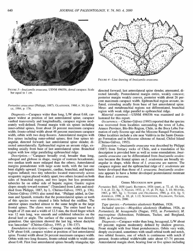
FIGURE 3—Imaizumila araucana, USNM 496556, dorsal carapace. Scale bar equal to 1 cm.
Portunites araucanus (Philippi, LE, 1984, p. 176.
1887). GLAESSNER, 1960, p. 30; QUAY-
Diagnosis.—Carapace wider than long, LAV about 0.68, carapace widest at position of last anterolateral spine; carapace vaulted transversely and longitudinally, carapace regions moderately well-defined. Frontal margin with six spines including inner-orbital spines, front about 24 percent maximum carapace width; fronto-orbital width about 48 percent maximum carapace width, orbits with two deep fissures. Anterolateral margins with five spines including outer-orbital spines; first four spines triangular, directed forward; last anterolateral spine slender, directed anterolaterally. Epibranchial region an arcuate ridge, extending axially from base of last anterolateral spine. Branchial region with low ridge paralleling epibranchial ridge.
Description,—"Carapace broadly oval, broader than long, subequal and globose in shape, margin of rostrum hexadentate, two median teeth more enlarged than the others; Anterolateral margins pentadentate with large acute teeth, with the last one bent slightly upward. Urogastric, cardiac, branchial, and hepatic regions inflated; two tiny tubercles located transversely across urogastric region placed widely apart; two others located on both sides of branchial region, and two larger, but less prominent tubercles lie on either side of hepatic region; Gastric region slopes steeply toward rostrum" (Translated from Latin and modified from Philippi, 1887, by L. Chirino-Galvez, 1993, p. 136). Chirino-Galvez (1993) also noted that, "Philippi added, in German and Spanish, remarks that the posterolateral spines or teeth of this species were situated a little behind the midline. The anterior spines reached almost to the same height as the large frontal spines. The claws were strong. The entire hand has a length of 25 nrni and a width of 12.5 mm, the fixed finger which was 12 mm long, was smooth and exhibited tubercles on the dorsal keel or angle. The surface of the carapace was densely granulose. One specimen showed the last leg, whose dactylus was lanceolate" (Chirino-Galvez, 1993, p. 136-137).
Emendation to description.—Carapace ovate, wider than long, LAV about 0.68, carapace widest at position of last anterolateral spine. Frontal width about 24 percent maximum carapace width. Orbits with two long fissures, fronto-orbital width to width ratio about 0.48. First four anterolateral spines broadly triangular, tips
FIGURE 4—Line drawing of Imaizumila araucana.
directed forward; last anterolateral spine slender, attenuated, directed laterally. Posterolateral margin entire, weakly concave; posterior margin weakly convex, posterior width about 26 percent maximum carapace width. Epibranchial region arcuate, inflated, extending axially from base of last anterolateral spine. Meso- and metabranchial regions not differentiated, branchial region with weak ridge parallel to epibranchial ridge.
Material examined.—USNM 496556 was examined and illustrated for this report.
Occurrence.—Chirino-Galvez (1993) reported that the species was recovered from localities surrounding the town of Lebu, Arauco Province, Bio-Bio Region, Chile, in the Boca Lebu Formation of early Eocene age and the Miocene Ranquil Formation. Other localities include a site near Valdivia in the Santo Domingo Formation and in Miocene siltstone of Ancud, Chiloe Island (Chirino-Galvez, 1993).
Discussion.—Imaizumila araucana was described by Philippi (1887) from Tertiary rocks of Chile, and a translation of his description is provided here as well as some emendations. Imaizumila araucana can be differentiated from Imaizumila sexden-tata because the frontal spines on /. sexdentata are broadly triangular in shape, while those of /. araucana are narrow. The carapace regions and the epibranchial ridge of /. sexdentata are better developed than those of /. araucana. Imaizumila sexdentata appears to have a better developed posterolateral reentrant than does /. araucana.
Genus MEGOKKOS new genus Portunites Bell, 1858 (part). RATHBUN, 1926 (part), p. 72, pi. 18, figs.
3-4, pi. 22, fig. 3; NAGAO, 1932, p. 15, pi. IV, figs. 3, 14; MUSTOE, 1983, p. 987, fig. 1; TUCKER AND FELDMANN, 1990, p. 414, fig. 5; ANSFIELD, 1998, p. 10, pi. 1.12; SCHWEITZER, FELDMANN, TUCKER, AND BERGLUND, 2000.
Type species.—Portunites alaskensis Rathbun, 1926. Included species.—Megokkos alaskensis (Rathbun, 1926, as
Portunites); M. hexagonalis (Nagao, 1932, as Portunites); M. macrospinus (Schweitzer, Feldmann, Tucker, and Berglund, 2000, as Portunites).
Diagnosis.—Carapace wider than long, hexagonal, LAV about 65 percent; regions moderately defined by shallow grooves. Front straight with four blunt protuberences. Orbits very wide, deeply excavated, sometimes with small orbital tooth and notch, two orbital fissures, one of which is continuous with notch if present; fronto-orbital width/width ratio about 67-74 percent. Anterolateral margin short, bearing four or five spines including
SCHWEITZER AND FELDMANN—NEW FOSSIL PORTUNIDS FROM WASHINGTON AND ARGENTINA 641
outer-orbital spine, last spine usually longest. Protogastric and hepatic regions with transverse ridges. Epibranchial region arcuate, extending axially from last anterolateral spine. Branchial regions weakly inflated. Posterolateral reentrant wide, well-developed. Chelae heterochelous, stout, fingers with large blunt denticles on occlusal surface. Dactyl of fifth pereipod paddlelike.
Etymology.—Greek roots mega, "large," and okkos, "eye," refer to the broad and deeply excavated orbits of members of the genus. The gender is masculine.
Material examined.—Megokkos alaskensis, USNM 354169 (paratype), 431258-62. M. hexagonalis, UHR 4456 (holotype), Department of Geology and Mineralogy, Hokkaido University, Sapporo, Japan. Specimens of M. macrospinus are given for the species.
Discussion.—Rathbun (1926) orginally assigned Portunites alaskensis to Portunites based upon the development of carapace regions and the epibranchial ridge. Nagao (1932) described P. hexagonalis as being most similar to P. alaskensis, and examination of type material by the authors indicates that these two species are indeed congeneric because they both possess broad, deeply excavated orbits; broad fronto-orbital widths; short anterolateral margins; and ridges on the protogastric, hepatic, and epibranchial regions. Schweitzer and Feldmann (1999) suggested that these two species may belong to a genus other than Portunites based upon their large and deeply excavated orbits. Schweitzer et al. (2000) subsequently named P. macrospinus from Eocene rocks of Washington and argued that P. alaskensis, P. hexagonalis, and P. macrospinus should remain within Portunites.
Re-examination of these three species indicates that they differ from Portunites s.s in several, regards and are therefore re-ferrable to a new genus, Megokkos. Members of Portunites s.s. have wide fronto-orbital widths as do members of Megokkos; however, the orbits of the three species referred to Megokkos are much more deeply excavated. The front of members of Megokkos possesses blunt projections, while in Portunites s.s., the front has spines. Some members of Megokkos possess an orbital tooth and notch, which are never seen in species of Portunites s.s. Species of Portunites s.s. typically lack transverse ridges on the hepatic and protogastric regions which members of Megokkos possess; and species of Portunites s.s. possess longitudinal ridges on the branchial regions which members of Megokkos lack. The anterolateral spines are sharper and more attenuated in species of Megokkos than in Portunites s.s. Members of Megokkos have a markedly hexagonal carapace, while that of species of Portunites s.s. is more ovate. Species of Megokkos have well-developed posterolateral reentrants while in Portunites s.s. they are shallow or lacking. Species of Portunites s.s. typically have tubercles on the branchial regions which members of Megokkos lack. Finally, the carapace regions are less inflated and the grooves are less distinct in species of Megokkos than of Portunites s.s.
Species of Megokkos can be differentiated from Imaizumila and Minohellenus because Megokkos has much broader, more deeply excavated orbits than those two genera and possesses transverse ridges on the hepatic regions which members of Imaizumila and Minohellenus lack. Members of Proterocarcinus have much broader orbits than do species of Megokkos and also have a much broader fronto-orbital width to maximum width ratio.
Species within Megokkos are differentiated based upon the length of the anterolateral spines, the development of the protogastric and hepatic transverse ridges, the ornamentation of the orbital margin, and the length of the spines on the frontal margin. There is some degree of variation within species of the
genus with regard to the number of anterolateral spines. The anterolateral spine at the base of the outer-orbital spine can be missing, poorly developed, or well-developed within a single species. Therefore, that character is not diagnostic for species within the genus.
Species of Megokkos are known only from the North Pacific Rim. Megokkos macrospinus is the earliest known member of the genus, from middle to late Eocene rocks of Washington. Megokkos hexagonalis has been reported from late Eocene rocks of Japan, and M. alaskensis has been collected from Oligocene rocks of Alaska, British Columbia, Washington, and Oregon. The genus appears to have evolved and dispersed only within the North Pacific Ocean.
MEGOKKOS MACROSPINUS (Schweitzer, Feldmann, Tucker, and Berglund, 2000) new combination
Figure 5 Portunites macrospinus SCHWEITZER, FELDMANN, TUCKER, AND BER
GLUND, 2000.
Diagnosis.^Carapace rounded-hexagonal in outline; orbits broad, with small triangular projection and shallow notch at mid-width; outer orbital spine long, sharp; anterolateral margin usually with five spines including outer orbital spine, last anterolateral spine longest, attenuated; protogastric and hepatic regions with weak transverse ridges; first pereiopods heterochelate, major chela short, stout; dactyl of fifth periopod paddle-like.
Emendation to description.—Orbits with sinuous margin; very small triangular projection at midwidth, projection directed anterolaterally; shallow notch just distal to projection, notch continuous with short, closed orbital fissure.
Pterygostomial region granular. Third maxillipeds granular, longer than wide.
Male abdomen triangular, lateral margins concave. Somite 1 not well-known, much wider than long. Somite 2 much wider than long, anterior and posterior margins sinuous. Somites 3-5 fused, anterior and posterior margins nearly straight, lateral margins concave. Somite 6 square, lateral and posterior margins nearly straight, anterior margin slightly concave. Telson a rounded-triangle, posterior margin convex, lateral margins slightly convex.
First pereiopods heterochelate. Manus of major cheliped not much longer than high, widest distally; outer surface vaulted, with broad, medial keel; inner surface flattened, with broad, medial keel; upper and lower margins nearly straight; fixed finger highest proximally, occlusal surface with several large, blunt denticles; movable finger strongly arched proximally and nearly straight distally, occlusal surface with blunt denticles. Minor chela of same general shape as major but with more slender fingers. Meri of second through fifth pereiopods much longer than high. Carpus of fourth pereiopod not much longer than high, widened distally. Dactyl of fifth pereiopod longer than high, oblanceolate.
Material examined.—New material includes USNM 507775-507810. The holotype, CM45833, and paratypes CM45834-45836, were also examined.
Occurrence.—Specimens USNM 507775-507792 were collected from the late Eocene Quimper Sandstone, at locality RB40 from
642 JOURNAL OF PALEONTOLOGY, V. 74, NO. 4, 2000
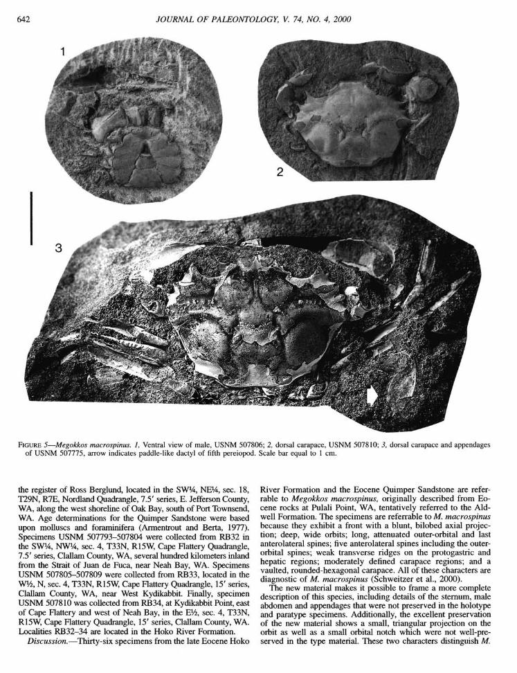
FIGURE 5—Megokkos macrospinus. 1, Ventral view of male, USNM 507806; 2, dorsal carapace, USNM 507810; 3, dorsal carapace and appendages of USNM 507775, arrow indicates paddle-like dactyl of fifth pereiopod. Scale bar equal to 1 cm.
the register of Ross Berglimd, located in the SW 4̂, NE^4, sec. 18, T29N, R7E, Nordland Quadrangle, 7.5' series, E. Jefferson County, WA, along the west shoreline of Oak Bay, south of Port Townsend, WA. Age determinations for the Quimper Sandstone were based upon molluscs and foraminifera (Armentrout and Berta, 1977). Specimens USNM 507793-507804 were coUected from RB32 in the SW14, NWl^, sec. 4, T33N, R15W, Cape Flattery Quadrangle, 7.5' series, Clallam County, WA, several hundred kilometers inland from the Strait of Juan de Fuca, near Neah Bay, WA. Specimens USNM 507805-507809 were collected from RB33, located in the WV2, N, sec. 4, T33N, R15W, Cape Flattery Quadrangle, 15' series, Clallam County, WA, near West Kydikabbit. Finally, specimen USNM 507810 was collected from RB34, at Kydikabbit Point, east of Cape Flattery and west of Neah Bay, in the EVi, sec. 4, T33N, R15W, Cape Flattery Quadrangle, 15' series, Clallam County, WA. Localities RB32-34 are located in the Hoko River Formation.
Discussion.—Thirty-six specimens from the late Eocene Hoko
River Formation and the Eocene Quimper Sandstone are referrable to Megokkos macrospinus, originally described from Eocene rocks at Pulali Point, WA, tentatively referred to the Aid-well Formation. The specimens are referrable to M. macrospinus because they exhibit a front with a blunt, bilobed axial projection; deep, wide orbits; long, attenuated outer-orbital and last anterolateral spines; five anterolateral spines including the outer-orbital spines; weak transverse ridges on the protogastric and hepatic regions; moderately defined carapace regions; and a vaulted, rounded-hexagonal carapace. All of these characters are diagnostic of M. macrospinus (Schweitzer et al., 2000).
The new material makes it possible to frame a more complete description of this species, including details of the sternum, male abdomen and appendages that were not preserved in the holotype and paratype specimens. Additionally, the excellent preservation of the new material shows a small, triangular projection on the orbit as well as a small orbital notch which were not well-preserved in the type material. These two characters distinguish M.
SCHWEITZER AND FELDMANN—NEW FOSSIL PORTUNIDS FROM WASHINGTON AND ARGENTINA 643
macrospinus from all other species of the genus. Also, the excellent preservation of the new material shows that the small anterolateral spine located at the base of the outer orbital spine, which was described as sometimes being absent in the original description, is almost always present and well-developed.
Genus MINOHELLENUS Karasawa, 1990
Charybdis (Minohellenus) KARASAWA, 1990, p. 21, pi. 6, figs. 7-8; KARASAWA, 1993, p. 56, pi. 13, figs. 3a,b, pi. 14, figs. 3a-c. Minohellenus Karasawa, 1990. KATO AND KARASAWA, 1994, p. 53, fig. 2, pi. 4; KATO AND KARASAWA, 1996, p. 31, pi. 10;
Type species.—Charybdis (Minohellenus) quinquedentata Karasawa, 1990.
Other species.—M. chichibuensis (Kato, 1996, as Itoigawaia) M. macrocheilus Kato and Karasawa, 1994; M. minoensis (Karasawa, 1990, as Portunites); M. triangulum (Rathbun, 1926, as Portunites); M. umemotoi (Karasawa, 1993, as Itoigawaia).
Diagnosis.—Carapace hexagonal, longer than wide, LAV about 0.60-0.75, widest at position of last anterolateral spine, carapace regions typically poorly defined. Frontal margin with six triangular, well-developed spines including inner orbital spines. Orbits circular, directed forward, with two orbital fissures, fronto-orbital width to width ratio about 0.4-0.5. Anterolateral margin with five spines including outer orbital spines, spines long, triangular, sharp; last spine typically longest. Epi-branchial region an arcuate ridge, extending from base of fifth anterolateral spine to axis, ridge poorly to moderately well-defined. Protogastric region weakly inflated, hepatic region flattened. Chelae heterochelate, smooth or with granular keels on outer surface. Dactyls of fifth pereiopods paddle-like.
Material examined.—Charybdis (Minohellenus) quinquedentata, MFM9030 (holotype); MFM9031 (paratype). M. macrocheilus, KMNH IvP 300, 020 (holotype); KMNH IvP 300, 022-24 (paratypes). Itoigawaia minoensis, MFM9035 (holotype); MFM9037 (paratype). /. umemotoi, MFM 9039 (holotype). Specimens of M. triangulum are given for the species.
Discussion.—Karasawa (1990) originally named Minohellenus as a subgenus of Charybdis and referred C. (M.) quinquedentata from the early Miocene of Japan to it. Subsequently, Kato and Karasawa (1994) elevated Minohellenus to generic status and named M. macrocheilus, a new species from the late Oligocene of Japan. The genus Itoigawaia Karasawa, Sakumoto, and Takayasu, 1992 was erected to acconmiodate material originally assigned to Portunites by Karasawa (1990). In their 1994 and 1996 papers, Kato and Karasawa noted that Minohellenus and Itoigawaia were similar to each other and that differentiating between the two was problematic. Schweitzer and Feldmann (1999) suggested that Minohellenus and Itoigawaia may be synonymous.
Review of these taxa by the authors and by Karasawa (personal commun.) has brought each independently to consider Itoigawaia to be synonymous with Minohellenus, Itoigawaia becoming the junior subjective synonym. The two genera share numerous characteristics, including a similar carapace shape; a front with six well-developed spines including the inner-orbital spines; a fronto-orbital width to width ratio of 0.40-0.50; an
anterolateral margin with five large, triangular, sharp spines including the outer-orbital spine; carapace regions that are weakly to moderately developed; a weakly inflated protogastric region; a depressed hepatic region; an arcuate epibranchial ridge extending from the last anterolateral spine; nearly straight to weakly convex posterolateral margins; and heterochelate chelae with mani that are longer than high and with large, blunt denticles on the fingers. The only difference between the two genera is that in members of Itoigawaia, the penultimate anterolateral spine is sometimes longest instead of the last anterolateral spine (Schweitzer and Feldmarm, 1999). This sole difference does not constitute sufficient grounds for distinguishing the two taxa as separate genera, so they are herein united as a single genus, Minohellenus.
Minohellenus is easily distingushed from Portunites, Megok-kos, Imaizumila and Proterocarcinus. Members of Portunites have much better developed carapace regions, much smaller anterolateral spines, and fewer frontal spines than do species of Minohellenus. Furthermore, members of Minohellenus lack the longitudinal ridges and tubercles on the branchial regions diagnostic of species of Portunites. Members of Minohellenus differs from species of Megokkos because Megokkos has much wider orbits and a much broader fronto-orbital width to width ratio of over 0.66 as compared to 0.40-0.50 in Minohellenus. Additionally, members of Megokkos have four blunt projections on the frontal margin, while species of Minohellenus have six spines on the frontal margin. Species of Megokkos have distinctive transverse ridges on the protogastric and hepatic regions which members of Minohellenus lack. Finally, the arcuate epibranchial ridge is much better developed in species of Megokkos than in those of Minohellenus. Minohellenus was distinguished from Imaizumila above. Species within Minohellenus are differentiated based upon the development of carapace regions, the ornamentation of carapace regions, the length of the anterolateral spines, the development of the frontal spines, and the relative width of the fronto-orbital margin. Species of Proterocarcinus have much more broad orbits and a much wider fronto-orbital width to width ratio than do species of Minohellenus.
Minohellenus is known only from Japan and the Pacific Northwest of North America. Species from Japan range in age from Oligocene to middle Miocene, and the sole North American species is Oligocene-early Miocene in age. The genus apparently evolved in the North Pacific Ocean and subsequently dispersed only along the North Pacific rim; it is not possible to determine whether the dispersal was east to west or west to east.
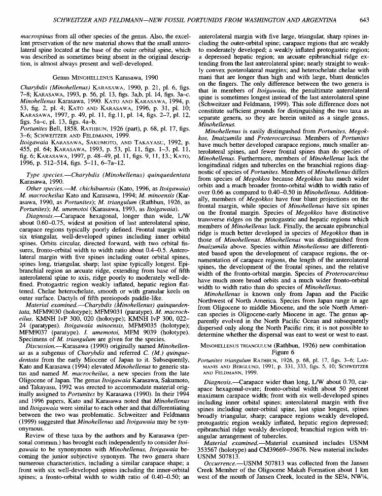
MINOHELLENUS TRIANGULUM (Rathbun, 1926) new combination Figure 6
Portunites triangulum RATHBUN, 1926, p. 68, pi. 17, figs. 3-6; LAS-MANIS AND BERGLUND, 1991, p. 331, 333, figs. 5, 10; SCHWEITZER AND FELDMANN, 1999.
Diagnosis.—Carapace wider than long, LAV about 0.70, carapace hexagonal-ovate; fronto-orbital width about 50 percent maximum carapace width; front with six well-developed spines including inner orbital spines; anterolateral margin with five spines including outer-orbital spine, last spine longest, spines broadly triangular, sharp; carapace regions weakly developed, protogastric region weakly inflated, hepatic region depressed; epibranchial ridge weakly developed; branchial region with triangular arrangement of tubercles.
Material examined.—Material examined includes USNM 353567 (holotype) and CM39669-39676. New material includes USNM 507813.
Occurrence.—USNM 507813 was collected from the Jansen Creek Member of the Oligocene Makah Formation about 1 km west of the mouth of Jansen Creek, located in the SEi/4, NW^A,
644 JOURNAL OF PALEONTOLOGY, V. 74, NO. 4, 2000
FIGURE 6—Minohellenus triangulum. USNM 507813, dorsal carapace. Scale bar equal to 1 cm.
sec. 26, T33N, R14W, Clallam Bay Quadrangle, 15' series, Clallam County, WA.
Discussion.—Rathbun (1926) originally described Portunites triangulum from Oligocene rocks of Washington and Oregon, and Schweitzer and Feldmann (1999) reported it from late Oligocene to early Miocene rocks of the Pysht Formation, Olympic Peninsula, WA. Re-examination of this species indicates that it is not referrable to Portunites s.s. Portunites triangulum has a narrower fronto-orbital width to width ratio than most members of Portunites s.s., and P. triangulum has six spines on the frontal margin rather than four as in Portunites s.s. The carapace regions of P. triangulum are much more poorly developed and the anterolateral spines are larger and sharper than those of species of Portunites s.s. Portunites triangulum also lacks the longitudinal ridges on the branchial region diagnostic of Portunites s.s.
Portunites triangulum exhibits all of the diagnostic characters of Minohellenus and is herein referred to that genus. Those diagnostic characters include six spines on the frontal margin; a fronto-orbital width to width ratio of 0.50; five large, sharp anterolateral spines; poorly developed carapace regions; an inflated protogastric and depressed hepatic region; and a weak epibran-chial ridge extending axially from the base of the last anterolateral spine. The specimen was badly sheared, making it impossible to take accurate measurements or to make an accurate reconstruction of the animal. Therefore, the specimen has not been described for this report. However, the broad, sharp, anterolateral spines have not before been seen in unbroken form.
Minohellenus triangulum differs from all other species of the genus in possessing a triangular arrangement of tubercles on the branchial regions of the carapace. The anterolateral spines are sharper and more attenuated at their tips than those of other species in the genus. The chelae of M. triangulum have granular keels on the outer surface of the manus, which have not been described for other species within the genus.
This is the first report of Minohellenus triangulum from the middle Oligocene Makah Formation, Olympic Peninsula, WA, which extends neither the geographic nor the geologic range of
the species, previously reported from Oligocene rocks of Washington and Oregon (Rathbun, 1926) and from the late Oligocene to early Miocene Pysht Formation of the Olympic Peninsula (Schweitzer and Feldmann, 1999).
Genus PROTEROCARCINUS Feldmann, Casadio, Chirino-Galvez, and Aguirre-Urreta, 1995
Type species.—Proterocarcinus lophos Feldmann, Casadio, Chirino-Galvez, and Aguirre-Urreta, 1995.
Other species.—Proterocarcinus latus (Glaessner, 1933, as Archaeogeryon).
Diagnosis.—Carapace transversely ovoid to hexagonal, wider than long, 0.60-0.76 times as long as wide; front narrow, downtumed, with four spines including inner-orbital spines; orbits extremely broad, rimmed, fronto-orbital width to width ratio 0.70-0.95, two orbital fissures; anterolateral margin short, with four spines including outer-orbital spines; epibranchial ridge elevated, granular, terminating at base of fourth anterolateral spine; branchial region swollen, with longitudinal ridge parallel to long axis of cardiac region; first pereiopods isochelous; propodus and dactyl of fifth pereiopod paddle-like.
Discussion.—Proterocarcinus was first described from Dan-ian rocks of the Roca Formation in the Neuquen Basin, Rio Negro Province, Argentina. The sole specimen upon which the description was based lacked the frontal regions and, therefore, description of that critical region was not possible. Recognition of a second species of the genus herein provides important information on that area as well as morphological details of the propodi and dactyli of the fifth pereiopod, confirming the placement within the Portunidae and the Polybiinae. Comparison of the genus with others in the subfamily has been discussed above, in reference to Portunites.
PROTEROCARCINUS LATUS (Glaessner, 1933) new combination Figures 7, 8
Diagnosis.—Carapace trapezoidal in outline, LAV about 0.76; carapace regions moderately well-defined, often coarsely pustulose; front narrow, downtumed, with four spines including inner orbital spines; orbits extremely wide, shallow, rimmed, with two orbital fissures, fronto-orbital width to width ratio about 0.95; anterolateral margin short, with four spines including outer orbital spine; hepatic region elevated, granular, with central node; epibranchial ridge arcuate, granular, extending from base of fourth anterolateral spine; branchial region with longitudinal, granular ridge parallel to long axis of cardiac region; first pereiopods isochelous; propodi and dactyli of fifth pereiopod paddle-like.
Description.—Carapace trapezoidal in outline, length about 0.76 times maximum width measured at broken tips of fourth anterolateral spine; nearly flat transversely, weakly vaulted longitudinally; carapace regions moderately well defined by tumid, often coarsely pustulose elevations and narrow, smooth grooves.
Front narrow, rostrate, about 17 percent maximum width, downtumed almost at right angles to carapace surface, sulcate axially, projected slightly in advance of orbits in mature individuals and strongly projected in immature (smaller) forms, terminating in four spines including inner-orbital spines, outer pair stronger and divergent distally, inner two narrower and more closely spaced. Orbits very wide, shallow, finely denticulate, fronto-orbital width about 95 percent total width, bounded by outer rostral spine axially and prominent, long, slender, antero-laterally directed outer orbital spine laterally; with two shallow
SCHWEITZER AND FELDMANN—NEW FOSSIL PORTUNIDS FROM WASHINGTON AND ARGENTINA 645
.^.,.....^m^:r "̂'̂ -ifev,.
FIGURE 7—Proterocarcinus latus. 1, Dorsal carapace, GHUNLPam 16818; 2, mold of pereiopods 3-5 showing flattened, paddle-like propodus and dactylus on pereiopod 5, GHUNLPam 16821; 3, dorsal carapace, GHUNLPam 16816. Scale bar equal to 1 cm.
orbital fissures, innermost fissure located about 40 percent width of orbit measured from base of rostrum to base of outer orbital spine, with narrow swollen rim axially and broader, elevated orbital rim adaxially; outer orbital fissure near base of outer orbital spine. Anterolateral margin about 40 percent total length of carapace, slightly divergent posteriorly, bearing four spines, including outer orbital spine; second and third spines smaller, directed anterolateral^, acute; fourth with broad base at outer end of epibranchial ridge, broken in all specimens but apparently large and laterally directed. Posterolateral margins straight to weakly concave, convergent posteriorly. Posterolateral reentrants long, strongly convergent, concave. Posterior margin about 38 percent total width, straight, weakly rimmed.
Mesogastric region extends as subtle, posteriorly widening elevation from rostral sulcus to abruptly widening posterior region bearing two subtle nodes at widest point, bounded anteriorly by subtle circular, granular epigastric swellings and at midlength by
large, ovoid, prominent, granular protogastric swellings. Uro-gastric region narrow, poorly defined as transverse swelling. Cardiac region and urogastric regions bounded laterally by deep, narrow, branchiocardiac furrow. Cardiac widest of axial regions, about 26 percent total width, narrowing posteriorly to poorly defined intestinal region. Hepatic regions elevated, with central node and granular surface; second anterolateral spine defines outer extremity of hepatic region. Epibranchial region a prominent elevated ridge with granular surface terminating distally at fourth anterolateral spine. Branchial regions defined as swollen adaxial elevation paralleling branchiocardiac groove. Mesobran-chial and metabranchial regions not distinguishable, with prominent granular ridge extending longitudinally along midline of branchial regions.
Sternum ovoid, slightly longer than wide, greatest width at stemite five or six, lateral margins a series of arcuate elements defined by smoothly curved lateral somite margins and narrow
646 JOURNAL OF PALEONTOLOGY, V. 74, NO. 4, 2000
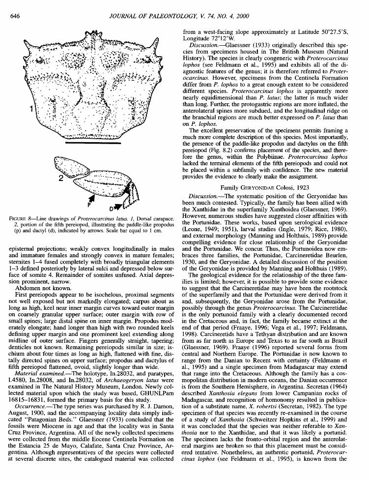
FIGURE 8—Line drawings of Proterocarcinus latus. 1, Dorsal carapace. 2, portion of the fifth pereiopod, illustrating the paddle-like propodus (p) and dactyl (d), indicated by arrows. Scale bar equal to 1 cm.
epistemal projections; weakly convex longitudinally in males and immature females and strongly convex in mature females; stemites 1-4 fused completely with broadly triangular elements 1-3 defined posteriorly by lateral sulci and depressed below surface of somite 4. Remainder of somites unfused. Axial depression prominent, narrow.
Abdomen not known. First pereiopods appear to be isochelous, proximal segments
not well exposed but not markedly elongated; carpus about as long as high, keel near inner margin curves toward outer margin on coarsely granular upper surface; outer margin with row of small spines; large distal spine on inner margin. Propodus moderately elongate; hand longer than high with two rounded keels defining upper margin and one prominent keel extending along midline of outer surface. Fingers generally straight, tapering; denticles not known. Remaining pereiopods similar in size; ischium about four times as long as high, flattened with fine, dis-tally directed spines on upper surface; propodus and dactylus of fifth pereiopod flattened, ovoid, slightly longer than wide.
Material examined.—The holotype, In.28032, and paratypes, 1.4580, In.28008, and In.28032, of Archaeogeryon latus were examined in The Natural History Museum, London. Newly collected material upon which the study was based, GHUNLPam 16815-16831, formed the primary basis for this study.
Occurrence.—The type series was purchased by R. J. Damon, August, 1900, and the accompanying locality data simply indicated "Patagonian Beds." Glaessner (1933) concluded that the fossils were Miocene in age and that the locality was in Santa Cruz Province, Argentina. All of the newly collected specimens were collected from the middle Eocene Centinela Formation on the Estancia 25 de Mayo, Calafate, Santa Cruz Province, Argentina. Although representatives of the species were collected at several discrete sites, the catalogued material was collected
from a west-facing slope approximately at Latitude 50°27.5'S, Longitude 72°12'W.
Discussion.—Glaessner (1933) originally described this species from specimens housed in The British Museum (Natural History). The species is clearly congeneric with Proterocarcinus lophos (see Feldmann et al., 1995) and exhibits all of the diagnostic features of the genus; it is therefore referred to Proterocarcinus. However, specimens from the Centinela Formation differ from P. lophos to a great enough extent to be considered different species. Proterocarcinus lophos is apparently more nearly equidimensional than P. latus; the latter is much wider than long. Further, the protogastric regions are more inflated, the anterolateral spines more subdued, and the longitudinal ridge on the branchial regions are much better expressed on P. latus than on P. lophos.
The excellent preservation of the specimens permits framing a much more complete description of this species. Most importantly, the presence of the paddle-like propodus and dactylus on the fifth pereiopod (Fig. 8.2) confirms placement of the species, and therefore the genus, within the Polybiinae. Proterocarcinus lophos lacked the terminal elements of the fifth pereiopods and could not be placed within a subfamily with confidence. The new material provides the evidence to clearly make the assignment.
Family GERYONIDAE Colosi, 1923
Discussion.—The systematic position of the Geryonidae has been much contested. Typically, the family has been allied with the Xanthidae in the superfamily Xanthoidea (Glaessner, 1969). However, numerous studies have suggested closer affinities with the Portunidae. These works, based upon serological evidence (Leone, 1949; 1951), larval studies (Ingle, 1979; Rice, 1980), and external morphology (Manning and Holthuis, 1989) provide compelling evidence for close relationship of the Geryonidae and the Portunidae. We concur. Thus, the Portunoidea now embraces three families, the Portunidae, Carcineretidae Beurlen, 1930, and the Geryonidae. A detailed discussion of the position of the Geryonidae is provided by Manning and Holthuis (1989).
The geological evidence for the relationship of the three families is limited; however, it is possible to provide some evidence to suggest that the Carcineretidae may have been the rootstock of the superfamily and that the Portunidae were derived from it and, subsequently, the Geryonidae arose from the Portunidae, possibly through the genus Proterocarcinus. The Carcineretidae is the only portunoid family with a clearly documented record in the Cretaceous and, in fact, the family became extinct at the end of that period (Fraaye, 1996; Vega et al., 1997; Feldmann, 1998). Carcineretids have a Tethyan distribution and are known from as far north as Europe and Texas to as far south as Brazil (Glaessner, 1969). Fraaye (1996) reported several forms from central and Northern Europe. The Portunidae is now known to range from the Danian to Recent with certainty (Feldmann et al., 1995) and a single specimen from Madagascar may extend that range into the Cretaceous. Although the family has a cosmopolitan distribution in modem oceans, the Danian occurrence is from the Southern Hemisphere, in Argentina. Secretan (1964) described Xanthosia elegans from lower Campanian rocks of Madagascar, and recognition of homonomy resulted in publication of a substitute name, X. robertsi (Secretan, 1982). The type specimen of that species was recently re-examined in the course of a study of Xanthosia (Schweitzer Hopkins et al., 1999) and it was concluded that the species was neither referable to Xanthosia nor to the Xanthidae, and that it was likely a portunid. The specimen lacks the fronto-orbital region and the anterolateral margins are broken so that this placement must be considered tentative. Nonetheless, an authentic portunid, Proterocarcinus lophos (see Feldmann et al., 1995), is known from the
SCHWEITZER AND FELDMANN—NEW FOSSIL PORTUNIDS FROM WASHINGTON AND ARGENTINA 647
Danian Roca Formation in Argentina. A second species of that genus, P. latus, described above, coexisted with, and may have been the rootstock for, Chaceon peruvianus (d'Orbigny, 1842) which is described below.
The morphological similarity between the carapace features of Proterocarcinus latus and Chaceon peruvianus is amazing, particularly when comparing juvenile specimens of C. peruvianus and adult specimens of P. latus. Sufficient differences in the fronto-orbital and anterolateral regions distinguish the two species; however, these areas are frequently broken or poorly preserved in which case it is impossible to make a species determination. The development of carapace regions, distribution of granular surfaces, and expression of carapace grooves are so similar, especially between adult individuals of P. latus and juveniles of C peruvianus, that it was originally concluded by the authors that only a single species was present. However, when more completely preserved specimens were studied it became clear that the two could be distinguished. Adult Proterocarcinus latus are characterized by broader fronto-orbital regions, ranging from about 84 to 91 percent maximum width, and only four anterolateral spines whereas the fronto-orbital region ranges from 80-82 percent maximum width and there are five anterolateral spines on juvenile specimens of Chaceon peruvianus. The juvenile specimens of the latter species are in the same size range of adult P. latus whereas the adults of C. peruvianus are three or more times as large. Furthermore, adults of Chaceon peruvianus exhibit a proportionately narrower fronto-orbital region, ranging from 52-63 percent maximum width, and much more subdued carapace morphology than do the juveniles.
Based upon these observations, it is possible to suggest that Chaceon peruvianus, the oldest member of the Geryonidae, may have evolved from Proterocarcinus latus, or an unknown closely related species, by a process of peramorphosis in which the adult characters of the latter species were expressed in the juvenile form of Chaceon peruvianus. During the ontogeny of C. peruvianus, the fronto-orbital width decreases, proportional to maximum width, and the sculpturing of the carapace becomes more subdued so that the adult morphology of this species bears much less resemblance to that of adult Proterocarcinus latus. Manning and Holthuis (1989) described the pereiopods of the Geryonidae as lacking segments that were flattened or ovate as an adaptation for swimming. The ancestral taxon for the Geryonidae proposed here, Proterocarcinus, has propodi and dactyli of the fifth pereiopod that are ovate and flattened. However, this difference in appendages may be the result of abandonment of the swimming habit within the primarily deep-water Geryonidae and may be convergent with the loss of paddle-like swimming appendages in the portunid subfamily Carcininae Macleay, 1838. In any case, the remarkable similarities in dorsal carapace morphology clearly suggests that Proterocarcinus bears an ancestral link to the Geryonidae.
For the above reasons, much confusion has resulted from the generic assignments of the geryonids and portunids from Patagonia. To resolve some of these problems, the history of assignment of the most confusing taxa, including Chaceon peruvianus, Archaeogeryon fuegianus, Lebucarcinus tyro, and Proterocarcinus latus will be discussed in more detail.
Chaceon peruvianus has variously been assigned to the Portun-idae, Xanthidae, Cancridae, and Geryonidae, as documented by the synonymy presented below. The primary reason that the species was assigned to genera now in four diifferent families probably resulted from lack of knowledge of details of the walking legs and incomplete knowledge of details of carapace morphology. The present assignment to Chaceon and the Geryonidae conforms closely to the definition of those taxa (Manning and Holthuis, 1989) and
reinforces affinities with the genus Geryon Kroyer, 1837, first suggested by Ortmann (1900). The junior subjective synonymy of Cancer patagonicus Philippi, 1887, has been generally agreed upon since first noted by Ortmann (1902).
Archaeogeryon fuegianus Colosi, 1923, was named as the type species of that genus and was characterized by greatly thickened orbital margins, strong hepatic spines, three strong and two tiny anterolateral spines, and subtle longitudinal branchial ridges. In other morphological features the species bears a close resemblance to species referrable to Chaceon, including C. peruvianus. Aguirre Urreta (1987, p. 469) assigned the genus to Coeloma as C. (Coeloma) fueguianum (sic); however, Coeloma A. Milne Edwards, 1865, typically bears four anterolateral spines, not an odd number, and lacks the elongate ridges typical of most geryonids, including Chaceon spp. The family placement of Coeloma remains in question and it currently seems unlikely that it is a geryonid as suggested by Glaessner (1969). The development of the orbital ridges, hepatic spines, and anterolateral spines, and the suppression of the branchial ridges are taken to be sufficiently important to constitute distinction of Ar-chaeogeryon and Chaceon at the generic level. Examination of the specimens studied by Glaessner (1933) in the Natural History Museum, London (In.28022, In.28033, and In.28027-30) confirms the generic placement and the unique nature of the species. Therefore, A. fuegianus, from rocks of Argentina, remains the sole species within the genus. It should be noted that, although the labels accompanying the specimens in the Natural History Museum record the material as being either Tertiary or Oligocene in age, Glaessner (1933, p. 23) concluded that they date from the Miocene. This may have been based upon the opinion that the Patagonian Beds were of that age. That age assignment must be independently tested.
Aguirre Urreta (1987) £dso assigned Lebucarcinus tyro (Philippi, 1887) to the Geryonidae. However, presence of a trifid rostrum, only two anterolateral spines, and absence of an outer orbital spine rule out assignment to the family. Chirino-Galvez (1993) referred the genus to the Goneplacidae, with which we concur.
Glaessner described Archaeogeryon latus based upon four specimens from, "Miocene, Patagonian Beds, Santa Cruz, Argentina" (1933, p. 24). His description of the species, including reference to four anterolateral spines, development of carapace ridges and granular ornamentation suggests that the species is quite different from other species referred to Archaeogeryon. Assignment of A. latus to Coeloma (Coeloma) by Aguirre Urreta (1987) conformed to her conviction that Archaeogeryon was a junior synonym of Coeloma. Examination of the type series of A. latus confirms that it is not a geryonid at all but that it is a portunid, referrable to Proterocarcinus Feldmann et al., 1995. Thus, P. latus (Glaessner) becomes the second species referrable to Proterocarcinus.
The morphological differences between P. latus, A. fuegianus, and Chaceon peruvianus were suggested to be reflective of an evolutionary continuum (Glaessner, 1933, p. 24) in which Proterocarcinus latus was the most derived. That relationship continues to be recognized but, as described above, it is probable that P. latus was the rootstock for origin of the Geryonidae. The Geryonidae as defined by Manning and Holthuis (1989) is now recognized to embrace four genera with the addition oi Archaeogeryon known only from the fossil record, Chaceon, Geryon, and Zariquieyon Manning and Holthuis, 1989. Chaceon peruvianus constitutes the oldest known member of the genus and the family.
Genus CHACEON Manning and Holthuis, 1989
Type species.—Geryon fenneri Manning and Holthuis, 1984.
648 JOURNAL OF PALEONTOLOGY, V. 74, NO. 4, 2000
CHACEON PERUviANUS (d'Orbigny, 1842) new combination Figures 9, 10
Portunus peruvianus D'ORBIGNY, 1842, p. 107, pi. 6, fig. 17. Podopilumnus peruvianas (d'Orbigny). MCCOY, 1849, p. 166. Carcinus peruvianus (d'Orbigny). A. MILNE EDWARDS, 1860, p. 269,
pi. 8, figs 1-c. Cancer patagonicus PHILIPPI, 1887; HATCHER, 1897, p. 337. Geryon! peruvianus (d'Orbigny). ORTMANN, 1900, p. 381; ORTMANN,
1902, p. 255, pi. 38, fig. 6; STEINMANN AND WILCKENS, 1908, p. 70. Geryon peruvianus (d'Orbigny). ROVERETO, 1921, p. 25; FRENGUELLI,
1927, p. 205, figs, l(a-b), ll(a-b); GLAESSNER, 1929, p. 182; AGUIRRE URRETA, 1987, p. 464, pi. 1, pi 2, figs A-C, pi. 3, figs. A-E, pi. 4, fig. B.
Archaeogeryon peruvianus (d'Orbigny). GLAESSNER, 1933, p. 22, pi. 5, figs. 1-2; CAMACHO, 1966, p. 477, pi. 16.13, figs a-b; GLAESSNER, 1969, p. R524, fig. 332, 5a-b; DE LA FUENTE, 1977, p. 310; MORRIS, 1980, p.l; BRIGGS ET AL., 1985, p. 203, pi. 9.2.10 A-B.
Emended description.—Moderately large for genus; outline hexagonal, length about 78 percent maximum width measured at broken tips of fifth anterolateral spines; weakly vaulted transversely and longitudinally; carapace regions well defined in juvenile (smaller) specimens by pustulose elevations, regions subdued in adults.
Front rostrate, about 18 percent maximum width, downtumed slightly, sulcate axially, projected in advance of orbits, terminating in four subequal spines of which outer pair are slightly stronger and divergent distally and inner pair are narrower, dis-tally divergent, and closely spaced. Orbits broad, arcuate, fronto-orbital width varies from about 82 percent maximum width in juveniles to 52 percent in mature forms, orbits bounded by outer rostral spines axially and strong, triangular outer orbital spines laterally; with two shallow orbital fissures, innermost one at about midpoint of orbit, orbital rim narrow, smooth axially and denticulate laterally; outer orbital fissure near base of outer orbital spine. Anterolateral margin about 47 percent total length, convex, bearing five spines, including outer orbital spine; first four spines similar in size, directed radially, fifth spine longest, directed laterally and dorsally. Posterolateral margin straight to weakly concave, smooth, convergent posteriorly. Posterolateral comers long, strongly convergent, concave. Posterior margin about 33 percent maximum width, straight, weakly rimmed.
Mesogastric region extends as subtle, posteriorly widening elevation from rostral sulcus to abruptly widened posterior region bearing two subtle swellings at widest point; granular protogas-tric swellings large, ovoid, prominent. Urogastric region narrow, poorly defined as transverse swelling. Cardiac region and urogastric regions bounded laterally by moderately deep, narrow, branchiocardiac furrow. Cardiac region widest of axial regions, narrowing posteriorly to poorly defined intestinal region. Hepatic regions elevated, with central node and granular surface; third anterolateral spine defines outer extremity of hepatic region. Epibranchial region a prominent elevated ridge with granular surface terminating distally at fifth anterolateral spine. Branchial regions defined as swollen adaxial elevation paralleling branchiocardiac groove. Mesobranchial and metabranchial regions not distinguishable, with prominent granular ridge arising as circular elevation and extending longitudinally along midline of branchial regions.
Sternum ovoid, shghtly longer than wide, greatest width at ster-nite six, lateral margins a series of arcs defined by smoothly curved lateral somite margins and narrow epistemal projections; weakly convex longitudinally in males and immature females and strongly convex in mature females; stemites 1-4 fused completely with broadly triangular elements 1-3 defined posteriorly by lateral sulci and depressed below surface of somite 4. Remainder of somites unfused. Axial depression prominent, narrow.
Abdomen of male with unfiised segments and triangular telson, widest at somite 2, margins straight or concave axially. Female abdominal segments unfused, convex laterally, widest at somite 5; somite 2 shortest; somite 6 longest with concave distal margin into which telson is set. Telson a parallelogram. Immature female abdomen about 44 percent maximum stemal width; mature female abdomen about 75 percent maximum stemal width.
Third maxiUipeds with ischium with narrow proximal termination widening to parallel-sided distal region, slightly longer than wide; exopod narrower, longer than ischium, tapering distally.
First pereiopod strong, moderately heterochelous, carpus rectangular with smooth platform on upper surface, prominent distal spine on inner surface. Propodus with inflated hand, about 62 percent as high as long, smooth to weakly keeled outer surface, slightly flattened upper surface; fixed finger straight, dactylus slightly curved, elongate, bearing dome-shaped denticles on occlusal surface. Remaining pereiopods moderately long, slender, ischia of pereiopods 2-5 long, slender, possibly slightly compressed; meri poorly preserved, circular to ovoid in cross-section; distal segments poorly known; propodus of pereiopod 5 flattened at least on immature specimen, dactylus lanceolate.
Material examined.—The material basis for Glaessner's comments on the species, In.27384-87, In.28001, In.28002, In.28004-7, In.28024, In.28026, In.28036-36, and In.28288, were studied in The Natural History Museum. Additionally, the J. B. Hatcher Collection, including 16 specimens catalogued as P.774-79 (nine specimens collected by Hatcher and identified by Ortmann, and P. 780, six specimens collected by R. Hauthal and identified by Ortmann) were studied at Kent State University. This collection will be deposited in the U.S. National Museum of Natural History upon completion of the research. Newly collected material that formed the primary basis for the study, GHUNLPam 16804-16814, provided important details to expand on the description of the species.
Occurrences.—All of the specimens were collected from the middle Eocene Centinela Formation on the Estancia 25 de Mayo, near Calafate, Santa Cruz Province, Argentina. GHUNLPam 16804-16809 were collected from Laritude 50°30.716'S, Longitude 72°15.303'W. GHUNLPam 16810-16811 were collected across the stream and just to the north from that locahty. GHUNLPam 16812-16814 were collected about 10 km north from those localities approximately at Latitude 50°27.5"S, Longitude 72°12"W.
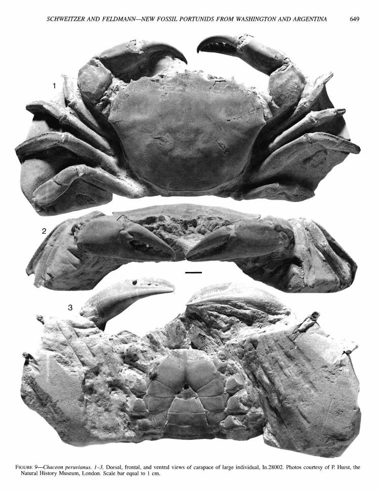
Discussion.—Living representatives of Chaceon inhabit deep water (Manning and Holthuis, 1989) and are characterized by long, slender walking legs. The fossils from South America conform closely to the definition of the genus in all other regards; however, the legs tend to be only moderately long and the fifth pereiopod bears a flattened propodus although the dactylus is styliform. The flattening of the propodus has been recognized by Glaessner (1933) and Aguirre Urreta (1987) but neither author illustrated the limb. The nature of the fifth pereiopod is clearly seen in Figure 10.1. Thus, it would appear that the earliest of the geryonids, adapted to a nearshore, shallow water habitat possessed a modified portunid-like appendage, possibly adapted to swimming and burrowing, and that full development of the elongate pereiopods may have been an adaptation to life in deeper habitats.
Recognition of two different size classes of individuals in this species suggests that there may have been seasonal recmitment of young although this suggestion must be taken as highly speculative. The difference in size between small, "juvenile" specimens and large, "adult" specimens is great enough to suspect that the large individuals may be much older than the small ones. Perhaps more significant is the observation that growth of the frontal and orbital regions are allometric; the fronto-orbital width
SCHWEITZER AND FELDMANN—NEW FOSSIL PORTUNIDS FROM WASHINGTON AND ARGENTINA 649
FIGURE 9—Chaceon peruvianas. 1-3, Dorsal, frontal, and ventral views of carapace of large individual. In.28002. Photos courtesy of P. Hurst, the Natural History Museum, London. Scale bar equal to 1 cm.
650 JOURNAL OF PALEONTOLOGY, V. 74, NO. 4, 2000
FIGURE 10—Chaceon peruvianus, 1, Dorsal carapace, GHUNLPam 16812; 2, dorsal carapace, GHUNLPam 16811; 3, sternum and partial abdomen of male, GHUNLPaml6813; 4, stereoscopic view of sternum and abdomen of female, GHUNLPam 16814. Scale bar equal to 1 cm.
relative to the maximum width does change with increased overall size and presumably with age.
DISCUSSION
The Carcineretidae exhibits a primarily tropical distribution, having been reported from localities in Mexico, Brazil, the Gulf Coast of North America, the Caribbean, and Europe (Glaessner, 1969; Fraaye, 1996; Vega et al., 1997). All of the occurrences are from Late Cretaceous age rocks and the family may have become extinct as a result of the Chicxulub impact (Feldmann, 1998). The distinguishing features of the family, including the
quadrate outline of the carapace, development of carapace regions, and development of a swimming paddle on the fourth, not the fifth, pereiopod serve to distinguish the representatives of this group from the other members of the superfamily. It is likely that some member of the family, possibly within the genus Carcineretes Withers, 1922, was the progenitor of the Portuni-dae. The earliest portunid known, Proterocarcinus lophos has a rectilinear carapace, ridges on the carapace, and extremely long orbits as do representatives of Carcineretes. If that is so, the superfamily first appeared in low latitude regions of the Caribbean, dispersed southward, radiated into species assignable to
SCHWEITZER AND FELDMANN—NEW FOSSIL PORTUNIDS FROM WASHINGTON AND ARGENTINA 651
the Portunidae, and subsequently experienced further radiation and dispersal.
Several important biogeographic patterns emerge from description and re-evaluation of the portunid and geryonid genera discussed herein. The earliest occurrence of authentic Polybi-inae, and indeed the Portunidae, is Proterocarcinus from Danian rocks of southern Argentina, suggesting that the family arose in the high southern latitudes. Additionally, Pororaria Glaessner, 1980, a member of the Polybiinae, is known from Eocene rocks of New Zealand (Feldmann and Maxwell, 1990), supporting a high southern latitude origin. The generic reassignment of several polybiine species previously assigned to Portunites sensu lato and the recognition of Imaizumila from the southern hemisphere has broad implications for understanding the evolutionary history of the Polybiinae. The biogeographic range of Portunites sensu stricto is unchanged by the removal of some taxa from the genus; it is reported from the Eocene of northern Europe, the west coast of North America, and Japan. The new genus Megokkos, from Eocene to Oligocene rocks, and Minohellenus, reported from Oligocene to Miocene rocks, are both restricted to the North Pacific rim, while Imaizumila has a disjunct, am-phitropical distribution in Japan and Chile, having been reported from Eocene to Miocene rocks. The subfamily may have arisen in the high southern latitudes and subsequently dispersed northward into the North Atlantic region and to the North Pacific Ocean via the Straits of Panama. Alternatively, the subfamily may have dispersed to the North Atlantic Ocean and then to the Pacific Ocean via the Tethys. The limited age constraints on some of the taxa, and the widely overlapping age ranges of the five genera, make it difficult to speculate on just exactly how the subfamily was distributed globally from its apparent origin in the southern hemisphere. The subfamily could have arisen from a carcineretid ancestor which dispersed from the tropics to the middle southern latitudes, resulting in the evolution of the Polybiinae. Subsequently, the subfamily would have dispersed northward into the middle northern latitudes. Modem members of the Polybiinae, and the Portunidae, have a cosmopolitan distribution (Rathbun, 1930a; Glaessner, 1969; Sakai, 1976; Manning and Holthuis, 1981; Dai and Yang, 1991).
In addition to constituting the area of origin for the Portunidae, specifically, the Polybiinae, southern South America was the probable area of origin for the Geryonidae, as discussed above. Unfortunately, little else in known about the geologic distribution of the family. Modem geryonids are known from deep water sites around the world. Geryon is restricted to the North Atlantic, Zariquieyon is known only from the Mediterranean, and Chaceon is cosmopolitan.
ACKNOWLEDGMENTS
Material from the west coast of North America was collected and generously donated by R. E. and M. Berglund, Bainbridge Island, Washington. H. Karasawa, Mizunami Fossil Museum, Mizunami, Japan, provided numerous helpful comments and assisted the authors with synonymy of Minohellenus and Itoiga-waia. He also provided access to the collections at his institution, and his assistance in all regards is gratefully acknowledged. L. Chirino-Galvez, Kent State University, assisted in the collection of South American material, provided translations of Philippi (1887), and assembled locality information for Imaizumila ar-aucana; his assistance is appreciated. H. Kato provided access to material at the Natural History Museum and Institute, Chiba, Japan. R. B. Manning, United States National Museum of Natural History, Washington, DC, generously provided access to the biological collections at that institution. A. Ross, The Natural
History Museum, London, and M. Dorling and R. Long, Sedgwick Museum, Cambridge University, Cambridge, provided access to decapod material at their respective institutions. W Blow, United States National Museum of Natural History, Washington, DC, kindly loaned fossil material from the collection at that institution. Karasawa and Kato provided thoughtful reviews of the manuscript; we thank them for their useful suggestions. Material in Argentina was collected by Feldmann with the assistance of Silvio Casadio, Universidad Nacional de La Pampa and Maria Aguirre Urreta, Universidad de Buenos Aires, and was funded by National Science Foundation Grant OPP 9417697 and National Geographic Society Grant 5588-95 to Feldmann. Travel for examination of Japanese fossil material and material deposited in museums in the United Kingdom was funded by National Geographic Society Grant 6265-98 to Feldmann and Schweitzer. Travel to examine specimens of Xanthosia robertsi was funded by National Science Foundation Grant OPP 9526252 to Feldmann.
REFERENCES
AGUIRRE URRETA, M. B. 1987. La familia Geryonidae (Crustacea, Brachyura) en el Terciario de Patagonia y Tierra del Fuego, Argentina. IV Congreso Latinoamericano de Paleontologia, Bolivia, 1:459-484.
ANSFIELD, V. J. 1998. The Eocene Kydikabbit Point sedimentary breccias of the Lower Hoko River Formation, Northwestern Olympic Peninsula, Washington, and their contained invertebrates, p. 1-2 L In J. E. Martin (ed.), Contributions to the Paleontology and Geology of the West Coast. Thomas Burke Memorial Washington State Museum Research Report 6, Burke Museum, Seattle.
ARMENTROUT, J. M., AND A. BERTA. 1977. Eocene-Oligocene forami-niferal sequence from the northeast Olympic Peninsula, Washington. Journal of Foraminiferal Research, 7(3):216-233.
BELL, T. 1858. A monograph of the fossil malacostracous Crustacea of Great Britain, Pt. I, Crustacea of the London Clay. Palaeontographical Society, London, 44 pp.
BEURLEN, K. 1930. Vergleichende Stammesgeschichte Grundlagen, Methoden, Probleme unter Besonderer Beruchsichtingung der hoh-eren Krebse. Fortschrift Geologic und Paleontologie, 8:317-586.
BRIGGS, D. E. G., A. T. THOMAS, AND R. A. FORTEY. 1985. Chapter 9, Arthropods, p. 199-229. In J. W Murray (ed.). Atlas of Invertebrate Fossils. Longman and the Palaeontological Association/Halsted Press, New York.
CAMACHO, H. H. 1966. Invertebrados fdsiles. Manuales de Eudeba. Editorial Universitaria de Buenos Aires (EUDERA), Buenos Aires, 707 P-
CASADIO, S.; R. M. FELDMANN, AND K. POLAND. In press. ''̂ Ar/ '̂Ar age of the Centinela Formation, southwestern Argentina: an Eocene age for crustacean-rich "Patagonian" beds. Journal of South American Earth Sciences.
CHIRINO-GALVEZ, L. 1993. Cenozoic crabs from Chile. Unpublished M.S. thesis, Kent State University, Kent, Ohio, 280 pp.
COLOSi, G. 1923. Una specie fossile de Gerionide (Decapodi brachiuri). Bolettino della Societa dei Naturalist! in Napoli, 35 (series 2, volume 15), 37:248-255.
DAI, A., AND S. YANG. 1991. Crabs of the China Seas. Springer-Verlag, New York, New York.
FELDMANN, R. M. 1998. Cretaceous extinction of the Tethyan crab family Carcineretidae: a victim of the impact? Geological Society of America Abstracts with Programs, 7:A286.
, AND P. A. MAXWELL. 1990. Late Eocene decapod Crustacea from North Westland, South Island, New Zealand. Journal of Paleontology, 64:779-797.
, S. CASADIO, L. CHIRINO-GALVEZ, AND M. AGUIRRE URRETA. 1995. Fossil decapod crustaceans from the Jaguel and Roca formations (Maastrichtian-Danian) of the Neuquen Basin, Argentina. The Paleontological Society Memoir 43, 22 p.
, K. L. BICE, C. SCHWEITZER HOPKINS, E. W. SALVA, AND K. PiCKFORD. 1998. Decapod crustaceans from the Eocene Castle Hayne
652 JOURNAL OF PALEONTOLOGY, V. 74, NO. 4, 2000
Limestone, North Carolina:Paleoceanographic implications. The Pa-leontological Society Memoir 48, 28 pp.
FERUGLIO, E . 1949-1950. Descripcion de la Patagonia. Direccion General de Yacimientos Petroliferos Fiscales, Buenos Aires. (Volume I (1949):xv+334 p., figs. 1-78, pis. 1-60; volume II (1949): 349 pp., figs. 79-160, pis. 61-80; volume III (1950): 431 pp., figs. 161-266, pis. 81-119).
FRAAYE, R . H . B . 1996. Late Cretaceous swimming crabs: radiation, migration, competition, and extinction. Acta Geologica Polonica, 46: 269-278.
FRENGUELLI, J. 1927. El entrerrienes de Golfo Nuevo en el Chubut. Boletin de la Academia Nacional de Ciencias, Cordoba, 29:191-270.
FUENTE, M . S., DE LA. 1977. Algunas Consideraciones paleoecologicas sobre Archaeogeryon (Crustacea, Decapoda) registrado en el Terciaro Marino de Patagonia y Tierra del Fuego. Ameghiniana, 14:310-311.
GLAESSNER, M . F 1929. Crustacea Decapoda, p. 1-464. In J. F Pom-peckj (ed.), Fossilium Catalogus: Animalia, pars 41. W. Junk, Berlin.
. 1933. New Tertiary crabs in the collection of the British Museum. Annals and Magazine of Natural History, Ser. 10 (12): 1-28, pis. 1-6.
. 1960. The fossil decapod Crustacea of New Zealand and the evolution of the order Decapoda. New Zealand Geological Survey Paleontological Bulletin 31:1-79.
1969. Decapoda, p. R400-R566. In R. C. Moore (ed.). Treatise on Invertebrate Paleontology, Pt. R, Arthropoda 4. Geological Society of America and University of Kansas Press, Lawrence.
-. 1980. New Cretaceous and Tertiary crabs (Crustacea: Brachyura) from Australia and New Zealand. Transactions of the Royal Society of South Australia, 104:171-192.
HATCHER, J. B. 1897. On the geology of southern Patagonia. American Journal of Science, 4:327-354.
HOFFSTETTER, R . , H . FUENZALIDA, AND G. CECIONI. 1957. Chile-Chili.
Lexique Stratigraphique International, Centre National de la Recherche Scientifique, vol. 5:Latin America, Fascicule 7, 444 p.
ILLIES, H . 1960. Geologic der Gegend von Valdivia/Chile. Neues Jahr-buch fiir Geologic und Palaontologie, Abhandlungen, 111:30-110.
INGLE, R . W. 1979. The larval and post-larval development of the brach-yuran crab Geryon tridens Kroyer (Family Geryonidae) reared in the laboratory. Bulletin of the British Museum (Natural History), Zoology, 36:217-234.
KARASAWA, H . 1990. Decapod crustaceans from the Miocene Mizunami Group, Central Japan, Pt. 2, Section Oxyryncha, Cancridea, and Bra-chyryncha. Bulletin of the Mizunami Fossil Museum, 17:1-34, pis. 1-8.
. 1992. Fossil decapod crustaceans from the Manda Group (Middle Eocene), Kyushu, Japan. Transactions and Proceedings of the Pa-lacontological Society of Japan, N.S., 167:1247-1258.
-. 1993. Cenozoic decapod Crustacea from southwest Japan. Bulletin of the Mizunami Fossil Museum, 20:1-92, 24 pis.
1997. A monograph of Cenozoic stomatopod, decapod, isopod. and amphipod Crustacea from west Japan. Monograph of the Mizunami Fossil Museum, 8:1-81, 30 pis.
-., T. SAKUMOTO, AND K. TAKAYASU. 1992. A new portunid genus Itoigawaia (Decapoda: Crustacea) from the Miocene of southwest Japan. Bulletin of the Mizunami Fossil Museum, 19:455-457.
KATO, H . 1996. Miocene decapod crustaceans from the Chichibu Basin, Central Japan. Transactions and Proceedings of the Palaeontological Society of Japan, N. S., 183:500-521.
, AND H. KARASAWA. 1994. Minohellenus macrocheilus sp. nov. (Decapoda: Crustacea) from the Oligocenc Ashiya Group, Kyushu, Japan. Bulletin of the Kitakyushu Museum of Natural History, 13: 51-58.
, AND . 1996. An additional record of Minohellenus macrocheilus Kato and Karasawa, 1994 (Crustacea, Decapoda, Portunidae). Bulletin of the Kitakyushu Museum of Natural History, 15:31-33.
KROYER, H . 1837. Geryon tridens, en ny Krabbe. Naturhistorisk Tidssk-rift, 1:10-21, pi. 1. '
LASMANIS, R. , AND R. E. BERGLUND. 1991. Tertiary crustaceans: Collecting localities in western Washington state. Rocks and Minerals, 66:330-334.
LEONE, C . A. 1949. Comparative serology of some brachyuran Crustacea and studies in hemocyanin correspondence. The Biological Bulletin, 97:273-286.
. 1951. A serological analysis of the systematic relationship of the brachyuran crab Geryon quinquedens. The Biological Bulletin, 100: 44-48.
L6RENTHEY, E., AND K. BEURLEN. 1929. Die fossilen Dekapoden der Lander der ungarischen Krone. Geologica Hungarica, Series Palaeon-tologica, 420 p., 16 pis.
MACLEAY, W. S . 1838. On the Brachyurous Decapod Crustacea brought from the Cape by Dr. Smith, p. 53-71, pi 2. In Illustrations of the Annulosa of South Africa; being a portion of the objects of Natural History chiefly collected during an expedition into the interior of South Africa, under the direction of Dr. Andrew Smith, in the years 1834, 1835, and 1836; fitted out by "The Cape of Good Hope Association for Exploring Central Africa." London.
MANNING, R . B . , AND L . B . HOLTHUIS. 1981. West African Brachyuran Crabs (Crustacea: Decapoda). Smithsonian Contributions to Zoology, 306:1-379.
, AND . 1984. Geryon fenneri, a new deep-water crab from Florida (Crustacea: Decapoda: Geryonidae). Proceedings of the Biological Society of Washington, 97:666-673.
, AND . 1989. Two new genera and nine new species of ger-yonid crabs (Crustacea, Decapoda, Geryonidae). Proceedings of the Biological Society of Washington, 102:50-77.
MCCOY, F. 1849. On the classification of some British fossil Crustacea, with notices of the new forms in the University collection at Cambridge. Annals and Magazine of Natural History, 2nd Series, 4(21): 161-179.
MILNE EDWARDS, A. 1860. Histoire des Crustaces Podophthalmaires fossiles. Annales des Sciences Naturelles (Zoologie et Botanic), 4ieme serie Zoologie, 14(3):129-293, pis. 1-10.
. 1862-1865. Monographic des crustaces de la famille canceriens. Annales des Sciences Naturelles (Zoologie et Botanic), Series 4, Volume 18 (1862):31-85, pis. 1-10; Volume 20 (1863):273-324, pis. 5 -12; Series 5, Volume 1 (1864):31-88, pis. 3-9; Volume 3 (1865): 297-351, pis. 5-13.
MORRIS, S. F. 1980. Catalogue of the Type and Figured Specimens of Fossil Crustacea (excl. Ostracoda), Chclicerata, and Pycnogonida in the British Museum (Natural History). British Museum, London, 53 P-
MusTOE, G. E. 1983. Fossil crabs in glacial drift at Bellingham, Washington: Evidence of mid-Tertiary marine deposition. Canadian Journal of Earth Science, 20:987-990.
NAGAO, T 1932. Two Tertiary and one Cretaceous Crustacea from Japan. Journal of the Faculty of Science, Hokkaido Imperial University, Scries 4, Geology and Mineralogy, 2(l):15-22, pi. IV.
ORBIGNY, A . , D ' . 1842. Voyage dans I'Amerique meridional, 1826-1833 (III). Geologic et Paleontologie. Paris, France, 561 p.
ORTMANN, A . E . 1893. Die Decapoden-Krebse des Strassburger Museum. VII. Theil. Abtheilung: Brachyura (Brachyura genuina Boas) II. Unterabtheilung: Cancroidea, 2. Section: Cancrinea, 1. Gruppe: Cyclometopa. Zoologischcn Jahrbuchem. Abtheilung fiir Systematic, Geographic und Biologic der Thiere, 7:411-495.
. 1900. Synopsis of the Collection of invertebrate fossils made by the Princeton Expedition to southern Patagonia. American Journal of Science, series 4, 10:368-381.
. 1902. Part II. Tertiary Invertebrates, p. 45-332, pi. 38. In W B. Scott (ed.). Reports of the Princeton University Expeditions to Patagonia, 1896-1899. Princeton University Press, Stuttgart.
PHILIPPI, R . A . 1887. Los fosiles terciarios i cuartarios dc Chile. Brock-haus, Leipzig and Santiago, 256 p.
QuAYLE, W. J. 1984. A new crab, Portunites stintoni (Crustacea, Decapoda) from the London Clay. Tertiary Research, 5:173-178.
, AND J. S. H. COLLINS. 1981. New Eocene crabs from the Hampshire Basin. Palaeontology, 24:733-758.
RAFINESQUE, C . S. 1815. Analyse de la nature, ou tableau de I'univers et des corps organises. Palermo, 224 p.
RATHBUN, M . J. 1926. The Fossil Stalk-eyed Crustacea of the Pacific Slope of North America. United States National Museum Bulletin 138:1-155.
. 1930a. The Cancroid Crabs of America of the Families Euryali-dae, Portunidae, Atelecyclidae, Cancridae, and Xanthidae. United States National Museum Bulletin 152:1-609.
. 1930b. Fossil decapod crustaceans from Mexico. Proceedings of the United States National Museum, 78:1-10, pis. 1-6.