Integrin a1b1 participates in chondrocyte transduction of osmotic stress Christina L. Jablonski a , Samuel Ferguson b , Ambra Pozzi c,d , Andrea L. Clark a,e,⇑ a Faculty of Kinesiology, University of Calgary, Calgary, AB, Canada b Faculty of Science, University of Victoria, Victoria, BC, Canada c Department of Medicine, Vanderbilt University, Nashville, TN, USA d Department of Medicine, Veterans Affairs Hospital, Nashville, TN, USA e Department of Surgery, Faculty of Medicine, University of Calgary, Calgary, AB, Canada article info Article history: Received 20 January 2014 Available online 2 February 2014 Keywords: Integrin a1b1 Osmolarity Chondrocytes Intracellular calcium transients TRPV4 abstract Background/purpose: The goal of this study was to determine the role of the collagen binding receptor integrin a1b1 in regulating osmotically induced [Ca 2+ ] i transients in chondrocytes. Method: The [Ca 2+ ] i transient response of chondrocytes to osmotic stress was measured using real-time confocal microscopy. Chondrocytes from wildtype and integrin a1-null mice were imaged ex vivo (in the cartilage of intact murine femora) and in vitro (isolated from the matrix, attached to glass coverslips). Immunocytochemistry was performed to detect the presence of the osmosensor, transient receptor potential vanilloid-4 (TRPV4), and the agonist GSK1016790A (GSK101) was used to test for its function- ality on chondrocytes from wildtype and integrin a1-null mice. Results/interpretation: Deletion of the integrin a1 subunit inhibited the ability of chondrocytes to respond to a hypo-osmotic stress with [Ca 2+ ] i transients ex vivo and in vitro. The percentage of chondrocytes responding ex vivo was smaller than in vitro and of the cells that responded, more single [Ca 2+ ] i transients were observed ex vivo compared to in vitro. Immunocytochemistry confirmed the presence of TRPV4 on wildtype and integrin a1-null chondrocytes, however application of GSK101 revealed that TRPV4 could be activated on wildtype but not integrin a1-null chondrocytes. Integrin a1b1 is a key participant in chondrocyte transduction of a hypo-osmotic stress. Furthermore, the mechanism by which integrin a1b1 influences osmotransduction is independent of matrix binding, but likely dependent on the chondrocyte osmosensor TRPV4. Ó 2014 Elsevier Inc. All rights reserved. 1. Introduction Articular cartilage forms a mechanically functional surface for articulating bones due to the complimentary physical properties of its three major components; interstitial fluid, proteoglycans and type II collagen [1]. Chondrocytes are the solitary cells of cartilage and are responsible for maintaining the extracellular matrix. Proteoglycans within the cartilage matrix contain fixed negative charges that are responsible for maintaining the resulting ionic imbalance and consequently, the elevated osmolarity of the interstitial fluid [3]. In addition to type II collagen, type VI collagen is found in articular cartilage and is most abundant in the pericel- lular matrix that encapsulates chondrocytes, forming the chondron [1,4]. The specificity of type VI collagen to the pericellular matrix suggests the importance of this collagen and its receptors in medi- ating structural interactions and signaling potentials between the chondron and its surroundings [4]. Integrins are transmembrane receptors for extracellular matrix components composed of an a and a b subunit. Upon binding to the matrix milieu, integrins activate via an ‘outside-in’ signaling mechanism enabling various intracellular molecular pathways and thus controlling a variety of cell responses [5,6]. Integrins have been shown to modulate the activity, function, and expression of various growth factor receptors, transporters, and the Cav1.2 calcium channel [7,8]. Within the integrin family, integrins a1b1 and a2b1 are two of the major collagen II receptors [5,9]. In addi- tion, integrin a1b1 serves as the primary mediator of chondrocyte http://dx.doi.org/10.1016/j.bbrc.2014.01.157 0006-291X/Ó 2014 Elsevier Inc. All rights reserved. Abbreviations: [Ca 2+ ] i , intracellular calcium; TRPV4, transient receptor potential vanilloid-4; GSK101, GlaxoSmithKline1016790A; DMEM, Dulbecco’s Modified Eagle Medium; PBS, phosphate buffered saline; CTRL, control; DIC, differential interference contrast. ⇑ Corresponding author. Address: KNB304, Human Performance Laboratory, University of Calgary, 2500 University Drive NW, Calgary, AB T2N 1N4, Canada. E-mail addresses: [email protected](C.L. Jablonski), [email protected](S. Ferguson), [email protected](A. Pozzi), [email protected](A.L. Clark). Biochemical and Biophysical Research Communications 445 (2014) 184–190 Contents lists available at ScienceDirect Biochemical and Biophysical Research Communications journal homepage: www.elsevier.com/locate/ybbrc
Transcript
Biochemical and Biophysical Research Communications 445 (2014) 184–190
Contents lists available at ScienceDirect
Biochemical and Biophysical Research Communications
journal homepage: www.elsevier .com/locate /ybbrc
Integrin a1b1 participates in chondrocyte transduction of osmotic stress
http://dx.doi.org/10.1016/j.bbrc.2014.01.1570006-291X/� 2014 Elsevier Inc. All rights reserved.
Christina L. Jablonski a, Samuel Ferguson b, Ambra Pozzi c,d, Andrea L. Clark a,e,⇑a Faculty of Kinesiology, University of Calgary, Calgary, AB, Canadab Faculty of Science, University of Victoria, Victoria, BC, Canadac Department of Medicine, Vanderbilt University, Nashville, TN, USAd Department of Medicine, Veterans Affairs Hospital, Nashville, TN, USAe Department of Surgery, Faculty of Medicine, University of Calgary, Calgary, AB, Canada
a r t i c l e i n f o
Article history:Received 20 January 2014Available online 2 February 2014
Background/purpose: The goal of this study was to determine the role of the collagen binding receptorintegrin a1b1 in regulating osmotically induced [Ca2+]i transients in chondrocytes.Method: The [Ca2+]i transient response of chondrocytes to osmotic stress was measured using real-timeconfocal microscopy. Chondrocytes from wildtype and integrin a1-null mice were imaged ex vivo (in thecartilage of intact murine femora) and in vitro (isolated from the matrix, attached to glass coverslips).Immunocytochemistry was performed to detect the presence of the osmosensor, transient receptorpotential vanilloid-4 (TRPV4), and the agonist GSK1016790A (GSK101) was used to test for its function-ality on chondrocytes from wildtype and integrin a1-null mice.Results/interpretation: Deletion of the integrin a1 subunit inhibited the ability of chondrocytes to respondto a hypo-osmotic stress with [Ca2+]i transients ex vivo and in vitro. The percentage of chondrocytesresponding ex vivo was smaller than in vitro and of the cells that responded, more single [Ca2+]i transientswere observed ex vivo compared to in vitro. Immunocytochemistry confirmed the presence of TRPV4 onwildtype and integrin a1-null chondrocytes, however application of GSK101 revealed that TRPV4 couldbe activated on wildtype but not integrin a1-null chondrocytes. Integrin a1b1 is a key participant inchondrocyte transduction of a hypo-osmotic stress. Furthermore, the mechanism by which integrina1b1 influences osmotransduction is independent of matrix binding, but likely dependent on thechondrocyte osmosensor TRPV4.
� 2014 Elsevier Inc. All rights reserved.
1. Introduction
Articular cartilage forms a mechanically functional surface forarticulating bones due to the complimentary physical propertiesof its three major components; interstitial fluid, proteoglycansand type II collagen [1]. Chondrocytes are the solitary cells ofcartilage and are responsible for maintaining the extracellularmatrix. Proteoglycans within the cartilage matrix contain fixednegative charges that are responsible for maintaining the resulting
ionic imbalance and consequently, the elevated osmolarity of theinterstitial fluid [3]. In addition to type II collagen, type VI collagenis found in articular cartilage and is most abundant in the pericel-lular matrix that encapsulates chondrocytes, forming the chondron[1,4]. The specificity of type VI collagen to the pericellular matrixsuggests the importance of this collagen and its receptors in medi-ating structural interactions and signaling potentials between thechondron and its surroundings [4].
Integrins are transmembrane receptors for extracellular matrixcomponents composed of an a and a b subunit. Upon binding tothe matrix milieu, integrins activate via an ‘outside-in’ signalingmechanism enabling various intracellular molecular pathwaysand thus controlling a variety of cell responses [5,6]. Integrins havebeen shown to modulate the activity, function, and expression ofvarious growth factor receptors, transporters, and the Cav1.2calcium channel [7,8]. Within the integrin family, integrins a1b1and a2b1 are two of the major collagen II receptors [5,9]. In addi-tion, integrin a1b1 serves as the primary mediator of chondrocyte
C.L. Jablonski et al. / Biochemical and Biophysical Research Communications 445 (2014) 184–190 185
adhesion to type VI collagen [5] and its expression is upregulatedin the early stages of osteoarthritis disease [9,10]. Importantly,integrin a1-null mice demonstrate increased subchondral bonedensity and cartilage degradation at a younger age compared tocontrols [9,11], suggesting a critical role for integrin a1b1 in medi-ating chondrocyte–matrix interactions and cartilage remodelingoccurring with spontaneous knee osteoarthritis.
Intracellular calcium ([Ca2+]i) transients are one of the firstbiological responses of chondrocytes to osmotic stress [12]. Chon-drocytes possess the non-specific cation channel, transient recep-tor potential vanilloid-4 (TRPV4), an ion channel thought to bemost critical in chondrocyte osmoregulation involving [Ca2+]i tran-sients [13,14]. Inhibition of TRPV channels (including TRPV4) byruthenium red reduces the increase of [Ca2+]i from the extracellularsolution following a hypo-osmotic stress and the chondrocytesof trpv4�/� mice have impaired [Ca2+]i responses to osmoticstress [14,15].
TRP channels form large molecular complexes that associatewith the chondrocyte cytoskeleton [16] and the expression patternof TRPV4 is similar to that of b1 integrins [17] and chondrogenicmarkers such as type II collagen and aggrecan [18]. Interestingly,both trpv4-null and integrin a1-null mice demonstrate an osteoar-thritic phenotype, including full thickness loss of articular cartilageand an increase in subchondral bone thickness/volume at12 months of age relative to controls, suggesting a common protec-tive role against osteoarthritis by both TRPV4 and integrin a1b1.
The possible interactions between integrins and TRPV4 andtheir influence upon chondrocyte [Ca2+]i transients in response toosmotic stress are not very well understood. In this study weutilized integrin a1-null mice to investigate the role of integrina1b1 in regulating osmotically induced [Ca2+]i transients ex vivoand in vitro. Although wildtype and integrin a1-null chondrocytesexpress TRPV4, chondrocytes from integrin a1-null mice showedimpaired [Ca2+]i transient responses to osmotic stress and theTRPV4 agonist GSK1016790A (GSK101). These results suggest thatintegrin a1b1 plays an important role in chondrocyte osmotrans-duction in a manner that is independent of matrix binding, andprovide evidence of crosstalk between integrin a1b1 and TRPV4.
2. Materials and methods
2.1. Animals
All animal procedures were approved by the Life and Environ-mental Sciences Animal Care Committee of the University ofCalgary. Breeder pairs of heterozygous integrin a1-null [19] micewere backcrossed onto the Balb/C background strain for ten gener-ations. Genotype was confirmed by polymerase chain reaction ofear punch tissue as previously described [19]. Male or femaleskeletally mature integrin a1-null (n = 73, age = 21 ± 2 weeks,mass = 30 ± 4 g; mean ± sd) and wildtype (n = 83, age = 18 ± 3weeks, mass = 29 ± 4 g; mean ± sd) control mice were utilized forexperiments.
2.2. Media preparation
For all ex vivo experimental procedures, murine femora weresubmerged in iso-osmotic (300 mOsm) wash medium (Dulbecco’sModified Eagle Medium (DMEM)-High Glucose) (Invitrogen; GrandIsland, NY, USA). During isolation, in vitro chondrocytes were main-tained in iso-osmotic (380 mOsm, to account for the loss of proteo-glycan molecules [20–22]) feed medium (DMEM/F-12) (Invitrogen)containing 10% Fetal Bovine Serum and 2% Pen/Strep. During dyeincubation and all confocal microscope imaging, in vitro chondro-cytes were submerged in wash medium augmented with 1%
Kanamycin, 0.5% Fungizone and 0.1% Gentamycin. All media wasadjusted to pH 7.4.
During confocal imaging, chondrocytes were presented with ahypo-osmotic (�50 or �100 mOsm) challenge, hyper-osmotic(+50 or +100 mOsm) challenge, or 10 nM GSK1016790A (GSK101,Sigma, Oakville, ON) a potent activator of TRPV4 [23]. Appropriatecontrol experiments, iso-osmotic (final osmolarity 300 mOsmex vivo or 380 mOsm in vitro) and GSK vehicular control (Dimethylsulfoxide, Fisher Scientific, Ottawa, ON) (1:106), were alsoconducted. Medium osmolarity was adjusted using distilledwater/sucrose, and confirmed using a freezing point osmometer(Advanced Instruments; Norwood, MA, USA).
2.3. Tissue preparation
Mice were weighed and euthanized prior to the hindlimbsbeing harvested and the femora isolated by microdissection. Forex vivo experiments, Krazy Glue was used on the anterior side ofthe femoral shaft to fix the intact femora to a glass coverslip withcondyles facing up. The Krazy Glue was allowed to cure prior toplacement of the femora inside a heated (37.0 ± 1 �C) perfusionchamber and submersion in wash media. Femora were imagedthe same day as harvest. Cell viability assays were conducted toensure >90% cell viability was maintained after this procedure.
For in vitro experiments, cartilage was harvested from the med-ial/lateral condyles of the left/right femora of eight mice andpooled in wash media. Enzymatic isolation of chondrocytes wasachieved using sequential pronase (500 lg/mL) and collagenase(Type II; 300 lg/mL) application. Isolated chondrocytes were pla-ted on glass coverslips at a density of �700 cells/lL, incubated(37 �C) overnight in feed media and submerged in wash media ina heated (37.0 ± 1 �C) perfusion chamber the following day.
All chondrocytes were incubated with the calcium sensitivefluorescent dyes: Fura Red AM (60 M) and Fluo-4 AM (15 M) (Invit-rogen) for 40 min and washed for 20 min immediately prior toimaging. The [Ca2+]i transients were measured using an adaptationof a previously described ratio imaging technique: fluorescence ofFluo-4 AM divided by that of Fura Red AM [14,24].
2.4. Laser scanning microscopy
Ex vivo experiments were performed on an upright confocallaser scanning microscope (Olympus Fluoview FV1000, Shinjuku-ku, Tokyo, Japan) using a LUMPlanFl 40�/0.80 W dipping objectivelens (Olympus). In vitro chondrocytes were imaged with an in-verted confocal laser scanning microscope (LSM 7 Duo; Carl ZeissCanada Ltd., Toronto, ON) fitted with an incubation chamber usingan Apochromat 40�/1.1 NA water immersion objective lens (CarlZeiss). All chondrocytes were excited using an argon ion laser(488 nm), and fluorescence emission was recorded at 505–550 nm (Fluo-4) and at greater than 650 nm (Fura Red) with thepinhole opened to allow collection of the fluorescence over the en-tire diameter of the cells (10 lm). Images (512�512 pixels) wererecorded every 3.6 s for 12 min (Fig. 1A and B).
2.5. Data analysis and statistics
Cells were identified using a custom MATLAB program bymanually circling individual cells during the first scan of anexperiment. The average fluorescence ratio of Fluo 4/Fura Redwas calculated for each cell of each image over the 12 min exper-iment (Fig. 1C–E). Calcium transients were defined as events whenthe following two conditions were met: [1] both dyes responded ata given time and [2] the magnitude of the ratio of Fluo4/Fura Redresponse was >3.5 standard deviations above background noise.For all tests, chondrocytes experiencing transient changes in
Fig. 1. Murine chondrocytes loaded with Ca2+ sensitive dyes ex vivo (A) and in vitro (B) responded with single (C), multiple (D) and GSK (E) [Ca2+]i transients. Chondrocyteswere incubated with Fluo 4 (green) and Fura Red (red). Fluo 4 and Fura Red fluorescence are overlayed and the boxed area is magnified to demonstrate resolution (A). Adifferential interference contrast (DIC) image of isolated chondrocytes is shown separately and overlayed with Fluo 4 and Fura Red fluorescence (B). Traces are from in vitrointegrin a1-null chondrocytes stimulated with 280 mOsm hypo-osmotic media (C and D) and a wildtype chondrocyte stimulated with 10 nM GSK101 (E) with fluorescence inarbitrary units. (For interpretation of the references to color in this figure legend, the reader is referred to the web version of this article.)
186 C.L. Jablonski et al. / Biochemical and Biophysical Research Communications 445 (2014) 184–190
[Ca2+]i were reported as a percentage of the total number of chon-drocytes imaged. The percentage of cells responding with single,multiple or GSK transients was analyzed using a Chi-square (v2)test. Multiple regression analysis was applied to the results ofthe percentage of cells responding as well as the type of [Ca2+]i
transient (single/multiple) observed in the responding cells. Multi-variate analysis of variance with Tukey’s post hoc comparison wasperformed on the parametric data describing the characteristics ofthe [Ca2+]i transients with osmolarity, genotype and matrix asfactors.
2.6. Immunocytochemistry
Murine femora were fixed in 4% paraformaldehyde and decalci-fied (CalEX, Fisher Scientific, Ottawa, ON) prior to being submergedin 25% sucrose overnight. Samples were embedded in optimal cut-ting temperature compound and flash frozen. Sagittal sections(12 lm) were cut, washed (phosphate buffered saline (PBS)) andblocked (normal goat serum and Triton X (Sigma, Oakville, ON))before going through sequential wash (PBS and Tween 20 (Sigma,Oakville, ON)) and antibody application steps. A primary antibodyfor TRPV4 (1:200, Abcam, Cambridge, MA), the secondary antibodyAlexa Fluor� 647 (1:500, Invitrogen) and the nucleic acid stainHoescht 33342 (0.1 mM, Invitrogen) were applied to all sections.After staining, sections were mounted in ProLong� Gold (Invitro-gen,) and coverslipped.
Slides were imaged using an oil immersion lens (40�, 1.4 N.A.)on an LSM 7 DUO (Carl Zeiss) confocal microscope in either differ-ential interference contrast (DIC) or channel scanning confocalmodes; Hoescht (excitation 405 nm, emission 429–684 nm) andAlexa Fluor� 647 (excitation 633 nm, emission 638–755 nm). Atleast 30 cells located in the superficial and middle/deep zones ofthe cartilage were identified.
3. Results
3.1. Control experiments
Control experiments revealed that withdrawal and perfusion ofiso-osmotic media caused significantly (p < 0.001) more chondro-cytes to respond with [Ca2+]i transients in vitro (52% wildtype,74% integrin a1-null) compared to ex vivo (8% wildtype, 8% integrina1-null) (Fig. 2A and B), with the effect of genotype not reachingstatistical significance.
3.2. Osmotic affect
Application of 50 or 100 mOsm of hypo-osmotic stress resultedin a 1.8 or 1.9-fold increase in the percentage of ex vivo wildtypecells responding with [Ca2+]i transients (p = 0.05, p = 0.037), how-ever hyper-osmotic stress had no effect (Fig. 2A). Similarly, a
Fig. 2. The [Ca2+]i response of wildtype chondrocytes to hypo-osmotic stress andthe TRPV4 activator GSK is absent from integrin a1-null chondrocytes. Thepercentage of wildtype (black) and integrin a1-null (a1-null) (gray) chondrocytesresponding with single, multiple or GSK [Ca2+]i transients. Ex vivo (A) and in vitro (B,C) transients are expressed as a function of final osmolarity (starting osmolar-ity = 300 mOsm ex vivo and 380 mOsm in vitro), GSK101 or corresponding vehicularcontrol (CTRL). a = Significantly different from ex vivo wildtype 300 mOsm, p < 0.05;b = significantly different from ex vivo wildtype 300 mOsm, p = 0.05; c = signifi-cantly different from in vitro wildtype 380 mOsm, p < 0.01; d = significantlydifferent from wildtype control, p < 0.01. N P 3 independent experiments givingn P 25 cells for in vitro experiments and n P 139 cells for ex vivo experiments foreach bar.
C.L. Jablonski et al. / Biochemical and Biophysical Research Communications 445 (2014) 184–190 187
100 mOsm hypo-osmotic stress applied in vitro resulted in a1.6-fold increase in wildtype cells responding (p = 0.003), withhyper-osmotic stress having no effect (Fig. 2B). Together these re-sults suggest that chondrocyte [Ca2+]i response to hypo-osmoticstress is independent of attachment to the extracellular matrix.Interestingly, the induction of an osmotic stress (hypo- orhyper-) had no effect on the percentage of integrin a1-null cellsresponding with [Ca2+]i transients ex vivo or in vitro (Fig. 2A and
B), suggesting that integrin a1b1 is necessary for osmoregulationand that this mechanism is independent of attachment to theextracellular matrix.
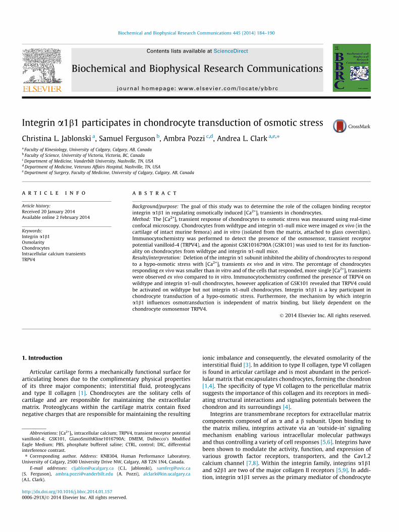
Both hypo- and hyper-osmotic stress delayed the [Ca2+]i tran-sients after withdrawal and infusion of the medium by 57 s(p = 0.04) or 122 s (p < 0.001), respectively relative to iso-osmoticcontrols (Fig. 3A). Additionally, the delay in [Ca2+]i transientsobserved after inducing a hyper-osmotic stress was significantlygreater (p = 0.003) than hypo-osmotically stressed cells (Fig. 3A).
3.3. Extracellular matrix effect
On average, the percentage of chondrocytes responding duringex vivo experiments was 6 times smaller than for in vitro experi-ments (p < 0.001) (Fig. 2). Furthermore, of the cells that responded,a larger proportion were single [Ca2+]i transients ex vivo (average73%) compared to in vitro (average 40%) (p = 0.007) (Fig. 2).
To determine how the presence of the extracellular matrixinfluenced the characteristics of chondrocyte [Ca2+]i transients,ex vivo and in vitro comparisons were made. [Ca2+]i transientsobserved in ex vivo chondrocytes were more than twice as largebut 4.3 s shorter than those in vitro (p < 0.001, p = 0.006, respec-tively) (Fig. 3B and C). The rise time of [Ca2+]i transients wasapproximately half the duration of the total transient suggestingthat the calcium waves were symmetrical about their peak (datanot shown). Finally, [Ca2+]i transients in ex vivo chondrocytes weredelayed by 113 s after withdrawal and infusion compared toin vitro chondrocytes (p < 0.001) (Fig. 3D).
Genotype had no effect on the characteristics of chondrocyte[Ca2+]i transients.
3.4. TRPV4
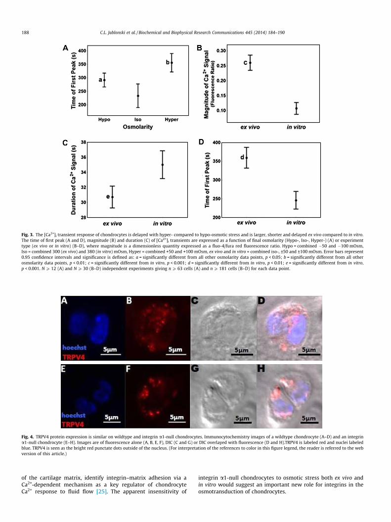
To assess whether loss of integrin a1b1 might affect TRPV4 lev-els and/or expression, immunocytochemistry was conducted.Immunostained full-thickness sections of cartilage showed similarexpression of TRPV4 in wildtype and integrin a1-null chondro-cytes, in terms of both staining intensity and punctate pattern(Fig. 4).
To determine if TRPV4 was functional as well as present on inte-grin a1-null and wildtype chondrocytes, the selective agonist ofTRPV4, GSK101 was utilized to stimulate in vitro chondrocytesand the resulting [Ca2+]i transients were measured. The additionof 10 nM GSK101 resulted in a 2-fold increase in the percentageof wildtype chondrocytes responding with [Ca2+]i transients(p = 0.004), however GSK101 had no effect on the percentage ofintegrin a1-null chondrocytes responding (Fig. 2C). Interestingly,the GSK101 [Ca2+]i transients of wildtype chondrocytes occurredwithin seconds and lasted throughout the 9 min experiment(Fig. 1E). In contrast, [Ca2+]i transients produced in the presenceof GSK101 in integrin a1-null chondrocytes were similar to the sin-gle or multiple transients produced by vehicular control or osmoticchallenge (Fig. 2C). Thus, the [Ca2+]i transient characteristics ofintegrin a1-null chondrocytes in the presence of GSK101 suggestthat chondrocytes were responding to infusion/withdrawal andnot the presence of GSK101.
4. Discussion
This study shows that integrin a1b1 plays a critical role in chon-drocyte [Ca2+]i transient response to hypo-osmotic stress, and thatintegrin a1b1 exerts this influence in a ligand-independentmanner. Previous studies on isolated bovine chondrocytes sus-pended in alginate hydrogels supplemented with Arg-Gly-Asp, anadhesion protein found on type VI collagen and other molecules
Fig. 3. The [Ca2+]i transient response of chondrocytes is delayed with hyper- compared to hypo-osmotic stress and is larger, shorter and delayed ex vivo compared to in vitro.The time of first peak (A and D), magnitude (B) and duration (C) of [Ca2+]i transients are expressed as a function of final osmolarity (Hypo-, Iso-, Hyper-) (A) or experimenttype (ex vivo or in vitro) (B–D), where magnitude is a dimensionless quantity expressed as a fluo-4/fura red fluorescence ratio. Hypo = combined �50 and �100 mOsm,Iso = combined 300 (ex vivo) and 380 (in vitro) mOsm, Hyper = combined +50 and +100 mOsm, ex vivo and in vitro = combined iso-, ±50 and ±100 mOsm. Error bars represent0.95 confidence intervals and significance is defined as: a = significantly different from all other osmolarity data points, p < 0.05; b = significantly different from all otherosmolarity data points, p < 0.01; c = significantly different from in vitro, p < 0.001; d = significantly different from in vitro, p < 0.01; e = significantly different from in vitro,p < 0.001. N P 12 (A) and N P 30 (B–D) independent experiments giving n P 63 cells (A) and n P 181 cells (B–D) for each data point.
Fig. 4. TRPV4 protein expression is similar on wildtype and integrin a1-null chondrocytes. Immunocytochemistry images of a wildtype chondrocyte (A–D) and an integrina1-null chondrocyte (E–H). Images are of fluorescence alone (A, B, E, F), DIC (C and G) or DIC overlayed with fluorescence (D and H).TRPV4 is labeled red and nuclei labeledblue. TRPV4 is seen as the bright red punctate dots outside of the nucleus. (For interpretation of the references to color in this figure legend, the reader is referred to the webversion of this article.)
188 C.L. Jablonski et al. / Biochemical and Biophysical Research Communications 445 (2014) 184–190
of the cartilage matrix, identify integrin–matrix adhesion via aCa2+-dependent mechanism as a key regulator of chondrocyteCa2+ response to fluid flow [25]. The apparent insensitivity of
integrin a1-null chondrocytes to osmotic stress both ex vivo andin vitro would suggest an important new role for integrins in theosmotransduction of chondrocytes.
C.L. Jablonski et al. / Biochemical and Biophysical Research Communications 445 (2014) 184–190 189
The characteristics of integrin-mediated chondrocyte [Ca2+]i
signaling are important to consider within the context of TRPV4– the dominant channel by which chondrocytes sense and respondto an osmotic stress [14]. Immunocytochemistry confirmed asimilar expression pattern for TRPV4 on both wildtype and integrina1-null chondrocytes, both in terms of the punctate pattern andintensity of staining within cells. To further explore the relation-ship between integrin a1b1 and TRPV4, we presented in vitro chon-drocytes from wildtype and integrin a1-null mice with the TRPV4agonist, GSK101 and measured the resulting [Ca2+]i transients.When presented with GSK101, over twice as many wildtype cellsresponded with [Ca2+]i transients compared to vehicular controls.GSK101 transients occurred within seconds and lasted throughoutthe entire 9 min experiment. Both of these phenomena wereabsent in integrin a1-null chondrocytes. Taken together, the re-sults above may suggest that integrin a1b1 has a high position inthe hierarchy of osmotic signal transduction, similar to TRPV4,and/or some influence on TRPV4.
Although the levels of this channel are similar in both geno-types, we provide evidence that its activity is decreased in the inte-grin 1-null chondrocyte suggesting that integrin a1b1 directlyparticipates in osmotic signal transduction. Further evidencesupporting a role for TRPV4 in integrin-mediated mechanotrans-duction are the findings that anti-integrin a2 antibody reducesthe TRPV4-mediated response to hypotonicity and that TRPV4coimmunoprecipitates with the integrin a2 subunit in cultureddorsal root ganglion neurons of mice and rats [17].
Our finding that hypo-osmotic stress induces [Ca2+]i transientsin chondrocytes both in vitro and ex vivo agrees well with previousstudies [14,26]. In agreement with previous ex vivo studies [14] butin contrast to in vitro studies [27], we measured no effect of50 mOsm or 100 mOsm of hyper-osmotic stress on [Ca2+]i tran-sients ex vivo or in vitro. A recent study showed porcine chondro-cytes exposed to >140 mOsm of hyper-osmotic stress respondedwith [Ca2+]i transients in vitro [27]. These data together with thepresent study may suggest there is a minimum threshold require-ment for a hyper-osmotic response to occur in vitro. It is importantto note that the osmolarity of the iso-osmotic control media usedby Erickson et al. was 310 mOsm compared to 380 mOsm – thesurface zone maximum and deep zone minimum osmolaritymeasured in human tissue – in our study.
Additionally, we provide a parallel comparison of ex vivo andin vitro measurements in osmotically stressed chondrocytes fromthe same species for the first time enabling us to measure the effectof the extracellular matrix in decreasing the percentage of chon-drocytes responding with [Ca2+]i transients and limiting the char-acteristics of those signals to single [Ca2+]i transients. Theobserved calcium peaks of ex vivo experiments were larger in mag-nitude and both shorter in duration and delayed compared toin vitro experiments. The duration of the calcium peaks we ob-served ex vivo agree well with previously reported ex vivo studiesutilizing compression-stimulated lapine chondrocytes or osmoti-cally-stimulated murine chondrocytes [14,28] and the magnitudeand delay of the [Ca2+]i transients we observed was comparableto the previously reported murine study [14]. Together theseobservations suggest that chondrocytes utilize contrasting signal-ing modalities in response to the type of stress (osmotic ormechanical) applied in addition to when surrounded by/isolatedfrom the extracellular matrix. As such, the [Ca2+]i transient re-sponse may be modulated by cytoskeleton/extracellular matrixinteractions perhaps involving ligand binding by integrins.
In conclusion we have shown that integrin a1b1 is necessary forchondrocytes to transduce a hypo-osmotic stress into an [Ca2+]i
transient, and that this influence is independent of its adhesionto the extracellular matrix. We suggest that the influence of inte-grin a1b1 on chondrocyte transduction of osmotic stress is made
via a mechanism that likely involves TRPV4. Furthermore, we showthat preservation of chondrocyte/extracellular matrix interactionsex vivo influences both the number of cells responding and thesignaling modalities harnessed by the chondrocyte. Together, theseobservations suggest a critical role for integrins in chondrocyteosmotransduction and thus in the homeostasis of articular carti-lage. Further analysis of integrin a1b1 expression, activation andfunction, particularly in relation to TRPV4 and other ion channelson the chondrocyte, will provide new insights into healthy chon-drocyte function, as well as the degenerative processes involvedin the onset and progression of osteoarthritis.
Acknowledgments
This work was supported by the University of Calgary Faculty ofKinesiology (A.C.), the Hunter Family Foundation (A.C.), NSERC Dis-covery Grant 371276-2010 (A.C.), AIHS Alberta Osteoarthritis TeamGrant (C.J.), Veterans Affairs Merit Reviews 1l01BX002025-01(A.P.) and the National Institutes of Health grants DK095761(A.P.). The authors would like to thank Carin Pihl for help withall animal procedures, Azim Jinha for assistance with MATLABanalysis, Jessica Chan, Danielle Curry, Charlie Shin and Rikesh Pare-kh for assistance with developing protocols.
References
[1] M. Huber, S. Trattnig, F. Lintner, Anatomy, biochemistry, and physiology ofarticular cartilage, Invest. Radiol. 35 (2000) 573–580.
[2] P.G. Bush, A.C. Hall, The osmotic sensitivity of isolated and in situ bovinearticular chondrocytes, J. Orthop. Res. 19 (2001) 768–778.
[3] V.C. Mow, C.C. Wang, C.T. Hung, The extracellular matrix, interstitial fluid andions as a mechanical signal transducer in articular cartilage, Osteoarthrit.Cartil. 7 (1999) 41–58.
[4] C.A. Poole, S. Ayad, J.R. Schofield, Chondrons from articular cartilage: I.Immunolocalization of type VI collagen in the pericellular capsule of isolatedcanine tibial chondrons, J. Cell Sci. 90 (Pt. 4) (1988) 635–643.
[5] R. Loeser, S. Sadiev, L. Tan, et al., Integrin expression by primary andimmortalized human chondrocytes: evidence of a differential role foralpha1beta1 and alpha2beta1 integrins in mediating chondrocyte adhesionto types II and VI collagen, Osteoarthrit. Cartil. 8 (2000) 96–105.
[6] D.E. Ingber, Integrins, tensegrity, and mechanotransduction, Gravit. Space Biol.Bull. 10 (1997) 49–55.
[7] K.-L. Chen, W.-H. Liu, Y.-Y. Yang, et al., Characterization of novel transforminggrowth factor-beta type I receptors found in malignant pleural effusion tumorcells, BMC Mol. Biol. 8 (2007) 72.
[8] J.-T. Chao, P. Gui, G.W. Zamponi, et al., Spatial association of the Cav1.2 calciumchannel with a5b1-integrin, Am. J. Physiol. Cell Physiol. 300 (2011) C477–C489.
[9] M. Zemmyo, E.J. Meharra, K. Kühn, et al., Accelerated, aging-dependentdevelopment of osteoarthritis in alpha1 integrin-deficient mice, ArthritisRheum. 48 (2003) 2873–2880.
[10] R. Loeser, C. Carlson, M. McGee, Expression of beta 1 integrins by culturedarticular chondrocytes and in osteoarthritic cartilage, Exp. Cell Res. 217 (1995)248–257.
[11] S. Shin, A. Pozzi, A. Clark, Spontaneous osteoarthritis develops earlier andbegins in the subchondral bone of the Integrin alpha1-null mouse knee, in:58th Annu. Meet. Orthop. Res. Soc., 2012: p. 788.
[13] M.N. Phan, H.A. Leddy, B.J. Votta, et al., Functional characterization of TRPV4 asan osmotically sensitive ion channel in porcine articular chondrocytes,Arthritis Rheum. 60 (2009) 3028–3037.
[14] A.L. Clark, B.J. Votta, S. Kumar, et al., Chondroprotective role of the osmoticallysensitive ion channel transient receptor potential vanilloid 4: age- and sex-dependent progression of osteoarthritis in Trpv4-deficient mice, ArthritisRheum. 62 (2010) 2973–2983.
[15] J.C. Sánchez, T.A. Danks, R.J. Wilkins, Mechanisms involved in the increase inintracellular calcium following hypotonic shock in bovine articularchondrocytes, Gen. Physiol. Biophys. 22 (2003) 487–500.
[16] K. Clark, M. Langeslag, B. van Leeuwen, et al., TRPM7, a novel regulator ofactomyosin contractility and cell adhesion, EMBO J. 25 (2006) 290–301.
[17] N. Alessandri-Haber, O.A. Dina, E.K. Joseph, et al., Interaction of transientreceptor potential vanilloid 4, integrin, and SRC tyrosine kinase in mechanicalhyperalgesia, J. Neurosci. 28 (2008) 1046–1057.
[18] S. Muramatsu, M. Wakabayashi, T. Ohno, et al., Functional gene screeningsystem identified TRPV4 as a regulator of chondrogenic differentiation, J. Biol.Chem. 282 (2007) 32158–32167.
190 C.L. Jablonski et al. / Biochemical and Biophysical Research Communications 445 (2014) 184–190
[19] H. Gardner, J. Kreidberg, V. Koteliansky, et al., Deletion of integrin alpha 1 byhomologous recombination permits normal murine development but givesrise to a specific deficit in cell adhesion, Dev. Biol. 175 (1996) 301–313.
[20] A.C. Hall, E.R. Horwitz, R.J. Wilkins, The cellular physiology of articularcartilage, Exp. Physiol. 81 (1996) 535–545.
[21] J.P. Urban, A.C. Hall, K.A. Gehl, Regulation of matrix synthesis rates by the ionicand osmotic environment of articular chondrocytes, J. Cell. Physiol. 154 (1993)262–270.
[22] A. Maroudas, Physicochemical Properties of Articular Cartilage, Adult ArticularCartilage, Pitman Medical Publishing, 1979.
[23] K.S. Thorneloe, A.C. Sulpizio, Z. Lin, et al., N-((1S)-1-{[4-((2S)-2-{[(2,4-dichlorophenyl)sulfonyl]amino}-3-hydroxypropanoyl)-1-piperazinyl]carbonyl}-3-methylbutyl)-1-benzothiophene-2-carboxamide(GSK1016790A), a novel and potent transient receptor potential vanilloid 4channel agonist induces urinar, J. Pharmacol. Exp. Ther. 326 (2008) 432–442.
[24] P. Lipp, E. Niggli, Modulation of Ca2+ release in cultured neonatal rat cardiacmyocytes. Insight from subcellular release patterns revealed by confocalmicroscopy, Circ. Res. 74 (1994) 979–990.
[25] S. Degala, W.R. Zipfel, L.J. Bonassar, Chondrocyte calcium signaling in responseto fluid flow is regulated by matrix adhesion in 3-D alginate scaffolds, Arch.Biochem. Biophys. 505 (2011) 112–117.
[26] G.R. Erickson, D.L. Northrup, F. Guilak, Hypo-osmotic stress induces calcium-dependent actin reorganization in articular chondrocytes, Osteoarthrit. Cartil.11 (2003) 187–197.
[27] G.R. Erickson, L.G. Alexopoulos, F. Guilak, Hyper-osmotic stress inducesvolume change and calcium transients in chondrocytes bytransmembrane, phospholipid, and G-protein pathways, J. Biomech. 34(2001) 1527–1535.
[28] S.-K. Han, W. Wouters, A. Clark, et al., Mechanically induced calcium signalingin chondrocytes in situ, J. Orthop. Res. 30 (2012) 475–481.