33
| Date post: | 23-Mar-2018 |
| Category: |
Documents |
| Upload: | truongkiet |
| View: | 215 times |
| Download: | 1 times |


Samar K Basak MBBS MD (AIIMS) DNB FRCS (Glasgow)Diplomate of National Board of Examiners in Ophthalmology, India
Disha Eye Hospitals Pvt. LimitedBarrackpore, Kolkata, West Bengal, India
New Delhi | London | Philadelphia | Panama The Health Sciences Publisher
Essentials ofOphthalmology
Prelims.indd 3 02-12-2015 16:38:09
Jayp
ee B
rothe
rs

Features
How this Book is Useful?
Ideal book for medical students, residents and health professionals for core information in ophthalmology.
New edition based on international standard meeting US guidelines like the use of American Spelling.
Bulleted and easy-to-read format.
More than 700 high-quality and accurate photographs and 250 line diagrams for a clear representation of detailed ophthalmic structures.
Updated with latest medical and surgical management techniques of the diseases.
Newer updates on latest investigations.
Includes an interactive DVD-ROM including important ophthalmic surgery videos.
Jayp
ee B
rothe
rs

Preface to the Sixth Edition
In the world of medicine, there is only one constant—change. Therefore, it has been essential to bring out the sixth edition of this book. Another constant—the appreciation and feedback from the readers, who are not just undergraduates, but also postgraduates and practising ophthalmologists from all around the Indian subcontinent, has encouraged me to improve and update the book. As a teacher, it has been a pleasure to see the readership of Essentials of Ophthalmology grow and now after eighteen years, and with over 75,000 copies sold, the book is stepping into adulthood! Ophthalmology has changed considerably over the years, especially with the advancement of technology, which reflect the improvement in surgical techniques, innovation in treatment regimens, and even in the way we diagnose and manage diseases. The highlights of this edition are—plenty of new colored diagrams and photographs, latest medical and surgical management of some diseases, and updates on newer investigations. The new look and the use of American spelling is to make the book reach out to a wider range of international readers. However, a few things have not changed in this edition, like its predecessors, is in four color and comes with a CD of major surgeries. The bulleted format of the book, detailed illustrations, a selection of slit-lamp examination, and such features which facilitate revision have been retained. Finally, this edition marks the handover of the book from Current Books International to Jaypee Brothers Medical Publishers. I take this opportunity to thank my old publisher for taking care of this book for so many years, and extend my thanks to my new publisher as well, who has published four of my ophthalmology books previously. Thanks are also due to all the doctors and staff members of Disha Eye Hospitals for their continued support and involvement. A special thanks to Dr Aditya Pradhan, my cornea fellow who has helped me with some notes. Dr Soham Basak, new budding ophthalmologist, has also helped me with his insights and suggestions and as always, thanks to my wife Dr Bani and my daughter Sohini. I hope this edition enhances your understanding of the subject. Readers are welcome to send in more constructive criticism.
Disha Eye Hospitals Pvt. Limited
Barrackpore, Kolkata - 700 120, West Bengal, India Samar K BasakE-mail: [email protected] [email protected]
Prelims.indd 7 02-12-2015 16:38:10
Jayp
ee B
rothe
rs

Contents
1. Embryology and Anatomy ...........................................................................................................................1 2. Physiology of the Eye .................................................................................................................................. 25 3. Physiology of Vision ..................................................................................................................................... 31 4. Neurology of Vision ..................................................................................................................................... 41 5. Ocular Pharmacology .................................................................................................................................. 46 6. Refraction of the Normal Eye .................................................................................................................... 62 7. Refractive Errors ............................................................................................................................................ 66 8. Accommodation and its Disturbances.................................................................................................. 82 9. Estimation and Correction of Refractive Errors .................................................................................. 86 10. History-taking and Examination of an Eye Case ................................................................................ 92 11. Diseases of the Eyelids ..............................................................................................................................115 12. Diseases of the Conjunctiva ....................................................................................................................138 13. Diseases of the Cornea and Sclera ........................................................................................................167 14. Diseases of the Uvea ..................................................................................................................................210 15. Diseases of the Lens ...................................................................................................................................242 16. Glaucoma .......................................................................................................................................................271 17. Diseases of the Vitreous............................................................................................................................308 18. Diseases of the Retina ...............................................................................................................................315 19. Diseases of the Optic Nerve ....................................................................................................................346 20. Intraocular Tumors .....................................................................................................................................359 21. Diseases of the Orbit .................................................................................................................................370 22. Diseases of the Lacrimal Apparatus .....................................................................................................386 23. Ocular Motility: Squint ..............................................................................................................................402 24. Ocular Injuries ..............................................................................................................................................427 25. Common Eye Surgeries ............................................................................................................................448 26. Blindness and its Prevention ..................................................................................................................484 27. Surgical Instruments ..................................................................................................................................498 28. Miscellaneous ..............................................................................................................................................535
Index ........................................................................................................................................................................ 559
Prelims.indd 13 02-12-2015 16:38:10
Jayp
ee B
rothe
rs
EVALUATION OF CORNEAL DISEASES
Symptoms z Pain: The cornea is richly supplied by sen-
sory nerve endings. In corneal abrasions or bullous keratopathy, the direct stimulation of bare nerve endings causes severe pain
z Decreased visual acuity: It is caused by loss of central corneal transparency
z Halos: They are due to diffraction of light by epithelial and subepithelial edema
z Photophobia z Lacrimation: It is due to reflex stimulation
of the corneal nerve.
SignsThe precise localization of corneal defects is best done by slit-lamp biomicroscopy.
z Epithelium: Punctate erosions, edema, fila-ments, superficial punctate keratitis, and staining with fluorescein and Rose-Bengal.
z Stroma: Infiltrates, edema, vascularization, deposits, and scarring (opacities).
z Descemet’s membrane: Breaks, folds, local ized thickening (excrescences), etc.
z Deposition of pigments: Corneal pigment deposition is associated with a variety of disorders (Table 13.1).
z Corneal thickness (by pachymetry) either by the optical or ultrasonic pachymeter.
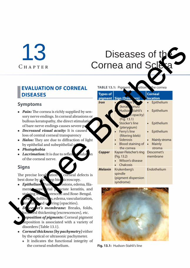
� It indicates the functional integrity of the corneal endothelium. Fig. 13.1: Hudson-Stahli’s line
TABLE 13.1: Pigment deposition in the cornea
Types of pigment Sign (Disorders)
Corneal location
Iron • Fleischer’s ring (keratoconus)
• Epithelium
• Hudson-Stahli’s line (old opacity) (Fig. 13.1)
• Epithelium
• Stocker’s line (pterygium)
• Epithelium
• Ferry’s line (filtering bleb)
• Epithelium
• Siderosis • Mainly stroma • Blood staining of
the cornea • Mainly
stromaCopper Kayser-Fleischer’s ring
(Fig. 13.2) • Wilson’s disease • Chalcosis
Descemet’s membrane
Melanin Krukenberg’s spindle(pigment dispersion syndrome)
Endothelium
Diseases of the Cornea and Sclera
13C h a p t e r
Chapter-13.indd 167 24-11-2015 10:20:23
Jayp
ee B
rothe
rs
� Ultrasonic pachymetry is useful before laser assisted in situ keratomileusis (LASIK) and other keratorefractive opera tions.
z Corneal sensation: Loss of corneal sensa-tion is an important sign to diagnose viral keratitis and neuroparalytic keratitis.
Laboratory diagnosis: Material for labor-atory analysis is obtained by scraping of the ulcer, from its base and margin. It is then plated onto the glass slides for Gram-staining and potassium hydroxide (KOH) preparation to identify the bacteria and fungus respectively. Part of the material is inoculated onto the following culture media.
z Blood agar mainly for aerobic bacteria. z Sabouraud’s media for fungus.
CONGENITAL ANOMALIES
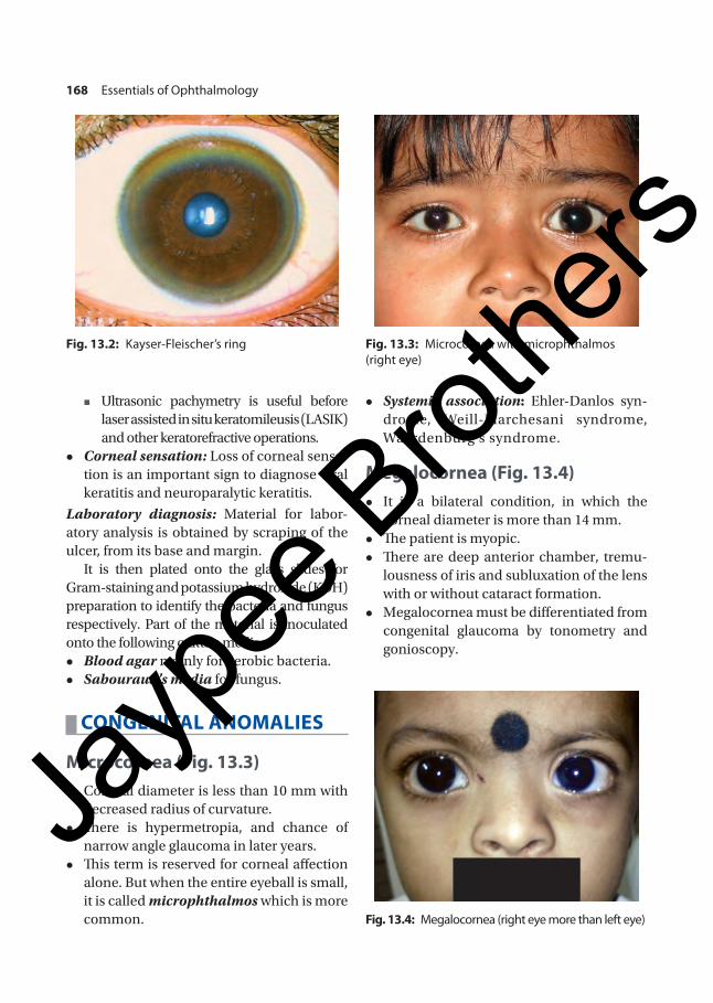
Microcornea (Fig. 13.3) z Corneal diameter is less than 10 mm with
decreased radius of curvature. z There is hypermetropia, and chance of
narrow angle glaucoma in later years. z This term is reserved for corneal affection
alone. But when the entire eyeball is small, it is called microphthalmos which is more common.
z Systemic association: Ehler-Danlos syn-drome, Weill-Marchesani syndrome, Waardenburg’s syndrome.
Megalocornea (Fig. 13.4) z It is a bilateral condition, in which the
corneal diameter is more than 14 mm. z The patient is myopic. z There are deep anterior chamber, tremu-
lousness of iris and subluxation of the lens with or without cataract formation.
z Megalocornea must be differentiated from congenital glaucoma by tonometry and gonioscopy.
Fig. 13.2: Kayser-Fleischer’s ring Fig. 13.3: Microcornea with microphthalmos (right eye)
Fig. 13.4: Megalocornea (right eye more than left eye)
168 Essentials of Ophthalmology
Chapter-13.indd 168 24-11-2015 10:20:23
Jayp
ee B
rothe
rs

Corneal Opacities z Central corneal opacity (Peter’s anomaly)
is due to defect in embryogenesis, due to incomplete separation of the lens from the surface ectoderm.
z It is usually associated with other anomalies in the iris or angle of the anterior chamber, with glaucoma.
Posterior Embryotoxon z An unusual prominence of Schwalbe’s
line which is the peripheral termination of Descemet’s membrane.
z It appears as a ring opacity in the deeper layer of the cornea.
CORNEAL EDEMA (FIGS 13.5A AND B)
z The integrity of both epithelium and the endothelium is necessary to maintain cor-nea in its relatively dehydrated state.
z It is associated with increased corneal thick-ness, and a variable decrease in corneal transparency.
z Source of fluid is either the aqueous humor (via endothelium) or the tears (via epithelium).
Pathology z Fluid accumulates between the basal epi-
thelial cells → then, in between the lamella and around the nerve fibers of the stroma → total haziness.
z In long standing cases, the epithelium tends to be raised into large vesicles or bullae, leading to bullous keratopathy (Fig. 13.6).
Etiology z Inflammatory: Corneal ulcer, erosions,
acute iridocyclitis (due to endothelial damage).
Figs 13.5A and B: Corneal edema. A. Diffuse illumination; B. Slit section
A B
Fig. 13.6: Bullous keratopathy in pseudophakic eye in slit section
Chapter 13 Diseases of the Cornea and Sclera 169
Chapter-13.indd 169 24-11-2015 10:20:24
Jayp
ee B
rothe
rs

z Traumatic: Mechanical trauma and post-surgical—due to endothelial damage, parti-cularly when vitreous remains adherent to it.
z Increased intraocular pressure: Acute edema in angle-closure glaucoma and epi-demic dropsy glaucoma. Chronic edema in long standing cases—as in absolute glaucoma and buphthalmos.
z Dystrophic condition of the cornea, e.g. Fuchs’ endothelial dystrophy.
z Hypoxia of the cornea: As in contact lens wearer due to epithelial edema, as a result of prolonged deprivation of atmospheric oxygen.
Symptoms z Colored halos around the light with red
color being outside, and blue inside. z Decreased visual acuity—due to corneal
haziness and irregular astigmatism.
Treatment z Use of hot air (by hairdryer). z Frequent instillation of concentrated
sodium chloride (5%) solution, or ointment containing 6% or 15% sodium chloride.
z Glycerine drop (to clear edema quickly and temporarily), but it is irritant.
z Bandage soft contact lenses, specially in case of ruptured bullae.
z Epithelium is stripped off, and is to be replaced with a thin conjunctival flap.
z Full-thickness penetrating keratoplasty to improve visual status.
INFLAMMATION OF THE CORNEA (KERATITIS)
Modes of Infection z Exogenous infections: Most common,
and cornea is primarily affected by viru-lent organisms.
z From the ocular tissues: Owing to direct anatomical continuity, the diseases of—
� Conjunctiva—spread to the corneal epithelium.
� Sclera—to the stroma. � Uveal tract—to the endothelium.
z Endogenous infections: Rare, as the cornea is avascular. They are typically allergic in nature.
Keratitis or inflammation of the cornea may be classified as follows:Superficial keratitis
z Infective keratitis � Bacterial � Fungal � Viral � Acanthamoebal.
z Non-infective keratitis � Central
− Exposure keratitis − Neurotrophic keratitis − Atheromatous keratitis.
� Peripheral − Marginal keratitis − Phlyctenular keratitis − Mooren’s ulcer − Terrien’s degeneration − Rosacea keratitis − Keratitis associated with collagen
diseases.Deep keratitis
z Interstitial keratitis z Disciform keratitis z Sclerosing keratitis.
In strict sense, keratitis and corneal ulcer are not always synonymous. A corneal ulcer is defined as a loss of cor-neal epithelium with underlying stromal infil-tration and suppuration associated with signs of inflammation with or without hypopyon.
BACTERIAL KERATITIS
Predisposing FactorsIntact corneal epithelium cannot be penetrated by any organism except, Neisseria gonorrhea, Neisseria meningitidis and Corynebacterium diphtheriae. In these cases, suppurative kera-
170 Essentials of Ophthalmology
Chapter-13.indd 170 24-11-2015 10:20:24
Jayp
ee B
rothe
rs

titis is always associated with a purulent con-junctivitis. With the other bacteria, keratitis is invariably associated with one or more of the following predisposing factors:
z Trauma to the corneal epithelium: By injury, foreign body, trichiasis, entropion, contact lens wear, etc.
z Underlying corneal diseases: Herpetic keratitis, corneal erosions, bullous kera-topathy, keratomalacia, etc.
z Neurotrophic or exposure keratopathy z Dry eyes z Chronic dacryocystitis or blepharitis z Use of topical steroids z Lowering of general body resistance, e.g.
malnutrition, diabetes, systemic immuno-suppressive therapy, etc.
PathologyUncomplicated, localized corneal ulcer is having four stages—stages of infiltration, progression, regression and cicatrization (Fig. 13.7).
Stage of Infiltration and Progression
z Edema and localized necrosis of the anterior part of the corneal stroma.
z Necrotic tissue is cast off leaving a saucer shaped defect, known as ulcer.
z Epithelium is sloughed off simultaneously with an involvement of larger area.
z Dense infiltration of polymorphs followed by macrophages. This infiltration extends for some distance around the ulcer.
z There is an associated iritis due to toxin being absorbed into the anterior chamber.
z This reaction is so toxic that leucocytosis takes place, and polymorphs poured out of the blood vessels pass into the aqueous, and gravitate at the bottom of anterior chamber. This is hypopyon and this pus remains sterile, so long as the Descemet’s membrane is intact.
Stage of Regression z The defect becomes larger, as there is diges-
tion of the necrotic tissue by the polymorphs. z Line of demarcation appears surrounding
the ulcer. z Infiltrations at the edges and the base start
to disappear, and polymorphs are replaced by mononuclear cells.
z The ulcerated area becomes smooth and relatively clear.
Stage of Cicatrization z Healing of the defect occurs by formation
of the fibrous tissue. z The fibrous tissue is derived from:
� Invading mononuclear cells � Keratocytes and � Endothelial cells of the new vessels.
Fig. 13.7: Pathological stages of corneal ulcer
Chapter 13 Diseases of the Cornea and Sclera 171
Chapter-13.indd 171 24-11-2015 10:20:24
Jayp
ee B
rothe
rs

z The epithelium grows from the edges of the defect to form a permanent covering.
Clinical Features
Symptoms z Acute pain, redness and lacrimation z Photophobia z Decreased visual acuity z White spot on the cornea.
Signs z Marked blepharospasm z Lid edema z Ciliary congestion of the conjunctiva z Ulcer usually starts as a grayish-white,
circumscribed infiltration, with edema of the surrounding tissue. There is absence of window reflex. The margins of the ulcer are overhanging, and the floor is covered by necrotic material. The extent of the ulcer may be detected by fluorescein staining and seen with blue filter. Ulcer area stains as brilliant green.
Later, the ulcer becomes gradually smooth in the regressive stage, and ultimately, scar tissue begins to appear the edges and the floor of the ulcer.
z Some degree of features of iritis. z Hypopyon may be present (Fig. 13.8). z Intraocular pressure (IOP) may be raised
in presence of hypopyon. But if the ulcer perforates, IOP decreases.
Certain bacteria produces characteristic response: • Staphylococcus aureus and Streptococcus
pneumonae—produce oval, yellowish-white, dense, opaque, stromal ulcer surrounded by relatively clear cornea.
• Pseudomonas—mucopurulent sticky and green-ish exudate, irregular deep ulcer with ‘ground-glass’ appearance of surrounding stroma. Ulcer progresses very rapidly, and results in corneal perforation within 48 hours.
• Enterobacteriaceae (E. coli, Proteus, Klebsiella, etc.) produce a shallow ulcer with pleomorphic grayish-white necrotic area. Sometimes, they produce ring-shaped corneal infiltrates.
Healing of a Corneal Ulcer z Healing of the corneal ulcer is taken place
by the formation of fibrous tissue. z The new fibers are not arranged regularly
as in normal lamellae, and they refract the light irregularly. Therefore, the scar is more or less opaque.
z Bowman’s membrane is never regenerated, and once it has been damaged, it leaves some degree of permanent corneal opacity.
z Scar tissue may not fill the gap exactly as level of surface, which is then little flat-tened—leading to the formation of corneal facets.
z If the corneal scar is thin, the resulting opacity is slight, it is called nebular. If rather more dense—it is called macular, and if still more dense and white—it is called a leucomatous (Figs 13.9A to C).
Old central leucoma, sometimes shows a horizontal pigmented line (Hudson-Stahli’s line) at the palpebral aperture.
z A thin, diffuse nebula on the pupillary area, interferes more with the vision than a strictly localized dense leucoma, so long as the latter does not block the whole pupillary area. The reason is, leucoma stops all the light which fall upon it, whereas the nebula refracts the light irregularly causing an irregular astigmatism.
Fig. 13.8: Bacterial corneal ulcer with hypopyon
172 Essentials of Ophthalmology
Chapter-13.indd 172 24-11-2015 10:20:24
Jayp
ee B
rothe
rs
Complications z Ectatic cicatrix: Thinned, scarred cornea
bulges under the influence of normal IOP. z Descemetocele (Figs 13.10A and B): Her-
niation of the elastic Descemet’s membrane through the ulcer as a transparent vesicle. It may persist, surrounded by a white cicatri-cial ring, or it may eventually rupture.
z Perforation and its effects � It is caused by sudden exertion (sneez-
ing, coughing, etc.) → acute rise in IOP → weak floor of the ulcer is unable to support this pressure → gives way.
� Once the ulcer perforates → sudden escape of aqueous → sudden fall in IOP → iris-lens diaphragm moves forwards
and comes in contact with the back of the cornea.
� The advantages of perforation − Pain is alleviated due to lowering of
IOP. − Better nutrition, and more antibody
or antibiotics will reach the ulcer area due to diffusion of fluid.
− Rapid healing of the ulcer. � Complications of perforation: They
vary according to the location and the size of the perforation.
− Anterior synechia: If the perfora-tion is very small, the iris becomes gummed down the opening, and the adhesion organizes.
Figs 13.9A to D: Corneal opacity. A. Nebular (arrows); B. Macular; C. Leucomatous; D. Adherent leucoma (arrows)
A
C
B
D
Chapter 13 Diseases of the Cornea and Sclera 173
Chapter-13.indd 173 24-11-2015 10:20:25
Jayp
ee B
rothe
rs
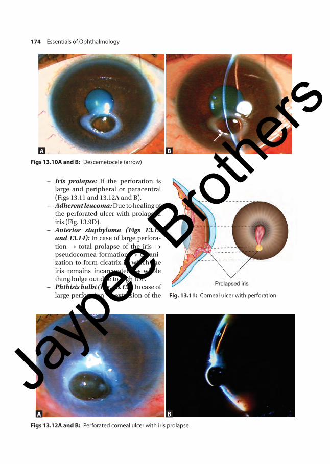
− Iris prolapse: If the perforation is large and peripheral or paracentral (Figs 13.11 and 13.12A and B).
− Adherent leucoma: Due to healing of the perforated ulcer with prolapsed iris (Fig. 13.9D).
− Anterior staphyloma (Figs 13.13 and 13.14): In case of large perfora-tion → total prolapse of the iris → pseudocornea formation → organi-zation to form cicatrix in which the iris remains incarcerated → whole thing bulge out due to high IOP.
− Phthisis bulbi (Fig. 13.15): In case of large perforation → extrusion of the Fig. 13.11: Corneal ulcer with perforation
A B
Figs 13.12A and B: Perforated corneal ulcer with iris prolapse
Figs 13.10A and B: Descemetocele (arrow)
A B
174 Essentials of Ophthalmology
Chapter-13.indd 174 24-11-2015 10:20:26
Jayp
ee B
rothe
rs

A B
Figs13.16A and B: A. Corneal fistula; B. With positive Seidel’s test
Fig. 13.15: Phthisis bulbi
Fig. 13.14: Total anterior staphylomaFig. 13.13: Partial anterior staphyloma
contents of the eyeball → shrinkage of the globe with low IOP.
− Subluxation or dislocation of the lens: Due to sudden forward move-ment of the lens.
− Anterior capsular cataract: When the perforation is at the center and small, the lens remains long in contact with the ulcer.
− Corneal fistula (Figs 13.16A and B): Due to repeated perforation → sealing of perforation by the exudate → re-opening due to strain → fistula formation.
Chapter 13 Diseases of the Cornea and Sclera 175
Chapter-13.indd 175 24-11-2015 10:20:27
Jayp
ee B
rothe
rs

− Expulsive hemorrhage: Due to sudden lowering of IOP → sudden dilatation of the choroidal vessels.
− Purulent acute iridocyclitis and its complications.
− Panophthalmitis (Fig. 13.17): Due to spread of infection into the interior of the eye.
ManagementA bacterial ulcer is a sight-threatening disease that demands urgent identification and eradi-cation of the causative organism. This is best performed with hospitalization of the patient.
z Identification of the organisms: This is done by smear preparation, and culture and sensitivity test of the scrapings, taken from the base and margin of the ulcer as described earlier.
Meanwhile, the aggressive treatment with antibiotics is started, till the reports are available, when it may be changed accord-ingly.
z Antibiotics � Gram ‘+ve’ organisms usually respond
to cephazoline, ciprofloxacin, vanco-mycin, gatifloxacin and moxifloxacin.
� Gram ‘–ve’ organisms are sensitive to gentamycin, tobramycin, polymyxin B, amikacin, ciprofloxacin, etc.
� Route of administration − Topical commercial preparation:
The drops are instilled at half hourly intervals round the clock for the first few days. Then, depending upon the response, the frequency of instilla-tion may be reduced.
− Topical fortified drop (concentrated) preparation: It is the most effective way to maintain a high and sustained level of antibiotics at the site of infection, e.g. fortified cefa-zolin, fortified tobramycin. Fortified tobramycin = 2 mL of parenteral tobramycin (80 mg) + 5 mL bottle of commercially available tobramy-cin eye drop. The resultant solution contains 14 mg/mL, and is stable for upto 30 days.
− Subconjunctival injections are for moderate to severe cases. It may be mixed with injection atropine. Gen-tamycin (20–40 mg) is usually pre-ferred.
− Systemic antibiotics: It is usually not necessary except, marginal bacterial ulcer, perforating corneal ulcer, and if the sclera is involved.
There is no need to change the initial antibiotics, if the response is good. But, if it is not so, the subsequent therapy is depended upon the sensitivity pattern.
z Atropine sulfate (1%) eye drops, 3 times daily to prevent ciliary spasm, and to control iritis.
z Hot fomentation is to improve circulation and to relieve pain.
z Analgesics with antacids for pain. z Removal of local predisposing factor if
any, like trichiasis, foreign body, chronic dacryocystitis, etc.
z Tab acetazolamide or timolol maleate eye drop (0.5%) to control IOP in selected cases.
z If the ulcer does not respond with these treatment, debridement and cauterization
Fig. 13.17: Corneal ulcer—panophthalmitis
176 Essentials of Ophthalmology
Chapter-13.indd 176 24-11-2015 10:20:27
Jayp
ee B
rothe
rs

of the ulcer under topical anesthesia may be done.
� Debridement means removal of nec- rotic material by scraping the floor with a spatula or no. 15 Bard-Parker blade.
� Cauterization is done by pure car-bolic acid or by iodine (1% iodine in potassium iodide solution).
� Carbolic acid penetrates more deeply, and extends its antibacterial property more widely.
z If the ulcer still does not respond, and if there is a threat of perforation—therapeutic penetrating keratoplasty is the best choice.
z Treatment of complications � Descemetocele: Rest and pressure
bandage, followed by therapeutic kera-toplasty.
� If the ulcer perforates: Alternate pres-sure bandage and medical treatment, or temporary tarsorrhaphy.
In case of small or impending perfora-tion—tissue adhesive with bandage contact lens (BCL).
� In leucoma or adherent leucoma: Pen-etrating keratoplasty.
� A leucoma with no personalized lamellar (PL): Treated either by tattooing or cosmetic contact lens.
� Anterior staphyloma: Staphylectomy or enucleation, then artificial eye.
� Panophthalmitis: Evisceration and then artificial eye.
� Corneal fistula: (i) Cyanoacrylate glue (tissue adhesive) application with BCL; (ii) Penetrating keratoplasty.
Corneal abscess (Fig. 13.18): It is localized collec-tion of pus in the cornea. Here, the epithelium is usually intact. It is similar to corneal ulcer. But in the treatment—evacuation of pus is done first, by a fine needle or knife, before starting the topical antibiotic therapy. The response of treatment will then be appreciated.
HYPOPYON CORNEAL ULCER (ULCUS SERPENS) (FIG. 13.19)
It is a typical bacterial ulcer which has a ten-dency to creep over the cornea in a serpigi-nous fashion, and associated with hypopyon and violent iridocyclitis.
Etiology z It occurs commonly in old debilitated
persons or the alcoholics. z Causative agent—Pneumococcus in most
of the cases. z Source of infection—mostly chronic dacr-
yocystitis.
Fig. 13.18: Corneal abscess
Fig. 13.19: Hypopyon corneal ulcer
Chapter 13 Diseases of the Cornea and Sclera 177
Chapter-13.indd 177 24-11-2015 10:20:27
Jayp
ee B
rothe
rs

SymptomsSame as the bacterial corneal ulcer, but there is remarkably little pain during the initial stage, hence, the treatment is often unduly delayed.
Signs z Grayish-white, disc-shaped ulcer near the
center of the cornea. z The ulcer is more at the edges than at the
center, and is particularly well-marked in one specific direction.
z A cloudy-gray area surrounds the ulcer area and again, more marked in the same direction.
z On one side the ulcer spreads, while on the other side it may be undergo simultaneous cicatrization, and the edges may be covered by fresh epithelium.
z The whole cornea may become hazy. z Violent iridocyclitis with a definite hyp o-
pyon. z Conjunctival and ciliary congestion. z Lids are edematous.
It must be noted that any corneal ulcer may be associated with hypopyon. But a corneal ulcer with hypopyon (due to other cause) is not equivalent to hypopyon corneal ulcer or ulcus serpens which has the above typical features (Fig. 13.20).Corneal ulcer with hypopyon is found in: • Pneumococcal infection • Pseudomonas pyocyanea • Gonococcal, Staphylococcal, Streptococcal,
Moraxella, etc. infection • Mycotic hypopyon corneal ulcer, as with
Aspergillus, Fusarium or Candida species.
Course z Great tendency for early perforation of the
cornea with its sequelae. z Hypopyon increases in size very rapidly.
leading to secondary glaucoma. z Ultimately, panophthalmitis may occur.
Treatment
Treatment is almost same as the treatment of a corneal ulcer. Special points are:
z Overhanging edges of the ulcer are due to be excised, and the floor is to be scraped with a spatula.
z Paracentesis to evacuate the pus if the hypopyon is massive.
z Secondary glaucoma is treated with anti-glaucoma agent like Tab. acetazolamide and timolol maleate (0.5%) eye drop.
z If there is any evidence of chronic dacryocystitis:
� Temporary punctal cautery � Dacryocystectomy.
MYCOTIC KERATITIS, KERATOMYCOSIS OR FUNGAL CORNEAL ULCER
Causative Agents z Filamentous fungus: Aspergillus and
Fusarium are common in our country. They are most prevalent among agricultural workers.
z Yeast: Candida albicans, frequently affects the immunocompromized host.
Fig. 13.20: Hypopyon corneal ulcer—bacterial
178 Essentials of Ophthalmology
Chapter-13.indd 178 24-11-2015 10:20:28
Jayp
ee B
rothe
rs

Predisposing Factors z Same as bacterial keratitis. z Indiscriminate use of topical steroids. z Immunocompromized subjects.
Mode of InfectionMycotic keratitis is typically preceded by ocu-lar trauma (often trivial in nature), mainly by agricultural and vegetable matters.
SymptomsSame as for the bacterial ulcer, but the symptoms are less prominent than an equal-size bacterial ulcer.
Signs z Dry looking, yellowish-white lesion with
indistinct margin. z Filamentous fungus keratitis has delicate,
feathery, finger-like projection into the adjacent stroma (Fig. 13.21). It may be surrounded by grayish-halo and mutiple satellite lesions (Fig. 13.22). Sometimes, it may be ring-shaped (Fig. 13.23).
z Candida keratitis—typically appears as collar-button abscess without any feathery edge (Fig. 13.24)
z The overlying epithelium is elevated and may be intact.
z Some degree of iridocyclitis and massive dense hypopyon are common. This hypo-pyon is immobile with upper convex border (Fig. 13.25).
Fig. 13.21: Fungal keratitis by filamentous fungus
Fig. 13.24: Fungal keratitis by Candida
Fig. 13.23: Ring-shaped fungal corneal ulcer
Fig. 13.22: Fungal corneal ulcer
Chapter 13 Diseases of the Cornea and Sclera 179
Chapter-13.indd 179 24-11-2015 10:20:28
Jayp
ee B
rothe
rs

Unlike bacterial ulcer, the hypopyon may ot be sterile, as the fungi can penetrate the Descemet’s membrane into the anterior chamber even without perforation.
z Vascularization usually does not occur.
Investigations z Ten percent potassium hydroxide (KOH)
mount preparation of the smear, to demon-strate causative fungus microscopically (Fig. 13.26).
z Culture in Sabouraud’s media.
Treatment z Scraping and debridement of the ulcer. z Atropine eye ointment—3 times daily. z Antifungal drugs
� Topical − Natamycin (5%) eye drop, 1 hourly. It is
effective against most common fungi mainly Fusarium and Aspergillus spp.
− Topical amphotericin B (0.15–0.25%), 1 hourly, effective against Aspergillus and Candida.
− Voriconazole eye drop—1–2 hourly. It is more effective against Aspergillus.
− Nystatin eye ointment, 5 times daily. It is only effective against Candida, and less potent.
Fig. 13.26: Filamentous fungus in KOH mount preparation
Fig. 13.25: Fungal keratitis—convex hypopyon
Topical antifungals are to be instilled for a long time, as the response is often delayed.
� Systemic: If the ulcer is deep, or mar - g inal, or if it is perforated, Tab. ketocon-azole or Tab. fluconazole may also be tried for 2–3 weeks.
z Cauterization of the ulcer may be done in non-responsive cases.
z Therapeutic full-thickness keratoplasty is much better solution in fungal keratitis.
ACANTHAMOEBA KERATITIS (FIG. 13.27)
Acanthamoeba is a free-living protozoa which has a trophozoite and a cystic form. It is found in stagnant water (pond, swimming pool, bath tub, etc.) and contact lens solution and contact lens cases.
Modes of Infection z Contact lens wearer—via contaminated
solution. z Noncontact lens wearer—direct conta-
mination after a minor trauma.
Clinical Features z Frequently misdiagnosed as herpetic or
fungal keratitis for many weeks.
180 Essentials of Ophthalmology
Chapter-13.indd 180 24-11-2015 10:20:28
Jayp
ee B
rothe
rs

z Severe pain, and it is out of proportion to the degree of inflammation (radial keratoneuritis).
z Progressive chronic stromal keratitis with recurrent breakdown of corneal epithelium.
z Frequent development of paracentral ring-shaped ulcer or abscess which may perforate.
z A nodular scleritis is a frequent finding. z It may be associated with limbal inflam-
mation (limbitis). z It does not respond to conventional anti-
microbial treatment.Diagnosis by calcofluor white staining of the smear for amoebic cyst. Acanthamoeba is cultured in special media—E. coli enriched nutrient agar plate.
Treatment z Propamidine isethionate (Brolene) (0.02%)
drops and ointment z Neomycin drops and ointment z Polyhexamethylene biguanide (PHMB)
(0.001%) drops is also be helpful z Chlorhexidine (0.02%) eye drop (prepared
from mouthwash)—1 hourly z In resistant cases, a therapeutic penetrating
keratoplasty may be required.
VIRAL KERATITIS
Herpes Simplex Viral KeratitisInfection with herpes simplex virus (HSV) is extremely common, and in the majority of the cases, it is subclinical.
Herper simplex virus (HSV) is of two types:1. HSV1 causes infection above the waist
(lips and eyes), and is acquired by kissing.2. HSV–2 causes infection below the waist
(genital herpes), and is acquired venereally. Primary infection may be subclinical or may cause mild ocular problems. Following the primary infection, the virus travels upto the trigeminal ganglion, and lies in a latent state. During recurrences, the virus travels, down along the sensory nerve to its target tissues, and causes recurrent lesions.Predisposing factors for recurrences
z Poor general health z Common cold and fever z Menstruation z Mild trauma z Topical and systemic corticosteroids z Immunosuppressive agents, etc.
Primary Herpetic Keratitis z Typically it occurs between 6 months to
5 years of age. z The typical lesion is an acute follicular
conjunctivitis. z Fine epithelial keratitis may be present,
which sometimes progresses into dendritic figure.
z Vesicular eruptions and edema of the lids. z It seldom causes serious problem, and
is treated by acyclovir eye ointment, five times a day for 3 weeks.
RECURRENT HSV KERATITISDendritic Keratitis (Fig. 13.28)
Symptoms z Acute pain, redness and lacrimation. z Photophobia and blurring of vision.
Signs z Circumciliary congestion. z Initially, numerous whitish plaques of
epithelial keratitis appear on the cornea,
Fig. 13.27: Acanthamoeba keratitis
Chapter 13 Diseases of the Cornea and Sclera 181
Chapter-13.indd 181 24-11-2015 10:20:29
Jayp
ee B
rothe
rs

known as superficial punctate keratitis (SPKs), and they quickly desquamate to form erosions.
z These erosions coalesce with each other and spread in all directions, and send out lateral branches with knobbed ends, to form ‘dendritic’ or ‘tree-like’ figure and this is pathognomonic.
z The bed of the ulcer stains with fluorescein, and diseased cell at the margin takes up Rose-Bengal stain.
z Corneal sensation is diminished or absent.
Geographical Keratitis (Ulcer) (Fig. 13.29)This is much larger epithelial lesion with typi-cal ‘geographical’ or ‘amoeboid’ configuration which occurs as a continued enlargement of dendritic ulcer. This is likely to occur following inadvertent use of topical steroids.
Treatment z Debridement of the ulcer. z Antiviral agents (anyone)
� Acyclovir (3%) eye ointment, 5 times daily for 14 days.
− It is the drug of choice.
− Prolong use of acyclovir ointment causes superficial punctate kerato-pathy.
� IDU (idoxuridine) 0.1% drop, every hour during day time, and every 2 hours (or ointment) during night.
or IDU (0.5%) ointment, 5 times a day.
� Trifluorothymidine (TFT) (1%) drop, every 2 hours during the day. It is more toxic than acyclovir.
� Adenine arabinoside (Ara-A) (3%) ointment, 5 times daily. It is as potent as IDU but less toxic.
� Oral acyclovir (200–400 mg), 1 tab 4 times daily for 5 days may be added.
z Atropine (1%) or homatropine (2%) eye drop, 2–3 times daily.
z Improvement of general health. z Cryocautery or iodine cautery of the ulcer
in resistance cases.
Stromal Necrotic Keratitis z Caused by active viral invasion and
destruction. z Cheesy and necrotic appearance of the
stroma. z Associated with anterior uveitis, called
herpetic keratouveitis.
Fig. 13.28: Herpes simplex virus dendritic keratitis Fig. 13.29: Herpes simplex virus geographical keratitis
182 Essentials of Ophthalmology
Chapter-13.indd 182 24-11-2015 10:20:29
Jayp
ee B
rothe
rs

z Vascularization, scarring and even per-foration may occur.
z Treatment by oral acyclovir, topical antiviral, atropine and judicious use of corticosteroids.
Metaherpetic Keratitis (Trophic Ulcer)
z It is due to persistent defects in the basement membrane of the corneal epithelium.
z It is not an active viral disease. z The margin is gray and thickened due to
heaped-up epithelium. z Treatment by artificial tears, patching or
bandage contact lens.
Disciform Keratitis (Figs 13.30A and B)Disciform keratitis is a deep keratitis with disc-like edema, mainly caused by herpes virus (and sometimes due to vaccinia and herpes zoster virus).
PathogenesisA delayed type of hypersensitivity reaction to HSV-antigen → low grade stromal inflamma-tion with damage to the underlying endothelium → passage of aqueous into the corneal stroma.
Clinical Features z In mild form, only focal disc-shaped stro-
mal edema with fine keratic precipitates (KPs). There is no necrosis or no neovas-cularization.
z In severe form � Stromal edema is more diffuse � Presence of Descemet’s folds � Deep vascularization � Focal bullous keratopathy � Wessely’s immune ring surrounding the
edema � Marked anterior uveitis. � Absent or diminished corneal sensation.
Treatment z The first aim is to heal any associated
epithelial lesion. z Topical corticosteroid drops (4–5 times
daily) under antiviral cover (acyclovir oint-ment 2–5 times daily) are given. The use of diluted steroid drops reduces the inci-dence of any steroid-related complication.
z Cycloplegic, like atropine eye ointment. z In majority of the cases, it resolves over a
period of several weeks.
A B
Figs 13.30A and B: Herpes simplex virus disciform keratitis (slit section)
Chapter 13 Diseases of the Cornea and Sclera 183
Chapter-13.indd 183 24-11-2015 10:20:29
Jayp
ee B
rothe
rs

HERPES ZOSTER OPHTHALMICUS
z Herpes zoster ophthalmicus (HZO) is a common infection caused by the varicella-zoster virus.
z The infection mainly affects the elderly patients.
z It is more common in immunocom-promised hosts (as in HIV).
Pathogenesis z After initial exposure, the virus remains
latent in the trigeminal ganglion. z Under the stress, it becomes reactivated,
replicates, and migrates down the oph-thalmic division of the trigeminal nerve to develop ocular complications.
Hutchinson’s sign (rule) (Fig. 13.31): When the tip of the nose is involved, the eye will also be involved, since both are supplied by the nasociliary nerve.
Symptoms z Vesicular eruptions around the eye,
forehead and scalp. z Severe pain along the distribution of the
ophthalmic division of 5th nerve. z Photophobia and lacrimation.
SignsThere are three stages:Stage I: Acute lesions—which develops with-in 3 weeks.Stage II: Chronic lesions—may persist for upto 10 years.Stage III: Recurrent lesions—which may reappear after 10 years.
Stage I: Acute Ocular Lesions z Lids: Redness, edema and vesicular erup-
tions. z Conjunctiva: Acute mucopurulent con-
junctivitis. z Sclera: Episcleritis and scleritis. z Cornea
� Punctate epithelial keratitis. � Microdendrites: Small, fine, multiple
dendritic or stellate lesions
D/D from HSV dendrite—in herpes zoster, the dendrites are peripheral, broader, plaque-like, raised from the surface, more frequently stellate-shaped, and without any terminal knobs.
� Nummular keratitis (Fig. 13.32): Mul-tiple granular lesions, surrounded by a halo of stromal haze.
� Disciform keratitis. � Sensation is diminished or absent.
Fig. 13.31: Herpes zoster ophthalmicus— Hutchinson’s sign
Fig. 13.32: Nummular keratitis
184 Essentials of Ophthalmology
Chapter-13.indd 184 24-11-2015 10:20:29
Jayp
ee B
rothe
rs

z Iris: Acute iridocyclitis with hyphema (hyp-eracute iritis) and patches of iris atrophy.
z Intraocular pressure may be raised. z Retina: Acute retinal necrosis. z Neuroophthalmological: Optic neuritis
and cranial nerve palsies—affecting the 3rd (most common), 4th and 6th nerves.
Stage II: Chronic Ocular Lesions z Ptosis, due to scarring of the lid z Trichiasis, entropion and lid notching may
also occur z Mucus-secreting conjunctivitis z Scleritis z Nummular and disciform keratitis z Seven percent of the patients present with
postherpetic neuralgia, which is worse at night and aggravated by touch and heat.
Stage III: Recurrent Ocular LesionsLike episcleritis, scleritis, mucus-plaque ker-atitis, nummular keratitis, iritis and second-ary glaucoma.
Treatment z Oral acyclovir: 800 mg 5 times daily for
7 days. It reduces pain, accelerates healing, and curtails vesiculation. It is also effective in anterior uveitis. It has no benefit on postherpetic neuralgia.
z Pain is relieved by strong analgesics, or even by injection pethidine.
z Antibioticcorticosteroid preparation for skin lesions. Calamine lotion promotes crust formation and is better avoided.
z Topical steroids in presence of corneal involvement, and anterior uveitis or scleritis.
z Topical antibiotics to prevent secondary infection.
z Cycloplegic for iritis or severe keratitis. z Systemic steroids—indications:
� To reduce the severity of postherpetic neuralgia
� Optic neuritis � Cranial nerve palsy.
z Artificial tears and bandage soft contact lens for persistent epithelial defects.
z Penetrating keratoplasty in case of dense scarring of the cornea.
LAGOPHTHALMIC KERATITIS EXPOSURE KERATITIS
This is due to exposure of the cornea when it remains insufficiently covered by the lids.
z The epithelium of the exposed cornea becomes dessicated and the stroma becomes hazy.
z Finally, owing to drying, the epithelium is cast off, and the raw area may be invaded by the micro-organisms.
Clinical Features z It ranges from minimum epithelial erosions
to severe ulceration, secondary infection and even perforation.
z The lower-third of the cornea is commonly affected, as this part remains more exposed (Figs 13.33A and B).
Other features and treatment have already been described earlier (see page 122).
NEUROTROPHIC KERATITIS z It occurs in an anesthetic cornea. z It appears that corneal sensation is very
important to maintain the health of the corneal epithelium.
z The loss of sensory influence alters the metabolic activity of the epithelium, there-by causes edema and exfoliation of the epithelial cells.
Etiology z Congenital: Very rare. Riley-Day syndrome,
anhidrotic ectodermal dysplasia, conge-nital insensitivity to pain may be the cause.
z Acquired � Section or affection of the 5th nerve � Herpes simplex virus and herpes zoster
virus keratitis
Chapter 13 Diseases of the Cornea and Sclera 185
Chapter-13.indd 185 24-11-2015 10:20:30
Jayp
ee B
rothe
rs

� Diabetes mellitus � Leprosy.
Neuroparalytic keratitis: It is due to paralysis of the motor nerve that closes the eyelids, and is normally a sequel to facial palsy. The picture is similar to ‘exposure keratitis’.
Clinical Features z Punctate epithelial erosions involving the
interpalpebral area. z Exfoliation of the epithelial cells followed
by central ulceration. z Corneal sensation is absent.
Treatment z Routine treatment of corneal ulcer z Ointments and patching z Amniotic membrane transplantation z Tarsorrhaphy for several months. No anes-
thesia is required during tarsorrhaphy, as the sensation of the lids and conjunctiva are absent.
ATHEROMATOUS ULCER z It develops over an old leucoma with
degenerative changes.
z It may start spontaneously or after a minor trauma.
z The degenerated tissue breaks away from the surface, and an ulcer develops.
z The ulcer progresses rapidly with a little tendency to heal.
z It gets easily infected and perforation may occur.
Treatment z Ointments and patching z Bandage contact lens and tear substitutes z If the eye is blind and painful—enucleation
or evisceration is better.
PERIPHERAL KERATITIS (THINNING)
This is a group of diseases, characterized by keratitis and/or melting of the corneal periphery.
Marginal Keratitis (Catarrhal Ulcer) (Fig. 13.34)It is caused by hypersensitivity reaction to staphylococcal exotoxins, and prevalent in patients having chronic staphylococcal blepharitis.
A B
Figs 13.33A and B: Bell’s palsy with corneal ulcer—right eye
186 Essentials of Ophthalmology
Chapter-13.indd 186 24-11-2015 10:20:30
Jayp
ee B
rothe
rs

Symptoms z Mild ocular irritation z Lacrimation and photophobia.
Signs z Subepithelial infiltrates at the periphery,
mostly at 4–8 o’clock position, or at 10–2 o’clock position.
z They are separated from the limbus by a clear zone of cornea.
z The lesions spread circumferentially and form a fluorescein staining ulcer.
z Corneal sensation is unaffected.
Treatment z Topical corticosteroids, 3–4 times daily for
a few days. z Mild cycloplegic, like cyclopentolate or
tropicamide. z Simultaneous treatment of blepharitis to
prevent recurrence.
Mooren’s Ulcer (Chronic Serpiginous Ulcer) (Fig. 13.35)It is a chronic progressive peripheral ulcer of unknown etiology. It may be due to an ischemic necrosis resul-ting from vasculitis of the perilimbal vessels.
Very rarely, it occurs following cataract surgery and hookworm infestation.There are two types of Mooren’s ulcer.1. A limited form: Unilateral and usually
affects the elderly people.2. A progressive form: Bilateral, relentlessly
progressive and affects the younger people.
Symptoms z Severe pain z Photophobia and lacrimation z Decreased vision due to irregular astigma-
tism and involvement of the visual axis in late stage.
Signs z Ulcer usually starts at the interpalpebral
area as patches of gray infiltrates at the margin.
z It spreads slowly undermining the epithe-lium and superficial stroma.
z Advancing border of the ulcer is having an overhanging edge.
z Later, it involves the entire circumference of the cornea.
z It also spreads towards center of the cor-nea, and also invades the sclera.
z Healing takes places from the periphery, and the healed area becomes vascularized, thinned and opaque.
z Perforation may occur with minor trauma.
Fig. 13.34: Marginal keratitis Fig. 13.35: Mooren’s ulcer
Chapter 13 Diseases of the Cornea and Sclera 187
Chapter-13.indd 187 24-11-2015 10:20:30
Jayp
ee B
rothe
rs
Treatment z Topical corticosteroids—at hourly inter-
vals. z Cycloplegics like atropine drops. z Conjunctival excision (peritomy): A 3 mm
collar of conjunctiva is excised from the limbus and parallel to the ulcer.
z Immunosuppressive therapy: Especially in bilateral cases—where the prognosis is worse.
� Systemic corticosteroids � Cytotoxic drugs, like cyclophosphamide,
azothioprim, or methotrexate. z Bandage soft contact lens z Lamellar sclerocorneal patch graft, if there
is threat of perforation.
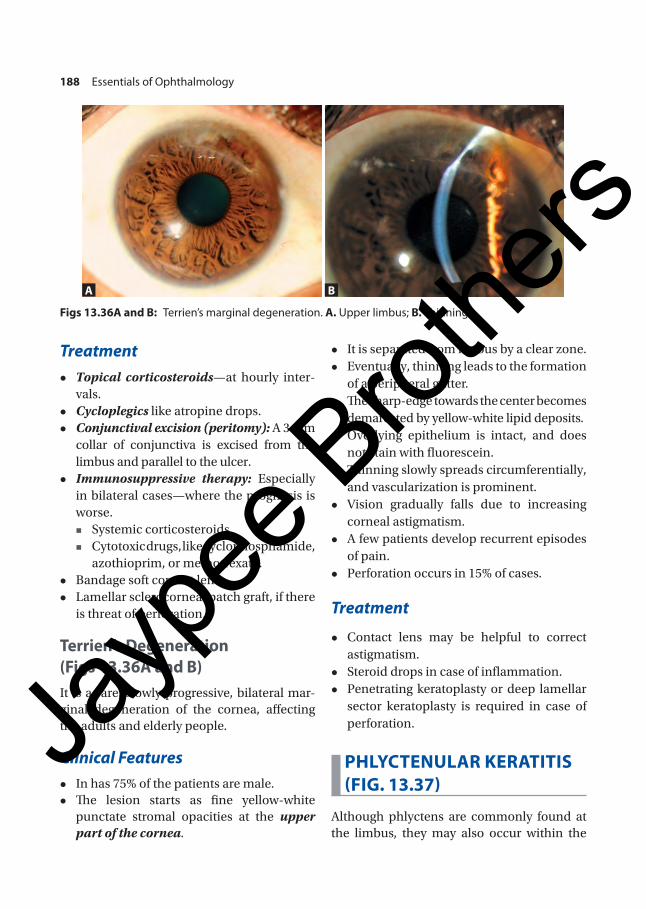
Terrien’s Degeneration (Figs 13.36A and B)It is a rare, slowly progressive, bilateral mar-ginal degeneration of the cornea, affecting the adults and elderly people.
Clinical Features z In has 75% of the patients are male. z The lesion starts as fine yellow-white
punctate stromal opacities at the upper part of the cornea.
z It is separated from limbus by a clear zone. z Eventually, thinning leads to the formation
of a peripheral gutter. z The sharp-edge towards the center becomes
demarcated by yellow-white lipid deposits. z Overlying epithelium is intact, and does
not stain with fluorescein. z Thinning slowly spreads circumferentially,
and vascularization is prominent. z Vision gradually falls due to increasing
corneal astigmatism. z A few patients develop recurrent episodes
of pain. z Perforation occurs in 15% of cases.
Treatment
z Contact lens may be helpful to correct astigmatism.
z Steroid drops in case of inflammation. z Penetrating keratoplasty or deep lamellar
sector keratoplasty is required in case of perforation.
PHLYCTENULAR KERATITIS (FIG. 13.37)
Although phlyctens are commonly found at the limbus, they may also occur within the
A B
Figs 13.36A and B: Terrien’s marginal degeneration. A. Upper limbus; B. Thinning
188 Essentials of Ophthalmology
Chapter-13.indd 188 24-11-2015 10:20:31
Jayp
ee B
rothe
rs
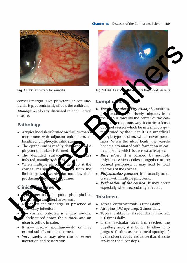
corneal margin. Like phlyctenular conjunc-tivitis, it predominantly affects the children.
Etiology: As already discussed in conjunctival disease.
Pathology z A typical nodule is formed on the Bowman’s
membrane with adjacent epithelium, as localized lymphocytic infiltrations.
z The epithelium is readily destroyed and a phlyctenular ulcer is formed.
z The denuded surface easily becomes infected, usually by Staphylococci.
z When multiple phlyctens develop at the corneal margin, blood vessels from the limbus grow towards the nodules, thus producing phlyctenular pannus.
Clinical Features z Symptoms include—pain, photophobia,
lacrimation and blepharospasm. z Mucopurulent discharge in presence of
secondary infection. z The corneal phlycten is a gray nodule,
slightly raised above the surface, and an ulcer is yellow in color.
z It may resolve spontaneously, or may extend radially onto the cornea.
z Very rarely, it may give rise to severe ulceration and perforation.
Complications z Fascicular ulcer (Fig. 13.38): Sometimes,
phlyctenular ulcer slowly migrates from the limbus towards the center of the cor-nea in a serpiginous way. It carries a leash of blood vessels which lie in a shallow gut-ter formed by the ulcer. It is a superficial allergic type of ulcer, which never perfo-rates. When the ulcer heals, the vessels become attenuated with formation of cor-neal opacity which is densest at its apex.
z Ring ulcer: It is formed by multiple phlyctens which coalesce together at the corneal periphery. It may lead to total necrosis of the cornea.
z Phlyctenular pannus: It is usually asso-ciated with multiple phlyctens.
z Perforation of the cornea: It may occur especially when secondarily infected.
Treatment z Topical corticosteroids, 4 times daily. z Atropine (1%) eye drop, 2 times daily. z Topical antibiotic, if secondarily infected,
4–6 times daily. z If the fascicular ulcer has reached the
pupillary area, it is better to allow it to progress further, as the corneal opacity left by the ulcer tract, is less dense than the site at which the ulcer stops.
Fig. 13.37: Phlyctenular keratitis Fig. 13.38: Fascicular ulcer (note the blood vessels)
Chapter 13 Diseases of the Cornea and Sclera 189
Chapter-13.indd 189 24-11-2015 10:20:31
Jayp
ee B
rothe
rs

Marginal Corneal Ulcer Associated with Systemic Collagen Vascular DiseasesMarginal corneal ulceration and thinning may occur in four main diseases:1. Rheumatoid arthritis2. Systemic lupus erythematosus3. Polyarteritis nodosa4. Wegener’s granulomatosis.The peripheral corneal changes are: • Peripheral keratitis • Peripheral corneal guttering (contact lens
cornea): Thinning or guttering occurs along the entire corneal periphery. As the central part of the cornea remains of normal thickness, the appearance resembles a hard contact lens placed on the eye—hence, the term
• Peripheral keratolysis, resulting in desceme-tocele formation and even perforation
• Sclerosing keratitis • Variable degree of corneal scarring and vascu-
larization • Other ocular manifestations include—scleritis,
choroidal vasculitis, retinal vasculitis, kerato-conjunctivitis sicca, anterior ischemic optic neuropathy, etc.
Treatment • Systemic and topical corticosteroids • Immunosuppressive agents • Conjunctival excision (peritomy) • Peripheral tectonic keratoplasty.
INTERSTITIAL KERATITIS
Interstitial keratitis (IK) is an inflammation affecting chiefly the corneal stroma, without primary involvement of the epithelium or endothelium.Causes: Three causes are congenital syphilis, tuberculosis, Cogan’s syndrome.
Syphilitic Interstitial Keratitis
Pathology
The disease is fundamentally a uveitis, and the keratitis, which clinically masks the uveitis, is secondary. These reactions are
largely allergic, since the spirochetes have never been detected in the cornea or uvea.
Clinical FeaturesIt is a late manifestation of congenital syphilis developing between 5 and 15 years. It is usually bilateral, and unilateral cases occur in delayed type, after the age of thirty, parti-cularly in acquired syphilis.
Symptoms z Initially, irritation and haziness of the
cornea. z Later pain, lacrimation, photophobia and
severe blepharospasm with increased hazi-ness of the cornea.
z Profound loss of vision.
SignsOcular signs
z Circumcorneal congestion. z The lesion starts as one or more hazy
patches in deeper cornea from the margin towards the center (Fig. 13.39A).
z They fuse together in 2–4 weeks, and the whole cornea becomes hazy with a steamy surface, giving rise to groundglass appear ance.
z Meanwhile, deep vascularization devel-ops, and the vessels meet in the center of the cornea. Since, the vessels are covered by a layer of hazy cornea, they appear as a dull, reddish-pink patch, known as salmon patch of Hutchinson.
z As a rule, there is always an associated iridocyclitis.
z After 2–4 months, the cornea begins to clear from periphery towards the center, and the vessels become nonperfused (only remain as fine lines of ghost vessels) (Fig. 13.39B). If there is re-inflammation of the cornea, the vessels may refill with blood.
z The clearing of the cornea takes many weeks or months, but little improvement
190 Essentials of Ophthalmology
Chapter-13.indd 190 24-11-2015 10:20:31
Jayp
ee B
rothe
rs

can be expected after eighteen months. Ultimately, majority of the patients regain some useful vision.
General signs z Hutchinson’s triad
� Interstitial keratitis (IK) � Permanent deafness � Hutchinson’s teeth (notching of the two
upper permanent incisors). z Prominent frontal eminences z Flatness of the nasal bridge z Rhagades at the angle of the mouth z Shotty cervical lymph nodes z Periosteal nodules on the tibia.
Treatment z Systemic penicillin: Its role is doubtful. It
may shorten the course of the disease. z Topical corticosteroids as drops or oint-
ment, 4–6 times daily. z Atropine (1%) ointment, 2 times daily. z Penetrating keratoplasty, if there is any
permanent dense corneal opacity.
Tuberculous Interstitial KeratitisThis is similar to the syphilitic interstitial kera-titis, but is more often unilateral and sectorial. Treatment is with anti-tubercular drugs, topical steroids and cycloplegics.
Cogan’s SyndromeIt is very rare, non-syphilitic interstitial kera-titis associated with vestibulocochlear dis-turbances (tinnitus, vertigo and deafness). It typically affects middle-aged patients. Treatment is with systemic steroids (to prevent permanent deafness), topical steroids and cycloplegics.
CORNEAL DEGENERATIONS AND DYSTROPHIES
Degeneration and dystrophy are not syno-nymous. Degeneration means, that the normal cells of a tissue undergo some patho-logical changes under the influences of some abnormal circumstances. Dystrophy means, the cells have some inborn defects which may cause pathological changes with passage of time (Table 13.2).
DEGENERATIONS
• Arcus senilis • Arcus juvenilis • Band-shaped
keratopathy • Lipid keratopathy
• Pellucid marginal • Terrien’s marginal • Salzmann’s nodular • Limbal girdle of Vogt
A B
Figs 13.39A and B: Interstitial keratitis. A. Lesion at the center; B. Ghost vessels
Chapter 13 Diseases of the Cornea and Sclera 191
Chapter-13.indd 191 24-11-2015 10:20:31
Jayp
ee B
rothe
rs

Arcus Senilis (Gerontoxon) (Fig. 13.40)
z It is bilateral lipid degeneration of the peri-pheral cornea, affecting the most elderly persons.
z It starts in the superior and inferior peril-imbal cornea, and then progresses circum-ferentially to form a band.
z The band is about 1 mm wide and its central border is diffuse, whereas the peri pheral edge is sharp.
z The peripheral sharp-edge is separated from the limbus by a clear zone of cornea, which may become thinned (senile furrow).
z Histologically: The lipid is first deposited anterior to the Descemet’s membrane and then in the anterior stroma, just beneath the Bowman’s layer.
z It does not affect the vision.
Arcus Juvenilis (Anterior Embryotoxon) (Fig. 13.41)It is similar to arcus senilis, except it occurs under the age of 40 years. The condition is usually associated with systemic hyper-lipidemias with raised serum cholesterol. So, a serum lipid profile is indicated in these patients.
Band-shaped Keratopathy (BSK) (Fig. 13.42)
z It is caused by deposition of calcium salts in the subepithelial layer of the cornea, and characterized by a horizontal band-shaped opacity.
TABLE 13.2: Difference between corneal degen-eration and corneal dystrophy
Corneal degeneration Corneal dystrophy
Usually unilateral and asymmetrical
Usually bilateral and symmetrical
Located peripherally Located centrally
Accompanied by vascularization
No vascularization
No inheritance pattern Hereditary (usually autosomal dominant)
Onset in middle life or later
Relatively early in onset
Secondary to some compromising factors, e.g. aging, inflammation, chemicals, trauma or systemic diseases
Unrelated to any systemic or local disease, or condition
Fig. 13.40: Arcus senilis with mature cataract Fig. 13.41: Arcus juvenilis
192 Essentials of Ophthalmology
Chapter-13.indd 192 24-11-2015 10:20:32
Jayp
ee B
rothe
rs
z The calcific band is largely at the palpebral fissure, and separated from the limbus by a clear zone.
z The band begins in the periphery at 3 and 9 o’clock position and affects the central area later on.
z There are numerous holes, where the corneal nerves penetrate the Bowman’s layer.
z As the band progresses, the epithelium becomes irregular and subsequently, its breakdown may be very painful.
z Pathologically: The calcium (mainly hydro-xyapatite) is deposited in the Bowman’s layer, in the epithelial basement membrane, and most superficial part of the stroma. The remaining layers of the cornea are clear.
Etiology z Childhood chronic iridocyclitis, especially
with juvenile rheumatoid arthritis (JRA) z Other ocular conditions: Phthisis bulbi
absolute glaucoma, interstitial keratitis z Systemic hypercalcemia z Idiopathic—in the elderly.
Treatment z Scraping of the epithelium overlying the
opacity, and application of 0.01 M solu-tion of disodium EDTA (ethylenediamine-tetraacetic acid) for 10 minutes. Then the
eye is patched with antibiotics and cyclo-plegics till the cornea is re-epithelialized.
z Lamellar keratoplasty for optical reason.
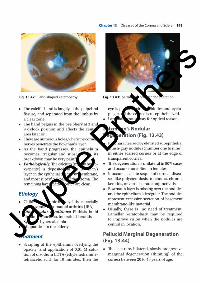
Salzmann’s Nodular Degeneration (Fig. 13.43)
z It is characterized by elevated subepithelial bluish-gray nodules (number one to nine), in either scarred cornea or at the edge of transparent cornea.
z The degeneration is unilateral in 80% cases and occurs more often in females.
z It occurs as a late sequel of corneal disea-ses like phlyctenulosis, trachoma, chronic keratitis, or vernal keratoconjunctivitis.
z Bowman’s layer is missing over the nodules and the epithelium is irregular. The nodules represent excessive secretion of basement membrane-like material.
z Usually, there is no need of treatment. Lamellar keratoplasty may be required to improve vision when the nodules are central in location.
Pellucid Marginal Degeneration (Fig. 13.44)
z This is a rare, bilateral, slowly progressive marginal degeneration (thinning) of the cornea between 20 to 40 years of age.
Fig. 13.42: Band-shaped keratopathy Fig. 13.43: Salzmann’s nodular degeneration
Chapter 13 Diseases of the Cornea and Sclera 193
Chapter-13.indd 193 24-11-2015 10:20:32
Jayp
ee B
rothe
rs
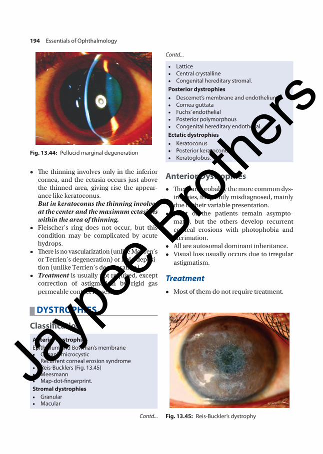
z The thinning involves only in the inferior cornea, and the ectasia occurs just above the thinned area, giving rise the appear-ance like keratoconus.
But in keratoconus the thinning involves at the center and the maximum ectasia is within the area of thinning.
z Fleischer’s ring does not occur, but this condition may be complicated by acute hydrops.
z There is no vascularization (unlike Mooren’s or Terrien’s degeneration) or lipid deposi-tion (unlike Terrien’s degeneration).
z Treatment is usually not required, except correction of astigmatism by rigid gas permeable contact lenses.
DYSTROPHIES
ClassificationAnterior dystrophiesEpithelium and Bowman’s membrane • Cogan’s microcystic • Recurrent corneal erosion syndrome • Reis-Bucklers (Fig. 13.45) • Meesmann • Map-dot-fingerprint.
Stromal dystrophies • Granular • Macular
• Lattice • Central crystalline • Congenital hereditary stromal.
Posterior dystrophies • Descemet’s membrane and endothelium • Cornea guttata • Fuchs’ endothelial • Posterior polymorphous • Congenital hereditary endothelial.
Ectatic dystrophies • Keratoconus • Posterior keratoconus • Keratoglobus.
Anterior Dystrophies z These are probably the more common dys-
trophies, frequently misdiagnosed, mainly due to their variable presentation.
z Most of the patients remain asympto-matic, but the others develop recurrent corneal erosions with photophobia and lacrimation.
z All are autosomal dominant inheritance. z Visual loss usually occurs due to irregular
astigmatism.
Treatment z Most of them do not require treatment.
Fig. 13.44: Pellucid marginal degeneration
Fig. 13.45: Reis-Buckler’s dystrophyContd...
Contd...
194 Essentials of Ophthalmology
Chapter-13.indd 194 24-11-2015 10:20:32
Jayp
ee B
rothe
rs


