Metal–Organic Frameworks Porphyrin-Based Metal–Organic Frameworks for Biomedical Applications Jiajie Chen, Yufang Zhu,* and Stefan Kaskel* A ngewandte Chemi e Keywords: biosensing · metal–organic frameworks · porphyrins · tumor therapy A ngewandte Chemi e Reviews How to cite: International Edition: doi.org/10.1002/anie.201909880 German Edition: doi.org/10.1002/ange.201909880 &&&& # 2020 The Authors. Published by Wiley-VCH GmbH Angew. Chem. Int. Ed. 2020, 59, 2 – 28 Ü Ü These are not the final page numbers!
Transcript
Metal–Organic Frameworks
Porphyrin-Based Metal–Organic Frameworks forBiomedical ApplicationsJiajie Chen, Yufang Zhu,* and Stefan Kaskel*
Porphyrins are macromolecular heterocyclic compoundscomposed of porphin (C20H14N4) substituted by variousfunctional groups at the meso-position or b-position.[1] Inaddition, free-base porphyrins can be coordinated withnumerous metal ions at the porphyrin center to form metalcomplexes, also known as metalloporphyrins.[2] In fact, manyporphyrins and metalized porphyrin derivatives, such ascytochrome, heme, and chlorophyll, exist in nature and playsignificant roles in organisms. The family of porphyrinsdiscovered up to now have been comprehensively studied.[3]
Due to their large p-aromatic system, porphyrins displayexcellent chemical and thermal stability as well as distinctphotophysical and electrochemical properties, which can beregulated by the substitution patterns on porphin and thecoordinated metal ions. Besides the coordination of metalions at the porphyrin center, the periphery of porphyrins canalso be bound to metal ions.[4] Intricate binding modes enableporphyrins to form desirable molecular cages or frameworksolids.[5] Porphyrins and porphyrin derivatives are an impor-tant class of organic chromophores with considerable absorp-tion in the visible section of the electromagnetic spectrum.Typically, the absorption spectrum shows the strongestabsorption around 400–450 nm (Soret band) and a series ofabsorption bands between 500 and 700 nm (Q-bands) withgradually reduced intensity.[1]
Porphyrins and porphyrin derivatives have been used invarious applications due to their characteristics and versatilefunctions (Figure 1). For instance, owing to their pronouncedvisible-light absorption and energy-transfer properties, por-phyrins can be used in light-harvesting, solar cells, andmolecular electronics.[6] The chemical catalytic activity ofporphyrins, especially metalloporphyrins, render them effi-cient photocatalysts, electrocatalysts, and biomimetic cata-
lysts.[7] The regulation of the opticaland electronic properties of porphyrinsby the coordination of metal ions andthe axial coordination of moleculesenable their application in molecularrecognition and metal-ion sensing.[8]
More importantly, porphyrins are cru-cial in many biological processes. Theyhave potent biological properties, such
as biocompatibility, effective clearance, long residence time intumors, few side effects, and the mimicking of variousbiological functions, which are extremely useful for biomed-ical applications.[9] For example, porphyrins have been widelydeveloped as photosensitizers for photodynamic therapy(PDT). At the same time, their fluorescence characteristicsmake porphyrin-based photosensitizers valuable systems for
Porphyrins and porphyrin derivatives have been widely explored forvarious applications owing to their excellent photophysical and elec-trochemical properties. However, inherent shortcomings, such asinstability and self-quenching under physiological conditions, limittheir biomedical applications. In recent years, metal–organic frame-works (MOFs) have received increasing attention. The construction ofporphyrin-based MOFs by introducing porphyrin molecules intoMOFs or using porphyrins as organic linkers to form MOFs cancombine the unique features of porphyrins and MOFs as well asovercome the limitations of porphyrins. This Review summarizesimportant synthesis strategies for porphyrin-based MOFs includingporphyrin@MOFs, porphyrinic MOFs, and composite porphyrinicMOFs, and highlights recent achievements and progress in the devel-opment of porphyrin-based MOFs for biomedical applications intumor therapy and biosensing. Finally, the challenges and prospectspresented by this class of emerging materials for biomedical applica-tions are discussed.
[*] J. Chen, Prof. Y. ZhuState Key Laboratory of High Performance Ceramics and SuperfineMicrostructureShanghai Institute of Ceramics, Chinese Academy of Sciences1295 Dingxi Road, Shanghai 200050 (China)andSchool of Materials Science and EngineeringUniversity of Shanghai for Science and Technology516 Jungong Road, Shanghai 200093 (China)E-mail: [email protected]
Prof. Y. ZhuHubei Key Laboratory of Processing and Application of CatalyticMaterialsCollege of Chemical Engineering, Huanggang Normal UniversityHuanggang, Hubei, 438000 (China)
Prof. S. KaskelProfessur f�r Anorganische Chemie IFachrichtung Chemie und LebensmittelchemieTechnische Universit�t DresdenBergstrasse 66, Dresden, 01062 (Germany)E-mail: [email protected]
The ORCID identification number(s) for the author(s) of this articlecan be found under:https://doi.org/10.1002/anie.201909880.
� 2020 The Authors. Published by Wiley-VCH GmbH. This is anopen access article under the terms of the Creative CommonsAttribution License, which permits use, distribution and reproduc-tion in any medium, provided the original work is properly cited.
AngewandteChemieReviews
&&&&Angew. Chem. Int. Ed. 2020, 59, 2 – 28 � 2020 The Authors. Published by Wiley-VCH GmbH www.angewandte.org
fluorescence imaging-guided therapy.[9b] However, the draw-backs of most porphyrins and porphyrin derivatives, inparticular instability, enzymatic degradation, nonspecifictargeting, and propensity to self-quench under physiologicalconditions, seriously limit their biomedical applications. Toovercome these problems, various carriers have been devel-oped to encapsulate, physically adsorb, or covalently bindporphyrins and porphyrin derivatives, such as micelles,[10]
Metal–organic frameworks (MOFs) are a category ofhybrid porous coordination polymers with two- or three-dimensional (2D/3D) topologies assembled by the coordina-tion of metal ions/secondary building units (SBUs) withorganic linkers. As an emerging class of material, MOFs haverecently attracted increasing attention because of their multi-ple merits including ultrahigh porosity,[15] tunable pores,adjustable structure and composition, unsaturated metalsites, and functional diversity.[16] Furthermore, the particlesize of MOFs can be easily decreased down to the nanoscale,and nanoscale MOFs (NMOFs) are suitable as deliverysystems. Compared with other types of carriers, NMOFs aremore promising as versatile nanoplatforms for biomedicalapplications, such as drug delivery, tumor therapy, bioimag-ing, and biosensing.[17]
Accordingly, it was proposed that the integration ofporphyrins into MOFs could lead to multifunctional carriershaving promising characteristics and versatile functionalcomponents. From this perspective, porphyrin-based MOFsare ideal biomaterials and their full potential has not yet beentapped. Moreover, the conceptual construction of porphyrin-based MOFs matches well with advanced design concepts forhighly active biomaterials that maximize integrated func-tional building units while minimizing inactive constituents.[18]
Therefore, much effort has been devoted to the developmentof various porphyrin-based MOF platforms for biomedicalapplications. The loading of porphyrins into MOF channelsand the decoration of porphyrins on the MOF surface areefficient strategies for the synthesis of porphyrin@MOFs thatenhance the stability of the porphyrin and facilitate potentialapplications.[19] Interestingly, free-base porphyrins and metal-loporphyrins can also be used as organic linkers to assembleporphyrinic and metalloporphyrinic MOFs, respectively. In
Jiajie Chen is currently studying MaterialsScience and Engineering at University ofShanghai for Science and Technology andhas been in the Master’s program since2017. His research interests focus on thesynthesis of multifunctional porphyrinicMOFs for biomedical applications in cancertherapy and biosensing.
Yufang Zhu studied Materials Physics andChemistry and received his PhD degree atShanghai Institute of Ceramics, ChineseAcademy of Sciences in 2006. He worked asan Alexander von Humboldt research fellowat Technical University Dresden (Germany)and an ICYS postdoctoral fellow at NationalInstitute for Materials Science (Japan) from2006 to 2010. Since 2011 he has beenProfessor of Materials Science and Engineer-ing at University of Shanghai for Scienceand Technology, and Professor at ShanghaiInstitute of Ceramics, Chinese Academy ofSciences since 2019.
Stefan Kaskel studied Chemistry andreceived his PhD degree in T�bingen in1997. After a postdoctoral stay as a FeodorLynen fellow of the Alexander von Humboldtfoundation in the group of J. D. Corbett, heobtained his habilitation degree in 2003 atBochum University on the design and func-tionality of new porous materials. From2002 to 2004 he was also a group leader atthe Max Planck Institute for Coal Research.Since 2004 he has been Professor of Inor-ganic Chemistry at Dresden University ofTechnology and since 2008 he has also
headed the Department of Chemical Surface Technology at the FraunhoferInstitute for Material and Beam Technology (IWS), Dresden.
Figure 1. Prominent applications of porphyrins and porphyrin deriva-tives.
AngewandteChemieReviews
&&&& www.angewandte.org � 2020 The Authors. Published by Wiley-VCH GmbH Angew. Chem. Int. Ed. 2020, 59, 2 – 28� �
this way, the self-aggregation and self-quenching of theporphyrins can be prevented and the physicochemical proper-ties improved. Due to the highly active frameworks andimproved performance of porphyrins in MOFs, porphyrin-based MOFs present a promising opportunity for biomedicalapplications.
Numerous excellent reviews in the past two decades haveseparately addressed porphyrins and MOFs, and their respec-tive potential biomedical applications. However, so fara comprehensive review linking porphyrins to MOFs forbiomedical applications has been missing. In this Review, wesummarize the latest achievements and progress in the field ofporphyrin-based MOFs, from synthesis strategies all the wayto functions and applications. We discuss numerous biomed-ical applications to provide a concise overview and motivationfor further work in this promising direction. Furthermore, wealso discuss the challenges and prospects for biomedicalapplications of porphyrin-based MOFs.
2. Synthesis of Porphyrin-Based MOFs
A tremendous number of MOFs, for example ZIFs(zeolite imidazolate frameworks),[20] MILs (Mat�riaux del’Institut Lavoisier),[21] and UiOs (Universitetet i Oslo),[22]
have been successfully synthesized, and provide efficientplatforms to encapsulate or deliver pharmaceuticals, imagingagents, and enzymes. For developing biomedical nanoplat-forms, the downsizing of MOFs to the nanoscale (10–100 nm)is essential because of the significant influence on the size-dependent function and biodistribution of administeredparticles. To date, a variety of strategies have been proposedfor the synthesis of NMOFs and have been summarized inseveral reviews.[23] Particularly for the synthesis of NMOFs,several efficient methods have also been developed, includinghydrothermal/solvothermal, sonochemical, and mechano-chemical methods, microwave-assisted synthesis, and reversemicroemulsion. However, there have been no review articleson the synthesis of porphyrin-based MOFs.
Typically, porphyrin-based MOFs can be categorized asporphyrin@MOFs and porphyrinic MOFs, according to thedistinct structures assembled from porphyrins and MOFs. Forporphyrin@MOFs, free-base porphyrins or metalloporphyr-ins are integrated as guest molecules into MOFs viaencapsulation in the pores or through adsorption or graftingon the surface. In porphyrinic/metalloporphyrinic MOFs,free-base porphyrins/metalloporphyrins act as organic linkersand coordinate with metal ions or SBUs. In addition,porphyrinic/metalloporphyrinic MOFs can be combinedwith other functional components, like magnetic nanoparti-cles, photothermal agents, and fluorescent quantum dots, toform multifunctional platforms. The morphology, structure,and composition of porphyrin-based MOFs are vital forbiomedical applications, and much effort has been devoted tosynthesizing porphyrin-based MOFs to meet the require-ments of biomedical applications.
2.1. Synthesis of Porphyrin@MOFs
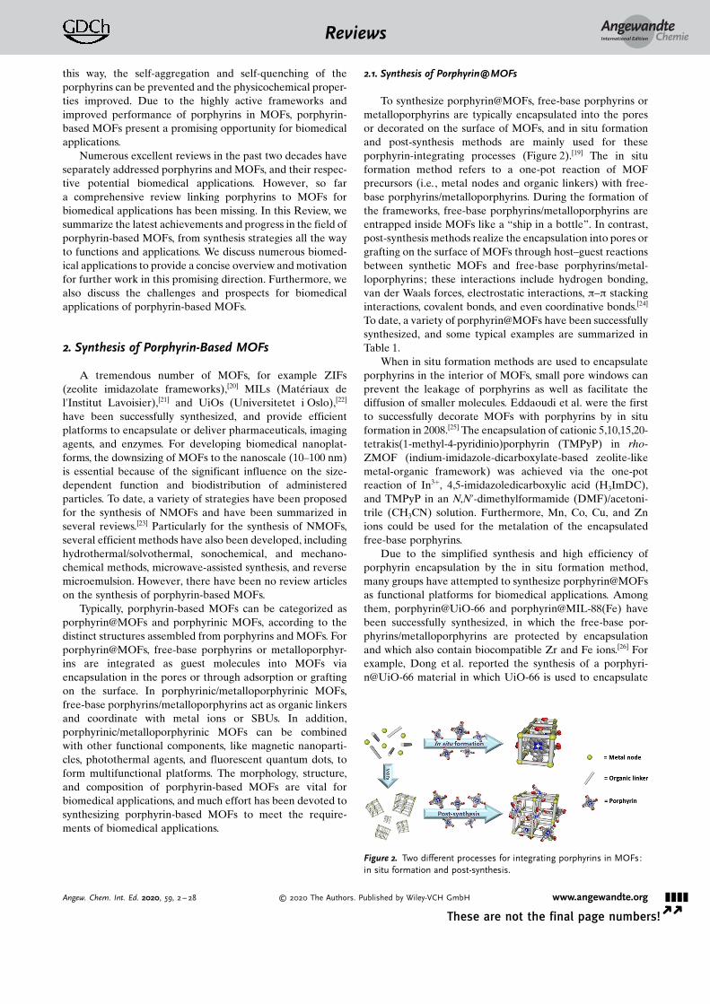
To synthesize porphyrin@MOFs, free-base porphyrins ormetalloporphyrins are typically encapsulated into the poresor decorated on the surface of MOFs, and in situ formationand post-synthesis methods are mainly used for theseporphyrin-integrating processes (Figure 2).[19] The in situformation method refers to a one-pot reaction of MOFprecursors (i.e., metal nodes and organic linkers) with free-base porphyrins/metalloporphyrins. During the formation ofthe frameworks, free-base porphyrins/metalloporphyrins areentrapped inside MOFs like a “ship in a bottle”. In contrast,post-synthesis methods realize the encapsulation into pores orgrafting on the surface of MOFs through host–guest reactionsbetween synthetic MOFs and free-base porphyrins/metal-loporphyrins; these interactions include hydrogen bonding,van der Waals forces, electrostatic interactions, p–p stackinginteractions, covalent bonds, and even coordinative bonds.[24]
To date, a variety of porphyrin@MOFs have been successfullysynthesized, and some typical examples are summarized inTable 1.
When in situ formation methods are used to encapsulateporphyrins in the interior of MOFs, small pore windows canprevent the leakage of porphyrins as well as facilitate thediffusion of smaller molecules. Eddaoudi et al. were the firstto successfully decorate MOFs with porphyrins by in situformation in 2008.[25] The encapsulation of cationic 5,10,15,20-tetrakis(1-methyl-4-pyridinio)porphyrin (TMPyP) in rho-ZMOF (indium-imidazole-dicarboxylate-based zeolite-likemetal-organic framework) was achieved via the one-potreaction of In3+, 4,5-imidazoledicarboxylic acid (H3ImDC),and TMPyP in an N,N’-dimethylformamide (DMF)/acetoni-trile (CH3CN) solution. Furthermore, Mn, Co, Cu, and Znions could be used for the metalation of the encapsulatedfree-base porphyrins.
Due to the simplified synthesis and high efficiency ofporphyrin encapsulation by the in situ formation method,many groups have attempted to synthesize porphyrin@MOFsas functional platforms for biomedical applications. Amongthem, porphyrin@UiO-66 and porphyrin@MIL-88(Fe) havebeen successfully synthesized, in which the free-base por-phyrins/metalloporphyrins are protected by encapsulationand which also contain biocompatible Zr and Fe ions.[26] Forexample, Dong et al. reported the synthesis of a porphyri-n@UiO-66 material in which UiO-66 is used to encapsulate
Figure 2. Two different processes for integrating porphyrins in MOFs:in situ formation and post-synthesis.
AngewandteChemieReviews
&&&&Angew. Chem. Int. Ed. 2020, 59, 2 – 28 � 2020 The Authors. Published by Wiley-VCH GmbH www.angewandte.org
5,15-di(4-carboxyphenyl)-10,20-bis(4-iodophenyl)porphyrinzinc(II) (DTPP(Zn)-I2) by the in situ formation method.[26a]
The loading of DTPP(Zn)-I2 in UiO-66 was estimated to beca. 1.4 wt%, which was much higher than that obtained by thenormal impregnation method (0.06 wt %).
For post-synthesis functionalization, the construction ofporphyrin@MOFs generally requires appropriate interactionsbetween the porphyrins and MOFs, and the following issuesrequire consideration:
1) MOFs should be activated to eliminate solvent mole-cules in the pores or channels before decoration.
2) The size and shape of the porphyrins should match thedimensions of the MOFs� pores or channels; this allows theentrance of porphyrins throughout the integration process.
3) Bond formation should be triggered between theporphyrin and the framework.
Demel et al. developed a phosphinate-based MOF (ICR-2, Inorganic Chemistry Rez No. 2), which could be decoratedwith anionic 5,10,15,20-tetrakis(4-R-phosphinatophenyl)por-phyrins (TPPPi(R), R = methyl, isopropyl, phenyl) by inter-action with the unsaturated metal sites on the ICR-2 sur-face.[27] The fraction of TPPPi(R) decorated on the MOFs washigher than that possible with the commercially available5,10,15,20-tetrakis(4-carboxyphenyl)porphyrin (TCPP). Thismight be explained by the fact that the bond between themetal ions and the phosphinic groups in TPPPi(R) is strongerthan that between the metal ions and the carboxylic groups inTCPP.
2.2. Synthesis of Porphyrinic MOFs
Porphyrins are macromolecular heterocyclic compoundsthat can be used as organic linkers to coordinate with metalions or SBUs to form porphyrinic MOFs. Such porphyrinicMOFs feature porphyrin functionality but also provide highporosity to host further secondary functional components. In1991 Robson et al. described the first porphyrinic MOFs,[28]
which were assembled from 5,10,15,20-tetrakis(4-pyridyl)por-
phyrin palladium(II) (TPyP(Pd)) as linkers and Cd2+ ions asnodes. Interestingly, SBUs, also termed metal clusters, couldbe formed through stable metal–oxygen bonds or metal–nitrogen bonds. Such units play a key role in improvingstructural stability, generating more coordination sites, pro-moting framework extension, preventing structural inter-penetration, and expanding pores in MOFs. Therefore, SBUsas nodes can be used to construct robust porphyrinic MOFswith high stability, and recently increasing attention has arisendue to their potential biomedical applications.[29] In 2002,Suslick et al. reported the first stable porphyrinic MOF(PIZA-1, porphyrinic Illinois zeolite analogue No. 1) withSBUs as nodes,[30] which was synthesized via self-assembly oftrinuclear CoII-carboxylate clusters and CoIII-metallopor-phyrin linkers through a solvothermal treatment. Theseporphyrinic MOFs have large and refillable tridirectionalchannels and exhibit significant hydrophilic character.
The rational choice of porphyrins plays a vital role inregulating the pores, shapes, and sizes of porphyrinic MOFsduring the synthesis.[31] Carboxy-based porphyrins, such asTCPP and its metalized molecules (TCPP(M)), have beenwidely used as linkers to synthesize porphyrinic MOFs.[31a]
Generally, free-base porphyrins can form porphyrinic MOFswithout metal chelation, while the cores of free-base por-phyrins can be pre-metalized or metalized by in situ chelationor post-synthesis chelation with various metal ions togenerate metalloporphyrinic MOFs. On the other hand, it isinteresting that weakly coordinated metal ions can bereplaced by other more strongly binding metal ions,[32] andthereby there are alternative approaches for the synthesis ofstable porphyrinic MOFs.
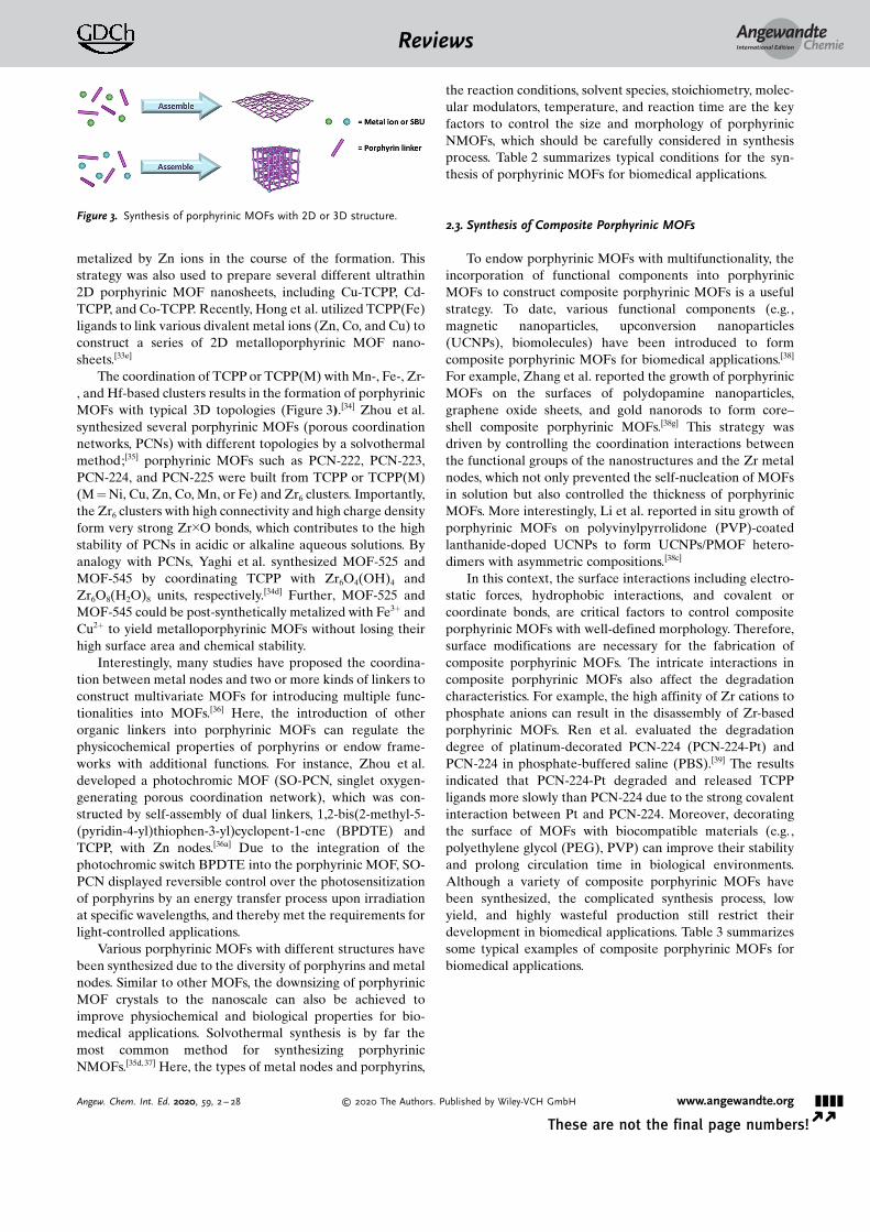
To date, various metal nodes, such as Cu, Zn, Co, and Cdions, have been used to assemble with TCPP or TCPP(M)linkers to form 2D MOFs (Figure 3).[6e, 33] For example, Zhanget al. reported the first surfactant-assisted method to synthe-size homogeneous ultrathin 2D MOF nanosheets (Zn-TCPP)with a thickness of less than 10 nm.[33c] In 2D Zn-TCPPnanosheets, one TCPP ligand is linked to four Zn paddle-wheel metal nodes (Zn2(COO)4) and the TCPP ligand is
Table 1: Typical examples of porphyrin@MOFs for biomedical applications.
MOF Porphyrin Method Biomedical application Ref.
UiO-66 DTPP(Zn)-I2 in situ formation PDT [26a]UiO-66 TCPP in situ formation PDT [26b,111]HKUST-1 TMPyP in situ formation PDT; fluorescence imaging [45]HKUST-1 TCPP(Fe) in situ formation DNA sensing [86a]HKUST-1 hemin in situ formation detection of H2O2, glucose [112]Fe-MIL-88 hemin in situ formation detection of TB [26c]Fe-MIL-88 hemin in situ formation detection of the FGFR3 gene mutation [26d]Fe-MIL-88 hemin in situ formation immunoassay of PSA [89]Tb-MOF hemin in situ formation colorimetric immunoassay of AFP [113]TTA-UC MOF TCPP(Pd) in situ formation ultralow-power in vivo imaging [77]rho-ZMOF TMPyP(Pt) in situ formation anion-selective sensing [8c]UiO-66 TPP-SH post-synthesis PDT [111]UiO-AM TAPP post-synthesis PDT [114]ICR-2 TPPPi(R), R =methyl, isopropyl, phenyl post-synthesis PDT [27]MIL-101(Al)-NH2 hemin post-synthesis detection of H2O2, glucose [93]Cu-MOF-74 hemin post-synthesis 2,4,6-trichlorophenol sensing [115]
[a] TB = thrombin; PSA =prostatic specific antigen; AFP= alpha-fetoprotein.
AngewandteChemieReviews
&&&& www.angewandte.org � 2020 The Authors. Published by Wiley-VCH GmbH Angew. Chem. Int. Ed. 2020, 59, 2 – 28� �
metalized by Zn ions in the course of the formation. Thisstrategy was also used to prepare several different ultrathin2D porphyrinic MOF nanosheets, including Cu-TCPP, Cd-TCPP, and Co-TCPP. Recently, Hong et al. utilized TCPP(Fe)ligands to link various divalent metal ions (Zn, Co, and Cu) toconstruct a series of 2D metalloporphyrinic MOF nano-sheets.[33e]
The coordination of TCPP or TCPP(M) with Mn-, Fe-, Zr-, and Hf-based clusters results in the formation of porphyrinicMOFs with typical 3D topologies (Figure 3).[34] Zhou et al.synthesized several porphyrinic MOFs (porous coordinationnetworks, PCNs) with different topologies by a solvothermalmethod;[35] porphyrinic MOFs such as PCN-222, PCN-223,PCN-224, and PCN-225 were built from TCPP or TCPP(M)(M = Ni, Cu, Zn, Co, Mn, or Fe) and Zr6 clusters. Importantly,the Zr6 clusters with high connectivity and high charge densityform very strong Zr�O bonds, which contributes to the highstability of PCNs in acidic or alkaline aqueous solutions. Byanalogy with PCNs, Yaghi et al. synthesized MOF-525 andMOF-545 by coordinating TCPP with Zr6O4(OH)4 andZr6O8(H2O)8 units, respectively.[34d] Further, MOF-525 andMOF-545 could be post-synthetically metalized with Fe3+ andCu2+ to yield metalloporphyrinic MOFs without losing theirhigh surface area and chemical stability.
Interestingly, many studies have proposed the coordina-tion between metal nodes and two or more kinds of linkers toconstruct multivariate MOFs for introducing multiple func-tionalities into MOFs.[36] Here, the introduction of otherorganic linkers into porphyrinic MOFs can regulate thephysicochemical properties of porphyrins or endow frame-works with additional functions. For instance, Zhou et al.developed a photochromic MOF (SO-PCN, singlet oxygen-generating porous coordination network), which was con-structed by self-assembly of dual linkers, 1,2-bis(2-methyl-5-(pyridin-4-yl)thiophen-3-yl)cyclopent-1-ene (BPDTE) andTCPP, with Zn nodes.[36a] Due to the integration of thephotochromic switch BPDTE into the porphyrinic MOF, SO-PCN displayed reversible control over the photosensitizationof porphyrins by an energy transfer process upon irradiationat specific wavelengths, and thereby met the requirements forlight-controlled applications.
Various porphyrinic MOFs with different structures havebeen synthesized due to the diversity of porphyrins and metalnodes. Similar to other MOFs, the downsizing of porphyrinicMOF crystals to the nanoscale can also be achieved toimprove physiochemical and biological properties for bio-medical applications. Solvothermal synthesis is by far themost common method for synthesizing porphyrinicNMOFs.[35d, 37] Here, the types of metal nodes and porphyrins,
the reaction conditions, solvent species, stoichiometry, molec-ular modulators, temperature, and reaction time are the keyfactors to control the size and morphology of porphyrinicNMOFs, which should be carefully considered in synthesisprocess. Table 2 summarizes typical conditions for the syn-thesis of porphyrinic MOFs for biomedical applications.
2.3. Synthesis of Composite Porphyrinic MOFs
To endow porphyrinic MOFs with multifunctionality, theincorporation of functional components into porphyrinicMOFs to construct composite porphyrinic MOFs is a usefulstrategy. To date, various functional components (e.g.,magnetic nanoparticles, upconversion nanoparticles(UCNPs), biomolecules) have been introduced to formcomposite porphyrinic MOFs for biomedical applications.[38]
For example, Zhang et al. reported the growth of porphyrinicMOFs on the surfaces of polydopamine nanoparticles,graphene oxide sheets, and gold nanorods to form core–shell composite porphyrinic MOFs.[38g] This strategy wasdriven by controlling the coordination interactions betweenthe functional groups of the nanostructures and the Zr metalnodes, which not only prevented the self-nucleation of MOFsin solution but also controlled the thickness of porphyrinicMOFs. More interestingly, Li et al. reported in situ growth ofporphyrinic MOFs on polyvinylpyrrolidone (PVP)-coatedlanthanide-doped UCNPs to form UCNPs/PMOF hetero-dimers with asymmetric compositions.[38c]
In this context, the surface interactions including electro-static forces, hydrophobic interactions, and covalent orcoordinate bonds, are critical factors to control compositeporphyrinic MOFs with well-defined morphology. Therefore,surface modifications are necessary for the fabrication ofcomposite porphyrinic MOFs. The intricate interactions incomposite porphyrinic MOFs also affect the degradationcharacteristics. For example, the high affinity of Zr cations tophosphate anions can result in the disassembly of Zr-basedporphyrinic MOFs. Ren et al. evaluated the degradationdegree of platinum-decorated PCN-224 (PCN-224-Pt) andPCN-224 in phosphate-buffered saline (PBS).[39] The resultsindicated that PCN-224-Pt degraded and released TCPPligands more slowly than PCN-224 due to the strong covalentinteraction between Pt and PCN-224. Moreover, decoratingthe surface of MOFs with biocompatible materials (e.g.,polyethylene glycol (PEG), PVP) can improve their stabilityand prolong circulation time in biological environments.Although a variety of composite porphyrinic MOFs havebeen synthesized, the complicated synthesis process, lowyield, and highly wasteful production still restrict theirdevelopment in biomedical applications. Table 3 summarizessome typical examples of composite porphyrinic MOFs forbiomedical applications.
Figure 3. Synthesis of porphyrinic MOFs with 2D or 3D structure.
AngewandteChemieReviews
&&&&Angew. Chem. Int. Ed. 2020, 59, 2 – 28 � 2020 The Authors. Published by Wiley-VCH GmbH www.angewandte.org
Porphyrin-based MOFs offer a variety of attractivefeatures making use of the integration of porphyrins intoMOFs, in particular excellent photophysical and electro-chemical properties, porous structure, modular functionaliza-tion, and biocompatibility. These characteristics are beneficialfor applications of porphyrin-based MOFs in biomedicine,which have attracted more and more attention. Nowadays,numerous interesting biomedical applications, such as drugdelivery, tumor therapy, bioimaging and biosensing, havebeen developed for porphyrin-based MOFs. In the following,we summarize recent progress in the area of biomedicalapplications of porphyrin-based MOFs, including photody-namic therapy (PDT), synergistic therapy, imaging-guidedtherapy, and biosensing (Figure 4).
3.1. Photodynamic Therapy with Porphyrin-Based MOFs
PDT is an efficient tumor therapy strategy that requiresthree essential factors: photosensitizer, light, and molecularoxygen in cells.[40] As a result, PDT generates highly cytotoxicreactive oxygen species (ROS) under light irradiation. ROSincluding singlet oxygen (1O2), superoxide anion radical(O2
�C), and hydroxyl radical (COH) can induce tumor celldeath through apoptosis and/or necrosis, and even tumorimmunity. By intravenous injection of photosensitizers foraccumulation in tumor tissue and local exposure of tumorsites to light, PDT can selectively destroy tumor cells whiledamage to the surrounding normal cells and tissues isminimized.[41] This approach profits from being non-invasiveand has fewer side effects (e.g., radiation damage, drugtoxicity) than conventional surgery, radiotherapy, and chemo-therapy.
Table 2: Typical examples of porphyrinic MOFs for biomedical applications.
MOF Metalion
Porphyrin Other components Biomedical application Ref.
Porphyrin and its derivatives are excellent photosensi-tizers for PDT and several have been approved for clinicaltrials. For porphyrin-based MOFs, the high porphyrin loadingcapacity and free diffusion of oxygen and ROS in the porousmaterial allow for highly efficient PDT. Furthermore, thebiocompatibility and biodegradability of MOFs result in
enhanced biosafety during PDT. Since the first report onporphyrinic NMOFs for potential tumor PDT by Lin et al. in2014,[42] numerous porphyrin-based MOFs have beendesigned for PDT. The most important advances are sum-marized in Tables 1– 3.
Table 3: Typical examples of composite porphyrinic MOFs for biomedical applications.
Composite PorphyrinicMOF
Functional components Biomedical application Ref.
PCN-224-Pt PCN-224 Pt nanoparticles PDT [39]mCGP PCN-224 GOx, catalase, tumor cell membrane cancer targeted starvation, PDT [52b]l-Arg@PCN@Mem PCN-224 l-arginine, tumor cell membrane gas therapy, PDT [52c]Dox/UCMOFs PCN-224 UCNPs chemotherapy, PDT [38c]MOF-UCNP PCN-224 UCNPs PDT [38d]UiO-66(OH)2@PCN PCN-224 UiO-66(OH)2 Cu2+ sensing [98]RB-PCN PCN-224 Fe3O4, RBITC broad-range pH sensor for fluorescence imaging [102]AuNR@MOFs@CPT Zr-TCPP Au nanorods, CPT PDT, PTT, chemotherapy; fluorescence imaging [38a]PDA@MOF Zr-TCPP PDA nanoparticles PTT, PDT [38g]Fe3O4@C@PMOF Zr-TCPP Fe3O4@C nanospheres PTT, PDT; MRI, fluorescence imaging [79]NMOF-SNO Zr-TCPP(Mn) S-nitrosothiol gas therapy, PTT; MRI [81]MOF-525-PEDOT NTs MOF-525 poly(3,4-ethylenedioxythiophene) nanotubes detection of dopamine [95]MOF-525/MPC MOF-525 macroporous carbon detection of luteolin [119]Cu-TCPP(Co)/MWCNTs Cu-TCPP(Co) multi-walled carbon nanotubes detection of nitrite and H2O2 [38f ]Cu-TCPP(Fe)/GOx Cu-TCPP(Fe) GOx antibacterial activity and in vivo wound healing [104]Au NPs/Cu-TCPP(M) Cu-TCPP(M),
M = Fe, CoAu nanoparticles detection of glucose [38b]
Ag/Cu-TCPP(Cu) Cu-TCPP(Cu) Ag nanoparticles antibacterial activity and in vivo wound healing [103]MOFs/CS-rGO Cu-hemin chitosan-functionalized reduced graphene oxide detection of H2O2 [120]Cu-H MOFs/NECF Cu-hemin nitrogen-containing melamine carbon foam detection of trichlorfon [96]CHC-PZM@HA Zr-TCPP a-cyano-4-hydroxycinnamate, HA PDT [51]L/AuNP/(Fe-P)n-MOF Fe-TCPP Au nanoparticles, DNA T4 PNK activity detection [121]
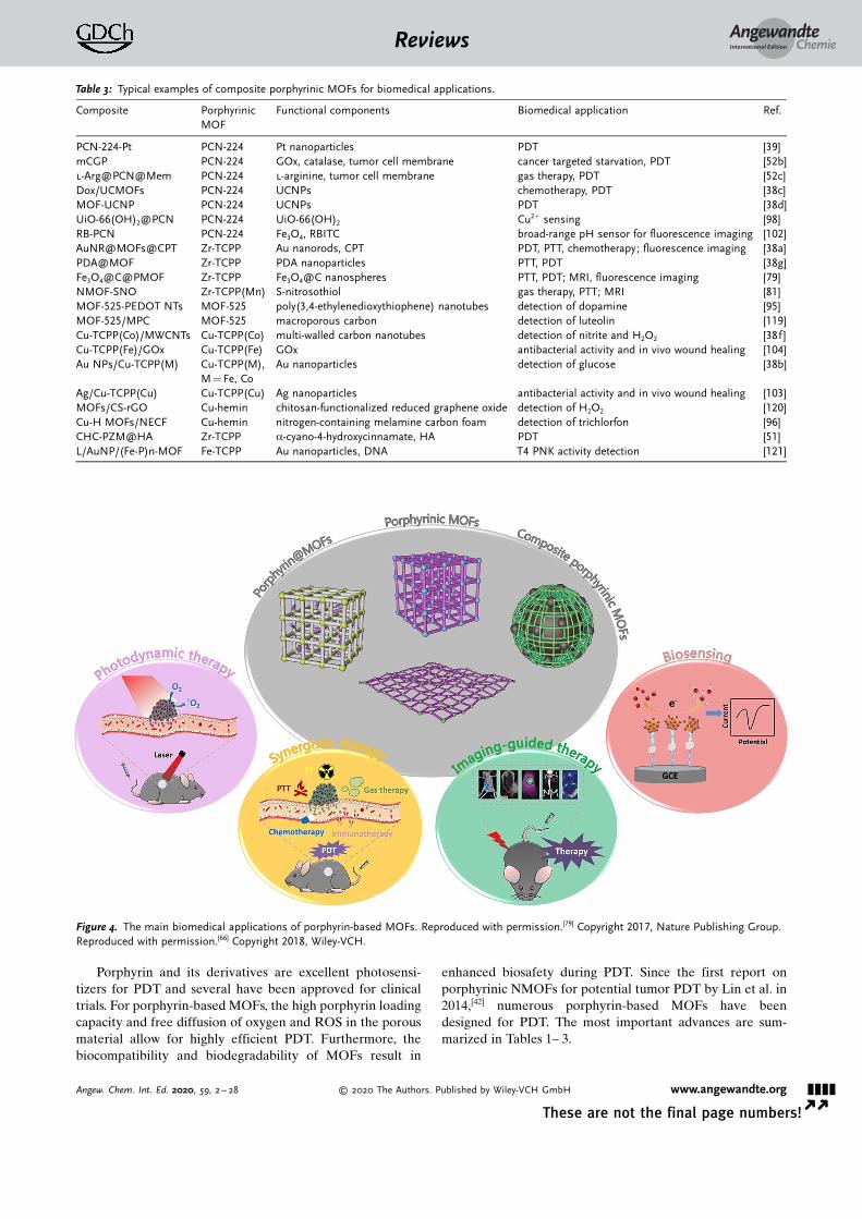
Figure 4. The main biomedical applications of porphyrin-based MOFs. Reproduced with permission.[79] Copyright 2017, Nature Publishing Group.Reproduced with permission.[66] Copyright 2018, Wiley-VCH.
AngewandteChemieReviews
&&&&Angew. Chem. Int. Ed. 2020, 59, 2 – 28 � 2020 The Authors. Published by Wiley-VCH GmbH www.angewandte.org
In general, the key factor for PDT is the quantity of 1O2
generated by photosensitizers upon light irradiation, whichcan be regulated by the efficiency of the intersystem crossing(ISC) of the photosensitizers from the singlet state (S1) to thetriplet state (T1).[43] Interestingly, the introduction of heavyatoms into photosensitizers is known to enhance ISC and isthe “heavy atom effect”. In porphyrin-based MOFs, theinteractions between porphyrins and metal ions can improvethe photodynamic properties of porphyrins on account of the“heavy atom effect”, and thereby increase the production of1O2.
[40a]
The first report on porphyrinic MOFs for PDTwas the useof DBP-UiO nanoplatelets, which were assembled from 5,15-di(p-benzoato)porphyrin (DBP) as linkers and Hf4+ ions asmetal nodes (Figure 5).[42] DBP-UiO nanoplates had a highporphyrin loading capacity of 77 wt %, and showed at leasttwice as efficient 1O2 production as free DBP. Correspond-ingly, DBP-UiO nanoplates presented highly enhanced PDTefficacy against human head and neck tumor cells (SQ20B)compared to free DBP. Therefore, the coordination ofporphyrin linkers with heavy metal nodes in porphyrinicMOFs lead to an enhancement on PDT, caused by the “heavy
atom effect”, and the same phenomenon was observed inother porphyrinic MOFs.[33d, 39,44] For example, Gd-TCPPMOF nanosheets, which were linked by TCPP with Gd ions,showed improved photosensitive activity compared to freeTCPP.[33d] On the other hand, the encapsulation of porphyrinsin MOFs can also enhance ISC together with the 1O2
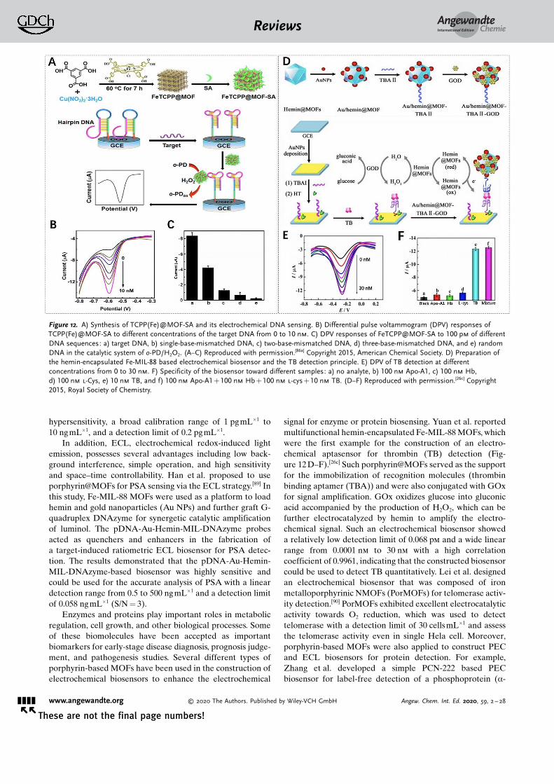
generation. Lei et al. encapsulated TMPyP into HKUST-1 and found that the narrowed S1–T1 energy gap of TMPyPcaused by the interaction between the encapsulated TMPyPand Cu2+ nodes enhanced the ISC. As a result, TMPyP@H-KUST-1 exhibited higher 1O2 production capability comparedto free TMPyP, and thereby induced higher phototoxicity totumor cells.[45]
To optimize the photophysical properties and PDTefficacy of porphyrin-based MOFs, the reduction of porphyr-ins to chlorins by the hydrogenation of the cross-conjugateddouble bond of porphyrin ring is also an effective strategy,which could result in a red-shift of absorption and an increaseof the extinction coefficient.[46] Lin et al. described the firstchlorin-based NMOFs (DBC-UiO), which were synthesizedfrom Hf4+ nodes and 5,15-di(p-benzoato)chlorin (DBC), andfound an enhanced PDT effect for colon tumors.[47] There wasa 13 nm red shift in absorption and an 11-fold augmentation inthe extinction coefficient of the longest-wavelength Q band.Consequently, DBC-UiO NMOFs were three times asefficient as DBP-UiO NMOFs in the generation of 1O2 andexhibited a much stronger PDTeffect in dual colon tumor celllines (CT26 and HT29). Therefore, the use of chlorins toconstruct NMOFs provides nanoplatforms for realizing highlyefficient PDT owing to the improved photophysical proper-ties of the reduced porphyrins.
Generally, the accumulation of photosensitizers in tumortissues and further uptake by tumor cells facilitates efficientPDT. It is well known that the size of NMOFs is of crucialimportance during cellular uptake.[48] In many studies theMOF particle size has been optimized to further enhancePDT efficacy. For example, Zhou et al. synthesized PCN-224with sizes ranging from 30 to 190 nm to investigate the effectof particle size on cellular uptake and PDT efficacy.[35d] ThesePCN-224 nanoparticles showed different uptake levels inHela cells, and the 90 nm sized PCN-224 had a maximalcellular uptake amount, as determined by inductively coupledplasma mass spectrometry (ICP-MS). Furthermore, the PDTfor Hela cells indicated that the 90 nm-sized PCN-224induced 81 % cell apoptosis after PDT treatment, which wasmuch higher than that induced by other sized PCN-224,suggesting that the size-dependent cellular uptake of NMOFsdetermines PDT efficacy.
3.1.2. Light-Controlled Photodynamic Therapy
Controllable generation of 1O2 is useful for enhancingPDT efficacy, which requires the release of cytotoxic 1O2 onlyin tumor sites and less damage to normal cells and tissues.[49]
Light, as an essential exogenous condition for PDT, has beenutilized to control the generation of 1O2 from porphyrin-based MOFs in PDT, and thereby enhance PDT efficacy andreduce toxic side effects. For example, Zhou et al. synthesizedphotochromic SO-PCN MOFs by the integration of the
Figure 5. A) Synthesis of DBP-UiO NMOFs and generation of 1O2.B) 1O2 generation of DBP-UiO, H2DBP, and H2DBP + HfCl4 detected bySinglet Oxygen Sensor Green (SOSG) assay. C) In vitro PDT cytotox-icity of different components (PpIX= protoporphyrin IX). D) In vivotumor volume changes after PDT in the presence of different compo-nents (black and red arrows refer to the injection and irradiation timepoints, respectively). E) Photographs of the mice and the correspond-ing tumors after PDT. (A–E) Reproduced with permission.[42] Copyright2016, American Chemical Society.
AngewandteChemieReviews
&&&& www.angewandte.org � 2020 The Authors. Published by Wiley-VCH GmbH Angew. Chem. Int. Ed. 2020, 59, 2 – 28� �
photochromic switch molecule BPDTE (1,2-bis(2-methyl-5-(pyridin-4-yl)thiophen-3-yl)cyclopent-1-ene) into porphyrinicMOFs. The SO-PCN MOFs showed a reversible control of1O2 production via a competitive energy transfer pathwayupon specific wavelength irradiation.[36a] Here, the open andclosed state of BPDTE can be reversibly transformed uponphotoirradiation, resulting in different photophysical proper-ties (i.e., different energy levels). Upon irradiation of the SO-PCN MOFs at l> 450 nm, the open state of BPDTE wasinduced, and energy transfer occurred from the TCPP excitedtriplet state to 3O2, stimulating the generation of 1O2.However, if SO-PCN is irradiated at l = 365 nm, BPDTE istransformed into the closed state, which is lower in energythan the TCPP, and thereby energy transfer happens to theclosed BPDTE, resulting in the quenching of 1O2 generation.Therefore, the introduction of BPDTE in porphyrinic MOFscan modulate 1O2 regeneration and this strategy shows greatpotential for controllable PDT.
The absorption spectra of porphyrins are mostly in thevisible light range, resulting in poor tissue penetration, andthereby there are limits for in vivo PDT with porphyrin-basedMOFs. Near-infrared (NIR) light has certain advantages overvisible light including deeper tissue penetration, and highersensitivity and resolution. This makes it attractive to harvestNIR light with porphyrin-based MOFs for NIR-triggeredPDT. Some studies demonstrated that lanthanide-dopedUCNPs can transform NIR light to visible light and therebyexcite photosensitizers under NIR irradiation for enhancedPDT.[50]
Recently, several studies reported the combination ofporphyrin-based MOFs with UCNPs for NIR-triggered PDTwith enhanced therapeutic efficacy.[38c,d] Li et al. reportedheterodimers (UCMOFs) that were composed of PCN-224and PVP-coated lanthanide-doped UCNPs for NIR-triggeredtumor therapy.[38c] UCNPs absorbed NIR light and emittedvisible light for energy transfer to TCPP in PCN-224, resultingin 1O2 generation. The results revealed that the cytotoxic 1O2
could be generated from UCMOFs instead of PCN-224 upon980 nm laser irradiation. Moreover, in vivo antitumor activitytests confirmed the significant NIR-triggered PDT ability ofUCMOFs. This strategy enables the application of porphyrin-based MOFs with NIR light harvesting functionality for NIR-triggered PDT of large or deep tumors. He et al. alsodeveloped MOF-UCNP core–shell nanoparticles by DNA-mediated assembly of PCN-224 with UCNPs.[38d] Upon NIRlaser irradiation, MOF-UCNP nanoparticles could produce1O2 and the yield increased with the loading of UCNPs. Theresults showed that less cell death was observed after 980 nmlaser irradiation of PCN-224 without UCNPs, but a reductionin cell viability of 63.7 % occurred after NIR irradiation ofMOF-UCNP nanoparticles. This indicates the great potentialof MOF-UCNP nanoparticles for NIR-triggered PDT.
3.1.3. Positive Targeted Photodynamic Therapy
In general, nanoparticles have a passive targeting abilityfor improving accumulation of their cargos at the tumor sitedue to the enhanced permeability and retention (EPR) effect.However, surface modification can endow nanoparticles with
an active targeting ability, facilitating precise positioning atthe targeted site and promoting cellular uptake. Due toabundant functional groups and metal nodes on the surface,porphyrin-based MOFs can be easily modified by host–guestreactions, and now various porphyrin-based MOFs have beenmodified with targeting moieties for enhanced PDT.[35d, 36e,51]
Zhou et al. modified PCN-224 NMOFs with folic acid(FA), of which the receptor (FAR) is overexpressed in tumorcells,[35d] through the coordination between the carboxylategroups of FA and Zr6 clusters. The results showed that FA-modified PCN-224 NMOFs had better cellular uptake byFAR-positive Hela cells than PCN-224 NMOFs due to the FAreceptor-mediated endocytosis, and further improved PDTefficacy against Hela cells. Zhang et al. reported hyaluronicacid (HA)-coated ZrIV-based porphyrinic NMOFs (PZM) forenhanced PDT due to the CD44-targeting of HA to CD44-overexpressed tumor cells.[51] The in vivo results confirmedthat more HA-coated PZM NMOFs were distributed inCD44-positive tumor sites compared to PZM NMOFs, andthey exhibited remarkable tumor growth inhibition.
In addition, modification of porphyrin-based MOFs withspecific biomolecules, such as cell membranes, DNA, andantibodies, also results in active targeting ability.[38d,52] Forinstance, Zhang�s group and Cheng�s group decorated por-phyrin-based NMOFs with tumor cell membranes forimmune escape and homologous targeting.[52b,c] Tumor cellsdisplay immune tolerance and tumor homologous binding,which are closely associated with their specific plasmamembrane proteins. Interestingly, Zhao et al. establisheda universal method for DNA functionalization of MOFs viasurface coordination chemistry, and further modified PCN-224 NMOFs with DNA aptamers (AS1411), which endowedPCN-224 NMOFs with specific molecular recognition capa-bility for specific targeting of human breast cancer cellsMDA-MB-231.[52a]
Porphyrins show great potential for PDT, but the oxygendependence limits the therapeutic efficacy of PDT owing tothe hypoxia conditions in most tumor cells/tissues.[53] More-over, the oxygen consumption in PDT exceeds the oxygensupply by tumor blood vessels, which further aggravateshypoxia in tumor cells/tissues, and thereby decreases PDTefficacy.[54] To overcome hypoxia in the tumor microenviron-ment, combining MOFs with other adjuvants (e.g., oxygencarriers, peroxidase, and interference agents of oxygenconsumption[51, 55]) or using its own components (e.g., Cu,Fe, Mn metal ions in MOF skeletons for Fenton-likereactions[56]) is an efficient strategy to increase the intra-tumoral O2 level and thereby achieve better therapeuticefficacy for PDT.
Ren et al. reported platinum-decorated PCN-224 (PCN-224-Pt) NMOFs for enhanced PDT (Figure 6).[39] Due to thehigher level of H2O2 in the tumor microenvironment, Ptnanoparticles with catalase-like activity could catalyze thedecomposition of intratumoral H2O2 to generate O2, whichcould facilitate the further generation of cytotoxic 1O2 to kill
AngewandteChemieReviews
&&&&Angew. Chem. Int. Ed. 2020, 59, 2 – 28 � 2020 The Authors. Published by Wiley-VCH GmbH www.angewandte.org
tumor cells. In vitro results showed that PCN-224-Pt pre-sented much higher lethality upon 638 nm laser irradiationthan that of PCN-224 under the hypoxia conditions. The invivo antitumor potential of PCN-224-Pt in an H22 tumor-bearing mice model confirmed that tumor growth wascompletely inhibited after the injection of PCN-224-Pt andsubsequent irradiation, but only partial tumor inhibition wasobserved for PCN-224 after PDT. Lin et al. proposed to use5,10,15,20-tetra(p-benzoato)porphyrins (TBP) as linkers andFe3O clusters as metal nodes for the synthesis of porphyrinicNMOFs (Fe-TBP), in which the Fe3O clusters could decom-pose the intratumoral H2O2 through the Fenton reaction toproduce more O2 for PDT and thereby effectively circumventcellular hypoxia.[56a] Compared to free TBP and Hf-TBPNMOFs constructed from Hf-based clusters and TBP linkers,Fe-TBP NMOFs showed highest PDT efficacy under both
normoxic and hypoxic conditions. Obviously, Fe-TBPNMOFs are promising for enhanced PDT in hypoxia due tothe use of inherent ingredients in porphyrinic MOFs withoutthe addition of other adjuvant agents.
In addition, the increase of the ROS level can be achievedby decreasing the glutathione (GSH) level in tumor cells,because GSH can weaken the ROS production from thephotosensitizers and further decrease PDT efficacy.[57] Todecrease the GSH level in tumor cells, Li et al. constructedmetalloporphyrinic NMOFs (MOF-2) that assembled fromAl3+ nodes and TCPP(Cu) linkers.[58] As the active center ofMOF-2, Cu2+ can specifically bind and absorb GSH and thusdecrease the GSH level, thereby increasing the ROS level.Compared to NMOFs without Cu2+ centers (MOF-1), MOF-2had a GSH-binding role and generated higher concentrationof ROS. In vitro results showed that the therapeutic effect ofMOF-2 was comparable to that of the antineoplastic drugcamptothecin (CPT). These results offer an interesting ideafor controlling the tumor microenvironment by directlydecreasing the intracellular GSH level to enhance PDTefficacy.
Specific tumor microenvironment-associated controllable1O2 release is also an attractive strategy for enhanced PDT.[59]
For instance, Tang et al. developed a bimetallic porphyrinicMOF (NP-1), which could be activated by hydrogen sulfide(H2S) signaling molecules to control 1O2 release for PDT inthe microenvironment of a colon adenocarcinoma tumor.[59a]
NP-1 was synthesized by the self-assembly of TCPP(Zn)linkers and Cu2+ nodes. In vitro results showed that NP-1 NMOFs were activated by H2S in cells to induce apoptosisof tumor cells upon irradiation due to the release of Cu2+ fromNP-1 NMOFs. In vivo antitumor results also demonstratedthat the high H2S levels in colon adenocarcinoma tumorssignificantly enhanced the antitumor efficacy owing to theH2S-responsive PDT.
More recently, Jiang et al. found that 2D Cu-TCPP MOFnanosheets exhibited the selective generation of 1O2 in thetumor microenvironment and the depletion of GSH, present-ing augmented tumor therapeutic efficacy.[60] Here, the TCPPlinkers in the nanosheets could be peroxided in the presenceof H2O2 and the acidic pH in the tumor microenvironment,and be further reduced to peroxyl radicals (ROOC) with thehelp of peroxidase-like Cu-TCPP MOF nanosheets and Cu2+
ions. 1O2 could be generated in the spontaneous recombina-tion reaction of ROOC according to the Russell mechanism.Furthermore, GSH could be depleted and converted intooxidized glutathione (GSSG) by the cycle conversion of Cu2+
and Cu1+ in Cu-TCPP MOF nanosheets. Consequently, Cu-TCPP MOF nanosheets selectively killed tumor cells withoutside effects both in vitro and in vivo. These therapeuticstrategies based on the tumor microenvironment can avoidthe oxygen dependence and light penetration limitationsinherent in PDT, offering inspiration for other tumor treat-ments.
Figure 6. A) Synthesis of PCN-224-Pt NMOFs for enhanced PDT.B) Transmission electron microscopy (TEM) image of PCN-224-PtNMOFs. C) Top: High-angle annular dark-field scanning transmissionelectron microscopy (HAADF-STEM) image of PCN-224-Pt NMOFs;bottom: the corresponding elemental mappings of the Zr-L edge (left)and Pt-L edge (right) signals. D) In vitro PDT cytotoxicity of PCN-224and PCN-224-Pt under different conditions. E) Changes of relativetumor volume in vivo after various treatments. F) Photographs of thecorresponding tumors after various treatments. (A–F) Reproducedwith permission.[39] Copyright 2018, American Chemical Society.
AngewandteChemieReviews
&&&& www.angewandte.org � 2020 The Authors. Published by Wiley-VCH GmbH Angew. Chem. Int. Ed. 2020, 59, 2 – 28� �
To date, numerous therapy modalities based on nanoplat-forms, including chemotherapy, PDT, photothermal therapy(PTT), radiotherapy (RT), immunotherapy, and gas therapy,have been developed as effective techniques for treatingmalignant tumors.[59c,61] However, single therapy modalityoften fails to achieve ideal therapeutic efficacy due tolimitations, such as multidrug resistance, oxygen dependence,nonspecific heating, and serious side effects. Hence, mucheffort has been devoted to developing multifunctional nano-platforms, which integrate two or more therapy modalitiesinto a single system. Porphyrin-based MOFs exhibit excellentPDT efficacy due to the photophysical properties of porphyr-ins, and porous structures endow them with controlled drugdelivery. Furthermore, porphyrin-based MOFs can integrateother functional components to obtain multifunctionality.Hence porphyrin-based MOFs are able to combine two ormore efficient therapy modalities for tumor therapy that canachieve synergistic effects for maximizing therapeutic effi-cacy.
3.2.1. Synergistic Chemo- and Photodynamic Therapy
The combination of PDT and chemotherapy is an efficientdual-modality therapy approach that has shown enhancedtherapeutic efficacy and negligible toxicity.[62] Here, PDT cangenerate toxic ROS in tumor cells/tissues that induceoxidative damage to intracellular protein and DNA, makingthe tumor cells highly sensitive to toxic chemotherapeuticdrugs.[56b, 63] Thus, the use of lower doses of chemotherapeuticdrugs could bring the desired antitumor effect with reducedtoxicity for normal cells/tissues. The open porosity, stablestructure, and low cytotoxicity of porphyrin-based MOFsmake them suitable drug delivery vehicles for encapsulatingchemotherapeutics, and the intrinsic nature of porphyrin-based MOFs supports satisfactory drug-release behavior toenhance therapeutic efficacy.[64] Hence, drug-loaded porphy-rin-based MOFs show great potential for synergistic chemo-and photodynamic therapy.
Yin et al. reported biocompatible zirconium porphyrinicNMOFs (NPMOFs) for synergistic chemotherapy andPDT.[37a] NPMOFs showed a doxorubicin (DOX) loadingefficiency as high as 109 % (w/w), which was attributed to thenoncovalent interactions between DOX and NPMOFs, suchas p–p stacking effects, hydrophobic interactions, and electro-static interactions. Drug release profiles showed a very slowDOX release rate in a normal biological environment (only10.3% of DOX released at pH 7.0 after 72 h), but a fast DOXrelease in the tumor microenvironment (58.5% of DOXreleased at pH 5.0 after 72 h). This drug release behavior isbeneficial for high therapeutic efficacy against tumor cellswith few side effects for normal cells. At the same time, thehigh porphyrin content of NPMOFs (59.8%) resulted inexcellent PDT efficacy. A tumor cell apoptosis rate of 90%was observed upon synergistic therapy with DOX@NPMOFsupon 655 nm laser irradiation. In vivo therapy for HepG2tumor-bearing mice achieved an enhanced therapeutic effi-cacy for DOX@NPMOFs compared to chemotherapy or PDT
alone, indicating that biocompatible porphyrin-based MOFsare highly promising for synergistic chemotherapy and PDT.
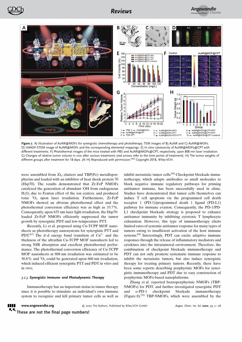
For light-controlled release of toxic chemotherapeuticdrugs and enhanced PDT, Zhang et al. developed core–shellcomposite nanoparticles (AuNR@MOFs) by the growth ofporphyrinic MOFs on gold nanorods (AuNR) (Figure 7).[38a]
These nanoparticles were used for synergistic chemotherapyand phototherapy due to the well-defined pore structure asthe drug carrier, TCPP as the photosensitizer, and AuNR asthe photothermal agent. With camptothecin (CPT) as a modeldrug, AuNR@MOFs achieved controllable CPT release upon808 nm laser irradiation. On the other hand, AuNR@MOFsexhibited an enhanced ability to produce 1O2 compared tosingle porphyrinic NMOFs upon 660 nm laser irradiation dueto the enhanced light absorption and strong electromagneticfield effect of the AuNR surface. Consequently, AuNR@-MOFs with NIR-triggered drug release and phototherapyshowed significant synergistic efficacy for killing tumor cellsin vitro and inhibiting tumor growth and metastasis in vivo.
3.2.2. Synergistic Photothermal and Photodynamic Therapy
Similar to PDT, PTT is also a targeted and localtherapeutic method with minimal invasiveness and highefficacy, which uses photothermal agents to generate heatupon NIR irradiation to ablate tumor cells and tissues.[41] Incontrast to PDT, PTT is an oxygen-independent and ROS-free process mediated by photothermal agents upon NIRirradiation. Hence, the combination of PDT and PTT fortumor treatment could synergistically enhance the therapeu-tic effect and reduce side effects.[65] On the one hand, the heatgenerated by PTT can improve blood flow as well as oxygensupply, and thereby intensify the sensitivity of tumor cells tooxygen-dependent PDT; on the other hand, the ROSproduced by PDT can interfere with tumor physiology andchange the microenvironment, and thereby increase the heatsensitivity of the tumor cells.[66]
Recently, several studies on porphyrin-based MOFs forsynergistic PDT and PTT have been reported. Zhang et al.proposed to assemble porphyrinic MOFs on polydopamine(PDA) nanoparticles to form core–shell nanoparticles(PDA@MOF),[38g] and the photodynamic activity of porphyr-inic MOFs and the photothermal effect of PDA nanoparticlescould contribute to PDT and PTT for tumors. The resultsindicated that PDA@MOF nanoparticles efficiently gener-ated intracellular ROS upon irradiation with 630 nm laserlight, and had excellent photothermal conversion uponirradiation with 808 nm laser light. Only 18% of the cellswere viable after incubation with PDA@MOF nanoparticlesand irradiation with 630 nm and 808 nm laser light. This wasmuch lower than the viability following single laser irradi-ation, suggesting synergistic tumor inhibition.
Besides composite porphyrinic NMOFs, porphyrinicNMOFs without the integration of other PTT agents alsohave been developed for synergistic PTT and PDT, whichshorten the synthesis procedures and improve biosafety. Forexample, Yang et al. reported novel multifunctional metal-loporphyrinic NMOF (Zr-FeP) shuttles for PDT and low-temperature PTT synergistic treatment.[66] Zr-FeP NMOFs
AngewandteChemieReviews
&&&&Angew. Chem. Int. Ed. 2020, 59, 2 – 28 � 2020 The Authors. Published by Wiley-VCH GmbH www.angewandte.org
were assembled from Zr6 clusters and TBP(Fe) metallopor-phyrins and loaded with an inhibitor of heat shock protein 70(Hsp70). The results demonstrated that Zr-FeP NMOFscatalyzed the generation of abundant COH from endogenousH2O2 due to Fenton effect of the ion centers, and producedtoxic 1O2 upon laser irradiation. Furthermore, Zr-FePNMOFs showed an obvious photothermal effect and thephotothermal conversion efficiency was as high as 33.7 %.Consequently, upon 635 nm laser light irradiation, the Hsp70-loaded Zr-FeP NMOFs efficiently suppressed the tumorgrowth by synergistic PDT and low-temperature PTT.
Recently, Li et al. proposed using Cu-TCPP MOF nano-sheets as phototherapy nanosystems for synergistic PTT andPDT.[67] The d–d energy band transition of Cu2+ and thethickness of the ultrathin Cu-TCPP MOF nanosheets led tostrong NIR absorption and excellent photothermal perfor-mance. The photothermal conversion efficiency of Cu-TCPPMOF nanosheets at 808 nm irradiation was estimated to be36.8% and 1O2 could be generated upon 660 nm irradiation,which induced efficient synergistic PTT and PDT in vitro andin vivo.
3.2.3. Synergistic Immuno- and Photodynamic Therapy
Immunotherapy has an important status in tumor therapysince it is possible to stimulate an individual�s own immunesystem to recognize and kill primary tumor cells as well as
inhibit metastatic tumor cells.[68] Checkpoint blockade immu-notherapy, which adopts antibodies or small molecules toblock negative immune regulatory pathways for primingantitumor immune, has been successfully used in clinic.Studies have demonstrated that tumor cells themselves caninduce T cell apoptosis via the programmed cell deathreceptor 1 (PD-1)/programmed death 1 ligand (PD-L1)pathway for immune evasion. Consequently, the PD-1/PD-L1 checkpoint blockade strategy is proposed to enhanceantitumor immunity by inhibiting cytotoxic T lymphocyteexhaustion. However, this type of immunotherapy elicitslimited rates of systemic antitumor response for many types oftumors owing to insufficient activation of the host immunesystems.[69] Interestingly, PDT can excite adaptive immuneresponses through the release of inflammatory mediators andcytokines into the intratumoral environment. Therefore, thecombination of checkpoint blockade immunotherapy andPDT can not only promote systematic immune response toinhibit the metastatic tumors, but also induce synergistictherapy for treating primary tumors. Recently, there havebeen some reports describing porphyrinic MOFs for syner-gistic immunotherapy and PDT due to easy construction ofporphyrinic MOFs-based nanoplatforms.
Zhang et al. reported benzoporphyrinic NMOFs (TBP-NMOFs) for PDT, and further investigated synergistic PDTand a-PD-1 checkpoint blockade immunotherapy(Figure 8).[70] TBP-NMOFs, which were assembled by the
Figure 7. A) Illustration of AuNR@MOFs for synergistic chemotherapy and phototherapy. TEM images of B) AuNR and C) [email protected]) HAADF-STEM image of AuNR@MOFs and the corresponding elemental mappings. E) In vitro cytotoxicity of AuNR@MOFs@CPT withdifferent treatments. F) Photothermal images of the mice treated with PBS and AuNR@MOFs@CPT, respectively, upon 808 nm laser irradiation.G) Changes of relative tumor volume in vivo after various treatments (red arrows refer to the time points of treatment). H) The tumor weights ofdifferent groups after treatment for 18 days. (A–H) Reproduced with permission.[38a] Copyright 2018, Wiley-VCH.
AngewandteChemieReviews
&&&& www.angewandte.org � 2020 The Authors. Published by Wiley-VCH GmbH Angew. Chem. Int. Ed. 2020, 59, 2 – 28� �
coordination of tetrabenzoporphyrin linkers with 10-con-nected Zr6 clusters, exhibited efficient 1O2 generation in thehypoxic microenvironment for PDT. The results demon-strated that TBP-NMOF-induced PDT could not only kill the4T1 murine breast tumor cells, but also augment thepresentation of tumor-activated antigens, further stimulatingadaptive immune response for initiating the secretion ofinflammatory cytokines (IFN-g, TNF-a) and recruitingtumor-infiltrating T cells (CD4+, CD8+). Further, they inves-tigated the antitumor effect of low O2-dependent PDT and a-PD-1 in vivo. When PD-1 antibodies were injected, TBP-NMOF-induced PDT dramatically inhibited the growth of4T1 tumors without body weight loss. Furthermore, thecombination therapy was more efficient than PDT alone in
preventing tumor recurrence and remarkably prevented lungmetastasis of 4T1 tumors for 22 days. Similarly, Lin et al. alsoutilized porphyrinic MOFs (Fe-TBP) and a-PD-L1 forsynergistic PDT and immunotherapy.[56a] It is interesting thatthe Fe3O clusters in Fe-TBP NMOFs could overcome hypoxiaby the Fenton reaction and enhance immunogenic PDT forpriming tumor immunotherapy.
Obviously, the use of porphyrinic MOFs for synergisticimmuno- and photodynamic therapy has shown great poten-tial for the therapy of many difficult-to-treat tumors andinhibition of metastatic tumor cells, because immunogenicPDT can prime tumor immunotherapy to promote theresponse rates, and thereby significantly promote immuno-therapeutic efficacy. In addition to checkpoint blockadeimmunotherapy based on antibodies or small-moleculeagents, it can be imagined that the use of porphyrinic MOFplatforms to combine PDT with other immunotherapeuticstrategies would be attractive.[71]
3.2.4. Other Synergistic Therapies
Radiotherapy (RT), which uses ionizing irradiation suchas X-rays to destroy localized solid tumors, is a commontherapy modality in clinic. Numerous nanoplatforms contain-ing high-Z elements, which are able to interact with ionizingradiation to produce photo/auger electrons and then producereactive free radicals to destroy tumor cells, have beenapplied for RT of tumors.[72] Biocompatible and biodegrad-able porphyrinic MOFs are formed by the coordination ofporphyrin linkers and metal nodes, which is ideal to introducehigh-Z metal elements into MOFs for use in synergistic PDTand RT. Liu et al. reported Hf-TCPP NMOFs assembled fromHf4+ and TCPP through a solvothermal process,[44] in whichTCPP linkers exhibited good PDT ability. The Hf4+ metalcenters, as a high-Z element, served as a radio-sensitizer toenhance RT. Due to the ISC enhanced by heavy Hf4+ nodes,Hf-TCPP NMOFs were extremely effective in producing 1O2
upon 661 nm irradiation for PDT. In vivo results on 4T1breast tumor-bearing mice showed that synergistic PDT andRT with Hf-TCPP NMOFs greatly inhibited tumor growthand had enhanced therapeutic efficacy compared to PDT orRT alone.
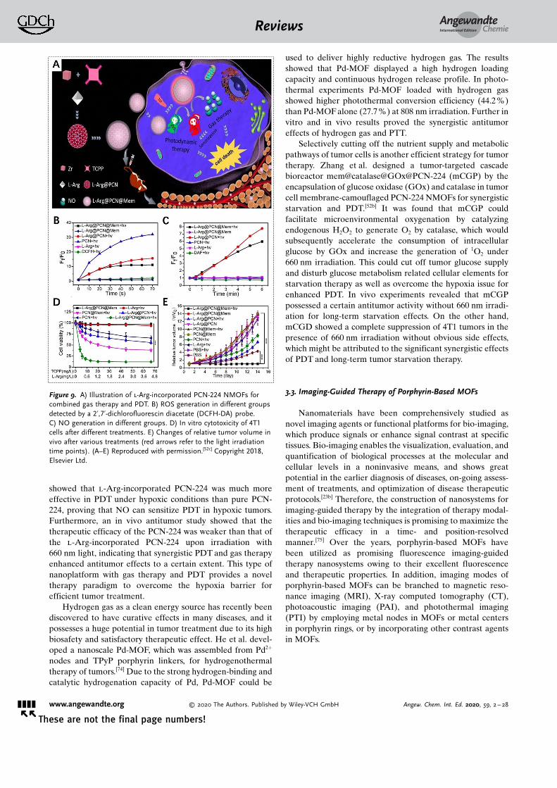
Gas therapy also has been developed for tumor therapydue to its negligible side effects and enhanced therapeuticefficacy. Various gaseous transmitters, such as hydrogensulfide (H2S), nitric oxide (NO), and carbon monoxide(CO), can be utilized for gas therapy, which can acceleratetumor cell apoptosis, prevent tumor cell proliferation, andselectively protect the activity and physiological function ofnormal cells.[73] Recently, Cheng et al. reported synergisticNO gas therapy and PDT to treat tumors based on l-arginine(l-Arg)-incorporated PCN-224 NMOFs (Figure 9).[52c] Here,l-Arg, a natural NO donor, could release NO for gas therapyvia oxidization through ROS, which were provided by PDTwith PCN-224 upon 660 nm irradiation. An in vitro MTTassay showed that the viability of 4T1 cells treated with l-Arg-incorporated PCN-224 was lower than that of cellstreated with PCN-224 upon 660 nm irradiation. Of specialnote, a cell apoptosis assay executed by flow cytometry
Figure 8. A) The proposed mechanism of antitumor immuneresponses induced by the combination of PDT and a-PD-1. B) Syner-gistic therapy using TBP-nMOFs. C) Comparative analysis after varioustreatments (from left to right): the tumor weight after 22 days, thepercentage of CD8+ cells, the generation of IFN-g and TNF-a in micesera obtained on the 12th day. D) Bioluminescence imaging of themice and the lung metastatic sites of the luciferase-4T1 (luc-4T1)tumors corresponding to the above five treatments. (A–D) Reproducedwith permission.[70] Copyright 2018, American Chemical Society.
AngewandteChemieReviews
&&&&Angew. Chem. Int. Ed. 2020, 59, 2 – 28 � 2020 The Authors. Published by Wiley-VCH GmbH www.angewandte.org
showed that l-Arg-incorporated PCN-224 was much moreeffective in PDT under hypoxic conditions than pure PCN-224, proving that NO can sensitize PDT in hypoxic tumors.Furthermore, an in vivo antitumor study showed that thetherapeutic efficacy of the PCN-224 was weaker than that ofthe l-Arg-incorporated PCN-224 upon irradiation with660 nm light, indicating that synergistic PDT and gas therapyenhanced antitumor effects to a certain extent. This type ofnanoplatform with gas therapy and PDT provides a noveltherapy paradigm to overcome the hypoxia barrier forefficient tumor treatment.
Hydrogen gas as a clean energy source has recently beendiscovered to have curative effects in many diseases, and itpossesses a huge potential in tumor treatment due to its highbiosafety and satisfactory therapeutic effect. He et al. devel-oped a nanoscale Pd-MOF, which was assembled from Pd2+
nodes and TPyP porphyrin linkers, for hydrogenothermaltherapy of tumors.[74] Due to the strong hydrogen-binding andcatalytic hydrogenation capacity of Pd, Pd-MOF could be
used to deliver highly reductive hydrogen gas. The resultsshowed that Pd-MOF displayed a high hydrogen loadingcapacity and continuous hydrogen release profile. In photo-thermal experiments Pd-MOF loaded with hydrogen gasshowed higher photothermal conversion efficiency (44.2%)than Pd-MOF alone (27.7%) at 808 nm irradiation. Further invitro and in vivo results proved the synergistic antitumoreffects of hydrogen gas and PTT.
Selectively cutting off the nutrient supply and metabolicpathways of tumor cells is another efficient strategy for tumortherapy. Zhang et al. designed a tumor-targeted cascadebioreactor mem@catalase@GOx@PCN-224 (mCGP) by theencapsulation of glucose oxidase (GOx) and catalase in tumorcell membrane-camouflaged PCN-224 NMOFs for synergisticstarvation and PDT.[52b] It was found that mCGP couldfacilitate microenvironmental oxygenation by catalyzingendogenous H2O2 to generate O2 by catalase, which wouldsubsequently accelerate the consumption of intracellularglucose by GOx and increase the generation of 1O2 under660 nm irradiation. This could cut off tumor glucose supplyand disturb glucose metabolism related cellular elements forstarvation therapy as well as overcome the hypoxia issue forenhanced PDT. In vivo experiments revealed that mCGPpossessed a certain antitumor activity without 660 nm irradi-ation for long-term starvation effects. On the other hand,mCGD showed a complete suppression of 4T1 tumors in thepresence of 660 nm irradiation without obvious side effects,which might be attributed to the significant synergistic effectsof PDT and long-term tumor starvation therapy.
3.3. Imaging-Guided Therapy of Porphyrin-Based MOFs
Nanomaterials have been comprehensively studied asnovel imaging agents or functional platforms for bio-imaging,which produce signals or enhance signal contrast at specifictissues. Bio-imaging enables the visualization, evaluation, andquantification of biological processes at the molecular andcellular levels in a noninvasive means, and shows greatpotential in the earlier diagnosis of diseases, on-going assess-ment of treatments, and optimization of disease therapeuticprotocols.[23b] Therefore, the construction of nanosystems forimaging-guided therapy by the integration of therapy modal-ities and bio-imaging techniques is promising to maximize thetherapeutic efficacy in a time- and position-resolvedmanner.[75] Over the years, porphyrin-based MOFs havebeen utilized as promising fluorescence imaging-guidedtherapy nanosystems owing to their excellent fluorescenceand therapeutic properties. In addition, imaging modes ofporphyrin-based MOFs can be branched to magnetic reso-nance imaging (MRI), X-ray computed tomography (CT),photoacoustic imaging (PAI), and photothermal imaging(PTI) by employing metal nodes in MOFs or metal centersin porphyrin rings, or by incorporating other contrast agentsin MOFs.
Figure 9. A) Illustration of l-Arg-incorporated PCN-224 NMOFs forcombined gas therapy and PDT. B) ROS generation in different groupsdetected by a 2’,7’-dichlorofluorescin diacetate (DCFH-DA) probe.C) NO generation in different groups. D) In vitro cytotoxicity of 4T1cells after different treatments. E) Changes of relative tumor volume invivo after various treatments (red arrows refer to the light irradiationtime points). (A–E) Reproduced with permission.[52c] Copyright 2018,Elsevier Ltd.
AngewandteChemieReviews
&&&& www.angewandte.org � 2020 The Authors. Published by Wiley-VCH GmbH Angew. Chem. Int. Ed. 2020, 59, 2 – 28� �
Fluorescence imaging has been a powerful imaging modefor both in vitro and in vivo imaging because it is noninvasiveand offers high signal sensitivity. Fluorescence imaging relieson the properties of fluorophores that absorb the photonenergy in a certain band of wavelengths and then emit newphoton energy in a band of longer wavelengths. Fluorescenceimaging is rapid and suitable for high-throughput screen-ing.[76] Porphyrins as popular fluorophores have been incor-porated into MOFs for both in vitro and in vivo fluorescenceimaging.[52a,77] The use of fluorescence imaging to guidetherapy allows the visualization of the distribution andevolution of the tumors/particles during treatment, andthereby ensures selectively controllable therapy with highefficacy and safety. Fluorescence imaging-guided therapynanosystems based on porphyrin-based MOFs can be easilyconstructed for efficient therapy due to the combination offluorescence and photosensitivity from porphyrins.
In 2017, Yin et al. developed the first biocompatibleporphyrinic NMOFs for fluorescence imaging-guided tumortherapy (Figure 10).[37a] Porphyrinic NMOFs (NPMOFs) witha high TCPP content (59.8%) achieved efficient fluorescenceimaging. The results showed that the fluorescence of TCPPwas observed at 651 nm with a weaker shoulder at 710 nmwhen excited at 514 nm, but only one emission peak at 689 nm
for NPMOFs.[78] The strong red emission and long Stokes shiftof NPMOFs resulted in a high signal-to-noise ratio andresolution in fluorescence imaging due to the low excitationinterference, autofluorescence, and scattering light from thebiological tissue. In a mouse model, the absorption, distribu-tion, metabolism, and excretion (ADME) processes ofNPMOFs were observed by tracking the fluorescent trajec-tory, indicating the biocompatibility of NPMOFs in mammals.Furthermore, in HepG2 tumor-bearing mice, the in vivofluorescence of NPMOFs clearly confirmed the distributionsof NPMOFs in tumors with high resolution and signal-to-noise ratio, suggesting that porphyrinic NMOFs are promisingas fluorescence imaging-guided therapy platforms for earliertumor diagnosis and enhanced therapy. On the other hand,composite porphyrinic NMOFs have also been constructedfor fluorescence imaging-guided therapy.[38a, 79] For example,Zhang et al. assembled porphyrinic NMOFs on AuNRs toform core–shell nanocomposites for fluorescence imaging-guided PDT, PTT, and chemotherapy.[38a] The introduction ofporphyrinic NMOFs made the nanocomposites suitable forPDT, chemotherapy, and fluorescence imaging, while AuNRsexhibited excellent PTT properties.
In addition, fluorescence imaging-guided therapy can alsobe used for real-time monitoring and assessment of thetherapeutic efficacy according to the changes of fluorescencesignals, and can be used to further improve the therapeuticefficacy and avoid under- or overtreatment. Lei et al.designed a photosensitive and caspase-responsive porphyr-in@MOF nanoprobe, which was assembled from porphyrin(TMPyP), folate targeting-motif, and a caspase-sensitivefluorescent dye, i.e., Cy3-labeled caspase-3 substrate peptide,in MOFs.[45] Here, the fluorescence of Cy3 was quenched untilthe activated caspase-3 specifically cleaved the peptide andCy3 was released from the MOF for fluorescence imaging,resulting in a caspase-responsive sensing strategy for mon-itoring cell apoptosis. These results demonstrated thatbiocompatible TMPyP@MOFs provided enhanced 1O2 yieldand folate targeting tumor therapy as well as the ability for insitu imaging of therapeutic efficacy via caspase-3 activation.The switch-on signal provides an effective way to imageintracellular caspase activity for real-time monitoring andevaluation of treatment effects.
3.3.2. MRI-Guided Therapy
MRI is another noninvasive medical method to obtainimages of the human anatomy and physiological processeswith high spatial resolution. It utilizes an external magneticfield to detect radiofrequency signals produced by protons,typically the hydrogen atoms in water from fat and tissues.The acquired signals can be utilized to compare differenttissues, distinguish lesions from healthy tissues, and constructanatomical maps of the human body.[80] Compared tofluorescence imaging, MRI possesses the advantages ofbetter spatial resolution, higher soft tissue contrast, andunlimited penetration depth, but exhibits the disadvantage oflow sensitivity. MRI contrast agents are typically used toreduce proton relaxation times and further improve the imagequality. For porphyrin-based MOFs, the metal nodes of the
Figure 10. Fluorescence images of A) the mouse (yellow dotted linesrefer to the liver region, red dotted lines refer to the intestine region,and green dotted lines refer to the lymph node) and B) the tumor-bearing mouse (yellow arrows refer to the small lymph node, bluearrows refer to the subcutaneous transplantable tumor) recorded atexcitation of 530 nm and emission of 700 nm after injection ofNPMOFs. C) Fluorescence images of dissected organs of a tumor-bearing mouse. (A–C) Reproduced with permission.[37a] Copyright 2017,Wiley-VCH.
AngewandteChemieReviews
&&&&Angew. Chem. Int. Ed. 2020, 59, 2 – 28 � 2020 The Authors. Published by Wiley-VCH GmbH www.angewandte.org
MOF skeleton, the chelation of paramagnetic metal ions inporphyrin rings, and the incorporation of other contrastagents in MOFs make MRI-guided therapy possible.
Recently, Wang et al. reported that gadolinium porphyr-inic NMOF sheets (Gd-TCPP) displayed a high relaxationrate (40.8 mm
�1 s�1) and 1O2 production upon 660 nm irradi-ation due to the high Gd3+ content in the MOFs and the TCPPphotosensitizer.[33d] Therefore, Gd-TCPP nanosheets arepotent for T1-weighted MRI-guided PDT. Yin et al. proposedto bind paramagnetic metal ions into porphyrin rings bychelation to develop metalloporphyrinic MOFs for T1-weighted MRI-guided therapy.[81] Through the chelation ofparamagnetic Mn ions in porphyrin rings, metalloporphyrinicMOFs possessed excellent T1-weighted MR contrast capacityand high photothermal conversion and heat-responsive NOrelease. In vivo MRI experiments showed that metallopor-phyrinic MOFs were efficiently accumulated at the tumor siteafter intravenous injection into mice, and tumor growth wascompletely inhibited when exposed to 808 nm laser for PTTand NO gas therapy, indicating the potential for MRI-guidedtherapy.
3.3.3. Multimode Imaging-Guided Therapy
Single-mode bio-imaging is usually insufficient for com-prehensive imaging and cannot provide the complete charac-terization needed to diagnose diseases due to its inherentlimitations, such as limited signal sensitivity, tissue penetra-tion depth, and spatial resolution. To overcome these prob-lems, various studies have concentrated on incorporatingmultimode imaging properties in a single MOF-basedsystem.[82] For porphyrinic MOFs, the use of functionalcomponents of the MOFs themselves or the assembly ofother imaging agents can achieve multimode imaging-guidedtherapy, which could avoid inherent limitations of single-mode imaging-guided therapy and provide more accuratelocalization of lesions/particles as well as guidelines oftherapy.
For example, Yin et al. developed a biocompatible nano-composite (Fe3O4@C@PMOF) with fluorescence imaging andMRI functions for dual-mode imaging-guided phototherapy(Figure 11).[79] Specifically, Fe3O4@C cores were selected asT2-weighted MRI contrast agents and photothermal agents,and porphyrinic MOF shells had fluorescence imaging andPDT abilities. Such dual-mode imaging nanoplatforms ach-ieved high sensitivity of fluorescence imaging as well as deeppenetration and great spatial resolution of MRI, and moreprecise in vivo information could be provided for enhancingthe safety and therapeutic efficacy. Following intravenousinjection of Fe3O4@C@PMOF nanocomposites in MCF-7tumor-bearing mice, 22 h later the fluorescence imagingshowed that nanocomposites were primarily localized in theliver region rather than in other organs. The tumor areaslowly brightened and became the brightest site in the miceafter 26 h, indicating that the accumulation of nanocompo-sites at the tumor site had gradually increased. The resultswere also confirmed by dramatic dimming at the tumor site inT2-weighted MR imaging. Owing to their high tumor accu-mulation, Fe3O4@C@PMOF nanocomposites were used for
imaging-guided phototherapy; an obvious PTT/PDT syner-getic effect was observed for MCF-7 breast tumor but thetoxicity for normal tissues was low.
Clearly, it is also valuable to develop porphyrinic MOFswithout additional components for multimode imaging-guided therapy due to its simplicity and efficiency. Wu et al.reported ultrathin 2D Cu-TCPP MOF nanosheets for dual-mode imaging-guided phototherapy.[67] The Cu2+ nodes in Cu-TCPP nanosheets offered excellent photothermal propertiesfor PTT, PTI, and T1-weighted MRI. Meanwhile, porphyrinTCPP provided the ability to produce 1O2 for PDT. Theresults indicated that Cu-TCPP nanosheets have tremendouspotential for synergistic PTT and PDT, guided by PTI andMRI.
CT is an important medical imaging technique, which isbased on the attenuation of X-rays by a specimen resulting in3D images with high spatial resolution.[83] The high-Z numberelements in MOFs can be chosen as CT contrast agents. Inaddition, PAI is a recently developed imaging technique thatintegrates optical excitation and ultrasound detection.[84] Thistechnique displays high selectivity and deep penetration, andcan be used to acquire high-resolution and strong-contrasttissue images. To combine the merits of PTI, CT, and PAI,Yang et al. developed multifunctional metalloporphyrinicNMOF (Zr-FeP) shuttles as an “all-in-one” nanoplatform torealize trimode imaging-guided therapy.[66] In vivo PAIexperiments demonstrated that a much stronger PA signalwas generated in the tumor tissue 2 h after tail vein injectionof nanoshuttles into mice, indicating the considerable accu-mulation of nanoshuttles in the tumor. Zr-FeP MOF nano-shuttles exhibited good CT capability due to the high-Zcomponent for CT imaging. Additionally, the outstandingphotothermal performance of Zr-FeP MOF nanoshuttles
Figure 11. A) Fluorescence images of the tumor-bearing mouse beforeand after injection of Fe3O4@C@PMOF (red arrow refers to the liverregion, yellow arrow refers to the tumor region). B) T2-weighted MRI ofthe tumor-bearing mouse (upper red dot lines refer to the liver region,lower red dot lines refer to the tumor region). C) Infrared thermalphotographs of tumor-bearing mice in different groups (black arrowsrefer to the tumor region). D) Changes of relative tumor volume invivo after various treatments (black and red arrows refer to theinjection and irradiation time points, respectively). (A–D) Reproducedwith permission.[79] Copyright 2017, Nature Publishing Group.
AngewandteChemieReviews
&&&& www.angewandte.org � 2020 The Authors. Published by Wiley-VCH GmbH Angew. Chem. Int. Ed. 2020, 59, 2 – 28� �
endowed remarkable PTI ability in vivo, and after treatmentwith Zr-FeP MOF nanoshuttles the tumor site temperaturerapidly increased by 17 8C upon exposure to 635 nm laser for5 min. Therefore, the fascinating trimode imaging capabilityof Zr-FeP MOF nanoshuttles would be a powerful tool forprecise tumor diagnosis and imaging-guided therapy.
3.4. Biosensing
A large number of physiological species play a key role inregulating cellular functions and physiological activities, ofwhich the changes in the concentrations and types indicatedisruptions or pathological changes in physiological environ-ments. Therefore, specific and sensitive biosensing systemshave been increasingly used to detect and monitor thevariations for disease diagnosis and biological processes inliving organisms. Porphyrins and their derivatives have beenwidely studied as small-molecule biosensors, due to theirexcellent physicochemical characteristics that can be used forsignal interaction with host molecules. Moreover, porphyrinscan mimic many biological functions. For example, porphyrinscan reversibly bind with gaseous compounds and can undergophotophysical and/or redox processes mediated by the targetanalytes.[8a] Interestingly, hemin, which is also called ironprotoporphyrin IX, has been utilized for biosensing, becauseit can act not only as a redox mediator based on theelectrochemical activity of the FeIII/FeII reversible redox pair,but also as a catalyst based on its peroxidase-like activity. Inrecent years, MOFs have also been extensively employed toconstruct biosensors due to their unique merits.[17b, 76a, 85] Inparticular, the porous structure and high specific surface areaof MOFs provide high catalytic activity. In addition, thedesigned shape and size as well as specific chemical inter-actions between guest molecules and MOFs endow them withhigh selectivity for their target molecules. Incorporatingporphyrins into MOFs can remit the self-quenching ofporphyrins and combine the multiple functionalities ofMOFs and porphyrins for biosensing with high sensitivity,resolution, and precision. To date, porphyrin-based MOFshave been utilized for biosensing applications includingelectrochemical (EC), fluorescence, photoelectrochemical(PEC), electrochemiluminescence (ECL), and colorimetricbiosensors. Various studies have demonstrated the possibilityof the porphyrin-based MOF-based biosensors for detectingdifferent biological substances and states, mainly includingbiomacromolecules, small molecules, ions, and pH values.Some typical examples of porphyrin-based MOFs for bio-sensing are summarized in Tables 1–3.
3.4.1. Biosensing of Biomacromolecules
DNA is the most essential genetic material of livingbeings, and various strategies have been proposed to detectDNA for diagnosis of diseases and biological processes.Among these strategies, EC biosensing has been extensivelyadopted for the detection of specific DNA owing to its highsensitivity and selectivity.[85] For DNA electrochemical bio-sensors, porphyrin-based MOFs with excellent electrocata-
lytic activity can be utilized as external probes for theproduction and amplification of the electrochemical sig-nal.[26d, 86] For example, due to the peroxidase-like catalyticactivity of TCPP(Fe) metalloporphyrins, Lei et al. encapsu-lated TCPP(Fe) into HKUST-1 and subsequently conjugatedit with streptavidin (SA) to construct an smart signal-trans-duction platform for hairpin DNA detection (Figure 12 A–C).[86a] Here, a label-free electrochemical DNA biosensor wasdesigned by using TCPP(Fe)@MOF-SA as a signal probe andhairpin DNA as an allosteric switch. The allosteric switch ofhairpin DNA on the glassy carbon electrode (GCE) surfacecould form SA aptamer that could selectively bind thesynthetic TCPP(Fe)@MOF-SA via biorecognition affinity.Thus, the conjugated TCPP(Fe)@MOF-SA on the GCEsurface acted as a mimetic catalyst; in the presence of H2O2,it could greatly enhance the oxidation of o-phenylenediamine(o-PD) to 2,2’-diaminoazobenzene, which could serve asa good electrochemical signal readout indicator. Sucha “signal-on” electrochemical biosensor was proved todetect hairpin DNA with a detection limit down to 0.48 fm(S/N = 3), with a linear range over six orders of magnitudeand good feasibility in complex serum matrixes.
On the other hand, porphyrinic MOFs for fluorescencebiosensing have also been developed for DNA detection byutilizing the fluorescence quenching mechanism.[33c,87] Forexample, Wang et al. used ultrathin Cu-TCPP MOF nano-sheets to design a fluorescence biosensor for rapid, sensitive,and multiplex detection of DNAs.[87] Due to the large surfacearea and large number of accessible active sites on thenanosheets, three dye-labeled ssDNA probes could be easilyabsorbed on the surface, leading to fluorescence quenching ofthe dye-labeled ssDNA by Fçrster resonance energy transfer(FRET). When a specific target was hybridized with thessDNA probes to form dsDNA, the formed dsDNA detachedfrom Cu-TCPP MOF nanosheets owing to the weak physicaladsorption between Cu-TCPP MOF nanosheets and dsDNA,and thereby the fluorescence “turned on”. Consequently, Cu-TCPP MOF nanosheets exhibited excellent performance forquantitatively detecting pathogenic DNA with a detectionlimit in the pm range.
Immunoassays are of great interest for the clinical earlydiagnosis of various tumors, such as prostate specific antigen(PSA) analysis for diagnosis of prostate cancer. PEC bio-analysis is a potential bioanalytical method based on thephotoinduced electron transfer (PET) processes amonganalyte, photoactive species, and electrode upon photoirra-diation, which has been widely used in the detection ofvarious biological molecules. Recently, Zhang et al. reporteda new non-enzyme PEC immunoassay using DNA-mediatednanoscale PCN-222 to detect PSA.[88] The ss-DNA-taggedantibody (Ab-DNA) was conjugated on PCN-222 to form Ab-DNA-functionalized NMOF complexes (Ab-DNA-NMOFs).Ab-DNA-NMOFs, as an active photocathode PEC substrate,displayed a remarkably enhanced photocurrent response inthe presence of dopamine (DA) in O2-containing aqueousmedia. Further functionalized with polyamidoamine dendri-mer, Ab-DNA-NMOFs could serve as an efficient PEC signaltransduction system for non-enzyme PSA immunoassays with
AngewandteChemieReviews
&&&&Angew. Chem. Int. Ed. 2020, 59, 2 – 28 � 2020 The Authors. Published by Wiley-VCH GmbH www.angewandte.org
hypersensitivity, a broad calibration range of 1 pg mL�1 to10 ng mL�1, and a detection limit of 0.2 pgmL�1.
In addition, ECL, electrochemical redox-induced lightemission, possesses several advantages including low back-ground interference, simple operation, and high sensitivityand space–time controllability. Han et al. proposed to useporphyrin@MOFs for PSA sensing via the ECL strategy.[89] Inthis study, Fe-MIL-88 MOFs were used as a platform to loadhemin and gold nanoparticles (Au NPs) and further graft G-quadruplex DNAzyme for synergetic catalytic amplificationof luminol. The pDNA-Au-Hemin-MIL-DNAzyme probesacted as quenchers and enhancers in the fabrication ofa target-induced ratiometric ECL biosensor for PSA detec-tion. The results demonstrated that the pDNA-Au-Hemin-MIL-DNAzyme-based biosensor was highly sensitive andcould be used for the accurate analysis of PSA with a lineardetection range from 0.5 to 500 ng mL�1 and a detection limitof 0.058 ngmL�1 (S/N = 3).
Enzymes and proteins play important roles in metabolicregulation, cell growth, and other biological processes. Someof these biomolecules have been accepted as importantbiomarkers for early-stage disease diagnosis, prognosis judge-ment, and pathogenesis studies. Several different types ofporphyrin-based MOFs have been used in the construction ofelectrochemical biosensors to enhance the electrochemical
signal for enzyme or protein biosensing. Yuan et al. reportedmultifunctional hemin-encapsulated Fe-MIL-88 MOFs, whichwere the first example for the construction of an electro-chemical aptasensor for thrombin (TB) detection (Fig-ure 12D–F).[26c] Such porphyrin@MOFs served as the supportfor the immobilization of recognition molecules (thrombinbinding aptamer (TBA)) and were also conjugated with GOxfor signal amplification. GOx oxidizes glucose into gluconicacid accompanied by the production of H2O2, which can befurther electrocatalyzed by hemin to amplify the electro-chemical signal. Such an electrochemical biosensor showeda relatively low detection limit of 0.068 pm and a wide linearrange from 0.0001 nm to 30 nm with a high correlationcoefficient of 0.9961, indicating that the constructed biosensorcould be used to detect TB quantitatively. Lei et al. designedan electrochemical biosensor that was composed of ironmetalloporphyrinic NMOFs (PorMOFs) for telomerase activ-ity detection.[90] PorMOFs exhibited excellent electrocatalyticactivity towards O2 reduction, which was used to detecttelomerase with a detection limit of 30 cells mL�1 and assessthe telomerase activity even in single Hela cell. Moreover,porphyrin-based MOFs were also applied to construct PECand ECL biosensors for protein detection. For example,Zhang et al. developed a simple PCN-222 based PECbiosensor for label-free detection of a phosphoprotein (a-
Figure 12. A) Synthesis of TCPP(Fe)@MOF-SA and its electrochemical DNA sensing. B) Differential pulse voltammogram (DPV) responses ofTCPP(Fe)@MOF-SA to different concentrations of the target DNA from 0 to 10 nm. C) DPV responses of FeTCPP@MOF-SA to 100 pm of differentDNA sequences: a) target DNA, b) single-base-mismatched DNA, c) two-base-mismatched DNA, d) three-base-mismatched DNA, and e) randomDNA in the catalytic system of o-PD/H2O2. (A–C) Reproduced with permission.[86a] Copyright 2015, American Chemical Society. D) Preparation ofthe hemin-encapsulated Fe-MIL-88 based electrochemical biosensor and the TB detection principle. E) DPV of TB detection at differentconcentrations from 0 to 30 nm. F) Specificity of the biosensor toward different samples: a) no analyte, b) 100 nm Apo-A1, c) 100 nm Hb,d) 100 nm l-Cys, e) 10 nm TB, and f) 100 nm Apo-A1+ 100 nm Hb + 100 nm l-cys + 10 nm TB. (D–F) Reproduced with permission.[26c] Copyright2015, Royal Society of Chemistry.
AngewandteChemieReviews
&&&& www.angewandte.org � 2020 The Authors. Published by Wiley-VCH GmbH Angew. Chem. Int. Ed. 2020, 59, 2 – 28� �
casein).[91] Shan et al. utilized zinc metalloporphyrinic MOFs(MOF-525(Zn)) to develop an ECL biosensor for a highlysensitive protein kinase A (PKA) activity assay.[92]
3.4.2. Biosensing of Small Molecules
Various porphyrin-based MOF biosensors with perox-idase-like activity have been developed for the detection ofH2O2 and glucose, which are important biological indicatorsin clinical medicine. For instance, Liu et al. synthesized MIL-101(Al)-NH2 MOFs to anchor hemin for the formation ofH@M composites, which showed peroxidase-like activity forcatalyzing oxidation of the peroxidase substrate 3,3,5,5-tetramethylbenzidine (TMB) by H2O2 to produce a bluecolor in solution.[93] Thus, H@M composites offered a simple,sensitive, and selective colorimetric approach for H2O2
determination, and the results showed that H2O2 could bemeasured at concentrations as low as 2 mm and the linearrange was from 5 mm to 200 mm with a correlation coefficientof 0.997. Furthermore, by introducing GOx as a catalyst intoH@M composites for the oxidation of glucose, a MOF-basedcolorimetric biosensor was constructed for quantitativeanalysis of glucose in aqueous solution. The glucose biosens-ing was observed to have a linear range from 10 mm to 300 mm
with a correlation coefficient of 0.996.Monitoring and quantifying some gas levels (e.g., O2, H2S)
in living cells is of great importance in clinical medicine,because the gas concentrations are correlated with manyhuman physiological and pathological processes. For example,Lin et al. developed novel phosphorescence/fluorescencedual-emissive NMOFs (R-UiO) for ratiometric intracellularO2 quantification (Figure 13);[36b] this was the first example ofa MOF-based intracellular O2 biosensor. R-UiO NMOFswere synthesized by the coordination of the PtII-porphyrinlinker DBP(Pt) as an O2-sensitive probe and amino-function-alized quaterphenyldicarboxylate linkers (QPDC-NH2) withHf ions. Then Rhodamine B isothiocyanate (RBITC) wascovalently attached as an O2-insensitive reference probe viathiourea bonds. DBP(Pt) and RBITC shared the sameexcitation energy, but the phosphorescence/fluorescenceemissions did not interfere with each other, which allowedfor the determination of intracellular O2 levels. When mousecolon carcinoma CT26 cells were treated with R-UiONMOFs under different O2 pressure (pO2) conditions at 4mmHg (hypoxia), 32 mmHg (normoxia), and 160 mmHg(aerated condition), the analytic data from ratiometricluminescence images were 5.1� 2.5 mmHg, 27.3�3.1 mmHg, and 158� 11 mmHg, respectively. The resultsmatched well with the preset pO2, suggesting the wide rangeand high accuracy of R-UiO NMOFs as intracellular oxygenbiosensors. Tang et al. reported aluminum–copper mixed-metal porphyrinic MOFs (PAC) assembled from Al3+ nodesand TCPP(Cu) linkers as a heterogeneous fluorescence probefor the sensing of H2S in living cells.[94] The Cu2+ centers inMOF frameworks induced the fluorescence quenching ofTCPP porphyrins, which could be used as the H2S-respondingsite for fluorescence “turn-on” sensing of H2S. Consequently,this biocompatible fluorescence probe exhibited high selec-
tivity and sensibility as well as low toxicity in in situ sensing ofH2S in a physiological environment.
As a biochemical messenger, dopamine (DA) playsa critical role in neurotransmitters and neuromodulators inorganisms that affect human activity. For measuring DArelease from living cells, Yamauchi et al. designed an electro-chemical biosensor using composite porphyrinic MOFs(MOF-525-PEDOT NTs),[95] which was made up of conduc-tive poly(3,4-ethylenedioxythiophene) nanotubes (PEDOTNTs) coated with MOF-525 nanocrystals. The synergisticconductivity and catalytic performance of MOF-525-PEDOTNTs enhanced the detection capability for DA with a linearconcentration range (2 mm–270 mm) and detection limit(0.04 mm).
Figure 13. A) Synthesis of mixed-ligand R-UiO. B) TEM image of R-UiO. C) Emission spectra and D) phosphorescent decay of R-UiO inHank’s Balanced Salt Solution (HBSS) buffer under different oxygenpartial pressures with excitation wavelengths of 514 nm and 405 nm.E) Calibration curve of the phosphorescence/fluorescence intensity ofR-UiO on confocal laser scanning microscopy (CLSM) under differentO2 pressures. F) Ratiometric luminescence images of CT26 cells aftertreatment with R-UiO under hypoxia, normoxia, and aerated conditions(from left to right) with an excitation wavelength of 514 nm. (A–F) Reproduced with permission.[36b] Copyright 2016, American Chem-ical Society.
AngewandteChemieReviews
&&&&Angew. Chem. Int. Ed. 2020, 59, 2 – 28 � 2020 The Authors. Published by Wiley-VCH GmbH www.angewandte.org
The rapid and sensitive detection of organophosphoruspesticides has gained more and more attention due to theenvironmental pollution and human health risks. Su et al.synthesized Cu-H MOFs/NECF composites consisting of ball-flower-like Cu-hemin MOFs grown on the fibers of 3Dnitrogen-containing melamine carbon foam (NECF) andimmobilized AChE on Cu-H MOFs/NEC for trichlorfondetection.[96] The Cu-H MOFs/NECF-based biosensor exhib-ited great performance for trichlorfon detection with a broadlinear range (0.25–20 ngmL�1), low detection limit(0.082 ngmL�1), and good stability, and thereby facilitatedtrace detection of organophosphorus pesticides.
3.4.3. Ion Sensing
Several metal ions are essential elements in the humanbody and play vital roles in cellular activities, while theirexcess would be harmful for health. In particular, heavy metalions are easily accumulated in the human body via the foodchain, which thereby results in adverse effects. Obviously, it isof great importance to selectively detect metal ions in cells orin the environment. For porphyrin-based MOFs, the coordi-nation capability of metal ions in the porphyrin rings endowsthem with the ability to construct fluorescence “turn-off” or“turn-on” biosensors. In 2013, Li et al. proposed to useporphyrinic MOF-525 as a fluorescence “turn-off” biosensorfor Cu2+ sensing.[97] The porphyrin moieties in MOF-525provided accessible recognition sites for fluorescence quench-ing through energy/charge transfer to determine the amountof Cu2+ ions. MOF-525 fluorescent probes showed greatpotential for rapid and specific detection of Cu2+ in practicalwater samples and for the study of copper-related diseaseslike Wilson�s and Alzheimer�s diseases. Recently, a novelfluorescent porphyrinic MOF probe, where the UiO-66(OH)2
MOF was encapsulated into PCN-224, was developed by Luet al. for detecting Cu2+ in complex water samples.[98]
Similarly, due to the fluorescence quenching ability of Cu2+
toward PCN-224, this fluorescent probe exhibited excellentCu2+ detection with a limit-of-detection (LOD) value as lowas 0.068 nm, which is lower than the Cu2+ concentration limitin drinking water regulated by the WHO.
Compared to fluorescence “turn-off” biosensors, fluores-cence “turn-on” biosensors can avert the wrong response tosensing signals and more conveniently identify in a darkbackground.[36c] For example, Jiang et al. developed a fluores-cence “turn-on” biosensor based on PdII metalloporphyrinicMOFs (PCN-222(Pd)) for Cu2+ sensing.[99] Here, PCN-222-(Pd) was used as a catalytic Heck reaction system to producehighly fluorescent “turn-on” signal in the presence of Cu2+,due to the stronger binding affinity of Cu2+ over Pd2+ to thenitrogen atoms in the porphyrin and the conversion of non-fluorescent aniline to fluorescent ring-closing product via Pd0-catalyzed Heck reaction. Consequently, the PCN-222(Pd)biosensor exhibited highly selective and sensitive sensing ofCu2+ in aqueous solution with a very low detection limit. Inaddition, porphyrinic MOFs were also utilized in the con-struction of electrochemical biosensors for detecting metalions.[100]
Additionally, although anion sensing is more challenging,selective sensing of anions by porphyrin-based MOFs has alsobeen reported. For instance, Cao et al. developed Zr-basedMOFs with the mixed linkers 1,3,6,8-tetra(4-carboxylphe-nyl)pyrene (TBAPy) and TCPP (Zr(TBAPy)5(TCPP)) asa fluorescence “turn-on” biosensor for S2� sensing in aqueousenvironments.[36c] The results showed that the fluorescencesignal of Zr(TBAPy)5(TCPP) was intensified remarkablyafter addition of S2�, but other anions including SO4
2�, CNS� ,COOH� , Br� , I� , IO3
� , F� , HSO3� , Cl� , and NO3
� did notalter its fluorescent intensity. Eddaoudi et al. encapsulatedTMPyP(Pt) porphyrins in rho-ZMOF for the selective sensingof various anions in aqueous and methanolic solutions.[8c]
3.4.4. pH Sensing
Generally, pH plays an important role in maintaining thestability, normal morphology, and function of cells andorganisms. Due to the spectral characteristics that changeupon protonation/deprotonation of the pyrrole ring inporphyrins with a change in pH,[101] porphyrin-based MOFscould be used to construct pH sensors, showing great potentialin pH monitoring, disease diagnosis, and even evaluatingtherapeutic efficacy. In 2013, Zhou et al. reported for the firsttime Zr-based porphyrinic MOF PCN-225 and metallopor-phyrinic MOF PCN-225(Zn) for pH sensing.[35b] The resultsdemonstrated that the fluorescence intensity of PCN-225 hasa strong correlation with the pH of the solution in the range ofpH 0–10.2. The most acidic solution showed the weakestfluorescence, while the pH 10.2 solution gave the highestintensity. Furthermore, pH 7–10 was the most sensitive pHrange for intensity response. Lu et al. developed an effectivedual-emission fluorescent MOF composite probe (RB-PCN)that was constructed by the encapsulation of Fe3O4 nano-particles in PCN-224 followed by modification withRBITC.[102] The results showed that RB-PCN could sensi-tively detect a broad range of pH changes. It could also beused to monitor the pH changes in living cells by fluorescenceconfocal imaging and determine the pH values in actual watersamples by fluorescence spectroscopy.
3.5. Other Applications of Porphyrin-Based MOFs
In addition to the biomedical applications mentionedabove, porphyrin-based MOFs also have valuable applica-tions in other biologically related fields. For example, Guoet al. synthesized spherical Cu-TCPP(Cu) MOFs, and sub-sequently encapsulated Ag nanoparticles in MOFs to formAg/Cu-TCPP(Cu) composites for antibacterial applica-tion.[103] The results showed that antibacterial effect of Ag/Cu-TCPP(Cu) composites was superior to that of penicillin,and the cytotoxicity of Ag/Cu-TCPP(Cu) composites waslower than that of naked Ag nanoparticles and Ag ions invitro. Furthermore, Ag/Cu-TCPP(Cu) composites could alsosignificantly promote wound healing in vivo.
More recently, Qu et al. utilized ultrathin Cu-TCPP(Fe)MOF nanosheets to physically adsorb GOx to constructa composite nanocatalyst for in vivo wound healing.[104] Here,
AngewandteChemieReviews
&&&& www.angewandte.org � 2020 The Authors. Published by Wiley-VCH GmbH Angew. Chem. Int. Ed. 2020, 59, 2 – 28� �
Cu-TCPP(Fe)/GOx could continuously convert nontoxicglucose into gluconic acid and H2O2 through the action ofGOx, and the subsequent pH decrease from 7 to 3–4 coulddramatically activate the peroxidase-like activity of Cu-TCPP(Fe) MOF nanosheets. Subsequently, the producedH2O2 could be further transformed to toxic COH radicals bythe Fenton reactions of Cu and Fe ions. In vitro and in vivoexperiments showed that this cascade catalytic conversion ofnontoxic glucose to toxic COH radicals endowed the Cu-TCPP(Fe)/GOx nanocatalyst with robust antibacterial activ-ity and negligible biotoxicity.
4. Toxicity and Safety Concerns
Regarding porphyrin-based MOFs for biomedical appli-cations, their potential toxicity is of high concern. Consideringthe construction of MOFs from metal nodes and organiclinkers, it is a priority to choose nontoxic or hypotoxicbuilding components to improve the biocompatibility andreduce the toxicity. Fortunately, porphyrins as biocompatiblemotifs are beneficial for the MOFs� biosafety. However, thetoxicity of MOFs was found to be closely related to theirmetal nodes.[76a] The biocompatible metal ions (e.g., Zr, Fe,Zn, Ca, Mg) are possible nodes for the construction ofbiofriendly porphyrin-based MOFs due to their acceptabletoxicity for biomedical applications.[106] Especially for thesynthesis of composite porphyrinic MOFs, the additionalfunctional components also need to be considered forpotential damage to the tissues and organisms. In additionto the composition of porphyrin-based MOFs, the physico-chemical natures of MOFs including size distribution, shape,surface chemistry, and dose-dependent properties are alsoimportant factors in terms of toxicology.[106, 107] All theseparameters strongly affect the absorption, distribution,metabolism and excretion, biodegradation, and clearance ofporphyrin-based MOFs, and further determine their potentialtoxicity in vivo. To date, these aspects of porphyrin-basedMOFs have not been fully investigated, while the multi-functionality has been deeply explored in most biomedicalstudies.
Understanding the intricate interactions between thephysicochemical properties, biological performance, and bio-safety could contribute to the successful translation topractical biomedical applications. Therefore, the relevantstudies of porphyrin-based MOFs in biomedical applicationsneed to be developed in depth. Firstly, it is important to usevarious progressive characterization techniques to accuratelydetermine the physicochemical parameters of MOFs (e.g.,size, shape, surface chemistry, porosity) and reasonablyevaluate their merits and limitations in order to understandand ultimately modulate the structure–function relationshipfor MOFs.[108] Next, investigating the correlation betweenMOFs and biological systems is the basis for correlating theMOFs� features with the toxicity results. Recently, Faria et al.proposed the MIRIBEL (Minimum Information Reporting inBio-Nano Experimental Literature) standard for differentstudies on bio–nano interactions, which normalizes materialcharacterization, biological characterization, and experimen-
tal details for enhancing reproducibility and quantitativecomparisons of bio–nano materials.[109] In the development ofporphyrin-based MOFs for biomedical applications, suchstandard should be referenced to facilitate safety evaluationand material optimizations. Furthermore, great progress hasbeen made with respect to mechanism-based hazard assess-ment of nanomaterials and advanced tools have emerged fortheir safety evaluation.[110] Therefore, not only for porphyrin-based MOFs, researchers need to accelerate the use ofemerging methods and tools, combining research results withbioinformatics, to scientifically and quantitatively correlatethe data outcome of biomaterials and their toxicity results fortheir practical application in biomedicine.
5. Conclusions and Perspectives
Porphyrins and their derivatives have been often used asphotosensitizers for PDT, fluorophores for fluorescenceimaging, and sensors for biosensing due to their excellentphotophysical properties. As a new type of porous materials,MOFs possess high porosity, tunable structures, multifunc-tionality, biocompatibility, and biodegradability. Studies havedemonstrated that the construction of porphyrin-based MOFscan not only overcome the limitations of free porphyrins likeinstability and self-quenching issues in physiological environ-ments, but also integrate the functionalities of MOFs andporphyrins as well as improve the physicochemical properties.In this Review, recent achievements and progress in theconstruction of porphyrin-based MOFs and their biomedicalapplications have been highlighted and discussed.
To date, various methods have been used to constructporphyrin@MOFs, porphyrinic MOFs, and composite por-phyrinic MOFs in order to meet the requirements ofbiomedical applications. Among them, many strategiesincluding heavy-atom substitution of porphyrinic MOFs,controlling the particle size, controllable design, and modifi-cation with targeting moieties or functional components havebeen proposed to enhance the therapeutic efficacy ofporphyrin-based MOFs in PDT. Additionally, the rationaldesign of functional components in MOFs or adjuvant agentsmakes it possible to interfere with survival mechanisms andalter the tumor microenvironment to achieve better results interms of PDT efficacy. Obviously, the optimized constructionof porphyrin-based MOFs shows great potential for enhancedPDT of tumors.
Another efficient strategy for enhancing therapeuticefficacy is the implementation of multimodality therapy ina single system to overcome the limitations of single-modalitytherapy. Porphyrin-based MOFs have exhibited a greatcapacity for combining PDT with other therapy modalities,including chemotherapy, PTT, immunotherapy, and gastherapy, to achieve synergistic effects for maximizing ther-apeutic efficacy and improving safety. However, the majorityof current studies focus on dual-modality therapies, but thereare few reports on tri- or more modality therapy based onporphyrin-based MOFs, which might offer more significanttherapeutic effects.
AngewandteChemieReviews
&&&&Angew. Chem. Int. Ed. 2020, 59, 2 – 28 � 2020 The Authors. Published by Wiley-VCH GmbH www.angewandte.org
Porphyrin-based MOFs have also been developed forimaging-guided therapy to further optimize the therapeuticprocess and thereby enhance therapeutic efficacy. Due to theexcellent fluorescent properties and photosensitization ofporphyrins, porphyrin-based MOFs could realize fluores-cence imaging-guided therapy. On the other hand, metalnodes in MOFs or the incorporation of contract agents inMOFs endow porphyrin-based MOFs with the potential forMRI-, CT-, PAI- or PTI-guided therapy. Such “all-in-one”nanoplatforms provide more accurate diagnosis and guidancefor treatment, and are of great attractive for tumor therapy.
Besides the exploration of tumor therapy functions,porphyrin-based MOFs have been utilized to constructbiosensors for detecting various biological species, such asbiomacromolecules, small molecules, and ions as well as pHvalues and gases. Owing to the combined excellent physico-chemical properties of MOFs and porphyrins, porphyrin-based MOFs types of biosensors show significant perfor-mance in biosensing with high sensitivity and selectivity,acceptable reproducibility, good stability, and biocompatibil-ity. Some porphyrins including hemin, TCPP, and its metalderivatives are often used to construct porphyrin-based MOFtype biosensors. However, it can be anticipated that thebiosensing performance could be further improved with moreinteresting porphyrins. On the other hand, porphyrin-basedMOF type biosensors for cell sensing and in vivo sensing needfurther investigation for the development of biosensors inliving biological systems.
Despite the growing number of impressive progressreports on porphyrin-based MOFs, their development inbiomedical applications is still at the early stage and facesmany challenges. For porphyrin@MOFs, the porphyrin load-ing capacity and premature leakage and self-quenching ofporphyrins should be considered during the synthesis process.Actually, porphyrinic MOFs are more promising candidatesfor biomedical applications. However, the influence of size,shape, composition, and surface chemistry of porphyrin-basedMOFs on therapeutic efficacy and biosensing ability shouldbe further optimized to meet the requirements of personal-ized therapy, diagnosis, and detection. On the other hand, thedesign of “all-in-one” nanoplatforms based on compositeporphyrinic MOFs would be an efficient strategy to enhancetherapeutic efficacy in tumor therapy and biosensing ability.Actually, researchers should establish straightforward syn-thesis protocols for these designed MOF materials, as thesimpler the preparation, the more likely it is that the productwill be relevant for biomedical applications in the real world.Finally, it should be re-emphasized that thoroughly anddeeply investigating the correlation between the fundamentalparameters of porphyrin-based MOFs and their biosafetyincluding the pharmacokinetics, biodistribution, biocompati-bility, long-term toxicity, and clearance from the body, iscrucial for possible clinical translations. In conclusion, por-phyrin-based MOFs have great potential for applications intumor therapy and biosensing, and more advances andprogress in the biomedical field will be achieved in the future.
Acknowledgements
We gratefully acknowledge support from the NationalNatural Science Foundation of China (No. 51572172,51872185), the Science and Technology Commission ofShanghai Municipality (NO. 17060502400), the University ofShanghai for Science and Technology (No. 2018KJFZ016),and the Alexander von Humboldt Foundation. Open accessfunding enabled and organized by Projekt DEAL.
Conflict of interest
The authors declare no conflict of interest.
[1] J. M. Gottfried, Surf. Sci. Rep. 2015, 70, 259 – 379.[2] A. D. Adler, F. K. F. R. Longo, J. Kim, J. Inorg. Nucl. Chem.
1970, 32, 2443 – 2445.[3] a) K. M. Kadish, K. M. Smith, R. Guilard, The Porphyrin
Handbook, Vols. 1 – 20, Academic Press, San Diego, 2000 –2003 ; K. M. Kadish, K. M. Smith, R. Guilard, Handbook ofPorphyrin Science Vols. 1 – 35, World Scientific Publishing Co.,2010 – 2014.
[4] J.-F. Longevial, S. Cl�ment, J. A. Wytko, R. Ruppert, J. Weiss, S.Richeter, Chem. Eur. J. 2018, 24, 15442 – 15460.
[5] a) S. Durot, J. Taesch, V. Heitz, Chem. Rev. 2014, 114, 8542 –8578; b) I. Goldberg, Chem. Commun. 2005, 1243 – 1254.
[6] a) R. P. Steer, Dalton Trans. 2018, 47, 8517 – 8525; b) L. L. Li,E. W. Diau, Chem. Soc. Rev. 2013, 42, 291 – 304; c) S. Goswami,M. Chen, M. R. Wasielewski, O. K. Farha, J. T. Hupp, ACSAppl. Mater. Interfaces 2018, 10, 34409 – 34417; d) J. Yu, X. Li,P. Deria, ACS Sustainable Chem. Eng. 2019, 7, 1841 – 1854;e) G. Ding, Y. Wang, G. Zhang, K. Zhou, K. Zeng, Z. Li, Y.Zhou, C. Zhang, X. Chen, S.-T. Han, Adv. Funct. Mater. 2019,29, 1806637.
[7] a) A. Rezaeifard, M. Jafarpour, Catal. Sci. Technol. 2014, 4,1960 – 1969; b) Y.-M. Zhao, G.-Q. Yu, F.-F. Wang, P.-J. Wei, J.-G. Liu, Chem. Eur. J. 2019, 25, 3726 – 3739; c) P. Li, X. Zhang, C.Hou, Y. Chen, T. He, Appl. Catal. B 2018, 238, 656 – 663; d) K.Ladomenou, M. Natali, E. Iengo, G. Charalampidis, F. Scan-dola, A. G. Coutsolelos, Coord. Chem. Rev. 2015, 304 – 305, 38 –54.
[8] a) R. Paolesse, S. Nardis, D. Monti, M. Stefanelli, C. D. Natale,Chem. Rev. 2017, 117, 2517 – 2583; b) H. Lee, K.-I. Hong, W.-D.Jang, Coord. Chem. Rev. 2018, 354, 46 – 73; c) D. Masih, V.Chernikova, O. Shekhah, M. Eddaoudi, O. F. Mohammed, ACSAppl. Mater. Interfaces 2018, 10, 11399 – 11405.
[9] a) S. Rojas, T. Devic, P. Horcajada, J. Mater. Chem. B 2017, 5,2560 – 2573; b) M. Ethirajan, Y. Chen, P. Joshi, R. K. Pandey,Chem. Soc. Rev. 2011, 40, 340 – 362; c) A. R. Monteiro, C. I. V.Ramos, S. Fateixa, N. M. M. Moura, M. G. P. M. S. Neves, T.Trindade, ACS Omega 2018, 3, 11184 – 11191.
[10] a) Y. Zhai, H. J. Busscher, Y. Liu, Z. Zhang, T. G. V. Kooten, L.Su, Y. Zhang, J. Liu, J. Liu, Y. An, L. Shi, Biomacromolecules2018, 19, 2023 – 2033; b) M. Managa, J. Britton, E. Prinsloo, T.Nyokong, Polyhedron 2018, 152, 102 – 107.
[11] X. Wang, F. Yan, X. Liu, P. Wang, S. Shao, Y. Sun, Z. Sheng, Q.Liu, J. F. Lovell, H. Zheng, J. Controlled Release 2018, 286,358 – 368.
[12] P. Manickam, R. E. Fernandez, Y. Umasankar, M. Gurusamy, F.Arizaleta, G. Urizar, S. Bhansali, Sens. Actuators B 2018, 274,47 – 53.
[13] P. Huang, X. Qian, Y. Chen, L. Yu, H. Lin, L. Wang, Y. Zhu, J.Shi, J. Am. Chem. Soc. 2017, 139, 1275 – 1284.
AngewandteChemieReviews
&&&& www.angewandte.org � 2020 The Authors. Published by Wiley-VCH GmbH Angew. Chem. Int. Ed. 2020, 59, 2 – 28� �
[14] a) D. Cui, W. Xie, S.-R. Zhang, Z.-X. Liu, C.-B. Liu, Y.-H. Xu,Mater. Lett. 2018, 232, 18 – 21; b) W. Zhang, J. Jin, Y. Zhu, Z.Zhang, Angew. Chem. Int. Ed. 2018, 57, 4891 – 4896; Angew.Chem. 2018, 130, 4985 – 4990.
[15] I. M. Hçnicke, I. Senkovska, V. Bon, I. A. Baburin, N. Bçnisch,S. Raschke, J. D. Evans, S. Kaskel, Angew. Chem. Int. Ed. 2018,57, 13780 – 13783; Angew. Chem. 2018, 130, 13976 – 13979.
[16] a) S. Kaskel, The Chemistry of Metal-Organic Frameworks:Synthesis Characterization, and Applications, Vol. 1, Wiley-VCH, Weinheim, 2016 ; b) O. M. Yaghi, M. J. Kalmutzki, C. S.Diercks, Introduction to Reticular Chemistry: Metal-OrganicFrameworks and Covalent Organic Frameworks, Wiley-VCH,Weinheim, 2019.
[18] R. Freund, U. L�chelt, T. Gruber, B. R�hle, S. Wuttke, ACSNano 2018, 12, 2094 – 2105.
[19] J. Chakraborty, I. Nath, F. Verpoort, Coord. Chem. Rev. 2016,326, 135 – 163.
[20] a) B. Wang, A. P. C�t�, H. Furukawa, M. O�Keeffe, O. M.Yaghi, Nature 2008, 453, 207 – 211; b) R. Banerjee, A. Phan, B.Wang, C. Knobler, H. Furukawa, M. O�Keeffe, O. M. Yaghi,Science 2008, 319, 939 – 943.
[21] P. Horcajada, T. Chalati, C. Serre, B. Gillet, C. Sebrie, T. Baati,J. F. Eubank, D. Heurtaux, P. Clayette, C. Kreuz, J.-S. Chang,Y. K. Hwang, V. Marsaud, P.-N. Bories, L. Cynober, S. Gil, G.F�rey, P. Couvreur, R. Gref, Nat. Mater. 2010, 9, 172 – 178.
[22] J. H. Cavka, S. Jakobsen, U. Olsbye, N. Guillou, C. Lamberti, S.Bordiga, K. P. Lillerud, J. Am. Chem. Soc. 2008, 130, 13850 –13851.
[23] a) S. Beg, M. Rahman, A. Jain, S. Saini, P. Midoux, C. Pichon,F. J. Ahmad, S. Akhter, Drug Discovery Today 2017, 22, 625 –637; b) W. Cai, C.-C. Chu, G. Liu, Y.-X. Wang, Small 2015, 11,4806 – 4822; c) W. Lin, W. J. Rieter, K. M. L. Taylor, Angew.Chem. Int. Ed. 2009, 48, 650 – 658; Angew. Chem. 2009, 121,660 – 668; d) A. M. Spokoyny, D. Kim, A. Sumrein, C. A.Mirkin, Chem. Soc. Rev. 2009, 38, 1218 – 1227.
[24] H. Cai, Y.-L. Huang, D. Li, Coord. Chem. Rev. 2019, 378, 207 –221.
[25] H. Alkordi, Y.-L. Li, R. W. Larsen, J. F. Eubank, M. Eddaoudi,J. Am. Chem. Soc. 2008, 130, 12639 – 12641.
[26] a) L.-L. Zhou, Q. Guan, Y.-A. Li, Y. Zhou, Y.-B. Xin, Y.-B.Dong, Inorg. Chem. 2018, 57, 3169 – 3176; b) J. Park, Q. Jiang,D. Feng, H.-C. Zhou, Angew. Chem. Int. Ed. 2016, 55, 7188 –7193; Angew. Chem. 2016, 128, 7304 – 7309; c) S. Xie, J. Ye, Y.Yuan, Y. Chai, R. Yuan, Nanoscale 2015, 7, 18232 – 18238; d) J.Chen, C. Yu, Y. Zhao, Y. Niu, L. Zhang, Y. Yu, J. Wu, J. He,Biosens. Bioelectron. 2017, 91, 892 – 899.
[27] J. Hynek, S. Jurik, M. Koncosova, J. Zelenka, I. Krizova, T.Ruml, K. Kirakci, I. Jakubec, F. Kovanda, K. Lang, J. Demel,Beilstein J. Nanotechnol. 2018, 9, 2960 – 2967.
[28] B. F. Abrahams, B. F. Hoskins, R. Robson, J. Am. Chem. Soc.1991, 113, 3606 – 3607.
[29] a) C. Zou, C. D. Wu, Dalton Trans. 2012, 41, 3879 – 3888; b) B. J.Burnett, P. M. Barron, W. Choe, CrystEngComm 2012, 14,3839 – 3846.
[30] M. E. Kosal, J.-H. Chou, S. R. Wilson, K. S. Suslick, Nat. Mater.2002, 1, 118 – 121.
[31] a) S. Huh, S.-J. Kim, Y. Kim, CrystEngComm 2016, 18, 345 –368; b) W.-Y. Gao, M. Chrzanowski, S. Ma, Chem. Soc. Rev.2014, 43, 5841 – 5866; c) S. Matsunaga, N. Endo, W. Mori,Chem. Lett. 2017, 46, 937 – 939.
[32] X.-S. Wang, M. Chrzanowski, L. Wojtas, Y.-S. Chen, S. Ma,Chem. Eur. J. 2013, 19, 3297 – 3301.
[33] a) S. Motoyama, R. Makiura, O. Sakata, H. Kitagawa, J. Am.Chem. Soc. 2011, 133, 5640 – 5643; b) G. Xu, T. Yamada, K.Otsubo, S. Sakaida, H. Kitagawa, J. Am. Chem. Soc. 2012, 134,
16524 – 16527; c) M. Zhao, Y. Wang, Q. Ma, Y. Huang, X.Zhang, J. Ping, Z. Zhang, Q. Lu, Y. Yu, H. Xu, Y. Zhao, H.Zhang, Adv. Mater. 2015, 27, 7372 – 7378; d) Y. Zhao, Y. Kuang,M. Liu, J. Wang, R. Pei, Chem. Mater. 2018, 30, 7511 – 7520;e) H. Ang, L. Hong, ACS Appl. Mater. Interfaces 2017, 9,28079 – 28088.
[34] a) Z. Guo, D. Yan, H. Wang, D. Tesfagaber, X. Li, Y. Chen, W.Huang, B. Chen, Inorg. Chem. 2015, 54, 200 – 204; b) K. Wang,D. Feng, T.-F. Liu, J. Su, S. Yuan, Y.-P. Chen, M. Bosch, X. Zou,H.-C. Zhou, J. Am. Chem. Soc. 2014, 136, 13983 – 13986; c) H.Cui, Y. Wang, Y. Wang, Y.-Z. Fan, L. Zhang, C.-Y. Su,CrystEngComm 2016, 18, 2203 – 2209; d) W. Morris, B. Volos-skiy, S. Demir, F. Gandara, P. L. McGrier, H. Furukawa, D.Cascio, J. F. Stoddart, O. M. Yaghi, Inorg. Chem. 2012, 51,6443 – 6445; e) M. L. Kelty, W. Morris, A. T. Gallagher, J. S.Anderson, K. A. Brown, C. A. Mirkin, T. D. Harris, Chem.Commun. 2016, 52, 7854 – 7857; f) R. Dai, F. Peng, P. Ji, K. Lu,C. Wang, J. Sun, W. Lin, Inorg. Chem. 2017, 56, 8128 – 8134.
[35] a) D. Feng, Z.-Y. Gu, J.-R. Li, H.-L. Jiang, Z. Wei, H.-C. Zhou,Angew. Chem. Int. Ed. 2012, 51, 10307 – 10310; Angew. Chem.2012, 124, 10453 – 10456; b) H.-L. Jiang, D. Feng, K. Wang, Z.-Y. Gu, Z. Wei, Y.-P. Chen, H.-C. Zhou, J. Am. Chem. Soc. 2013,135, 13934 – 13938; c) D. Feng, Z.-Y. Gu, Y.-P. Chen, J. Park, Z.Wei, Y. Sun, M. Bosch, S. Yuan, H.-C. Zhou, J. Am. Chem. Soc.2014, 136, 17714 – 17717; d) J. Park, Q. Jiang, D. Feng, L. Mao,H.-C. Zhou, J. Am. Chem. Soc. 2016, 138, 3518 – 3525.
[36] a) J. Park, D. Feng, S. Yuan, H.-C. Zhou, Angew. Chem. Int. Ed.2015, 54, 430 – 435; Angew. Chem. 2015, 127, 440 – 445; b) R.Xu, Y. Wang, X. Duan, K. Lu, D. Micheroni, A. Hu, W. Lin, J.Am. Chem. Soc. 2016, 138, 2158 – 2161; c) L. Guo, M. Wang, D.Cao, Small 2018, 14, 1703822; d) S. Yuan, J.-S. Qin, J. Li, L.Huang, L. Feng, Y. Fang, C. Lollar, J. Pang, L. Zhang, D. Sun,A. Alsalme, T. Cagin, H.-C. Zhou, Nat. Commun. 2018, 9, 808;e) X. Zhao, Z. Zhang, X. Cai, B. Ding, C. Sun, G. Liu, C. Hu, S.Shao, M. Pang, ACS Appl. Mater. Interfaces 2019, 11, 7884 –7892; f) X. Zheng, L. Wang, M. Liu, P. Lei, F. Liu, Z. Xie, Chem.Mater. 2018, 30, 6867 – 6876.
[37] a) W. Liu, Y. M. Wang, Y.-H. Li, S.-J. Cai, X.-B. Yin, X.-W. He,Y.-K. Zhang, Small 2017, 13, 1603459; b) J. Yang, X. Chen, Y.Li, Q. Zhuang, P. Liu, J. Gu, Chem. Mater. 2017, 29, 4580 – 4589.
[38] a) J.-Y. Zeng, M.-K. Zhang, M.-Y. Peng, D. Gong, X.-Z. Zhang,Adv. Funct. Mater. 2018, 28, 1705451; b) Y. Huang, M. Zhao, S.Han, Z. Lai, J. Yang, C. Tan, Q. Ma, Q. Lu, J. Chen, X. Zhang,Z. Zhang, B. Li, B. Chen, Y. Zong, H. Zhang, Adv. Mater. 2017,29, 1700102; c) Y. Li, Z. Di, J. Gao, P. Cheng, C. Di, G. Zhang,B. Liu, X. Shi, L.-D. Sun, L. Li, C.-H. Yan, J. Am. Chem. Soc.2017, 139, 13804 – 13810; d) L. He, M. Brasino, C. Mao, S. Cho,W. Park, A. P. Goodwin, J. N. Cha, small 2017, 13, 1700504;e) Q. Lu, M. Zhao, J. Chen, B. Chen, C. Tan, X. Zhang, Y.Huang, J. Yang, F. Cao, Y. Yu, J. Ping, Z. Zhang, X.-J. Wu, H.Zhang, Small 2016, 12, 4669 – 4674; f) L.-M. Shi, J.-X. Pan, B.Zhou, X. Jiang, J. Mater. Chem. B 2015, 3, 9340 – 9348; g) J.-Y.Zeng, X.-S. Wang, M.-K. Zhang, Z.-H. Li, D. Gong, P. Pan, L.Huang, S.-X. Cheng, H. Cheng, X.-Z. Zhang, ACS Appl. Mater.Interfaces 2017, 9, 43143 – 43153.
[39] Y. Zhang, F. Wang, C. Liu, Z. Wang, L. Kang, Y. Huang, K.Dong, J. Ren, X. Qu, ACS Nano 2018, 12, 651 – 661.
[40] a) M. Lismont, L. Dreesen, S. Wuttke, Adv. Funct. Mater. 2017,27, 1606314; b) A. P. Castano, T. N. Demidova, M. R. Hamblin,Photodiagn. Photodyn. Ther. 2004, 1, 279 – 293.
[41] G. Lan, K. Ni, W. Lin, Coord. Chem. Rev. 2019, 379, 65 – 81.[42] K. Lu, C. He, W. Lin, J. Am. Chem. Soc. 2014, 136, 16712 –
16715.[43] J. K. Aoife Gorman, C. O�Shea, T. Kenna, W. M. Gallagher,
D. F. O�Shea, J. Am. Chem. Soc. 2004, 126, 10619 – 10631.[44] J. Liu, Y. Yang, W. Zhu, X. Yi, Z. Dong, X. Xu, M. Chen, K.
Yang, G. Lu, L. Jiang, Z. Liu, Biomaterials 2016, 97, 1 – 9.
AngewandteChemieReviews
&&&&Angew. Chem. Int. Ed. 2020, 59, 2 – 28 � 2020 The Authors. Published by Wiley-VCH GmbH www.angewandte.org
[45] L. Zhang, J. Lei, F. Ma, P. Ling, J. Liu, H. Ju, Chem. Commun.2015, 51, 10831 – 10834.
[46] a) E. D. Sternberg, D. Dolphin, Tetrahedron 1998, 54, 4151 –4202; b) M. O. Senge, J. C. Brandt, Photochem. Photobiol.2011, 87, 1240 – 1296.
[47] K. Lu, C. He, W. Lin, J. Am. Chem. Soc. 2015, 137, 7600 – 7603.[48] C. Orellana-Tavra, S. A. Mercado, D. Fairen-Jimenez, Adv.
Healthcare Mater. 2016, 5, 2261 – 2270.[49] W. Wu, X. Shao, J. Zhao, M. Wu, Adv. Sci. 2017, 4, 1700113.[50] a) F. Wang, Y. Han, C. S. Lim, Y. Lu, J. Wang, J. Xu, H. Chen, C.
Zhang, M. Hong, X. Liu, Nature 2010, 463, 1061 – 1065; b) S.Lu, D. Tu, P. Hu, J. Xu, R. Li, M. Wang, Z. Chen, M. Huang, X.Chen, Angew. Chem. Int. Ed. 2015, 54, 7915 – 7919; Angew.Chem. 2015, 127, 8026 – 8030.
[52] a) W. Ning, Z. Di, Y. Yu, P. Zeng, C. Di, D. Chen, X. Kong, G.Nie, Y. Zhao, L. Li, Small 2018, 14, 1703812; b) S.-Y. Li, H.Cheng, B.-R. Xie, W.-X. Qiu, J.-Y. Zeng, C.-X. Li, S.-S. Wan, L.Zhang, W.-L. Liu, X.-Z. Zhang, ACS Nano 2017, 11, 7006 –7018; c) S.-S. Wan, J.-Y. Zeng, H. Cheng, X.-Z. Zhang,Biomaterials 2018, 185, 51 – 62.
[53] a) D.-W. Zheng, B. Li, C.-X. Li, J.-X. Fan, Q. Lei, C. Li, Z. Xu,X.-Z. Zhang, ACS Nano 2016, 10, 8715 – 8722; b) J.-N. Liu, W.Bu, J. Shi, Chem. Rev. 2017, 117, 6160 – 6224.
[54] C. Qian, J. Yu, Y. Chen, Q. Hu, X. Xiao, W. Sun, C. Wang, P.Feng, Q. D. Shen, Z. Gu, Adv. Mater. 2016, 28, 3313 – 3320.
[55] a) Z. Chen, L. Liu, R. Liang, Z. Luo, H. He, Z. Wu, H. Tian, M.Zheng, Y. Ma, L. Cai, ACS Nano 2018, 12, 8633 – 8645; b) H.-J.Cai, T.-T. Shen, J. Zhang, C.-F. Shan, J.-G. Jia, X. Li, W.-S. Liu,Y. Tang, J. Mater. Chem. B 2017, 5, 2390 – 2394.
[56] a) G. Lan, K. Ni, Z. Xu, S. S. Veroneau, Y. Song, W. Lin, J. Am.Chem. Soc. 2018, 140, 5670 – 5673; b) Y. Li, Z. Gao, F. Chen, C.You, H. Wu, K. Sun, P. An, K. Cheng, C. Sun, X. Zhu, B. Sun,ACS Appl. Mater. Interfaces 2018, 10, 30930 – 30935.
[57] C. Liang, L. Xu, G. Song, Z. Liu, Chem. Soc. Rev. 2016, 45,6250 – 6269.
[58] W. Zhang, J. Lu, X. Gao, P. Li, W. Zhang, Y. Ma, H. Wang, B.Tang, Angew. Chem. Int. Ed. 2018, 57, 4891 – 4896; Angew.Chem. 2018, 130, 4985 – 4990.
[59] a) Y. Ma, X. Li, A. Li, P. Yang, C. Zhang, B. Tang, Angew.Chem. Int. Ed. 2017, 56, 13752 – 13756; Angew. Chem. 2017,129, 13940 – 13944; b) S. S. Wan, Q. Cheng, X. Zeng, X. Z.Zhang, ACS Nano 2019, 13, 6561 – 6571; c) F. Hu, D. Mao,Kenry, Y. Wang, W. Wu, D. Zhao, D. Kong, B. Liu, Adv. Funct.Mater. 2018, 28, 1707519.
[60] C. Wang, F. Cao, Y. Ruan, X. Jia, W. Zhen, X. Jiang, Angew.Chem. Int. Ed. 2019, 58, 9846 – 9850; Angew. Chem. 2019, 131,9951 – 9955.
[61] a) Z. Zou, S. Li, D. He, X. He, K. Wang, L. Li, X. Yang, H. Li, J.Mater. Chem. B 2017, 5, 2126 – 2132; b) W. Wang, L. Wang, Y.Li, S. Liu, Z. Xie, X. Jing, Adv. Mater. 2016, 28, 9320 – 9325;c) M. E. Werner, J. A. Copp, S. Karve, N. D. Cummings, R.Sukumar, C. Li, M. E. Napier, R. C. Chen, A. D. Cox, A. Z.Wang, ACS Nano 2011, 5, 8990 – 8998; d) F. Duan, X. Feng, X.Yang, W. Sun, Y. Jin, H. Liu, K. Ge, Z. Li, J. Zhang,Biomaterials 2017, 122, 23 – 33; e) P. Zhao, Z. Jin, Q. Chen, T.Yang, D. Chen, J. Meng, X. Lu, Z. Gu, Q. He, Nat. Commun.2018, 9, 4241.
[62] G. Cantia, A. Nicolina, R. Cubeddub, P. Taronib, G. Bandier-amontec, G. Valentini, Cancer Lett. 1998, 125, 39 – 44.
[63] a) G. Y. Liou, P. Storz, Free. Radic. Res. 2010, 44, 479 – 496;b) Z. Gao, Y. Li, C. You, K. Sun, P. An, C. Sun, M. Wang, X.Zhu, B. Sun, ACS Appl. Bio Mater. 2018, 1, 270 – 280.
[64] W. Lin, Q. Hu, K. Jiang, Y. Yang, Y. Yang, Y. Cui, G. Qian, J.Solid State Chem. 2016, 237, 307 – 312.
[65] a) H. Gong, Z. Dong, Y. Liu, S. Yin, L. Cheng, W. Xi, J. Xiang,K. Liu, Y. Li, Z. Liu, Adv. Funct. Mater. 2014, 24, 6492 – 6502;b) B. Jang, J.-Y. Park, C.-H. Tung, I.-H. Kim, Y. Choi, ACSNano 2011, 5, 1086 – 1094.
[66] K. Zhang, X. Meng, Y. Cao, Z. Yang, H. Dong, Y. Zhang, H. Lu,Z. Shi, X. Zhang, Adv. Funct. Mater. 2018, 28, 1804634.
[67] B. Li, X. Wang, L. Chen, Y. Zhou, W. Dang, J. Chang, C. Wu,Theranostics 2018, 8, 4086 – 4096.
[68] W. Song, S. N. Musetti, L. Huang, Biomaterials 2017, 148, 16 –30.
[69] a) K. Lu, C. He, N. Guo, C. Chan, K. Ni, R. R. Weichselbaum,W. Lin, J. Am. Chem. Soc. 2016, 138, 12502 – 12510; b) P.Sharma, J. P. Allison, Science 2015, 348, 56 – 61.
[71] H. Qian, B. Liu, X. Jiang, Mate. Today Chem. 2018, 7, 53 – 64.[72] C. Zhang, K. Zhao, W. Bu, D. Ni, Y. Liu, J. Feng, J. Shi, Angew.
Chem. Int. Ed. 2015, 54, 1770 – 1774.[73] C. Szabo, Nat. Rev. Drug Discovery 2016, 15, 185 – 203.[74] G. Zhou, Y.-S. Wang, Z. Jin, P. Zhao, Y. Wen, H. Zhang, Q. He,
Nanoscale Horiz. 2019, 4, 1185 – 1193.[75] a) O. Keunen, T. Taxt, R. Gr�ner, M. Lund-Johansen, J.-C.
Tonn, T. Pavlin, R. Bjerkvig, S. P. Niclou, F. Thorsen, Adv. DrugDelivery Rev. 2014, 76, 98 – 115; b) L. Zeng, Y. Pan, S. Wang, X.Wang, X. Zhao, W. Ren, G. Lu, A. Wu, ACS Appl. Mater.Interfaces 2015, 7, 16781 – 16791.
[76] a) K. Lu, T. Aung, N. Guo, R. Weichselbaum, W. Lin, Adv.Mater. 2018, 30, 1707634; b) C. He, D. Liu, W. Lin, Chem. Rev.2015, 115, 11079 – 11108.
[77] J. Park, M. Xu, F. Li, H. C. Zhou, J. Am. Chem. Soc. 2018, 140,5493 – 5499.
[78] S. Patwardhan, G. C. Schatz, J. Phys. Chem. C 2015, 119, 24238 –24247.
[79] H. Zhang, Y.-H. Li, Y. Chen, M.-M. Wang, X.-S. Wang, X.-B.Yin, Sci. Rep. 2017, 7, 44153 – 44163.
[80] V. S. Khoo, D. P. Dearnaley, D. J. Finnigan, A. Padhani, S. F.Tanner, M. O. Leachd, Radiother. Oncol. 1997, 42, 1 – 15.
[81] H. Zhang, X.-T. Tian, Y. Shang, Y.-H. Li, X.-B. Yin, ACS Appl.Mater. Interfaces 2018, 10, 28390 – 28398.
[82] a) W. Shang, C. Zeng, Y. Du, H. Hui, X. Liang, C. Chi, K. Wang,Z. Wang, J. Tian, Adv. Mater. 2017, 29, 1604381; b) W. Cai, H.Gao, C. Chu, X. Wang, J. Wang, P. Zhang, G. Lin, W. Li, G. Liu,X. Chen, ACS Appl. Mater. Interfaces 2017, 9, 2040 – 2051.
[83] D. J. Brenner, E. J. Hall, N. Engl. J. Med. 2007, 357, 2277 – 2284.[84] S. Mallidi, S. Kim, A. Karpiouk, P. P. Joshi, K. Sokolov, S.
Emelianov, Photoacoustics 2015, 3, 26 – 34.[85] L. Liu, Y. Zhou, S. Liu, M. Xu, ChemElectroChem 2018, 5, 6 –
19.[86] a) P. Ling, J. Lei, L. Zhang, H. Ju, Anal. Chem. 2015, 87, 3957 –
3963; b) P. Ling, J. Lei, H. Ju, Biosens. Bioelectron. 2015, 71,373 – 379.
[87] Q. Qiu, H. Chen, S. Ying, S. Sharif, Z. You, Y. Wang, Y. Ying,Microchim. Acta 2019, 186, 93 – 99.
[88] G. Zhang, D. Shan, H. Dong, S. Cosnier, K. A. Al-Ghanim, Z.Ahmad, S. Mahboob, X. Zhang, Anal. Chem. 2018, 90, 12284 –12291.
[89] K. Shao, B. Wang, A. Nie, S. Ye, J. Ma, Z. Li, Z. Lv, H. Han,Biosens. Bioelectron. 2018, 118, 160 – 166.
[90] P. Ling, J. Lei, H. Ju, Anal. Chem. 2016, 88, 10680 – 10686.[91] G.-Y. Zhang, Y.-H. Zhuang, D. Shan, G.-F. Su, S. Cosnier, X.-J.
Zhang, Anal. Chem. 2016, 88, 11207 – 11212.[92] G.-Y. Zhang, C. Cai, S. Cosnier, H.-B. Zeng, X.-J. Zhang, D.
Shan, Nanoscale 2016, 8, 11649 – 11657.[93] F.-X. Qin, S.-Y. Jia, F.-F. Wang, S.-H. Wu, J. Song, Y. Liu, Catal.
Sci. Technol. 2013, 3, 2761 – 2768.[94] Y. Ma, H. Su, X. Kuang, X. Li, T. Zhang, B. Tang, Anal. Chem.
2014, 86, 11459 – 11463.
AngewandteChemieReviews
&&&& www.angewandte.org � 2020 The Authors. Published by Wiley-VCH GmbH Angew. Chem. Int. Ed. 2020, 59, 2 – 28� �
[95] T.-Y. Huang, C.-W. Kung, Y.-T. Liao, S.-Y. Kao, M. Cheng, T.-H.Chang, J. Henzie, H. R. Alamri, Z. A. Alothman, Y. Yamauchi,K.-C. Ho, K.-C. Wu, Adv. Sci. 2017, 4, 1700261.
[96] Y. Song, B. Shan, B. Feng, P. Xu, Q. Zeng, D. Su, RSC Adv. 2018,8, 27008 – 27015.
[97] L. Li, S. Shen, R. Lin, Y. Bai, H. Liu, Chem. Commun. 2017, 53,9986 – 9989.
[98] J. Chen, H. Chen, T. Wang, J. Li, J. Wang, X. Lu, Anal. Chem.2019, 91, 4331 – 4336.
[99] Y.-Z. Chen, H.-L. Jiang, Chem. Mater. 2016, 28, 6698 – 6704.[100] a) X. Wang, C. Yang, S. Zhu, M. Yan, S. Ge, J. Yu, Biosens.
Bioelectron. 2017, 87, 108 – 115; b) L. Cui, J. Wu, J. Li, H. Ju,Anal. Chem. 2015, 87, 10635 – 10641.
[101] B. K. Manna, S. C. Bera, K. K. Rohatgi-Mukherjee, Spectro-chim. Acta Part A 1995, 51, 1051 – 1060.
[102] H. Chen, J. Wang, D. Shan, J. Chen, S. Zhang, X. Lu, Anal.Chem. 2018, 90, 7056 – 7063.
[103] X. Guo, B. Guo, Y. Wang, S. Guan, Mater. Sci. Eng. C 2017, 80,698 – 707.
[104] X. Liu, Z. Yan, Y. Zhang, Z. Liu, Y. Sun, J. Ren, X. Qu, ACSNano 2019, 13, 5222 – 5230.
[105] J. Wang, Y. Fan, Y. Tan, X. Zhao, Y. Zhang, C. Cheng, M. Yang,ACS Appl. Mater. Interfaces 2018, 10, 36615 – 36621.
[106] P. Horcajada, R. Gref, T. Baati, P. K. Allan, G. Maurin, P.Couvreur, G. F�rey, R. E. Morris, C. Serre, Chem. Rev. 2012,112, 1232 – 1268.
[107] a) S. Wuttke, A. Zimpel, T. Bein, S. Braig, K. Stoiber, A.Vollmar, D. Muller, K. Haastert-Talini, J. Schaeske, M. Stiesch,G. Zahn, A. Mohmeyer, P. Behrens, O. Eickelberg, D. A.Bolukbas, S. Meiners, Adv. Healthcare Mater. 2017, 6, 1600818;b) H. F. Krug, Angew. Chem. Int. Ed. 2014, 53, 12304 – 12319;Angew. Chem. 2014, 126, 12502 – 12518; c) P. Rivera-Gil,D. J. D. Aberasturi, V. Wulf, B. Pelaz, P. D. Pino, Y. Zhao,J. M. D. L. Fuente, I. R. D. Larramendi, T. Rojo, X.-J. Liang,W. J. Parak, Acc. Chem. Res. 2013, 46, 743 – 749.
[108] M. M. Modena, B. R�hle, T. P. Burg, S. Wuttke, Adv. Mater.2019, 31, 1901556.
[109] M. Faria, M. Bjçrnmalm, K. J. Thurecht, S. J. Kent, R. G.Parton, M. Kavallaris, A. P. R. Johnston, J. J. Gooding, S. R.Corrie, B. J. Boyd, P. Thordarson, A. K. Whittaker, M. M.Stevens, C. A. Prestidge, C. J. H. Porter, W. J. Parak, T. P. Davis,E. J. Crampin, F. Caruso, Nat. Nanotechnol. 2018, 13, 777 – 785.
[110] B. Fadeel, L. Farcal, B. Hardy, S. Vazquez-Campos, D.Hristozov, A. Marcomini, I. Lynch, E. Valsami-Jones, H.Alenius, K. Savolainen, Nat. Nanotechnol. 2018, 13, 537 – 543.
[111] J.-L. Kan, Y. Jiang, A. Xue, Y.-H. Yu, Q. Wang, Y. Zhou, Y.-B.Dong, Inorg. Chem. 2018, 57, 5420 – 5428.
[112] F. Luo, Y. Lin, L. Zheng, X. Lin, Y. Chi, ACS Appl. Mater.Interfaces 2015, 7, 11322 – 11329.
[113] L. Zhang, C. Fan, M. Liu, F. Liu, S. Bian, S. Du, S. Zhu, H.Wang, Sens. Actuators B 2018, 266, 543 – 552.
[114] X. Zheng, L. Wang, Q. Pei, S. He, S. Liu, Z. Xie, Chem. Mater.2017, 29, 2374 – 2381.
[115] T. Zhang, L. Wang, C. Gao, C. Zhao, Y. Wang, J. Wang,Nanotechnology 2018, 29, 074003.
[116] B. J. Deibert, J. Li, Chem. Commun. 2014, 50, 9636 – 9639.[117] D. Buzek, J. Zelenka, P. Ulbrich, T. Ruml, I. Kr�zov�, J. Lang, P.
Kub�t, J. Demel, K. Kirakci, K. Lang, J. Mater. Chem. B 2017, 5,1815 – 1821.
[118] C.-W. Kung, T.-H. Chang, L.-Y. Chou, J. T. Hupp, O. K. Farha,K.-C. Ho, Electrochem. Commun. 2015, 58, 51 – 56.
[119] M. Cao, X. Yin, X. Bo, L. Guo, J. Electroanal. Chem. 2018, 824,153 – 160.
[120] L. Wang, H. Yang, J. He, Y. Zhang, J. Yu, Y. Song, Electrochim.Acta 2016, 213, 691 – 697.
[121] W. Song, W. Yin, Z. Zhang, P. He, X. Yang, X. Zhang,Mikrochim. Acta 2019, 186, 149 – 156.
Manuscript received: August 4, 2019Revised manuscript received: November 4, 2019Accepted manuscript online: January 27, 2020Version of record online: && &&, &&&&
AngewandteChemieReviews
&&&&Angew. Chem. Int. Ed. 2020, 59, 2 – 28 � 2020 The Authors. Published by Wiley-VCH GmbH www.angewandte.org
Porphyrin-Based Metal–OrganicFrameworks for Biomedical Applications
Porphyrin-based metal–organic frame-works can be designed and constructedfor powerful biomedical applications.Recent progress and future expectationsare discussed in this Review.
AngewandteChemieReviews
&&&& www.angewandte.org � 2020 The Authors. Published by Wiley-VCH GmbH Angew. Chem. Int. Ed. 2020, 59, 2 – 28� �