Vertebroplasty is a term that describes a surgical therapy that has been performed as an open operative procedure for decades, using bone graft, cement, or metal implants to modify or reconstruct damaged or destroyed vertebra.1–12 In these procedures, polymethylmethacrylate (PMMA) has been the cement most often used for reconstruction and augmentation of bone damaged by trauma or tumor invasion.1,3,11,12
Shortly after Galibert et al.13 performed the first Percutaneous Vertebroplasty (PV) in 1984 (by injecting PMMA into a C2 vertebra that had been destroyed by an aggressive hemangioma), Dusquenel adapted the procedure to treat the pain resulting from the compres-sion fractures associated with osteoporosis and malignancy; this was reported by Lapras and colleagues in 1989.14 A small series followed in 1991 by Debussche-Depriester and coworkers, who reported good pain relief in five osteoporotic compression fractures treated with PV.15 Even though the procedure was known to be useful in osteoporotic compres-sion fractures, its early use in Europe focused on the treatment for pain resulting from tumor invasion of the spine.
In 1993, PV was introduced into the United States at the University of Virginia by Dion and colleagues. These investigators focused their work primarily on osteoporotic compression fractures and subsequently provided the first clinical series from the United States in which PV was used.16 Their report noted significant pain relief in 85–90% of patients treated for painful osteoporotic compression fractures. This was similar to the early reports about PV from Europe. Since that time, the procedure has grown in popularity and is now becoming the standard of care for pain produced by osteoporotic compression fractures of the spine.17
The osteoporotic population at risk of fracture is huge, with between 700,000 and 1,200,000 vertebral compression fractures a year in the United States resulting from osteoporosis alone.18 The incidence of compression fracture exceeds that of hip fracture, and the direct
12Percutaneous Vertebroplasty
John M. Mathis and Charles Cho
249
250 Chapter 12 Percutaneous Vertebroplasty
costs of fractures yearly in the United States due to osteoporosis is in excess of $15 billion.18–20 Osteoporosis is greatest in elderly Caucasian females, and the number of affected individuals is growing yearly.20 Additionally, significant numbers of fractures occur in males and in patients receiving steroids for conditions such as cancer, collagen vascular disease, transplant therapy, and severe allergy or asthma.
Percutaneous vertebroplasty is indicated in patients who exhibit pain resulting from Vertebral Compression Fractures (VCFs) due to the weakening associated with bone mineral loss secondary to osteoporo-sis and who are not effectively treated by medical or conservative ther-apy (i.e., analgesics, bed rest, external bracing, etc.).16,17,21–33 Without PV, chronic pain in these individuals typically lasts from 2 weeks to 3 months.34 The chronic debilitation, limitation of activity, and decline in quality of life resulting from these fractures have been shown to result in depression, loss of self-esteem, and physical impairment. Recent data reveal that vertebral compression fractures are associated with an increased mortality of 25–30% compared with age-matched controls.35
Though less common than osteoporosis, neoplastic disease is well known as a cause of painful VCFs. These fractures can be produced by primary malignant or metastatic lesions, myeloma, and aggressive benign tumors such as hemangiomas. Painful compression fractures may have a clinical picture similar to that of the osteoporotic variety. If the etiology is in question, biopsy should precede or accompany the PV, which will not alter or impair other therapeutic measures such as chemotherapy or radiotherapy. The risk of cement leak is higher with a tumor etiology for VCF than with osteoporosis, generally because the vertebra is less intact. The risk of significant cement leak (or tumor extrusion by the cement) is increased with destruction of the posterior wall of the vertebra. With tumor extension into the spinal canal (even without symptoms), PV will have a high risk of creating or exacerbating neural compression and should generally be avoided.
Patient Selection and Workup
Some osteoporotic fractures may generate only mild pain, or there may be a rapid decrease in the initially severe pain after VCF. In either of these situations, PV is usually not indicated. However, persistent pain that limits the activities of daily living or requires narcotic analgesics (with or without hospitalization), may be rapidly diminished with the use of PV. The time between fracture and PV may be prolonged by failed attempts at conservative management or delayed referral. Patients with severe disability requiring hospitalization and parenteral analgesics should be treated immediately. There is no definite medical requirement for delay of therapy with PV if significant benefit to the patient is to be gained by its use. Some patients may present late with chronic, persistent pain, and limitation of normal activity. There are no absolute exclusion criteria based on the time between fracture and PV. However, old fractures (>3 months) are less likely to have beneficial results from PV unless one can show signs of nonunion or signs of
Patient Selection and Workup 251
recurrent fracture (Figure 12.1). Nonunion is indicated by persistent motion noted on fluoroscopy and can signify osteonecrosis (Kummell’s disease). Also, the finding of persistent marrow edema on Magnetic Resonance Imaging (MRI) scans (which may indicate new or recurrent fracture) is a good indication for PV.
Preoperative augmentation of vertebra prior to instrumentation and routine prophylactic use of PV are not validated for benefit or safety at this time, and these measures should be used with extreme caution and only under investigational protocols.
On physical examination, the patient’s pain location should be consistent with the anatomical location of the fracture considered for treatment with PV. The patient’s pain should not be radicular, since this suggests nerve root compression. However, it is not uncommon
Figure 12.1. (A) Extreme vertebral compression with the patient in expiration. The vertebral height at the location measured is 8 mm. (B) In inspiration, the vertebral height increases to 11 mm. This motion, though small, is consistent with nonunion and usually associated with chronic, severe pain. This pain will not subside without treatment such as PV (Reprinted with the kind permission of Springer Science + Business Media from Mathis JM ed. Image-Guided Spine Interventions. New York: Springer Science + Business Media, 2004).
252 Chapter 12 Percutaneous Vertebroplasty
to have referred pain, and this should not be considered a contrain-dication to treatment (i.e., referred intercostal pain associated with a thoracic vertebral fracture or referred hip pain associated with a lower lumbar fracture). It is often helpful to place a metallic marker at the site of maximal pain and to correlate fluoroscopically the anatomical loca-tion of the pain and the compression fracture. It should be remembered that pain localization is limited to no better than plus or minus one vertebral level in most patients.
Simple clinical situations in which physical findings are well correlated with recent radiographic exams may be treated without the addition of complex studies, such as MRI, Computed Tomography (CT), or nuclear medicine (Figure 12.2). However, one may miss bone injury (minimal fracture) that contributes to pain but that can not be recognized by simple radiographs or CT. Routine screenings, therefore, require recent MRI imaging. An MRI should be obtained on all patients
Figure 12.2. Lateral radiograph show-ing a typical osteoporotic compression fracture (arrow). Compression is typi-cally more in the anterior two thirds of the vertebra, with sparing of pos-terior wall height (Reprinted with the kind permission of Springer Science + Business Media from Mathis JM ed. Image-Guided Spine Interventions. New York: Springer Science + Business Media, 2004).
Patient Selection and Workup 253
when possible. This MRI should be as recent as possible (never older than 30 days).
Patients with multiple fractures or nonfocal pain often pose diagnostic dilemmas and require a more complex imaging evaluation. These patients should have magnetic resonance imaging in addition to a recent, stand-ard radiographic evaluation. Acute fractures will be easily demonstrated on T1-weighted sagittal images as having loss of signal in the affected vertebral marrow space (Figure 12.3). Short-Tau Inversion Recovery (STIR) images with fat suppression offer high sensitivity for recent fracture and marrow edema (represented by an abnormal bright signal in the involved region) . Images made with T2 weighting occasionally give additional information, as these sequences can show fluid-filled clefts that can result after fracture. These findings are important because the clefts or spaces should be filled with cement for dependable pain relief.
On T1-weighted MRI sequences, normal marrow will exhibit high (bright) signal, including any vertebra that were previously compressed and have undergone healing. One should be reluctant to perform PV for pain based on MRI unless an acute fracture or persistent marrow abnormality can be demonstrated.
If MRI cannot be performed or leaves doubt with respect to the need for therapy, a Nuclear Medicine (NM) bone scan may be utilized. However, NM may not be as useful as MRI for primary screening because the former has poorer anatomical resolution (even when Single-Photon-Emission Computed Tomography (SPECT) is used) and does not give information about conditions such as spinal stenosis, disc herniation, or tumor extension into the epidural space. Also, abnormal activity on a bone scan may persist long after healing has been dem-onstrated on MRI. A low-level positive NM scan may indicate only normal, progressive healing, which in turn might mislead a physician about the possible benefit of PV.36 However, there is a definite place for NM in patient evaluation. Some patients cannot tolerate MRI, and NM becomes the next best alternative. Rarely, information from the MRI will be insufficient to accurately localize an acute fracture. This usu-ally happens in very heterogeneous marrow (which may be found as a normal variation in the elderly or with conditions such as myeloma). Then, NM will usually add sufficient information to identify an acute fracture or determine the need for treatment (Figure 12.4).
Computed tomography offers anatomical information (as do standard radiographs), but it is unable to distinguish acute from chronic fractures under most circumstances. Therefore, CT is not a part of the routine initial patient workup. It may be very helpful to evaluate the cause of complications that are possible after PV, such as a cement leak outside the vertebral body. This mode of diagnosis should be used immediately if symptoms worsen or new symptoms present after PV.
The degree of compression does not correlate with the quantity of local pain. Minimal compressions, as measured radiographically, may cause incapacitating pain to some individuals. Even with minimal deformity, acute fractures are easily identified on MRI because they demonstrate local marrow edema. MRI may also show more than one acute compression injury (Figure 12.5). This finding will indicate
Patient Selection and Workup 255
Figure 12.4. Nuclear medicine bone scan showing increased uptake at T12 (arrow) resulting from an osteoporotic compres-sion fracture (Reprinted with the kind permission of Springer Science + Business Media from Mathis JM ed. Image-Guided Spine Interventions. New York: Springer Science + Business Media, 2004).
Figure 12.3. Three sagittal views. (A) The T1-weighted MRI shows an acute vertebral compression (arrow) with low signal in the marrow space. Chronic (healed) compressions have normal (bright) marrow signal (stars). (B) The STIR MRI reveals high signal in the marrow space of the acutely fractured vertebra (arrow). (C) The T2-weighted MRI demonstrates a high signal zone below the superior endplate in a recently fractured vertebra (arrow). This is believed to represent a fluid-filled cleft. Filling of the cleft with cement is essential for pain relief (Reprinted with the kind permission of Springer Science + Business Media from Mathis JM ed. Image-Guided Spine Interventions. New York: Springer Science + Business Media, 2004).
a need for therapy at each of the involved and painful levels. As the amount of compression increases, the degree of technical difficulty of performing the PV may increase as well. This is particularly true when the compression exceeds 70%. With complete or nearly complete vertebral collapse, the likelihood of successful PV is reduced but not eliminated.37,38 Before one attempts PV in a nearly complete collapse,
256 Chapter 12 Percutaneous Vertebroplasty
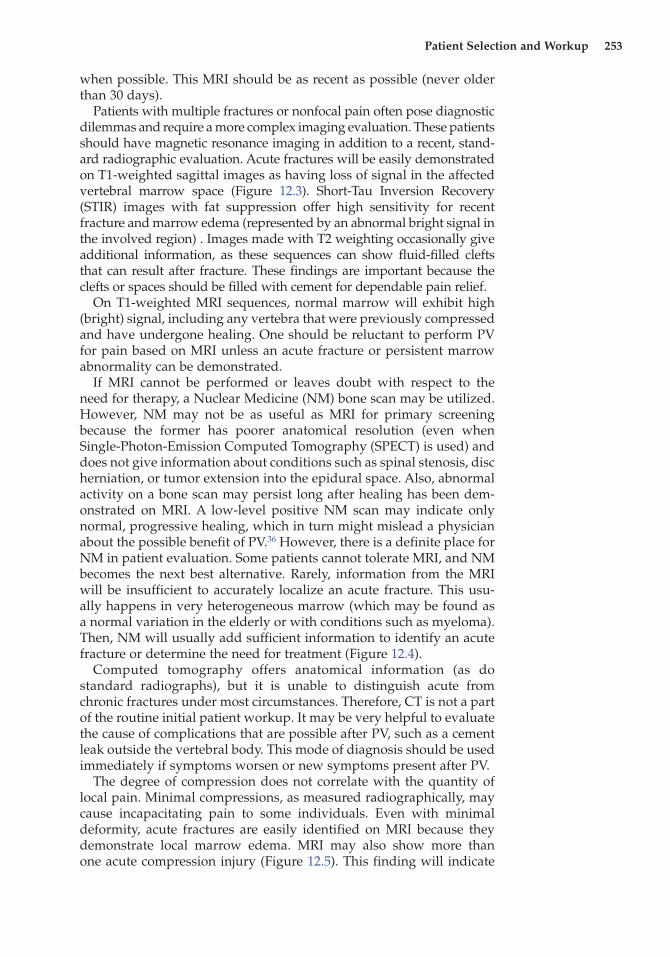
one should obtain an MRI that indicates no additional cause of pain. The same MRI allows one to evaluate the quantity of residual marrow space in the vertebra to be treated. Often, severe collapse is greatest centrally, while sparing residual marrow space laterally that can be successfully treated with PV (Figure 12.6). Patients with these lesions should be made aware that there may be a reduced chance of pain relief (in comparison to a modestly compressed vertebral fracture) and higher risk of complication.
Figure 12.5. Sagittal T1-weighted MRI revealing two acute fractures (arrows) at different locations in the spine (Reprinted with the kind permission of Springer Science + Business Media from Mathis JM ed. Image-Guided Spine Interventions. New York: Springer Science + Business Media, 2004).
Cement Selection and Preparation 257
Although PV has been shown to be very durable, on rare occasions, one may see a refracture with progressive height loss after PV. This usu-ally occurs when the patient has had a less than optimum fill during an initial treatment (even with good initial pain relief) or in the situation of an extremely fragile vertebra. In either case, the amount of cement introduced probably was not sufficient to restore adequate strength to resist recurrent compression. Pain relief and cement filling are poorly correlated. Recurrence of pain, marrow edema, and additional verte-bral collapse may indicate the rare need for repeat treatment.
Cement Selection and Preparation
The first bone cement used for PV was the PMMA Simplex™ P (Stryker Howmedica Osteonics, Mahwah, NJ).13 This was the only cement approved by the U.S. Food and Drug Administration (FDA)
Figure 12.6. (A) Sagittal T1-weighted MRI (midline) reveals extreme compression of the center of the L1 vertebral body (arrow). (B) Images along the lateral edge of L1 reveal less compression and more residual marrow space, which can accept bone cement (arrow) (Reprinted with the kind permission of Springer Science + Business Media from Mathis JM ed. Image-Guided Spine Interventions. New York: Springer Science + Business Media, 2004).
258 Chapter 12 Percutaneous Vertebroplasty
for use in the treatment of pathological fractures in the spine. It was not specifically approved for PV. There were no cements approved for PV in the early days of the procedure. Multiple non-approved PMMA cements were used for PV and seem to have had similar clinical results.16,17,33 It is important to note that bone cement is not treated as a pharmaceutical by the FDA but rather as a device. Alterations in the composition are therefore equivalent to making a new (non-approved) material. It has been suggested by other authors that such alterations constitute “off-label” use.39 Off-label use would be correctly applied if an unaltered cement were used in a non-indicated application or location. Alteration in the ratio of monomer to copolymer (liquid to powder) or addition of other materials (opacification agents or antibi-otics) results in the creation of a new material, and the FDA approval no longer exists. Patients should be informed that such alterations in the cement are to be used, and the reasons and consequences behind these changes should be discussed. Fortunately, there are now multi-ple FDA-approved bone cements for both PV and balloon-assisted PV. Under all but the most unusual circumstances, cement should be used as supplied and not modified.
Inherent in performing PV safely is the need to accurately monitor the injection of cement in real time.33 This is usually accomplished with fluoroscopy and requires that the cement be opacified so that it may be adequately seen in small quantities during introduction. It has been determined that barium sulfate, in quantities of 30% by weight mixed with the PMMA, will provide an adequate level of opacification.33,40,41 All FDA-approved cements have appropriate opacification for fluoro-scopic monitoring.42
Some investigators add antibiotics routinely to PMMA prior to injec-tion, the most common antibiotic being tobramycin.16,33 However, the infection rate with PV is very low, and the efficacy of adding antibiotics to the cement has not been scientifically substantiated in normal, uninfected patients. One report in the orthopedic literature did show reduced infection rates in hip replacements in which cement-containing antibiotics were used for immunosuppressed patients.43 As there is no scientific indication to add antibiotics in otherwise normal patients and because this addition alters the cement and negates its FDA-approved composition, the authors do not recommend the addition of antibiotics to cements except in the situation of immunocompromise.
Adequate precaution should be used during cement mixing to main-tain sterility. Cement manufacturers should provide closed, vacuum-mixing devices that aid in maintaining a sterile environment. Open mixing, which increases the risk of cement contamination and reduces the cement strength by the inclusion of air bubbles, should be avoided.
Thick PMMA seems to limit leaks and these leaks, when large, are associated with complications. However, small leaks (not clinically significant) are simply technical events like small, non-significant blood loss at surgery.
Though traditional percutaneous vertebroplasty and Kyphoplasty (KP) have been performed with PMMA, new cements (non-PMMA) are being developed in an attempt to improve on short comings know
Image Guidance 259
to exist with PMMA. These problems include: 1.) no intrinsic radio-opacity, requiring the addition of non-structural materials like barium, 2.) toxic monomer leaching and a very high exotherm during polym-erization (both of these are capable of killing adjacent cells within the bone), 3.) bone treats PMMA as a foreign body (actually forming a scar adjacent to it) with no local bonding. The first of these biologic materi-als has now been approved by the FDA for use in both PV and KP. This material is named Cortoss (Orthovita; Malvern, Pa). A multi-year, ran-domized trial was performed against PMMA and demonstrated that Cortoss was equivalent to PMMA in relieving pain and safety. It had better outcome profiles in long term improvement in patient function as well as in lower subsequent and adjacent fractures.
Cortoss is a modern, improved cement for bone augmentation. It contains a bioactive ceramic which bone bonds to forming a tight con-nection and eliminating loosing. It has no monomer leaching and low exotherm eliminating the toxic reactions seen with PMMA. Because its’ biomechanical properties are more similar to native bone, it requires a smaller amount to restore strength and stiffness. As less needs to be injected, leaks may be reduced. Finally, its mixing system is totally dif-ferent from PMMA. It uses a dual cartridge device that only combines the two active elements of the cement when needed (mix on demand) eliminating the short work time issues faced with PMMA. These changes should markedly improve PV and KP.
Informed Consent
Written permission for the procedure is recommended, following a complete discussion of the procedure, including the risks and complica-tions, with the patient and/or the patient’s representative. This must include a discussion of any intent (or need) to modify the cement from its FDA approved mixture. These type of modifications (or the perform-ance of non-standard or investigational procedures) should be used only under the supervision of an IRB and approved investigational protocol.
Image Guidance
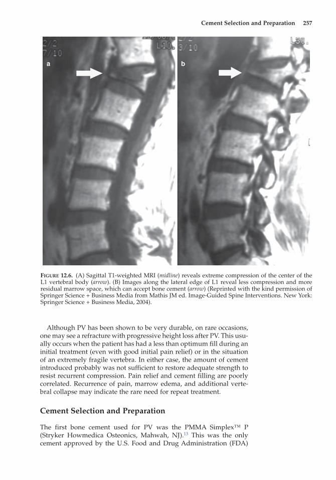
Since the first PV procedure,13 fluoroscopy has been the preferred method of image guidance for performing PV, although CT has been used infre-quently as a primary or adjunctive tool.44,45 Because this procedure was initiated and popularized by interventional neuroradiologists, biplane fluoroscopic equipment was commonly available and often used. This equipment allows multiplanar, real-time visualization for cannula intro-duction and cement injection, and permits rapid alternation between imaging planes without complex equipment moves or projection rea-lignment (Figure 12.7). However, this type of radiographic equipment is expensive and is not commonly available in interventional suites or operative rooms unless it is used for neurointerventional procedures.
It takes longer to acquire two-plane guidance and monitoring infor-mation with a single-plane than with a biplane system. However, it is feasible and safe to use a single-plane fluoroscopic system as long as the
260 Chapter 12 Percutaneous Vertebroplasty
operating physician recognizes the necessity of orthogonal projection visualization during the PV to ensure safety. With a single-plane system for PV, these C-arm moves will mean a slower procedure than that offered by a biplane system.
Gangi et al.45 introduced the concept of using a combination of CT and fluoroscopy for PV. This method gained a brief period of popularity in the United States with the study published by Barr et al.44 Barr and colleagues subsequently abandoned CT for routine PV. Although the contrast resolution with CT is superior to that of fluoroscopy, with CT, one gives up the ability to monitor needle placement and cement injection in real time. Even so, CT may be acceptable for needle place-ment, particularly if a small-gauge guide needle is first placed to ensure accurate and safe location before a large-bore bone biopsy sys-tem is introduced. However, CT certainly is not optimal for monitoring the injection of cement. For this reason, Gangi et al.45 and Barr et al.44 used fluoroscopy in the CT suite during cement introduction. CT does not afford one the opportunity to watch the cement as it is being injected or to alter the injection volume in real time if a leak occurs. Also, unless a large section is scanned with each observation, leaks can occur outside the scan plane, and they may be missed if one is looking only locally in the middle of the injected body. Barr et al.44 used general
Figure 12.7. Biplane fluoroscopy/angiography room. The ability to perform fluoroscopy in two projections without having to move equipment greatly speeds and simplifies vertebroplasty (Reprinted with the kind permission of Springer Science + Business Media from Mathis JM ed. Image-Guided Spine Interventions. New York: Springer Science + Business Media, 2004).
Anesthesia 261
anesthesia with CT guided surgery because of the need to minimize patient motion. This was successful but added a small additional risk to the procedure and considerable complexity and cost. For all these reasons, CT has not found a primary role in image guidance for PV; it is reserved for extremely difficult cases.
Laboratory Evaluations
Coagulation test results should be normal, and the patient should not be taking Coumadin® (Dupont-Merck, Wilmington, DE). Coumadin® may be discontinued and replaced with enoxaparin sodium (Lovenox®, Rhône-Poulenc Rorer Pharmaceuticals, Inc., Collegeville, PA), given once or twice a day on an outpatient basis. Coumadin® may also be stopped and replaced with heparin, but this medication must be administered intravenously, requiring hospital admission. Both enoxaparin sodium and heparin can be reversed with protamine sulfate before PV and restarted postoperatively. Aspirin use is not a contraindication to the procedure, but most interventional groups now take a much more conservative approach to these drugs as well. (The Society of Interventional Radiology recommends stopping anti-platelet drugs 5 days prior to interventional procedures).
PV is not recommended for patients with signs of active infection, but elevated white blood cell counts clearly associated with medical conditions such as myeloma or secondary to steroid use are not con-traindications.
Antibiotics
For PV, as for other surgical procedures that implant devices into the body, intravenous antibiotics are routinely given before (usually 30 min) the procedure is begun. The most common antibiotic used in this applica-tion is cefazolin (1 g).46 If an alternative must be used because of allergy, ciprofloxacin (500 mg orally, two times daily) may be substituted and continued for 24 h after the completion of the procedure. Optimally, an oral antibiotic should be started 12 h before the PV procedure.
As mentioned earlier, antibiotics are added to the cement itself only in the situation of immunocompromise (and this renders the cement no longer FDA approved).
Anesthesia
During PV, it is common to use both local anesthetics and conscious seda-tion to make the patient comfortable and relaxed. Patients who request not to receive intravenous sedation or cannot have it for safety reasons still can be treated with only mild discomfort if appropriate attention is given to local anesthetic placement. To reduce the sting and discomfort associated with locally administered anesthetics (lidocaine, etc.), one may buffer the anesthetic by the addition of a mixture of 1 mL of bicarbonate and 9 mL
262 Chapter 12 Percutaneous Vertebroplasty
of lidocaine. This mixture reduces, but does not eliminate, the anesthetic sting. The authors commonly use a mixture that includes both bicarbonate and Ringer’s lactate (Table 12.1), which essentially eliminates the sting of the local anesthetic. At the authors’ institution, this mixture is prepared daily for all procedures requiring local anesthetics. The excess is discarded at the end of each day, as it contains no preservative. This preparation has a low concentration of lidocaine (0.5%) and allows the use of a more gener-ous volume locally with less risk of toxicity.
Whatever the chosen local anesthetic preparation, the skin, subcutane-ous tissues along the expected needle tract, and periosteum of the bone at the bone entry site must be thoroughly infiltrated. When this has been accomplished, the patient will experience only mild discomfort while the bone needle is being placed, regardless of whether conscious sedation is used.
Conscious sedation has become a common adjunctive method of pain and anxiety control in awake patients who undergo minimally invasive procedures. The authors use a combination of intravenous midazolam (Versed®, Roche, Manati, PR) and fentanyl (Sublimase®, Abbott Labs, Chicago, IL). To decrease anxiety and diminish the discomfort associ-ated with positioning, it may be helpful to begin these medications before the patient is placed on the operating table. Dosages are chosen according to patient size and medical condition. The final amount is determined with titration while observing the patient’s response.
General anesthesia is rarely needed for PV, but it is used occasionally for patients in extreme pain who cannot tolerate the prone position used in PV or for patients with psychological restrictions that preclude a con-scious procedure. It is not needed for routine PV and should be avoided when possible because it adds a mild risk and considerable cost to the procedure. As described earlier, Barr et al.44 used general anesthesia routinely with CT-guided procedures to ensure minimum patient motion.
Needle Introduction and Placement
The original choice of a device for percutaneous cement introduc-tion was based on device availability. The size of the needles was empirically chosen to allow the viscous PMMA cement to be injected.
Reprinted with the kind permission of Springer Science + Business Media from Mathis JM ed. Image-Guided Spine Interventions. New York: Springer Science + Business Media, 2004.aSolution 1 makes a “sting-free” local anesthetic with 0.5% lidocaine. Solution 2 is “sting free” with 0.5% lidocaine and 1:200,000 epinephrine. These preparations should be mixed daily and discarded at the end of the day. The total volume of each mix is 30 mL.
Needle Introduction and Placement 263
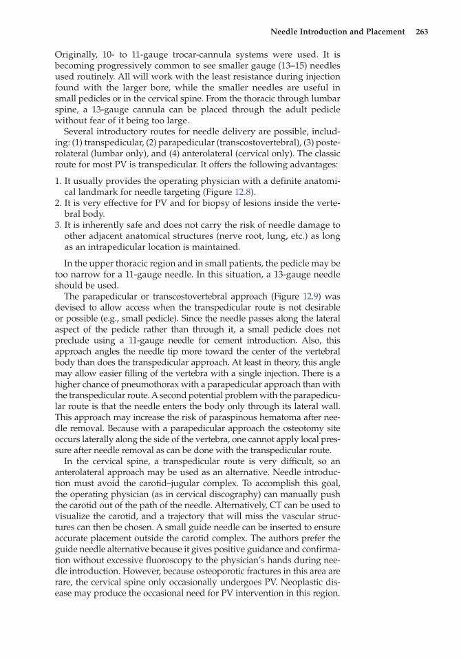
Originally, 10- to 11-gauge trocar-cannula systems were used. It is becoming progressively common to see smaller gauge (13–15) needles used routinely. All will work with the least resistance during injection found with the larger bore, while the smaller needles are useful in small pedicles or in the cervical spine. From the thoracic through lumbar spine, a 13-gauge cannula can be placed through the adult pedicle without fear of it being too large.
Several introductory routes for needle delivery are possible, includ-ing: (1) transpedicular, (2) parapedicular (transcostovertebral), (3) poste-rolateral (lumbar only), and (4) anterolateral (cervical only). The classic route for most PV is transpedicular. It offers the following advantages:
1. It usually provides the operating physician with a definite anatomi-cal landmark for needle targeting (Figure 12.8).
2. It is very effective for PV and for biopsy of lesions inside the verte-bral body.
3. It is inherently safe and does not carry the risk of needle damage to other adjacent anatomical structures (nerve root, lung, etc.) as long as an intrapedicular location is maintained.
In the upper thoracic region and in small patients, the pedicle may be too narrow for a 11-gauge needle. In this situation, a 13-gauge needle should be used.
The parapedicular or transcostovertebral approach (Figure 12.9) was devised to allow access when the transpedicular route is not desirable or possible (e.g., small pedicle). Since the needle passes along the lateral aspect of the pedicle rather than through it, a small pedicle does not preclude using a 11-gauge needle for cement introduction. Also, this approach angles the needle tip more toward the center of the vertebral body than does the transpedicular approach. At least in theory, this angle may allow easier filling of the vertebra with a single injection. There is a higher chance of pneumothorax with a parapedicular approach than with the transpedicular route. A second potential problem with the parapedicu-lar route is that the needle enters the body only through its lateral wall. This approach may increase the risk of paraspinous hematoma after nee-dle removal. Because with a parapedicular approach the osteotomy site occurs laterally along the side of the vertebra, one cannot apply local pres-sure after needle removal as can be done with the transpedicular route.
In the cervical spine, a transpedicular route is very difficult, so an anterolateral approach may be used as an alternative. Needle introduc-tion must avoid the carotid–jugular complex. To accomplish this goal, the operating physician (as in cervical discography) can manually push the carotid out of the path of the needle. Alternatively, CT can be used to visualize the carotid, and a trajectory that will miss the vascular struc-tures can then be chosen. A small guide needle can be inserted to ensure accurate placement outside the carotid complex. The authors prefer the guide needle alternative because it gives positive guidance and confirma-tion without excessive fluoroscopy to the physician’s hands during nee-dle introduction. However, because osteoporotic fractures in this area are rare, the cervical spine only occasionally undergoes PV. Neoplastic dis-ease may produce the occasional need for PV intervention in this region.
Figure 12.8. (A) Typical transpedicular route for needle placement into the vertebral body. (B) Anterior–posterior radiograph demonstrates the placement of the needle through the pedicle, which is seen as a well-circumscribed oval (arrow). In this projection, the needle is initially positioned during fluoroscopy while being held with a clamp (arrowhead) to avoid x-ray exposure to the operator’s hands. (C) Lateral fluoroscopic image demonstrates the final needle position beyond the midline of the ver-tebra (Reprinted with the kind permission of Springer Science + Business Media from Mathis JM ed. Image-Guided Spine Interventions. New York: Springer Science + Business Media, 2004).
Needle Introduction and Placement 265
Once the needle route is chosen, local anesthesia is administered, and a small dermatotomy incision is made with a no. 11 scalpel blade. The trocar–cannula system is introduced through the skin incision and subcutaneous tissue to the periosteum of the bone. This introduction can be facilitated with a sterile clamp to guide the needle during fluor-oscopy, thus avoiding radiation to the operating physician’s hands (Figure 12.8b). In osteoporotic bone, penetrating the bone cortex and advancing the needle into the vertebral body is usually very easy. In a patient with neoplastic disease, the bone still may be very dense and strong (except where it has been destroyed by a tumor). The use of a mallet to advance the needle through very dense bone is a technique clearly superior to manual advancement. Regardless of whether a transpedicular or parapedicular route has been chosen, the tip of the needle should lie beyond the vertebral midpoint as viewed from the lateral projection. The authors usually try to obtain an even more anterior position by placing the needle tip at the junction of the anterior and middle thirds of the vertebra.
Two needles are routinely placed, usually via the transpedicular approach. This takes minimally longer than a single needle placement and affords a large margin of safety for being able to dependably com-plete a vertebral fill with a single mix of cement. There is no question that a single needle placement can give an adequate fill in a large number
Figure 12.9. (A) Needle location for parapedicular (extrapedicular) place-ment. (B) Lateral projection demonstrat-ing that the needle must enter above the transverse process on the para-pedicular approach (Reprinted with the kind permission of Springer Science + Business Media from Mathis JM ed. Image-Guided Spine Interventions. New York: Springer Science + Business Media, 2004).
266 Chapter 12 Percutaneous Vertebroplasty
of cases. However, the single-needle method fails to produce uniform fills more often than the double-needle technique and may oblige the operator to accept a larger cement leak during filling (if the second needle is not in place as an alternate injection route).
Venography
Venography was never used much in Europe and was introduced in the United States in an attempt to discover potential leak sites prior to injecting cement. However, this technique worked poorly because the contrast material and the bone cement are very different in viscosity. The authors discontinued using venography in 1996 and have found no disadvantage or added risk without its use.47 Other long-term proponents have belatedly stopped its use in routine PV as they found no safety benefit.48
Cement Injection
Cement is prepared only after all needles are placed, as described in the earlier section of this chapter on “Cement Selection and Preparation.” Cement with an appropriate opacification is prepared and injected using small syringes (typically 1 mL) or devices made specifically for injection (Figure 12.10) (no actual advantage to a specific PMMA
Figure 12.10. Cement injection with a 1 mL syringe. Note bipedicular needle placement prior to beginning cement injection (Reprinted with the kind permis-sion of Springer Science + Business Media from Mathis JM ed. Image-Guided Spine Interventions. New York: Springer Science + Business Media, 2004).
Cement Injection 267
versus another has been shown. The newly FDA approved biologic cement Cortoss (Orthovita; Malvern, Pa) does give great promise to improve many of the short coming seen with PMMA). Either the cement injection should be monitored in real time, or small quantities (i.e., 0.1–0.2 mL) should be injected and the result visualized before additional cement is introduced. The latter approach, which allows one to step back from the fluoroscopy beam during visualization, mini-mizes radiographic exposure to the operator.
Any cement leak outside the vertebral body is an indication to stop the injection. When using a rapidly polymerizing cement, halting the injection may be necessary only for a minute or two while the injected cement hardens. Restarting the injection may then redirect flow into other areas of the vertebra. If leakage is still seen, it is advisable to terminate the cement injection through this needle and move to the second needle. This will usually allow completion of the vertebral fill without further leakage, since the original leak now will be occluded by the initial cement, which will have hardened. One should work through a single needle at a time. This avoids contamination of both needles at once and preserves a route for subsequent injection if a leak is encountered. Cement can still be introduced beyond the point at which most injection devices begin to fail. The trocar is useful to push additional thick cement from the cannula into the vertebra. Bone fillers are special cannula-plunger systems that fit through the existing bone cannula. They allow the injection of very thick cement (beyond what can be injected with a syringe or injector). They also prevent cement from touching the introductory cannula and precluding cannula clo-sure by hardening cement. This allows an additional margin of safety as the cannula remains open allowing multiple injections as needed to complete the fill and minimize leaks. The on demand mix of the new cement Cortoss makes this option much more viable.
The 5 in., 13-gauge cannula holds 0.5 mL, and the 5 in., 11-gauge cannula holds 0.9 mL. Reintroducing the trocar will push the residual cement in the cannula into the vertebra. This is done only if the addi-tional amount of cement is desired. The cannula can be removed safely without reintroduction of the trocar when the cement has hardened beyond the point at which it can be injected. Simply twisting the needle through several revolutions will break the cement at the tip of the cannula and will prevent leaving a trail of cement in the soft tissues. However, removing the cannula before the cement has hardened suffi-ciently can allow cement to track backward from the bone into the soft tissues and may create local pain.
The amount of cement needed to produce pain relief has not been accurately documented in available clinical reports. The authors believe that pain relief is related to fracture stabilization (not to a chemical or thermal effect), and thus the amount of cement needed to restore the initial vertebral body’s mechanical integrity should also give an approximation of the quantity needed to relieve pain clinically. In an in vitro study, the authors showed that the initial pre-fracture strength and stiffness of a vertebra could be restored by injecting 2.5–4 mL of PMMA in the thoracic vertebra, while 6–8 mL provided
268 Chapter 12 Percutaneous Vertebroplasty
similar augmentation in the lumbar region. (a smaller quantity of Cortoss will be required to cover the same region of the vertebra com-pared to PMMA).49 A reasonable guideline for the quantity of any cement to be injected is the amount that is needed to fill 50–70% of the residual volume of the compressed vertebra (determined by visual estimate during cement introduction). These amounts should not be taken as absolute but rather as a guide. The previously described study suggests that relatively small amounts of cement are needed to restore initial biomechanical strength and that these amounts vary with the relative vertebral level in the spine, as well as with individual vertebral body size and the degree of vertebral collapse.
The authors also have demonstrated that significant strength resto-ration is provided to the vertebral body with a unipedicular injection, where cement filling crosses the midline of the vertebral body.50 This would imply that unipedicular fills that achieve adequate cement injec-tion volumes are likely to be successful at achieving pain relief. This fact notwithstanding, there is a higher likelihood of achieving more uniform fills, with fewer leaks, when two needles are used rather than one (Figure 12.11).
Postoperative Care
After adequate vertebral filling has been accomplished, the needle is removed. Occasionally, venous bleeding is experienced at the needle entry site. Hemostasis is easily achieved with local pressure for 5 min. The entry site is dressed with Betadine ointment and a sterile bandage. The patient is maintained recumbent for 1–2 h after the procedure and monitored for changes in neurological function or for signs of any other clinical change or side effects. Table 12.2 lists typical postopera-tive orders.
Any sign of adverse events should trigger the use of appropriate imaging modalities (usually CT) in the search for an explanatory cause. It is well known that 1–2% of patients will have a transient period of benign increase in local pain following PV. However, this is a diagnosis of exclusion and should prompt extended monitoring (or hospitalization if the pain is severe and requires aggressive therapy) and imaging evaluation to exclude other causes for the pain (such as cement extravasation). Pain alone will usually be adequately treated with analgesics, nonsteroidal anti-inflammatory drugs (such as Toradol®, Roche Pharmaceuticals, Nutley NJ), or local steroid injections adjacent to affected nerve roots or in the epidural space. Large cement leaks (Figure 12.12) or neurological dysfunction should prompt an immediate surgical consultation.
PV is easily performed on an outpatient basis, with the patient dis-charged after 1–2 h of uneventful recovery. Table 12.2 gives typical discharge instructions. Follow-up is indicated to monitor the results of therapy and should be incorporated into a quality management program. Reports of complications and results should be maintained by the facility as well as for each individual provider. Additional informa-
Postoperative Care 269
Figure 12.11. (A) Anterior–posterior radiograph showing a good bipedicular vertebral fill of bone cement. (B) Lateral radiographs show the same vertebra. Note that the entire central volume of the vertebra is not filled (Reprinted with the kind per-mission of Springer Science + Business Media from Mathis JM ed. Image-Guided Spine Interventions. New York: Springer Science + Business Media, 2004).
270 Chapter 12 Percutaneous Vertebroplasty
Table 12.2. Sample postoperative orders and discharge instructions
Postoperative orders
Bed rest 1 h (may roll side to side)May sit up after 1 h with assistanceVital signs and neurological examinations (focused on the lower extremities)
every 15 min for the first hour, then every 30 min for the second hourRecord pain level (Visual Analog Scale, 1–10) at end of procedure and at
2 h postoperatively (before discharge). Compare with baseline values and notify physician if pain increases above baseline
May have liquids by mouth if no nauseaDiscontinue oxygen (if used) after procedure (if saturation is normal)Discontinue intravenous drips after 1 h if recovery is otherwise uneventfulDischarge patient home with adult companion after 2 h if recovery is
uneventful
Discharge instructions
Return home; bed rest or minimal activity for next 24 hMay resume regular diet and medicationsKeep operative site covered for 24 h. Bandages may then be removed and
site washed with a damp cloth. Do not soakNotify physician or facility if there is increasing pain, redness, swelling,
or drainage from the operative siteNotify physician or facility if there is difficulty with walking, changes in
sensation in hips or legs, new pain, or problems with bowel or bladder function
The area of the procedure will be tender to the touch for 24–48 hThis is to be expectedIf there is pain similar to that before the procedure, prescribed pain
medications may be continued as needed
Reprinted with the kind permission of Springer Science + Business Media from Mathis JM ed. Image-Guided Spine Interventions. New York: Springer Science + Business Media, 2004.
tion and recommendations about the credentialing and quality man-agement for PV can be found in the American College of Radiology manual on standards of practice (ACR.org/StandardsofPractice).
Results
To date there are no substantial prospective, randomized trials evaluating PV published in the literature. However, Zoarski and colleagues pre-sented a small prospective (nonrandomized) evaluation of the effective-ness of PV for relieving pain.51 This report utilized the MODEMS method to establish that 22 of 23 patients improved after PV and remained satisfied during the 15- to 18-month follow-up. Additionally, numerous retrospective series are available and uniformly report good pain relief and reduced requirements for analgesics following PV.16,17,22,27,44 This is especially true of pain related to compression fractures produced by oste-oporosis, where significant pain relief of between 80 and 90% has been observed. This pain relief is persistent with no reports of additional com-pression of vertebra previously treated with PV. Additional fractures at other levels remain a possibility and source of morbidity. If osteoporotic compression fracture occurs, every effort to minimize future bone loss
Results 271
medically should be made. Also, modifications in lifestyle should be attempted to minimize mechanical stress on the spine and thereby lessen the risk of additional fractures (NEJM articles discussed below.)
Much discussion has been given as to whether there is an advan-tage of balloon-assisted vertebroplasty (kyphoplasty) compared to traditional vertebroplasty. Again, there are no good randomized trials comparing the two techniques. Indeed, the mechanical stabilization used in both is the same (cementation). Safety is probably similar, though a higher permanent complication and death rate is reported with kyphoplasty. A consensus statement by multiple societies using both techniques (American Society of Neurological Surgeons/Congress of Neurological Surgeons, American Society of Spine Radiology, and American Society of Interventional and Therapeutic Neuroradiologist) was issued in 2007 and states: “After reviewing the published literature on kyphoplasty, the Societies have determined that the clinical response rate in individuals treated with kyphoplasty is equivalent to that seen in patients treated with vertebroplasty. There is no proved advantage of
Figure 12.12. CT scan of a patient who experienced paraplegia following vertebroplasty as a result of a large cement leak. The cement (stars) occupies a large amount of the spinal canal at the level of the CT scan and creates cord compression (Reprinted with the kind permission of Springer Science + Business Media from Mathis JM ed. Image-Guided Spine Interventions. New York: Springer Science + Business Media, 2004).
272 Chapter 12 Percutaneous Vertebroplasty
kyphoplasty relative to vertebroplasty with regard to pain relief, verte-bral height restoration, or complication rate.”52
There has been growing concern over the risk of secondary vertebral fracture after treatment, especially at adjacent levels. However, the natu-ral history of vertebral fractures treated conservatively is approximately 20% at 1 year. With the incidence of two fractures, incidence increases to 24%. Fifty percent of these new fractures will be in adjacent levels.53 This compares very well with reported incidence of new and adjacent levels of fractures occurring following treatment with PV or KP.54
Complications
Complications, though initially considered to be uniformly low, are unfortunately higher for inexperienced physicians or those who attempt the procedure without adequate image guidance or cement opacification. Appropriate training needs to be completed before the procedure is attempted. Recommendations can be obtained from the American College of Radiology Standards of Practice on Percutaneous Vertebroplasty (ACR.org/StandardsofPractice).
In osteoporosis-induced vertebral fractures, clinical reports of com-plications are around 1%.16,17,22,27 Many of these are transient and include increase in local pain after cement introduction (nonradicular and not associated with neurological deficit). This is usually easily treated with nonsteroidal anti-inflammatory drugs and resolves within 24–48 h. Uncommonly, cement leaking from the vertebra adjacent to a nerve root will produce radicular pain. Analgesics combined with local steroid and anesthetic injections usually provide adequate relief. A trial of this type of therapy is warranted as long as there are no associated motor deficits. The discovery of a motor deficit (or bowel or bladder dysfunction) should initiate an immediate surgical consultation. This type of severe complication will almost always be associated with large-volume leaks that have resulted in neurological compression. Correction of the complication surgically should be considered a medical emergency.
Cement leaks also have been implicated in producing pulmonary embolus.16 These are usually not symptomatic but rarely have pro-duced the clinical symptoms accompanying pulmonary infarct. With a right-to-left shunt, this can result in cerebral infarct.55 Likewise, infec-tion has been reported but is rare with PV.
The complication rate found when treating compression fractures resulting from malignant tumors is considerably higher.22,26,29,30,56 This occurs because there are frequently lytic areas involving the vertebral cortex and a greater propensity for cement to leak into the surrounding tissues or vessels. Cement leaks causing symptoms in this setting occur in 5–10% of patients; again, most are transient.
Much is made of cement leaks, but often without distinguishing the type of leaks. A small leak that has no clinical consequence should be thought of as a “technical event,” much like a small and insig-nificant blood loss at surgery. Only the rare (and usually large) leak
Pain relief after Vertebroplasty: Real or Sham 273
causes patient injury and is then termed as a “clinical complication.” Unfortunately, these distinctions have rarely been made in the verte-broplasty/kyphoplasty literature. Indeed, marketing has selectively used these “technical events” to try to gain advantage routinely.
Death is a rare complication associated with PV and KP. Though the exact details usually are not known, the likely cause seems to be pulmonary compromise, which is suspected to be due to fat (from the vertebral marrow) or cement emboli. A safe number of vertebrae to treat at one time has yet to be definitely established. Mathis and colleagues reported treating seven vertebrae in a 35-year-old patient with multiple fractures associated with steroid use for lupus.46 This patient’s therapy occurred in three treatment sessions. Because the introduction of cement is a hydraulic event with as much marrow pushed out of the inter-trabecular space as cement injected, there is concern about fat emboli in large volume cement injections. For reasons described earlier, the authors recommend treating no more than three vertebrae in any one session in healthy individuals and less if there is known cardiopulmonary disease. Those with increased risk should be appropriately warned as part of their consent process. There are no data that support the prophylactic use of PV to treat vertebra that are believed to be at risk of fracture. Except for prophylactic use, there is little conceivable reason to perform PV on large numbers of vertebrae at one time. No safety is gained with kyphoplasty, as the balloon also displaces marrow products that go to the lungs.
Any deviation from an expected good result (such as increased pain or neurological compromise) should initiate an immediate imaging search with CT to look for a cause of the clinical change. Unremitting or progressive symptoms may require surgical or aggressive medical intervention, and outpatients should be hospital-ized and monitored.
Pain relief after Vertebroplasty: Real or Sham
With over 1000 positive peer reviewed papers about Percutaneous Vertebroplasty (PV) or balloon assisted vertebroplasty or (Kyphoplasty) and with hundreds of thousands cases performed, it seems obvious to those of us that have performed these procedures that pain relief should be considered very real. However, 2009 articles in the New England Journal of Medicine described sham therapy that was equiva-lent to PV for pain relief in compression fractures of the spine.57,58
The NEJM articles would not have gotten nearly as much interest if good, randomized studies of PV against conservative therapy (the traditional, historic therapy used for compression fractures prior to PV) had been initially performed. However, these were not accomplished for numerous reasons. The questions raised by the NEJM articles there-fore had a magnified effect.
Both NEJM articles had a limited number of patients enrolled despite a prolonged acquisition period (over 4 years for the Kallmes study, the largest by a factor of 2). The Kallmes study intended to enroll 250 patients but cut its study off after 4.5 years with 131 total
274 Chapter 12 Percutaneous Vertebroplasty
patients.59 It found (but did not emphasize) a trend toward a “clini-cally meaningful improvement in pain for PV compared to sham therapy” (64% of PV patients vs. 48% of sham treated patients). This fell short of statistical significance with a P=.06 ( P=.05 was needed to be statistically significant). Obviously, the P value lacked only .01 reaching significance for PV to be better than sham. If one simply continues the proportions of improved patients in each group, an additional 19 patients were needed to reach this clinical significance of PV over sham. This would have been a total number of patients of 150, still far below the intended number of 250 which were initially planned for. This simple change in the study would have nullified most of the importance as it would have shown PV better than sham in this category. The 131 number did not provide the sensitivity to predict this outcome.
Patients in the sham group did get initial pain relief equal to PV. However, it must be remembered that this sham therapy is not con-servative therapy. The sham procedure consisted of injecting anesthetic into the periosteum of the fractured bone (along its posterior cortex where the pedicle entry point would be if PV had been performed). One may think this therapy should be insignificant and very transient in effect. However, similar injections have been used for years in pain management for spine pain such as facet blocks and for other pain control like occipital nerve blocks for migraine headaches. For those of us that perform such procedures, we commonly see weeks and sometimes months of relief from problems like migraine headaches with these simple anesthetic injections. Therefore, it is not surpris-ing that pain relief was seen with this “sham” procedure. However, very telling is the fact that crossover from one group to another found 43% of the sham patients crossing over to the PV group after 1 month (regardless of the amount of pain relief these patients claimed initially, it obviously did not last). Only 12 % of PV patients crossed over (this is exactly expected as we find 85–90% of patients getting good pain relief traditionally after PV.60 The crossover numbers were highly significant with a P=.001.
So sham therapy was associated with initial pain relief but failed to be persistent in many treated in this group. Is this sham a real therapy or a placebo? Probably we will never know for sure and from a clinical point of view, it may matter little. New evidence published in Science (2009) found functional MRI evidence of changes in uptake in the dorsal columns (sensory nerve region) when a placebo was applied to the skin in regions of pain.61 This paper shows the strong and real effect of placebo, long known to be active in most pain management treatments.
The NEJM articles found real or placebo effects of pain reduction with “sham” (not conservative) therapy. They found that the sham therapy was not long lasting with a statistically significant number of patients crossing over to the PV group after one month. Finally, if these papers had completed there initially advertised patient accrual (250 patients for Kallmes), a clinically significant improvement in PV over Sham would have been established as well.
Conclusion 275
Conclusion
Percutaneous vertebroplasty has been shown to be very effective at relieving the pain associated with compression fractures of vertebra caused by both primary (age-related) and secondary (steroid-induced) osteoporosis. It also has substantial benefit in neoplastic-induced ver-tebral compression fracture pain but with a higher chance of associated complication. PV is rapidly becoming the standard of care for com-pression fracture pain that does not respond to conservative medical therapy. However, this simple procedure must be treated with respect, for its application without appropriate judgment and physician train-ing can quickly result in increased pain, permanent neurological injury, and even death.
Consensus has now been reported to show no benefit of kyphoplasty over vertebroplasty with regard to pain relief, vertebral height restoration, or complication rate.52 Due to the high cost of kyphoplasty compared to vertebroplasty, kyphoplasty therefore seems rarely warranted for general medical use.
References
1. Alleyne CH Jr, Rodts GE, Jr, Haid RW. Corpectomy and stabilization with meth-ylmethacrylate in patients with metastatic disease of the spine: a technical note. J Spinal Disord 1995;8:439–443.
2. Cortet B, Cotten A, Deprez X, Deramond H, Lejeune JP, Leclerc X, Chastanet P, Duquesnoy B, Delcambre, B. [Value of vertebroplasty combined with surgical decompression in the treatment of aggressive spinal angioma. Apropos of 3 cases.] Rev Rheum Ed Fr 1994;61:16–22.
3. Cybulski GR. Methods of surgical stabilization for metastatic disease of the spine. Neurosurgery 1989;25:240–252.
4. Harrington KD. Anterior decompression and stabilization of the spine as a treat-ment for vertebral collapse and spinal cord compression from metastatic malig-nancy. Clin Orthop 1988;233:177–197.
5. Harrington KD, Sim FH, Enis JE, Johnston JO, Diok HM, Gristina AG. Methylmethacrylate as an adjunct in internal fixation of pathological fractures. Experience with three hundred and seventy-five cases. J Bone Joint Surg (Am) 1976;58:1047–1055.
6. Knight G. Paraspinal acrylic inlays in the treatment of cervical and lumbar spondylosis and other conditions. Lancet 1959;2:147–149.
7. Kostuik JP, Errico TJ, Gleason TF. Techniques of internal fixation for degenerative conditions of the lumbar spine. Clin Orthop 1986;203:219–231.
8. Mavian GZ, Okulski CJ. Double fixation of metastatic lesions of the lumbar and cervical vertebral bodies utilizing methylmethacrylate compound: report of a case and review of a series of cases. J Am Osteopath Assoc 1986;86:153–157.
9. O’Donnell RJ, Springfield DS, Motwani HK, Ready JE, Gebhardt MC, Mankin HJ. Recurrence of giant-cell tumors of the long bones after curettage and packing with cement. J Bone Joint Surg (Am) 1994;76:1827–1833.
10. Persson BM, Ekelund L, Lovdahl R, Gunterberg B. Favourable results of acrylic cementation for giant cell tumors. Acta Orthop Scand 1984;55:209–214.
11. Scoville WB, Palmer AH, Samra K, Chong G. The use of acrylic plastic for vertebral replacement or fixation in metastatic disease of the spine. Technical note. J Neurosurg 1967;27:274–279.
276 Chapter 12 Percutaneous Vertebroplasty
12. Sundaresan N, Galicich JH, Lane JM, Bains MS, McCormack P. Treatment of neoplastic epidural cord compression by vertebral body resection and stabilization. J Neurosurg 1985;63:676–684.
13. Galibert P, Deramond H, Rosat P, Le Gars D. [Preliminary note on the treatment of vertebral angioma by percutaneous acrylic vertebroplasty.] Neurochirurgie 1987;33:166–168.
14. Lapras C, Mottolese C, Deruty R, Lapras C Jr, Remond J, Duquesnel J. [Percutaneous injection of methyl-methacrylate in osteoporosis and severe verte-bral osteolysis (Galibert’s technic).] Ann Chir 1989;43:371–376.
15. Debussche-Depriester C, Deramond H, Fardellone P, Heleg A,. Sebert JL, Cartz L, Galibert P. Percutaneous vertebroplasty with acrylic cement in the treatment of osteoporotic vertebral crush fracture syndrome. Neuroradiology 1991;33(Suppl):149–152.
16. Jensen ME, Evans AJ, Mathis JM, Kallmes DF, Cloft HJ, Dion JE. Percutaneous polymethylmethacrylate vertebroplasty in the treatment of osteoporotic vertebral body compression fractures: technical aspects. Am J Neuroradiol 1997;18:1897–1904.
17. Mathis JM, Barr JD, Belkoff SM, Barr MS, Jensen ME, Deramond H. Percutaneous vertebroplasty: a developing standard of care for vertebral compression fractures. Am J Neuroradiol 2001;22:373–381.
18. Riggs BL, Melton LJ. The worldwide problem of osteoporosis: insights afforded by epidemiology. Bone 1999;17:505S–511S.
19. Ray NF, Chan JK, Thamer M, Melton III LJ. Medical expenditures for the treat-ment of osteoporotic fractures in the United States in 1995: report from the National Osteoporosis Foundation. J Bone Miner Res 1997;12(1):24–35.
20. Cooper C, Atkinson EJ, O’Fallon WM, Melton LJ. Incidence of clinically diag-nosed fractures: a population based study in Rochester, Minn. J Bone Miner Res 1992;7:221–227.
21. Bostrom MP, Lane JM. Future directions. Augmentation of osteoporotic vertebral bodies. Spine 1997;22:38S–42S.
22. Chiras J, Depriester C, Weill A, Sola-Martinez MT, Deramond H. [Percutaneous vertebral surgery. Technics and indications.] J Neuroradiol 1997;24:45–59.
23. Cortet B, Cotten A, Boutry N, Flipo RM, Duquesnoy B, Chastanet P, Delcambre B. Percutaneous vertebroplasty in the treatment of osteoporotic vertebral compres-sion fractures: an open prospective study. J Rheumatol 1999;26:2222–2228.
24. Cotten A, Boutry N, Cortet B, Assaker R, Demondion X, Leblond D, Chastanet P, Duquesnoy B, Deramond H. Percutaneous vertebroplasty: state of the art. Radiographics 1998;18:311–323.
25. Cotten A, Deramond H, Cortet B, Lejeune JP, Leclerc X, Chastanet P, Clarisse J. Preoperative percutaneous injection of methyl methacrylate and N-butyl cyanoacrylate in vertebral hemangiomas. AJNR Am J Neuroradiol 1996;17:137–142.
26. Cotten A, Dewatre F, Cortet B, Assaker R, Leblond D, Duquesnoy B, Chastanet P, Clarisse J. Percutaneous vertebroplasty for osteolytic metastases and myeloma: effects of the percentage of lesion filling and the leakage of methyl methacrylate at clinical follow-up. Radiology 1996;200:525–530.
27. Cyteval C, Sarrabere MP, Roux JO, Thomas E, Jorgensen C, Blotman F, Sany J, Taourel P. Acute osteoporotic vertebral collapse: open study on percutaneous injection of acrylic surgical cement in 20 patients. AJR Am J Roentgenol 1999;173:1685–1690.
28. Deramond H, Depriester C, Galibert P, Le Gars D. Percutaneous vertebroplasty with polymethylmethacrylate. Technique, indications, and results. Radiol Clin North Am 1998;36:533–546.
29. Deramond H, Depriester C, Toussaint P. [Vertebroplasty and percutaneous inter-ventional radiology in bone metastases: techniques, indications, con-tra-indications.] Bull Cancer Radiother 1996;83:277–282.
31. Deramond H, Galibert P, Debussche C, Pruvo J, Heleg A, Hodes J. Percutaneous vertebroplasty with methylmethacrylate: technique, method, results [abstract]. Radiology 1990;177P:352–352.
32. Dousset V, Mousselard H, de Monck d’User L, Bouvet R, Bernard P, Vital JM, Senegas J, Caille JM. Asymptomatic cervical haemangioma treated by percutane-ous vertebroplasty. Neuroradiology 1996;38:392–394.
33. Mathis JM, Eckel TS, Belkoff SM, Deramond H. Percutaneous vertebroplasty: a therapeutic option for pain associated with vertebral compression fracture. J Back Musculoskelet Rehabil 1999;13:11–17.
34. Silverman SL. The clinical consequences of vertebral compression fracture. Bone 1992;13(Suppl):27–31.
35. Kado DM, Browner WS, Palermo L, Nevitt MC, Genant HK, Cummings SR. Vertebral fractures and mortality in older women: a prospective study. Study of Osteoporotic Fractures Research Group. Arch Intern Med 1999;159:1215–1220.
36. Maynard AS, Jensen ME, Schweickert PA, Marx WF, Short JG, Kallmes DF. Value of bone scan imaging in predicting pain relief from percutaneous vertebroplasty in osteoporotic vertebral fractures. Am J Neuroradiol 2000;21(10):1807–1812.
37. O’Brien JP, Sims JT, Evans AJ. Vertebroplasty in patients with severe vertebral com-pression fractures: a technical report. Am J Neuroradiol 2000;21(8):1555–1558.
38. Peh WC, Gilula LA, Peck DD. Percutaneous vertebroplasty for severe osteoporotic vertebral body compression fractures. Radiology 2002;223:121–126.
39. Jensen ME, Dion JE. Percutaneous vertebroplasty in the treatment of osteoporotic compression fractures. Imaging Clin North Am 2000;10(3):547–568.
40. Belkoff SM, Maroney M, Fenton DC, Mathis JM. An in vitro biomechanical evalua-tion of bone cements used in percutaneous vertebroplasty. Bone 1999;25:23S–26S.
41. Jasper L, Deramond H, Mathis JM, Belkoff SM. Material properties of various cements for use with vertebroplasty. J Mater Sci Mater Med 2002;13:1–5.
42. Jasper LE, Deramond H, Mathis JM, Belkoff SM. The effect of monomer-to-powder ratio on the material properties of cranioplastic. Bone 1999;25:27S–29S.
43. Norden CW. Antibiotic prophylaxis in orthopedic surgery. Rev Infect Dis 1991;10:S842–S846.
44. Barr JD, Barr MS, Lemley TJ, McCann RM. Percutaneous vertebroplasty for pain relief and spine stabilization. Spine 2000;25:923–928.
45. Gangi A, Kastler BA, Dietemann JL. Percutaneous vertebroplasty guided by a combination of CT and fluoroscopy. Am J Neuroradiol 1994;15:83–86.
46. Mathis JM, Petri M, Naff N. Percutaneous vertebroplasty treatment of steroid-induced osteoporotic compression fractures. Arthritis Rheum 1998;41:171–175.
47. Wong W, Mathis JM. Commentary: is intraosseous venography a significant safety measure in performance of vertebroplasty? J Vasc Interv Radiol 2002;13:137–138.
48. Gaughen JR, Jensen ME, Schweickert PA, Kaufmann TJ, Marx WF, Kallmes DF. Relevance of antecedent venography in percutaneous vertebroplasty for the treat-ment of osteoporotic compression fractures. Am J Neuroradiol 2002;23:594–600.
49. Belkoff SM, Mathis JM, Jasper LE, Deramond H. The biomechanics of vertebroplasty: the effect of cement volume on mechanical behavior. Spine 2001;26:1537–1541.
50. Tohmeh AG, Mathis JM, Fenton DC, Levine AM, Belkoff SM. Biomechanical efficacy of unipedicular versus bipedicular vertebroplasty for the management of osteoporotic compression fractures. Spine 1999;24:1772–1776.
51. Zoarski GH, Snow P, Olan WJ, Stallmeyer M, Dick B, Hebel J, De Deyne M. Percutaneous vertebroplasty for osteoporotic compression fracture: quantitative pro-spective evaluation of long-term outcomes. J Vasc Interv Radiol 2002;13:139–148.
52. Jensen ME, McGraw JK, Cardella JF, Hirsch JA. Position Statement on per-cutaneous vertebral augmentation: A consensus statement developed by the American Society of Interventional and Therapeutic Neuroradiology, Society of Interventional Radiology, American Association of Neurological Surgeons/
278 Chapter 12 Percutaneous Vertebroplasty
Congress of Neurological Surgeons, and American Society of Spine Radiology. J Vasc Interv Radiol 2007;18:325–330.
53. Lindsay R, Silverman SL, Cooper C, Hanley DA,Barton I, Broy SB,Licata A, Benhamou L, Geusens P, Flowers K, Stracke H, Seeman E. Risk of new vertebral fracture in the year following a fracture. JAMA 2001;285:320–323.
54. Voormolen MHJ, Lohle PNM, Juttmann JR, van der Graaf Y, Fransen H, Lampmann L. The risk of new osteoporotic vertebral compression fractures in the year after percutaneous vertebroplasty. J Vasc Interv Radiol 2006;17:71–76.
55. Scroop R, Eskridge J, Britz GW. Paradoxical cerebral arterial embolization of cement during intraoperative vertebroplasty: case report. Am J Neuroradiol 2002;23:868–870.
56. Weill A, Chiras J, Simon J, Rose M, Sola-Martinez T, Enkaoua E. Spinal metas-tases: indications for and results of percutaneous injection of acrylic surgical cement. Radiology 1996;199:241–247.
57. Kallmes DF, Comstock BA, Heagerty PJ, et al. A randomized trial of vertebropla-seoporotic spinal fractures. NEJM 2009,361:569–579.
58. Buchbinder R, Osborne RH, Ebeling PR, et al. A randomized trial of vertebro-plasty for painful osteoporotic vertebral fractures. NEJM 2009, 361:557–568.
59. Gray LA, Jarvik JG, Heagerty PJ, et al. Investigational vertebroplasty efficacy and safety trial (INVEST): a randomized controlled trial of percutaneous vertebro-plasty. BMC Musculoskel Disorders 2007, 8:126–134.
60. Mathis JM, Barr JB, Belkoff SM, et al. Percutaneous vertebroplasty: a developing standard of care for vertebral compression fractures. AJNR 2001, 22:373–381.
61. Eippert F, Finsterbusch J, Bingel U, Buchel C. Direct evidence for spinal cord involvement in placebo analgesia. Science 2009, 326:404.