JOHNS HOPKiNS CIRCULARS Pub/is/zed wit/i t/ie approt$atioiz of t/ie Board o Trustees NOL. YJ.—No. 5~.] BALTIMORE, DECEMBER, 1886. [PRIcE, 10 CENTS. THE ZOOLOGICAL WORK OF THE)OHNS HOPKINS UNIVERSITY, 1878-86. A REPORT BY W. K. BROOKS, PH. li, DIRECTOR OF THE MARINE LABORATORY.* To the President of the Johns Hopkins University: Sir :—At your request, I submit herewith a report of the zoiilogical work of the University during the last nine years — In natural science the policy of the University is to promote the study of life, rather than to accumulate specimens: and since natural laws are best studied in their simplest manifestations, much attention has been given to the investigation of the simpler forms of life, with confidence that this will ultimately contribute to a clearer insight into all vital phenomena. The oldest forms of life are marine: every great group of animals is represented in the ocean, while many important and instructive groups have no terrestrial representatives; omitting the insects, more than four- fifths of the known species of animals are marine, and the total amount of animal life in the ocean is incomparably greater than that upon the land. In a word, the ocean is now, as it has been at all stages in the earth’s history, the home of life; and it is there, and there only, that we find the living representatives of the oldest fossils, and are thus enabled to study the continuous history of life from its simplest to its most complex manifestations. On the sand flats at the mouth of the Chesapeake Bay, we find, living side by side, animals like Lingula, Amphioxus, Limulus and Balanoglos- sus, which are the representatives of some of the oldest and most primitive types of animal life; and all attempts to trace out the natural relationships of any group of animals, lead us at once to forms which are found only in the ocean. The animals which have contributed most extensively to the formation of the earth’s crust, the corals and foraminifera and radiolarians, abound in the ocean to-day, and it is only by studying their life by observations at the seashore, that we can understand and interpret their geological influence. Nearly every one of the great generalizations of morphology is based upon the study of marine animals, and most of the problems which are now awaiting a solution roust be answered in the same way. For these reasons our chief aim in zoi5logy and animal morphology has been to provide means for research upon the marine animals of the Atlantic coast, and for nine years, successive parties, composed of instructors, fellows and students in this department, together with instructors and advanced students from other institutions have spent at the seashore all the months in which marine work is practicable. Their time~and energy have been devoted to research rather than to the preservation of collections, and the wisdom of this course can be estimated by examination of the a.ccompanying list of publications; all of which are based, either in part or entirely, upon researches which we have carried on at the seashore. The wisdom of our policy is well illustrated by the fact that the leading naturalist of America, himself the head of one of the largest scientific col- lections in the world, says in his annual report for 1884,t that the expenses of an immense natural-history collection are so great that it would be far cheaper, with the present facilities and the cost of travel, to supply the student with the necessary funds for valuable researches, than to go on for years spending in salaries of curators and the care of collections, sums of money which, if spent in a different manner, in promoting original investi- gation in the field or in the laboratory and in providing means for the publication of such origis~ial researches, would do far more towards the pro- motion of natural history than our past methods of spending our resources. This fact has become widely recognized during the last ten years, as is shown by the establishment of marine laboratories by several of the European institutions of learning; and in the summers of 1883 and 1884 we had with us at our laboratory a young English naturalist who had been provided by the iRoyal Society of London with funds for his researches, the results of which have recently been published in England. The Johns Hopkins University was among the first to recognize and act upon this new departure in zoillogy, and our little marine station is almost as old as the great Naples laboratory. Briefly stated its history is as follows. In 1878 a small appropriation was made to enable a party of biologists from the University to spend a few weeks at the seashore in the study of marine zo6logy. Through the influence of Maj. Gen. Q. A. Gilimore, the Secretary of War permitted us to occupy the vacant building at Fort Wool. Prof. Spencer F. Baird also exerted his influence with the Secretary of War in our behalf, and aided us in many other ways; furnishing us with dredg- ing apparatus and with three small row-boats. The scientific results of our season’s work were printed in an illustrated volume, the cost of publishing which was borne by the following citizens of Baltimore: Samuel M. Shoe- maker, John W. Garrett, John NV. McCoy, Enoch Pratt, P. R. Uhler,T.B. Ferguson, Dr. Geo. Reuling, President Gilman, Professor Martin and others. In 1879 tlse appropriation for the maintenance of the laboratory was renewed, and in order to present an opportunity for studying the oyster * Generally known as the Chesapeake ZoPlogical Lahoratory. 1- Report of the Museum of Comparative Zodlogy, Cambridge, Mass.
Transcript
JOHNS HOPKiNS
CIRCULARSPub/is/zedwit/i t/ie approt$atioizoft/ie Board o Trustees
THE ZOOLOGICAL WORK OF THE)OHNS HOPKINS UNIVERSITY, 1878-86.A REPORT BY W. K. BROOKS, PH. li, DIRECTOR OF THE MARINE LABORATORY.*
To the Presidentof theJohnsHopkinsUniversity:
Sir :—At your request,I submitherewitha reportof the zoiilogical workof theUniversity duringthelast nineyears —
In natural sciencethe policy of theUniversity is to promotethestudyoflife, ratherthanto accumulatespecimens:and sincenatural laws are beststudied in their simplestmanifestations,muchattention hasbeengiventotheinvestigationofthesimpler formsof life, with confidencethat thiswillultimately contributeto a clearerinsight into all vital phenomena.
The oldest formsof life are marine: everygreatgroup of animalsisrepresentedin the ocean,while many important and instructive groupshaveno terrestrial representatives;omitting the insects,more than four-fifths of theknownspeciesof animalsaremarine, and the total amount ofanimal life in theoceanis incomparablygreaterthanthat upon the land.In a word, the oceanis now, as it hasbeenat all stagesin the earth’shistory, the homeof life; and it is there,and thereonly, that we findthe living representativesof the oldest fossils, and are thus enabledtostudy the continuoushistory of life from its simplest to its mostcomplexmanifestations.
On the sand flats at the mouthof the ChesapeakeBay, we find, livingside by side,animalslike Lingula,Amphioxus, Limulus and Balanoglos-sus,which are therepresentativesof someof theoldestandmostprimitivetypesof animallife; and all attemptsto trace out thenaturalrelationshipsof any group of animals,lead us at once to formswhich are found onlyin theocean.
The animalswhich have contributed mostextensivelyto theformationof theearth’scrust,thecoralsand foraminiferaandradiolarians,aboundinthe oceanto-day, and it is only by studying their life by observationsat the seashore,that we can understandand interpret their geologicalinfluence.
Nearly every one of the greatgeneralizationsof morphology is basedupon thestudyof marine animals,andmostof theproblemswhicharenowawaitinga solutionroust beansweredin thesameway.
For thesereasonsour chief aim in zoi5logy and animal morphologyhasbeento providemeansfor researchuponthemarineanimalsoftheAtlanticcoast,andfor nineyears,successiveparties,composedof instructors,fellowsand studentsin this department,together with instructorsand advancedstudentsfrom otherinstitutions have spentat theseashoreall themonths
in which marinework is practicable. Their time~and energyhave beendevotedto researchratherthan to the preservationof collections,and thewisdomof this coursecan beestimatedby examinationof thea.ccompanyinglist of publications; all of which arebased,either in part or entirely, uponresearcheswhich we havecarried on at theseashore.
The wisdomof our policy is well illustrated by thefact that theleadingnaturalistof America, himself the headof oneof the largestscientific col-lectionsin theworld, saysin his annualreport for 1884,tthat theexpensesof an immensenatural-historycollection are so greatthat it would be farcheaper,with the presentfacilities and the costof travel, to supply thestudentwith thenecessaryfunds for valuable researches,thanto go on foryears spendingin salariesof curatorsand the careof collections,sums ofmoneywhich, if spentin a differentmanner,in promotingoriginal investi-gation in the field or in the laboratoryand in providing meansfor thepublicationof suchorigis~ialresearches,would do farmoretowardsthepro-motionof naturalhistory thanour pastmethodsof spendingour resources.
This fact has becomewidely recognizedduring the last ten years,asis shown by the establishmentof marine laboratoriesby severalof theEuropeaninstitutionsof learning; andin thesummersof 1883 and 1884wehadwith us at our laboratorya young English naturalistwho had beenprovidedby theiRoyal Societyof Londonwith fundsfor his researches,theresultsof which haverecentlybeenpublishedin England.
TheJohns Hopkins Universitywasamongthefirst to recognizeandactupon this new departurein zoillogy, and our little marine stationis almostasold asthegreatNapleslaboratory. Briefly stateditshistoryis asfollows.
In 1878 a small appropriationwas madeto enablea party of biologistsfrom theUniversity to spenda few weeksat the seashorein the studyofmarine zo6logy. Throughtheinfluenceof Maj. Gen. Q. A. Gilimore, theSecretaryof War permittedus to occupythevacantbuilding at FortWool.Prof.SpencerF.Baird alsoexertedhis influencewith theSecretaryof Warin our behalf,andaided us in manyother ways; furnishing us with dredg-ing apparatusandwith threesmall row-boats. The scientificresultsof ourseason’swork were printedin an illustrated volume, thecostof publishingwhich was borneby thefollowing citizens of Baltimore: SamuelM. Shoe-maker,JohnW. Garrett,JohnNV. McCoy, EnochPratt, P. R.Uhler,T.B.Ferguson,Dr. Geo.Reuling,PresidentGilman, ProfessorMartin andothers.
In 1879 tlse appropriationfor the maintenanceof the laboratorywasrenewed,and in order to presentan opportunityfor studying the oyster
* Generallyknown asthe ChesapeakeZoPlogicalLahoratory. 1- Report of the Museumof ComparativeZodlogy, Cambridge,Mass.
~IOHNS HOPKINS
bedsof Maryland,the laboratorywas openedin threeof thebargesof theMaryland Fish Commissionat Crisfield, Maryland,a point ~vhich provedto beveryunfavorable. Maj. T. B. Ferguson,theStateFish Commissioner,not only provided the bargesfor our accommodation,but healso fitted thesteamyacht Lookout with dredging apparatus,and renderedus valuablehelp in dredging and collecting. Through his influencea small steamlaunchwasalsodetailed from the U. S. Navy for our use.
Thenext yearthe Trnsteesof theUniversityvotedto continuethelabor-~ory for three yearsmore, 1880—1—2, and they provided a liberal annualappropriationof $1,000 for currentexpenses,whichwas renewedannuallyin1883—4—5-6, and was expendedin rent, wages,fuel, laboratory supplies,repairs,&c. They also appropriatedthesumof $4,500 for permanentout-fit, and mostof this was used in the purchaseof two boats; a Herreshohisteamlaunch twenty-sevenfeet long and eight feet beam, and a centre-boardsloop forty-sevenfeetlong andfourteenfeet beam.
After anexaminationof all theavailablelocalitiesthetown of Beaufort,N. C., aboutfour hundredmiles south of Baltimorewasselectedasthesitefor thelaboratory,and a vacanthouse,suitablefor theaccommodationof asmall party was found and rentedas a laboratory and lodgings for theparty, and it has been occupied during the seasonsof 1880—1—2—4—5and by two studentsin 1886. As the directorwas, in 1883,a memberofthe MarylandOysterCommission,theoutfit of thelaboratorywasthat yearmovedfrom Beaufortinto theChesapeakeBay,and we occupieda buildingwhich we rentedfromthe NormalSchool at Hampton,Va. As hamptonprovedto be a very unfavorableplacefor ourwork we returnedto Beaufortthe nextyear,andwe haveaccordinglyspentfive seasonsatBeaufort.
During the seasonof 1886 thezoLilogical studentsof theUniversitywerestationedat threewidelyseparatedpointsof theseacoast.A party of sevenunder my direction visited the BahamaIslands,two wereat Beaufort,andoneoccupiedtheUniversity tableat. thestationof theU. S. FishCommis-sion atWools Holl.
At Beaufort, Mr. II. V. Wilson studied thedevelopmentof theparasiticlarvaeof Cunina. The degradationof structurewhich is producedby aparasitic life is oneof themostinterestingphenomenain thewhole fieldof zoblogy. Parasitismis very rareamongthemedusae,but in thecaseofCuninathelarvae are often found as parasiteswithin thebodiesof othermedusne,as was first discoveredat Charleston,S. C., by Prof. MeCrady,who foundin a Turritopsis,a mednsabelonging to quitea different group,parasiticlarva.ewhich he provedt.o beyoungCuninas.
~Te have had manyopportunities,at Beaufort,to verify McCradysdis-covery,asspecimensof Turritopsiswith theparasiticlarvaeareoften foundthere,althoughtheyhave been observednowhereelsein theworld exceptat Charleston. In the Mediterranean,Cuninalarvaehave been found asparasitesin other Cuninas,andalsoon Geryonids,but noneof theselarvaehad ever been observedon our coast,exceptin Turritopsis, until this sum-mer Mr. Wilson obtained,at Beaufort,Geryonidsand Cuninaswith para-sitic larvae as well as thosein Turritopsis. He hasbeenable to makea very thorough study of the histological and anatomical detailsof theprocessof developmentby whichthelarvabecomeschangedinto theadultmedusae,and he is now preparingfor publicationan illustratedaccountofhisresearches,anabstractof which is givenin anotherpart of this Gircular.
Mr. Ilaldeman studied at Beaufortthe metamorphosisof the Tornarialarvaof Balano~lossus,asthe observationswhichwere madetheretwo yearsago by Mr. Batesonupon thedevelopmentof a specieswhich hasno Tor-nariastagehasthrown newlight upon thevexedquestionof theaffinitiesof theseanimalsand hasren(lereda completehistory of theTornarialarvapeculiarly desirable. Mr. Haldemanhasthis summer tracedthe meta-morphosisof the Tornariainto theyoung Balanoglossus,and he hasalso
l)reserveda good supplyof specimensof thevariousstagesand he is nowengagedin studying by sections the minute detailsof the processofdevelopment. An abstractof someof the resultsof his work is given inanotherpart of this Girculer.
Dr. Brucestudied atWoodsRoll theearlystagesin thedevelopmentofthe Scjuid andthe origin of theblastoderm,andhehasprovedthat all thecellsof thebody, thoseof theendodermaswell asthoseof theother germlayers, are directly derivedfrom the blastoderm,by ordinarycell division.An illustratedaccountof his work is now in preparation.
The party which visited the Bahamasconsistedof seven persons,andour expeditionoccupiedtwo months,abouthalf of this beingconsumedbythejourney.
The seasonwhich is mostsuitablefor our work endsin July, andwe hadhoped to reach the Islandsin time for tenor twelve weeksof work there,but the difficulty which I experiencedin my attemptsto obtain a propervesseldelayedus in Baltimore, and aswe met with manydelaysafterwestarted,we werenearlythreeweeksin reachingour destination.
We stoppedat Beaufort to ship our laboratoryoutfit and furniture,butthevessel,a schoonerof 49 tons, wasso small that all the availablespacewas neededfor our accommodation,and we were forced to leavepart ofour outfit behindatBeaufort.
We reachedour dest.ination,GreenTurtle Key,on June2d, andremainedthereuntil July 1st. The faunaproved to be so rich and variedand soeasilyaccessiblethat we were able to do good work, notwithstandingtheshortnessof our stayand thevery primitive characterof our laboratory.Thiswas a small dwelling housewhich we rented. It was not very welladaptedfor our purposes,andwe occupiedaslodgings theroomswhich weusedaswork rooms.
One of themostinterestingresultsof ourseasonwas the captureof twovery young Cubo-mednsae,a group regarding the life history of whichnothingwhateverwas previouslyknown. The specimens,which areprob-ably the young of Cheiropsalamus,were at two stages of development.Carefuldrawingsof them weremadewhile they werealive, andthey werethen hardenedandpreservedfor morecompleteexamination,and anillus-trated accountof them will soonbereadyfor publication.
My own desire to visit thetropicsthis summerwasdueto thehope thatI might beableto procurethe eggs of someStomatopodand to tracetheirdevelopmentby actual observation,for notwithstandingthe fact that thead4sltsaredistributedall over the world, and areabundanton all shoresofthetropical andsub-tropicalocean,nothingwhateverhaseverbeen learnedregardingtheir earlystagesby actualobservation,althoughwehavein theonehundredormore larval typeswhich havebeendescribed,abundantmaterialfor tracingout whatevercanbe learnedfrom thissourceof indirectevidence.
As the adults do not, like other Crustaeea,carry their eggs aboutwiththem, attachedto their bodies,but depositthemat thebottomof their deepandinacessibleburrows, it is almost uselessto attempt to procuretheminthecaseof any of our common species;but asit haslongbeenknownthatmanyspecies live in the limestoneof coral reefs, wherethe hardnessofthe material preventsthem from burrowingvery deeply, I hopedthat, byvisiting a coralisland,I might beableto obtain their eggs. This hopewascompletelygratified, for I not only procuredanabundantsupplyof theeggsof Gonodactyluscheiragraatall stages,but I alsohatchedhundredsof larvaefrom theeggsandrearedthem throughseveralmoults.
Soonafterour arrival at GreenTurtle Key, Mr. Andrews,while break-ing a water-wornfragmentof coralrock, found a specimenof Gonodactylushiddenin a fissure, and with it a massof eggswhich were clearly those ofsomeCrustacean. Theseeggswereshownto melate in theafternoon,andas thetide was out I went at onceto a spotnear tIme laboratorywhere thebeachis coveredwith fragmentsfrom an overhangingcliff In thewaterI founda largehoney-combedfragmentanddashedit againsta largerrockto breakit. It wasnearlydark but light enough to enablemeto seeseveralspecimensof Gonodactyluscreeping out of the broken fragmentsandswimmiug away,and tIme large rock which I had used as an anvil wasspatteredwith yellow eggswhich I gatheredup aswell as I could in thedarkness. A few days later some of theseeggs hatched into the firstStomatopodswhichm had everbeen rearedin captivity. I procureda goodseriesof drawingsof thevariousstagesof development,aswell as a supplyof preservedmaterialwhich I amnow engagedin studying.
Mr. Herrick, who has beenatwork for thelast eighteen monthmson theembryologyof Alphmeus,procuredat GreenTurtle Key theeggsandyoungof severalspecies,and his abstract,which is givenin anotherpart of timisOirculor, containsthe mostimportantresultsof his work.
The published resultsof our work at the seashorefrom 1879 to 1880,includeninety-ninetitles, which are givenin thefollowing list. They havebeen printedin thefollowimig journals: Studiesfrom theBiological Labora-tory; University Circulars; AmnericanNaturalist; American Journal ofScience;Memoirs,BostonSociety of NaturalHistory; ZoblogiseherAnacin-ger; QuarterlyJournalof MicroscopicalScience,London; ProceedingsandI~hilosophical Transactionsof the Royal Society, London. Thirty-four ofthem arebooksor illustratedpapers;sixteenof them wereoriginally pub-hishedimm Englaudor Germany; andtranslationsor abstractsof forty-six ofthmemn haveappearedin thezohlogicaljournalsof England,Franceor Ger-
38 [No. 54.
DECEMBER, 1886.] UNJVERSITYCII?C([LA ]?S.
many. One of thepaperson the list has receivedthe medalof the firstclassof the Socjiti d’Aecfirnctatio’n of Paris, and anotherwhich is now iii
press,andwill appearin a few weeks(The Life-History of Tholossime,byH. XV. Cono) was,in 1883, awardedone of the Walker prizesof the Bos-ton Society of Natural History.
Thefollowing papersmaybe mentionedThe Developmentof Lucifer, by W. K. Brooks (Phil. Trans. Royal Soc.,
London,1552, pp.57—1 37, elevenplates),givesan illustrated accountoftheonly Decapod Nauplius which has ever been rearedfrom the egg, andtraced through its metamorphosisby the actual observationof captivespecimens; Fritz Mfiller’s well-known researchesupon Penacuslarva.ewhich hecapturedin theopenoceanhad shown,beyondreasonabledoubt,that someof the DecapodCrustaceado passthrougha Naupliusstage,butthis had neverbeenproved beyond dispute,until the larvaof Lucifer wasrearedfrom the egg. The, paperis amply illustrated by eleven plates•and beautifully executedlithographsfrom theoriginal drawingshavealsobeen publishedin the Memoirsof theMuseumof ComparativeZoblogy ofHarvard College,Vol. IX, No. 1, PlateX.
The Developmentof Reejila, by F. B. Wilson (Phil. Trans. Royal Soc.,London, III, 1883, pp. 723—815, plates52—67). Renilla is a communityorcolony of poiyps, consistingof an axial polyp and a large nuruberofsecondarypolyps producedby budding from the axial or primary indi-vidual. The colony has the form of a pond-lily with a long flexiblepeduncleor stem by which it is rooted in the sand. The polyps arearrangedin radiating lines over the upper surfaceof the leaf,and thereis considerablepolymorphismand division of laboramong the constituentindividuals. As the shapeand sizeof the community, and the arrange-mentof the polyps are all definite the laws accordingto whichthe com-munity is developedby budding from a primary polyp arealsodefinite•andDr. Wilson hasnot only tracedout thedevelopmentof theeggandt.heorigin oftheprimary polyp.but hehasalsotracedout, by actualobservationunderthemicroscope,thecomplicatedlawsaccordingto which the growthof thecommunity takesplace. He hassupplementedthis by thestudy atBeaufortof a clearlyallied form, Leptogorgia,and he hasalsocarriedonresearchesuponsimilar subjectsatNaples.
As Renilla lays its eggs about six in the morning and Leptogorgiabetweenthreeand four o’clock, theseresearchescouldnot havebeen car-ried out unlesshe had beenin the laboratoryat all hours. The eggsofLucifer arelaid about9 p. ma., andasdevelopmentgoeson very rapidly allmy work upon theearlystageswasdonebetween9 p. m. and 7 a. in., duringwhich time theeggswere kept nuder constantobservation. As therearemany marine animals which can be studiedonly at definite hoursof thedayor night, a marine laboratoryfor researchmust, like an astronomicalobservatory,containor be close to the lodgings of the student, and thegreatsuccesswhich we have hadatBeaufortin obtainingrarelarvaeandembryos,has been ilue, in greatmeasureto thefact thatwe have lived inthe laboratorybuilding.
The Emhryologyof Limulus,by W. K. Brooks and A. T. Bruce (not yet
published). Limulus is, so far as xve areaware,theoldestliving memberof the Arthropoda,a groupwhich includes more than three-fifthsof thewholeanimalkingdom. Fossil remainsof animalswhich wereverycloselyrelatedto it arefound in everyancient geological formation, andas it haspersisted,almost without change, for a very considerableportion of theperiod duringwhich life has existedon the earth,a knowledgeof its em-bryologyis greatly to be desired,but thereare numerousdifficulties, andalthough the animals are very abundantalong our coast,and have longattractedtheattentionof naturalists,very little wasaccomplisheduntil Mr.H. L. Osborndiscoveredat Beaufortin 1884, that theeggscanbe fertilizedartificially and that there is no difficulty in thus obtaining an abundantsupplyof theearlystages.
One of the most important and interestingof our studies is the workwhich hasbeen carried on for severalseasonson the natural history ofLimulus. In 1884, Mr. Osborndiscoveredthat theeggsof Limuluscan befertilized artificially, and that thereis no difficulty in thus obtaininganabundantsupplyof the early stages. From theeggswhich werethus fer-tilized Mr. Osbornreareda numberof larvae,from which hemadea seriesof valuabledrawingsof theexternalfeatures. In 1885, Mr. Bruceandmy-selfhavecontinuedthe studyand haveobtaineda very completehistory ofthedevelopmentof Limulus,anabstractof which appearedin theUniver-sity Circulars, No. 43, October,1885. The most interestingresult of this
39
studyis theevidencewhich it furnishesto showthat theembryonichistoryof Limulus exhibits the most striking similarity to that of the ordinaryArachnida. A week after the publication of this abstractour libraryreceived the cu~rentnumber of the QuarterlyJournal of MicroscopicalScience,in which thereis an illustrated paper on the embryology ofLimulus, by Mr. Kingsley, of Salem, Mass., who hasreacheda similarconclusion. Dr. Howell devoteda part of theseasonof 1885 to thestudyoftheblood of Limulus,Mr. Jenkinsstudiedthehistologyof its digestivetract,.and Mr. Wilson its nervoussystem,and theresultsof their investiaationsare either ready for publication or well advanced. Mr. Bruce andI studiedthe early stageswith greatcare, and as Mr. Kingsley’s illustrated paperdealsmore especiallywith tIre later stages,it is to be Ii opedthat our ownwork maysoon bepublished.
The Early Stagesin the Developmentof Balanoylossus,by Win. Bateson(Quart. JournalMic. Science,London, 1884 and 1885). The researcheswhich Mr. Batesonhascarriedon, duringtwo seasons,upon the embryologyof this most remarkableorganism are soon to be reissuedin Englandinbookform. Balanoglossusis a worm-like animrmal, which lies buriedin thesandbelowlow-tide mark. Mr. Bateson’sstmmdiesshowthat thereare manyreasonsfor believing that it is a vertebrate,and perhapsthe oldest andmostprimitive representationof this group.
The Developmentof the American Oyster,by W. K. Brooks (Studies Biol.Lab.,Vol. I, pp. 81, 10 plates). This paper,which givesthefirstpublishedaccountof thereproductionof our oyster,hasfurnished thebasisfor all theexperimentswhich havebeencarriedon duringthelast five yearsin oysterpropagation. Yoursrespectfully,
BAaTrmrmorcE, December,1886.
W. K. BROOKS,Director of the ]liarine Laboratory.
ROLL OF THE CHESAPEAKE ZObLOGICAL LABORATORY, 1878—86.
The followiug list includesthenamnesof fifty personswho haveattendedthe marine laboratory,but as many of them have spentmore than oneseasontherethe total attendancefor the nine yearshasbeenninety-five,or anaverageof aboutelevenfor eachyear.
Director, 2878-86.
XV. K. Baooxs,Ph. D., AssociateProfessoroflllorphology, J. H. U.
Assistant, i88m.
S.F. Clarke, Ph. D., now Professor’ of NaterralHistory in Witliams College.
Assistant, 1882.E. B. Wiloon, Ph. D., now AssociateProfessorof Biology in Bryn Mawr’ College.
Assistant in charge, 1884.H. W. Coun, Ph. B., now Professor’of ZoOlogyin WesleganUniversity, (ova.
Members.E. A. Androwo, Ph. B., Fellow, J. H. U.—
(1884, 1885, 1886.)J. E. Armstrong, Assistarrt,Illinois Iridas-
trial Unicersity.—(1851).B. XV. Barton, hi. B., Baltimore.—(1879).W. Bateson,Fellooc, St.John’sCollege Uni-
Fellow, J. if U.—(1883,1884,1885).J. Nelson,A. B., Feltore, J. if (—(1884).W. L. Norris, Arliriglorn, Ill.—(1881).E. A. Nmmnn, Professor’, Wellesley College,
Masr.—(1879).H. C. Ocr, Ph.B.—(1886).H. L. Oshorn, Ph. B., Fellow, J. II. U.—
The Artificial Fertilizalion of Oyster Eggs and the Propagationof the AmericanOyslor. (Ane.Jster. of Science,1879, lS,p. 425; ScieteceFetes,I, li,pp. 249-201;Aeon.and Play.Nat. lust. (5) Vol. 5, Jate.,pp. 82-83.)
TheAcqoisition andLoss of aFood-Yolk in MolloscanEggo. (Biot. Sleedics,I,pp. 107-115,ptateNJ; abstractin ZoSt.Juh’resbertchl,1880, Vol. iii; p. 8.)
The Developmentof the AmericanOystor. (Reportof theitharyland Fish C’oteemission,1879, repricetedin Blot. Studies,I, pp. 1-81,len plates; absteactin Zoil. Jahresber’icht,1880, J,p. 117, and 1880,hIp. 8.)
The Bevelopenentof the Cophalopodaand the Homnolony of the CephalopotlFoot.(Ace. Joter. of Science,20, Oct.,1880, p. 288-291,threefiyttrcs.)
Time Rhythmical Characterof theProcessof Segmnentatioms.(Ant.Jour, of Science,20,Oct p. 293; Asses.asPPlay. Nat. Hist. (1), Vol. ip. 408.)
Embryology and Metansorphosisof the Sergestidac. (ZoSt. .4sezeiyer,Nor., I8iO,pp.563-367.)
The Vonogof the CrustaceanLocifer, aNauplins. (Am. Nat.,Nor’., 1880,pp. 806-808;ZoSt.Jahresbcrichl,1880, lIp. 19.)
The Pevelopmaent01 the Sqsmid. (Asonie.PIece.,BostonSoc. Nat. hEist.,March, 1881, 92pp., threeplates; abstractits ZoSt.Jahresbericht,1880,III, p. 25.)
Altermeation of Periodsof Restwith Periodsof Activity us the SegmentingEggsofVertebrates. (lust. Steedies,1881,lIp. 117,ptaleELI].)
The First Zocaof Porcellana. (With E. B. Wiloon.) (Riot. Slesdiee,11,1881,p. 58,plates VI- VIIJ.)
List of theMedsmoaeof Beaufort,N. C. I. (Riot. Stmsdies,II, no. 2,188’?,pp.135-146.)Origime ofthe Eggsof Salpa. (Biot. Steedies,II, 1882,pp. 302-313,plateNXIV; Arch. d.
ZoSt.exper.,T. 10,no.4, plate LNII.)Lucifer: A Stsmdyin Morphol%y. (Proc.RoyalSoc.,London,1881, Vol. 39,pp. 46-48.)TheDevelopmnentof Locifer. (Phil. Trans.RoyalSoc.,London,Part I, 1882,pp.57-117,
Hamedhookof InvertebrateZoOlogy. (Bostoes,Cassino,1882.)ChamissoandtheDiscoveryofAltermialion ofGeneration. (ZoSt..4’sezeiyer,lSSI,p.212.)TheI)evelopneentoftheDigestiveTract1mm Mollsmscs. (Proc. BostonSoc.Nat. lust.,1879.)The Metamorphosisof Alphens. (Univ. Oircettars, 17, p. 247; Ann. and Play. Nat.
Ilist., Vol. II, p. 435.)Notesomm tlseMedmesacof Beaufort,N. C. H. (Riot. Stssdies,1881, H, no. 4,pp. 465-473).Time Law of Heredity. (Baltimore, Msssjshy,1883.)Abstract of Observationsome the Emnbryolo~yof Lirnulus Polyphemus. (With A. F.
Bruce.) (Unie. Oircntars,43, Oct., 1885,seithonecut.)On the Artificial Propa~ationamid Cultivation of Oystersin Floats. (Unie. Ciresslars,
On theLarval History ofEretimna,andon Radial andBilateral Synemnetryin Hydroids.(ZeSt. Anseiyer,7, no. 184,p. 709; Jostres.RoyalliRe. Soc.,Londoss,(2), Vol. 5, P. 2,p.709.)
Metamorpisosisof Pensens. (Univ. Otreutars,19,pp. 6-7; Ann. and May. Nat. P1st.,(5), 3~ol. ii, Feb.,pp. 117-9.)
* Dm1. Studies Stoedies froesm the Biological Laboratory of the Johns HopkimesPeel-veraity.
On theOrigin ofAltermealionof Gemmerationin Ihe Hydro-Eledusac. (Uttie. Oirculars,22,p.71; Asses.assdMesy.Fret. lust., (5), Vol.11,Jaee.,pp.438-9; Jottrn.RoyalMic. Soc.,Loesdoee,(2), Vol. 3, P. ‘1,p. 515.)
Amupisioxims seedLingsmla at the month of theChesapeakeBay. (Ant.Nat., 13, l,pp.
44,45.)Is SalpaamaExample of Altermeatiomaof Generation1 (Natesre,Vol.30,see.776,p. 463.)Developnsemstand Alternation of Cemeerationof the Ilydro-Medusac. (Proceedietys
TheStomatopodaof the ChallengerCollection. (Univ. Circtelars,49, May, 1886.)Time Anatomy amed ]Isevelopneentof the Salpa-Chain. (Biol. Stssdies,Vol.111,pp.451-
475,ptalesXl VLII, XXI.l)
IF. BATESON.Abstractof Observationson the Developmentof Balanoglossus. (flsis. Gircutars,27,
The Early Stsgesline the Developmnentof Balanogloosus. (Quart.Joures.Mic. Sc.,no.xcie,p. 208,withfoterplates; Jsttree.RoyalMic. See,Lotedots,(2), Vol.4, P. l,p. 388.)
IT. G. DRYER.(With H. IE Cbnn.) The NervousSyslemnof Porpita. (Biot. Steedies,II, pp. 433-444,p1.
XXXV; Joserse.RoyalMic. Soc.,(2), Vol. 4, P. hp. 64.)
E. A. Penner.Notes on the Developmentof PanopacusSayl. (Biol. Studies,11,pp.411-426,plates
XXX-XXXIJL)The First ZocaStageof Pinnotiseusostreum. (Aess.Nat., Vol. 16, Julyp. 589.)
A. F. BRUCE.Observatiomeson the Embryology ofInsectsandArachnids. (Uteir. Circulars,49.)Origims of theeEndodermin Lepidoptera. (Univ. flirculass,43.)Segmmeemshaliommof mite Egg and Forniation of the GermLayers of the Squid. (Unit.
Circtslttrs, 34.)Observalionson the NervousSystemof InsectsamadSpiders,etc. (Ussir. Olrctstaes,54.)(515th IV. N. Breoi:s.) Abstractof Observationson the Emnbryologyof Limulus pohy-
phemnus. ( Univ. Cisceslats,43.)
S. F. CLAnecme.New Hydroids from the ChesapeakeBay. (Mem.RostessSoc. Nat Ilist., III, 4,pp.135-
142,threei)lates.)
B. P. COLTON.(hVith IL German.) SomaseNoteson the Developneentof Arbaciapunctulata. (Riot.
Studies,11,pp. 247-253,plates XVII-XVIIL)
H. IF. CONN.litevelopusentof Tubulteriacrislata. (Univ. Circttlaes,17,p. 247; repriettedin Zs5l. Ast-
zeiycr,1882,p. 483; Joetrn.RoyalPlic. Ssc.,Lsndsee,(2), t~sl. 2, P. 5, p. 634.)On Radialamid Bilateral Symmetry in Aninmals. (Ussiv.Gircetlars, 22,p. 73; Ann. and
Play. Nat. Hist., Vsl. 12,p. 69; Jsnin.RepelMic. Ssc.,Londsst, Vol. 3,p. 633.)Ama Imistauceof SexualVariatioma in the Crsestaeea. (f/stir. Oircutess,27; Ansi. and
T. WESLEY MILLs.On thePhysiologyof the Heartof the Alligator. (tioiv. Cirenlars,45.)The CardiacRhythmof Fisisesand the action on the sameof certainDrugs and
H. L. OsnonN.The Structureand Growthof the Shell of theOyster. (Blot. Studies,II, pp. 427-412
plateXXXIV)On theGrowth ofthe MolluscanShell. (Univ. C’irculars, 27; Ann. and lJIag.Nat. Mist.,
Vol. 11,p. 149; Jonrn. RoyalMic. Soc.,London,Vol.3, p. 195; Amer.Nat.,Vol. l7,p.96.)Developmentof the Gill in Fasciolaria. (Blot. Studies,III, pp. 217-225,plate XIIL)On the Gill in Neptunca. (fistic. Ci’rculars, 35,p. 16; Jourco.RoyalSi/lie. Soc.,London,
(2), Vol. 5, P. 2,p.226.)
41
TheItolluseanBody Cavity. (Amer.Nat., Vol. 18, Deep. 1271.)On theGill in someforms of ProsobranehiateMolluscs. (Riot. Studies,III, no. 1, pp.
37-45,threeplates.)(With B. B. and J. SI. Wilson.) Variation in the Segmentationof the Eggof iRenilla.
H. V. WILSON.TheParasiticCuninasof Beaufort. (Univ. Ciroulars,54.)
J. M. WILSON.
(IVith B. B. IVilson and H. L. Osborn.) Variation in theYolk-cleavageof Renilla.(ZeSt.Auzeiger,1882,no. l23,p.545).
SELECTED MORPHOLOGIC AL MONOGRAPHS.
The PublicationAgency of the Johns Hopkins University will issue early in 1887, VOltlme One of a series of Selected Mosploological Monographsby membersof the University under the editorial direction of W. K. BRooKs, Ph. D., Associate Professor of Morphology and Director of theChesapeakeZoSlogical Laboratory. The volume will be handsomelybound in cloth and will contain 368 pages and fifty plates, quarto.
Only one hundred copies in all will be issued. Tue price is fixed at eight dollars net.
CONTENTS.
I. Lucifer: a Studyin Morphology,with 11 Plates. By W. K. BROOKS.
(Reprinted fromthe PhilosophicalTransactionsof the RoyalSocietyof London,1882,PartI).
II. The Developmentof IRenilla, with 16 Plates. By B. B. WILsoN.(Reprintedfrom the PhilosophicalTransactionsof the Royal Societyof London,1883,
PartIII).
III. The Life-I-Jistory of theHydro-Medusae:a Discussionof theOn-gtn of theMedusae,andof theSignificanceof Metagenesis,with 8 Plates,By W. K. BRooKs.
(Reprintedfrom the Memoirsof theBostonSocietyof NaturalHistory, 1886: VolumeItI, NumuherXII).
IV. Reporton theStomatopoda,with 15 Plates. By W. K. BROOKS.(Reprintedfrom the Reporton the Scientific Resultsof the Voyageof H. M. S. Chal-
STUDIES FROM THE BIOLOGICAL LABORATORY.(Including the ChesapeakeZoological Laboratory.)
The publicationof thesepaperscommencedin 1879,underthedirectionof ProfessorMartin, with the assistanceof Dr. Brooks. Two volumesofabout500pages,octavo,and40 plateseach,havebeencompleted.
Eiglst numbersof thethird volume have been issued. The contentsofrecentnumbersare asfollows
VOL. III, No. 8. Pcice $1.00.
A Contribution to the Embryologyof the ProcobranelsGssteropods. By J. PLAYFAIRMcMcREsdu. With PlatesXXIV, XXV, XXVI, XX VII.
TheAnatomy and Developmentof the Salpa-Chain. By IV. K. BROOKS. With PlatesXX VIII andXXIX.
Revolving Automatic Microtome. Embryographfor Use with ZeissMicroscopes.
VOL. III, No. 7. Price 30 cents.
Life history of Thalassema. By H. XV. CONN, Ph.D. With PlatesXX, XXI, XXII, andXXIII.
VOL. III, No. 6. Price 60 cents.
Observationsome,theBlood of Limulus Polyphemus,CallinectesHastatus,andaspeciesofHolothurian. By W. H. HOWELL, Ph.D. With PlateXVIII.
Noteon tIme Presenceof Haemoglobinin theEchinoderms. By XV. H. HOWELL, Ph.D.On the So-called“New Element~~of the Blond andits Relationto Coagulation. By GEO.
T. KEMP, A. B. With PlatoXIX.
The Studiesare issuedfrom time to time andcontainthemajority of theoriginal scientific paperspublishedby membersof theBiological Depart-snoutof theUniversity.
The subscriptionprice, for thevolumeof about500 pages,is $5. Singlenumbersmay alsobe purchased,at a pricevarying with their Sizeandthenumberof platestheycontain.
Subscriptionsand propositionsfor exchangeshould be addressedto thePUBLICATION AGENCY OF THE JOHNS HOPKINS UNIVERSITY.
JOHNSHOPKINS
NOTES OF WORK IN THE MARINE LABORATORY.
siEssIcML~f QIB’ 1886.
Notes on the Embryology of Aipheus and otherCrustacea and on the Development of the CompoundEye. By F. H. HERRICK.
A study of thedevelopmentof decapodcrustacea,of which this paperisan abstract,was nndertakenover a yearago at the Marine LaboratoryatBeaufort,N. C., andcontinuedat the BahamaIslandsthis summer.
The commonspeciesofAipheus (A. heterochelisand minus)werechosenfor special study,at the suggestionof Dr. Brooks, who hasalreadytracedthemetamorphosisof both tiseseforms. Theeggsof Hippa talpoides,Pal-aemonitesvnlgaris and severalbrachynrahave also beenexaminedwithmore or less completeness. Those of Alpheusprovedquite favorableforembryologicalwork, notwithstanding the refractory naturo which they,in common with the eggsof many Crustacea,exhibit when it is desiredto prodncevery thin sections. I was enabledthis snmmer to procurea greater nnmber of the earlier stagesof someof theseforms, whereinthe Beaufortmaterialwas deficient,and hope soon to makethe followingacconntmnchmorecomplete.
The origin anddevelopmentof ovarianeggshasbeen stndiedwith mostdetailin the lobster. In this instancetheyare the direct descendantsofepithelialnuclei. The germmassor germogenformstheaxisof thelobessurroundedby the vitellogenor maturer eggs. The nuclei passing to thegermogenare imbeddedin a plasma,wherethey grow, becomesphericalandacqnirethecharacteristicgerminal dot. They now closelyresembleagerminalvesicle. Each is then envelopedby a layer of granularproto-plasm,in which yolk spberulesappearto arise endogenously. A delicateyolk sac can alsobe distinguished,and the egg is further enclosedby athin coatof follicular epithelium. As the eggs increasein size,theypassgraduallyto the peripheryof the ovary. In Alpheusand Palinurustheovarianeggsoriginatein a similar manner.
Thefertile eggof Alphensminus hasa largesegmentationnucleuswhichat anearlyperiod becomesan ill-defincd mass of chromatin threadsandgranules. This is centrallyembeddcdin anareaof protoplasmwhichformsa network throughouttheegg,andenclosesinnumerable,homogeneousyolkspheres. After 2 or 4 partsarisefrom thedivision of thefirst nucleus,thechromatinof eachsegmentationmassis concentratedatvariouspoints,giv-ing rise to swarmsof small nuclearbodies,eachxx ith distinctcell wall andgranular contents. A few caseshave beennoticed, in which total seg-mentationof theyolk followed this state,the egg dividing into two equalhalves,but the processwas not followed further. It is thereforepossiblethat in this speciestheblastodermis formedasin Eupagurus.
In thenextstage,whichwasobservedin thelargeAlphens,theyolk hadundergonepartial segmentationinto pyramids,giving the surface theappearanceof a mosaicof hexagonalplates. This occursalso in Palae-monites,Hippa,and probably in a greatmany other forms. In the lastinstancethe resemblanceto a morulais very marked. The whole interiorof the egg is occupiedby unsegmentedfood yolk, the pyramidalstructurebeingmainly superficial. A large flat nucleuswhich occupiesthe baseofeach segmentis suspendedin an irregular protoplasmicbody which inPalaemonitesis drawnout into long radial threads. These are united tothethin septabetweenthe segmentsandto thereticulum of the yolk. Atthis point thesecrustaceacloselyresemblePalaemon,asBobretzkydescribesit, but differ from the Crayfishin wanting the “dotterkern~~ and completepyramidalstructure.
Theeggsof anundeterminedspeciesof Alpheus,foundvery abundantlyattheBahamaIslands,whereit livesin certainsponges,haveshownsomepointsof interest. Theyolk segmentsinto ~)yran1idsasin thecasejust described.A blastodermarisesin the samexvay, and the pyramidal structureis par-tially lost. Theeg~thenappearsto be fleckedwith largeblastodermiccells.It is found,when studied hy meansof sections,that thesesuperficialcellsare widely separated. They lie slightly beloxv the surfaceand some aresunkeninto the yolk. Moreover,numerousdeep staining nucleiare foundni the vicinity of thesecellsand in all l)arts of the food-yolk. As no in-vaginationhastakenplaceatthesurface,it is thereforeprobablethat these
yolk nucleiarederivatives from thesuperficialcells, and form a portionoftheen(loderm.
In the next stage,obtained in Alpheusheterochelis,a blastodermwasalreadyformed andon it a slight thimble-shapedinvagination. Thecellsaroundtheblastoporehavedefinite cell-walls andprominentnuclei. Thosein thewallsof thearchenteronhavelessdefinitecell-boundaries,andappearto sendout processesinto theyolk. The cellsat thebottomof this invagi-nation multiply andsend into theinterior of theegg nuclei, which form aportionat leastof theinnergerminallayerorendoderm. The earlyhistoryof this layer, so far as we have followed it, differs essentiallyfrom thatdescribedfor Astacus or Palaemon. It is impossibleto drawa sharplinebetweenthecellular elementsof theeggjust considered. The endodermalnucleiscatteredthroughtheyolk staina trifle deeperthan therest~,andaremore irregular in outline. rihere is the greatestaccumulationof nucleion one sideof the blastoporebeloxv the blastoderm. A part if not all oftheseareto he regardedas theforerunnersof themesoderm. The groxvthof theembryo properbeginsat this point which markstheabdominalpro-cess. Thearchenteronis obscured,andis neverincluded in theyolk. Twothickenedpatchesof epithelium appearin front of theabdominalprocess,a. short distanceapart. Theseare theoptic areasor rudimentsof thepro-cephaliclobes.
Thesegerminalpatchesunite andformasomewhattriangularorV-shapedarea. If we examinesectionsof theeggat this period,we find a continuouslayer of largeepidermiccellsextendingover theembryonic disc. This ismorethanonecell thick intheoptic region,whileadeepcell-massmarkstheabdominalprocess. Thesuperficialcellsarecloselycrowdedandcolumnarin shapeespeciallyin the regionsjust named. Their nuclei are usuallyspherical,andlie at theinner endof the cell. Each nucleushasnumerouslargegranulesresemblingnucleoli. Thesuperficialcellsmultiply, andgiverise to daughtercellswhich aredriven belowthesurface,xvhere theydividein turn in thesameway.
The cell-masswhich constitutesthe abdominalprocessrepresentsin alargedegreeat leastthemesoderm,which by subsequentdevelopmentfillsthespacebetweenthebody wall and hind-gut in theabdomen,and extendsforward undertheembryo, to thefolds of theappendages.
If theseobservationsarecorrect, it is thereforeprobablethat themeso-dormhasnot a simpleorigin, but arises partly from invaginatedectodermat the gastrula stage,and partly from the superficialepidermisata laterperiod.
The embryonicepithelium passesinto a thin and almost discontinuousmembraneover therestof theegg. The food-yolk appearsin preparationsto befilled with vacuoles,andis disposedin irregularsegments,a conditionwhich is maintainedfor a long period. Yolk nuclei are most abundantunder the embryonicdisc. Someof these have all the characteristicsofmesodermcells. The abdominalprocessat this time is elevatedfrom thesurroundingsurfaceand markedby a slight fold.
The threepairsof nauphiarappendagesgrow out assimplelobesbetweenthe optic and abdominalregions. They are thefirst distinctly embryonicfeaturesvisible from thesurface,butsectionsshowthat therudimentof theabdomenis really the first to appear. The labrum arisesnearly simul-taneouslywith theappendagesof theNaupliusandformsa crescenticridgeon a line betweenthe first andsecondpairsof antennae. Theinvaginationof theoesophagusoccursbeneathit as a short straight tube. The latterremainsfor a long time in a rudimentarycondition, and persistsasa blindpouch until about the time the embryo entersupon its larval life. Theproctodneuniis formeda little later andconimtmnicateswith the spaceoccu-pied by thefood-yolk.
In anembryowhich hasjust passedtheNaupliusstage,we find a deepventral groove,and restingin it theabdomen,which has nowgrown for-wardasfar asthemandiblesandwhich is bibbedat its tip. On a medianline the ventral groove is but onecell thick, but it is borderedon eitherside by a lateral string of mesodermcells, which extendforward from theabdomen. The rudimentarybrain is representedby a massof undifferen-tiatedectodermlying above theoesophagus.It is continuouswith exactly
42 [No. 54.
DECEMBER, 1886.] UNIVEPSITY CIRCULAPS.
sin]ilar cell-masseswhich arch over the hrain on either side,and are therudimentsof theoptic lobesandeyes.
Mesodermcellsextendforwardto thehrain,and passunderthehody wallon either sideof theembryo at right anglesto its long axis,wherethefirsttracesof the carapacenppear. They also fill the pouchesof the appen-dages. The entire eggis now enclosedhy a continuousblastoderm.
Numerousverysmall nucleiarefound in theyolk of tbeearlierNaupliusembryos,often close to the ventral surface. These bodies may representsecondarymesodermelements,suchas are met with in severalcrustacea.Theirorigin andhistory, however,hasnot been traced.
The developmentof theventral nervoussystem hasnot heen followedthrough all its stages,hut we maysafelyconcludethat it arisesfromthelateral stringsof mesodermand from theectodermwhich linestheventralgroove.
When the emhryo possesses3—4 pairs of appendagessucceedingthemandibles,the ventral nervechain is constricted into cubical blocks, thefuture ganglia. The endodermoccupiesa peripheralpositionenclosingtheyolk, and certainof its cellswhich are aggregatedoppositet.heabdominalflexure, are the first tracesof the liver. The heartis formed a little laterwhen a clear plasmafills the spacebetweentheliver andbody wall, andcovers also thenervoussystem. It arisesasanoutgrowthof cellshetweenthe hind-gut and surfaceepithelium,and is thusprobablyof mesodermicorigin.
The endoderm,now a membraneof largecubical cells, screensthe heartfrom the food yolk, is continuouswith the hindgnt, andgrowsboth dor-sally, andventrally over thenervoussystem. A medianfold finally passesdownward,and unites with a similar one below, thusforming two cavities,therudimentaryhepaticlobes,whichextendposteriorlyundertheintestine.A thin layer of mesodermcellsscreenstheventralnervoussystemandtheplasmacoveringit from thefood-yolk. This membraneis continuedante-riorly above theoesophagusand envelopsthebrain and ganglia. Its cellshaveslenderfusiform outlines.
The developmentof theeyehasbeentracedthroughall thelater stagesin both Alpheusand Palaemonites,in which theprocessis preciselysimilar,hut theearlystepsin eachcasehavenot been sofully studied.
We havealreadyseen that in the Naupliusembryothe brain and opticgangliaare representedby a continuousmassof ectodermcells.’ Thesecellsappearto ariseby successivecell-proliferation from superficialectoderin.
Kingsleydescribesthe compoundeye of Crangonasoriginating from aninvagination of the optic epithelium (Zoiil. Anz., No. 234, 1886). Threelayers are thus formed, the superficial epithelium, a retinal layerwhichforms the outerwall of theenclosedpouch,and a gan
0lionic layerwhicharisesfrom theinner wall.
The absenceof anything like an invaginatedcavityin thestagesalreadyconsideredrendersit probablethat the eyesof Alpheusoriginatein a dif-ferentmanner. To settlethis point however,it will be necessaryto studya greaternumberof the earlierembryos.
When theembryopossessesthe appendagesandtherudimentaryorganslast described,the optic lobesarch over the brain and nearlymeet on amedianline. Eachlobeconsistsof a mass of ganglioncells in closeprox-imity to thebrain,and in contactwith a superficial layerof muchelongated,columnar,ectodermcells. This last layerrepresentsthe rudimentof theeye,and from it alone thewholeof the eye proper(exceptingits nervoustissue) is subsequentlydeveloped. Theseelongatedoptic cellsattain theirgreatestlength over the centreof the ganglion,where they first be~in todivide by splitting in thedirectionof their minor axis. A concavo-convexcellular disc arises by this process,composedof strings of cells exactlysimilar to eachother,and radially arrangedwith respectto a mathemati-cal centrein theoptic lobe. A similar processtakesplacein theganglion,producinga like result. Its cells,which aremoresphericaloroblongthancolumnar,aredisposedin radii correspondingto thecell-stringsof theeye.
The eye of Ligea, a largespeciesof Isopod which aboundsalong thebeachand wharvesat I3eaufort, wasexaminedat a stageclosely agreeingwith theone we haveconsidered. This eyeconsistsof a greatly protruded,thickenedfold of ectoderm,forming a pouch,the cavity of which is filledwith closelypackedganglion cells. Thesearejoined by broad commissuresto the brain. The wall of this pouch,fromwhich theeye is derived,con-sistsalsoof radially arrangedcell-strings. It thins out on the bordersoftheeye to a unicellular layer,continuouswith thesurroundingepidermis.The radial arrangementis not so apparentin theganglioncells. Nusbaum
The characteristicwhite nervoussubstance“punct-substanz”hasalreadyappearedin thecentralpartsof thenervoussystemof theAlpheusembryo.It arisesfrom a metamorphosisof ganglion cells. Theseswell out to anenormoussize; the chromatin disappears,and the cell protoplasmstainsfeebly. The cell-walls, which have becomevery thin, finally collapse,leavinga white granularresiduebehind.
The threecomponentgangliaof the optic lobeare graduallymarked offand becomevery distinctwhenpigment is first depositedin theeye. Wewill speakof these,passingfi-om thebrain outwards,asthecentral,median~andterminalganghiarespectively.
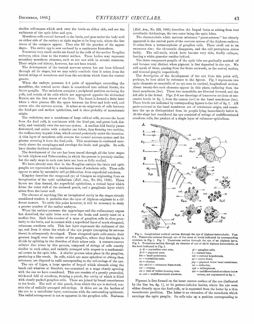
The descriptionof the developmentof the eye from this point will,perhaps,be bestaided by referenceto the figures. Fig. 1 representstwooptic elementsor ammatidiaof an eyeseenin vertical,longitudinal section.About twenty-fivesuchelementsappearin this plane, radiatingfrom thebasalmembrane(bin). Thesetwo omenatidiaare directedforward,and theleft sideis thedorsal. Figs.2—6 are drawingsof transversesectionsatsuc-cessivelevels in fig. 1, from thecornea(car) to the basalmembrane(bin).Theselevelsare indicatedby correspondingfiguresto theleft of fig. 1. Allpartsexternalto thebasalmembraneare of ectodermicorigin, and consti-tute the eye as distinguishedfrom its ganglia lying immediately below it.At thestagelast consideredtheeye consistedof strings of undifferentiatedectoderencells, theproductof a single layerof columnarepithelium.
Fig. 1. Longitudinalvertical sectionthroughtheeyeof Aipheusheterochelis. Figs.2—6. Transversesectionsthrougheyeof the sameat levelsindicated by correspondingnumbersin Fig. 1. Fig. 7. Transversesectionthrough the eyeof an Alpheuslarva.Fig. 5. Transversesectionthroughan element of eyeof adultAipheusheterochelis,atthelevelindicatedin Fig. 1.
A-B = crystalline conearea.B—C--= pigmentzone.inn basalmembrane.cc crystallinecone.ch chorion.chi: chi 1-4 = chitenonsframework.cer cornea.cw = wall of bodiesfarmingcone.cc: ecd undifferentiatedectoderm.
Pigmentis first formedon theinner convexsurfaceof theeye (indicatedby the line bin, fig. 1), at its postero-inferiorborder,wherethe eye restseitherdirectly upon thefood-yolk, or is separatedfrom thelatterby a thinmesodermicpa.rtition. The latteris anextensionof themesodermwhichenvelopsthe optic ganglia. Its cells take up a position correspondingto
JOHNSHOPKINS
thebasilar membrane,but soonbecomeobscuredby pigment. They havelong slenderoutlines and lie at right angles to the cell-stringsof theeyeandganglion.
The pigmentof the eyeappears,when seen throughthe eggshell fromthesurface,first asa narrowline, andthensuccessivelyasa lentienlar,oval,andronadedblack spot. Thepigmentcellsor retinulac (ret figs. 1, 6) growoutward in the directionof thecell-strings,spreadingover thewhole innersurfaceof theeye,and eventuallyfill abouthalf thespacewhich it occupies.In longitudinalsectionstheypresenta broadblack zone(B—c’, fig. 1),whichis composedof radiating bundlesof linear cells. Two suchbundlesarerepresentedin thefigure (ret, fig. 1). Eachbundleconsistsof sevengreatlyelongatedpigment-cellsorretinulae,symmetricallyarrangedabouta centralaxis,thus forming a tube (a, b, fig. 6). Sevenretin,ilaeinvariably occur inthe eyes of Alpheus,Palaemonites,Callinectes, Penacus,and in severalbrachynrawhich havebeenexamined.
As thegrowthof pigment proceeds,changesrapidly occurin the gangli-onic layer(gi, fig. 1), andin the peripheralportion of theeye (A—B,fig. 1).The mesodermbetweenthe eyeand ganglionbecomesobscuredby the de-positof pigment (pig, fig. 1), and thereis probablyat this time a gradualoutgrowthof nervous matter into the eye itself. The following changesoccur in the external half of theeye (A—B, fig. 1). The outermost cellsseparateslightly from the others, and subsequentlyform an independentlayer, the corneal hypoderrnia (rnb, fig. 1, 2), which secretesthe cornea (v.Pattenon the “Eyes of Molluscs andArthropods,’ ]liittheii. Neapel,6 Bd.,1886). A stratumof greatlyelongatedcells, the retinophorae(rph, figs. 1, 3),lies below the corneal hypodermis,hut at this period it is not in contactwith it. These cells appearto bearrangedin pairs, anda white granularsubstanceis seen betweeneach pair. We see by transversesectionsthattheretinophoraea.re really arrangedin groupsof fours (rph, fig. 3). Thewhite structurelessmaterial which theyenclose,is a secretionproduct ofthecells, and is the rudimentof the crystalline cone (cc, figs. 1—6). Eachconelies in a radius,which if producedwould correspondwith that aboutwhich the circle of retinulac(ret, fig. 6) aregroupedto form a bundle. Wehave,therefore,elevencells(fourretinophoraeandsevenretinulac)in eachprimitive optic elementor onunatidium.
The coneis formed by theapproximationof four bodiessimilar to eachotherin both shapeandsize,eachbeingtheproductof a retinophoralcell.Their boundarywalls (en, figs. 1, 4) are earlyrecognizable,andremaindis-tinct throughoutembryonicand adult life. The coneis speedilyprolongedinwards and finally is enclosedby a bundleof retinulac (cc, fig. 6, in aof this figure pigment of retinulaeis not represented). Theinter-omma-tidial spacebetweenthecorneaand theretinophoraeon theonehand,andbetweenthe latter and the pigment zone, is occupiedby undifferentiatedectodermncells (cc, ecd, sch, figs. 1—5).
The eye now developsrapidly with the growthof the embryo, theleastapparentchangebeing in the pigment zone (B—C’, fig. 1). A few narrowcellssometimesoccur betweenthebundlesof retinulae. Thesearedoubt-lessectodermcells, preciselysimilar to thoseoccupyingtime spacebetweenthe cones(ecd, fig. 1).
The cells of the cornealhypodermisgrow to a relatively largesize, andform avery conspicuouslayer. Eachcell containsa largegranularnucleus,which fills abouthalf thecell-space. Thereappearsto be txvo suchcells toeach optic elementarrangedlike the ectodermcells (a, b) in fig. 2. Eachommatidinmnis apparentlyunited to the cornealhypodermisby a delicatemembrane. A spaceis thus enclosedin which theapex of theconelies.
The eyerepresentedin figs. 1—6 differsbut little fro,n that of thelarva.Thecornea(car, fig. 1) is now a chitenousmembrane,constricted into cir-cularfacets. Eachfacetis a biconvexlens,with greatestconvexityon itsinner side,which is applied to theoptic element. Time cornealhypodermisof each optic element appearsin transversesections,like two crescentichodiessurroundingthe apex of the cone. (In fig. 2, mb, these crescenticcellsarebrokenup.) Time crystallinecone (cc,figs. 1—5) is now enormouslyenlarged,and rcachesits greatestdiameterwhere it is surroundedby theretinophorae(rp/m, figs. 1, 3). It passesrapidly outward to a blunt apex,whichrestsagainstt.hecorneallens,buttapersgraduallyinwardto aslenderstalk which entersabundleof retinulae(cc, fig. 6). TIme retinophorae(rph,-figs. 1, 3) have elongatedpeculiarcell-bodies,and nucleicontaining largevacuoles. Timey partially surroundthecone,but can be tracedin thin sec-tionsonly a short distanceinwards (cc, figs. 1—6). Pattenhas shownthatin time adult Penaeustheretinophoralcellsentirely surroundthecone, and
extend from thecornealhmypodermisto time basalmembrane. Thepart ofthecone batweenthe retinophmoraeand corneais finely granularandstainsreadily, while time reverseof this is trueof theinner portion.
The retinulae (met, figs. 1, 6) are very long, linear cells of nearlysolidpigment, without nuclei. Theytapergraduallyto thebasalumembrane,andappearto becontinueda short distancebelow it. Timey receivenervefibres(nf, fig. 1) from time ganghion. Time individualcells areseparatedfrom eachotherbut united in the adult (ret, fig. 8). Here each of the fused cells ispartially divided alongits long axisandtime wholebundleis heavilycoatedwith pigment.
A complicated chitenonsframework (figs. 1, 6, chi, 1—4), tracesof whichappearatanearlierstage,nowsupportstime outerportionsof time retinulae,andis continuedbelowthebasalmembrane(chi 4, fig. 1). This is bestseenin an eyewhich hasbeen depigmentedby nitric acid It farmsa honey-combstructure,time cellsortubesof which enclosetheretinulee,at a pointwhere time latter receivetime delicateendsof thecones. In transversesec-tion (chi, fig. 6) this structurehas the appearanceof a net, eachmeshofwhich enclosesa bundleof retinulae.
Theanatomyof theoptic tract canbeonly briefly referredto here. Theoptic lobeconsistsof threesolid ganghiaof wimite spongytissuecovered bya thick nmassof ganghioncells. Theyarepartiallyseparatedfrom eachotherby musclefibres andconnectivetissue. Nervefibres passdirectly from tImecentral to tIme medianganglion,but deemissatein
0oing frona the latter totime terminalganghion. In traversingthis, theyentersuccessivelytwo strataof elougmutedplugsof nervoustissue,which areradiallyarrangedandappearto correspondwith time optic elements. Similar, but lessmarked layers ofnerve-tissueoccur in time medianganglion. A layer of columnarganghioncells, agreeingin arrangementandposition with the plugs just mentioned,coverstime convex surfimeeof theguinglion. The interveningspacebetweenthe latter and time eye is occupiedlargely by ordinary ganglion cells (gl,fig. 1).
When pigment first appearsin theeye,thecentralganghiaof theoptictract are unitedby commissuresto thebrain.
We have to mentiononemore importantchange,which takesplaceintheeye, just astime youngAipheusis enteringupon larval life. Time retin-ulaenowgrow rapidly outwardandcompletelyenveloptheconesand inter-vening cells in black pigment,extendingnearly to tlme cornea. Fig. 7representsa transversesectionthrough time eyeof anAlpheusjust hatched,andshowsthreeoptic elenmentswith the surroundingretinulae and undif-ferentiatedcells. In timis animal time ommatidia are thins isolatedandactasseparmuteorgansassoon astheeye is functional.
We haveseen in the above accountthatall partsof time eye,exceptingits nerve tissue are developedindependentlyof the brain and ganghia.Each optic elementis prinmitivehy a string of undifferentiatedectodermcells, which arosefrom simple columnar epithmelium. These cell-stringsare closelypackedtogether and are without separatingenvelopesof anykind. Theprimitive ommatidiumdoesnot thereforeresemblean ocelfas,nor doesthedevelopmentof this compoundeye favor the suppositionthatit hasarisenby a gradualfusiomi of ocehhi.
A fuller accountof time eye, thenervoussystem,andotherfeaturesin thedevelopmentof theembryo mustbe left for a longerarticle.
Notes on Tornaria and Balanoglossus. By G. B.HALPEMAN.
In addition to the Tornariadescribedby Alex. Agassizasoccurringonour Atlantic coast,there is anotheroccasionallytaken in time tow-net atBeanfort, N. C., which more closely resemblesthe Mediterraneanformn.At thesuggestionof Dr. W. K. Brooks I imaveundertakenthestudy of itstransformationinto theBalanoglossus.
In generalappearanceit is much like theTornaria describedby Met-schnikofl with whicim it agreesin thepossessionof a secondaryanal bandof cilia, and in tImeformation of thegill-slits asa single pair of outgrowthsfrom theoesophagus.
Time poreof thewatervpsselopensto theleft of thedorsalmedianline.Time apical thickening on which tIme “eye-spots”aresituatedis providedwith a small flagellumafterwardsreplacedby a tuft ofcilia. Time digestivetract appearsto be ciliated throughout. Thereis a patchof much larger
44 [No. 54.
DECEMBER, 1~86.] UNIVERSITY CIIICULA PS.
cilia on a slight elevationof thestomachwall ventral to thevesicularvalvethat closestheentranceof theoesophagos.The anuswhenopenedextrudesa circle of cilia but theseare perhapsnot distinct from the ciliated liningof the intestine.
The processof digestionis somewhatas follows:An Annelid larvawas observedto passbetweenthelongitudinal bands,
which seemedto selectthefood, throughtheopened oesophagusand valveinto thestomachwhere the paralyzed larvawas soon resolvedinto refrac-tive globules and setae. The patch of largecilia guardedthevalve fromcontactwith the coarserparticles. The seteewere collectedandexpelledin a bundle.
The transformationof theTornariainto a Balanoglossusbe~inswhenthewater-vesselmeasuresabouttwo-thirdsof thedistancefrom stomach to apex.Thecliverticulaof thefirst pair of gill-slits then becomenoticeableby theactivemovementof theircilia. Migratorymesoblastcellsbecomenumerous.Vigorouscontractionsof themesoblasticbandaccompanythe enlargementof the water-vessel. TheyoungBalanoglossusis lessthanone-halfthesizeof theTornariaandso opaquethat sectionsare indispensablefor followingtheinternalchanges. Henceit proved advisableto kill mostof my speci-mensduringor at short intervalsaftertheirtransformation. Severalwhichwere kept for six weeksafterchangingto larval Balanoglossialpearedtohave but two pairs of gili-slits. Thoughnot old enough to identify, thelarvaeprobablybelongto B. B’rooks’ii, acommon speciesat Beaufort,whosecastsarea characteristicfeatureof the sandflats at low tide.
Recentspeculationsregardingthephylogenyof the Euteropuenstahavebeen modified by the prominencewhich is given to secondarylarva.l char-acters. The basis for Metschnikoff’s classification of the Euteropuenstawith Echinodermataas “Ambulacralia” would beseriouslyimpaired if itwereprovedthat thepointsof similarity betweenTornariaand Bipannariawere the result of secondaryadaptationsto the samesurroundings. Buthowever this question may be ultimately decided,the homology of thewatem-vesselin thetwo larvaeappearsto hegenuineandindicatesrelation-ship betweenthetwo groups.
Huxley includes Balanoglossuswith the Tunicates in his division of“Phmaryngopacusta,’and draws attentionto “tIme extraordinarysimilarityin thestructureof theperforatedpharyngealsacin thelarvaeof Tunicatesandof Balanoglossus.”
The larva of B. Kowelem’skiias elucidatedby Batesonshows additionalpoints of resemblanceto the Chordata,especiallyto Amphioxus in. thenotochord,atria, etc.
A closer study of tIme transformedTornaria in regard to its internalchancesand later developmentwould show how far it confirmstheseaffini-ties of B. Kewolevskii. But whetherTornaria or Batesonslarva is to heconsideredasmorenearlyrepresentingtheancestraltype, thereis still thepossibility that eachmay possesscertaimaphylogeneticcharactersthat havebecomeobsoletein time other.
The Parasitic Cuninas of Beaufort. By H. V. WILSON.
Therehave beendescribedthreeclassesof parasiticCuninasor (Hacekel)Narco-medusae. The first gromapincludestime Cuninasfoundin thesubum-brellar cavity of Turritopsis (CunoctanthaoctonariaH.). To time secondbelongstheeiglmt tentacledCuninathat developsin time stomacimof certainGeryonidae, and in tIme third group come those Cuninas found in thestomachmsof otimer Cuninas. In time last division tlae host and parasitea.greein somecasesmmnd differ in otimers, in time numberof tentacles. Theparasitismof time contained medusaeis beyond question in the first twogroups, and though not definitely settled is very probablein the tlmirdgroup. TIme Turritopsis Cuninawasdiscoveredlong agoin theCharlestonImarborby MeCradyandwasre-discoveredby Dr. Brooksin 1882 atBeaufort.I again observedit timis summer. TIme Cunina found in the Geryonidmme(Carmmmrina, Geryonia) Imas been observedon our coast in but a singleinstance, and then Fritz Muller timought the little Cunina had beenswallowedby the Liriope. DuringJuly and Augmist I fommtid a nuamberofLiriope (the comnmon Geryonid at Beaufort)with Cuninasin the gastriccavity. My yommngeststagewasa thin two-lmmyered sacwith a. singlenmouthopening. Time nextwas a similar sacwith two tentacularprotuberancesbywhich it wmms attached,and threemouths. In the nextstageat tIme pointof eachmoutla,a medusabud was developed. Thmesestagesdiffer slightly
45
from Metschmnikoff’s description, in that thereis no sign of the centralpseudopodialcell, timomigh in Metsclmnikoff’s Cmmninas it persistedup to astageconsiderablylater than my third. Another point of differencei~;in time American Cimnina tImenmedusabudsare sproutedout overthegenermilsurfaceof time sac, timere being no part of the sac specializedas a stolonfrom which a closeagglomerationof budscould be produced,asMetsclmni-koff figures. The Cuninasparasiticimm Cuninahave imitherto beenobservedonly in the Mediterranean. ThroughAugust, I got in time tow-neta num-ber of eighmt temmtacledCuninasdifferimmg from Cunoctanthaoctonariaonly inbeing perfectly transparent,and in having a much smallermouthwith nopigmentroundit. No parasiteswereobservedin timis medusauntil time lastof August, when I obtained a single specimen with half a dozen littleCuninasin its stomacim. The parasitic Cuninas were lying free in thestomacim,wereperfectly trammsparent,andimad twelve to fifteen tentacles.
Segmentationof the Eggand Formation of the GermLayers of the Squid (Loligo Pealii). By A. T. BRUCE.
The segmentationof time egg and formation of the germ layers of theCephalopodmshave beenobservedby severalwell-known investigators.
Sonmepoints,however,relatingto theorigin of layers and,particularly,oftheinner lmsyeror endodermarestill doubtful.
Lastsummerwhile enjoying time facilities for study offered by the tableof theJohnsHopkins Universityat tImeU. S.FishCommissionLaboratoryat Wood’s Hohl, Mass.,a good opportmmnitywas offered for time studyof timedevelopmentof thecommon Squid(Lohigo Peahii).
Two Squidskept in anaquariumlaid eggson thenight of July 3d. Myattentionwascalled to thais by Prof. JohnA. Byder.
The earlieststagewas studied by meansof sectionsof eggsimardenedtimenextday. Subsequentstagesup to time timeof Imatchingwerealsopreservedandstudied. For time studyof segmentationand formation of germlayersthesectionalnmetimocl on manyaccountsis preferableto superficialobserva-tion. This metimod demonstratedin a very satisfactorymannertime originof time germlayers.
The youngesteggs studiedhad tIme protoplasm segregatedat one poleforming a germinaldisk. This disk in eggswhich probablyhadnot beenlaid twelve imours beforeimardeningwas alreadysegmented.
TIme se~mentsdivided thedisk into a numberof rectangularareas. Timecleavageplanesbetweenthesegmentsextendedthroughtime entire thicknessof the germinaldisk.
In older eggswhich were probably imardenedabouttwenty-four hoursafterdepositionthegerminaldisk by a processof delaminationhad becomedivided into txvo layers. This delaminationdoes not affect the entiresur-faceof thegerminaldisk.
One axisof thedisk probablycorrespondedto the long axis of the em~bryo wasmarkedby anareabut onecell thaick. This areaor axisdoesnothoweverextend the entire lengthm of the germinal disk. Transversesee-tions beyondthepolesof theaxisaretwo cells timick throughout. Sectionstlaroughtheaxis are two cells timick at thelateralmarginsbetweenwimicimtheareaof the axisis a single cell thick. The mesodermor deeperlayerof time disk is consequentlyin the region of the axis separatedinto twomassesor hands.
In theregion of the axis,and probablyanteriorandposteriorto it, alsocertaincellswere observedpartially separatedfrom the mesodermcells.Timesecells from their spindleshapeandoval nucleiarereadily recognizedas the endodermcells wimich form the inner coatingof time yolk sackanddigestivetract in moreadvancedeumbryos.
No nucleiwere observedin the yolk at this or subsequentstages. Itseemsprobable,then,that no endodermcellsoriginatespontaneouslyin theyolk, butthat all are derivedby growthsanddivisionfrom time earliestendo-dermarisingasdescribedfrom beneatimtime mesoderm
In eggsabout thirty-six Imours old the mesodermbandshad becomefurtherdivided andwere two cells timick. The endodermat this stage bythe growthanddivision of its cells formedathin stratumover the innerconcavesurfaceof thegerminaldisk restingupon tIme yolk.
In eggsabomatsixty hoursold time ectodermamm(l endodermImad extendeAsomuchbeyondtheareacomprisingtheoriginalgerminaldisk asto enclosenearlyall theyolk. The mesodermat timis stagedid imot cover muchmore
JOHNSHOPKINS
than half the egg, its terminal margin forming a line a little belowtheeqnatorof the egg. Conseqnentlythe egg at this stage may bedividedinto two hemispheres:an upper hemispherecovered by ectoderm,meso-derm, and endoderm,and a lower hemispherecovered by ectodermandendoderm. The upper hemisphereis the embryonic area,and the lowerhemisphereis the areaof theyolk sack. The line separatingthe hemi-spheresandmarkingtheterminaledgeof themesodermwasmarkedexter-nally by aslight depression.
At this stagea slight eminencein the middle of the embryonic areamarkedthefirst traceof themantle. The separationofthemesodermintotwo bandswas apparentlynot retainedat this stage. Theectodermwasnotsharplymarkedoff from the mesoderm. The developmentof organsbe-longsto later stages. The embryoshatchedon the18th of July.
Notes on the Flora of Abaco and adjoining islands.By F. H. HERRICK.
The flora of Abaco, so far as we observedit, differswidely from thatofthesmaller islandsor keyswhich form its inner reef. The whole islandis coveredfrom end to end by a rank forestgrowth, coumposedof a verygreatvariety of treesandshrubs.
The Pine,probablyPlaysCebensis,is theprincipal forest tree,andstrik-ingly resemblesthe Plaus taedaor Loblolly pine of the SouthernStates.The formerand theRed Cedar,Jmniiper’es Virglnicmna, were the only coni-fers which we saw. Cedarprevails in the vicinity of certain swampstowardsthe north-easternextremityof the island. Above this point thepinereachesits greatestsize,attaininga height of 50 feet. Other common
- andimportant treesare theMastic, “Dogwood,” WhiteTorch, Bullet, and“Poison” woods. The last is prohablya speciesof Rhus. It hasa scalylight brown bole,greenish flowers and pinnateleaves,the jnice of whichstainstheskin black,and producesan inflammation similar to that causedby our common PoisonIvy, Rhus To. icodeiic/roa. The low, swampypartsof all theislandsarealmost exclusivelyoccupiedby the Mangrove,Rhiro-pliora ]liaay/e, which forms dense, impassible thickets. This shrub hadpassedits flowering by the first of June,and thepeculiar,elongatedseedswerein all stagesof development.
The mostinterestingfeatureof Abaco,however,is thegreatvariety andbeautyof its epiphyticplants,—Orchids,Ferns,and Bromeliads. In someplaceseverytreeand.shrub,howeverinsignificant in size,is ornamentedbyone, oftenby severalspeciesof theseattractiveplants.
TheKeys, abouta dozenof which we visited, were characterizedby theabsenceof the conspicuousforesttreesseenon Abaco. Manyof thesmallerislands,which possessa very scantysoil, are carpetedwith a heavymat-ting of vines, twining plants,and a few grassesand small shrubs. Amongthesewe find “Rock Samphire,”or “Turtle grass” (Gornphrena),runningprostrateplantswith turgid stemsandleaves: £bnvo/va/iior Morning Glo-ries of severalspecies,oneof which hasvery largereddishflowers: Garna-va/ia, a pea vine- having long rope-like stems and small pink blossoms.Echitesvuberecta,which belongsto a genusof tropical plants,is avery comn-mon and showy climber, often completelycovering the lower shrubbery.It is distinguishedby its largelemon-coloredflowers,shining leaves,andmilky juice. A beautifulmaroon-coloredpassionflower, Pasnj/oracupraca,was foundgrowingon PawpawKey.
Joe’s Key, off Little Abaco,differed somewhatfrom anyof the islandswhich we visited. It containeda small grassymeadowdrainedby a man-groveswampon either side,a grove of silver-toppedpalms; a sandybeachon theleewardside,andhigh cliffs facingthesea. Unio/a panicu/atcm, Spike-Grass(common along our Southernsea-coast),with severaltall sedgeswasgrowing alongthe sandbeach,and alsoa delicateplant, Sabbatiagraci/is,which haslargerose-coloredflowers.
The Pancretiam, or “West Indian Lily,” a member of the Amarillisfamily, a plant thebulbsof which areregularlyimportedby florists, growscommonly along the sandymargins of all theseislands. Its flowers are
impenetrablegrowth of shrubsandsmall trees,including severalspeciesofpulm,anda largebushycactus(Opuntia). Perhmapsthemostcharacteristicshrub is Rhacica//isrupestris,called“Seaweed”by thenatives. It is pros-
trate or partially erect,and has dark green spmayey foliage, and minutesaffron flowers. It is usuallyconfinedto therocksalongshore. Thie Cono-corpus or “Button-wood,” often time only shrubof considerablesizeon thesmallerislands,everywhereabounds,andis conspicuousby itssilveryfoliage.Besidesthesewe constantlymeet with the Wild Sapodilla,SapotaAc/nay,tImefruit of which resemblesasmall rusty apple; Genipac/avmfo/iaor “Seven-yearApple,” which hasfragrant cream-coloredflowers and a hard greenfruit aboutthesizeof awalnut; Borrichia orboresceas,“Lavender,”a yellowcompositewith tall brittle stems and hoary leavds, which are used formedicinalpurposes.
GreenTurtleKey probablyaffordsamorediversifiedflora thananyof theislandsof equal area. Its habitationby manhasled to the introductionofvarious foreign fruits and a hostof comumonweeds. Some fine specimensof tIme CocoanmmtPalmmaybe seen here. This palm alsoflourishesahongtheshoresof Abacoandon someof the lessexposedishands. A small groveofthesetreeson Allons Key wascompletelyprostratedby thegreathurri-caneof September7, 1884. Such fruits as tIme Banana,Orange,Lemon,Shaddock,Sapodilla, Mango, Mammee, CustardApple or Papaw,hogPluma, Tamarind,DatePalm, Fig (the two last representedby a few sh)eci-mansonly), havebeenintroducedandarenow growingspontaneouslyovertIme island.
Thme C’occo/obcm or “Sea Grape” formns a. low contortedshrub, or as fre-quently attainstImehmeighmt of a smuall tree. It is remarkablefor its thick,nearlycircular leaves,andlong pendulousracemesof greenflowersor fruit.
By far tIme moststriking hmerb on the Key just mentionedis the Manillaplant, a speciesof Agave,whosesolitary flower stalk attainsa height ofupwardsof 20 feet,and forms a prominentfeaturein tIme landscape.
TIme Pine-apple,Aaanossavotive, is grown extensivelyon Abacoin clear-ings along the shores,and is the only fruit exportedfrom this portion oftheBahamas.
List of Plants from Abaco Island, Bahama. By D.C. EATON and W. A. SETCHELL, from collections made by F.H. HERRICK.
Prof. D. C. Eatonof Yale College,New Ilaven,Ct.,very kindly consentedto examinethis collection,andto him and to Mr. XV. A. Setchellof thesameinstitution we are indebtedfor the deteruminationof nearlyall time plantsincludedin tIme list below.
We were unfortunatelyunableto preservesuitablespecimensof a greatmuanyh)lants,andsuchi aswe obtainedwere often too fragmentaryto admitof certainidentification.
About forty naturalomders or familiesare representedby tIme sixty oddspeciesgiven in thelist. Tlmose plantsmarkedby a ~, abouta third of thewhole number,are not knownto occur in time SouthernStatesor on tImeFlorida Keys,andare probaimlypeculiarto the WestIndies. Plantsfoumidonly on AbacoIslandare thusindicated,andthoseobservedonly on GreenTurtle Key aremarkedby thelettersG.T. Localpopularnamesareplacedin quotationmarks. F. H. H.
FLOWERING PLANTS.
1. Agerempm (?). “Manilla Plant.”
t 2. Allermma,mtlmereflavescens,Moquimi.ArgenemmeJlfexicaae,L. Prickly Pop-
py. (G.T.)4. Artenisieemmlgaris,L. CommonMug-
wort. (G. ‘V.)t 5. A. hispida, Pnrsh. “Bastard Gin-
aimmom.6. Asciepinipeupereimle,Michx. Milk-
weed.7. Bidenslemmcaalhcm,W. Beggars’Ticks.5. Borriehia arboreiceas, D. L~. “Bay
Lavender.”
t 9. Brenfelsie.~—(?). Tall shrmmb.10. Gammseee/’iaebtesmfelieID. C. Coarse
vine (Pei family).
Ill. ~etepsis minions,Grim. (?). EpiphyticBronmeliad. “Wild Pine.” (Abaco).
112. Cemmcheras him-soles L. “CourageBash.”
~la. 6’emmclmrmmstribuleicles,L. (Grass).
~14. Cledimmme mecideetale, Sebrader.(Sedge).
t15. Ceecelebae.ufere. Shrmmb or smalltree. “SeaGrape.”
16. Cmmmeca~peserectus L. Y. ereetims,Grim.Butlon—woed.”
Observationson the NervousSystemof InsectsandSpidersandsomePreliminaryObservationson Phry-nus. By A. T. BRUCE.
Fromacareful studyof thenervoussystemof someinsects(Acridium andThyridopteryx) and of spidersit appearsthat the supra~sophagealgan-glion of insectsandArachnidsis distinctly double. Eachganglionof theganglionatedchain of insectsand spidersis closelyinvestedby a sheathof flattenedmesoblastcells, which may be describedas tine perineserium.Theperineurium in insectsalso invests the transverseand long corumes-suresbetweengangliaand thenervetrunks.
Theperineuriurnextendsbetweenadjacentgangliaforming aclosesheathfor each ganglion. Consequentlyadjacent ganglia are separatedby in-growths of perinenrium. The perineuriumoriginatesin part asa medianingrowth along the medianventral line. The modeof origin of this i’n-growthwasdescribedin Circulor No. 49, p. 85. All the medianingrowthbecomesperineuriumexceptportionsof it betweenadjacentpairs of nervegangliawhere it persistsascolumnsof cellsextendingto the dorsal limitof thenervoussystem.
It is apparentlyby theextensionandunion of coluninssuchasthesethattheendosterniteof Limulus andArachnidsis formed. The nerveganghiaarefirst solid massesof cells. By thebreakingdown of cells in the centreof eachganghionthe commissuralor punctsubstanceis formed.
Bythe extensionof thepunctsubstancelaterally and internally thecrossor transversecommissuresare formed, and by a similar extensionof thepunctsubstanceanteriorly andposteriorlytine long commissuresare formed.Thecentralportion of eachganghionconsistingas describedof punctsub-stanceis continuousexternallyand laterallywith tine nervefibres. It is bythebreakingdown of the cells of the originally solid nervecord that thenervefibre is formed. All statesin tIneformationof commissuresandfibreswerenot observed.
In comparingthesupra~sophagea1ganglion of insectsand spiderswithothcr gangliaof thechainit appearsthat thesupraocsophagealganghionofinsectsand spidersis distinctly double. Eachdivision is seriallyhomolo-gouswith a ganghionpair for succeedinnbody somites. In both insectsandspiderstIne anteriordivision of thesupracesophagealganglionis distinctlyseparatedfrom theposteriordivision by theingrowthof theinvesting pen-
47
neurium. In insects (Thyridopteryx) tine columns of cells describedasexisting betweenotlnerpairsof gangliaexistsbetweenthetwo divisionsoftine suprac&sophagealganglion.
In Thyridopteryxtheanteriordivision of thesupracesophagealganglionis markedexternallyby an incision of ectodermasare tbesucceedingbodysegments. Therecan be no doubt tinen hut that the brain of insectsandspidersconsistsof two pairsof gangliaserially Inomologouswith otherpairsof ganglia.
The anteriordivision of the supracesophagealganglionof insectsis tinepair of gangliabelonging to the antennalsomite. The posterior divisionof thesupracesophagealganglion of insectsis tine ganghionof tine segment
of tine upper lip. Theupperlip is a pairedstructurein TinyridopteryxandAcridium. It receivesnervesfromthegangliaof its somite. Tine posteriordivision of the supracesophagealganghionof insectsby its backward ex-tension formsnaostof the circunacesophagealcommissure,consequentlythelabrum is innervatedfrom thecommissureasis tine secondantennaof ems-tacea. TIne anteriordivision of thespider’s brain probablyininervatestherostrum. Tine lateralpartsoftherostrunnformdownwardprojectionsoneachsideandcorrespondin positionto tine antennaeof insects. Tinese antennal(?) projectionsarewell shownin transversesections. TIne posteriordivisionof thespider’sbrain innervatesthe labmuns,which is distinctly groovedinthe median line andhastheappearanceof a pairedstrnsctnre. The man-dibular ganghionof spiderslies below the posteriordivision of the supra-cesoplnagealganglion, andis partly overlappedby it laterally.
Tinere appears,then, to be a special homology existing betweenthelabrum in spidersand insectsand the recurrent antennaof crust.acea.There is also a correspondencebetweenthe antennaof insects, tine firstantennaof crustacca,andtine lateralpartsof tine rostrumin spiders.
The eyesof insects(Acridiuns andTisyridopteryx) appearfirst asthick-eningsof ectodermon the antero-lateralpartsof theInead. In Tinyridop-teryx this areabecomesfurtinerspecializedto form distinct optic elennentsor ocehli, formed, primarily as solid areasor cones. Subsequently,in allprobability,by thebreakingdownof tlne centralcellsof thecone,thepunet-substanceor granularsubstanceof the ocellus is formed,surroundedby alayerof cellswhich, at theperipinery,is double. Theseelementsof theeye,or ocelli, in Thyridopteryx are, in the advancedembryos,separatefromeachotherand not connectedwith thecentralnervoussystem. Thedevel-opment of theeye in the grassinopper(Acridium) wasnot followed beyondthevery earlieststages.
Some observationswere made in a pedipaip (Pinrynus) with embryosbrought from the BahamaIslands by Dr. Ocr. Tine ventral surface oftine thoraxof tine adult pedipaipclosely resemblestine ventral surfaceofLimulus. A striking point of likenessis theexistenceof a curvedprocesson tine inner sideof the cocal joint of the last thoracicappendage,corre-spondingto a similar processon tine cocaljoint of the last appendageofLimulus. Tracesof similar processesalsoappearin the fourth and fifththoracicappendages.
Another interesting likeness to Limulus is the existenceof a pairedstructureposteriorto thelast pair of thoracicappendages,correspondinginposition to thechiloria or metastomaof Limulus. Eachof the threeiastpairsof thoracic linnbs possessepisternaat tlneir bases,betweenwhich is amediandark-coloredsternum,that for thelast pair of thoracic limbsbeingdouble. The metastomaof the Pedipaiphasanteriorto it a pairedstruc-ture, which is, perhaps,a pairedsternite,and at its basea pairedstructurewhicin is anepisternite.
Tlnese points of likenesstogetlnerwith themanyfamiliar ones,and theinvolution of the abdominal feetof thespider to form the lung book de-scribed by mein arecentnumberof theAmericaxNatseraiint,maketherela-tionship of Limulus andtheArachnidsvery close.
Theembryopedipalphasanamnionlike thatdescribedby mefor spiders.Onthecoealjoint of the4th appendagea senseorganwas veryconspicuousin theyoungpedipaip. The hypodermiccellsof tinat joint becomecolum-nar, unlike otherhypodermiccells,which are of irregularcontour. Tiseseelongatedcellsare continuedexternally to form filaments,severalof whichentera singlepair, which is theexternalpart of thesenseorgan.
JOHNSHOPKINS UWIVEJ?SITY011WULA PS. [No. 54.
PUBLICATiONS ISSUED UNDER THE AUSPICES OF THE UNIVERSITY.I. American Journal of Mathematics.
The publication of this journal commencedin 1878, under theeditorial direction of ProfessorSylvester. It is now conductedby ProfessorSimon Newcomb, as Editor, and Dr. T. Craig, asAssociateEditor. Eight volumesof about400 pageseachbavebeen issued,and theninth is in progress. It appearsquarterly,in thequarto form. Subscription$5 per year. Single numbers$1.50.
II. American chemical Journal.This journal was commencedin 1879, with ProfessorRemsen
as Editor. Sevenvolumes of about450 pageseachhave beenissued, and the eighth is in progress. It appearsbi-monthly.Subscription$3 peryear. Singlenumbers50 cents.
III. American Journal of Philoloqy.
The publicationof this journal commencedin 1880,under theeditorial direction of Profcssor Gildersleeve. Six volumes ofabout570pageseachhavebeenissued,andtheseventhis in pro-gress. It appearsfour times yearly. Subscription$3 pervolume.Singlenumbers 1.00.
IV. Studiesfrom the Biological Laboratory.(Including thechesapeakeZobVagiccetLaboratory.)
The publication of thesepaperscommencedin 1879, underthedirection of ProfessorMartin, with the assistanceof Dr. W. K.Brooks. Two volumes of about500 pagesoctavo, and40 plateseach,havebeenissued,andthethird is in progress. Subscription$5.00per volume. (Seepage41 of this Circular.)
V. Studies in Historical and Political Science.
The publication of thesepaperswasbegunin 1882, under theeditorial direction of Dr. H. B. Adams. Four seriesof twelvemonthly partseachhavebeenissued,andthe fifth is in progress.Subscription$3.00pervolume. Extra volumes,bound in cloth,a±ealsoissuedfrom time to time.
VI. Johns Hopkins University Girculars.
The publicationof theCircularsbeganin December,1879,andfifty-four numbershavesincebeenissued.
The Circularsarepublishedat convenientintervals during theacademicyear for the purposeof communicatingintelligenceto
thevariousmembersof theuniversity in respectto work which isherein progress,as well asfor thepurposeof promulgatingofficialannouncementsfrom thegoverningarid teachingbodies. Duringthecurrentacademicyear,circulars will be issuedin successivemonths,to be followed at the closeof theyear by an index.
Although thesecircularsare designedfor the membersof theuniversity, they have frequently beencalled for by institutionsandlibraries at adistance,andalsoby individualswho are inter-ested in the literary and scientific activity of this university.Subscriptionsandexchangesarethereforereceived. Subscription$1.00 per year. Subscribersto the Circulars will also receivetheAnnualRegisterand the Annual Report of theUniversity.
Vii. Annual Report.
Presentedby the Presidentto the Board of Trustees,reviewingtheoperationsof theUniversity during thepastacademicyear.
VIII. Annual Reqister.
Giving the list of theofficers andstudents,andcontainingdetailedstatementsasto the regulationsandwork of theUniversity.
Announcementsof proposedlectures,coursesof instruction,etc.,appearin the University Circulars, or are separatelyissuedasProgrammesfrom time to time.
The following separatepublications have also been recentlyissuedby thePublicationAgencyof the University —
Photograph of theNormal SolarSpectrum. Made by ProfessorH. A. Rowland. Set of sevenplates unmounted$10, mounted$12; singleplatesunmounted$2, mounted$2.25.
Reproductionin Phototgpeof a SyriacMS.with theAntilegomenaEpistles. (Includingseventeenphototypepages,eachon aseparateleaf,besidesadditional pagescontainingdescriptiveandexplana-tory matter.) Editedby Dr. I. H. Hall. In paper $3; in cloth $4.
SelectedMorphological Monographs. Edited by Dr. XV. K.Brooks. In cloth $8. (See announcementon page 41 of thisOircular.) ______________
Communications in respect to e chanycs and remit-tancesmay besent to the JohnsHopkins University (Pub-lication Ayency,) Baltimore, Maryland.
CONTENTS.
~oo1ogie~IWork of the JohnsHopkins University, 1878.86:Reportof the Directorof theMarine Laboratory, - -
Roll of Membersof theLaboratory,PublishedResultsof ScientificResearch,
notes of Work in the Marine Laboratory, 1886:
On theEmbryologyof Aipheusandother Crustaceaandon theDevelopmentof theCompoundEye. By F. H. HERmaN,
On TornariaandBalanoglossus. By G. B. HALDEMAN, - -
The ParasiticCuninasof Beaufort. By H. V. WILsoN, - -
PAGE
- 37—393940
424445
Segmentationof the Eggand Formationof theGerm Layersof theSquid. By A. T. BRUcE,
On theFlora of Abacoandadjoining islands. By F. H. HER-
lucK,
List of Plantsfrom Abaco Island, Bahama. By ID. C. EATON
and W. A. SETCHELL, from collections made by F. H.HERRICK,
Observationson theNervousSystemof Insectsand Spiders,etc.By A. T. BRUcE,
University Publications
PAGE
45
46
46
47- 41,48
48
The JohnsHopkinsUniversity Circulars are printed by Messrs.JOHN MURPHY& 00., 182 WestBaltimore Street,Baltimore,from whomsingle copiesmaybeobtained; they may also be procuredfrom Messrs.CUSHINGS& BAILEY, No. 262 WestBaltimore Street,Baltimore. Subscriptioas,$1.00 a year, maybe addressedto the PUBLICATION AGENCY OF THE JOHNS HOPKINS UNIVERSITY, BALTIMORE; single copies will be sentby mail for ten cents each.