Trans-activation of the DNA-damage signalling protein kinase Chk2 by T-loop exchange Antony W Oliver 1, *, Angela Paul 2 , Katherine J Boxall 3 , S Elaine Barrie 3 , G Wynne Aherne 3 , Michelle D Garrett 3 , Sibylle Mittnacht 2 and Laurence H Pearl 1, * 1 Cancer Research UK DNA Repair Enzymes Group, Section of Structural Biology, The Institute of Cancer Research, Chelsea, London, UK, 2 Cancer Research UK Centre for Cell and Molecular Biology, The Institute of Cancer Research, Chelsea, London, UK and 3 Cancer Research UK Centre for Cancer Therapeutics, The Institute of Cancer Research, Haddow Laboratories, Sutton, Surrey, UK The protein kinase Chk2 (checkpoint kinase 2) is a major effector of the replication checkpoint. Chk2 activation is initiated by phosphorylation of Thr68, in the serine– glutamine/threonine–glutamine cluster domain (SCD), by ATM. The phosphorylated SCD-segment binds to the FHA domain of a second Chk2 molecule, promoting dimerisa- tion of the protein and triggering phosphorylation of the activation segment/T-loop in the kinase domain. We have now determined the structure of the kinase domain of human Chk2 in complexes with ADP and a small-molecule inhibitor debromohymenialdisine. The structure reveals a remarkable dimeric arrangement in which T-loops are exchanged between protomers, to form an active kinase conformation in trans. Biochemical data suggest that this dimer is the biologically active state promoted by ATM-phosphorylation, and also suggests a mechanism for dimerisation-driven activation of Chk2 by trans-phos- phorylation. The EMBO Journal (2006) 25, 3179–3190. doi:10.1038/ sj.emboj.7601209; Published online 22 June 2006 Subject Categories: genome stability & dynamics; structural biology Keywords: cancer; CHEK2; CHK2; inhibitor; kinase Introduction DNA double-strand breaks, generated by ionising radiation, genotoxic chemicals or collapsed replication forks, are the most serious type of DNA damage with which the eukaryotic cell must contend. Survival depends on a coordinated re- sponse whereby the DNA lesion is repaired, and the progress of the cell cycle is halted to allow that repair to occur. Key to this response is the activation of a signalling network, dependent on the phosphatidylinositol kinase-like kinases (PIKKs), ATM and ATR, which are primary sensors of DNA damage (McGowan and Russell, 2004; Lavin et al, 2005) (Figure 1A). ATM/ATR driven signals modulate transcription through stabilisation of p53, and directly regulate cell cycle progression by promoting the degradation of the Cdc25 phosphatases that activate Cdk2/CyclinE complexes (Falck et al, 2001). An immediate downstream target of the ATM branch of the network is checkpoint kinase 2 (Chk2) (Cds1), a Ser/Thr kinase consisting of an N-terminal serine–gluta- mine/threonine–glutamine cluster domain (SCD), a middle phosphothreonine-binding FHA domain (Li et al, 2002) and a C-terminal catalytic domain (Figure 1B). Activated Chk2 in turn phosphorylates a range of downstream targets including Cdc25A and Cdc25C, BRCA1 and p53 (Bartek and Lukas, 2003). Chk2 activation is a multistep process, initiated by ATM, which phosphorylates Chk2 on Thr68 in a segment of the protein upstream of the FHA domain (Ahn et al, 2000; Matsuoka et al, 2000; Melchionna et al, 2000). Once phos- phorylated, the pThr68 segment of Chk2 can bind in trans to the FHA domain of another molecule promoting dimerisation and trans-activating phosphorylation of Thr383 and Thr387 in the activation segment or ‘T-loop’ of the catalytic domain (Ahn et al, 2002; Xu et al, 2002). However, the mechanism by which dimerisation promotes T-loop phosphorylation is not understood. The role played by Chk2 in mediating the cellular response to DNA damage gives it an important function as a tumour suppressor (McGowan, 2002). Truncations and missense mutations of Chk2 have been identified in a wide range of tumours, particularly breast (reviewed in Bartek and Lukas, 2003). Although Chk2 is formally a tumour suppressor, there is a growing interest in its inhibition in the treatment of cancer, both to radio-sensitise tumour cells already defective in other damage checkpoints (e.g. p53 /), and to inhibit radiation induced apoptosis in sensitive collateral tissues (Bartek and Lukas, 2003; Collins and Garrett, 2005; Pommier et al, 2005). Towards understanding the molecular mechanism for Chk2 activation and to facilitate development of specific inhibitors, we have now determined the crystal structure of a catalytic domain construct of human Chk2 in complex with Mg-ADP, and with the ATP-competitive kinase inhibitor debromohymenialdisine (DBQ) (Meijer et al, 2000; Sharma and Tepe, 2004). The structure of the Chk2 kinase domain reveals an unusual dimeric arrangement involving exchange of T-loops, and suggests a mechanism for dimerisation-driven activation of Chk2 by trans-phosphorylation. Results Chk2 kinase domain constructs As attempts to crystallise full-length Chk2 were unsuccessful, we sought smaller constructs that would encapsulate the catalytic domain. Initial trials, based on sequence threading predictions, used an N-terminally His 6 -tagged construct run- ning from Ser210 to the natural C-terminus at Leu543, but Received: 8 February 2006; accepted: 31 May 2006; published online: 22 June 2006 *Corresponding authors. AW Oliver or LH Pearl, Cancer Research UK DNA Repair Enzymes Group, The Institute of Cancer Research, 237 Fulham Road, Chelsea, London SW3 6JB, UK. Tel.: þ 44 20 7153 5571; Fax: þ 44 20 6153 5457; E-mails: [email protected] or [email protected]The EMBO Journal (2006) 25, 3179–3190 | & 2006 European Molecular Biology Organization | All Rights Reserved 0261-4189/06 www.embojournal.org & 2006 European Molecular Biology Organization The EMBO Journal VOL 25 | NO 13 | 2006 EMBO THE EMBO JOURNAL THE EMBO JOURNAL 3179
Transcript
Trans-activation of the DNA-damage signallingprotein kinase Chk2 by T-loop exchange
Antony W Oliver1,*, Angela Paul2,Katherine J Boxall3, S Elaine Barrie3,G Wynne Aherne3, Michelle D Garrett3,Sibylle Mittnacht2 and Laurence H Pearl1,*1Cancer Research UK DNA Repair Enzymes Group, Section of StructuralBiology, The Institute of Cancer Research, Chelsea, London, UK, 2CancerResearch UK Centre for Cell and Molecular Biology, The Institute ofCancer Research, Chelsea, London, UK and 3Cancer Research UK Centrefor Cancer Therapeutics, The Institute of Cancer Research, HaddowLaboratories, Sutton, Surrey, UK
The protein kinase Chk2 (checkpoint kinase 2) is a major
effector of the replication checkpoint. Chk2 activation
is initiated by phosphorylation of Thr68, in the serine–
glutamine/threonine–glutamine cluster domain (SCD), by
ATM. The phosphorylated SCD-segment binds to the FHA
domain of a second Chk2 molecule, promoting dimerisa-
tion of the protein and triggering phosphorylation of the
activation segment/T-loop in the kinase domain. We have
now determined the structure of the kinase domain of
human Chk2 in complexes with ADP and a small-molecule
inhibitor debromohymenialdisine. The structure reveals
a remarkable dimeric arrangement in which T-loops are
exchanged between protomers, to form an active kinase
conformation in trans. Biochemical data suggest that
this dimer is the biologically active state promoted by
ATM-phosphorylation, and also suggests a mechanism
for dimerisation-driven activation of Chk2 by trans-phos-
phorylation.
The EMBO Journal (2006) 25, 3179–3190. doi:10.1038/
through stabilisation of p53, and directly regulate cell cycle
progression by promoting the degradation of the Cdc25
phosphatases that activate Cdk2/CyclinE complexes (Falck
et al, 2001). An immediate downstream target of the ATM
branch of the network is checkpoint kinase 2 (Chk2) (Cds1),
a Ser/Thr kinase consisting of an N-terminal serine–gluta-
mine/threonine–glutamine cluster domain (SCD), a middle
phosphothreonine-binding FHA domain (Li et al, 2002) and
a C-terminal catalytic domain (Figure 1B). Activated Chk2 in
turn phosphorylates a range of downstream targets including
Cdc25A and Cdc25C, BRCA1 and p53 (Bartek and Lukas,
2003). Chk2 activation is a multistep process, initiated by
ATM, which phosphorylates Chk2 on Thr68 in a segment of
the protein upstream of the FHA domain (Ahn et al, 2000;
Matsuoka et al, 2000; Melchionna et al, 2000). Once phos-
phorylated, the pThr68 segment of Chk2 can bind in trans to
the FHA domain of another molecule promoting dimerisation
and trans-activating phosphorylation of Thr383 and Thr387
in the activation segment or ‘T-loop’ of the catalytic domain
(Ahn et al, 2002; Xu et al, 2002). However, the mechanism by
which dimerisation promotes T-loop phosphorylation is not
understood.
The role played by Chk2 in mediating the cellular response
to DNA damage gives it an important function as a tumour
suppressor (McGowan, 2002). Truncations and missense
mutations of Chk2 have been identified in a wide range of
tumours, particularly breast (reviewed in Bartek and Lukas,
2003). Although Chk2 is formally a tumour suppressor, there
is a growing interest in its inhibition in the treatment of
cancer, both to radio-sensitise tumour cells already defective
in other damage checkpoints (e.g. p53�/�), and to inhibit
radiation induced apoptosis in sensitive collateral tissues
(Bartek and Lukas, 2003; Collins and Garrett, 2005;
Pommier et al, 2005).
Towards understanding the molecular mechanism for
Chk2 activation and to facilitate development of specific
inhibitors, we have now determined the crystal structure of
a catalytic domain construct of human Chk2 in complex with
Mg-ADP, and with the ATP-competitive kinase inhibitor
debromohymenialdisine (DBQ) (Meijer et al, 2000; Sharma
and Tepe, 2004). The structure of the Chk2 kinase domain
reveals an unusual dimeric arrangement involving exchange
of T-loops, and suggests a mechanism for dimerisation-driven
activation of Chk2 by trans-phosphorylation.
Results
Chk2 kinase domain constructs
As attempts to crystallise full-length Chk2 were unsuccessful,
we sought smaller constructs that would encapsulate the
catalytic domain. Initial trials, based on sequence threading
predictions, used an N-terminally His6-tagged construct run-
ning from Ser210 to the natural C-terminus at Leu543, butReceived: 8 February 2006; accepted: 31 May 2006; published online:22 June 2006
*Corresponding authors. AW Oliver or LH Pearl, Cancer Research UKDNA Repair Enzymes Group, The Institute of Cancer Research,237 Fulham Road, Chelsea, London SW3 6JB, UK.Tel.: þ 44 20 7153 5571; Fax: þ 44 20 6153 5457;E-mails: [email protected] or [email protected]
The EMBO Journal (2006) 25, 3179–3190 | & 2006 European Molecular Biology Organization |All Rights Reserved 0261-4189/06
www.embojournal.org
&2006 European Molecular Biology Organization The EMBO Journal VOL 25 | NO 13 | 2006
EMBO
THE
EMBOJOURNAL
THE
EMBOJOURNAL
3179
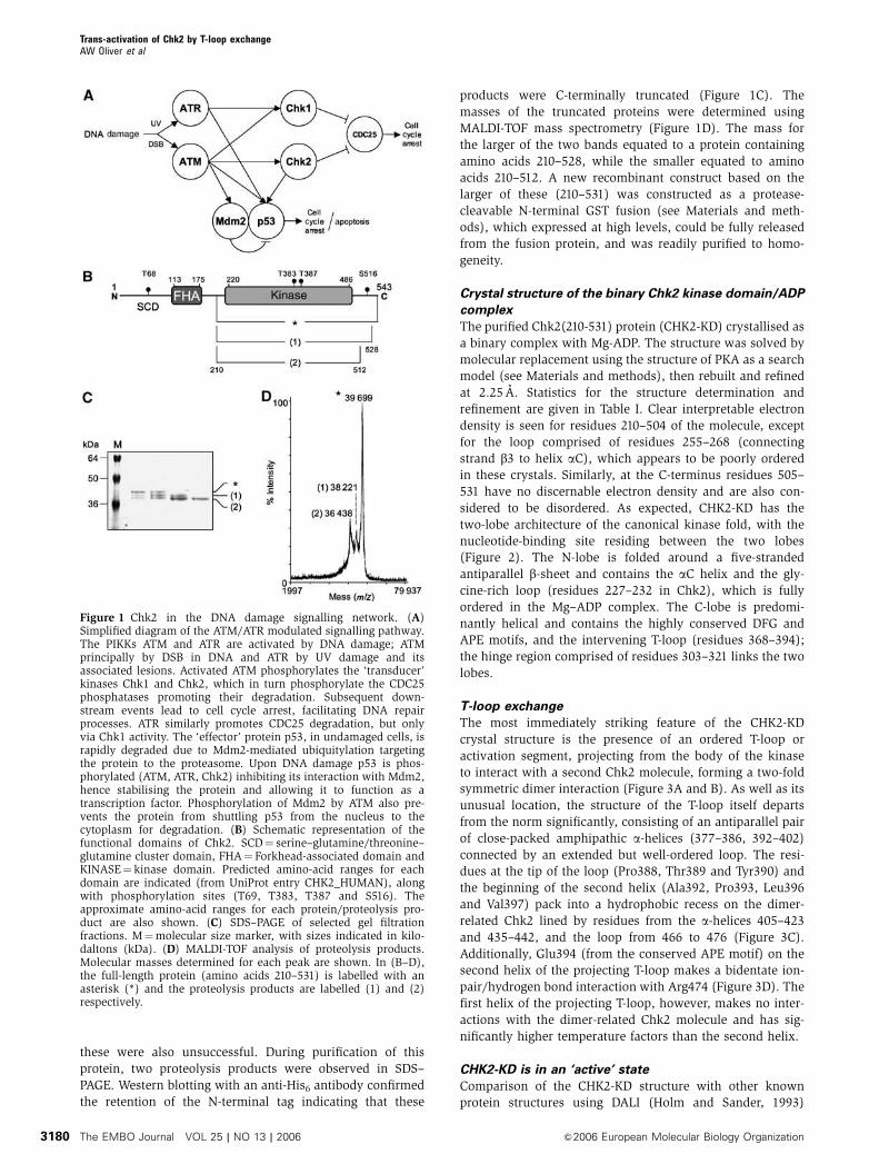
these were also unsuccessful. During purification of this
protein, two proteolysis products were observed in SDS–
PAGE. Western blotting with an anti-His6 antibody confirmed
the retention of the N-terminal tag indicating that these
products were C-terminally truncated (Figure 1C). The
masses of the truncated proteins were determined using
MALDI-TOF mass spectrometry (Figure 1D). The mass for
the larger of the two bands equated to a protein containing
amino acids 210–528, while the smaller equated to amino
acids 210–512. A new recombinant construct based on the
larger of these (210–531) was constructed as a protease-
cleavable N-terminal GST fusion (see Materials and meth-
ods), which expressed at high levels, could be fully released
from the fusion protein, and was readily purified to homo-
geneity.
Crystal structure of the binary Chk2 kinase domain/ADP
complex
The purified Chk2(210-531) protein (CHK2-KD) crystallised as
a binary complex with Mg-ADP. The structure was solved by
molecular replacement using the structure of PKA as a search
model (see Materials and methods), then rebuilt and refined
at 2.25 A. Statistics for the structure determination and
refinement are given in Table I. Clear interpretable electron
density is seen for residues 210–504 of the molecule, except
for the loop comprised of residues 255–268 (connecting
strand b3 to helix aC), which appears to be poorly ordered
in these crystals. Similarly, at the C-terminus residues 505–
531 have no discernable electron density and are also con-
sidered to be disordered. As expected, CHK2-KD has the
two-lobe architecture of the canonical kinase fold, with the
nucleotide-binding site residing between the two lobes
(Figure 2). The N-lobe is folded around a five-stranded
antiparallel b-sheet and contains the aC helix and the gly-
cine-rich loop (residues 227–232 in Chk2), which is fully
ordered in the Mg–ADP complex. The C-lobe is predomi-
nantly helical and contains the highly conserved DFG and
APE motifs, and the intervening T-loop (residues 368–394);
the hinge region comprised of residues 303–321 links the two
lobes.
T-loop exchange
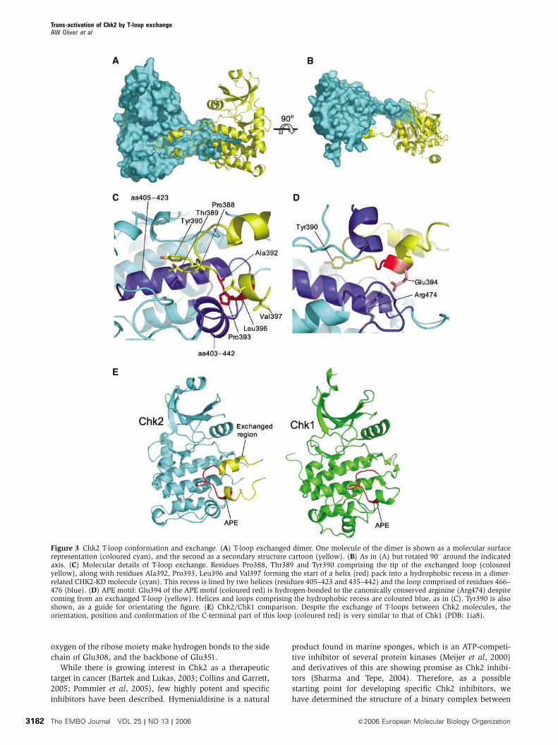
The most immediately striking feature of the CHK2-KD
crystal structure is the presence of an ordered T-loop or
activation segment, projecting from the body of the kinase
to interact with a second Chk2 molecule, forming a two-fold
symmetric dimer interaction (Figure 3A and B). As well as its
unusual location, the structure of the T-loop itself departs
from the norm significantly, consisting of an antiparallel pair
of close-packed amphipathic a-helices (377–386, 392–402)
connected by an extended but well-ordered loop. The resi-
dues at the tip of the loop (Pro388, Thr389 and Tyr390) and
the beginning of the second helix (Ala392, Pro393, Leu396
and Val397) pack into a hydrophobic recess on the dimer-
related Chk2 lined by residues from the a-helices 405–423
and 435–442, and the loop from 466 to 476 (Figure 3C).
Additionally, Glu394 (from the conserved APE motif) on the
second helix of the projecting T-loop makes a bidentate ion-
pair/hydrogen bond interaction with Arg474 (Figure 3D). The
first helix of the projecting T-loop, however, makes no inter-
actions with the dimer-related Chk2 molecule and has sig-
nificantly higher temperature factors than the second helix.
CHK2-KD is in an ‘active’ state
Comparison of the CHK2-KD structure with other known
protein structures using DALI (Holm and Sander, 1993)
Figure 1 Chk2 in the DNA damage signalling network. (A)Simplified diagram of the ATM/ATR modulated signalling pathway.The PIKKs ATM and ATR are activated by DNA damage; ATMprincipally by DSB in DNA and ATR by UV damage and itsassociated lesions. Activated ATM phosphorylates the ‘transducer’kinases Chk1 and Chk2, which in turn phosphorylate the CDC25phosphatases promoting their degradation. Subsequent down-stream events lead to cell cycle arrest, facilitating DNA repairprocesses. ATR similarly promotes CDC25 degradation, but onlyvia Chk1 activity. The ‘effector’ protein p53, in undamaged cells, israpidly degraded due to Mdm2-mediated ubiquitylation targetingthe protein to the proteasome. Upon DNA damage p53 is phos-phorylated (ATM, ATR, Chk2) inhibiting its interaction with Mdm2,hence stabilising the protein and allowing it to function as atranscription factor. Phosphorylation of Mdm2 by ATM also pre-vents the protein from shuttling p53 from the nucleus to thecytoplasm for degradation. (B) Schematic representation of thefunctional domains of Chk2. SCD¼ serine–glutamine/threonine–glutamine cluster domain, FHA¼ Forkhead-associated domain andKINASE¼ kinase domain. Predicted amino-acid ranges for eachdomain are indicated (from UniProt entry CHK2_HUMAN), alongwith phosphorylation sites (T69, T383, T387 and S516). Theapproximate amino-acid ranges for each protein/proteolysis pro-duct are also shown. (C) SDS–PAGE of selected gel filtrationfractions. M¼molecular size marker, with sizes indicated in kilo-daltons (kDa). (D) MALDI-TOF analysis of proteolysis products.Molecular masses determined for each peak are shown. In (B–D),the full-length protein (amino acids 210–531) is labelled with anasterisk (*) and the proteolysis products are labelled (1) and (2)respectively.
Trans-activation of Chk2 by T-loop exchangeAW Oliver et al
The EMBO Journal VOL 25 | NO 13 | 2006 &2006 European Molecular Biology Organization3180
(www.ebi.ac.uk/dali), highlighted the Ser/Thr protein ki-
nases PKA and Chk1 as the most similar, with root-mean-
squared deviations of 1.64 and 1.75 A between 205 and 204
Ca positions, respectively (Krissinel and Henrick, 2004)
(PDBs: 1cdk and 1ia8).
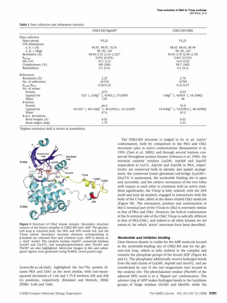
The CHK2-KD structure is judged to be in an ‘active’
conformation, both by comparison to the PKA and Chk1
structures (also in active conformations (Bossemeyer et al,
1993; Chen et al, 2000)) and through several features con-
served throughout protein kinases (Johnson et al, 1996): the
essential catalytic residues Lys249, Asp368 and Asp347
(equivalent to Lys72, Asp166 and Asp184 in PKA, respec-
tively) are conserved both in identity and spatial arrange-
ment, the conserved lysine–glutamate salt-bridge (Lys249—
Glu273) is maintained, the nucleotide binding site is open
and accessible, and the relative orientation of the two lobes
with respect to each other is consistent with an active state.
Most significantly, the T-loop is fully ordered, with the APE
motif and loop tip properly engaged in interactions with the
body of the C-lobe, albeit in the dimer-related Chk2 molecule
(Figure 3E). The orientation, position and conformation of
this C-terminal part of the T-loop in Chk2 is extremely similar
to that of PKA and Chk1. However, the helical conformation
of the N-terminal side of the Chk2 T-loop is radically different
to that of PKA/Chk1, and indeed to all other kinases we are
aware of, for which ‘active’ structures have been described.
Nucleotide and inhibitor binding
Clear electron density is visible for the ADP molecule located
in the nucleotide-binding site of CHK2-KD and for the gly-
cine-rich loop, which is fully ordered in the complex and
contacts the phosphate groups of the bound ADP (Figure 4A
and C). The phosphates additionally receive hydrogen bonds
from the side chains of Lys249, Asp368 and Asn352, and are
coordinated by one of the two magnesium ions located in
the catalytic site. The phenylalanine residue (Phe369) of the
adjacent DFG motif is in a ‘flipped out’ conformation. The
adenine ring of ADP makes hydrogen bonds to the backbone
groups of hinge residues Glu302 and Met304, while the
Table I Data collection and refinement statistics
CHK2-KD/MgADP CHK2-KD/DBQ
Data collectionSpace group P3221 P3221Cell dimensionsa, b, c (A) 90.87, 90.87, 92.91 88.65, 88.65, 88.90a, b, g (deg) 90, 90, 120 90, 90, 120
aHighest resolution shell is shown in parenthesis.
Figure 2 Structure of Chk2 kinase domain. Secondary structurecartoon of the binary complex of CHK2-KD with ADP. The glycine-rich loop is coloured pink, the DFG and APE motifs red, and theT-loop yellow. Secondary structure elements corresponding tob-strands are coloured blue and a-helices cyan. ADP is shown asa ‘stick’ model. The catalytic residue Asp347, conserved residuesLys249 and Glu373, and autophosphorylation sites Thr383 andThr387 are also highlighted. Molecular images in this and subse-quent figures were generated using PyMOL (www.pymol.org).
Trans-activation of Chk2 by T-loop exchangeAW Oliver et al
&2006 European Molecular Biology Organization The EMBO Journal VOL 25 | NO 13 | 2006 3181
oxygen of the ribose moiety make hydrogen bonds to the side
chain of Glu308, and the backbone of Glu351.
While there is growing interest in Chk2 as a therapeutic
target in cancer (Bartek and Lukas, 2003; Collins and Garrett,
2005; Pommier et al, 2005), few highly potent and specific
inhibitors have been described. Hymenialdisine is a natural
product found in marine sponges, which is an ATP-competi-
tive inhibitor of several protein kinases (Meijer et al, 2000)
and derivatives of this are showing promise as Chk2 inhibi-
tors (Sharma and Tepe, 2004). Therefore, as a possible
starting point for developing specific Chk2 inhibitors, we
have determined the structure of a binary complex between
Figure 3 Chk2 T-loop conformation and exchange. (A) T-loop exchanged dimer. One molecule of the dimer is shown as a molecular surfacerepresentation (coloured cyan), and the second as a secondary structure cartoon (yellow). (B) As in (A) but rotated 901 around the indicatedaxis. (C) Molecular details of T-loop exchange. Residues Pro388, Thr389 and Tyr390 comprising the tip of the exchanged loop (colouredyellow), along with residues Ala392, Pro393, Leu396 and Val397 forming the start of a helix (red) pack into a hydrophobic recess in a dimer-related CHK2-KD molecule (cyan). This recess is lined by two helices (residues 405–423 and 435–442) and the loop comprised of residues 466–476 (blue). (D) APE motif: Glu394 of the APE motif (coloured red) is hydrogen-bonded to the canonically conserved arginine (Arg474) despitecoming from an exchanged T-loop (yellow). Helices and loops comprising the hydrophobic recess are coloured blue, as in (C). Tyr390 is alsoshown, as a guide for orientating the figure. (E) Chk2/Chk1 comparison. Despite the exchange of T-loops between Chk2 molecules, theorientation, position and conformation of the C-terminal part of this loop (coloured red) is very similar to that of Chk1 (PDB: 1ia8).
Trans-activation of Chk2 by T-loop exchangeAW Oliver et al
The EMBO Journal VOL 25 | NO 13 | 2006 &2006 European Molecular Biology Organization3182
CHK2-KD and DBQ at 2.7 A resolution; a compound with an
IC50 of 3.5 mM (Curman et al, 2001).
As expected from biochemical studies, the compound
occupies the nucleotide-binding pocket, making hydrogen
bonds to several key residues (Figure 4B and D).
Principally, two hydrogen bonds are made to the backbone
of hinge residues Glu302 and Met304, mimicking the inter-
actions made by the adenine group of bound ADP, a binding-
mode equivalent to that reported for other kinase/DBQ
complexes (Meijer et al, 2000; Lougheed et al, 2004).
Hydrogen bonds are also made between the amino group of
the dihydroimidazolone moiety and the side-chains of Glu308
and Asn352, and with the backbone oxygen of Asn352,
similar to the contacts made by the ribose and phosphate
groups of bound ADP. An additional hydrogen bond is made
between the N4H position of DBQ and the side-chain of
Glu308. In contrast to other kinase/DBQ complexes, how-
ever, no polar interactions, direct or water-mediated, are
made to either Asp368 of the DFG motif, or its conserved
salt-bridge partner Lys249. Furthermore, no interactions are
made between DBQ and the glycine-rich loop, which is
disordered in the drug complex. One effect of the extensive
hydrogen-bonding network involving Glu308, Asn352 and
DBQ is to change the orientation of the dihydroimidazolone
moiety with respect to the rest of the DBQ molecule, com-
pared to the conformation in other kinases, a difference that
might be exploited in drug development.
Chk2 dimerisation and activation
The current model for Chk2 activation in response to DNA
damage involves two distinct phases of phosphorylation.
Initially, Chk2 is primed for activation by phosphorylation
Figure 4 Nucleotide and drug binding to Chk2 kinase domain. (A) Electron density for ADP bound to CHK2-KD, contoured at a level of 4.5sfrom an Fo�Fc omit map. (B) Electron density for the inhibitor DBQ bound to CHK2-KD, contoured at a level of 3s from an Fo�Fc omit map.(C) LIGPLOT diagram (Wallace et al, 1995) detailing the interactions made between ADP and CHK2-KD. (D) LIGPLOT diagram detailing theinteractions made between DBQ and CHK2-KD. In (A) and (B), residues involved in hydrogen bonds to the bound ligand are shown in ‘stick’representation. Water molecules are shown as grey spheres, and magnesium ions as green spheres. Secondary structure elementscorresponding to b-sheets are coloured blue, and a-helices cyan. In (C) and (D), green dotted lines represent hydrogen-bonds (o3.5 A)whereas brown ‘sunbursts’ represent hydrophobic-type interactions between the protein and ligand.
Trans-activation of Chk2 by T-loop exchangeAW Oliver et al
&2006 European Molecular Biology Organization The EMBO Journal VOL 25 | NO 13 | 2006 3183
of Thr68 within its SCD, principally by the ATM kinase
(Matsuoka et al, 2000; Melchionna et al, 2000). Thr68-
phosphorylation of Chk2 converts it to a state where it is
competent to autophosphorylate two residues in its T-loop,
Thr383 and Thr387 (Lee and Chung, 2001). Phosphorylation
of Thr68 had been proposed to activate Chk2 by promoting
allosteric changes in the kinase domain that facilitate auto-
phosphorylation (Bartek et al, 2001). However, a body of
subsequent data have shown that the main effect of Thr68
phosphorylation is to promote homo-dimerisation of Chk2
via interaction of the phospho-Thr68 segment of one mole-
cule with the FHA domain of the other (Ahn et al, 2002; Xu
et al, 2002). Once dimerised, one Chk2 molecule can phos-
phorylate the T-loop in the other Chk2 molecule, to give a
fully active species which may then be able to cis-phosphor-
ylate at Thr68 and Ser516 (Schwarz et al, 2003), although the
significance of this latter phosphorylation is unknown.
The activated conformation and dimeric interactions of
CHK2-KD in the crystal structure suggests that it is (or closely
resembles) the trans-phosphorylation-competent state engen-
dered by Thr68 phosphorylation. In support of this idea we
first sought to determine that the Chk2 kinase domain does
indeed have an inherent propensity to dimerise and that the
observed dimer is not merely the result of crystallisation. We
therefore incubated CHK2-KD in dilute solution (0.1mg/ml,
compared to 20mg/ml used for crystallisation) in the pre-
sence of increasing concentrations of a lysine-specific cross-
linking reagent, and analysed the result on SDS–PAGE as
previously described (Dajani et al, 2001). At all concentra-
tions of the crosslinker, we observed a consistent crosslinked
dimer band, but with no significant higher bands that would
indicate nonspecific aggregation of the protein at this con-
centration, suggesting that CHK2-KD does exist in a mono-
mer–dimer equilibrium in solution (Figure 5A). Although this
does not prove that the T-loop exchanged dimer in the
crystals is that which forms in solution, the surface area
buried by formation of this interface (1344 A2 per monomer,
2688 A2 in total) is in the range typically found for functional
interactions (Ponstingl et al, 2000) and significantly larger
than any other intermolecular contact observed in the crys-
tals. The equilibrium for the isolated kinase domain clearly
favours the monomer over the dimer at this low protein
concentration (8 mM). However in the intact protein, tether-
ing of two Chk2 molecules via pThr68–FHA domain inter-
actions would massively increase the effective concentration
of the associated kinase domains, shifting the equilibrium
significantly towards the dimerised state.
We next asked whether the dimeric arrangement of the
kinase domains was architecturally consistent with the pro-
posed activation mechanism in which the phosphorylated
Thr68 segments of each Chk2 protomer could bind to the
FHA domain of the other and promote kinase domain asso-
ciation. The visible C-terminus of the crystal structure of the
Chk2 FHA domain (Asp207) is only separated by a couple of
residues from the visible N-terminus of the CHK2-KD struc-
ture (Ser210), so that the juxtaposition of the two domains is
closely constrained, and the FHA domain and N-terminal lobe
of the kinase, are likely to be in contact. With this constraint,
we constructed a structure-based model for a Chk2 FHA-KD
segment dimer. Taking the pThr residue bound to the FHA
domain structure to represent pThr68, 20 residues separate
the C-terminal end of the phosphopeptide (equivalent to
Chk2 residue 72) bound to the FHA domain on one chain,
and the visible N-terminus of the FHA domain (Pro92) on the
other (Figure 6). The distance between residues 72 and 92
(B50–70 A) could be spanned comfortably by this amount of
polypeptide chain, so that the model is geometrically reason-
able. While this is of course a hypothetical model, that it fits
comfortably within the architectural constraints of Thr68-
driven dimerisation is reassuring, and further supports the
idea that the kinase-domain dimer in the crystals is biologi-
cally authentic.
We then sought to determine whether dimerisation per se
had any effect on the ability of Chk2 to autophosphorylate. To
do this, we compared the autophosphorylation activity of
CHK2-KD and a GST-CHK2-KD construct. GST is itself a dimer
and we verified that the GST-CHK2-KD fusion protein is also
strongly dimeric (Figure 5B). Examination of the crystal
structure of S. japonicum GST shows that the two C-termini
in the GST dimer present at the same face of the structure, so
that although they would not be optimally positioned, the
attached kinase domains would not be prevented from ex-
changing T-loops in the fusion protein. We reasoned that if
our model was correct, the surrogate dimerisation provided
by GST would enhance the ability of CHK2-KD to autophos-
phorylate to some degree, compared with the unconstrained
isolated CHK2-KD. Both constructs showed some ability to
autophosphorylate on incubation with g-32P-labelled ATP
(Figure 5C and D); however, consistent with our model, the
GST-fused kinase was at least four-fold more active in auto-
phosphorylation than the isolated kinase domain. To deter-
mine whether this enhanced activity was specific, we cleaved
the GST-CHK2-KD fusion protein postincubation at a rhino-
virus 3C-protease site between the C-terminus of the GSTand
the N-terminus of the Chk2 kinase domain, and analysed the
products by SDS–PAGE and autoradiography (Figure 5E and
F). All the incorporated 32P resided in the Chk2 component
and not the GST, indicating that dimeric tethering of the
kinase had promoted a specific autophosphorylation reac-
tion. We then compared the ability of the bacterially ex-
pressed and unphosphorylated CHK2-KD, GST-CHK2-KD
proteins and the full-length baculovirus expressed Chk2
(CHK2-FL) to phosphorylate a substrate peptide derived
from Cdc25C. The baculovirus expressed protein, which
contains high-levels of pThr68 (as measured by phospho-
specific antibody) and is therefore likely to be substantially
phosphorylated on T-loop residues Thr383 and Thr387, was
found to be very significantly (100-fold compared to GST-
CHK2-KD, 400-fold to CHK2-KD) more active than either
of the kinase domain constructs over the course of the assay
period (Figure 5G). However, with the unphosphorylated
proteins, the hetero-phosphorylation of the Cdc25C peptide is
dependent on progressive activation of the protein by autophos-
phorylation during the assay. The higher rate of Cdc25C
phosphorylation by the GST-dimerised protein is then fully
consistent with enforced dimerisation by GST enhancing spe-
cific T-loop trans-phosphorylation. Finally, we analysed the
ability of this activated full-length Chk2 to phosphorylate a
separate CHK2-KD. When incubated with ATP, CHK2-FL did
autophosphorylate to some degree, suggesting that not all
possible sites were fully phosphorylated. However, despite its
high activity against Cdc25C substrate, CHK2-FL showed no
ability to phosphorylate a separate Chk2 kinase domain pre-
sented as an untethered substrate (Figure 5H and I).
Trans-activation of Chk2 by T-loop exchangeAW Oliver et al
The EMBO Journal VOL 25 | NO 13 | 2006 &2006 European Molecular Biology Organization3184
Figure 5 Dimerisation, Auto- and substrate-phosphorylation of CHK2-KD. (A) SDS–PAGE analysis of CHK2-KD crosslinking. (B) SDS–PAGEanalysis of GST-CHK2-KD crosslinking. In each case, a fixed concentration of protein was incubated with increasing concentrations of thehomo-bifunctional crosslinker BS3. Mw¼molecular mass marker, O¼protein not exposed to crosslinking agent, D¼dimeric species,M¼monomeric species. (C) Autophosphorylation assay. Fixed concentrations of GST-CHK2-KD or CHK2-KD were incubated with g-32P-labelled ATP for increasing periods of time (as indicated). Samples were analysed by SDS–PAGE, visualised by phosphorimager andautoradiography. (D) Coomassie-stained SDS–PAGE gel of (C). (E) Autophosphorylation specifically occurs within the kinase domain ofChk2. Autophosphorylated samples of GST-CHK2-KD (Lane 1) and CHK2-KD (3) were incubated with rhinovirus 3C-protease (Lanes 2 and 4,respectively) then analysed by SDS–PAGE. Incorporation of g-32P-labelled ATP was visualised by autoradiography. (F) Coomassie-stained SDS–PAGE gel of (E). (G) DELFIA assay. Samples of CHK2-FL, GST-CHK2-KD and CHK2-KD at varying concentrations were incubated, in thepresence of ATP, with a substrate peptide (corresponding to the sequence flanking the Ser216 phosphorylation site of CDC25C).Phosphorylation was detected by incubation with a polyclonal anti-CDC25C(Ser216) antibody, followed by a Europium-labelled secondaryantibody. A fluorescent plate reader was then used to quantitate the level of peptide phosphorylation (as Europium counts). (H) Trans-phosphorylation assay. Increasing amounts of CHK2-FL were incubated with a fixed concentration of CHK2-KD (as indicated) in the presence ofg-32P-labelled ATP. Samples were analysed by SDS–PAGE, then visualised by phosphorimager and autoradiography. (I) Coomassie-stainedSDS–PAGE gel of (H).
Trans-activation of Chk2 by T-loop exchangeAW Oliver et al
&2006 European Molecular Biology Organization The EMBO Journal VOL 25 | NO 13 | 2006 3185
Discussion
Phosphorylation of residues within the T-loop/activation
segment is a key determinant of activation of many protein
kinases (Johnson et al, 1996), in particular those of the ‘RD’
group (Krupa et al, 2004) of which Chk2 is a member.
Typically, the unphosphorylated T-loop is disordered, or
is locked into an inactive alternative conformation.
Phosphorylation on one or more residues fixes the T-loop
into an active conformation in which it forms the substrate-
binding site. The conserved arginine of the RD motif,
immediately preceding the catalytic aspartate, interacts with
the phosphorylated residue(s) in the T-loop, orientating both
the DFG motif and other catalytic/substrate-binding residues
to facilitate the phospho-transfer reaction.
In many phospho-regulated kinases, such as those in MAP-
kinase cascades, phosphorylation of the T-loop is performed
by a different upstream kinase. However, there are a signifi-
cant number of enzymes, which like Chk2, have been shown
unambiguously to be responsible for their own T-loop phos-
phorylation; in some cases by intramolecular reactions
coupled to protein folding (Lochhead et al, 2005), but most
commonly by intermolecular reactions. Although earlier
models had suggested a mechanism of intramolecular auto-
phosphorylation allosterically triggered by phosphorylation
of Thr68 by ATM (Bartek et al, 2001), subsequent work
suggests that T-loop phosphorylation of Chk2 is an intermo-
lecular event dependent on pThr68-induced dimerisation
(Ahn et al, 2002; Xu et al, 2002). This latter model is strongly
supported by our observation that fusion of a heterologous
dimerisation domain (GST) to CHK2-KD gives at least a four-
fold increase in the rate of autophosphorylation.
Intermolecular autophosphorylation by a kinase that re-
quires phosphorylation for activity presents an interesting
logical paradox. This is conventionally resolved by assuming
that the T-loop in the unphosphorylated kinase can transi-
ently adopt the activated conformation rarely, but sufficiently
often, for one molecule to bind and phosphorylate the T-loop
of a second molecule, initiating a chain-reaction. In these
circumstances, the T-loop of the substrate kinase molecule
binds in the same way as a heterologous substrate and its
amino-acid sequence typically conforms to the same con-
sensus as other target sites (Johnson et al, 1996). However,
the sequences surrounding both phosphorylation sites
(Thr383 and Thr387) in the T-loop sequence of Chk2 present
very poor matches to the consensus sequences of known
Chk2 phosphorylation sites in targets such as E2F1, Cdc25A/
C, PML and BRCA1, and to consensus substrate peptides
derived there from, lacking in particular the preferred Arg at
�3 and Leu at �5 (Seo et al, 2003) (Figure 7A). Consistent
with this we find that the Chk2 kinase domain is not
significantly phosphorylated when presented as a conven-
tional substrate to an activated Chk2 molecule, compared to a
Cdc25C sequence that conforms to the specificity. This argues
strongly that the process of autophosphorylation proceeds
via a mechanism that is distinct from the process of hetero-
phosphorylation of a substrate.
The crystal structure of the Chk2 kinase domain and the
biochemical data described here suggest a mechanism for
dimerisation-driven trans-phosphorylation that simulta-
neously circumvents the problem of specificity mismatch by
the Chk2 T-loop, and the starting paradox that bedevils all
autophosphorylation mechanisms, as follows (summarised
in Figure 7B). Initially, phosphorylation of Chk2 Thr68
by ATM promotes interaction with the FHA domain of a
second Chk2 molecule, generating a symmetrical Thr68-FHA
Figure 6 Model of the trans-activating Chk2 dimer. Schematicmodel of a full-length Chk2 dimer. The X-ray structure of theChk2 FHA domain and its associated phospho-peptide (PDB:1GXC), along with the CHK2 kinase domain reported here, wereused to build a theoretical model for the full-length Chk2 molecule.Predictive modelling of the FHA/Kinase domain junction is facili-tated due to the short (2 amino acid) distance between the reportedvisible termini in each structure (Asp207 and Ser210 respectively),which suggests a spatially constrained arrangement. The position ofthe FHA-bound phosphothreonine is taken to represent Thr68. Thevisible C-terminus of the bound peptide therefore equates to Tyr72.The distance between this residue and the visible N-terminus of adimer-related FHA molecule (Pro92) is estimated to be between 50and 70 A. A connecting polypeptide chain of 30 amino acids wouldbe therefore be sufficient to connect the two termini. See Figure 1for SCD, FHA and KINASE abbreviations. N and C termini for eachpolypeptide chain are coloured blue and red, respectively.
Figure 7 Chk2 T-loop autophosphorylation sites do not resembleendogenous substrates. Alignment of endogenous Chk2 substratesequences (CDC25A, CDC25C, BRCA-1, E2F-1, PML) with an opti-mised substrate sequence (Chk2-TIDE) and T-loop autophosphor-ylation sites. For each sequence, the residue number of the firstamino acid is listed (res). The autophosphorylation sites of Chk2overlap, as indicated by the underlined regions. The ‘p’ columnheader indicates the phosphorylated amino acid.
Trans-activation of Chk2 by T-loop exchangeAW Oliver et al
The EMBO Journal VOL 25 | NO 13 | 2006 &2006 European Molecular Biology Organization3186
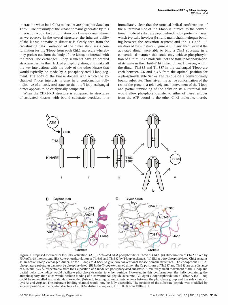
interaction when both Chk2 molecules are phosphorylated on
Thr68. The proximity of the kinase domains generated by this
interaction would favour formation of a kinase-domain dimer
as we observe in the crystal structure; the inherent ability
of the kinase domains to dimerise is clearly seen from the
crosslinking data. Formation of the dimer stabilises a con-
formation for the T-loop from each Chk2 molecule whereby
they project out from the body of one kinase to interact with
the other. The exchanged T-loop segments have an ordered
structure despite their lack of phosphorylation, and make all
the key interactions with the body of the other kinase that
would typically be made by a phosphorylated T-loop seg-
ment. The body of the kinase domain with which the ex-
changed T-loop interacts is also in a conformation fully
indicative of an activated state, so that the T-loop exchanged
dimer appears to be catalytically competent.
When the CHK2-KD structure is compared to structures
of activated kinases with bound substrate peptides, it is
immediately clear that the unusual helical conformation of
the N-terminal side of the T-loop is inimical to the conven-
tional mode of substrate peptide-binding by protein kinases,
which typically involves b-strand main-chain hydrogen bond-
ing between the activation segment and the þ 1 and þ 3
residues of the substrate (Figure 7C). In any event, even if the
activated dimer were able to bind a Chk2 substrate in a
conventional manner, this could only achieve phosphoryla-
tion of a third Chk2 molecule, not the trans-phosphorylation
of its mate in the Thr68-FHA linked dimer. However, within
the dimer, Thr383 and Thr387 in the exchanged T-loop are
each between 5.6 and 7.3 A from the optimal position for
a phosphorylatable Ser or Thr residue on a conventionally
bound substrate. Thus, given the active conformation of the
rest of the protein, a relatively small movement of the T-loop
and partial unwinding of the helix on its N-terminal side
would allow phosphoryl-transfer to either of these residues
from the ATP bound to the other Chk2 molecule, thereby
Figure 8 Proposed mechanism for Chk2 activation. (A) (i) Activated ATM phosphorylates Thr68 of Chk2. (ii) Dimerisation of Chk2 driven byFHA-pThr68 interactions. (iii) Auto-phosphorylation of Thr383 and Thr387 by T-loop exchange. (iv) Either auto-phosphorylated Chk2 remainsas an active T-loop exchanged dimer, or the T-loops fold back to give two conventional kinase domain structures. The endogenous CDC25phosphatase substrates can now be phosphorylated. (B) In the T-loop exchanged dimer, the Ca positions of Thr387 and Thr383 are at a distanceof 5.85 and 7.29 A, respectively, from the Ca position of a modelled phosphorylated substrate. A relatively small movement of the T-loop andpartial helix unwinding would facilitate phosphoryl-transfer to either residue. However, in this conformation, the helix containing theautophosphorylation sites would occlude binding of a conventional peptide substrate. (C) Upon autophosphorylation of Thr387, the T-loopcould be remodelled into a standard extended b-strand, forming canonical interactions between the phosphate group and the side chains ofLys373 and Arg346. The substrate binding channel would now be fully accessible. The position of the substrate peptide was modelled bysuperimposition of the crystal structure of a PKA-substrate complex (PDB: 1JLU) onto CHK2-KD.
Trans-activation of Chk2 by T-loop exchangeAW Oliver et al
&2006 European Molecular Biology Organization The EMBO Journal VOL 25 | NO 13 | 2006 3187
achieving trans-phosphorylation. Once phosphorylated,
especially at Thr383, the helical conformation for the
N-terminal side of the T-loop would be greatly destabilised
and the T-loop could adopt a conventional extended confor-
mation in which the phosphate of pThr383 interacts with the
side chains of Arg346 and Lys373 in an essentially identical
manner to the interaction of pThr197 with Arg165 and
Lys189 in PKA (Knighton et al, 1991) (Figure 7D).
Given retention of Thr68 phosphorylation and consequent
maintenance of N-terminal dimerisation, there is no clear
structural reason why dimerisation of the T-loop exchanged
kinase domains should not be maintained after trans-phos-
phorylation. With the T-loop stabilised in a conventional
active conformation consistent with productive binding of
hetero-substrates, and the rest of the protein in an active
conformation, each Chk2 molecule in the T-loop exchanged
dimer would in principle be a fully competent protein kinase
so long as the presence of the other Chk2 molecule did not
obstruct binding of a substrate protein (Figure 8).
Alternatively, the activated kinase domains might operate
independently despite N-terminal dimerisation, with the
phosphorylated T-loop segment folding back onto the body
of its own kinase domain to give a conventional protein
kinase structure. Further work will be required to distinguish
these possibilities.
During review of this manuscript, additional evidence
supporting our proposed model of Chk2 activation by
T-loop exchange has been published by Xu et al (2006) on
Cds1, the fission yeast homolog of Chk2.
Materials and methods
Mass spectroscopyMALDI-TOF experiments were carried out using standard protocolson a Voyager-DE STR Biospectrometry Workstation (AppliedBiosystems, Foster City, CA).
Cloning, expression and purificationThe IMAGE clone AU20-2, was used as a template for PCR, as itcontained a full-length human CHK2 cDNA. Primers were designedto amplify the region encompassing the kinase domain (residues210–531), with additional flanking restriction enzymes sites (NdeIand EcoRI) to facilitate cloning into the expression vector pTHREE-E(a GST-fusion vector modified to contain the multiple cloning siteof pET-17b, following a encoded rhinovirus 3C-protease site). Thevector pTHREE-Chk2 (210–531) was transformed into the Escher-ichia coli expression host Rosetta2 (DE3) pLysS (EMD Bioscience,Inc., San Diego, USA). A 250ml flask containing 50ml of L-broth(1% w/v tryptone, 0.5% w/v NaCl, 0.5% w/v yeast extract)supplemented with 50 mg/ml of carbenicillin and 34mg/ml ofchloramphenicol was inoculated with a single transformed bacterialcolony. This was grown in an orbital incubator set at 371C and225 r.p.m., until the A600 of the culture reached 0.6. This ‘starter’culture was then stored overnight at 41C. From the starter culture,10ml was used to inoculate a 2 l flask containing 1 l of L-brothsupplemented as before with antibiotics. The culture was grown inan orbital incubator set at 371C and 200 r.p.m. Once the A600 of thecell culture had reached 0.6, expression of the Chk2 kinase domainwas induced by the addition of IPTG to a final concentration of0.2mM. Cultures were grown for a further 19 h at a reducedtemperature of 201C, after which the cells were harvested bycentrifugation (5000 g, 10min, 101C). Cell pellets were stored at�801C until required.
The cell pellet arising from 3 l of culture was resuspended, on ice,in 100ml of buffer A (50mM HEPES NaOH pH 7.5, 250mM NaCl,1mM EDTA, 10mM DTT, supplemented with protease inhibitors(Roche, Lewes, East Sussex, UK)). Cells were then lysed bysonication (15� 5 s bursts, on ice, at 80% amplitude, with a largeparallel probe; Jencons Ultrasonic Processor), after which cell
debris and insoluble materials were removed by centrifugation(40 000 g, 60min at 101C). The supernatant arising from this stepwas filtered through a 4-mm filter (Whatman plc, Brentford,Middlesex, UK), then applied to a batch/gravity column containing25ml of Glutathione Sepharose 4 resin (Amerham Biosciences,Little Chalfont, Buckinghamshire, UK) equilibrated in Buffer A. Thecolumn containing the cell extract and resin was rotated/rolled at41C, for a period of 1 h to facilitate protein binding. The resin wasallowed to pack under gravity, and then washed with successiveapplications of buffer A (approximately 250ml in total). It was thenresuspended in buffer A to give an approximate 50% resin/bufferslurry, transferred to a 50ml Falcon tube (BD Biosciences, Le PontDe Claix, France), and 150ml of rhinovirus 3C-protease (PreScis-sion protease, Amersham Biosciences) added. This was incubatedovernight with constant rotation/rolling at 41C. The resin was againallowed to pack under gravity, and the flow-through collected.Fractions containing the Chk2 kinase domain (CHK2-KD) wereidentified by SDS–PAGE, then pooled and concentrated to a finalvolume of 10ml (Vivaspin 20, 10 kD MWCO; Vivascience AG,Hannover, Germany). The concentrated protein was applied toa Superdex 75 (Amersham Biosciences) size exclusion columnequilibrated in buffer B (10mM HEPES NaOH pH 7.5, 250mM NaCl,1mM EDTA, 10mM DTT). Again, fractions containing CHK2-KDwere identified by SDS–PAGE, then pooled and concentratedto a final concentration of 20mg/ml (as determined by UVspectroscopy). The purified protein was flash-frozen on dry-iceand stored at �801C until required.
CrystallisationInitial crystallisation trials were carried out at a concentration of20mg/ml in microbatch experiments, under oil, at 41C. Smallcrystals were observed, after a period of several days, in condition64 of the ‘The PEGs Suite’ from Nextal Biotechnologies (Montreal,Quebec, Canada). This condition was optimised in hanging dropexperiments at 41C, to mixing 1ml of protein (20mg/ml in 10mMHEPES NaOH pH 7.5, 250mM NaCl, 10mM DTT, 1mM EDTA)supplemented with either 2.5mM ADP or 2.5mM DBQ, with 1 ml ofprecipitant containing 0.1M HEPES NaOH pH 7.6, 0.2M Mg(NO3)2,22% w/v PEG 3350, 16% v/v ethylene glycol. The mother liquorfrom these experiments was sufficient to cryo-protect the crystalsduring data collection.
Data collection and processingData to 2.25 Awere collected for the CHK2-KD/ADP binary complexon station ID29 at the ESRF (European Synchrotron RadiationFacility, Grenoble, France) from a single crystal at 100K, recordedon an ADSC CCD detector. This complex crystallised in spacegroupP3221, with a single molecule comprising the asymmetric unit, andunit cell dimensions a, b¼ 90.87 A, c¼ 92.91 A. Data to 2.7 A werecollected for the CHK2-KD complex with DBQ on station 14.2 at theSRS (Synchrotron Radiation Source, Daresbury, UK) from a singlecrystal at 100K, recorded on an ADSC CCD detector. This complexalso crystallised in the spacegroup P3221, with a single molecule inasymmetric unit, but with a smaller unit cell of a, b¼ 88.65 A,c¼ 88.90 A. Images for both data sets were integrated usingMOSFLM (Leslie, 1995), and reduced/scaled using programs fromthe CCP4 suite (Collaborative Computational Project, 1994).
The structures of the Chk2 complexes were solved by molecularreplacement using PHASER (McCoy et al, 2005), with the X-raystructure of PKA as a search model (PDB: 1ATP). Difference mapswere used to extend and rebuild the initial models using Coot(Emsley and Cowtan, 2004). Iterative cycles of refinement(REFMAC5; Collaborative Computational Project, 1994) and man-ual intervention produced the current models for each complex,CHK2-KD/ADP: which contains 2271 protein atoms, 1 nitrate ion,2 magnesium ions, 1 ADP molecule, and 156 solvent atoms, andCHK2-KD/debromohymenisaldisine: which contains 2153 proteinatoms, 1 nitrate ion, 1 magnesium ion, 1 DBQ molecule and 44solvent atoms. Crystallographic statistics are given in Table I.Coordinates and structure factors for both complexes have beendeposited in the RSCB Protein Data Bank, with codes 2CN5 and2CN8 for the CHK2-KD/ADP and CHK2-KD/DBQ complexesrespectively.
BS3 crosslinkingExperiments were carried out at a protein concentration of 8 mM in20mM HEPES NaOH pH 7.5, 250mM NaCl. A 50mM stock solution
Trans-activation of Chk2 by T-loop exchangeAW Oliver et al
The EMBO Journal VOL 25 | NO 13 | 2006 &2006 European Molecular Biology Organization3188
of BS3 (Pierce Biotechnology Inc., IL, USA) was serially diluted,then added to each of several protein samples. Crosslinking wasallowed to proceed for 30min at room temperature, before thesamples were analysed by SDS–PAGE. Final BS3 concentrationsranged between 5mM and 8 mM. BS3 (Bis[sulfosuccinimidyl]suberate) is a homobifunctional primary-amine reactive cross-linking agent.
Phosphorylation assaysEquimolar amounts (60 nM) of either purified GST-CHK2-KD orCHK2-KD were incubated at 251C in a final reaction volume of 24 mlof assay buffer (40mM HEPESNaOH pH 7.4, 40mM KCl, 10mMDTT, 2mM MgCl2, 0.02% (v/v) Tween-20) containing 10mM ATPand 0.1mCi g-32P-labelled ATP. Reactions were stopped by theaddition of SDS loading buffer and proteins resolved by SDS–PAGEbefore autoradiography or analysis by phosphorimager.
For the rhinovirus 3C-protease digestions, phosphorylationreactions were incubated as before, for 20min, then stopped bythe addition of EDTA (50mM final concentration) and thephosphatase inhibitor NaF (10mM final concentration). Reactionswere split and incubated with or without protease for 30min on iceprior to addition of SDS loading buffer.
For the CHK2-FL trans-phosphorylation assay, increasingamounts of purified CHK2-FL were incubated with a fixed(120nM) concentration of CHK2-KD, at 251C, in assay buffercontaining 10mM ATP and 0.1mCi g-32P-ATP, for 20min.
DELFIA assaysKinase activity was measured in a DELFIAs assay that monitorsphosphorylation of a CDC25C peptide using a specificphospho-antibody. The enzyme reaction was carried out inpolypropylene plates (Greiner Bio-One Ltd, Gloucestershire,UK). The reaction mix (25ml) contained enzyme (10ml), peptide
(Biotin-KKKVSRSGLYRSPSMPENLNRPR) (1mM, 5ml), ATP (30mM,5 ml) in assay buffer (see phosphorylation assays). The reaction wasincubated for 30min at room temperature and stopped by theaddition of buffer (125 ml) containing 40mM EDTA, 0.05% v/vTween-20, 0.1% w/v BSA in TBS. An aliquot (100 ml) of the reactionmix was transferred to a black neutravidin-coated plate (PerbioScience UK Ltd, Northumberland, UK) and incubated for 1 h on ashaker at room temperature. The plates were washed four timeswith wash buffer (25mM Tris pH 8, 150mM NaCl and 0.1% v/vTween-20) and incubated for 1 h as before with antibody mix(100ml) consisting of anti-phospho CDC25C (1.25 nM, #9528, CellSignalling Technology Inc., MA, USA) and europium-labelled anti-rabbit IgG (0.3mg/ml, AD0105, PerkinElmer Life Sciences) diluted inDELFIA assay buffer (PerkinElmer Life Sciences, MA, USA). Theplates were washed a further four times with wash buffer before theaddition of enhancement solution (100 ml/well, PerkinElmer LifeSciences). The plate was read on a Victor2 1420 multilabel counter(PerkinElmer Life Sciences) using a time-resolved measurementmode reading fluorescence at 615 nm.
Acknowledgements
We are very grateful to our colleagues: Mark Roe for advice andassistance with data processing and model refinement. DavidKomander, Markus Hassler, Andy Dore, Mairi Kilkenny and WyattYue for assistance with synchrotron data collection, and MatthewTall for assistance with the DELFIA assays. We acknowledge boththe European Synchrotron Radiation Facility and the DaresburySynchrotron Radiation Source for provision of synchrotron radia-tion facilities. This work was supported by a Programme Grant fromCancer Research UK (LHP).
References
Ahn JY, Li X, Davis HL, Canman CE (2002) Phosphorylation ofthreonine 68 promotes oligomerization and autophosphorylationof the Chk2 protein kinase via the forkhead-associated domain.J Biol Chem 277: 19389–19395
Ahn JY, Schwarz JK, Piwnica-Worms H, Canman CE (2000)Threonine 68 phosphorylation by ataxia telangiectasia mutatedis required for efficient activation of Chk2 in response to ionizingradiation. Cancer Res 60: 5934–5936
Bartek J, Lukas J (2003) Chk1 and Chk2 kinases in checkpointcontrol and cancer. Cancer Cell 3: 421–429
Bossemeyer D, Engh RA, Kinzel V, Ponstingl H, Huber R (1993)Phosphotransferase and substrate binding mechanism of thecAMP-dependent protein kinase catalytic subunit from porcineheart as deduced from the 2.0 A structure of the complex withMn2+ adenylyl imidodiphosphate and inhibitor peptide PKI(5–24). EMBO J 12: 849–859
Chen P, Luo C, Deng Y, Ryan K, Register J, Margosiak S, Tempczyk-Russell A, Nguyen B, Myers P, Lundgren K, Kan CC, O’Connor PM(2000) The 1.7 A crystal structure of human cell cycle checkpointkinase Chk1: implications for Chk1 regulation. Cell 100: 681–692
Collaborative Computational Project, N (1994) The CCP4 suite:programs for protein crystallography. Acta Crystallographica, D50: 760–763
Collins I, Garrett MD (2005) Targeting the cell division cycle incancer: CDK and cell cycle checkpoint kinase inhibitors. CurrOpin Pharmacol 5: 366–373
Curman D, Cinel B, Williams DE, Rundle N, Block WD, GoodarziAA, Hutchins JR, Clarke PR, Zhou B-B, Lees-Miller SP, AndersonRJ, Roberge M (2001) Inhibition of the G2 DNA damage check-point and of protein kinases Chk1 and Chk2 by the marine spongealkaloid debromohymenialdisine. J Biol Chem 276: 17914–17919
Dajani R, Fraser E, Roe SM, Young N, Good V, Dale TC, Pearl LH(2001) Crystal structure of glycogen synthase kinase 3 beta:structural basis for phosphate-primed substrate specificity andautoinhibition. Cell 105: 721–732
Emsley P, Cowtan K (2004) Coot: model-building tools for molecu-lar graphics. Acta Crystallograph D 60: 2126–2132
Falck J, Mailand N, Syljuasen RG, Bartek J, Lukas J (2001) TheATM-Chk2-Cdc25A checkpoint pathway guards against radio-resistant DNA synthesis. Nature 410: 842–847
Holm L, Sander C (1993) Protein structure comparison by alignmentof distance matrices. J Mol Biol 233: 123–138
Johnson LN, Noble ME, Owen DJ (1996) Active and inactive proteinkinases: structural basis for regulation. Cell 85: 149–158
Knighton DR, Zheng J, Ten Eyck LF, Ashford VA, Xuong N-H, TaylorSS, Sowadski JM (1991) Crystal structure of the catalytic subunitof cyclic adenosine monophosphate-dependent protein kinase.Science 253: 407–413
Krissinel E, Henrick K (2004) Secondary-structure matching (SSM),a new tool for fast protein structure alignment in three dimen-sions. Acta Crystallograph D 60: 2256–2268
Krupa A, Preethi G, Srinivasan N (2004) Structural modes ofstabilization of permissive phosphorylation sites in protein ki-nases: distinct strategies in Ser/Thr and Tyr kinases. J Mol Biol339: 1025–1039
Lavin MF, Birrell G, Chen P, Kozlov S, Scott S, Gueven N (2005)ATM signaling and genomic stability in response to DNA damage.Mutat Res 569: 123–132
Lee CH, Chung JH (2001) The hCds1 (Chk2)-FHA domain isessential for a chain of phosphorylation events on hCds1that is induced by ionizing radiation. J Biol Chem 276:30537–30541
Li J, Williams BL, Haire LF, Goldberg M, Wilker E, Durocher D,Yaffe MB, Jackson SP, Smerdon SJ (2002) Structural andfunctional versatility of the FHA domain in DNA-damagesignaling by the tumor suppressor kinase Chk2. Mol Cell 9:1045–1054
Lochhead PA, Sibbet G, Morrice N, Cleghon V (2005) Activation-loop autophosphorylation is mediated by a novel transitionalintermediate form of DYRKs. Cell 121: 925–936
Trans-activation of Chk2 by T-loop exchangeAW Oliver et al
&2006 European Molecular Biology Organization The EMBO Journal VOL 25 | NO 13 | 2006 3189
Matsuoka S, Rotman G, Ogawa A, Shiloh Y, Tamai K, Elledge SJ(2000) Ataxia telangiectasia-mutated phosphorylates Chk2 invivo and in vitro. Proc Natl Acad Sci USA 97: 10389–10394
McCoy AJ, Grosse-Kunstleve RW, Storoni LC, Read RJ (2005)Likelihood-enhanced fast translation functions. ActaCrystallographica, D 61: 458–464
McGowan CH (2002) Checking in on Cds1 (Chk2): a checkpointkinase and tumor suppressor. Bioessays 24: 502–511
McGowan CH, Russell P (2004) The DNA damage response: sensingand signaling. Curr Opin Cell Biol 16: 629–633
Meijer L, Thunnissen AM, White AW, Garnier M, Nikolic M, TsaiLH, Walter J, Cleverley KE, Salinas PC, Wu YZ, Biernat J,Mandelkow EM, Kim SH, Pettit GR (2000) Inhibition of cyclin-dependent kinases, GSK-3beta and CK1 by hymenialdisine, amarine sponge constituent. Chem Biol 7: 51–63
Melchionna R, Chen XB, Blasina A, McGowan CH (2000) Threonine68 is required for radiation-induced phosphorylation and activa-tion of Cds1. Nat Cell Biol 2: 762–765
Pommier Y, Sordet O, Rao VA, Zhang H, Kohn KW (2005) Targetingchk2 kinase: molecular interaction maps and therapeutic ratio-nale. Curr Pharm Des 11: 2855–2872
Ponstingl H, Henrick K, Thornton JM (2000) Discriminatingbetween homodimeric and monomeric proteins in the crystallinestate. Proteins 41: 47–57
Schwarz JK, Lovly CM, Piwnica-Worms H (2003) Regulation of theChk2 protein kinase by oligomerization-mediated cis- and trans-phosphorylation. Mol Cancer Res 1: 598–609
Seo GJ, Kim SE, Lee YM, Lee JW, Lee JR, Hahn MJ, Kim ST(2003) Determination of substrate specificity and putativesubstrates of Chk2 kinase. Biochem Biophys Res Commun 304:339–343
Sharma V, Tepe JJ (2004) Potent inhibition of checkpoint kinaseactivity by a hymenialdisine-derived indoloazepine. Bioorg MedChem Lett 14: 4319–4321
Wallace AC, Laskowski RA, Thornton JM (1995) LIGPLOT: aprogram to generate schematic diagrams of protein-ligand inter-actions. Protein Eng 8: 127–134
Xu YJ, Davenport M, Kelly TJ (2006) Two stage-mechanism foractivation of the DNA replication checkpoint kinase Cds1 infission yeast. Genes Dev 20: 990–1003
Trans-activation of Chk2 by T-loop exchangeAW Oliver et al
The EMBO Journal VOL 25 | NO 13 | 2006 &2006 European Molecular Biology Organization3190


![CartemotoneigeSagLac2014-15 [Unlocked by ] sentier lac st-jean.pdf · 6.6 trans-quÉbec 83 trans-quÉbec 93 trans-quÉbec 93 trans-quÉbec 93 trans-quÉbec 93 trans-quÉbec 93 trans-quÉbec](https://static.documents.pub/doc/80x56/5b2cb5eb7f8b9ac06e8b5a01/cartemotoneigesaglac2014-15-unlocked-by-sentier-lac-st-jeanpdf-66-trans-quebec.jpg)


![Study of the behavior of the reduced activation ... · técnica de calorimetría diferencial de barrido [5] y enfocados en aspectos tales como la cinética de la trans- ... manteniendo](https://static.documents.pub/doc/80x56/5baf020609d3f2dd708b6703/study-of-the-behavior-of-the-reduced-activation-tecnica-de-calorimetria.jpg)


