Journal of Asian Ceramic Societies 2 (2014) 241–247
Contents lists available at ScienceDirect
Journal of Asian Ceramic Societies
j ourna l ho me page: www.elsev ier .com/ loca te / jascer
abrication, bioactivity, in vitro cytotoxicity and cell viability ofryo-treated nanohydroxyapatite–gelatin–polyvinyl alcoholacroporous scaffold
anjaya Kumar Swain, Debasish Sarkar ∗
epartment of Ceramic Engineering, National Institute of Technology, Rourkela 769008, Odisha, India
r t i c l e i n f o
rticle history:eceived 1 April 2014eceived in revised form 5 May 2014ccepted 7 May 2014vailable online 5 June 2014
eywords:ydroxyapatite
a b s t r a c t
Freeze casting and cryogenic treatment both low temperature process have been employed to fab-ricate nanobiocomposite hydroxyapatite (HA)–gelatin–polyvinyl alcohol (PVA) macroporous scaffoldsfrom synthesized three different spherical, rod and fibrous HA nanoparticles and composition optimizedvis-á-vis porosity architecture, content and compressive strength. A critical HA morphology, solid load-ing and liquid nitrogen interaction time have a significant effect to enhance the mechanical responseof developed scaffolds. Cryo-treated 40 wt.% nanorod HA–gelatin–PVA scaffold posses interconnectedpore structure with 80 vol.% porosity, average pore diameter 50–200 �m and highest 5.8 MPa compres-
sive strength. Different degree of the apatite deposition phenomenon in simulated body fluid solution at37 ◦C and pH ∼ 7.4 varies with respect to time. In vitro cytotoxicity and L929 mouse fibroblast cell culturein the presence of Dulbecco’s Modified Eagle Medium and 10% Fetal Bovine Serum at 37 ◦C and 5% CO2
atmosphere exhibit excellent cytocompatibility and cell viability at low extract concentration up to 25%.
Auto graft limitation and probability of allograft induced dis-ase transmission in recipient influence the artificial biomaterialemand for tissue engineering [1]. As a consequence, the inter-st has been attracted toward the use of synthetic implantableiomaterial that reproduces the bond and morphology of natu-al bone. Matured bone contains 65% hydroxyapatite (HA) mineral,5–30% collagen and rest being the matrix. Cancellous bone hasigh 50–80 vol.% porosity and average pore size ∼125 �m, in whichA nanocrystals provide rigidity through insertion within colla-en matrix, whereas high ordered fibrous collagen supports theensile strength and flexibility of bone [2]. Synthetic HA has excel-ent protein adsorption capability and biocompatibility consist of
similar mineral constituent of bone and teeth [3,4]. In another end,gelatin is a cytocompatible and biodegradable material which isnothing but an irreversibly hydrolyzed form of collagen and hassimilar chemical composition, easy availability and low cost. Inaddition, gelatin has several clinical utility factors such as tempo-rary defect filler and wood dressing [5]. Polymeric binder polyvinylalcohol (PVA) is a biocompatible, biodegradable, highly water sol-uble and chemical resistance material most widely employed forbiomedical application [6]. Thus, HA–gelatin–PVA nanobiocompos-ite has great potential to consider for the selection of suitableartificial biomaterial to mimic the nature cancellous bone com-position. This class of macroporous HA scaffold has extensive useto repair and in the reconstruction of the musculoskeletal systembecause of their excellent bioactivity and biocompatibility withnatural bone. High surface area, suitable pore size and intercon-nectivity simulate the human bone structure for the migrationand cell proliferation, vascularization and as a support material forgrowth of new bone [7,8]. In order to produce highly porous andmechanically robust bioceramics, different approaches are consid-ered such as gel casting, freeze casting, foaming, incorporation ofpore formers, dual phase mixing and salt leaching [9–12]. Freezecasting has attracted much more attention among other methodssince solvent water itself acts as porogen to avoid the use of other
organic pore formers. Low temperature comprises fabrication ofidentical bone as nanobiocomposite matrix made of HA and colla-gen, which can minimize the several fabrication steps comparedto other processes including high temperature assisted porogen
limination. The detailed process and advantages over formationf porous architecture are found elsewhere [13]. Heinemann et al.repared HA-collagen nanobiocomposite scaffold through freezerying technique to mimic the cancellous bone structure, whichxhibits 85 vol.% porosity, pore size in the range of 100–200 �m and0.085 MPa compressive strength [14]. In another study, Kane andoeder [15] have also employed a similar technique to fabricateA-collagen scaffold with 30 wt.% HA solid loading and achieved.02 MPa compressive strength when scaffold has 90 vol.% poros-
ty and 50 �m elongated pore. However, the morphological effectf different HA nanoparticles on the mechanical response, porositynd bioactivity of freeze casted scaffold are not well documentedo justify the efficacy of HA nanoparticles for the fabrication ofcaffolds. In a recent attempt, an optimized freeze casted scaf-old has been further cryo-treated to enhance the mechanicalroperties and considered for biological assessment [13]. Hulbertt al. [16] have demonstrated the influence of pore size on boneegeneration, where pore size less than 10 �m inhibits cellular in-rowth, while pore between 15 and 50 �m helps fibro-vascularolonization, pores between 50 and 150 �m determines osteoidrowth and pores higher than 150 �m facilitates internal regen-ration of mineralized bone. In vitro bioactivity has been assessedhrough the spherical apatite nucleation on the surface of macro-orous HA scaffold in simulated body fluid (SBF) solution [9].im et al. [17] investigated the in vitro cytocompatibility throughsteoblastic cells (MG63), under dynamic cell culture conditions.he differentiation and proliferation of the bone cells are mea-ured to be a higher degree on the gelatin–HA nanocompositecaffold compared to the pure HA scaffold. In another study Lit al. [18] also evaluated the cytotoxicity and cell viability on freezeasted nanoHA–chitosan composite scaffold through L929 mousebroblast cell line. The developed composite scaffold shows non-oxicity behavior to the L929 cells after 24 h incubation [18]. Inhis perspective, the efficiency of optimized cryo-treated nanorodA–gelatin–PVA macroporous scaffold has been evaluated throughioactivity in SBF solution, in vitro cytotoxicity and cell viability forhe future scope of clinical application.
. Experimental
.1. Synthesis and characterization of HA nanoparticles
Three different HA nanoparticles prepared from commonrecursors (CH3COO)2Ca and KH2PO4. Both the solutions wererepared in deionized (DI) water and slowly added into 1 L DIater with adjustment of solution pH and temperature. NH4OH
nd tris-buffer (tri-methylhydroxy aminomethane) solutions weresed to maintain the solution pH for spherical and rod morphology,espectively. Spherical HA nanoparticles (NHS), rod HA nanoparti-les (NHR) and fibroid HA nanoparticles (NHF) were prepared atH 12.25 and temperature = 298 K, pH 9.5 and temperature = 303 K,nd pH 5.25 and temperature = 353 K, respectively. Detailed pow-er preparation procedure could be found elsewhere [19]. Phasevaluation of HA nanoparticles was studied through room tem-erature powder X-ray diffraction, XRD (Philips PAN Analytical,etherlands) with filtered 1.540 A Cu K-� radiation. Samples were
canned in a continuous mode with a scanning rate of 0.02◦/s.A peaks were recognized by referring JCPDS file number 74-565. Morphologies of synthesized HA nanoparticles were studiedhrough transmission electron microscope (JEOL JEM-2100, TEM).
he TEM samples were prepared by dispersing a small amount ofowder in acetone using 20 kHz and 500 W ultrasonic energy for0 min. The dispersed suspension was dropped on a carbon coatedopper grid and dried to evaporate the solvent and images were
ramic Societies 2 (2014) 241–247
taken in bright field mode. The surface area of nanoparticles wasmeasured through BET surface area (Quanta chrome, Autosorb-I).
2.2. Fabrication and characterization of porous scaffold
Freeze casting technique was employed to fabricate porous HAscaffolds from three HA different morphologies such as NHS, NHRand NHF. PVA solution was prepared in DI water (10 wt.% PVA)at 80 ◦C by constant stirring. After the formation of a clear PVAsolution, the solution was cooled down to 30 ◦C. Gelatin (15 wt.%)was mixed with PVA solution and stirred for 2 h. HA nanoparticleswere slowly added into PVA–gelatin solution, and further continu-ously stirred for homogenous mixing. The resulting HA slurry wascasted into a glass mold and pre-freezed for 12 h at −5 ◦C insidea refrigerator, followed by freeze drying for 24 h at −53 ◦C and77 torr. Freeze casted HA–gelatin–PVA scaffolds were designed asHGPS, HGPR and HGPF for spherical, rod and fibroid morpholo-gies, respectively. Different grade of scaffolds was prepared withsolid loading variation of 30, 40 and 50 wt.%. Nanorod HA andtheir 40 wt.% solid loading scaffold exhibited highest compressivestrength with 70 vol.% porosity and hence considered as optimizedcomposition to enhance the mechanical strength through cryo-treatment at different time schedules. Optimized cryo-treatmenttime was considered for 5 h [13]. The cryo-treated macroporousHA–gelatin–PVA scaffolds were designed as HGPS05, HGPR05 andHGPF05 for HGPS, HGPR and HGPF scaffolds, respectively. Both asfreeze casted and cryo-treated HA–gelatin–PVA scaffold (ø-14 mm,h-16 mm) samples were uniaxially compressed by universal test-ing machine (H10 KS Tinius Olsen). Elastic modulus was calculatedfrom the slope of the stress–strain curve. HA solid content wasoptimized from mechanical response and pore size phenomenawere evaluated for the same. Surface morphology, microstructureand pore shape of cryo-treated HA–gelatin–PVA scaffold sampleswere studied through scanning electron microscope (SEM) (JEOLJSM 6480LV). The SEM images of platinum coated samples wereobserved in secondary electron mode at 20 kV. Porosity of scaffoldswere measured by applying Archimedes’ principle using ethanolas solvent, as well as cross checked through mercury intrusionporosimetry (Quantachrome, Pore master-33). Mercury intrusionporosimetry was also employed to measure pore size and distri-bution of all scaffolds. The HA scaffold samples were placed in apenetrometer and infused with mercury under increasing pressure(1.0–33,000 psi) with Hg contact angle 140◦.
2.3. In vitro bioactivity of the scaffold
A similar human blood plasma ion concentration was preparedat 7.4 solution pH and 37 ◦C temperature from different chemi-cals and designated as SBF solution [20]. The HGPR05 scaffolds(0.5 g, 3 × 6 × 10 mm3) were soaked in 20 mL of SBF solution insidea closed polystyrene (Tarson) petridis and kept in an incubator attemperature 37 ◦C and solution pH ∼ 7.4 for the time interval of 1,3 and 7 days to assess in vitro bioactivity. The SBF soaked sam-ples were repeatedly cleaned with DI water and dried at 40 ◦C for12 h prior to understanding the bioactivity. Feasibility of the apatitenucleation and deposition was studied through SEM attached withEDX.
2.4. Cytotoxicity assessment of scaffold
In vitro cytotoxicity test of steam sterilized HGPR05 scaffoldwas performed using mammalian mouse fibroblast cell line, L929
by direct contact method as per ISO-10993-5 guideline [21]. L929cells were used in the present study, because it can be easily cul-tured in a reproducible manner, and also this cell line is widelyused for preliminary cytotoxicity evaluation for a wide range of
ian Ceramic Societies 2 (2014) 241–247 243
bowsDimluucatt
2
iyyFtis2ospfo5atca
3
nipsptnp
S.K. Swain, D. Sarkar / Journal of As
iomaterials because of easy proliferation and adherence on mostf the biomaterial surface. In the beginning, guide line L929 cellsere subcultured, trypsinized and seeded on to multiwall tis-
ue culture plates. The L929 fibroblast cells were cultured withulbecco’s Modified Eagle Medium, 10% Fetal Bovine Serum and
ncubated at 37 ◦C in 5% CO2 atmosphere till formation of the cellonolayer. The test specimen (HGPR05) was incubated with mono-
ayer cells at 37 ◦C for 24–26 h. The HGPR05 surface was examinedsing phase contrast microscope for cellular response after the req-isite incubation. In vitro cytotoxicity of the test specimen wasompared with the negative control (high density polyethylene),
nontoxic material and positive control (stabilized PVA disk), aoxic material. Cell monolayer was examined microscopically forhe response around the test specimens.
.5. Cell viability study on scaffolds
The MTT assay was performed to measure the metabolic activ-ty of cells and estimated through ‘color-change’ phenomenon fromellow colored tetrazolium salt, MTT {3-(4,5-diamethyl thiazol-2-l)-2,5-diphenyltetrazolium bromide} to purple colored formazan.resh test specimens (HGPR05) were sterilized by steam steriliza-ion at 121 ◦C for 20 min and extract was prepared after 24–26 hncubation at 37 ± 1 ◦C in 1 mL culture medium with containingerum protein. The extract solution was further diluted to 50%,5% and 12.5% in same culture medium. Equal volume (100 �L)f extract, as obtained from HGPR05, negative control (high den-ity polyethylene), positive control (dilute phenol) and cell werelaced on the subconfluent monolayer of L929 cells and incubatedor 24 ± 2 h at 37 ± 1 ◦C. The cultured cells were treated with 50 �Lf MTT and further incubated at 37 ± 1 ◦C for 4 h in humidified and% CO2 atmosphere. The excess amount of MTT was removed byspiration and 100 �L of isopropanol was added in order to dissolvehe formazan crystals. Cytotoxicity tests were performed in tripli-ate. The color exchange was quantified by measuring absorbancet 570 nm using a spectrophotometer.
. Results and discussion
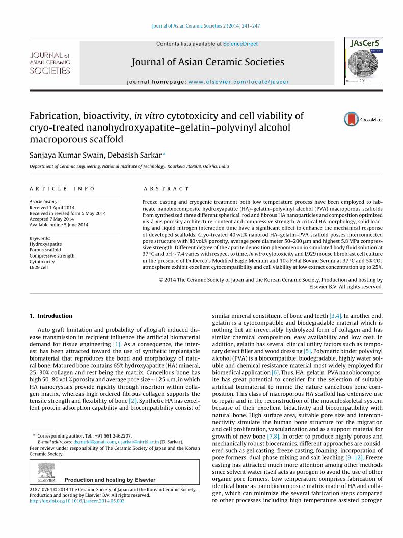
Phase content, purity and crystallinity of the synthesized HAanoparticles are evaluated through XRD patterns and represented
n Fig. 1. XRD pattern of NHS and NHR reveals the formation of HAure phase with semicrystalline behavior at 25 ◦C, whereas NHFhows high crystallinity after synthesis at relatively high 80 ◦C tem-
erature. The diffraction peaks for all HA nanoparticles confirmhe hexagonal structure of HA. Low temperature synthesized HAanoparticle appears without any secondary phase as tricalciumhosphates. The results reveal that the crystallinity of HA phase
Fig. 2. HRTEM micrographs of synthesized HA na
Fig. 1. XRD pattern of synthesized HA nanoparticles (A) NHS, (B) NHR and (C) NHF.
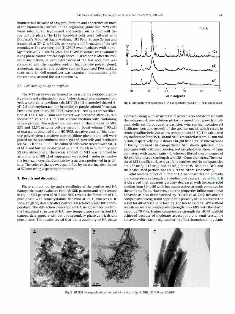
increases along with an increase in aspect ratio and decrease withthe solution pH. Low solution pH favors anisotropic growth of cal-cium deficient fibrous apatite particles, whereas high solution pHfacilitates isotropic growth of the apatite nuclei which result insemicrystalline behavior at low temperature (25 ◦C). The calculatedcrystallite size for NHS, NHR and NHF is recorded as 9 nm, 12 nm and60 nm, respectively. Fig. 2 shows a bright field HRTEM micrographsof the synthesized HA nanoparticles. NHS shows spherical mor-phologies with ∼20 nm diameter, rod morphologies show ∼15 nmdiameters with aspect ratio ∼5, whereas fibroid morphologies ofHA exhibits micron size length with 30–40 nm diameters. The mea-sured BET specific surface area of the synthesized HA nanoparticlesare 256 m2/g, 217 m2/g and 47 m2/g for NHS, NHR and NHF andtheir calculated particle size are 7, 9 and 70 nm, respectively.
Solid loading effect of different HA nanoparticles on porosityand compressive strength are studied and represented in Fig. 3. Itis observed that apparent porosity decreases with increase solidloading from 30 to 50 wt.%, but compressive strength enhances forthe same scaffolds. However, both the properties follow non-linearbehavior as also demonstrated by Fritsch et al. [22]. Reasonablecompressive strength and appropriate porosity of the scaffold is theresult for 40 wt.% HA solid loading. The freeze casted HGPR scaffoldreveals an average compressive strength of ∼2 MPa with the elastic
modulus 70 MPa. Higher compressive strength for HGPR scaffoldachieved because of moderate aspect ratio and semi-crystallinebehavior, which favors high anchoring effect throughout the gelatin
noparticles (A) NHS, (B) NHR and (C) NHF.
244 S.K. Swain, D. Sarkar / Journal of Asian Ceramic Societies 2 (2014) 241–247
r (A) H
mm
opfipaasHeo∼btpptlcasdt(msir
folds under compression are represented in Fig. 6. The property ofthe cryo-treated HA–gelatin–PVA porous scaffolds and their detailmechanical behavior is shown in Table 1. Under identical loading
Table 1Physical properties of cryo-treated HA–gelatin–PVA scaffold.
Sample ID Porosity (%) Compressive Elastic Pore size
Fig. 3. Variation of porosity and compressive strength fo
atrix. This optimized scaffold has been considered to enhanceechanical strength through cryo-treatment.SEM microstructure of 5 h cryo-treated HA–gelatin scaffolds for
ptimum 40 wt.% solid loading is represented in Fig. 4. Adequateorosity, uniform pore size and reasonable strength are the keyeatures for the selection of such a scaffold. HGPS05 scaffold resultsn irregular pore morphologies with partly connected open macro-orous structure and average pore diameter in the range ∼170 �ms illustrated in Fig. 4A. Open macroporous architecture with aver-ge pore diameter in the range ∼160 �m is observed for the HGPR05caffold (Fig. 4B). Fig. 4C represents the SEM microstructure ofGPF05 scaffold. HGPF05 scaffold has relatively larger pore diam-ter than the counterpart HGPR05 and HGPS05 scaffolds. Ellipticalpen macropores with average pore diameter observe in the range180 �m. HGPR05 scaffold demonstrates well pore size distri-ution with the reticulated open porous structure as comparedo HGPS05 and HGPF05, which is further confirmed by mercuryorosimeter analysis. Most of the pores are open macroporous andreferably have irregular elliptical shape. Different pore size, con-ent and morphologies are influenced by the HA surface area, solidoading and PVA molecular interaction phenomena during freezeasting process as well as cryo-treatment. Pore size distributionfter 5 h cryo-treated scaffold is analyzed through mercury intru-ion porosimetry, as demonstrated in Fig. 5. Mono-mode pore sizeistribution is observed for HGPS05 with average pore diameter inhe range ∼90 �m and some micro pores in the range ∼1–10 �mFig. 5A). Porosity of HGPS05 scaffold is measured 80 vol.% through
ercury intrusion porosimeter. Fig. 5B exhibits multimodal poreize distribution for HGPR05 scaffold with average pore diametern the range ∼85 �m, 78 vol.% porosity and micron size pores in theange ∼0.1–1 �m. The existence of micron size pore distribution is
GPS, (B) HGPR and (C) HGPF with respect to HA content.
attributed to the growth of tiny ice crystals while freezing process[23]. Fig. 5C illustrates the similar pore size distribution patternfor HGPF05 scaffold. The average pore diameter is observed in therange of ∼100 �m along with porosity of around 85 vol.%. Differentgradation of micro pore is observed in the range of ∼0.1–10 �m.Strong anchoring of HA particles in the gelatin matrix develops icecrystals during freeze casting followed by cryo-treatment that facil-itates diverse pore size distribution as revealed by porosimetry andSEM microstructure. Tiny closed pores are difficult to encounterthrough SEM microstructure, but mercury porosimeter evaluatesall range of open and closed pores diameter (0.1–200 �m) andcumulative pore size shifted to the lower region. Multimodal poresize along with variation in pore diameter may suitable for the pro-liferation of osteoblast and mesenchymal stem cell as well as theeasy passage of nutrients through the pores.
The optimum composition of HA–gelatin–PVA scaffold at liq-uid nitrogen environment shows the higher mechanical propertiescompared with the untreated HA scaffold. Stress–strain behavior of40 wt.% solid content after 5 h cryo-treated HA–gelatin–PVA scaf-
S.K. Swain, D. Sarkar / Journal of Asian Ceramic Societies 2 (2014) 241–247 245
F4
c5atw[hsps
face as compared to the dense surface because of their high surfacearea. Aforementioned data illustrate that the apatite preferentiallydeposits along the macroporous architecture of the scaffold.
ig. 4. SEM microstructure of scaffold (A) HGPS05, (B) HGPR05 and (C) HGPF05 for0 wt.% solid loading.
ondition, the cryo-treated HA–gelatin–PVA scaffold exhibits 5.2,.8 and 4.7 MPa stress and corresponding modulus are 155, 202nd 167 MPa for HGPS05, HGPR05 and HGPF05 scaffold, respec-ively. The rate of stain 5% is observed in HGPS05 and 8% in HGPR05,hereas 12% strain is calculated in HGPF05 scaffold. Asefnejad et al.
as 50–200 �m pore size, 80 vol.% porosity and 0.6 MPa compres-ive strength [24]. In another study, Isikli et al. [25] reportedorous chitosan–gelatin–HA scaffold have moderate compressivetrength ∼ 3 MPa and such scaffold also support attachment and
Fig. 5. Mercury intrusion pore size distribution of HA–gelatin–PVA scaffolds (A)HGPS05, (B) HGPR05 and (C) HGPF05 for 40 wt.% solid loading.
proliferation of bone cells. Low strain rate and high compressivestrength reveal that scaffold develops relatively ductile to brit-tle transition. Organic polymer matrix of the scaffold under goesductile behavior to the brittle nature through the development ofinternal stress which increases the compressive strength at thecryogenic temperature (77 K).
In vitro bioactivity of the HGPR05 scaffolds in SBF solution isevaluated and represented in Fig. 7. Fig. 7A illustrates the nucle-ated spherical apatite layer on the HGPR05 scaffold surface at37 ◦C and pH ∼ 7.4 after one day incubation. Fig. 7B represents theSEM microstructure after 3 days incubation of HGPR05 scaffoldin SBF solution. However, minimum seven days are required forthe complete deposition of near spherical apatite particles withoutexistence of any bare scaffold surface as demonstrated in Fig. 7C.The mechanism of the apatite layer formation on the HA scaffoldsurface influence by the surface charge of the HA scaffold thatabsorbs Ca2+ and PO4
3− from the metastable SBF solution and formsamorphous apatite layer [26]. In addition, the nucleated spheri-cal shaped apatite layer on the surface of the HA scaffold in SBF isalso confirmed through EDAX. EDAX images show the presence ofprime elements such as Ca, P, O and C along with some trace ele-ments of Mg and Na from SBF solution. Combined SEM and EDAXsupports the bioaccessibility of such macroporous HA–gelatin–PVAscaffolds. The deposition of apatite layer begins on the porous sur-
Fig. 6. Stress–strain behavior of the 5 h cryo-treated HA–gelatin–PVA scaffolds.
246 S.K. Swain, D. Sarkar / Journal of Asian Ceramic Societies 2 (2014) 241–247
olutio
vta
F
Fig. 7. Apatite nucleation on the HGPR05 scaffold in SBF s
The cell adhesion/proliferation, L929 mouse fibroblast cellsiability on HGPR05 scaffolds is investigated using phase con-rast microscopy and MTT analysis, respectively. The results arelso compared with positive control and negative control. Some
ig. 8. Phase contrast microscopic image revealing the adhesion of cultured L929 cells aft
n associated with EDX (A) 1 day, (B) 3 days and (C) 7 days.
representative microscopic images of cultured L929 cells are shownin Fig. 8. The cell density on the negative control (high densitypolyethylene) is very similar to the fibroblast-like morphologywith cell-to-cell contacts and filopodia extension. In contrast, the
er one day of incubation: (A) negative control, (B) positive control and (C) HGPR05.
S.K. Swain, D. Sarkar / Journal of Asian Ce
Lrd(tsorotLptcHcctmsnatcmoLpm
4
mdp
[
[
[[[
[[
[[[
[[[
[[
[[26] H.M. Kim, T. Himeno, T. Kokubo and T. Nakamura, Biomaterials, 26, 4366–4373
(2005).
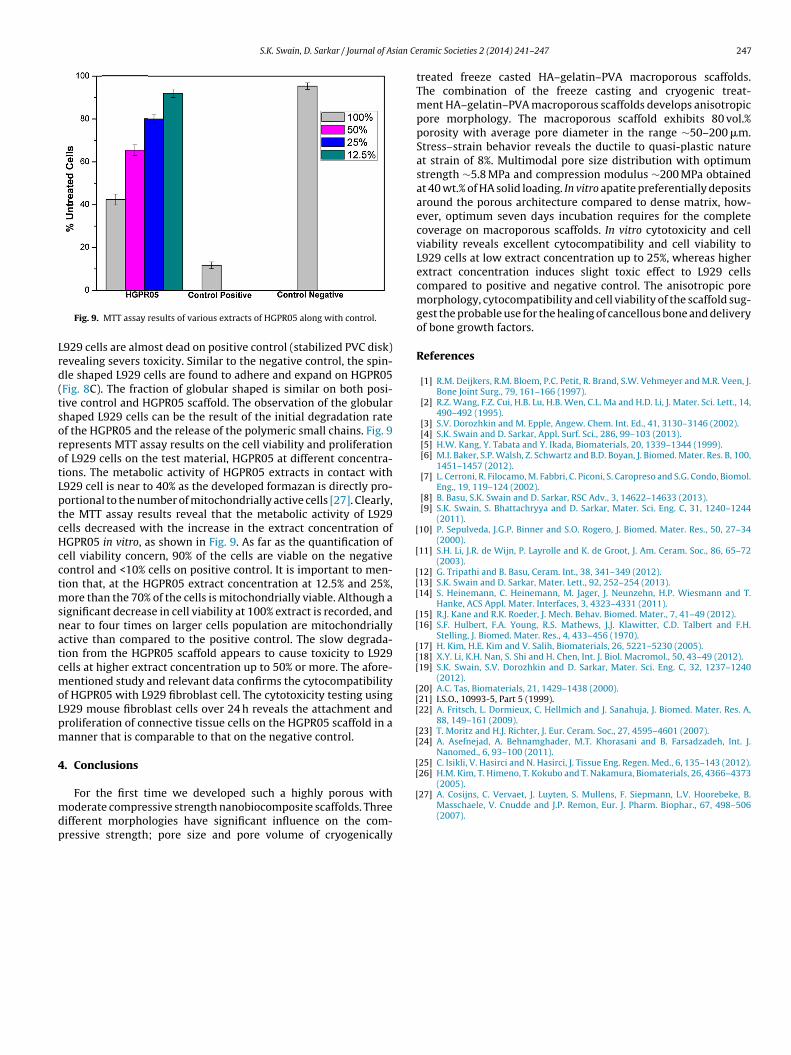
Fig. 9. MTT assay results of various extracts of HGPR05 along with control.
929 cells are almost dead on positive control (stabilized PVC disk)evealing severs toxicity. Similar to the negative control, the spin-le shaped L929 cells are found to adhere and expand on HGPR05Fig. 8C). The fraction of globular shaped is similar on both posi-ive control and HGPR05 scaffold. The observation of the globularhaped L929 cells can be the result of the initial degradation ratef the HGPR05 and the release of the polymeric small chains. Fig. 9epresents MTT assay results on the cell viability and proliferationf L929 cells on the test material, HGPR05 at different concentra-ions. The metabolic activity of HGPR05 extracts in contact with929 cell is near to 40% as the developed formazan is directly pro-ortional to the number of mitochondrially active cells [27]. Clearly,he MTT assay results reveal that the metabolic activity of L929ells decreased with the increase in the extract concentration ofGPR05 in vitro, as shown in Fig. 9. As far as the quantification ofell viability concern, 90% of the cells are viable on the negativeontrol and <10% cells on positive control. It is important to men-ion that, at the HGPR05 extract concentration at 12.5% and 25%,
ore than the 70% of the cells is mitochondrially viable. Although aignificant decrease in cell viability at 100% extract is recorded, andear to four times on larger cells population are mitochondriallyctive than compared to the positive control. The slow degrada-ion from the HGPR05 scaffold appears to cause toxicity to L929ells at higher extract concentration up to 50% or more. The afore-entioned study and relevant data confirms the cytocompatibility
f HGPR05 with L929 fibroblast cell. The cytotoxicity testing using929 mouse fibroblast cells over 24 h reveals the attachment androliferation of connective tissue cells on the HGPR05 scaffold in aanner that is comparable to that on the negative control.
. Conclusions
For the first time we developed such a highly porous withoderate compressive strength nanobiocomposite scaffolds. Three
ifferent morphologies have significant influence on the com-ressive strength; pore size and pore volume of cryogenically
[
ramic Societies 2 (2014) 241–247 247
treated freeze casted HA–gelatin–PVA macroporous scaffolds.The combination of the freeze casting and cryogenic treat-ment HA–gelatin–PVA macroporous scaffolds develops anisotropicpore morphology. The macroporous scaffold exhibits 80 vol.%porosity with average pore diameter in the range ∼50–200 �m.Stress–strain behavior reveals the ductile to quasi-plastic natureat strain of 8%. Multimodal pore size distribution with optimumstrength ∼5.8 MPa and compression modulus ∼200 MPa obtainedat 40 wt.% of HA solid loading. In vitro apatite preferentially depositsaround the porous architecture compared to dense matrix, how-ever, optimum seven days incubation requires for the completecoverage on macroporous scaffolds. In vitro cytotoxicity and cellviability reveals excellent cytocompatibility and cell viability toL929 cells at low extract concentration up to 25%, whereas higherextract concentration induces slight toxic effect to L929 cellscompared to positive and negative control. The anisotropic poremorphology, cytocompatibility and cell viability of the scaffold sug-gest the probable use for the healing of cancellous bone and deliveryof bone growth factors.
References
[1] R.M. Deijkers, R.M. Bloem, P.C. Petit, R. Brand, S.W. Vehmeyer and M.R. Veen, J.Bone Joint Surg., 79, 161–166 (1997).
[2] R.Z. Wang, F.Z. Cui, H.B. Lu, H.B. Wen, C.L. Ma and H.D. Li, J. Mater. Sci. Lett., 14,490–492 (1995).
[3] S.V. Dorozhkin and M. Epple, Angew. Chem. Int. Ed., 41, 3130–3146 (2002).[4] S.K. Swain and D. Sarkar, Appl. Surf. Sci., 286, 99–103 (2013).[5] H.W. Kang, Y. Tabata and Y. Ikada, Biomaterials, 20, 1339–1344 (1999).[6] M.I. Baker, S.P. Walsh, Z. Schwartz and B.D. Boyan, J. Biomed. Mater. Res. B, 100,
1451–1457 (2012).[7] L. Cerroni, R. Filocamo, M. Fabbri, C. Piconi, S. Caropreso and S.G. Condo, Biomol.
Eng., 19, 119–124 (2002).[8] B. Basu, S.K. Swain and D. Sarkar, RSC Adv., 3, 14622–14633 (2013).[9] S.K. Swain, S. Bhattachryya and D. Sarkar, Mater. Sci. Eng. C, 31, 1240–1244
(2011).10] P. Sepulveda, J.G.P. Binner and S.O. Rogero, J. Biomed. Mater. Res., 50, 27–34
(2000).11] S.H. Li, J.R. de Wijn, P. Layrolle and K. de Groot, J. Am. Ceram. Soc., 86, 65–72
(2003).12] G. Tripathi and B. Basu, Ceram. Int., 38, 341–349 (2012).13] S.K. Swain and D. Sarkar, Mater. Lett., 92, 252–254 (2013).14] S. Heinemann, C. Heinemann, M. Jager, J. Neunzehn, H.P. Wiesmann and T.
Hanke, ACS Appl. Mater. Interfaces, 3, 4323–4331 (2011).15] R.J. Kane and R.K. Roeder, J. Mech. Behav. Biomed. Mater., 7, 41–49 (2012).16] S.F. Hulbert, F.A. Young, R.S. Mathews, J.J. Klawitter, C.D. Talbert and F.H.
Stelling, J. Biomed. Mater. Res., 4, 433–456 (1970).17] H. Kim, H.E. Kim and V. Salih, Biomaterials, 26, 5221–5230 (2005).18] X.Y. Li, K.H. Nan, S. Shi and H. Chen, Int. J. Biol. Macromol., 50, 43–49 (2012).19] S.K. Swain, S.V. Dorozhkin and D. Sarkar, Mater. Sci. Eng. C, 32, 1237–1240
(2012).20] A.C. Tas, Biomaterials, 21, 1429–1438 (2000).21] I.S.O., 10993-5, Part 5 (1999).22] A. Fritsch, L. Dormieux, C. Hellmich and J. Sanahuja, J. Biomed. Mater. Res. A,
88, 149–161 (2009).23] T. Moritz and H.J. Richter, J. Eur. Ceram. Soc., 27, 4595–4601 (2007).24] A. Asefnejad, A. Behnamghader, M.T. Khorasani and B. Farsadzadeh, Int. J.
Nanomed., 6, 93–100 (2011).25] C. Isikli, V. Hasirci and N. Hasirci, J. Tissue Eng. Regen. Med., 6, 135–143 (2012).
27] A. Cosijns, C. Vervaet, J. Luyten, S. Mullens, F. Siepmann, L.V. Hoorebeke, B.Masschaele, V. Cnudde and J.P. Remon, Eur. J. Pharm. Biophar., 67, 498–506(2007).