Aptamer facilitated purification of functional proteins
Stanislav S. Beloborodov1, Jiayin Bao1, Svetlana M. Krylova, Agnesa Shala-Lawrence,Philip E. Johnson, Sergey N. Krylov⁎
Department of Chemistry and Centre for Research on Biomolecular Interactions, York University, Toronto, Ontario M3J 1P3, Canada
A R T I C L E I N F O
Keywords:Aptamer-facilitated protein purificationProtein activityAlkBMutSDemethylation assayMismatch-binding assay
A B S T R A C T
DNA aptamers are attractive capture probes for affinity chromatography since, in contrast to antibodies, they canbe chemically synthesized and, in contrast to tag-specific capture probes (such as Nickel-NTA or Glutathione),they can be used for purification of proteins free of genetic modifications (such as His or GST tags). Despite theseattractive features of aptamers as capture probes, there are only a few reports on aptamer-based protein pur-ification and none of them includes a test of the purified protein's activity, thus, leaving discouraging doubtsabout method’s ability to purify proteins in their active state. The goal of this work was to prove that aptamerscould facilitate isolation of active proteins. We refined a complete aptamer-based affinity purification procedure,which takes 4 h to complete. We further applied this procedure to purify two recombinant proteins, MutS andAlkB, from bacterial cell culture: 0.21mg of 85%-pure AlkB from 4mL of culture and 0.24mg of 82%-pure MutSfrom 0.5mL of culture. Finally, we proved protein activity by two capillary electrophoresis based assays: anenzymatic assay for AlkB and a DNA-binding assay for MutS. We suggest that in combination with aptamerselection for non-purified protein targets in crude cell lysate, aptamer-based purification provides a means of fastisolation of tag-free recombinant proteins in their native state without the use of antibodies.
1. Introduction
Purification of recombinant proteins is a key procedure in bio-chemical research [1] and development and production of biologics[2]. The gene, which encodes the protein, is cloned and over-expressedin cultured bacterial cells; the cells are lysed to obtain the cell lysateenriched with the target protein [3]. The target protein is then sepa-rated from the cell lysate based on its unique physicochemical prop-erties such as charge [4], size [5], or affinity to a capture probe (such asNickel-NTA or Glutathione) [6,7]. Affinity purification facilitates thehighest degree of purity, as it relies on very selective interaction of thetarget protein with the capture probe. Affinity purification typicallyrequires fusion of the target protein with an affinity tag (such as His orGST tag). An affinity tag may alter protein conformation and, thus,change its physicochemical properties [8], and its activity as a result.Although a tag can be enzymatically removed, the enzyme may cleavefragments of the target protein, rendering it inactive and causing ad-ditional contamination of the protein with tag-removal agents [9].Therefore, purification of some proteins requires tag-less affinity tech-niques.
A commonly used tag-less purification method involves antibodiesas capture probes that can specifically bind the target protein itself.
However, the utility of this method is restricted by the limitations ofantibodies. The development of antibodies is a lengthy and labor-in-tensive process that cannot be conducted in vitro. In addition, antibodiesare very sensitive to temperature changes and undergo denaturationover the time. Hence, tag-less purification method would benefit fromreplacing antibodies with capture probes, which can be easily synthe-sized and are more stable.
Antibodies can be replaced with oligonucleotide aptamers, whichare single-stranded DNA or RNA (ssDNA and ssRNA) that can selec-tively bind their target protein with high affinity and specificity [10].Aptamers are usually selected from random-sequence DNA (or RNA)libraries via the SELEX (Systematic Evolution of Ligands by EXponentialenrichment) process, which involves alternating steps of partitioning ofprotein-DNA complexes from unbound DNA and amplification of theaptamer-enriched DNA library by the polymerase chain reaction [11].Aptamers are more stable than antibodies and can be synthesized invitro. Aptamers can also be synthetically modified with a linker (such asan amine group, thiol group or biotin) [12], which allows their reliablenon-covalent or covalent conjugation with the solid-phase substrate.The idea of aptamer facilitated protein purification was introduced in1999 [13], and since then, a few proof-of-principle studies have beenconducted [14–17]. To the best of our knowledge, protein’s activity was
https://doi.org/10.1016/j.jchromb.2017.12.024Received 31 August 2017; Received in revised form 15 November 2017; Accepted 15 December 2017
⁎ Corresponding author.
1 These authors contributed equally to this work.E-mail address: [email protected] (S.N. Krylov).
not confirmed in any of the published studies. Also, some studies usedaptamers that specifically bind His-tag [18,19] (thus, still requiring anaffinity tag on a target protein) or involved the use of denaturing agentsat the elution step [20] (thus potentially leading to protein inactiva-tion). Therefore, our goal was to develop and validate a protocol foraptamer facilitated purification of tag-less proteins with preserved ac-tivity.
The developed purification procedure is applied to a crude bacterialcell lysate prepared by sonication and follows a general scheme shownin Fig. 1. The biotinylated aptamer is immobilized onto magnetic beadsvia streptavidin-biotin interaction. After immobilization, the beads areincubated with the cell lysate containing the target protein. During theincubation, the target protein binds to the immobilized aptamer. Theunbound components of the lysate are then removed. The bound pro-tein is eluted by a buffer containing a high concentration of a strongelectrolyte (NaCl or MgCl2) with physiological pH at +4 °C (the highionic strength induces reversible structural alteration of DNA aptamerwithout denaturing the target protein). The procedure takes 4 h tocomplete.
The developed procedure was validated with the AlkB [21] andMutS [22] proteins. Both proteins are involved in DNA repair: AlkB(23.9 kDa) catalyzes dealkylation of ssDNA, and MutS (95 kDa) re-cognizes and binds to mismatches in double stranded DNA (dsDNA).Protein activity after purification was confirmed by two capillaryelectrophoresis based assays: an enzymatic assay for AlkB and a DNA-binding assay for MutS. These assays showed that both proteins re-tained their specific activity. Thus, our protocol can be used for isola-tion of the tag-less proteins in their active state and can also be com-bined with a previously-developed approach of aptamer selection fornon-purified protein in a crude cell lysate [16]. This approach involvesmultiple rounds of the following alternating steps: incubation of theDNA library with the target-containing cell lysate, collection of thebound DNA, incubation of the collected DNA with target-free cell lysateand collection of the unbound DNA, followed by the PCR amplificationand purification. The selected polyclonal aptamers can then be used forpurification of the target protein.
2. Materials and methods
2.1. Chemicals and materials
E. coli strain MG1655 was kindly provided by Coli Genetic StockCenter (CT, USA). The pET-24a(+) vector and NovaBlue competentcells were purchased from EMD Millipore (PA, USA). E. coli BL21-Gold(DE3) competent cells were purchased from Agilent Technologies (CA,USA). A biotin labeled anti-AlkB aptamer [23] (biotin-AlkB-aptamer):5′-/5BioTEG/CTC CTC TGA CTG TAA CCA CGT GCC TAG CGT TTC ATTGTC CCT TCT TAT TAG GTG ATA ATA GCA TAG GTA GTC CAG AAG
CC-3′ and AlkB primers: forward 5′-GGTGGTCATATGTTGGATCTGTTTGCC-3′ and reverse 5′-GGTGGTGGATCCTTATTCTTTTTTACCTGC-3′were custom-synthesized by Integrated DNA Technologies (IA, USA).The AlkB methylated substrate TTCmTTTTTTTTTTTT3′-FAM (TTCm)was custom-synthesized by ATDBio (Southampton, UK). The restrictionenzymes NdeI and BamHI, blue prestained protein standard, and T4DNA ligase were purchased from New England Biolabs (MA, USA).
The pETMutS plasmid #13245 was obtained from Addgene (MA,USA). The E. coli Rossetta-gamiTM 2 (DE3) competent cells were pur-chased from EMD Millipore (PA, USA). A biotin labeled anti-MutS ap-tamer [24] (biotin-MutS-aptamer): 5′-/5BioTEG/CTT CTG CCC GCCTCC TTC CTG GTA AAG TCA TTA ATA GGT GTG GGG TGC CGG GCATTT CGG AGA CGA GAT AGG CGG ACA CT -3′ and fluorescently-la-beled dsDNA with a G-T mismatch (dsDNA-GT) with forward sequenceof 5′-CTT CTG CCC GCC TCC TTC CTT CCA ACC TTC ATC GGC CAC CC-3′ (the mismatch is indicated with an underline) were custom-synthe-sized by Integrated DNA Technologies (IA, USA). Streptavidin-coupledmagnetic beads (MagnaBind Streptavidin Beads) were purchased fromThermo Scientific (IL, USA). Mono Q anion exchange column (1mL bedbolume) was purchased from Amersham (Amersham Pharmacia Bio-tech, currently GE Healthcare Life Sciences, IL, USA).
The isopropyl-β-D-thiogalactoside (IPTG) was purchased from GoldBiotechnology (MO, USA). A protease inhibitor cocktail (cOmplete™)was purchased from Roche (CA, USA). A bicinchoninic acid (BCA) assaykit was obtained from Pierce Biotechnology (IL, USA). A fused silicacapillary (75 μm inner diameter, 360 μm outer diameter) was purchasedfrom Molex (AZ, USA). All other chemicals were purchased from Sigma-Aldrich (ON, Canada). All solutions were prepared with deionizedwater filtered through a 0.22 μm Millipore filter (ON, Canada).
2.2. Immobilization of biotinylated aptamer on streptavidin-beads
To immobilize the aptamers on magnetic beads we took 1mL ofstreptavidin-coated magnetic beads (which can bind 2 μg or 8.19 na-nomoles of biotin) and washed them 3 times with deionized water. Thewashed beads were mixed with 0.8mL deionized water and 105 μL of100 μM aptamer-biotin solution. After a 2-h incubation at room tem-perature, the supernatant was removed and the beads were washedwith deionized water three times and then resuspended in 0.5 mL ofdeionized water. The aptamer occupied 92% of the streptavidin mole-cules on the beads, which was determined by absorbance measurementsof the aptamer-containing solution at 260 nm before and after thebinding. In other words, 1 mL of the original beads suspension bound7.53 nanomoles of biotin and aptamer, accordingly. A suspension ofaptamer-free beads was prepared in a way similar to preparation of thesuspension of aptamer-containing beads, but the aptamer solution wasreplaced with water at the mixing step and the final suspension wasmade in 0.5 mL of the resuspension buffer instead of deionized water.
Fig. 1. Schematic illustration of aptamer-facilitated protein purification. Multiple copies of protein-specific ssDNA aptamer are attached to magnetic beads through the streptavidin-biotinbridge. The aptamers serve as capture probes for the target protein in a crude cell lysate. The beads with captured target protein are separated from the lysate by retaining the beads with amagnet and washing out the lysate. Finally, the target protein is eluted with phosphate-buffered saline (PBS) supplemented with an additional strong electrolyte (NaCl or MgCl2) toincrease the ionic strength.
S.S. Beloborodov et al. Journal of Chromatography B 1073 (2018) 201–206
202
2.3. Protein expression and preparation of cell lysate
AlkB was expressed as the following. The AlkB gene from E. colistrain MG1655 was amplified by PCR. The product of PCR amplificationwas then digested with NdeI and BamHI enzymes and cloned into thepET-24a(+) vector. The pET-24a(+)-AlkB vector was then transfectedinto NovaBlue cells, amplified, reextracted and purified by using theGenElute Plasmid Miniprep Kit. The purified plasmid was then trans-fected into E. coli BL21-Gold (DE3) competent cells. The culture wasfirst grown in 25mL Lysogeny Broth (LB) media with 50 μg/mL kana-mycin at 37 °C overnight (at 250 rpm shaking). Then 1mL of the culturewas transferred to 100mL of Terrific Broth (TB) media (with 50 μg/mLkanamycin) and was incubated with shaking at 250 rpm for 6 h at 37 °Cuntil the Optical Density value of 0.8 was achieved at 600 nm (OD600).Then, IPTG was added to 100mL of the cell culture in LB media toachieve 1mM final concentration, and the mixture was shaken over-night under room temperature at 250 rpm. Next, the cells were pelletedby centrifugation at ∼5000× g (Allegra 21R centrifuge with S4180rotor (Beckman Coulter, ON, Canada)) for 30min at 4 °C. The super-natant was removed, and the cells were resuspended in 10mL of theresuspension buffer (100mM 2-(N-morpholino)ethanesulfonic acid(MES), 1 mM Dithiothreitol (DTT) and 19 μL of protease P8340 for10mL solution, pH 5.8), and sonicated on ice with 15-s on/off intervalsfor 7min at 20% of maximum power (Fisher Scientific, SonicDismembrator, Model 500). Next, the mixture was centrifuged for30min at ∼20000× g (Eppendorf 5417R centrifuge with F45-30-11rotor (Fisher scientific, PA, USA)) at 4 °C. The supernatant was col-lected.
MutS was expressed in a similar way to that of AlkB expression[16,25]. The competent E. coli Rossetta-gami™ 2 (DE3) cells weretransformed with the plasmid pETMutS. The transformed cells weregrown in 1 L of LB medium in the presence of 50 μg/mL ampicillin at37 °C with 250 rpm shaking to reach an OD600 value of 0.8. Proteinexpression was induced by adding IPTG to 1mM in the cell culture. Theculture was shaken overnight at room temperature at 250 rpm. Thecells were pelleted by centrifugation at ∼5000× g (Allegra 21R cen-trifuge with S4180 rotor (Beckman Coulter, ON, Canada)) for 1 h at4 °C. The cell pellet was resuspended in 10mL Tris buffer (20mM tris-HCl pH 7.4) that contained a cOmplete protease inhibitor cocktail ta-blet and sonicated on ice with 15-s on and 55-s off intervals for 4min at60% of maximum power of the sonicator. The cell debris was pelletedby centrifugation at 5000× g for 1 h at 4 °C. The supernatant was in-cubated at 70 °C for 45min followed by a new round of centrifugationfor 1 h at 4 °C. For pre-affinity purification, the protein sample wasapplied onto a Mono Q anion exchange column, which was equilibratedwith Tris buffer, and eluted with a linear gradient of 0.1–1.0M NaCl ata flow rate of 1mL/min. The MutS protein was eluted at approximately0.4 M NaCl. Fractions containing MutS protein were pooled and dia-lyzed extensively against Tris buffer by using an Amicon Ultra-15Centrifugal Filter Unit with Ultracel-10 membrane (Millipore, Ireland).
2.4. Protein purification by aptamer-beads
In the case of AlkB purification, 4mL of protein-containing super-natant was mixed with 0.5mL of bead suspension (on ice) and in-cubated at continuous shaking for 2 h at 250 rpm. The beads were re-moved and sequentially incubated for 10min with 1mL of each of thefollowing buffers: resuspension buffer, 0.5 M NaCl solution (in PBS) and1M NaCl solution in PBS. The eluates were analyzed with CE by usingProteomeLab SDS-MW Analysis Kit (Beckman-Coulter) according to themanufacturer’s instructions. The resuspension buffer eluate containedtraces of the cell lysate. The eluate of 0.5M NaCl in PBS contained thelargest amount of pure AlkB and then was subjected to a buffer ex-change with AlkB protein storage buffer (50mM Tris-HCl, 500mMNaCl, 1 mM DTT, 0.02% NaN3) by using Amicon Ultra-0.5 CentrifugalFilter Unit with Ultracel-10 membrane (Millipore, Ireland). The eluate
of 1M NaCl in PBS contained only traces of AlkB and was not used.AlkB-containing cell supernatant was also used for the negative controlexperiments. In these experiments, 8 μL of the supernatant was mixedwith 200 μL of the aptamer-free beads suspension and incubated on icewith continuous shaking at 250 rpm for 2 h. The same was done for 100,50, 25 and 0 μL of the aptamer-free beads suspension with addition ofan appropriate volume of the resuspension buffer to keep the finalvolume of the solution equal to 208 μL. The beads were then removedand the supernatant was analyzed with CE by using ProteomeLab SDS-MW Analysis Kit (Beckman-Coulter) according to the manufacturer’sinstructions.
For purification of MutS protein, 0.5mL of MutS-protein-containingsolution was incubated with 0.5mL of aptamer-bead solution on ice for1 h under gentle agitation. After the incubation, the beads were col-lected and the clear solution was removed. For MutS protein elution,the beads were sequentially incubated (10min each) and eluted with500 μL of 0.25, 0.5, 0.75, 1, 2 and 3M MgCl2 solution at room tem-perature. The fraction which eluted with 2M MgCl2 contained thelargest amount of pure MutS and was further used. The eluted proteinsolutions were dialyzed extensively against Tris buffer (20mM tris-HClpH 7.4) by using Amicon Ultra-15 Centrifugal Filter Unit with Ultracel-10 membrane (Millipore, Ireland). The concentrations of both AlkB andMutS were determined by using the BCA assay with bovine serum al-bumin (BSA) as a standard.
2.5. Capillary electrophoresis
CE experiments were carried out with a P/ACE MDQ instrumentfrom SCIEX (Fullerton, CA, USA). Laser-induced fluorescence (LIF) de-tection with excitation at 488 nm and emission at 520 nm was used forAlkB enzymatic activity assay and dsDNA-MutS binding assay. Light-absorption detection at 214 nm was used for protein molecular weightanalysis in the SDS-gel electrophoresis mode (SDS-MW). New capil-laries were preconditioned by washing them with 10 capillary volumesof methanol. For CE experiments with LIF detection each run was fol-lowed by washing the capillary with 7 capillary volumes of each of100mM HCl, 100mM NaOH, deionized water (dd), and running buffer.Capillary coolant temperature was kept at +25 °C. An electric field of310 V/cm with a positive electrode at the injection end was used to runelectrophoresis. Uncoated fused silica capillaries with a total length of50 cm and a 40-cm distance from the inlet to the detection window andwith inner diameter of 75 μm were used for detection of the outputproducts of the AlkB enzymatic assay. Uncoated fused silica capillariesof a 30-cm total length and a 20-cm distance from the inlet to the de-tection window (50 μm inner diameter) were used for the MutS-DNAmismatch binding assay. In a case of SDS-MW analysis, the total lengthof capillaries was 30 cm (20 cm from the inlet to the detection window)and the inner diameter was 50 μm.
2.6. SDS-PAGE analysis
SDS-PAGE assay was conducted in the freshly made 12% poly-acrylamide gel. Twenty microliters of MutS-containing solutions (celllysate and MutS-containing eluates) were mixed with 80 μL of 4× SDS-PAGE loading buffer (62.5 mM Tris, 10% Glycerol, 0.05% BromphenolBlue, 2% SDS and 5% 2-mercaptoethanol) and 10 μL of each mixturewas injected into the gel-plate vials. SDS-PAGE electrophoresis wasconducted at 200 V for 50min. Coommassie Blue dye was used for vi-sualization of the protein bands.
2.7. AlkB enzymatic activity assay
The demethylation assay was carried out by mixing AlkB enzymewith the fluorescently labeled homopolymeric sequence TTCm at 20and 200 nM final concentrations, respectively, in 50mM Tris-HCl pH7.5 buffer in the presence of 4mM ascorbic acid, 0.16mM α-
S.S. Beloborodov et al. Journal of Chromatography B 1073 (2018) 201–206
203
Ketoglutaric acid (2OG), 0.05mM BSA, 2100 units of catalase and0.08mM Fe (II) sulphate. The aliquots of the mixture (15 μL each) wereadded to 5 μL of 20mM EDTA (iron chelator) at different time intervals(0, 0.5, 1, and 2min) and kept at 70 °C on a hot plate to denature theprotein and stop the reaction. Each sample was then injected into thecapillary by pressure pulse of 0.5 psi (3.45 kPa) for 6 s and analyzedwith CE at an electric field of 300 V/cm. The methylated DNA (un-reacted substrate) and demethylated DNA (product) were separated in aBorax buffer (20mM Borax, 60mM SDS, pH 8.2).
2.8. MutS-DNA mismatch binding assay based on plug–plug kineticcapillary electrophoresis (ppKCE)
A plug of 1 μM fluorescently-labeled dsDNA-GT solution was in-jected into the capillary by a 10 s pressure pulse of 0.5 psi (3.45 kPa).Next, a plug of the separation buffer was injected at 0.5 psi (3.45 kPa)for 30 s. Finally, the plug of 1.5 μM purified MutS protein solution wasinjected at 0.5 psi (3.45 kPa) for 10 s. The ends of the capillary wereinserted into the inlet and outlet reservoirs, filled with the runningbuffer (50mM Tris-HCl and 1mM MgCl2, pH 8.0) and electrophoreticseparation was conducted at an electric field of 300 V/cm. The controlexperiments were done at the same temperatures by using a solution ofdsDNA-GT void of MutS protein.
3. Results and discussion
3.1. AlkB expression and purification by aptamer-beads
The AlkB expression procedure was adapted from a previously de-veloped protocol [25]. The main deviation from the protocol was theuse of TB media instead of LB media. This replacement allowed us toincrease the amount of the expressed protein. The crude AlkB-con-taining cell lysate was analyzed with SDS-MW in CE (Fig. 2, bottomtrace). The AlkB peak was identified according to its molecular weightof 23.9 kDa, using molecular weight markers (Fig. 2, upper trace).Based on the area analysis it was established that AlkB is the mostabundant protein (30% of the total protein yield). By using BCA, it wasfound that the total protein concentration in the lysate was equal to20mg/mL (± 10%) and the concentration of AlkB protein was ap-proximately 6mg/mL. Next, we applied the developed purification
protocol to the crude cell lysate. By using 1mL of aptamer-saturatedbead suspension (containing 7.53 nanomoles of aptamer at 92% occu-pancy) we have obtained 0.21mg of pure AlkB protein in a form ofsolution with the total volume of 70 μL. The purified eluate was ana-lyzed by SDS-MW in CE (Fig. 2, middle trace). Based on the area ana-lysis of peaks in this trace, it was established that the purity of AlkBprotein was more than 85%. In theory, we could obtain only 0.18mg ofthe protein with this amount of the aptamer, but due to non-specificbinding of AlkB to the surface of the beads, we were able to extractmore protein. The non-specific binding was confirmed in a series ofnegative control experiments with aptamer-free beads, which showedthat these beads are capable of binding proteins in the cell lysate, but donot have specificity to any particular protein. According to SDS-MWelectropherograms (not shown), the areas corresponding to all theproteins were gradually decreasing with increasing the number of beadsin the system. It was found that approximately 200 μL of the aptamer-free beads suspension was sufficiant to bind all the proteins in 8 μL ofthe cell lysate.
3.2. AlkB activity assay
To confirm that the aptamer-purified AlkB protein retained its ac-tivity, we conducted a comparative demethylation assay. As a referencefor comparison, we used another AlkB protein, which was purified by apreviously published approach [25], based on cation exchange columnchromatography. The methylated DNA substrate was incubated withAlkB protein in the presence of Fe (II) and 2OG as cofactors. The con-centration of the DNA in a mixture was 10 times higher than the con-centration of the protein. The demethylation reaction was terminated atdifferent time intervals from reaction initiation. Then, CE was used toseparate the methylated DNA substrate from the demethylated DNAproduct. As follows from the electropherograms (Fig. 3) the con-centration of the methylated DNA is decreasing over the time, while theconcentration of demethylated product becomes greater, which in-dicates that both aptamer-purified and cation-exchange chromato-graphy-purified AlkB proteins were active. However, the AlkB that waspurified by the aptamers led to an almost complete demethylation ofthe substrate in 1min (Fig. 3B), while the reference protein demethy-lated less than a half of the substrate during the same time (Fig. 3A).
Fig. 2. CE based SDS-MW analysis of crude lysate and purified AlkBprotein, confirming expression and purity of the product. The traces cor-respond to the following samples (from bottom to top): crude cell lysate,AlkB-containing eluate, and a mixture of molecular weight markers. Thetraces are vertically offset for clarity of presentation.
S.S. Beloborodov et al. Journal of Chromatography B 1073 (2018) 201–206
204
3.3. MutS expression and purification by aptamer-beads
The MutS expression was induced with 1mM IPTG, similar to in-duction of AlkB expression. The cell lysate before and after inductionwas analyzed by slab-gel SDS-PAGE (Fig. 4, lanes b, c). A large amountof undesirable non-target proteins was removed by heating the celllysate at 70 °C for 45min, consequently denaturing those proteins(Fig. 4, lane d); MutS was not affected as it is a temperature-stableprotein. The soluble portion of heat treated lysate was loaded onto ananion exchange column. The anion exchange pre-purification wasperformed in order to minimize the nonspecific interactions betweenMutS and endogenous bacterial DNA (Fig. 4, lane e). The final step ofprotein isolation was done by incubating the MutS-containing eluatewith aptamer-beads on ice for 1 h. The target protein was retained by
aptamers on a surface of the beads, while the solution was removed. Weeluted the bound protein with a high-ionic-strength solution of salt. Wefirst tried the monovalent cation Na+, however, the interaction be-tween MutS and aptamer could not be disrupted even by 3M NaCl. Wethen tested the divalent cation Mg2+ and found that 2M MgCl2 couldelute 82%-pure MutS (based on the color densities of the bands in theSDS-PAGE slab). A total amount of obtained MutS was 0.24mg (ac-cording to the BCA assay).
3.4. MutS-DNA mismatch binding assay
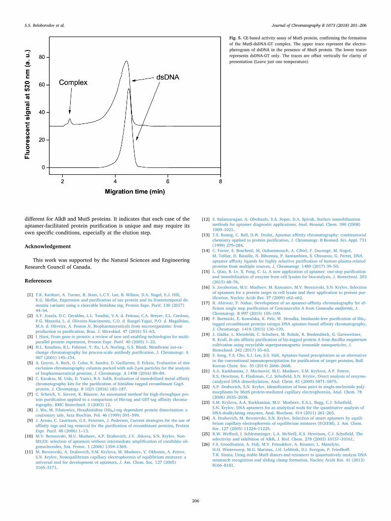
The DNA-binding activity of purified MutS was assessed by appKCE-based binding assay. Short plugs of dsDNA-GT and MutS weresequentially injected one after another by low-pressure pulses. Anelectric field was then applied, causing movement of MutS with a fastervelocity that dsDNA-GT. When passing through dsDNA-GT, MutS couldform a complex with it. As separation continued, the zones of thecomplex and unbound dsDNA-GT and MutS protein were separatedfrom each other (Fig. 5). Based on the area analysis, we found a KD
value of the complex of 326 ± 26 nM at 25 °C. The obtained value iswithin the range of previously reported KD values, lying between35 ± 6 nM (G in the mismatch is surrounded by G fragments only)[26] and 420 ± 30 nM (G in the mismatch is surrounded by AT frag-ments) [26]. Therefore, we conclude that aptamer-purified MutS pro-tein retained its binding activity.
4. Conclusions
To conclude, we have successfully developed and validated an ap-tamer facilitated protein purification protocol. Using AlkB protein andMutS protein as two models, we demonstrated that our approach couldbe used to achieve fast and reliable purification of active proteins. Toconfirm the AlkB activity, we have conducted the DNA demethylationassay and to confirm the functional activity of MutS protein, we haveconducted a CE-based dsDNA-GT binding assay. Both assays showedthat proteins retained their respective activities. Thus, the developedprotocol can be used for purification of tag-less proteins in their activestate. It should also be noted that despite the approximate equality ofthe dissociation constants, Kd, for the utilized AlkB and MutS aptamers(20 and 15 nM, respectively), the optimum elution procedures were
Fig. 3. Activity comparison of the reference AlkB (A) and aptamer-purified AlkB (B). The traces in each panel correspond to differentreaction times ranging from 0 to 2min. The percentage corresponds torelative amount of demethylated product formed by the time the re-action is stopped. The traces are offset vertically for clarity of pre-sentation. The additive concentration of the methylated and de-methylated DNA corresponds to 85 nM on all of theelectropherograms.
Fig. 4. Expression and aptamer facilitated purification of MutS protein. The lanes cor-respond to the following (from left to right): molecular weight markers (a), un-inducedcell culture (b), IPTG-induced cell culture (c), temperature-treated supernatant (d), aneluate after anion exchange column purification (e) and MutS eluted by MgCl2 (f).
S.S. Beloborodov et al. Journal of Chromatography B 1073 (2018) 201–206
205
different for AlkB and MutS proteins. It indicates that each case of theaptamer-facilitated protein purification is unique and may require itsown specific conditions, especially at the elution step.
Acknowledgement
This work was supported by the Natural Sciences and EngineeringResearch Council of Canada.
References
[1] T.K. Karikari, A. Turner, R. Stass, L.C.Y. Lee, B. Wilson, D.A. Nagel, E.J. Hill,K.G. Moffat, Expression and purification of tau protein and its frontotemporal de-mentia variants using a cleavable histidine tag, Protein Expr. Purif. 130 (2017)44–54.
[2] A.F. Jozala, D.C. Geraldes, L.L. Tundisi, V.A. d. Feitosa, C.A. Breyer, S.L. Cardoso,P.G. Mazzola, L. d. Oliveira-Nascimento, C.O. d. Rangel-Yagui, P.O. d. Magalhães,M.A. d. Oliveira, A. Pessoa Jr, Biopharmaceuticals from microorganisms: fromproduction to purification, Braz. J. Microbiol. 47 (2016) 51–63.
[3] I. Hunt, From gene to protein: a review of new and enabling technologies for multi-parallel protein expression, Protein Expr. Purif. 40 (2005) 1–22.
[4] H.L. Knudsen, R.L. Fahrner, Y. Xu, L.A. Norling, G.S. Blank, Membrane ion-ex-change chromatography for process-scale antibody purification, J. Chromatogr. A907 (2001) 145–154.
[5] A. Goyon, A. Beck, O. Colas, K. Sandra, D. Guillarme, S. Fekete, Evaluation of sizeexclusion chromatography columns packed with sub-3 μm particles for the analysisof biopharmaceutical proteins, J. Chromatogr. A 1498 (2016) 80–89.
[6] C. Karakus, M. Uslu, D. Yazici, B.A. Salih, Evaluation of immobilized metal affinitychromatography kits for the purification of histidine-tagged recombinant CagAprotein, J. Chromatogr. B 1021 (2016) 182–187.
[7] C. Scheich, V. Sievert, K. Büssow, An automated method for high-throughput pro-tein purification applied to a comparison of His-tag and GST-tag affinity chroma-tography, BMC Biotechnol. 3 (2003) 12.
[8] J. Wu, M. Filutowicz, Hexahistidine (His6)-tag dependent protein dimerization: acautionary tale, Acta Biochim. Pol. 46 (1999) 591–599.
[9] J. Arnau, C. Lauritzen, G.E. Petersen, J. Pedersen, Current strategies for the use ofaffinity tags and tag removal for the purification of recombinant proteins, ProteinExpr. Purif. 48 (2006) 1–13.
[10] M.V. Berezovski, M.U. Musheev, A.P. Drabovich, J.V. Jitkova, S.N. Krylov, Non-SELEX: selection of aptamers without intermediate amplification of candidate oli-gonucleotides, Nat. Protoc. 1 (2006) 1359–1369.
[11] M. Berezovski, A. Drabovich, S.M. Krylova, M. Musheev, V. Okhonin, A. Petrov,S.N. Krylov, Nonequilibrium capillary electrophoresis of equilibrium mixtures: auniversal tool for development of aptamers, J. Am. Chem. Soc. 127 (2005)3165–3171.
[12] S. Balamurugan, A. Obubuafo, S.A. Soper, D.A. Spivak, Surface immobilizationmethods for aptamer diagnostic applications, Anal. Bioanal. Chem. 390 (2008)1009–1021.
[13] T.S. Romig, C. Bell, D.W. Drolet, Aptamer affinity chromatography: combinatorialchemistry applied to protein purification, J. Chromatogr. B Biomed. Sci. Appl. 731(1999) 275–284.
[14] C. Forier, E. Boschetti, M. Ouhammouch, A. Cibiel, F. Ducongé, M. Nogré,M. Tellier, D. Bataille, N. Bihoreau, P. Santambien, S. Chtourou, G. Perret, DNAaptamer affinity ligands for highly selective purification of human plasma-relatedproteins from multiple sources, J. Chromatogr. 1489 (2017) 39–50.
[15] L. Qiao, B. Lv, X. Feng, C. Li, A new application of aptamer: one-step purificationand immobilization of enzyme from cell lysates for biocatalysis, J. Biotechnol. 203(2015) 68–76.
[16] S. Javaherian, M.U. Musheev, M. Kanoatov, M.V. Berezovski, S.N. Krylov, Selectionof aptamers for a protein target in cell lysate and their application to protein pur-ification, Nucleic Acids Res. 37 (2009) e62–e62.
[17] R. Ahirwar, P. Nahar, Development of an aptamer-affinity chromatography for ef-ficient single step purification of Concanavalin A from Canavalia ensiformis, J.Chromatogr. B 997 (2015) 105–109.
[18] F. Bartnicki, E. Kowalska, K. Pels, W. Strzalka, Imidazole-free purification of His3-tagged recombinant proteins usingss DNA aptamer-based affinity chromatography,J. Chromatogr. 1418 (2015) 130–139.
[19] J. Gädke, L. Kleinfeldt, C. Schubert, M. Rohde, R. Biedendieck, G. Garnweitner,R. Krull, In situ affinity purification of his-tagged protein A from Bacillus megateriumcultivation using recyclable superparamagnetic ironoxide nanoparticles, J.Biotechnol. 242 (2017) 55–63.
[20] S. Song, Y.S. Cho, S.J. Lee, S.S. Hah, Aptamer-based precipitation as an alternativeto the conventional immunoprecipitation for purification of target proteins, Bull.Korean Chem. Soc. 35 (2014) 2666–2668.
[21] A.A. Karkhanina, J. Mecinović, M.U. Musheev, S.M. Krylova, A.P. Petrov,K.S. Hewitson, E. Flashman, C.J. Schofield, S.N. Krylov, Direct analysis of enzyme-catalyzed DNA demethylation, Anal. Chem. 81 (2009) 5871–5875.
[22] A.P. Drabovich, S.N. Krylov, Identification of base pairs in single-nucleotide poly-morphisms by MutS protein-mediated capillary electrophoresis, Anal. Chem. 78(2006) 2035–2038.
[23] S.M. Krylova, A.A. Karkhanina, M.U. Musheev, E.A.L. Bagg, C.J. Schofield,S.N. Krylov, DNA aptamers for as analytical tools for the quantitative analysis ofDNA-dealkylating enzymes, Anal. Biochem. 414 (2011) 261–265.
[24] A. Drabovich, M. Berezovski, S.N. Krylov, Selection of smart aptamers by equili-brium capillary electrophoresis of equilibrium mixtures (ECEEM), J. Am. Chem.Soc. 127 (2005) 11224–11225.
[25] R.W. Welford, I. Schlemminger, L.A. McNeill, K.S. Hewitson, C.J. Schofield, Theselectivity and inhibition of AlkB, J. Biol. Chem. 278 (2003) 10157–10161.
[26] F.S. Groothuizen, A. Fish, M.V. Petoukhov, A. Reumer, L. Manelyte,H.H. Winterwerp, M.G. Marinus, J.H. Lebbink, D.I. Svergun, P. Friedhoff,T.K. Sixma, Using stable MutS dimers and tetramers to quantitatively analyze DNAmismatch recognition and sliding clamp formation, Nucleic Acids Res. 41 (2013)8166–8181.
Fig. 5. CE-based activity assay of MutS protein, confirming the formationof the MutS-dsDNA-GT complex. The upper trace represent the electro-pherogram of dsDNA in the presence of MutS protein. The lower tracesrepresents dsDNA-GT only. The traces are offset vertically for clarity ofpresentation (Leave just one temperature).
S.S. Beloborodov et al. Journal of Chromatography B 1073 (2018) 201–206