*Corresponding author: Norbert Jost, Division of Cardiovascular Pharmacology,Hungarian Academy of Sciences and Department of Pharmacology &Pharmacotherapy, Faculty of Medicine, University of Szeged, Dóm tér 12, P.O. Box 427, H-6701 Szeged, Hungary, Tel: (36-62) 546885; Fax: (36-62) 545680; E-mail:[email protected]
Received August 13, 2013; Accepted October 24, 2013; Published October 28, 2013
Citation: Muntean DM, Kohajda Z, Fazekas T, Jost N (2013) Atrial Remodeling in Permanent Atrial Fibrillation: Mechanisms and Pharmacological Implications. J Clin Exp Cardiolog 4: 273. doi: 10.4172/2155-9880.1000273
AbstractAtrial fibrillation (AF), the most prevalent rhythm disorder in clinical practice, is currently significantly contributing
to morbidity and mortality of the ageing population. In the past decades, a tremendous amount of research resulted in valuable insights into AF pathophysiology, with a primary focus on atrial remodeling. Defined as a persistent change in atrial function and structure, remodeling has the intrinsic properties to enhance the probability of focal (ectopic) and/or re-entrant pursuits, thus supporting AF persistence. The hallmark of structural remodeling is represented by atrial fibrosis, a multifactorial process involving an interaction between neurohormonal and cellular mediators. This paper provides a brief summary of the recent knowledge with respect to electrical and structural remodeling and novel insights into the pathogenesis of atrial fibrosis. Since current drug options for AF treatment are far from being optimal we also discuss the therapeutic principles and current alternatives for counteracting atrial fibrosis, and thus preventing arrhythmia recurrence.
Atrial Remodeling in Permanent Atrial Fibrillation: Mechanisms and Pharmacological ImplicationsDanina M Muntean1, Zsófia Kohajda2,3, Tamás Fazekas4 and Norbert Jost1-3*1Department of Pathophysiology, University of Medicine and Pharmacy, Timisoara, Romania2Division of Cardiovascular Pharmacology, Hungarian Academy of Sciences, Szeged, Hungary3Department of Pharmacology & Pharmacotherapy, Faculty of Medicine, University of Szeged, Hungary4First Department of Internal Medicine, Faculty of Medicine, University of Szeged, Hungary
IntroductionAtrial fibrillation (AF) has a prevalence of 1.5-2% in general
population and represents a major cause of morbidity and a socio-economic burden that is expected to grow worth in coming decades mainly in the developed countries. The arrhythmia is associated with high risk of stroke due to thromboembolism, of congestive heart failure and, also accounts for the highest rate of hospitalization among all types of arrhythmia, especially with advancing age [1].
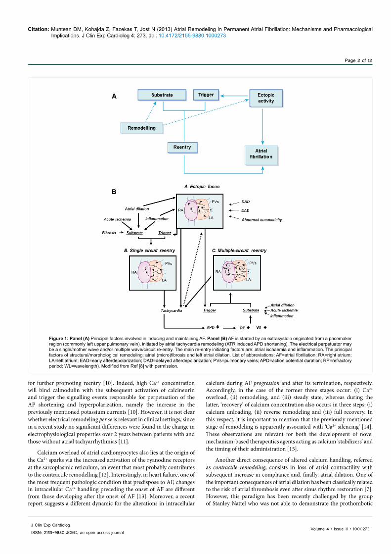
AF is initiated when an atrial ectopic beat encounters during propagation an anatomical and/or functional obstacle and triggers the re-entry of the excitation wavefront. The classic mechanisms underlying the ectopic activity are represented by: (i) increased automaticity and (ii) triggered activity expressed as early (EAD) or delayed afterdepolarizations (DAD). It is nowadays proven that atrial cardiomyocytes in the pulmonary sleeve veins may present increased automaticity/pacemaker activity; they represent ectopic foci responsible for the initiation of single or multiple reentry circuits [2,3]. There are 2 main theories that explain the persistence of AF, namely: (i) the ‘leading circle’ theory implying the existence of single/multiple reentry circuits [2] and, more recently, (ii) the cardiac electric rotors theory [4]. Figure 1 depicts the pathophysiological changes that are involved in the initiation of AF.
Persistent or recurrent AF is constantly associated with the phenomenon of atrial remodeling characterized by electrical and structural changes of cardiomyocytes that are responsible for arrhythmia self-perpetuation and resistance to sinus rhythm conversion. The changes that contribute to atrial remodeling in AF include: (i) alterations of ion channels, gap-junctions, and extracellular matrix and, (ii) neurohumoral dysregulation, in particular of the renin-angiotensin-aldosterone system (RAAS) and the autonomic nervous system.
The paper briefly presents novel insights into the pathophysiology of atrial remodeling with particular emphasis on atrial fibrosis. The
currently available therapeutic options and strategies for developing novel pharmacologic agents capable to prevent/treat atrial remodelling are also discussed.
Mechanisms of Atrial RemodelingRemodeling in AF lies at the very core of the progressive nature
of the arrhythmia. During the past decades the phenomenon has been thoroughly characterized at cellular level with respect of three major components: electrical, contractile and structural remodeling that synergistically contributes to the generation of the vulnerable substrate [5].
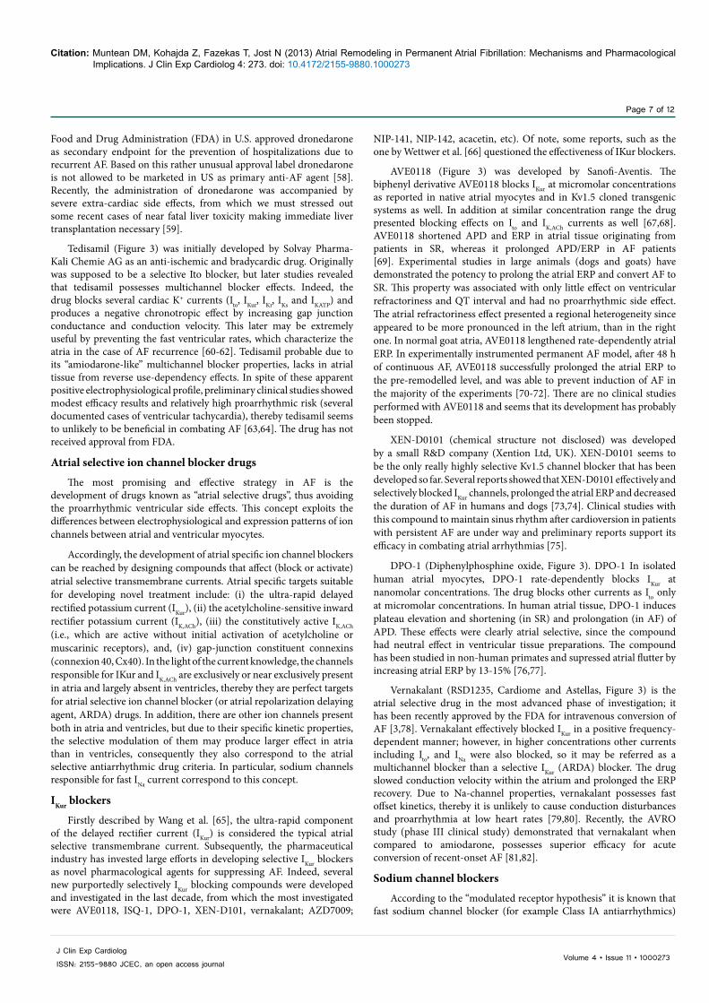
Electrical remodeling is due to alteration of several ion channels with the subsequent shortening of the action potential (AP) and membrane hyperpolarization [6]. The changes are due to the downregulation of the plateau currents and upregulation of several repolarizing currents (Figure 2). In this respect, three major changes have been described in the past decade to underlie the AP triangularization: (i) downregulation of inward current ICa,L, (ii) upregulation of the inward rectifier current IK1, and (iii) activation of IK,Ach, respectively [7-10].
Electrical remodeling (shortening of the atrial refractoriness) is reversible after the sinus rhythm restoration; however, with AF persistence, calcium overload is the main intracellular signal responsible
Journal of Clinical & Experimental CardiologyJo
urna
l of C
linica
l & Experimental Cardiology
ISSN: 2155-9880
Citation: Muntean DM, Kohajda Z, Fazekas T, Jost N (2013) Atrial Remodeling in Permanent Atrial Fibrillation: Mechanisms and Pharmacological Implications. J Clin Exp Cardiolog 4: 273. doi: 10.4172/2155-9880.1000273
Page 2 of 12
Volume 4 • Issue 11 • 1000273J Clin Exp Cardiolog
ISSN: 2155-9880 JCEC, an open access journal
calcium during AF progression and after its termination, respectively. Accordingly, in the case of the former three stages occur: (i) Ca2+ overload, (ii) remodeling, and (iii) steady state, whereas during the latter, ‘recovery’ of calcium concentration also occurs in three steps: (i) calcium unloading, (ii) reverse remodeling and (iii) full recovery. In this respect, it is important to mention that the previously mentioned stage of remodeling is apparently associated with ‘Ca2+ silencing’ [14]. These observations are relevant for both the development of novel mechanism-based therapeutics agents acting as calcium ‘stabilizers’ and the timing of their administration [15].
Another direct consequence of altered calcium handling, referred as contractile remodeling, consists in loss of atrial contractility with subsequent increase in compliance and, finally, atrial dilation. One of the important consequences of atrial dilation has been classically related to the risk of atrial thrombosis even after sinus rhythm restoration [7]. However, this paradigm has been recently challenged by the group of Stanley Nattel who was not able to demonstrate the prothombotic
for further promoting reentry [10]. Indeed, high Ca2+ concentration will bind calmodulin with the subsequent activation of calcineurin and trigger the signalling events responsible for perpetuation of the AP shortening and hyperpolarization, namely the increase in the previously mentioned potassium currents [10]. However, it is not clear whether electrical remodeling per se is relevant in clinical settings, since in a recent study no significant differences were found in the change in electrophysiological properties over 2 years between patients with and those without atrial tachyarrhythmias [11].
Calcium overload of atrial cardiomyocytes also lies at the origin of the Ca2+ sparks via the increased activation of the ryanodine receptors at the sarcoplasmic reticulum, an event that most probably contributes to the contractile remodelling [12]. Interestingly, in heart failure, one of the most frequent pathologic condition that predispose to AF, changes in intracellular Ca2+ handling preceding the onset of AF are different from those developing after the onset of AF [13]. Moreover, a recent report suggests a different dynamic for the alterations in intracellular
Figure 1: Panel (A) Principal factors involved in inducing and maintaining AF. Panel (B) AF is started by an extrasystole originated from a pacemaker region (commonly left upper pulmonary vein), initiated by atrial tachycardia remodeling (ATR induced APD shortening). The electrical perpetuator may be a single/mother wave and/or multiple wave/circuit re-entry. The main re-entry initiating factors are: atrial ischaemia and inflammation. The principal factors of structural/morphological remodeling: atrial (micro)fibrosis and left atrial dilation. List of abbreviations: AF=atrial fibrillation; RA=right atrium; LA=left atrium; EAD=early afterdepolarization; DAD=delayed afterdepolarization; PVs=pulmonary veins; APD=action potential duration; RP=refractory period; WL=wavelength). Modified from Ref [8] with permission.
Citation: Muntean DM, Kohajda Z, Fazekas T, Jost N (2013) Atrial Remodeling in Permanent Atrial Fibrillation: Mechanisms and Pharmacological Implications. J Clin Exp Cardiolog 4: 273. doi: 10.4172/2155-9880.1000273
Page 3 of 12
Volume 4 • Issue 11 • 1000273J Clin Exp Cardiolog
ISSN: 2155-9880 JCEC, an open access journal
effects of AF-associated remodeling in an elegant study performed in canines [16].
Chronic atrial stretch and geometric deformation are the major activators of the signalling pathways leading to cellular hypertrophy and diffuse and patchy interstitial fibrosis [17], collectively termed structural remodeling, the main mechanism responsible for the progression of AF. Structural remodeling refers to both the cellular and non-cellular components of the atrial tissue and results in local conduction heterogeneities that account for AF self-perpetuation. Predictably, structural remodeling will be less reversible as compared to the electrical one. However, it should be mentioned that structural remodeling is also the hallmark of heart failure and of other underlying chronic cardiac pathologies; therefore, the occurrence of AF is favoured in these settings.
In the past few years, with the rapid evolution of research in the field of microRNA (miRNA), a class of small, non-coding RNA molecules that silence gene expression at the post-transcriptional level, a role of miRNA in both AF associated electrical and structural remodeling has been described. Thus, in the case of the former, downregulation of miR-26 was associated with increased density of IK1, whereas upregulation of miR-328 elicited ICa,L reduction in both animal and human atrial samples [18]. As concerning the latter issue, Shan et al. [19] reported, in a dog model of AF induced by nicotine administration and rapid pacing, a profibrotic response characterized by significant upregulation of expression of TGF-beta1 and TGF-betaRII proteins together with a 60-70% reduction of the anti-fibrotic miR-133 and miR-590 levels. In the same line, knockdown of atrial miR-21 has been reported to suppress atrial fibrosis and duration of AF 8 weeks after experimental myocardial infarction in rats [20]. Recently, miR-499 was found to be significantly upregulated in atrial myocardium of 4 subjects with permanent AF, leading to the downregulation of the small-conductance calcium-activated potassium channel 3 (SK3) proteins [21]. Whether this finding might contribute to the electrical remodeling in AF remain to be established. In the same line, it has been recently reported that
miR29 was significantly decreased in patients with chronic heart failure and atrial fibrillation; moreover, miR29b knockdown in canine atrial fibroblasts elicited an increase in colagen expression thus contributing to atrial fibrotic remodeling [22]. Therefore the authors concluded that miR29 may be used in the future as potetential biomarker and/or therapeutic target.
The past decade witnessed a huge resurgence of interest in the role of mitochondria in cardiovascular health and disease. In this line, a recent prospective study reported an association between pre-operative atrial mitochondria dysfunction and the occurrence of AF in patients with metabolic syndrome undergoing CABG surgery [23]. In permeabilized cardiac fibres, these authors demonstrated a decreased respiration of permeabilized fibres in the presence of pyruvate-malate and palmitoyl-L-carnitine (but not of succinate) and an increased sensitivity to calcium overload. We have recently performed a similar study aimed at assessing the respiratory function in permeabilized fibres of human right atrial appendages harvested from patients with coronary heart disease vs. patients with valvular disease and preserved ejection fraction that underwent non-emergency cardiac surgery. Similarly to the previously mentioned data, we also found in coronary patients (but not in valvular ones) a significant decline for the oxidative phosphorylation capacity and respiratory control ratio for mitochondria energized with complex I (but not with complex II) substrates [24]. These observations are in the line with previous reports suggesting that treatments aimed at supporting cardiac mitochondria function might be able to mitigate electrical dysfunction in the heart [25].
Causes and Consequences of Atrial FibrosisIn the past decade the contributing role of the structural remodeling
(for example fibrosis) in initiating and maintaining and of AF has been extensively investigated, with a major emphasis on the occurrence of atrial fibrosis.
Tissue fibrosis represents the common endpoint in diseased hearts from hypertensive, valvular, coronary, diabetic and failing patients.
Figure 2: Major transmembrane ionic currents underlying atrial AP in sinus rhythm (SR) and in AF (ion channel remodeling). Left column presents ionic channel/current densities. Middle and right columns represent that AF induced alterations at the expression level current subunit forming proteins and genes, respectively. Pictograms show the course (magnitude and time duration) of the respective current. Magnitude and time course of the pictogram reflect approximately the real format proportions. Modified from Ref [9] with permission.
Citation: Muntean DM, Kohajda Z, Fazekas T, Jost N (2013) Atrial Remodeling in Permanent Atrial Fibrillation: Mechanisms and Pharmacological Implications. J Clin Exp Cardiolog 4: 273. doi: 10.4172/2155-9880.1000273
Page 4 of 12
Volume 4 • Issue 11 • 1000273J Clin Exp Cardiolog
ISSN: 2155-9880 JCEC, an open access journal
In 2/3 of cases AF is secondary to a pre-existent organic heart disease that contributes to the development of the vulnerable substrate for AF, whereas lone AF occurs in 1/3 of patients. Nevertheless, there is both experimental and clinical evidence that AF itself is able to promote fibrosis [26]. However, the timing of AF appearance is equally important since, in a recent study, prognosis in patients who developed AF before or consecutively with heart failure was less severe as compared with those who firstly developed heart failure [27].
Atrial fibrosis in the setting of AF is the result of complex interplay among profibrotic signalling pathways, inflammation and oxidative stress, the first two contributors being extensively studied. Indeed, profibrotic signalling pathways have been recently described and include the: (i) RAA system with angiotensin II acting on AT-1 receptors and aldosterone acting on the mineralocorticoid receptors, promoting both atrial and ventricular fibrosis, and the (ii) TGF-beta1 that stimulates collagen production via the SMAD pathway. Also, AF has been associated with high levels of inflammatory serum biomarkers and a positive effect of anti-inflammatory agents [28]. However, in an important population based case-control study, the use of non-aspirin NSAIDs has been reported to increase the relative risk of AF or flutter [29]. Atrial fibrosis results in electrical dissociation between adjacent muscle bundles with discontinuous transverse conduction and also,
between the epicardial layer and endocardial bundles leading to both focal and macro-reentry [30].
We will briefly mention some novel insights related to the role of oxidative stress in AF pathophysiology, since increased reactive oxygen species (ROS) production occurs with ageing as it does the AF prevalence. The major sources of ROS in the atria are: NADPH oxidases (NOX 1, 2 and 4), the electron transport chain (ETC), uncoupled NO synthase (NOS), and in some extent myeloperoxidase (MPO) [31,32]. Interestingly, the participation of ROS sources varies with the progression of the disease, an observation with important therapeutic implications. Thus, in both animals and humans, superoxide anion was reported to be NADPH oxidase (NOX2) dependent at 2 weeks of AF and NOS- and mitochondria-dependent after 6 months of AF [33]. These authors speculated that the early but transient increase of atrial NADPH oxidases may explain why statins (that inhibit NOX2) prevent postoperative AF but are less effective in the secondary prevention of the arrhythmia [33].
A second major source of superoxide anion which generates the highly reactive peroxynitrite is represented by NO synthases [34]. In a canine model of AF induced by seven days of tachypacing, induction of NOS isoform 2 (NOS2) has been reported [31].
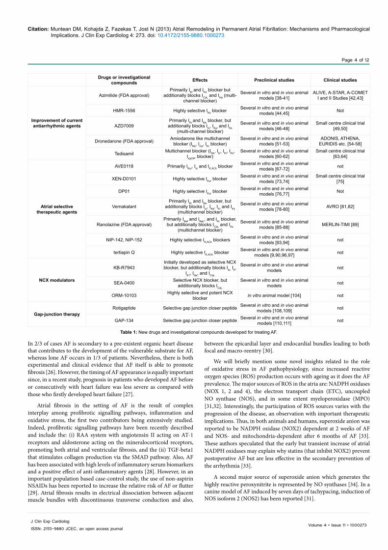
Drugs or investigational compounds Effects Preclinical studies Clinical studies
Improvement of current antiarrhythmic agents
Azimilide (FDA approval)Primarily IKr and IKs blocker but
additionally blocks ICaL and INa (multi-channel blocker)
Several in vitro and in vivo animal models [38-41]
ALIVE, A-STAR, A-COMET I and II Studies [42,43]
HMR-1556 Highly selective IKs blocker Several in vitro and in vivo animal models [44,45] Not
AZD7009Primarily IKr and INa blocker, but additionally blocks Ito, IKur and IKs
(multi-channel blocker)
Several in vitro and in vivo animal models [46-48]
Several in vitro and in vivo animal models [60-62]
Small centre clinical trial [63,64]
Atrial selective therapeutic agents
AVE0118 Primarily IKur, Ito and IK,ACh blocker Several in vitro and in vivo animal models [67-72] not
XEN-D0101 Highly selective IKur blocker Several in vitro and in vivo animal models [73,74]
Small centre clinical trial [75]
DP01 Highly selective IKur blocker Several in vitro and in vivo animal models [76,77] Not
VernakalantPrimarily IKr and INa blocker, but
additionally blocks Ito, INa, IKr and IKs (multichannel blocker)
Several in vitro and in vivo animal models [78-80] AVRO [81,82]
Ranolazine (FDA approval)Primarily INaf and INaL, and IKr blocker,
but additionally blocks ICaL and IKs (multichannel blocker)
Several in vitro and in vivo animal models [85-88] MERLIN-TIMI [89]
NIP-142, NIP-152 Highly selective IK,ACh blockers Several in vitro and in vivo animal models [93,94] not
tertiapin Q Highly selective IK,ACh blocker Several in vitro and in vivo animal models [9,90,96,97] not
NCX modulators
KB-R7943Initially developed as selective NCX blocker, but additionally blocks Ito, IK,
IK1, INa, and ICaL
Several in vitro and in vivo animal models not
SEA-0400 Selective NCX blocker, but additionally blocks ICaL
Several in vitro and in vivo animal models not
ORM-10103 Highly selective and potent NCX blocker in vitro animal model [104] not
Gap-junction therapyRotigaptide Selective gap junction closer peptide Several in vitro and in vivo animal
models [108,109] not
GAP-134 Selective gap junction closer peptide Several in vitro and in vivo animal models [110,111] not
Table 1: New drugs and investigational compounds developed for treating AF.
Citation: Muntean DM, Kohajda Z, Fazekas T, Jost N (2013) Atrial Remodeling in Permanent Atrial Fibrillation: Mechanisms and Pharmacological Implications. J Clin Exp Cardiolog 4: 273. doi: 10.4172/2155-9880.1000273
Page 5 of 12
Volume 4 • Issue 11 • 1000273J Clin Exp Cardiolog
ISSN: 2155-9880 JCEC, an open access journal
A third important source of ROS is the ETC at the inner mitochondrial membrane. In a mouse model of cardiac renin-angiotensin system activation (ACE8/8) presenting with a high rate of spontaneous ventricular tachycardia and reduction in connexin-43 level, administration of a mitochondrial antioxidant (MitoTempo) significantly decreased ventricular tachycardia inducibility, diminished elevated mitochondrial ROS, and increased connexin-43 and conduction at the gap junctions [35].
As for MPO, when released from polymorphs, it generates hypochlorous acid that further contributes to the tissue fibrosis via the activation of matrix metalloproteinases [31].
ROS elevation aggravates AF evolution by affecting ionic currents, gap junctions, inflammation and fibrosis (see [32] for a recent review); therefore, targeting oxidative stress is nowadays regarded as a potential “upstream therapy” [34], especially when considering the fact that many of the current pharmacologic therapies lack atrial specificity
or/and are proarrhythmic. Indeed, as recently suggested, primary prevention of AF and postoperative AF may benefit from NADPH oxidases inhibitors, whereas in persistent AF mitochondria-targeted antioxidants may prove to be most effective [32].
Modalities for Prevention and Therapy of Atrial Remodeling
Restoration of sinus rhythm versus rate control
The rhythm control is the optimal therapeutic option and intervention to supress atrial fibrillation, i.e. to re-establish the normal sinus rhythm (SR). The rate control can be reached by several therapeutic approaches including to prolong atrioventricular nodal refractoriness and/or to slow of AV node conduction. These can be assessed by application of various types and classes of antiarrhythmic drugs from which we highlight especially the β-blockers, the Ca2+-channel blockers or the golden standard, the amiodarone [36]. It was demonstrated that ectopic
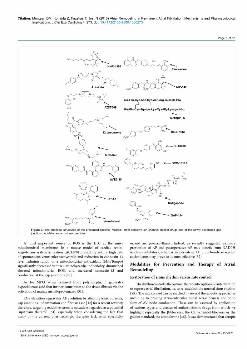
Figure 3: The chemical structures of the presented specific, multiple, atrial selective ion channel blocker drugs and of the newly developed gap-junction modulator antiarrhythmic peptides.
Citation: Muntean DM, Kohajda Z, Fazekas T, Jost N (2013) Atrial Remodeling in Permanent Atrial Fibrillation: Mechanisms and Pharmacological Implications. J Clin Exp Cardiolog 4: 273. doi: 10.4172/2155-9880.1000273
Page 6 of 12
Volume 4 • Issue 11 • 1000273J Clin Exp Cardiolog
ISSN: 2155-9880 JCEC, an open access journal
triggers originating from pulmonary veins are sources of extrasystoles and, consequently, of AF. Eliminating the excitability of these ectopic triggers can terminate AF and, hence, provide rhythm control. This goal can be reached by several classical antiarrhythmic drugs which include especially the Na+ channel blockers or by multiple ion channel blockers (for example amiodarone). Based on the commonly accepted wavelet concept [2], short effective refractory periods and slowed conduction will enhance the probability of inducing re-entries. According to this theory, by counteracting either of them the conduction in the respective tissue will be enhanced (consequently that re-entrant wavefront will find the tissue still in refractory status).
Novel pharmacological agents and investigational compounds surmounting AF
The palette of currently existing antiarrhythmic agents for the treatment of AF are in principle far from being ideal, since, many of them have serious problems regarding not only their efficacy but also safety concerns. In general, all antiarrhythmic drugs combating ventricular arrhythmias may successfully suppress AF by lengthening atrial ERP and by of slowing atrial tissue conduction, but it is expected that their atrial selectivity to minimize ventricular proarrhythmic effects. This was referred as the ‘atrial selective drug concept’, which means these drugs are expected to target currents existing in the atria but not or only minimal the ones present in the ventricles. In addition these drugs must lack cardiac or extra cardiac organ toxicity and should be tolerable in patients having heart complications as for example coronary artery disease.
There are several possibilities to design drugs for combating AF. The novel compounds can block: (i) specific or multiple ion channels, (ii) preferably in an atrial-selective manner, (iii) they can be directed at non-ion channel targets including upstream inflammatory or infiltrative processes or (iv) they may influence gap-junctions (the latter being considered the most modern pharmacological therapeutic approach of AF). We will briefly address further all these issues. At the end of this section Table 1 summarizes again all important data of the presented drugs.
Drugs with specific and multiple ion channel blocking effects
The principal target for these drugs is to lengthen repolarization (i.e., class III compounds). During last two decades there were reported several novel compounds or investigational drugs; however, majority of them have been abandoned, because of the risk of ventricular proarrhythmic potency, especially of inducing Torsades de Pointes type tachyarrhythmias.
Selective IKs blockers
Azimilide (Procter & Gamble, specific IKr and IKs blocker, Figure 3). The drug was designed based on an elegant developing idea from the mid-nineties (so called Sanguinetti’s hypothesis [37]). Accordingly, it was presumed that IKs blockers would be free of reverse rate-dependency. However, the data showed that azimilide blocked not only IKs but IKr as well. Based on these results it was expected to be specifically effective during tachycardia which commonly associates with AF. Some reports showed that azimilide, like amiodarone, possesses calcium and use-dependent sodium channel blocking effects [38,39]. However, we must emphasize that after some encouraging studies in AF [40,41], the initial optimism disappeared and the modest results of several studies (e.g., the ALIVE study) ended up with the conclusion that azimilide will never become a powerful tool for treating AF [42,43].
HMR-1556 (IKs blocker, Figure 3). HMR-1556 is a pure IKs selective blocker and was designed based on the Sanguinetti’s hypothesis [37], which turned the attention of the pharmaceutical industry to IKs blockers. HMR1556 is the first powerful and highly selective IKs blocker that was tested as novel antiarrhythmic drug, and the results demonstrated that indeed effectively blocks IKs, and only at at higher concentration also inhibits Ito, the sustained outward current Isus, and ICaL currents [44]. In a canine model of vagal AF, HMR1556 prolonged the atrial effective refractory period (AERP) and exerted a modest effect on the duration of induced AF only in the presence of intact β-adrenergic stimulation. However, we must emphasize that in the last years several reports made questionable the efficacy of IKs blockers, especially in certain conditions, such as the long QT syndrome 1 and in other circumstances where repolarization reserve is compromised [45].
AZD7009 (Astra Zeneca, IKr and INa blocker, Figure 3). The pharmacological profile of AZD7009 includes a combined block of IKr and rate-dependent block of INa at micromolar concentrations. In addition later studies revealed that, in higher concentrations, the compound blocked other repolarizing currents such as Ito, IKur and IKs [46,47]. In dog atria, AZD7009 concentration-dependently reduced Vmax and increased APD. In addition, the suppression of Vmax, but not APD prolongation, showed used frequency-dependence also [48]. New clinical trials demonstrated AZD7009 to be successful in SR conversion in persistent atrial fibrillation [49,50]. It is presumable that the latter effect is the result of the favourable ion channel blocker profile of the compound.
Amiodarone-like multichannel blockers
The undoubted success of amiodarone promoted the concept that a simultaneous blockade of several specific inward and outward currents may result in a more favourable electrophysiological profile than that obtained through the use of single channel blockers [51].
Dronedarone (Sanofi Aventis, Figure 3) is so far the most promising recently drug developed based on this idea. The originating point for designing the dronedarone molecule was amiodarone but without containing an iodine. It is generally accepted that the iodine molecule of amiodarone is responsible for the severe extra-cardiac (pulmonary, thyroid, hepatic and ocular) toxicity of amiodarone [51]. The in vitro electrophysiological results showed that dronedarone possesses quite similar acute and chronic electrophysiological effects as known to amiodarone [52,53]. In dog ventricular preparations, dronedarone indeed was multi-channel blocker. Accordingly the drug blocked ICa,L and IKr, and reduced the maximum upstroke velocity in a frequency-dependent manner [51]. Based on these promising preclinical investigations dronedarone has been recommended for combating atrial arrhythmias, including in intensive clinical treatment.
Two large trials, ADONIS and EURIDIS, also showed the superiority of dronedarone over placebo. Accordingly dronedarone did not significantly lengthen the QT interval and had very low ventricular proarrhythmic effects (lack of causing torsades de pointes [54,55] arrhythmias). Later ATHENA (phase III randomised trial) investigators reported also encouraging results, from which we should emphasize the prolonged time to first cardiovascular hospitalization or death from any cause (the primary endpoint) by 24% compared to placebo [56]. Despite of these positive reports, it should be emphasized that dronedarone is still far from being a new golden antiarrhythmic drug. Several clinical reports indicated that the antiarrhythmic potential of the starting molecule amiodarone remains net superior to that of dronedarone [57,58]. After the results of the ATHENA trial, the
Citation: Muntean DM, Kohajda Z, Fazekas T, Jost N (2013) Atrial Remodeling in Permanent Atrial Fibrillation: Mechanisms and Pharmacological Implications. J Clin Exp Cardiolog 4: 273. doi: 10.4172/2155-9880.1000273
Page 7 of 12
Volume 4 • Issue 11 • 1000273J Clin Exp Cardiolog
ISSN: 2155-9880 JCEC, an open access journal
Food and Drug Administration (FDA) in U.S. approved dronedarone as secondary endpoint for the prevention of hospitalizations due to recurrent AF. Based on this rather unusual approval label dronedarone is not allowed to be marketed in US as primary anti-AF agent [58]. Recently, the administration of dronedarone was accompanied by severe extra-cardiac side effects, from which we must stressed out some recent cases of near fatal liver toxicity making immediate liver transplantation necessary [59].
Tedisamil (Figure 3) was initially developed by Solvay Pharma-Kali Chemie AG as an anti-ischemic and bradycardic drug. Originally was supposed to be a selective Ito blocker, but later studies revealed that tedisamil possesses multichannel blocker effects. Indeed, the drug blocks several cardiac K+ currents (Ito, IKur, IKr, IKs and IKATP) and produces a negative chronotropic effect by increasing gap junction conductance and conduction velocity. This later may be extremely useful by preventing the fast ventricular rates, which characterize the atria in the case of AF recurrence [60-62]. Tedisamil probable due to its “amiodarone-like” multichannel blocker properties, lacks in atrial tissue from reverse use-dependency effects. In spite of these apparent positive electrophysiological profile, preliminary clinical studies showed modest efficacy results and relatively high proarrhythmic risk (several documented cases of ventricular tachycardia), thereby tedisamil seems to unlikely to be beneficial in combating AF [63,64]. The drug has not received approval from FDA.
Atrial selective ion channel blocker drugs
The most promising and effective strategy in AF is the development of drugs known as “atrial selective drugs”, thus avoiding the proarrhythmic ventricular side effects. This concept exploits the differences between electrophysiological and expression patterns of ion channels between atrial and ventricular myocytes.
Accordingly, the development of atrial specific ion channel blockers can be reached by designing compounds that affect (block or activate) atrial selective transmembrane currents. Atrial specific targets suitable for developing novel treatment include: (i) the ultra-rapid delayed rectified potassium current (IKur), (ii) the acetylcholine-sensitive inward rectifier potassium current (IK,ACh), (iii) the constitutively active IK,ACh (i.e., which are active without initial activation of acetylcholine or muscarinic receptors), and, (iv) gap-junction constituent connexins (connexion 40, Cx40). In the light of the current knowledge, the channels responsible for IKur and IK,ACh are exclusively or near exclusively present in atria and largely absent in ventricles, thereby they are perfect targets for atrial selective ion channel blocker (or atrial repolarization delaying agent, ARDA) drugs. In addition, there are other ion channels present both in atria and ventricles, but due to their specific kinetic properties, the selective modulation of them may produce larger effect in atria than in ventricles, consequently they also correspond to the atrial selective antiarrhythmic drug criteria. In particular, sodium channels responsible for fast INa current correspond to this concept.
IKur blockers
Firstly described by Wang et al. [65], the ultra-rapid component of the delayed rectifier current (IKur) is considered the typical atrial selective transmembrane current. Subsequently, the pharmaceutical industry has invested large efforts in developing selective IKur blockers as novel pharmacological agents for suppressing AF. Indeed, several new purportedly selectively IKur blocking compounds were developed and investigated in the last decade, from which the most investigated were AVE0118, ISQ-1, DPO-1, XEN-D101, vernakalant; AZD7009;
NIP-141, NIP-142, acacetin, etc). Of note, some reports, such as the one by Wettwer et al. [66] questioned the effectiveness of IKur blockers.
AVE0118 (Figure 3) was developed by Sanofi-Aventis. The biphenyl derivative AVE0118 blocks IKur at micromolar concentrations as reported in native atrial myocytes and in Kv1.5 cloned transgenic systems as well. In addition at similar concentration range the drug presented blocking effects on Ito and IK,ACh currents as well [67,68]. AVE0118 shortened APD and ERP in atrial tissue originating from patients in SR, whereas it prolonged APD/ERP in AF patients [69]. Experimental studies in large animals (dogs and goats) have demonstrated the potency to prolong the atrial ERP and convert AF to SR. This property was associated with only little effect on ventricular refractoriness and QT interval and had no proarrhythmic side effect. The atrial refractoriness effect presented a regional heterogeneity since appeared to be more pronounced in the left atrium, than in the right one. In normal goat atria, AVE0118 lengthened rate-dependently atrial ERP. In experimentally instrumented permanent AF model, after 48 h of continuous AF, AVE0118 successfully prolonged the atrial ERP to the pre-remodelled level, and was able to prevent induction of AF in the majority of the experiments [70-72]. There are no clinical studies performed with AVE0118 and seems that its development has probably been stopped.
XEN-D0101 (chemical structure not disclosed) was developed by a small R&D company (Xention Ltd, UK). XEN-D0101 seems to be the only really highly selective Kv1.5 channel blocker that has been developed so far. Several reports showed that XEN-D0101 effectively and selectively blocked IKur channels, prolonged the atrial ERP and decreased the duration of AF in humans and dogs [73,74]. Clinical studies with this compound to maintain sinus rhythm after cardioversion in patients with persistent AF are under way and preliminary reports support its efficacy in combating atrial arrhythmias [75].
DPO-1 (Diphenylphosphine oxide, Figure 3). DPO-1 In isolated human atrial myocytes, DPO-1 rate-dependently blocks IKur at nanomolar concentrations. The drug blocks other currents as Ito only at micromolar concentrations. In human atrial tissue, DPO-1 induces plateau elevation and shortening (in SR) and prolongation (in AF) of APD. These effects were clearly atrial selective, since the compound had neutral effect in ventricular tissue preparations. The compound has been studied in non-human primates and supressed atrial flutter by increasing atrial ERP by 13-15% [76,77].
Vernakalant (RSD1235, Cardiome and Astellas, Figure 3) is the atrial selective drug in the most advanced phase of investigation; it has been recently approved by the FDA for intravenous conversion of AF [3,78]. Vernakalant effectively blocked IKur in a positive frequency-dependent manner; however, in higher concentrations other currents including Ito, and INa were also blocked, so it may be referred as a multichannel blocker than a selective IKur (ARDA) blocker. The drug slowed conduction velocity within the atrium and prolonged the ERP recovery. Due to Na-channel properties, vernakalant possesses fast offset kinetics, thereby it is unlikely to cause conduction disturbances and proarrhythmia at low heart rates [79,80]. Recently, the AVRO study (phase III clinical study) demonstrated that vernakalant when compared to amiodarone, possesses superior efficacy for acute conversion of recent-onset AF [81,82].
Sodium channel blockers
According to the “modulated receptor hypothesis” it is known that fast sodium channel blocker (for example Class IA antiarrhythmics)
Citation: Muntean DM, Kohajda Z, Fazekas T, Jost N (2013) Atrial Remodeling in Permanent Atrial Fibrillation: Mechanisms and Pharmacological Implications. J Clin Exp Cardiolog 4: 273. doi: 10.4172/2155-9880.1000273
Page 8 of 12
Volume 4 • Issue 11 • 1000273J Clin Exp Cardiolog
ISSN: 2155-9880 JCEC, an open access journal
drugs displays preferential binding to open (activated) or closed (inactivated; resting) states of the Na+-channel [83]. Since fast sodium channel is highly expressed in ventricular tissue, it is not obviously to explain and understand the atrial selectivity properties of Na+ channel blockers. Based on the hypotheses formulated by Antzelevitch and co-workers we may assume that the atrial selectivity of INa blockers is probable due to two important diffences of the atrial tissue in comparison with the ventricles: (i) atrial tissue has a slightly more positive (depolarized) resting potential; (ii) a more negative potential for half-maximum inactivation of INa. Due to more depolarized membrane potential during diastole, unlike in ventricles, fewer Na+ channel will recover faster from inactivation in the atria; consequently INa blockers known to bind more preferentially to the inactivated channel state will exhibit larger Na+ channel blockade in the atria as compared to the ventricles [84].
Ranolazine (Figure 3) was initially developed as an antianginal drug, but was soon recognized also to successfully suppressing ventricular EADs and to reduce transmural dispersion of APD [85]. In vitro electrophysiological investigations revealed that ranolazine blocks primarily late INa but several other currents as IKr, IKs, and even possibly L-type calcium current (ICaL) [86].. Ranolazine was shown to shown to reduce ischemic intracellular sodium and calcium overload, and to effectively suppress triggered activity, such as EADs. These properties were associated with the late INa current inhibiting properties. In order to determine whether INaL blockade may be effective in suppressing arrhythmias, extensive investigations were performed in isolated canine single (ventricular myocytes) and multicellular (wedge) preparations [87]. Ranolazine reduced the transmural dispersion of repolarization, a known proarrhythmic substrate [84]. Recent experiments in canine isolated perfused atrial and ventricular preparations have suggested that ranolazine shows a stronger affinity to atrial sodium channels than that of ventricular ones [88]. However, we must emphasize that clinical studies showed only moderate results when testing the effect of ranolazine in counteracting AF [89].
Atrial acetylcholine-sensitive potassium current (IK,ACh) blockers
Blockade of an another atrial-selective current, the acetylcholine activated inward rectifier K+ current, IK,ACh, is expected to exert an useful effect in vagally induced atrial fibrillation. Since IK,ACh is also absent in ventricles, the IK.ACh blockers, similar to IKur blockers, are also real ARDAs. Numerous investigations reported that activation of IK,ACh by vagal stimulation will parallel shorten atrial ERP and increase the availability of the Na+ channel, thereby can create of re-entry substrate based on the dispersion of atrial repolarisation. At high frequencies characteristic to AF, these reentrys may create rotors, which consequently promote the increased duration of AF episodes [90]. Several investigations reported that vagal activity can contribute to the initiation of paroxysmal AF [91,92], thereby some authors proposed the hypothesis that blockade of parasympathetic activity may have beneficial effects and could help to maintain SR; therefore, selective IK,ACh blockade may be a promising atrial selective therapeutic strategy [90].
NIP-142 (Figure 3) and NIP-151 (chemical structures not disclosed). NIP-142 is a benzopyrane derivative compound, which was synthetized as highly selective blocker of IK,Ach. Accordingly with the hypothesis, investigational results demonstrated that NIP-142 could prevent the acetylcholine-induced, arrhythmogenic AP shortening [93]. The congener derivative NIP-151 seems to be even more potent and more selective compound than NIP-142. A recent investigation reported
performed in dogs, NIP-151, in an atrial selective manner, significantly lengthened atrial ERP and prevented vagally- and aconitine induced AF [94].
Constitutively active IK,ACh channels (CI-IK,ACh)
Recently, it has been shown that in atrial tissue originating from patients in permanent AF, the IK,ACh channels are opened and active without direct ligand stimulation, i.e. they are constitutively active (CC_IKA,CH) [90,95]. In this study it was hypothesised that in long term chronic AF constitutively active IK,ACh current is one of main responsible for APD abbreviation and triangularization, thus making the atria susceptible for reentry based tachyarrhythmias [90]. A logic conclusion of this observation was that selective blockade of CC_IK,ACh current may have potent antiarrhythmic/antifibrillatory effects [93]. However, due to lack of selective CI-IK,ACh antagonists, direct control of this hypothesis is not possible yet. However, in a recent study, this hypothesis was at least questioned. Indeed, in a experimentally induced tachypaced dog model of permanent AF (ATR), we have revealed that the real magnitude of the outward repolarizing CI_IK,Ach does not seem to be large enough to substantially contribute to the atrial ERP shortening and thus to the APD shortening [9,96,97]. Therefore, in this study [9] we have proposed the following new concept: in normal in vivo physiological condition a “background” vagal stimulation is always present, thereby, consequently it is probable that a acetylcholine-dependent IK,ACh exists in atrial myocytes either in SR or AF. In permanent AF the CI-IK,ACh can also be activated, and its effect will add to the vagally stimulated IKA,Ch, so the resulting net outward current may become large enough to contribute to the atrial ERP/APD shortening. Consequently, blockade of this combined basal IK,ACh and CC_IK,ACh current may prevent AF. Indeed, in a series of recent experiments we have clearly demonstrated the presence of CI-IK,ACh in atrial cardiomyocytes isolated from ATR dogs, near of a clear presence of cholinergic activated IK,ACh currents [9]. Selective blockade of combined IK,ACh current with low nanomolar concentrations of Tertiapin Q (Figure 3) successfully prevented experimentally induced AF in conscious ATR dogs [9,96,97]. However, further investigations are performed presently to test this hypothesis.
NCX modulators
The Na+/Ca2+ exchanger current (NCX) exchanges one intracellular Ca2+ ion for three extracellular sodium ions. During rapid atrial rates caused by AF or pacing, the larger entrant intracellular sodium relative to calcium may cause the bidirectional exchanger to work in the reverse mode, consequently elevating the intracellular Ca2+ level and thus contributing to the shortening of the action potential. These may increase the incidence of DAD (elicited by NCX1 activity) that in turn can trigger AF. Therefore, it has been postulated that NCX blockers as useful antiarrhythmic drugs [98,99]. Lack of potent and selective NCX blockers made impossible to unequivocally prove this hypothesis so far. During last decade several NCX blockers were developed and investigated but to date no true potent and selective NCX blocker has been reported.
KB-R7943 (Kanebo, Figure 3) preferentially inhibits the reverse mode of the NCX. Several studies revealed also that this effect was associated by a clear blocking potency on sodium, potassium and calcium channels (Ito, IK, IK1, INa, and ICaL). In anesthetized dogs, KB-R7943 prevented atrial ERP shortening caused by pacing-induced AF [100,101], but obviously this effect cannot be associated strictly to NCX blocking properties of the drug.
SEA0400 (Taisho Pharmaceutical, Figure 3) is another NCX
Citation: Muntean DM, Kohajda Z, Fazekas T, Jost N (2013) Atrial Remodeling in Permanent Atrial Fibrillation: Mechanisms and Pharmacological Implications. J Clin Exp Cardiolog 4: 273. doi: 10.4172/2155-9880.1000273
Page 9 of 12
Volume 4 • Issue 11 • 1000273J Clin Exp Cardiolog
ISSN: 2155-9880 JCEC, an open access journal
inhibitor which is more selective and potent than KB-R7943, and for several years this was the most investigated NCX blocker compound. It was reported that SEA0400 effectively blocks NCX in both forward and reverse mode at micromolar concentrations; however, the drug also effectively suppressed the L-type Ca current (ICaL) at comparable concentrations [102]. In a recent study in humans where SEA0400 has been applied as a tool to identify NCX current, it was reported that the NCX current is significantly upregulated in AF as compared to SR [103]. In spite of many published reports that showed that NCX blockade with SEA0400 may suppress ectopic automaticity in pulmonary veins, we must emphasize that these observations remains questionable due to the lack of trusted selective NCX blocking effect [99].
ORM-10103 (Orion Pharmaceutical, Figure 3) is a novel developed, and purportedly the most selective and highly potent NCX blocker. In a recent study it was reported that ORM-10103 significantly reduced both the inward and outward NCX currents at submicromolar concentration. The drug did not significantly change the main repolarisation K currents (Ito, IKr, IKs, IK1), the L-type Ca2+ current, the Na+/K+ pump or the maximum rate of depolarization (dV/dtmax), indicative of the fast inward Na+ current even at the high concentration of 10 µM ORM-10103, suggesting that, indeed, this is the firstly reported highly selective NCX blocker [104]. The amplitude of pharmacologically induced early and delayed afterdepolarizations were significantly decreased by ORM-10103 (3 and 10 μM) in a concentration-dependent manner [104].
Gap junctions modulators
Electrical and structural remodelling caused by permanent AF involves significant changes in junctions at the atrial intercalated discs. These may include fascia adherens, the desmosomes, and recently, the gap junction proteins (N-cadherin, desmoplakin, and connexins). Cardiac connexins present two major isoforms: one with the molecular weight of 40 and 43 kDa, respectively. Atrial myocardium and conduction system express particularly connexin 40, while connexin 43 is also present in conductive tissue [105]. There are some studies reporting that acute ischemia may close the gap junctions and slow conduction velocity. Therefore, it has been suggested that specific gap junctions’ modulators that prevent closing of cardiac gap junctions may possess antiarrhythmic effect against AF [106,107].
Rotigaptide (GAP-486, ZP123, Figure 3) is the firstly reported substance developed for protecting against the closing of the gap junctions. Rotigaptide is a peptide developed by applying the original antiarrhythmic peptide structure, where the d-isomers have been substituted for l-isomers. In acute coronary artery occlusion, rotigaptide was shown to effectively attenuate the conduction velocity slowing and ventricular arrhythmogenesis, while being neutral in controls [108,109].
GAP 134 (Figure 3) was designed with the specific aim to reduce atrial conduction velocity [110]. However, in a recent investigation performed in the acute dog model of permanent AF the drug did not prove to have either strong antiarrhythmic or AP lengthening potential [110,111]. We may summarize that gap junction modulators may be indeed novel and potentially efficacious pharmacological targets against AF, but several investigations are required to establish their real potential.
Other possible ion channel targets for novel antiarrhythmic drugs
There are several attempts for targeting other ion channels such as the: two pore-domain potassium channels [K2P] [112], transient receptor channels [TRP] [113], mechanosensitive, stretch activated
channels [114], calcium activated K+-channels [115] etc. Based on the information of several investigations in native (especially ventricular) and transgenic systems many of them may be useful targets for antiarrhythmic exploitation; however, until now there are no or very few promising results suggesting that modulators of these channels may provide beneficial effects in preventing AF.
Non ion-channel blockers – upstream therapy of AF
In addition to the ion channel-based AF therapy, there is a rapid development of non ion-channel approaches, aimed at reducing or reversing structural remodeling, inflammation, and oxidative stress injury associated with AF. These approaches are collectively referred to as “upstream therapies” [116,117].
As previously mentioned several pathophysiological processes, such as inflammation and oxidative injury, promote structural remodeling, including fibroblast proliferation, accumulation and/or redistribution of collagen, chamber dilation, and hypertrophy that are associated to AF. Proarrhythmic actions of atrial structural remodeling are generally related to conduction disturbances, which promote re-entrant arrhythmias. Several experimental and clinical studies have shown that the so-called “upstream therapy” drugs affecting structural remodeling, inflammation, and/or oxidative stress, such as angiotensin-converting enzyme inhibitors, angiotensin II receptor blockers, and statins may reduce the occurrence of AF [118,119]; while in contrary, other studies questioned the efficacy of such therapies in AF [120]. However, we must emphasize that numerous clinical trials proved that the use of statins was significantly associated with a decreased risk of atrial fibrillation in patients with sinus rhythm. The highest benefit was seen for the prevention of postoperative atrial fibrillation and in secondary prevention of atrial fibrillation, with a high heterogeneity, explained by differences in statin types, patient populations and surgery types [121,122].
We must also emphasize that the contribution of inflammation and oxidative stress in the development of AF is still not fully understood and varies significantly among different AF pathologies [36,123,124].
ConclusionsThe past decades witnessed an enormous amount of research aimed
at deciphering the mechanisms underlying atrial remodeling in AF and developing methods to counteract it. Both ion channel and non ion-channel therapeutic approaches are envisaged in order to improve the management of AF. A great deal of attention is currently paid to novel antiarrhythmic drugs that possess high affinity for atrial myocardium or target multiple ion channels. Whether the use of these antiarrhythmic agents will translate into improved prognosis of patients with AF remains to be confirmed in larger-scale prospective trials.
Acknowledgement
Supported by grants from the Hungarian Scientific Research Fund (K-82079 and NK-104331), the National Office for Research and Technology-Baross Programmes (REG-DA-09-2-2009-0115-NCXINHIB), the National Development Agency and co-financed by the European Social Fund (TÁMOP-4.2.2A-11/1/KONV-2012-0073 and TÁMOP-4.2.2.A-11/1/KONV-2012-0060), the Hungarian Academy of Sciences, and the National Research Council Project IDEAS - Programme “Exploratory Research Projects” PN-II-ID-PCE-2012-4-0512.
References
1. Camm AJ, Lip GY, De Caterina R, Savelieva I, Atar D, et al. (2012) 2012 focused update of ESC Guidelines for the management of atrial fibrillation: an update of the 2010 guidelines for the management of atrial fibrillation. Developed with the special contribution of the European Heart Rhtyhm Association. Europace 14: 1385-1413.
Citation: Muntean DM, Kohajda Z, Fazekas T, Jost N (2013) Atrial Remodeling in Permanent Atrial Fibrillation: Mechanisms and Pharmacological Implications. J Clin Exp Cardiolog 4: 273. doi: 10.4172/2155-9880.1000273
Page 10 of 12
Volume 4 • Issue 11 • 1000273J Clin Exp Cardiolog
ISSN: 2155-9880 JCEC, an open access journal
2. Allessie MA, Bonke FI, Schopman FJ (1977) Circus movement in rabbit atrial muscle as a mechanism of tachycardia. III. The „Leading Circle“ concept: a new model of circus movement in cardiac tissue without the involvement of an anatomical obstacle. Circ Res 41: 9-18.
3. Ravens U, Poulet C, Wettwer E, Knaut M (2013) Atrial selectivity of antiarrhythmic drugs. J Physiol.
4. Pandit SV, Jalife J (2013) Rotors and the dynamics of cardiac fibrillation. Circ Res 112: 849-862.
5. Dobrev D, Ravens U (2003) Remodeling of cardiomyocyte ion channels in human atrial fibrillation. Basic Res Cardiol 98: 137-148.
6. Nattel S, Maguy A, Le Bouter S, Yeh YH (2007) Arrhythmogenic ion-channel remodeling in the heart: heart failure, myocardial infarction, and atrial fibrillation. Physiol Rev 87: 425-456.
7. Allessie M, Ausma J, Schotten U (2002) Electrical, contractile and structural remodeling during atrial fibrillation. Cardiovasc Res 54: 230-246.
8. Nattel S, Burstein B, Dobrev D (2008) Atrial remodeling and atrial fibrillation: mechanisms and implications. Circ Arrhythm Electrophysiol 1: 62-73.
9. Jost N, Kohajda Z, Kristóf A, Kovacs PP, Husti Z, et al. (2011) Atrial remodeling and novel pharmacological strategies for antiarrhythmic therapy in atrial fibrillation. Current Medicinal Chemistry 18: 3675-3694.
10. Nattel S, Dobrev D (2012) The multidimensional role of calcium in atrial fibrillation pathophysiology: mechanistic insights and therapeutic opportunities. European Heart J 33: 1870-1877.
11. Healey JS, Israel CW, Connoly SJ, Hohnloser SH, Nair GM, et al. (2012) Relevance of electrical remodelling in human atrial fibrillation: results of the asymptomatic atrial fibrillation and stroke evaluation in pacemaker patients and the atrial fibrillation reduction atrial pacing trial mechanisms of atrial fibrillation study. Circ Arrhythm Electrophysiol 5: 626-631.
13. Greiser M, Lederer WJ, Schotten U (2011) Alterations of atrial Ca(2+) handling as a cause and consequence of atrial fibrillation. Cardiovasc Res 89: 722-733.
14. Greiser M, Schotten U (2013) Dynamic remodelling of intracellular Ca(2+) signalling during atrial fibrillation. J Mol Cell Cardiol 58: 134-142.
15. Voigt N, Dobrev D (2012) Cellular and molecular correlates of ectopic activity in patients with atrial fibrillation. Europace 5: v97-v105.
16. Nishida K, Chiba K, Iwasaki YK, Katsouras G, Shi YF, et al. (2012) Atrial fibrillation-associated remodeling does not promote atrial thrombus formation in canine models. Circ Arrhythm Electrophysiol 5: 1168-1175.
17. Schotten U, Neuberger HR, Allessie MA (2003) The role of atrial dilatation in the domestication of atrial fibrillation. Prog Biophys Mol Biol 82: 151-162.
18. Wang Z, Lu Y, Yang B (2011) MicroRNAs and atrial fibrillation: new fundamentals. Cardiovasc Res 89: 710-721.
19. Shan H, Zhang Y, Lu Y, Zhang Y, Pan Z, et al. (2009) Downregulation of miR-133 and miR-590 contributes to nicotine-induced atrial remodeling in canines. Cardiovasc Res 83: 465-472.
20. Cardin S, Guasch E, Luo X, Naud P, Le Quang K, et al. (2012) Role of microRNA-21 in atrial profibrillatory fibrotic remodeling associated with experimental postinfarction heart failure. Circ Arrhythm Electrophysiol 5: 1027-1035.
21. Ling TY, Wang XL, Chai Q, Lau TW, Koestler CM, et al. (2013) Regulation of SK3 channel by microRNA-499–potential role in atrial fibrillation. Heart Rhythm 10: 1001-1019.
22. Dawson K, Wakili R, Ördög B, Clauss S, Chen Y, et al. (2013) Circulation 127: 1466-1475.
23. Montaigne D, Marechal X, Lefebvre P, Modine T, Fayad G, et al. (2013) Mitochondrial dysfunction as an arrhythmogenic substrate: a translational proof-of-concept study in patients with metabolic syndrome developing post-operative atrial fibrillation. J Am Coll Cardiol 62: 1466-1473.
24. Duicu O, Juşcă C, Falniţă L, Mirică S, Maximov D, et al. (2013) Substrate-specific impairment of mitochondrial respiration in permeabilized fibers from patients with coronary heart disease vs. valvular disease. Mol Cell Biochem 379: 229-234.
25. Brown DA, O’Rourke B (2010) Cardiac mitochondria and arrhythmia. Cardiovasc Res 88: 241-249.
26. Burstein B, Nattel S (2008) Atrial fibrosis: mechanisms and clinical relevance in atrial fibrillation. J Am Coll Cardiol 51: 802-809.
27. Smit MD, Moes ML, Maass AH, Achekar ID, Van Geel PP, et al. (2012) The importance whether atrial fibrillation or heart failure develops first. Eur J Heart Fail 14: 1030-1040.
28. Pellman J, Lyon RC, Sheikh F (2010) Extracellular matrix remodeling in atrial fibrosis: mechanisms and implications in atrial fibrillation. J Mol Cell Cardiol 48: 461-467.
29. Schmidt M, Christiansen CF, Mehnert F, Rothman KJ, Sørensen HT (2011) Non-steroidal anti-inflammatory drug use and risk of atrial fibrillation or flutter: population based case-control study. BMJ 343: d3450.
30. Eckstein J, Verheule S, de Groot NM, Allessie M, Schotten U (2008) Mechanisms of perpetuation of atrial fibrillation in chronically dilated atria. Prog Biophys Mol Biol 97: 435-451.
31. Schillinger KJ, Patel VV (2012) Atrial fibrillation in the elderly: the potential contribution of reactive oxygen species. J Geriatr Cardiol 9: 379-388.
32. Sovari AA, Dudley SC (2012) Reactive oxygen species-targeted therapeutic interventions for atrial fibrillation. Front Physiol 3: 311.
33. Reilly SN, Jayaram R, Nahar K, Antoniades C, Verheule S, et al. (2011) Atrial sources of reactive oxygen species vary with the duration and substrate of atrial fibrillation: implications for the antiarrhythmic effect of statins. Circulation 124: 1107-1117.
34. Bonilla IM, Sridhar A, Györke S, Cardounel AJ, Carnes CA (2012) Nitric oxide synthases and atrial fibrillation. Frontiers Physiol 3: 105.
35. Sovari AA, Rutledge CA, Jeong EM, Dolmatova E, Arasu D, et al. (2013) Mitochondria oxidative stress, connexin-43 remodeling and sudden arrhythmic death. Circ Arrhythm Electrophysiol 6: 623-631.
37. Jurkiewicz NK, Sanguinetti MC (1993) Rate-dependent prolongation of cardiac action potentials by a methanesulfonanilide class III antiarrhythmic agent. Specific block of rapidly activating delayed rectifier K+ current by dofetilide. Circ Res 71: 75-83.
38. Salata JJ, Brooks RR (1997) Pharmacology of azimilide dihydrochloride (NE-10064), a class III antiarrhythmic agent. Cardiovasc Drug Rev 15: 137-156.
40. Karam R, Marcello S, Brooks RR, Corey AE, Moore A (1998) Azimilide dihidrochloride, a novel antiarrhythmic agent. Am J Cardiol 81: 40D-46D.
41. Connolly SJ, Schnell DJ, Page RL, Wilkinson WE, Marcello SR, et al. (2001) Dose–response relations of azimilide in the managementof symptomatic, recurrent, atrial fibrillation. Am J Cardiol 88: 974-979.
42. Camm AJ, Pratt CM, Schwartz PJ, Al-Khalidi HR, Spyt M, et al. (2001) Azimilide Post Infarct Survival Evaluation (ALIVE): azimilide does not affect mortality in post-myocardial infarction patients. Circulation 104: 1B.
43. Camm AJ, Pratt CM, Schwartz PJ, Al-Khalidi HR, Spyt MJ, et al. (2004) AzimiLide post Infarct surVival Evaluation (ALIVE) Investigators. Mortality in patients after a recent myocardial infarction: a randomized, placebo-controlled trial of azimilide using heart rate variability for risk stratification. Circulation 109: 990-996.
44. So PP, Backx PH, Dorian P (2008) Slow delayed rectifier K+ current block by HMR 1556 increases dispersion of repolarization and promotes Torsades de Pointes in rabbit ventricles. Br J Pharmacol 155: 1185-1194.
45. Jost N, Virág L, Comtois P, Ordög B, Szuts V, et al. (2013) Ionic mechanisms limiting cardiac repolarization-reserve in humans compared to dogs. Journal of Physiology epub.
46. Goldstein RN, Khrestian C, Carlsson L, Waldo AL (2004) AZD7009: a new antiarrhythmic drug with predominant effects on the atria effectively terminates and prevents reinduction of atrial fibrillation and flutter in the sterile pericarditis model. J Cardiovasc Electrophysiol 15: 1444-1450.
47. Persson F, Carlsson L, Duker G, Jacobson I (2005) Blocking characteristics of hKv1.5 and Kv4.3/hKChIP2.2 after administration of the novel antiarrhythmic compound AZD7009. J Cardiovasc Pharmacol 46: 7-417.
Citation: Muntean DM, Kohajda Z, Fazekas T, Jost N (2013) Atrial Remodeling in Permanent Atrial Fibrillation: Mechanisms and Pharmacological Implications. J Clin Exp Cardiolog 4: 273. doi: 10.4172/2155-9880.1000273
Page 11 of 12
Volume 4 • Issue 11 • 1000273J Clin Exp Cardiolog
ISSN: 2155-9880 JCEC, an open access journal
48. Wu Y, Carlsson L, Liu T, Kowey PR, Yan GX (2005) Assessment of the proarrhythmic potential of the novel antiarrhythmic agent AZD7009 and dofetilide in experimental models of torsades de pointes. J Cardiovasc Electrophysiol 16: 898-904.
49. Crijns HJ, Van Gelder IC, Walfridsson H, Kulakowski P, Rónaszéki A, et al. (2006) Safe and effective conversion of persistent atrial fibrillation to sinus rhythm by intravenous AZD7009. Heart Rhythm 3: 1321-1331.
50. Aunes-Jansson M, Edvardsson N, Stridh M, Sörnmo L, Frison L, et al. (2013) Decrease of the atrial fibrillatory rate, increased organization of the atrial rhythm and termination of atrial fibrillation by AZD7009. J Electrocardiol 46: 36-37.
51. Hondeghem LM, Snyders DJ (1990) Class III antiarrhythmic agents have a lot of potential but a long way to go. Reduced effectiveness and dangers of reverse use dependence. Circulation 81: 686-690.
52. Aimond F, Beck L, Gautier P, Chérif OK, Davy JM, et al. (2000) Cellular and in vivo electrophysiological effects of dronedarone in normal and postmyocardial infarcted rats. J Pharmacol Exp Ther 292: 415-424.
53. Varró A, Takács J, Németh M, Hála O, Virág L, et al. (2001) Electrophysiological effects of dronedarone (SR 33589), a noniodinated amiodarone derivative in the canine heart: comparison with amiodarone. Br J Pharmacol 133: 625-633.
54. Singh BN, Connolly SJ, Crijns HJ, Roy D, Kowey PR, et al. (2007) EURIDIS ADONIS Investigators. Dronedarone for maintenance of sinus rhythm in atrial fibrillation or flutter. N Engl J Med 357: 987-999.
56. Torp-Pedersen C, Crijns HJ, Gaudin C, Page RL, Connolly SJ, et al. (2011) ATHENA Investigators Impact of dronedarone on hospitalization burden in patients with atrial fibrillation: results from the ATHENA study. Europace 13: 1118-1126.
57. Burashnikov A, Belardinelli L, Antzelevitch C (2010) Acute dronedarone is inferior to amiodarone in terminating and preventing atrial fibrillation in canine atria. Heart Rhythm 7: 1273-1279.
58. Franz MR, Singh SN (2010) Amiodarone and dronedarone: the worker bee and the drone? Heart Rhythm 7: 1280-1281.
59. Joghetaei N, Weirich G, Huber W, Büchler P, Estner H (2011) Acute liver failure associated with dronedarone. Circ Arrhythm Electrophysiol 4: 592-593.
60. Beatch GN, Abraham S, MacLeod BA, Yoshida NR, Walker MJA (1991) Antiarrhythmic properties of tedisamil (KC 8857), a putative transient outward K+ current blocker. Br J Pharmacol 102: 13-18.
62. Jost N, Virág L, Hála O, Varró A, Thormählen D, et al. (2004) Effect of the antifibrillatory compound tedisamil (KC-8857) on transmembrane currents in mammalian ventricular myocytes. Curr Med Chem 11: 3219-3228.
63. Fischbach PS, Johnston PV, Friedrichs GS, Lucchesi BR (1999) Tedisamil in a chronic canine model of atrial flutter. J Cardiovasc Pharmacol 34: 212-218.
64. Fischbach PS, Barrett TD, Goyal R, Tran BC, Syed ZA, et al. (2001) Conversion of atrial fibrillation by the experimental antiarrhythmic drug tedisamil in two canine models. J Cardiovasc Electrophysiol 12: 1138-1144.
65. Wang Z, Fermini B, Nattel S (1993) Sustained depolarization-induced outward current in human atrial myocytes. Evidence for a novel delayed rectifier K+ current similar to Kv1.5 cloned channel currents. Circ Res 73: 1061-1076.
66. Wettwer E, Hála O, Christ T, Heubach JF, Dobrev D, et al. (2004) Role of IKur in controlling action potential shape and contractility in the human atrium: influence of chronic atrial fibrillation. Circulation 110: 2299-2306.
67. Wirth KJ, Paehler T, Rosenstein B, Knobloch K, Maier T, et al. (2003) Atrial effects of the novel K+-channel-blocker AVE0118 in anesthetized pigs. Cardiovasc Res 60: 298-306.
68. Christ T, Wettwer E, Voigt N, Hála O, Radicke S, et al. (2008) Pathology-specific effects of the IKur/Ito/IK,ACh blocker AVE0118 on ion channels in human chronic atrial fibrillation. Br J Pharmacol 154: 1619-1630.
69. Voigt N, Rozmaritsa N, Trausch A, Zimniak T, Christ T, et al. (2010) Naunyn Schmiedebergs Arch Pharmacol 381: 251-259.
70. Blaauw Y, Schotten U, van Hunnik A, Neuberger HR, Allessie MA (2007) Cardioversion of persistent atrial fibrillation by a combination of atrial specific and non-specific class III drugs in the goat. Cardiovasc Res 75: 89-98.
71. de Haan S, Greiser M, Harks E, Blaauw Y, van Hunnik A, et al. (2006) AVE0118, blocker of the transient outward current (I(to)) and ultrarapid delayed rectifier current (I(Kur)), fully restores atrial contractility after cardioversion of atrial fibrillation in the goat. Circulation 114: 1234-1242.
72. Oros A, Volders PG, Beekman JD, van der Nagel T, Vos MA (2006) Atrial-specific drug AVE0118 is free of torsades de pointes in anesthetized dogs with chronic complete atrioventricular block. Heart Rhythm 3: 1339-1345.
73. Rivard L, Shiroshita-Takeshita A, Maltais C, Ford J, Pinnock R, et al. (2005) Electrophysiological and atrial antiarrhythmic effects of a novel IKur/Kv1.5 blocker in dogs. Heart Rhythm 2: S180.
74. Shiroshita-Takeshita A, Ford J, Madge D, Pinnock R, Nattel S (2006) Electrophysiological and atrial antiarrhythmic effects of a novel IKur/Kv1.5 blocker in dogs with atrial tachycardia remodeling. Heart Rhythm 3: S183.
75. Ford J, Milnes J, Wettwer E, Christ T, Rogers M, et al. (2013) Human Electrophysiological and Pharmacological Properties of XEN-D0101: A Novel Atrial Selective Kv1.5/IKur Inhibitor. J Cardiovasc Pharmacol 61: 408-415.
76. Stump GL, Wallace AA, Regan CP, Lynch JJ Jr (2005) In vivo antiarrhythmic and cardiac electrophysiologic effects of a novel diphenylphosphine oxide IKur blocker (2-isopropyl-5-methylcyclohexyl) diphenylphosphine oxide. J Pharmacol Exp Ther 315: 1362-1367.
77. Lagrutta A, Wang J, Fermini B, Salata JJ (2006) Novel, potent inhibitors of human Kv1.5 K+ channels and ultrarapidly activating delayed rectifier potassium current. J Pharmacol Exp Ther 317: 1054-1063.
78. Wettwer E, Christ T, Endig S, Rozmaritsa N, Matschke K, et al. (2013) The new antiarrhythmic drug vernakalant: ex vivo study of human atrial tissue from sinus rhythm and chronic atrial fibrillation. Cardiovasc Res 98: 145-154.
79. Burashnikov A, Pourrier M, Gibson JK, Lynch JJ, Antzelevitch C (2012) Rate-dependent effects of vernakalant in the isolated non-remodeled canine left atria are primarily due to block of the sodium channel: comparison with ranolazine and dl-sotalol. Circ Arrhythm Electrophysiol 5: 400-408.
80. Dobrev D, Hamad B, Kirkpatrick P (2010) Vernakalant. Nat Rev Drug Discov 9: 915-916.
81. Cialdella P, Pedicino D, Santangeli P (2011) Novel agents for the acute conversion of atrial fibrillation: focus on vernakalant. Recent Pat Cardiovasc Drug Discov 6: 1-8.
82. Camm AJ, Capucci A, Hohnloser SH, Torp-Pedersen C, Van Gelder IC, et al. (2011) A randomized active-controlled study comparing the efficacy and safety of vernakalant to amiodarone in recent-onset atrial fibrillation. J Am Coll Cardiol 57: 313-321.
83. Hondeghem L M, Katzung BG (1984) Antiarrhythmic agents: the modulated receptor mechanism of action of sodium and calcium channel-blocking drugs. Annu Rev Pharmacol Toxicol 24: 387−423.
84. Antzelevitch C, Burashnikov A (2009) Atrial selective sodium channel block as a novel strategy for the management of atrial fibrillation. J Electrocardiol 42: 543-548.
85. Burashnikov A, Belardinelli L, Antzelevitch C (2012) Atrial-selective sodium channel block strategy to suppress atrial fibrillation: ranolazine versus propafenone. J Pharmacol Exp Ther 340: 161-168.
86. Szél TI, Koncz N Jost, I Baczkó, Z Husti, L Virág, et al. (2011) Class I/B antiarrhythmic property of ranolazine, a novel antianginal agent, in dog and human cardiac preparations. Eur J Pharmacol 668: 419-426.
87. Antzelevitch C, Belardinelli L, Zygmunt AC, Burashnikov A, Di Diego JM, et al. (2004) Electrophysiological effects of ranolazine, a novel antianginal agent with antiarrhythmic properties. Circulation 110: 904-907.
88. Wu L, Rajamani S, Li H, January CT, Shryock JC, Belardinelli L (2009) Reduction of repolarization reserve unmasks the proarrhythmic role of endogenous late Na(+) current in the heart. Am J Physiol Heart Circ Physiol 297: H1048-H1057.
89. Ravens U (2010) Antiarrhythmic therapy in atrial fibrillation. Pharmacol Ther 128: 129-145.
90. Dobrev D, Friedrich A, Voigt N, Jost N, Wettwer E, et al. (2005) The G-protein gated potassium current IK,ACh is constitutively active in patients with chronic atrial fibrillation. Circulation 112: 3697-3706.
91. Bettoni M, Zimmermann M (2002) Autonomic tone variations before the onset of paroxysmal atrial fibrillation. Circulation 105: 2753-2759.
Citation: Muntean DM, Kohajda Z, Fazekas T, Jost N (2013) Atrial Remodeling in Permanent Atrial Fibrillation: Mechanisms and Pharmacological Implications. J Clin Exp Cardiolog 4: 273. doi: 10.4172/2155-9880.1000273
Page 12 of 12
Volume 4 • Issue 11 • 1000273J Clin Exp Cardiolog
ISSN: 2155-9880 JCEC, an open access journal
92. Kovoor P, Wickman K, Maguire CT, Pu W, Gehrmann J, et al. (2001) Evaluation of the role of I(KACh) in atrial fibrillation using a mouse knockout model. J Am Coll Cardiol 37: 2136−2143.
93. Tanaka H, Hashimoto N (2007) A multiple ion channel blocker, NIP-142, for the treatment of atrial fibrillation. Cardiovasc Drug Rev 25: 342-356.
94. Hashimoto N, Yamashita T, Tsuruzoe N (2008) Characterization of in vivo andin vitro electrophysiological and antiarrhythmic effects of a novel IKACh blocker,NIP-151: a comparison with an IKr-blocker dofetilide. J Cardiovasc Pharmacol51: 162-169.
95. Makary S, Voigt N, Maguy A, Wakili R, Nishida K, et al. (2011) Differential protein kinase C isoform regulation and increased constitutive activity of acetylcholine-regulated potassium channels in atrial remodeling. Circ Res 109: 1031-1043.
96. Kohajda Z, Kristóf A, Kovács PP, Virag L, Varro A, et al. (2010) The properties of the transient outward and ultra-rapid delayed rectifier potassium currents in canine atrial myocytes. Cardiovasc Res 87: S51.
97. Husti Z, Chadaide Sz, Kohajda Z, Juhasz V, Saghy L, et al. (2011) Effects of the IK,ACh blocker tertiapin-Q on chronic atrial tachypacing-induced atrialfibrillation in conscious dogs. Europace 13: 223.
98. Pogwizd SM (2003) Clinical potential of sodium−calcium exchanger inhibitors as antiarrhythmic agents. Drugs 63: 439−452.
99. Tóth A, Kiss L, Varró A, Nánási PP (2009) Potential therapeutic effects of Na+/Ca2+ exchanger inhibition in cardiac diseases. Curr Med Chem 16: 3294-321.
100. Elias CL, Lukas A, Shurraw S, Scott J, Omelchenko A, et al. (2001)Inhibition of Na+/Ca2+ exchange by KB-R7943: transport mode selectivity andantiarrhythmic consequences. Am J Physiol, Heart Circ Physiol 281: H1334−H1345.
101. Birinyi P, Acsai K, Bányász T, Tóth A, Horváth B, et al. (2005) Effects of SEA0400 and KB-R7943 on Na+/Ca2+ exchange current and L-type Ca2+
current in canine ventricular cardiomyocytes. Naunyn Schmiedebergs ArchPharmacol 372: 63-70.
102. Nagy ZA, Virág L, Tóth A, Biliczki P, Acsai K, et al. (2004) Selective inhibition of sodium-calcium exchanger by SEA-0400 decreases early and delayedafterdepolarization in canine heart. Br J Pharmacol 143: 827-831.
103. Kovács PP, Simon J, Christ T, Wettwer E, Varró A, et al. (2010) NCX current is increased in human chronic atrial fibrillation: a possible explanation for contractile dysfunction? Cardiovasc Res 87: S50.
104. Jost N, Nagy N, Kohajda Z, Horváth A, Corici C, et al. (2013) ORM-10103, a novel specific inhibitor of the sodium/calcium exchanger, decreases early and delayed afterdepolarization in the canine heart. British Journal ofPharmacology 170: 768-778.
105. Dobrzynski H, Anderson RH, Atkinson A, Borbas Z, D‘Souza A, et al. (2013)Structure, function and clinical relevance of the cardiac conduction system,including the atrioventricular ring and outflow tract tissues. Pharmacol Ther 139: 260-288.
106. Papp R, Gönczi M, Kovács M (2007) Gap junctional uncoupling plays a trigger role in the antiarrhythmic effect of ischaemic preconditioning. Cardiovasc Res 74: 396-405.
107. Dhein S, Hagen A, Jozwiak J, Dietze A, Garbade J, et al. (2010) Improvingcardiac gap junction communication as a new antiarrhythmic mechanism: theaction of antiarrhythmic peptides. Naunyn Schmiedebergs Arch Pharmacol381: 221-234.
108. Shiroshita-Takeshita A, Sakabe M, Haugan K, Hennan JK, Nattel S(2007) Model-dependent effects of the gap junction conduction-enhancingantiarrhythmic peptide rotigaptide (ZP123) on experimental atrial fibrillation in dogs. Circulation 115: 310-318.
109. Guerra JM, Everett TH 4th, Lee KW, Wilson E, Olgin JE (2006) Effects of the gap junction modifier rotigaptide (ZP123) on atrial conduction and vulnerability to atrial fibrillation. Circulation 114: 110-118.
110. Butera JA, Larsen BD, Hennan JK, Kerns E, Di L, et al. (2009) Discoveryof (2S,4R)-1-(2-aminoacetyl)-4-benzamidopyrrolidine-2-carboxylic acidhydrochloride (GAP-134)13, an orally active small molecule gap-junctionmodifier for the treatment of atrial fibrillation. J Med Chem 52: 908-511.
111. Laurent G, Leong-Poi H, Mangat I (2009) Effects of chronic gap junctionconduction-enhancing antiarrhythmic peptide GAP-134 administration onexperimental atrial fibrillation in dogs. Circ Arrhythm Electrophysiol 2: 171-178.
112. Schmidt C, Wiedmann F, Schweizer PA, Katus HA, Thomas D (2012) Cardiac two-pore-domain potassium channels (K2P): Physiology, pharmacology, andtherapeutic potential. Deutsche Medizin Wochenschr 137: 1654-1658.
113. Zhang YH, Wu HJ, Che H, Sun HY, Cheng LC, et al. (2013) Functionaltransient receptor potential canonical type 1 channels in human atrialmyocytes. Pflügers Arch.
114. Ninio DM, Saint DA (2008) The role of stretch-activated channels in atrialfibrillation and the impact of intracellular acidosis. Prog Biophys Mol Biol 97: 401-416.
115. Yu T, Deng C, Wu R, Guo H, Zheng S, et al. (2012) Decreased expressionof small-conductance Ca2+-activated K+ channels SK1 and SK2 in humanchronic atrial fibrillation. Life Sci 90: 219-227.
116. Heidbüchel H (2003) A paradigm shift in treatment of atrial fibrillation: from electrical to structural therapy? Eur Heart J 24: 2077-2078.
117. Goette A, Bukowska A, Lendeckel U (2007) Non-ion channel blockers as anti-arrhythmic drugs (reversal of structural remodeling). Curr Opin Pharmacol 7:219-224.
118. Dąbrowski R, Szwed H (2012) Antiarrhythmic potential of aldosterone antagonists in atrial fibrillation. Cardiol J 19: 223-229.
119. Zhou X, Du JL, Yuan J, Chen YQ (2013) Statins therapy can reduce the riskof atrial fibrillation in patients with acute coronary syndrome: a meta-analysis. Int J Med Sci 10: 198-205.
120. Berkowitsch A, Neumann T, Kuniss M, Janin S, Wojcik M, et al. (2010) Therapy with Renin-Angiotensin system blockers after pulmonary vein isolation inpatients with atrial fibrillation: who is a responder? Pacing Clin Electrophysiol 33: 1101-1011.
121. Fauchier L, Clementy N, Babuty D (2013) Statin therapy and atrial fibrillation: systematic review and updated meta-analysis of published randomizedcontrolled trials. Curr Opin Cardiol 28: 7-18.
122. Fang WT Li, HJ Zhang H, Jiang S (2012) The role of statin therapy in theprevention of atrial fibrillation: a meta-analysis of randomized controlled trials. Br J Clin Pharmacol 74: 744-756.
123. Alegret JM, Aragonès G, Elosua R, Beltrán-Debón R, Hernández-Aguilera A, et al. (2013) The relevance of the association between inflammation and atrial fibrillation. Eur J Clin Invest 43: 324-331.
124. Youn JY, Zhang J, Zhang Y, Chen H, Liu D, et al. (2013) Oxidative stress inatrial fibrillation: An emerging role of NADPH oxidase. J Mol Cell Cardiol 62: 72-79.
Citation: Muntean DM, Kohajda Z, Fazekas T, Jost N (2013) Atrial Remodeling in Permanent Atrial Fibrillation: Mechanisms and Pharmacological Implications. J Clin Exp Cardiolog 4: 273. doi: 10.4172/2155-9880.1000273