Dietary correlates of temporomandibular joint morphology in New World primates Claire E. Terhune Department of Community and Family Medicine, Duke University Medical Center, Box 104780, Durham, NC 27710, USA article info Article history: Received 19 March 2011 Accepted 10 August 2011 Keywords: Geometric morphometrics Primate feeding behavior Masticatory morphology Mandible TMJ abstract Previous analyses of the masticatory apparatus have demonstrated that the shape of the temporo- mandibular joint (TMJ) is functionally and adaptively linked to variation in feeding behavior and diet in primates. Building on previous research, this study presents an analysis of the link between diet and TMJ morphology in the context of functional and dietary differences among New World primates. To evaluate this proposed relationship, I used three-dimensional morphometric methods to quantify TMJ shape across a sample of 13 platyrrhine species. A broad interspecific analysis of this sample found strong relationships among TMJ size, TMJ shape, and diet, suggesting that both size and diet are significant factors influencing TMJ morphology in New World primates. However, it is likely that at least some of these differences are related to a division of dietary categories along clade lines. A series of hypotheses related to load resistance capabilities and range of motion in the TMJ were then tested among small groups of closely related taxa with documented dietary differences. These pairwise analyses indicate that some aspects of TMJ morphology can be used to differentiate among closely related species with different diets. However, not all of my predictions were upheld. The anteroposterior dimensions of the TMJ were most strongly consistent with hypothesized differences in ingestive/ masticatory behaviors and jaw gape, whereas the predictions generated for variation in entoglenoid and articular tubercle height were not upheld. These results imply that while some features can be reliably associated with increased load resistance and facilitation of wider jaw gapes in the masticatory appa- ratus, other features are less strongly correlated with masticatory function. Ó 2011 Elsevier Ltd. All rights reserved. Introduction Temporomandibular joint (TMJ) form has been documented to vary both within humans (Sullivan, 1917; Weidenreich, 1943; Hinton and Carlson, 1979; Harvati, 2001; Lockwood et al., 2002; Terhune et al., 2007) and across primates as a whole (Weidenreich, 1943; Ashton and Zuckerman, 1954; Bouvier, 1986a,b; Wall, 1995; Vinyard, 1999; Lockwood et al., 2002). The more recent of these analyses have directly linked aspects of this variation to functional differences among taxa in the masticatory apparatus (e.g., Bouvier, 1986a,b; Wall, 1995, 1999; Vinyard, 1999; Vinyard et al., 2003). Building upon experi- mental analyses of masticatory function by Hylander and colleagues (e.g., Hylander, 1975, 1979; Hylander and Bays, 1978, 1979; Hylander and Crompton, 1980), Smith et al. (1983) and Bouvier (1986a,b) first analyzed the biomechanical scaling of the TMJ both across anthropoids (Smith et al., 1983) and separately within cercopithecoids (Bouvier, 1986a) and platyrrhines (Bouvier, 1986b). More recent work by Wall and Vinyard (Wall, 1995, 1999; Vinyard, 1999; Vinyard et al., 2003) has more explicitly tested functional hypotheses related to TMJ shape in both anthropoids and strepsirrhines. All of these studies suggest that the form of the TMJ covaries with differences in feeding behavior among primates, principally in the dimensions of the joint (and particularly the mandibular condyle) in relation to load resistance, use of the anterior or posterior dentition, and relative gape. Following on this research, it is the goal of this study to examine variation in TMJ morphology in the context of functional and die- tary differences among New World primates. Platyrrhines are an excellent group to test this proposed association between feeding behavior and TMJ morphology for a number of reasons. Taxa within this clade vary considerably in their feeding behavior and body size, ranging from an annual fruit intake of as little as 8% in Cebuella to as much as 86% in Ateles (Norconk et al., 2009), and with body sizes varying between 0.11 and 11.4 kg in Cebuella pygmaea and Alouatta pigra, respectively (Smith and Jungers, 1997). Importantly, masti- catory variation in this group has been extensively evaluated (e.g., Kinzey, 1974; Hylander, 1975; Kay, 1975; Rosenberger and Kinzey, 1976; van Roosmalen et al., 1988; Ayres, 1989; Rosenberger and E-mail address: [email protected]. Contents lists available at SciVerse ScienceDirect Journal of Human Evolution journal homepage: www.elsevier.com/locate/jhevol 0047-2484/$ e see front matter Ó 2011 Elsevier Ltd. All rights reserved. doi:10.1016/j.jhevol.2011.08.003 Journal of Human Evolution 61 (2011) 583e596
0047-2484/$ e see front matter � 2011 Elsevier Ltd.doi:10.1016/j.jhevol.2011.08.003
a b s t r a c t
Previous analyses of the masticatory apparatus have demonstrated that the shape of the temporo-mandibular joint (TMJ) is functionally and adaptively linked to variation in feeding behavior and diet inprimates. Building on previous research, this study presents an analysis of the link between diet and TMJmorphology in the context of functional and dietary differences among NewWorld primates. To evaluatethis proposed relationship, I used three-dimensional morphometric methods to quantify TMJ shapeacross a sample of 13 platyrrhine species. A broad interspecific analysis of this sample found strongrelationships among TMJ size, TMJ shape, and diet, suggesting that both size and diet are significantfactors influencing TMJ morphology in New World primates. However, it is likely that at least some ofthese differences are related to a division of dietary categories along clade lines.
A series of hypotheses related to load resistance capabilities and range of motion in the TMJ were thentested among small groups of closely related taxa with documented dietary differences. These pairwiseanalyses indicate that some aspects of TMJ morphology can be used to differentiate among closelyrelated species with different diets. However, not all of my predictions were upheld. The anteroposteriordimensions of the TMJ were most strongly consistent with hypothesized differences in ingestive/masticatory behaviors and jaw gape, whereas the predictions generated for variation in entoglenoid andarticular tubercle height were not upheld. These results imply that while some features can be reliablyassociated with increased load resistance and facilitation of wider jaw gapes in the masticatory appa-ratus, other features are less strongly correlated with masticatory function.
� 2011 Elsevier Ltd. All rights reserved.
Introduction
Temporomandibular joint (TMJ) form has been documented tovary both within humans (Sullivan, 1917; Weidenreich, 1943; Hintonand Carlson, 1979; Harvati, 2001; Lockwood et al., 2002; Terhuneet al., 2007) and across primates as a whole (Weidenreich, 1943;Ashton and Zuckerman, 1954; Bouvier, 1986a,b; Wall, 1995; Vinyard,1999; Lockwood et al., 2002). The more recent of these analyses havedirectly linkedaspects of this variation to functional differences amongtaxa in the masticatory apparatus (e.g., Bouvier, 1986a,b; Wall, 1995,1999; Vinyard, 1999; Vinyard et al., 2003). Building upon experi-mental analyses of masticatory function by Hylander and colleagues(e.g., Hylander, 1975, 1979; Hylander and Bays, 1978, 1979; Hylanderand Crompton, 1980), Smith et al. (1983) and Bouvier (1986a,b) firstanalyzed thebiomechanical scalingof theTMJbothacross anthropoids(Smith et al., 1983) and separately within cercopithecoids (Bouvier,1986a) and platyrrhines (Bouvier, 1986b). More recent work by Wall
All rights reserved.
and Vinyard (Wall,1995,1999; Vinyard,1999; Vinyard et al., 2003) hasmore explicitly tested functional hypotheses related to TMJ shape inboth anthropoids and strepsirrhines. All of these studies suggest thatthe form of the TMJ covaries with differences in feeding behavioramong primates, principally in the dimensions of the joint (andparticularly themandibular condyle) in relation to load resistance, useof the anterior or posterior dentition, and relative gape.
Following on this research, it is the goal of this study to examinevariation in TMJ morphology in the context of functional and die-tary differences among New World primates. Platyrrhines are anexcellent group to test this proposed association between feedingbehavior and TMJ morphology for a number of reasons. Taxa withinthis clade vary considerably in their feeding behavior and body size,ranging from an annual fruit intake of as little as 8% in Cebuella to asmuch as 86% in Ateles (Norconk et al., 2009), and with body sizesvarying between 0.11 and 11.4 kg in Cebuella pygmaea and Alouattapigra, respectively (Smith and Jungers, 1997). Importantly, masti-catory variation in this group has been extensively evaluated (e.g.,Kinzey, 1974; Hylander, 1975; Kay, 1975; Rosenberger and Kinzey,1976; van Roosmalen et al., 1988; Ayres, 1989; Rosenberger and
C.E. Terhune / Journal of Human Evolution 61 (2011) 583e596584
Strier, 1989; Kinzey, 1992; Kinzey and Norconk, 1993; Anapol andLee, 1994; Spencer, 1995; Wright, 2005; Constantino, 2007; Taylorand Vinyard, 2009; Norconk et al., 2009), and the material prop-erties of food items ingested by a number of species in this cladehave been comparatively well documented (Kinzey and Norconk,1990; Wright, 2005; Norconk et al., 2009; Wright and Wright,2010; Chalk et al., 2010). This study analyzed TMJ variation in thisclade in two ways. First, TMJ morphology across the entire samplewas quantified using geometric morphometric methods, and theextent to which this variation covaries with feeding behavior andbody size was examined. Second, three separate sets of phyloge-netically restricted pairwise comparisons were performed. Thesecomparisons were designed to test a series of morphologicalpredictions for TMJ shape based on existing experimental andbehavioral data. These morphological predictions and theirbiomechanical basis are outlined in the following section and theecological data for each of the comparative groups is reviewed.
Morphological predictions
Theoretical and experimental analyses of masticatory functionhave demonstrated that the magnitude of the joint reaction forcevaries depending upon the position of the bite point, themagnitudeand position of the muscle resultant force, position of the TMJ inrelation to the tooth row, as well as the overall geometry of themasticatory apparatus (e.g., Hylander, 1975, 1979, 2006; Greaves,1978; Smith, 1978; Hylander and Bays, 1979; Brehnan et al., 1981;Spencer, 1995, 1999). For example, more anterior bite pointsproduce relatively higher joint reaction forces (JRF), and the ratio ofthe JRF between the working and balancing sides becomes smalleras the bite forcemovesmore posteriorly. Histological, experimental,and comparative studies have also shown that the loading withinthe joint is unlikely to be uniform (e.g., Moffett et al., 1964;Hylander, 1979, 2006; Richards, 1987, 1988).
Bony morphology may also help to facilitate or limit movementsat the joint. These movements primarily occur in the ante-roposterior and mediolateral planes and include rotation andtranslation of the condyle, or a combination of these two move-ments. During simple opening and closing movements of themandible (as would occur with social display behaviors or incision,as well as at the beginning or end of a gape cycle during mastica-tion), motion of the right and left condyles should be roughly equal,and is typically comprised of a combination of translation androtation in primates (i.e., sagittal sliding). Cineradiographic analysesconducted by Wall (1995, 1999) indicate that sagittal sliding of thecondyle is strongly correlated with linear gape between the inci-sors. By contrast, mastication occurs along the postcanine dentitionand is characterized bymediolateral deviation of themandible. Thistype of movement typically involves only slight rotation and lateralmovement of the working side condyle with the balancing sidecondyle shifting downward (or forward) and medially along the(variably prominent) articular eminence (Byrd et al., 1978;Miyawaki et al., 2000; Komiyama et al., 2003; Hylander, 2006).Analyses of masticatory movements during the comminution offoods with different food material properties suggest that withmore resistant foods, the amount of lateral deviation increases(Byrd et al., 1978; Anderson et al., 2002; Komiyama et al., 2003;Wall et al., 2006; but see Reed and Ross, 2010).
Although there are many factors that are likely to affect bothforce and range of motion at the TMJ, this study focuses on three(non-mutually exclusive) influences: food material properties,location of the bite point, and gape requirements. First, the materialproperty of a food item influences the amount of muscle forcerequired to process the food, which, in turn, influences themagnitude of the joint reaction force at the TMJ (e.g., Lucas, 2004;
Williams et al., 2005). Second, the location of the bite point willinfluence the amount of muscle and bite force vs. joint reactionforce, as well as the distribution of the joint reaction force acrossthe balancing and working side condyles (e.g., Hylander, 1979;Hylander and Bays, 1979; Brehnan et al., 1981). Finally, behavioraland dietary demands associated with gape requirements (e.g.,social display behaviors such as canine displays and/or food objectsize) are particularly likely to influence range of motion at the TMJ(e.g., Lucas, 1981, 1982; Wall, 1995, 1999; Vinyard et al., 2003;Hylander and Vinyard, 2006; Hylander et al., 2008). Based on thisprevious research regarding TMJ biomechanics, I generate a seriesof predictions regarding how TMJ forms are expected to vary inassociation with these factors in three separate clades ofplatyrrhines:
1) The surface area of the mandibular condyle should be relativelylarger in taxa that masticate more resistant food objects and/oruse their anterior dentition extensively for food processing.Increased joint reaction forces at the TMJ (as a consequence ofincreased muscle resultant forces and/or use of the anteriordentition) should necessitate relatively larger joint surfaceareas in order to improve the load resistance capabilities of theTMJ by increasing the area over which force is applied(Hylander, 1979; Smith et al., 1983; Bouvier, 1986a,b; Taylor,2005).
2) The anteroposterior length of the TMJ will vary in associationwith the frequency of ingestive behavior. Taxa that use theiranterior teeth extensively for ingestive behaviors (particularlylarge object feeders that must also generate large jaw gapes)should have a relatively anteroposteriorly elongated cranialarticular surface (e.g., the glenoid should be anteroposteriorlylonger in relation to condylar length so that more sagittalsliding can occur at the joint) (Wall, 1995, 1999; Vinyard et al.,2003).
3) Themediolateral width of the TMJwill vary as a consequence ofthe frequency of masticatory behaviors. Studies of jointremodeling and dysfunction have suggested that the lateralaspect of the TMJ experiences higher stresses than otherportions of the joint (Moffett et al., 1964; Richards and Brown,1981; Hinton, 1981; Richards, 1988). These increased stressesmay be a result of twisting of the mandibular corpus andcompression of the lateral aspect of the condyle during thepower stroke and/or lateral shifting of the working sidecondyle during lateral deviation of the mandible (Hylander,1979; Hylander and Bays, 1979; Bouvier, 1986a,b; Taylor,2005, 2006). Consequently, it is predicted here that taxamasticating hard- and/or tough-food objects will exhibitmediolaterally expanded joint surfaces to withstand theincreased stresses generated by increased twisting and lateraldeviation of the mandible during mastication. This will beparticularly true of taxa where the frequency (rather than justthe magnitude) of the power stroke is increased (i.e., toughobject feeders) and/or where intensive unilateral masticationof food items is emphasized (as with taxa that masticate hard-and/or tough-food objects on the postcanine dentition). 4) Theentoglenoid process will be relatively larger in resistant-objectfeeders and taxa that use their postcanine dentition extensivelyfor unilateral mastication when compared with taxa that donot rely heavily on unilateral mastication on the posteriorteeth. Work by Wall (1995, 1999) demonstrated that themandibular condyle contacts the entoglenoid process duringmasticatory movement, and that size and shape of the ento-glenoid process and mandibular condyle were correlated.Functionally, Wall (1999) interpreted this congruence to indi-cate that the entoglenoid process acts to guide the mandibular
C.E. Terhune / Journal of Human Evolution 61 (2011) 583e596 585
condyle during sagittal sliding, and possibly to prevent exces-sive mediolateral movements. Thus, a relatively large entogle-noid process may serve to guide medial movements of thebalancing side condyle during mastication and to increase jointsurface area and reduce joint stresses. 5) The articular tubercleshould be relatively larger in resistant-object feeders comparedwith soft-object feeders. As the primary attachment site of thetemporomandibular ligament (TML), the size of the articulartubercle is likely a reflection of the size of the TML (Wall, 1995).This ligament has been suggested to function to maintaincontact between the mandibular and cranial components ofthe TMJ, therefore resisting tensile forces at the joint (Greaves,1978; Hylander, 1979; Spencer, 1995; Wall, 1995). Sun et al.’s(2002) analysis of the TMJ tissues of miniature pigsconcluded that the primary function of the lateral joint capsulewas to stabilize the TMJ when the condyle performs lateralmovements, such as during lateral deviation. It is thereforepredicted that relatively larger articular tubercles should befound in taxa that exhibit increased lateral deviation (e.g.,resistant-object feeders and taxa that use their postcaninedentition extensively for unilateral mastication) (Byrd et al.,1978; Anderson et al., 2002; Komiyama et al., 2003; Wallet al., 2006, but see Reed and Ross, 2010).
Dietary ecology, morphology, and predicted TMJ shape variation inthe comparative groups
Analyses of TMJ shape were performed by examining three setsof closely related taxa with different feeding behaviors and diets:atelines, cebines, and pitheciines. This approach allows for multiplepairwise comparisons of TMJ morphology among closely relatedtaxa in a single clade, thereby minimizing the effects of phylogenyon shape differences. The following section outlines the dietaryecology of the members of each of the comparative groups, reviewsprevious morphological findings for these species, and lays out theexpected variation in TMJ morphology in each clade. Thesepredictions rely heavily on previous descriptions of the dietaryecology of these species, and particularly on available dataregarding food material properties (e.g., Williams et al., 2005;Wright, 2005; Norconk et al., 2009).Atelines Five ateline species were studied in the pairwise analysis:Ateles geoffroyi, Lagothrix lagothrica, and three species of Alouatta(Alouatta seniculus, Alouatta belzebul, and Alouatta palliata). Amongthese taxa, A. geoffroyi and L. lagothrica are the most frugivorous,consuming between 74% and 87% fruit parts in their diets (Chapman,1987, 1989; Peres, 1994; Di Fiore, 2004; Russo et al., 2005). Thesespecies differ, however, in their relative consumption of seeds andinsect prey. Some data suggest that L. lagothrica is a seasonal seedpredator and relies more heavily on insect parts than doesA. geoffroyi (van Roosmalen and Klein, 1988; Peres, 1994; Di Fiore,2004; Russo et al., 2005). In contrast, Alouatta is the most folivorousof all New World primates, although there is considerable variationin this group. A. seniculus consumes approximately 50% leaves, witha preference for young rather than mature leaves. The remainder ofits diet is composed of fruit and flowers (Gaulin and Gaulin, 1982;Julliot, 1996). Food items opened or breached and/or masticated byAlouatta are substantially more resistant than those utilized byAteles, both in average and maximum toughness (Wright, 2004;Norconk et al., 2009). No comparable data are available forLagothrix. Another consideration in this clade is the highly derivednature of the vocal apparatus in Alouatta. Members of this genusare characterized by their distinctive vocal behaviors (e.g.,Carpenter, 1934; Hershkovitz, 1949; Altmann, 1959), which could beassociated with relatively larger gapes. However, no behavioral data
quantifying gape during vocalizations in these species are currentlyavailable.
Morphological differences in the masticatory apparatus of theatelines have also been extensively evaluated. The more folivorousAlouatta exhibits relatively higher occlusal relief and relativelygreater molar area compared with the more frugivorous Ateles andLagothrix (Hylander, 1975; Kay, 1975; Rosenberger and Kinzey,1976; Rosenberger and Strier, 1989; Anapol and Lee, 1994). Simi-larly, Alouatta has been argued to exhibit a more robust masticatoryapparatus (e.g., high TMJ, robust mandible, larger temporal fossa)comparedwith Ateles (Rosenberger and Strier,1989; Spencer,1995).
Given the ecological and morphological data, when comparedwith the relatively more gracile Ateles, Alouatta should have rela-tively larger condylar joint surface areas because of their heavyreliance on tough food objects. Increased use of the postcaninedentition in Alouatta predicts a relatively mediolaterally wider jointas well. However, Alouatta is also predicted to have relativelyanteroposteriorly longer joints to facilitate generating relativelywider jaw gapes during their distinctive vocal behaviors. Addi-tionally, Alouatta is predicted to have the relatively highest ento-glenoid process and articular tubercle to guide movements of thecondyle during masticatory and/or vocal behaviors. In contrast,Lagothrix and Ateles should have relatively smaller joint surfaceareas and processes, and also have relatively shorter joints in theanteroposterior dimension.Cebines Three species of Cebus (subfamily Cebinae) were includedin this study:Cebus capucinusandCebusalbifrons (non-apelloids), andCebus apella. Although primarily frugivorous, all three taxa consumevertebrates, invertebrates, leaves, and flowers to some extent (Izawaand Mizuno, 1977; Izawa, 1979; Freese and Oppenheimer, 1981;Chapman and Fedigan, 1990; Janson and Boinski, 1992). In addition,all three species use their anterior teeth during ingestive behaviors,including processing seeds (Terborgh, 1986; Janson and Boinski,1992). However, C. apella exploits relatively greater amounts ofresistant foods compared with the non-apelloid capuchins(Terborgh, 1983). In particular, C. apella spends a larger percentageof time feeding on Astrocaryum nuts, the hard outer husks of whichrequire either manual preparation and/or dental preparation, oftenin the form of the use of the canines as a wedge to furtherpropagate cracks (Izawa and Mizuno, 1977; Izawa, 1979; Terborgh,1983; Janson and Boinski, 1992).
Numerous morphological analyses of cebine masticatorymorphology are consistent with the finding that C. apella exploitstougher and stiffer food objects than non-apelloids, and that theserelatively large food items necessitate wide jaw gapes (Kinzey,1974; Rosenberger and Kinzey, 1976; Teaford, 1985; Bouvier,1986b; Cole, 1992; Daegling, 1992; Spencer, 1995; Wright, 2005;Norconk et al., 2009; Taylor and Vinyard, 2009). Interestingly,Spencer (1995) and Wright (2005) examined variation in Cebusmasticatory morphology in the context of Greaves’s (1978) model,with somewhat mixed results. Spencer (1995) did not find anyconsistent differences in masticatory configuration among speciesof Cebus and hypothesized instead that the ability of C. apella toutilize more resistant food objects was related to differences in softtissue anatomy. In contrast, Wright (2005) found that the masti-catory apparatus of C. apellawas more advantageous for generatingand dissipating higher masticatory forces (particularly along theanterior dentition) than other Cebus species, although these forcesmay be relatively infrequent.
Given these data, C. apella is expected to exhibit relatively largercondylar surface areas as a consequence of their assumed relativelylarger joint reaction forces (Table 1). In addition, increased use ofthe anterior dentition and ingestion and biting of relatively largefood objects along the postcanine dentition suggest C. apellashould have a relatively anteroposteriorly longer glenoid and
Table 1Predicted variation in aspects of TMJ shape in each comparative group examined in the dietary analyses.
Atelines Cebines Pitheciines
Articular tubercle height Alouatta > Lagothrix/Ateles C. apella > C. albifrons/C. capucinus Pithecia > Cacajao/ChiropotesEntoglenoid height Alouatta > Lagothrix/Ateles C. apella > C. albifrons/C. capucinus Pithecia > Cacajao/ChiropotesGlenoid length Alouatta > Lagothrix/Ateles C. apella > C. albifrons/C. capucinus Cacajao/Chiropotes > PitheciaGlenoid width Alouatta > Lagothrix/Ateles C. apella > C. albifrons/C. capucinus Pithecia > Cacajao/ChiropotesGlenoid area Alouatta > Lagothrix/Ateles C. apella > C. albifrons/C. capucinus Cacajao/Chiropotes > PitheciaPreglenoid length Alouatta > Lagothrix/Ateles C. apella > C. albifrons/C. capucinus Cacajao/Chiropotes > PitheciaCondyle width Alouatta > Lagothrix/Ateles C. apella > C. albifrons/C. capucinus Pithecia > Cacajao/ChiropotesCondyle length Alouatta > Lagothrix/Ateles C. apella > C. albifrons/C. capucinus Cacajao/Chiropotes > PitheciaCondyle area Alouatta > Lagothrix/Ateles C. apella > C. albifrons/C. capucinus Cacajao/Chiropotes > PitheciaGlenoid length/condyle length ratio Alouatta > Lagothrix/Ateles C. apella > C. albifrons/C. capucinus Cacajao/Chiropotes > Pithecia
C.E. Terhune / Journal of Human Evolution 61 (2011) 583e596586
mandibular condyle, which should facilitate achieving the largergapes necessary for food processing. This increased gape is alsoexpected to be accompanied by a relatively large entoglenoidprocess that acts to guide the movement of the condyle andfunction to increase the joint surface area of the TMJ, as well as anenlarged articular tubercle.Pitheciines Cacajao melanocephalus, Chiropotes satanas, and Pith-ecia pithecia are included from the subfamily Pitheciinae. All threespecies feed primarily on seeds, particularly when fruits are lessavailable (Buchannon et al., 1981; van Roosmalen et al., 1981, 1988;Ayres, 1989; Kinzey, 1992; Boubli, 1999). Collectively, these threetaxa have been identified as ‘sclerocarp harvesters’ (Kinzey, 1992).Kinzey (1992) noted that this type of foraging involves twodistinct stages: initial removal of the hard outer husk of seedswith the anterior dentition, and mastication by the posteriordentition of the softer inner seed parts. The first part of thisprocess requires the use of the pitheciine’s large wedge-shapedcanines and procumbent incisors to open tough food items (vanRoosmalen et al., 1988; Ayres, 1989; Kinzey, 1992). Of these taxa,Pithecia has been suggested to be the least specialized for seedpredation, primarily because of its more generalized masticatorymorphology, lower molar relief, and less well-developed canines(Kinzey, 1992; Kinzey and Norconk, 1993). Spencer (1995) testedthe hypothesis that Pithecia was more generalized than eitherChiropotes or Cacajao, and found that the mechanical advantageof the masticatory muscles in Pithecia was substantially lowerthan in Chiropotes and Cacajao. Additionally, Norconk et al. (2009)found that among pitheciines, the masticatory apparatus ofPithecia has the lowest mechanical advantage for biting on theanterior teeth. This may be associated with the greaterproportion of leaves in the diet of Pithecia compared withChiropotes and Cacajao. However, C. satanas and P. pithecia ingestand masticate foods of comparable average and maximumtoughness, and in fact, Pithecia has been documented to breachfood items that are considerably tougher than those processed byChiropotes (Wright, 2004; Norconk et al., 2009).
These data suggest that Cacajao and Chiropotes should both haverelatively larger condylar surface areas as a consequence of theirincreased reliance on seed predation (and therefore presumablylarger joint reaction forces) (Table 1). Both of these taxa may also beexpected to have relatively anteroposteriorly longer joint surfaceareas because of their extensive use of the anterior dentition. Incontrast, Pithecia should have relatively mediolaterally wider jointswith a larger articular tubercle and entoglenoid process as a resultof their increased use of the postcanine dentition.
To examine TMJ shape variation in the sample and to test thepredictions, three-dimensional (3D) coordinate data describing the
shape of the glenoid fossa and mandibular condyle were collectedon females and males from 13 platyrrhine species (Table 2). Datawere collected using aMicroscribe G2X digitizer (Immersion Corp.).The measured accuracy for the Microscribe G2X is �0.23 mm, andan analysis of intraobserver error for this dataset found an averageerror of approximately 0.03 mm for the skull and 0.04 mm for themandible (Terhune, 2010). Three-dimensional renderings of thearticular surfaces of the glenoid and mandibular condyle werecreated by collecting a dense cloud of 3D landmarks describingeach of these surfaces 3D surface areas were then calculated usingthese point clouds.
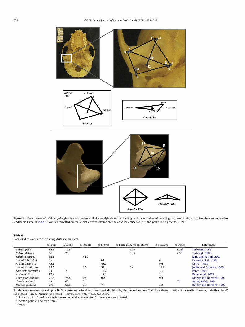
Following registration of the 3D coordinate data using Gener-alized Procrustes Analysis (GPA), a principal components analysis(PCA) was performed to summarize and evaluate variation in the3D datasets. The resulting principal component (PC) axes wereinterpreted as shape changes around a mean form, or consensusconfiguration, and the distribution of taxa along these axes thensummarizes information regarding shape variation within thesample (Slice et al., 1998; Zelditch et al., 2004). Variation along eachaxis was then visualized using wireframe diagrams. All configura-tionswere scaled to the same unit centroid size, and the size of eachconfiguration was retained as a separate variable (centroid size). Inthis study, shape variationwas examined both for the entire sampleand separately for each of the three comparative groups. This wasdone for both the glenoid and condylar configurations. Allgeometric morphometric data were analyzed using the programMorphologika (O’Higgins and Jones, 1998). Landmarks used in thegeometric morphometric analyses and their corresponding wire-frame diagrams are listed in Table 3 and illustrated in Fig. 1.
Analyses examining TMJ shape across the entire platyrrhine sample
Analyses of shape variation in the entire sample of 13 speciesincluded examining the extent to which shape variation is
Glenoid landmarks1 Most inferior point on entoglenoid process2 Most inferior point on articular tubercle3 Most inferior point on postglenoid process4 Deepest point in the mandibular fossa in the sagittal plane of
the postglenoid process point5 Most anterior point on the articular surface of the
glenoid fossa6 Most lateral point on the articular surface of the glenoid
at the end of the long axis of the articular eminence7 Most lateral point on the surface of the articular eminence8 Most medial point on the surface of the articular eminence9 Most medial point on the articular surface of the glenoid
at the end of the long axis of the articular eminence10 Midpoint of the crest of the articular eminence11 Most anterior point on the articular surface of the glenoid
along a line perpendicular to the long axis of thearticular eminence
12 Point on the posterior edge of the articular eminence alonga line perpendicular to the long axis of the articulareminence
Condyle landmarks13 Most lateral point on the articular surface of the
mandibular condyle14 Most medial point on the articular surface of the
mandibular condyle15 Midpoint of line connecting the medial and lateral poles
of the mandibular condyle16 Most posterior point on the articular surface of the
mandibular condyle at the midpoint of the mediolateralcurve
17 Most anterior point on the mandibular condyle at themidpoint of the mediolateral curve
C.E. Terhune / Journal of Human Evolution 61 (2011) 583e596 587
correlated with diet and/or TMJ size. These analyses were per-formed for females and males separately. To assess the rela-tionship between TMJ shape and size, Procrustes distancematrices describing shape were compared to size matrices thatcalculated the absolute difference in centroid size among groupsusing a Mantel test (Mantel, 1976; Smouse et al., 1986) in thefree Microsoft Excel add-on PopTools. To assess the relationshipbetween shape variation and diet (as a very rough proxy forfood material properties (FMPs)), I compiled published datareporting the percentage of particular food items included in thediets of 11 of the 13 species (Table 4) and created a Euclideandistance matrix using PopTools. Two dietary matrices werecreated. The first matrix calculated Euclidean distances amongtaxa when the percentages of less resistant (e.g., fruit, flower,insects) and more resistant (e.g., seeds, leaves, bark, roots) fooditems were summed. Notably, this matrix did not distinguishbetween tough and hard food items such as leaves and seeds. Asa result, I also computed a dietary matrix based on the summedpercentages of these more qualitative dietary categories (e.g.,fruits/insects, seeds, and leaves). Admittedly, these categories offood items are very rough proxies for food material properties,but many more data describing FMPs in platyrrhines (and acrossprimates) are necessary before a similar matrix incorporatingaverage and/or maximum fracture toughness or elasticity can bemade.
Where shape was significantly correlated with both size anddiet, partial Mantel tests were used to examine the correlationbetween shape and size while controlling for diet, and the corre-lation between shape and diet while controlling for size. PartialMantel tests were performed in the free program zt.exe (Bonnetand Van de Peer, 2002).
Pairwise comparisons
For each of the clades for which pairwise comparisons wereperformed, 3D geometric morphometric analyses first assisted indescribing the variation in each clade and its correlation to size.Further analyses of shape variation in the sample were performedusing linear measurements extracted from the coordinate datausing the programs MacMorph (Spencer and Spencer, 1993) andRhino 3D (Robert McNeel & Associates). Variables measuredincluded anteroposterior glenoid and condyle length as well asmediolateral glenoid and condyle width, 3D glenoid and condylarsurface area, preglenoid plane length (as a measure of functionalglenoid length), entoglenoid process height, and articular tubercleheight (Table 5). These variables were size adjusted by dividingeach variable by either mandibular length (as a measure of theload-arm during incision) or the distance from the TMJ to M1 (asa measure of the load-arm during mastication) (after Hylander,1985; Vinyard, 2008). For the atelines, variables were standard-ized using the distance from the TMJ to M1, whereas for the cebinesand pitheciines these biomechanical shape ratios were createdusing mandibular length. Behavioral data upon which thesedeterminations were made are outlined below.
Differences in these univariate measurements were analyzedusing a one-tailed Student’s t-test. Where these tests failed to findsignificant differences between the taxa, a two-tailed test wassubsequently performed to assess whether there were differencesin the direction opposite than that predicted. Taxa that were notspecifically predicted to vary (e.g., Ateles vs. Lagothrix, C. capucinusvs. C. albifrons, and Chiropotes vs. Cacajao) were not compared. Thecritical a ¼ 0.05 was further adjusted for multiple comparisonsusing the sequential Bonferroni method (Rice, 1989). These analyseswere performed separately for atelines, cebines, and pitheciines.Student's t-tests for significant differences between the sexes sug-gested that males and females do not significantly differ in mostaspects of TMJ shape, and since I have no a priori expectation offunctional differences between the sexes, their data were pooled.The t-tests were calculated using the program IBM SPSS (Version19).
Results
Platyrrhine TMJ variation
In the geometric morphometric analysis of the entire sample(Fig. 2), PC 1 separates the three Alouatta species from the rest ofthe sample. This PC explains 43% of the sample variance and issignificantly correlated with size in females (r2 ¼ 0.497, p ¼ 0.007)and males (r2 ¼ 0.680, p ¼ 0.007). Shape variation along this axis isprimarily associated with postglenoid process projection, and toa lesser degree, the relative anteroposterior and mediolateraldimensions of the joint. PC2 (which explains approximately 25% ofthe sample variance) is not significantly correlated with size andseparates the remaining atelines (Lagothrix and Ateles) and pith-eciines from the cebines (including Aotus and Saimiri), although thisseparation is marginal. Shape variation along this PC is associatedwith entoglenoid process projection and to a lesser extent,mediolateral width of the glenoid fossa.
TMJ shape and diet are significantly correlated in both sexes, butonly when seeds and leaves were summed separately and incor-porated into the dietary matrix (Table 6). There is also a significantcorrelation between the shape and size matrices in both femalesand males. Partial Mantel tests were also performed to determinethe relative influence of size and diet, and suggest that size is morestrongly correlated with TMJ shape when diet is held constant(Table 6). However, diet was still significantly (r¼ 0.3458, p¼ 0.011)
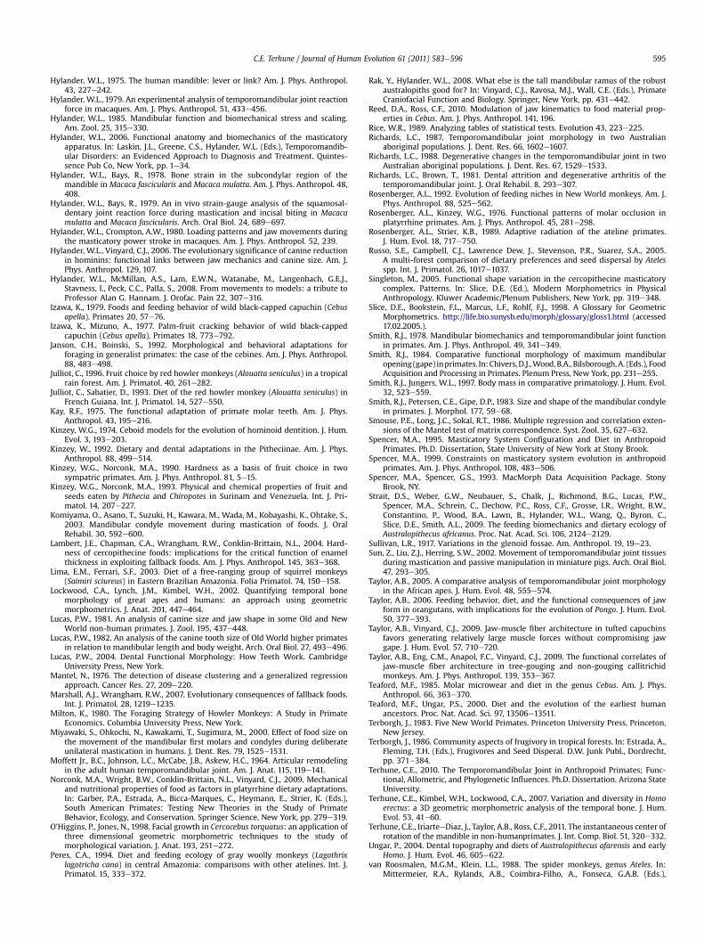
Figure 1. Inferior views of a Cebus apella glenoid (top) and mandibular condyle (bottom) showing landmarks and wireframe diagrams used in this study. Numbers correspond tolandmarks listed in Table 3. Features indicated on the lateral view wireframe are the articular eminence (AE) and postglenoid process (PGP).
Table 4Data used to calculate the dietary distance matrices.
% Fruit % Seeds % Insects % Leaves % Bark, pith, wood, stems % Flowers % Other References
Totals do not necessarily add up to 100% because some food items were not identified by the original authors. ‘Soft’ food items¼ fruit, animal matter, flowers, and other; ‘hard’food items ¼ seeds; ‘tough’ food items ¼ leaves, bark, pith, wood, and stems.
a Since data for C. melanocephalus were not available, data for C. calvus were substituted.b Nectar, petiole, and meristem.c Nectar.
C.E. Terhune / Journal of Human Evolution 61 (2011) 583e596588
Table 5Measurements included in the univariate analysis.
Name Definition Abbreviation
Articular tubercle height Inferior projection of the articular tubercle (Ldmk 2), as measured from the Frankfurt Horizontal plane ArtTubHtEntoglenoid height Inferior projection of the entoglenoid process (Ldmk 1), as measured from the Frankfurt Horizontal plane EntGlHtGlenoid length Linear distance between the most anterior point on the articular surface of the glenoid fossa to the most
inferior point on the postglenoid process (Ldmks 3 and 5)GlenLg
Glenoid width Linear distance between the most lateral and most medial points of the glenoid fossa (Ldmks 6 and 9) GlenWidGlenoid area Sum of the surface area of the polygons connecting a cloud of points covering the articular surface of the
glenoid fossa (calculated in the program Rhino 3D)3DGlenArea
Preglenoid plane length Linear distance between the apex of the articular eminence and the most anterior point on the articularsurface of the glenoid fossa (Ldmks 5 and 10)
PreglenLg
Condyle length Distance between the most anterior and most posterior points on the mandibular condyle (Ldmks 16 and 17) CondLgCondyle width Distance between the most medial and most lateral points on the mandibular condyle (Ldmks 13 and 14) CondWidCondyle area Sum of the surface area of the polygons connecting a cloud of points covering the articular surface of the
mandibular condyle (calculated in the program Rhino 3D)3DCondArea
Glenoid lengthecondyle length ratio Glenoid length divided by condyle length GlenLg/CondLgMandible length Distance from the center of the mandibular condyle to infradentale MandLgDistance from TMJ to M1 Distance from the center of the mandibular condyle to the lateral alveolar margin of mandibular M1 TMJ-M1
Table 6Results of the full and partial Mantel tests between the Procrustes distance (shape),centroid size, and dietary matrices.
Female shape Male shape
r p-value r p-value
Centroid size 0.692 <0.001 0.719 <0.001Diet (soft vs. resistant) 0.155 0.115 0.060 0.310Diet (soft vs. hard vs. tough) 0.488 <0.001 0.316 0.024Centroid size (controlling for diet) 0.6303 <0.001 0.6812 <0.001Diet (controlling for size) 0.3458 0.011 0.029 0.425
Significant correlations are shown in bold.
C.E. Terhune / Journal of Human Evolution 61 (2011) 583e596 589
correlated with female glenoid shape even after holding centroidsize constant.
Pairwise comparisons
Atelines PC1 1 accounts for 29% of the sample variation and issignificantly correlated with centroid size (r2 ¼ 0.57, p < 0.001)(Fig. 3). This PC separates all three species of Alouatta from Lagothrixand Ateles. Variation in shape along this axis is associated with therelative size of the postglenoid process (PGP), which is considerablylarger in Alouatta, but also with relative AP length of the glenoid.However, a separate geometric morphometrics analysis for thecondylar configurations (not shown) did not separate these taxa.
Several trends are apparent from the pairwise comparisons ofrelative differences in TMJ shape (Table 7). Atelines generally exhibitthe predicted differences in relative entoglenoid height, articulartubercle height, and some aspects of glenoid shape. Compared withAteles, Alouatta has significantly more projecting entoglenoidprocesses and articular tubercles, a relatively anteroposteriorlylonger glenoid, and a larger ratio of glenoid to condyle length. OnlyA. palliata was found to have relatively larger glenoid or condylarsurface areas than Ateles. Similarly, Alouatta has a relatively moreprojecting entoglenoid process and larger ratio of glenoid to
Figure 2. Bivariate plot of PC 1 (x-axis) and PC 2 (y-axis) from the PC analysis of theglenoid landmarks with wireframe diagrams illustrating shape variation in the sample.Platyrrhine females only included.
condylar length than does Lagothrix. But unlike the comparisonsbetween Alouatta and Ateles, Alouatta has relatively smaller condylarlength, condylar area, and (less consistently) glenoid width andpreglenoid plane length than Lagothrix.Cebines The PC analysis for the cebine glenoid configurationseparates C. apella from C. capucinus and C. albifrons along PC 1(27.2% of variation) and very slightly along PC 2 (15.2% of variation)(Fig. 4). No significant correlations were found between the top tenPC axes (which account for w90% of the sample variance) andcentroid size. The wireframe diagrams indicate that C. apelladiffers from the other species in anteroposterior length andmediolateral width of the glenoid, and in the relative size of thepostglenoid process. C. apella also separated from C. albifrons andC. capucinus in condylar morphology (Fig. 5), with C. apellaexhibiting a relatively anteroposteriorly shorter condyle that isless mediolaterally convex.
Figure 3. Bivariate plot of PC 1 (x-axis) and PC 2 (y-axis) from the PC analysis of theglenoid configurations in the Atelinae females. Wireframe diagrams illustrate theshape variation from the resistant to soft-object feeders in the sample.
Table 7Means, standard deviations (SD), and p-values for the Student’s t-tests among the atelines. Variables were scaled by the distance from the TMJ to the first molar (TMJ-M1).
Variable Prediction A. belzebul A. palliata A. seniculus Ateles Lagothrix
a p-values indicate the significance level for the one-tailed test, followed by a two-tailed, which was performed only if the one-tailed test failed to find a significantdifference. A significant p-value for the two-tailed test indicates a statistically significant difference in the direction opposite to that originally predicted. Bolded values indicatea significant p-value, whereas NS indicates no significant difference in shape between the taxa examined.
C.E. Terhune / Journal of Human Evolution 61 (2011) 583e596590
In contrast to the geometric morphometric analyses, mostpairwise comparisons between C. apella on the one hand, andC. capucinus and C. albifrons on the other, were not statisticallysignificant, and several that were significant were not in theexpected direction (Table 8). Thus, contrary to predictions, C. apellahas a relatively smaller articular tubercle and entoglenoid processcompared with C. albifrons and/or C. capucinus. However, C. apellatends to have relatively wider joints, and larger glenoid jointsurface area compared with C. albifrons and/or C. capucinus.Pitheciines For both glenoid and condylarmorphologies, PC analysisof the 3D landmarks failed to significantly distinguish amongC. melanocephalus, C. satanas, and P. pithecia (Figs. 6 and 7). However,centroid size was found to be significantly correlated with PC 1(r ¼ 0.278, p ¼ 0.003). In contrast, significant differences in TMJshape among these three species were identified in the univariate
Figure 4. Bivariate plot of PC 1 (x-axis) and PC 2 (y-axis) from the PC analysis of theglenoid configurations in the Cebinae females. Wireframe diagrams illustrate theshape variation from the resistant to soft-object feeders in the sample.
analyses (Table 8). As predicted, C. satanas and/or C. melanocephalushave relatively larger glenoid and condylar surface areas anddimensions compared with P. pithecia. Counter to expectations,Pithecia was not found to have a relatively wider joint comparedwith Cacajao and Chiropotes, or (at least in comparison to
Figure 5. Bivariate plot of PC 1 (x-axis) and PC 2 (y-axis) from the PC analysis of thecondylar configurations in the Cebinae females. Wireframe diagrams illustrate theshape variation from the resistant to soft-object feeders in the sample.
Table 8Means, standard deviations (SD), and p-values for the Student’s t-tests among the cebines (top) and pitheciines (bottom). Variables were scaled by mandible length.
Variable Prediction C. apella C. capucinus C. albifrons p-valuea
Mean SD Mean SD Mean SD C. apella vs. C. capucinus C. apella vs. C. albifrons
a p-values indicate the significance level for the one-tailed test, followed by a two-tailed, which was performed only if the one-tailed test failed to find a significantdifference. A significant p-value for the two-tailed test indicates a statistically significant difference in the direction opposite to that originally predicted. Bolded values indicatea significant p-value, whereas NS indicates no significant difference in shape between the taxa examined.
C.E. Terhune / Journal of Human Evolution 61 (2011) 583e596 591
Chiropotes) a more projecting articular tubercle. Glenoid andcondylar length also tended to be longer in Chiropotes and Cacajaothan in Pithecia.
Discussion
TMJ size and shape variation across platyrrhines
Analysis of TMJ variation across the platyrrhine sample high-lights the wide range of variation in glenoid shape present in thisclade. Importantly, this analysis found a relationship between sizeand shape of the TMJ and diet, particularly when seeds and leavesare considered separately. Assuming that dietary categories bearsome relationship to food material properties, the results of thisstudy further suggest that differentiating between seeds and leavesmay be important for understanding functional differences in TMJshape. However, one caveat regarding the dietary analyses thatshould be addressed is the imperfection of the data used to createthe dietary matrices. The ideal dataset for this analysis wouldinclude the food material properties for each food item ingested by
Figure 6. Bivariate plot of PC 1 (x-axis) and PC 2 (y-axis) from the PC analysis of theglenoid configurations in the Pitheciinae females.
the species included in this analysis. This type of analysis (seeWright, 2005) would then allow for the direct comparison of themasticatory demands of particular food items (or classes of fooditems) among taxa. However, these data are currently available forvery few primate species (Kinzey and Norconk, 1990, 1993; Elgart-Berry, 2004; Williams et al., 2005; Wright, 2005; Yamashita et al.,2009). Instead, the data used here represented the percentage offeeding records and/or the percentage of time spent feeding onfoods of a particular group (e.g., leaves, insect prey, fruit, etc.). Onemajor problemwith these data is that these categories of food typesare not standardized across analyses, and therefore may not becomparable with one another. In addition, some researchersdifferentiate among the parts of a specific food item (e.g., seeds vs.fruit pulp) or the relative maturation of food items (e.g., young vs.
Figure 7. Bivariate plot of PC 1 (x-axis) and PC 2 (y-axis) from the PC analysis of thecondylar configurations in the Pitheciinae females.
C.E. Terhune / Journal of Human Evolution 61 (2011) 583e596592
mature leaves), since these different categories may have differentfood material properties and/or require different masticatory abil-ities. As more data on food material properties become available,future work will be able to further refine the analyses performed inthis study.
One reason for the increased resolution in the dietary signal ofthe TMJ when the percentage of seeds and leaves in the diets ofthese taxa are analyzed separately may be that these dietarycategories separate along clade lines. For example, pitheciines areseed predators whereas the atelines and cebines have relativelylarger percentages of leaves and fruit pulp, respectively, in theirdiets (e.g., Izawa, 1979; Gaulin and Gaulin, 1982; Terborgh, 1986;Chapman, 1987; Ayres, 1989; Kinzey, 1992; Janson and Boinski,1992; Peres, 1994; Boubli, 1999). Thus, by separating these dietarycategories, the correlation between morphology and diet increases.This therefore suggests that TMJ morphology varies both asa function of diet and as a function of phylogenetic differentiationof these groups. The goal of the restricted pairwise comparisonswas tominimize this possible phylogenetic confound. Although notdirectly comparable analyses, the pairwise comparisons did notshowasmarked of a correlation between diet and TMJ shape as wasdemonstrated by the analysis across the entire platyrrhine sample.Thus, at lower taxonomic levels shape does not appear to be asstrongly correlated with diet. In some instances, this may representan overlap in the types of foods utilized by closely related taxa (e.g.,C. apella and C. albifrons), or it could indicate an insignificantknowledge of the dietary breadth of taxa in particular clades (e.g.,pitheciines). As a result, even though these pairwise comparisonsgive us the ability to specifically test biomechanical hypotheses offunction, future analyses seeking to assess the morphologicalcorrelates of diet or feeding behavior may also benefit from broaderinterspecific analyses of shape with strong phylogenetic controls.
Predictions for the components of the TMJ
TMJ size and shape The size and relative dimensions of theglenoid and condylar articular surfaces were predicted to differamong taxa that masticate more resistant food objects and/or usetheir anterior or posterior teeth during food processing or masti-cation. Resistant-object feeders, as well as taxa that utilize theiranterior dentition extensively, were expected to have relativelylarger condylar joint surface areas to improve the load resistancecapabilities of the TMJ. However, this prediction was onlysupported in the pitheciine sample, and to a much lesser extent,the atelines. Thus, these data do not support an overwhelmingpattern of differences in TMJ shape within cebines or atelinesrelated to force production. This result is curious in light of foodmaterial property data for these species (Norconk et al., 2009).Average and maximum toughness values for food items opened ormasticated by these taxa suggest that C. apella and A. seniculusgenerate considerably higher bite forces than do C. olivaceus andAteles paniscus, respectively. That these differences in bite forcemagnitude are apparently not linked to differences in loadresistance at the joint may imply that other morphological featuresof the mandible and/or TMJ (e.g., the articular eminence) act tomediate the magnitude of the joint reaction force in relation to thebite force, or that the TMJ is over designed and therefore does notreflect differences in joint loading.
Taxa were also predicted to differ significantly in the relativeanteroposterior and mediolateral dimensions of the joint surfaces,depending on whether they rely more extensively on their anteriorvs. posterior teeth for processing foods. Taxa that repetitivelymasticate tougher food items (e.g., leaves) on their posterior teethwere expected to have relatively mediolaterally wider joints thantaxa that use their anterior teeth more extensively to process food
items (Smith et al., 1983; Bouvier, 1986a,b). Mixed support wasfound for this prediction. The anteroposterior dimensions of thejoint differed predictably and most consistently among the atelinesand pitheciines. No differences in anteroposterior dimensions ofthe joint were found in the cebines. In the atelines, anteroposteriordimensions of the joint differed primarily between Alouatta andAteles. Interestingly, in comparison with Lagothrix, Alouatta waseither not significant or had smaller anteroposterior dimensions ofthe joint. However, despite these differences, Alouatta consistentlyhad a larger glenoid to condylar length ratio than Ateles and Lago-thrix. These data may therefore suggest subtle differences in thetranslatory potential of the TMJ among these species, such thatAlouatta may have the ability to generate wider jaw gapes thaneither Ateles or Lagothrix. Alouatta tends to rely heavily on thepostcanine dentition for the repetitive mastication of leaves, whichis not necessarily a feeding behavior that necessitates generatingrelatively wide jaw gapes (although this may depend on thequantity of leaves consumed in a single bite). Therefore, theimproved sagittal sliding and attendant capacity to generate rela-tively large jaw gapes could instead be related to the unique vocalbehaviors of Alouatta. Although it is currently unknown whetherAlouatta generates relatively wide jaw gapes during their vocalbehaviors, if these vocal behaviors do require generating relativelylarge gapes this behavior could further be facilitated by differencesin soft tissue anatomy, such as jaw adductor muscle position andfiber length.
Some significant differences in anteroposterior TMJ length werealso found in the pitheciines. In the females, both glenoid andcondylar length were larger in Chiropotes than in Pithecia. Thesedifferences could be indicative of masticatory specializations relatedto the extensive use of the anterior dentition to process hard ortough seeds in Chiropotes and Cacajao (e.g., Kinzey, 1992). However,the ratio between glenoid and condylar length did not vary signif-icantly between these species, suggesting that these differencesin the anteroposterior dimensions of the joint in the pitheciinesdo not necessarily translate into differences in gape capacity amongthese species (although other factors such as muscle architecture,muscle position, or mandibular morphology may also influencegape).
C. apella has also been documented to process food items usingits anterior dentition (e.g., the canines, premolars, and even firstmolar (Wright and Wright, 2010; Reed and Ross, 2010)). I thereforepredicted that this species would have relatively anteroposteriorlylonger TMJs than C. capucinus or C. albifrons. However, no signifi-cant differences were found for variables reflecting TMJ lengthamong these taxa. These data suggest that the species of Cebusexamined here maintain roughly similar capacities for sagittalsliding at the TMJ. In fact, coupled with the relatively increasedheight of the TMJ above the occlusal plane in C. apella (Constantino,2007), this finding may suggest that C. apella has relatively similar(or possibly smaller) maximum jaw gapes than C. capucinus orC. albifrons, since mandibular ramus height tends to be negativelycorrelated with gape (Herring and Herring, 1974; Smith, 1984;Singleton, 2005). Thus, minimally, these data suggest that TMJmorphology in this species is not adapted in such a way as tofacilitate relatively larger gapes in C. apella. Together with recentanalyses of masticatory muscle architecture in the cebines, thesedata further support previous suggestions that C. apella cangenerate relatively larger muscle and bite forces, while maintaininggapes similar to non-apelloid species (Spencer, 1995; Wright, 2005;Constantino, 2007; Taylor and Vinyard, 2009).
No significant differences in mediolateral width were found inthe atelines, which ran counter to the expectation that Alouattawould have a relatively wider TMJ than Ateles or Lagothrix. Simi-larly, few differences in relative TMJ width were found in the Cebus
C.E. Terhune / Journal of Human Evolution 61 (2011) 583e596 593
sample, although C. apella does tend to have awider glenoid and/orcondyle than either C. albifrons or C. capucinus, a finding that isconsistent with previous analyses by Bouvier (1986b). This maysuggest increased joint reaction forces on the lateral aspect of thejoint in C. apella, as would be consistent with increased unilateralmastication on the posterior dentition in this taxon. In the pith-eciines, it was predicted that Pithecia would exhibit the relativelymediolaterally widest TMJ as a result of their heavier use of thepostcanine dentition to masticate leaves. This was not the case,however, since all measures of TMJ width (and in fact, all measuresof joint size except the ratio between glenoid and condyle length)were significantly larger in Chiropotes and Cacajao than in Pithecia.This finding suggests that joint reaction forces may be orientedmoremediolaterally (as is expected to be the casewith axial torsionof the mandible) in Chiropotes and Cacajao in comparison to Pith-ecia. Furthermore, the significantly larger joint dimensions in Chi-ropotes and Cacajao may be indicative of globally higher jointreaction forces in these taxa.Entoglenoid process and articular tubercle shape The relative sizeof the entoglenoid process and articular tubercle were expected tovary as a function of joint reaction forces and range of motion at theTMJ. It was predicted that the relative size of both of these featureswould increase with increased joint reaction forces (i.e., to increasejoint surface area of the joint), and/or with increased range ofmotion (i.e., to guide movement of the condyle and counteracttensile forces at the joint). Thus, in resistant-object feeders and taxathat extensively use their postcanine dentition, the entoglenoidprocess and articular tubercle should be relatively larger than intaxa that masticate relatively softer food objects and/or do notextensively use their postcanine dentition.
Again, results were mixed. The more resistant-object feedersamong the atelines (e.g., Alouatta) and the pitheciines (e.g., Chiro-potes) do have relatively larger articular tubercles and/or entogle-noid processes. However, contrary to predictions, C. apella hasa smaller articular tubercle and entoglenoid process comparedwith the non-apelloids. Although no explicit comparisons weremade between Cacajao and Chiropotes, it is particularly notablethat the entoglenoid process is the only variable for which Cacajaoand Chiropotes depart from one another. For all other variables,these two genera differ in the same direction from Pithecia,whereas for this variable, Cacajao has a smaller entoglenoid thanPithecia and the values for Chiropotes are larger (although neitherspecies differs significantly from Pithecia). Differences in feedingecology between Cacajao and Chiropotes are poorly documented,however, and it is therefore unclear whether these morphologicaldifferences may be associated with differences in masticatory (oringestive) behavior.
Some of the observed variation in entoglenoid (and possiblyarticular tubercle) height may be related to the need for some ofthese taxa to generate relatively high jaw forces at relatively widejaw gapes. In Alouatta, it is presumed that higher forces aregenerally generated along the postcanine dentition while jawgapes are relatively low, whereas C. apella and all of the pitheciineshave been observed to process relatively large and resistant objectsusing the anterior dentition (e.g., Izawa and Mizuno, 1977;Terborgh, 1983; van Roosmalen et al., 1988; Ayres, 1989; Jansonand Boinski, 1992; Kinzey, 1992), which often necessitates largeingestive gapes (e.g., Norconk et al., 2009; Taylor and Vinyard,2009). The relatively less projecting entoglenoid processesobserved in these taxa may be advantageous for allowing increasedmediolateral movements of the mandible during ingestive behav-iors that involve the use of the anterior dentition. Experimentalanalyses of mandibular movements and glenoid morphology arenecessary to further explore this proposed function of the ento-glenoid process.
The TMJ and the masticatory apparatus as a whole
The TMJ is one portion of the much larger masticatory appa-ratus. Examining the functional morphology of the TMJ is infor-mative, but only to the extent that functional variation in themasticatory apparatus is reflected in this joint. Here I examinedmorphology of the TMJ as it is assumed to be related to differencesin force production and range of motion in the masticatory appa-ratus as a whole, and these data suggest that form of the TMJ doesvary as a consequence of feeding behavior. Some features of the TMJcan be linked to hypothesized differences in joint reaction forces aswell as movement at the joint, and these features should be takeninto consideration in future evaluations of the masticatory appa-ratus in living and extinct primates. For example, this studyprovides further (albeit indirect) evidence that the anteroposteriordimensions of the joint are linked to gape capacity (Wall, 1999;Vinyard et al., 2003). If this is the case, then (along with othermandibular dimensions) we may be able to further refine ourestimates of relative gape in fossil hominins such as Austral-opithecus or Paranthropus, which in turn has implications for dietand feeding behavior in these taxa (e.g., Demes and Creel, 1988;Teaford and Ungar, 2000; Ungar, 2004; Rak and Hylander, 2008;Strait et al., 2009).
The inferences that can be made using only TMJ morphology arelimited, however. Large joint surface areas may imply relativelyimproved load resisting ability, but cannot be used to infer whetherjoint forces are of infrequent but high magnitude, or high frequencyand low magnitude. This is of particular importance in consider-ations of critical function and fallback foods (e.g., Rosenberger andKinzey, 1976; Rosenberger, 1992; Lambert et al., 2004; Marshalland Wrangham, 2007), where especially resistant food items mayonly be utilized in times of resource scarcity. The seasonal use ofmore resistant food items has been documented for species in allthree of the clades examined here, and therefore these data indicatethat the morphologies of these taxa may be specialized for theutilization of food items during food shortages, but what impact theutilization of these food items has in comparison with morecommonly exploited foods is unclear. Further data comparing andcontrasting the material properties of foods used during resourcerich vs. resource poor times of the year are necessary to betterunderstand how this differential resource use may impact themorphology and function of the masticatory apparatus.
Just as inferences from the TMJ regarding forces in the masti-catory apparatus are limited, so are inferences regarding gapecapacity. The data presented here suggest that some taxa haveincreased the extent to which sagittal sliding can occur at the joint,and these morphological differences are likely the be linked todifferences in these taxa’s ability to generate wide jaw gapes (e.g.,Wall, 1999; Vinyard et al., 2003). However, a number of otherfeatures influence gape capacity. Height of the TMJ above theocclusal plane, jaw length, soft tissues structures in and around thejoint, and the position and internal architecture of the masticatorymuscles may all limit or facilitate wide jaw gapes (e.g., Herring andHerring,1974; Smith,1984;Wall, 1995,1999; Hylander and Vinyard,2006; Taylor and Vinyard, 2009; Taylor et al., 2009; Terhune et al.,2011), and it is therefore imperative that other features of themasticatory apparatus be considered when drawing definitiveconclusions regarding gape capacity in primates.
Conclusions
The data presented here suggest mixed correlations betweenfeeding behavior and the morphology of the TMJ in platyrrhineprimates. The correlations observed among the shape, size, anddietary matrices suggest relationships among all of these datasets,
C.E. Terhune / Journal of Human Evolution 61 (2011) 583e596594
and demonstrate that both size and diet are significant factorsinfluencing TMJ morphology in New World primates. Analyses of3D TMJ shape variation in each of the three comparative groupsfurther indicated that some aspects of TMJ morphology can be usedto differentiate among closely related species with different diets.Subsequent univariate analyses of TMJ shape demonstratedstatistically significant differences in multiple aspects of TMJ shapein each of these comparative groups. Results for some of theseanalyseswere consistent with the predictions outlined at the outsetof this study, while others were not. The anteroposterior dimen-sions of the TMJ were most strongly consistent with these initialpredictions, whereas the predictions generated for variation inentoglenoid and articular tubercle height were not upheld. Theseresults imply that while some features can be reliably associatedwith increased load resistance and facilitation of wider jaw gapesin the masticatory apparatus, other features are less stronglycorrelated with masticatory function. Further analyses, particularlyregarding the articular tubercle and entoglenoid process arenecessary to fully understand the functions of these specific features.
One major way in which we will be able to further evaluate TMJfunction is through a better understanding of the functionalimplications of anterior vs. posterior tooth use. The comparativegroups examined here present a mixture of taxa that use theirposterior teeth extensively for the repetitive mastication of foodobjects, as well as some taxa that use their anterior teeth for initialfood processing, but still likely need to generate high magnitudebite forces on their posterior dentition. This distinction is particu-larly important because the significance of high bite force magni-tudes vs. high bite force frequencies is poorly understood (e.g.,Yamashita, 2003; Taylor, 2006; Daegling and McGraw, 2007). Forexample, the magnitude of a single chew may be higher for bitingon the incisors in comparison to the molars (as shown by Hylander,1979; Hylander and Bays, 1979; Brehnan et al., 1981), but it isunclear how forces generated during repetitive processing of fooditems on the posterior teeth compare to these high magnitude butless frequent forces. As concluded by Daegling and McGraw (2007),more data on the use of the anterior vs. posterior dentition (in theircase, for mangabeys), coupled with detailed data regarding foodmaterial properties, are needed to adequately test modelsregarding the relative influence of anterior or posterior tooth useduring resistant-object feeding.
In sum, these data indicate that TMJ shape is influenced by thefunction of the masticatory apparatus, including variation in theuse of foods with presumably different material properties, use ofthe dentition, and jaw gape. These findings correspond well toprevious analyses of other aspects of the masticatory apparatus inmany of the same taxa examined here. Together, these data canprovide important insight into the adaptive response of themasticatory apparatus in New World primates. Further analyseswill explore these relationships in cercopithecoids and hominoidsand will seek to draw further links between TMJ morphology andmasticatory function.
Acknowledgments
Thanks go to all of the individuals and institutions that madecollections available for this research: AmericanMuseum of NaturalHistory, the Department of Primatology at the State Collection ofAnthropology and Paleoantomy, the National Museum of NaturalHistory, the Field Museum, and the Royal Museum for CentralAfrica. Valuable feedback was received from Bill Kimbel, GarySchwartz, Mark Spencer, Andrea Taylor, Christine Wall, Chris Vin-yard, David Begun and two anonymous reviewers. Any faultsremaining are my own. This research was supported by fundingfrom the National Science Foundation (BCS 0752661), the Leakey
Foundation, and the Graduate and Professional Student Associationof Arizona State University.
References
Altmann, S.A., 1959. Field observations on a howling monkey society. J. Mammal.40, 317e330.
Anapol, F., Lee, S., 1994. Morphological adaptation to diet in platyrrhine primates.Am. J. Phys. Anthropol. 94, 239e261.
Anderson, K., Throckmorton, G.S., Buschang, P.H., Hayasaki, H., 2002. The effects ofbolus hardness on masticatory kinematics. J. Oral Rehabil. 29, 689e696.
Ashton, E.H., Zuckerman, S., 1954. The anatomy of the articular fossa (fossa man-dibularis) in man and apes. Am. J. Phys. Anthropol. 12, 29e61.
Ayres, J.M., 1986. The White Uakaris and the Amazonian Flooded Forest. Ph.D.Dissertation, Cambridge University.
Ayres, J.M., 1989. Comparative feeding ecology of the uakari and bearded saki,Cacajao and Chiropotes. J. Hum. Evol. 18, 697e716.
Bonnet, E., Van de Peer, Y., 2002. zt: a software tool for simple and partial Manteltests. J. Stat. Soft. 7, 1e12.
Boubli, J.P., 1999. Feeding ecology of black-headed uacaris (Cacajao melanocephalusmelanocephalus) inPicodaNeblinaNationalPark,Brazil. Int. J. Primatol. 20,719e749.
Bouvier, M., 1986a. A biomechanical analysis of mandibular scaling in Old Worldmonkeys. Am. J. Phys. Anthropol. 69, 473e482.
Bouvier, M., 1986b. Biomechanical scaling of mandibular dimensions in New Worldmonkeys. Int. J. Primatol. 7, 551e567.
Brehnan, K., Boyd, R.L., Laskin, J.L., Gibbs, C.H., Mahan, P.E., 1981. Direct measure-ment of loads at the temporomandibular joint in Macaca arctoides. J. Dent. Res.60, 1820e1824.
Buchannon, D.B., Mittermeier, R.A., van Roosmalen, M.G.M., 1981. The saki monkeys,genus Pithecia. In: Coimbra-Filho, A.F., Mittermeier, R.A. (Eds.), Ecology andBehavior of Neotropical Primates. Academia Brasiliera de Ciencias, Rio deJaneiro, pp. 391e417.
Byrd, K.E., Milberg, D.J., Luschei, E.S., 1978. Human and macaque mastication:a quantitative study. J. Dent. Res. 57, 834e843.
Carpenter, C.R., 1934. A field study of the behavior and social relations of howlingmonkeys. Comp. Psychol. Monog. 10, 1e168.
Chalk, J., Wright, B.W., Lucas, P.W., Richmond, B.G., Fragaszy, D., Visalberghi, E.,Izar, P., Ottoni, E.B., 2010. Food mechanical property variation during ontogenyin Cebus libidinosus. Am. J. Phys. Anthropol. 141 (S50).
Chapman, C., 1987. Flexibility in the diets of three species of Costa Rican primates.Folia Primatol. 49, 90e105.
Chapman, C.A., 1989. Primate seed dispersal: the fate of dispersed seeds. Biotropica21, 148e154.
Chapman, C.A., Fedigan, L.M., 1990. Dietary differences between neighboring Cebuscapucinus groups: local traditions, food availability or responses to food prof-itability? Folia Primatol. 54,177e54,186.
Cole, T.M., 1992. Postnatal heterochrony of the masticatory apparatus in Cebusapella and Cebus albifrons. J. Hum. Evol. 23, 253e282.
Constantino, P.J., 2007. Primate Masticatory Adaptations to Fracture-ResistantFoods. Ph.D. Dissertation, George Washington University.
Daegling, D.J., 1992. Mandibular morphology and diet in the genus Cebus. Int. J.Primatol. 13, 545e570.
Daegling, D.J., McGraw, W.S., 2007. Functional morphology of the mangabeymandibular corpus: relationship to dental specializations and feeding behavior.Am. J. Phys. Anthropol. 134, 50e62.
DeSouza, L.L., Ferrari, S.F., DaCosta, M.L., Kern, D.C., 2002. Geophagy as a correlate offolivory in red-handed howler monkeys (Alouatta belzebul) from Eastern Bra-zilian Amazonia. J. Chem. Ecol. 28, 1613e1621.
Demes, B., Creel, N., 1988. Bite force, diet, and cranial morphology of fossil homi-nids. J. Hum. Evol. 17, 657e670.
Di Fiore, A., 2004. Diet and feeding ecology of woolly monkeys in a westernAmazonian rain forest. Int. J. Primatol. 25, 767e801.
Elgart-Berry, A., 2004. Fracture toughness of mountain gorilla (Gorilla gorillaberingei) food plants. Am. J. Primatol. 62, 275e285.
Freese, C., Oppenheimer, J.R., 1981. The capuchin monkeys, genus Cebus. In: Coim-bra-Filho, A.F., Mittermeier, R.A. (Eds.), Ecology and Behavior of NeotropicalPrimates. Academia Brasiliera de Ciencias, Rio de Janeiro, pp. 331e390.
Gaulin, S.J.C., Gaulin, C.K., 1982. Behavioral ecology of Alouatta seniculus in Andeancloud forest. Int. J. Primatol. 3, 1e32.
Greaves, W., 1978. The jaw lever system in ungulates: a new model. J. Zool. Lond.184, 271e285.
Harvati, K., 2001. The Neanderthal problem: 3-D geometric morphometric modelsof cranial shape variation within and among species. Ph.D. Dissertation, CityUniversity of New York.
Herring, S.W., Herring, S.E., 1974. The superficial masseter and gape in mammals.Am. Nat. 108, 561e576.
Hershkovitz, P., 1949. Mammals of northern Colombia. Preliminary report No. 4:monkeys (Primates), with taxonomic revisions of some forms. Proc. Am. Nat.Mus. 98, 323e427.
Hinton, R.J., 1981. Changes in articular eminence morphology with dental function.Am. J. Phys. Anthropol. 54, 439e455.
Hinton, R.J., Carlson, D.S., 1979. Temporal changes in human temporomandibularjoint size and shape. Am. J. Phys. Anthropol. 50, 325e334.
C.E. Terhune / Journal of Human Evolution 61 (2011) 583e596 595
Hylander, W.L., 1975. The human mandible: lever or link? Am. J. Phys. Anthropol.43, 227e242.
Hylander, W.L., 1979. An experimental analysis of temporomandibular joint reactionforce in macaques. Am. J. Phys. Anthropol. 51, 433e456.
Hylander, W.L., 1985. Mandibular function and biomechanical stress and scaling.Am. Zool. 25, 315e330.
Hylander, W.L., 2006. Functional anatomy and biomechanics of the masticatoryapparatus. In: Laskin, J.L., Greene, C.S., Hylander, W.L. (Eds.), Temporomandib-ular Disorders: an Evidenced Approach to Diagnosis and Treatment. Quintes-sence Pub Co, New York, pp. 1e34.
Hylander, W.L., Bays, R., 1978. Bone strain in the subcondylar region of themandible in Macaca fascicularis and Macaca mulatta. Am. J. Phys. Anthropol. 48,408.
Hylander, W.L., Bays, R., 1979. An in vivo strain-gauge analysis of the squamosal-dentary joint reaction force during mastication and incisal biting in Macacamulatta and Macaca fascicularis. Arch. Oral Biol. 24, 689e697.
Hylander, W.L., Crompton, A.W., 1980. Loading patterns and jaw movements duringthe masticatory power stroke in macaques. Am. J. Phys. Anthropol. 52, 239.
Hylander, W.L., Vinyard, C.J., 2006. The evolutionary significance of canine reductionin hominins: functional links between jaw mechanics and canine size. Am. J.Phys. Anthropol. 129, 107.
Hylander, W.L., McMillan, A.S., Lam, E.W.N., Watanabe, M., Langenbach, G.E.J.,Stavness, I., Peck, C.C., Palla, S., 2008. From movements to models: a tribute toProfessor Alan G. Hannam. J. Orofac. Pain 22, 307e316.
Izawa, K., 1979. Foods and feeding behavior of wild black-capped capuchin (Cebusapella). Primates 20, 57e76.
Izawa, K., Mizuno, A., 1977. Palm-fruit cracking behavior of wild black-cappedcapuchin (Cebus apella). Primates 18, 773e792.
Janson, C.H., Boinski, S., 1992. Morphological and behavioral adaptations forforaging in generalist primates: the case of the cebines. Am. J. Phys. Anthropol.88, 483e498.
Julliot, C., 1996. Fruit choice by red howler monkeys (Alouatta seniculus) in a tropicalrain forest. Am. J. Primatol. 40, 261e282.
Julliot, C., Sabatier, D., 1993. Diet of the red howler monkey (Alouatta seniculus) inFrench Guiana. Int. J. Primatol. 14, 527e550.
Kay, R.F., 1975. The functional adaptation of primate molar teeth. Am. J. Phys.Anthropol. 43, 195e216.
Kinzey, W.G., 1974. Ceboid models for the evolution of hominoid dentition. J. Hum.Evol. 3, 193e203.
Kinzey, W., 1992. Dietary and dental adaptations in the Pitheciinae. Am. J. Phys.Anthropol. 88, 499e514.
Kinzey, W.G., Norconk, M.A., 1990. Hardness as a basis of fruit choice in twosympatric primates. Am. J. Phys. Anthropol. 81, 5e15.
Kinzey, W.G., Norconk, M.A., 1993. Physical and chemical properties of fruit andseeds eaten by Pithecia and Chiropotes in Surinam and Venezuela. Int. J. Pri-matol. 14, 207e227.
Komiyama, O., Asano, T., Suzuki, H., Kawara, M., Wada, M., Kobayashi, K., Ohtake, S.,2003. Mandibular condyle movement during mastication of foods. J. OralRehabil. 30, 592e600.
Lambert, J.E., Chapman, C.A., Wrangham, R.W., Conklin-Brittain, N.L., 2004. Hard-ness of cercopithecine foods: implications for the critical function of enamelthickness in exploiting fallback foods. Am. J. Phys. Anthropol. 145, 363e368.
Lima, E.M., Ferrari, S.F., 2003. Diet of a free-ranging group of squirrel monkeys(Saimiri sciureus) in Eastern Brazilian Amazonia. Folia Primatol. 74, 150e158.
Lockwood, C.A., Lynch, J.M., Kimbel, W.H., 2002. Quantifying temporal bonemorphology of great apes and humans: an approach using geometricmorphometrics. J. Anat. 201, 447e464.
Lucas, P.W., 1981. An analysis of canine size and jaw shape in some Old and NewWorld non-human primates. J. Zool. 195, 437e448.
Lucas, P.W., 1982. An analysis of the canine tooth size of Old World higher primatesin relation to mandibular length and body weight. Arch. Oral Biol. 27, 493e496.
Lucas, P.W., 2004. Dental Functional Morphology: How Teeth Work. CambridgeUniversity Press, New York.
Mantel, N., 1976. The detection of disease clustering and a generalized regressionapproach. Cancer Res. 27, 209e220.
Marshall, A.J., Wrangham, R.W., 2007. Evolutionary consequences of fallback foods.Int. J. Primatol. 28, 1219e1235.
Milton, K., 1980. The Foraging Strategy of Howler Monkeys: A Study in PrimateEconomics. Columbia University Press, New York.
Miyawaki, S., Ohkochi, N., Kawakami, T., Sugimura, M., 2000. Effect of food size onthe movement of the mandibular first molars and condyles during deliberateunilateral mastication in humans. J. Dent. Res. 79, 1525e1531.
Moffett Jr., B.C., Johnson, L.C., McCabe, J.B., Askew, H.C., 1964. Articular remodelingin the adult human temporomandibular joint. Am. J. Anat. 115, 119e141.
Norconk, M.A., Wright, B.W., Conklin-Brittain, N.L., Vinyard, C.J., 2009. Mechanicaland nutritional properties of food as factors in platyrrhine dietary adaptations.In: Garber, P.A., Estrada, A., Bicca-Marques, C., Heymann, E., Strier, K. (Eds.),South American Primates: Testing New Theories in the Study of PrimateBehavior, Ecology, and Conservation. Springer Science, New York, pp. 279e319.
O’Higgins, P., Jones, N., 1998. Facial growth in Cercocebus torquatus: an application ofthree dimensional geometric morphometric techniques to the study ofmorphological variation. J. Anat. 193, 251e272.
Peres, C.A., 1994. Diet and feeding ecology of gray woolly monkeys (Lagothrixlagotricha cana) in central Amazonia: comparisons with other atelines. Int. J.Primatol. 15, 333e372.
Rak, Y., Hylander, W.L., 2008. What else is the tall mandibular ramus of the robustaustralopiths good for? In: Vinyard, C.J., Ravosa, M.J., Wall, C.E. (Eds.), PrimateCraniofacial Function and Biology. Springer, New York, pp. 431e442.
Reed, D.A., Ross, C.F., 2010. Modulation of jaw kinematics to food material prop-erties in Cebus. Am. J. Phys. Anthropol. 141, 196.
Rice, W.R., 1989. Analyzing tables of statistical tests. Evolution 43, 223e225.Richards, L.C., 1987. Temporomandibular joint morphology in two Australian
aboriginal populations. J. Dent. Res. 66, 1602e1607.Richards, L.C., 1988. Degenerative changes in the temporomandibular joint in two
Australian aboriginal populations. J. Dent. Res. 67, 1529e1533.Richards, L.C., Brown, T., 1981. Dental attrition and degenerative arthritis of the
temporomandibular joint. J. Oral Rehabil. 8, 293e307.Rosenberger, A.L., 1992. Evolution of feeding niches in New World monkeys. Am. J.
Phys. Anthropol. 88, 525e562.Rosenberger, A.L., Kinzey, W.G., 1976. Functional patterns of molar occlusion in
platyrrhine primates. Am. J. Phys. Anthropol. 45, 281e298.Rosenberger, A.L., Strier, K.B., 1989. Adaptive radiation of the ateline primates.
J. Hum. Evol. 18, 717e750.Russo, S.E., Campbell, C.J., Lawrence Dew, J., Stevenson, P.R., Suarez, S.A., 2005.
A multi-forest comparison of dietary preferences and seed dispersal by Atelesspp. Int. J. Primatol. 26, 1017e1037.
Singleton, M., 2005. Functional shape variation in the cercopithecine masticatorycomplex. Patterns. In: Slice, D.E. (Ed.), Modern Morphometrics in PhysicalAnthropology. Kluwer Academic/Plenum Publishers, New York, pp. 319e348.
Slice, D.E., Bookstein, F.L., Marcus, L.F., Rohlf, F.J., 1998. A Glossary for GeometricMorphometrics. http://life.bio.sunysb.edu/morph/glossary/gloss1.html (accessed17.02.2005.).
Smith, R.J., 1978. Mandibular biomechanics and temporomandibular joint functionin primates. Am. J. Phys. Anthropol. 49, 341e349.
Smith, R.J., 1984. Comparative functional morphology of maximum mandibularopening (gape) inprimates. In:Chivers,D.J.,Wood,B.A.,Bilsborough,A. (Eds.), FoodAcquisition and Processing in Primates. Plenum Press, New York, pp. 231e255.
Smith, R.J., Jungers, W.L., 1997. Body mass in comparative primatology. J. Hum. Evol.32, 523e559.
Smith, R.J., Petersen, C.E., Gipe, D.P., 1983. Size and shape of the mandibular condylein primates. J. Morphol. 177, 59e68.
Smouse, P.E., Long, J.C., Sokal, R.T., 1986. Multiple regression and correlation exten-sions of the Mantel test of matrix correspondence. Syst. Zool. 35, 627e632.
Spencer, M.A., 1995. Masticatory System Configuration and Diet in AnthropoidPrimates. Ph.D. Dissertation, State University of New York at Stony Brook.
Spencer, M.A., 1999. Constraints on masticatory system evolution in anthropoidprimates. Am. J. Phys. Anthropol. 108, 483e506.
Sullivan, L.R., 1917. Variations in the glenoid fossae. Am. Anthropol. 19, 19e23.Sun, Z., Liu, Z.J., Herring, S.W., 2002. Movement of temporomandibular joint tissues
during mastication and passive manipulation in miniature pigs. Arch. Oral Biol.47, 293e305.
Taylor, A.B., 2005. A comparative analysis of temporomandibular joint morphologyin the African apes. J. Hum. Evol. 48, 555e574.
Taylor, A.B., 2006. Feeding behavior, diet, and the functional consequences of jawform in orangutans, with implications for the evolution of Pongo. J. Hum. Evol.50, 377e393.
Taylor, A.B., Vinyard, C.J., 2009. Jaw-muscle fiber architecture in tufted capuchinsfavors generating relatively large muscle forces without compromising jawgape. J. Hum. Evol. 57, 710e720.
Taylor, A.B., Eng, C.M., Anapol, F.C., Vinyard, C.J., 2009. The functional correlates ofjaw-muscle fiber architecture in tree-gouging and non-gouging callitrichidmonkeys. Am. J. Phys. Anthropol. 139, 353e367.
Teaford, M.F., 1985. Molar microwear and diet in the genus Cebus. Am. J. Phys.Anthropol. 66, 363e370.
Teaford, M.F., Ungar, P.S., 2000. Diet and the evolution of the earliest humanancestors. Proc. Nat. Acad. Sci. 97, 13506e13511.
Terborgh, J., 1983. Five New World Primates. Princeton University Press, Princeton,New Jersey.
Terborgh, J., 1986. Community aspects of frugivory in tropical forests. In: Estrada, A.,Fleming, T.H. (Eds.), Frugivores and Seed Disperal. D.W. Junk Publ., Dordrecht,pp. 371e384.
Terhune, C.E., 2010. The Temporomandibular Joint in Anthropoid Primates; Func-tional, Allometric, and Phylogenetic Influences. Ph.D. Dissertation. Arizona StateUniversity.
Terhune, C.E., Kimbel, W.H., Lockwood, C.A., 2007. Variation and diversity in Homoerectus: a 3D geometric morphometric analysis of the temporal bone. J. Hum.Evol. 53, 41e60.
Terhune, C.E., IriarteeDiaz, J., Taylor, A.B., Ross, C.F., 2011. The instantaneous center ofrotation of the mandible in non-humanprimates. J. Int. Comp. Biol. 51, 320e332.
Ungar, P., 2004. Dental topography and diets of Australopithecus afarensis and earlyHomo. J. Hum. Evol. 46, 605e622.
van Roosmalen, M.G.M., Klein, L.L., 1988. The spider monkeys, genus Ateles. In:Mittermeier, R.A., Rylands, A.B., Coimbra-Filho, A., Fonseca, G.A.B. (Eds.),
C.E. Terhune / Journal of Human Evolution 61 (2011) 583e596596
Ecology and Behavior of Neotropical Primates, vol. 2. World Wildlife Fund,Washington D.C., pp. 455e538.
van Roosmalen, M.G.M., Mittermeier, R.A., Milton, K., 1981. The bearded sakis, genusChiropotes. In: CoimbraeFilho, A.F., Mittermeier, R.A. (Eds.), Ecology andBehavior of Neotropical Primates. Academia Brasiliera de Ciencias, Rio deJaneiro, pp. 419e441.
van Roosmalen, M.G.M., Mittermeier, R.A., Fleagle, J.G., 1988. Diet of the northernbearded saki (Chiropotes satanas chiropotes): a neotropical seed predator. Am. J.Primatol. 14, 11e35.
Vinyard, C., 1999. Temporomandibular Joint Morphology and Function in Strepsir-rhine and Eocene Primates. Ph.D. Dissertation, Northwestern University.
Vinyard, C.J., 2008. Putting shape to work: making functional interpretations ofmasticatory apparatus shapes in primates. In: Vinyard, C.J., Ravosa, M.J.,Wall, C.E. (Eds.), Primate Craniofacial Function and Biology. Springer Science,New York, pp. 357e386.
Vinyard, C.J., Wall, C.E., Williams, S.H., Hylander, W.L., 2003. Comparative functionalanalysis of skull morphology of tree-gouging primates. Am. J. Phys. Anthropol.120, 153e170.
Wall, C.E., 1995. Form and Function of the Temporomandibular Joint in AnthropoidPrimates, Ph.D. Dissertation, State University of New York at Stony Brook.
Wall, C.E., 1999. A model of temporomandibular joint function in anthropoidprimates based on condylar movements during mastication. Am. J. Phys.Anthropol. 109, 67e88.
Wall, C.E., Vinyard, C.J., Johnson, K.R., Williams, S.H., Hylander, W.L., 2006. Phase IIjaw movements and masseter muscle activity during chewing in Papio anubis.Am. J. Phys. Anthropol. 129, 215e224.
Weidenreich, F., 1943. The skull of Sinanthropus pekinensis: a comparative odon-tography of the hominids. Palaeontol. Sin. D 10, 1e484.
Williams, S.H., Wright, B.W., Truong, V.D., Daubert, C.R., Vinyard, C.J., 2005.Mechanical properties of foods used in experimental studies of primatemasticatory function. Am. J. Primatol. 67, 329e346.
Wright, B.W., 2004. Ecological Distinctions in Diet, Food Toughness, andMasticatory Anatomy in a Community of Six Neotropical Primates inGuyana, South America. Ph.D. Dissertation. Urbana-Champaign, Universityof Illinois.
Wright, B.W., 2005. Craniodental biomechanics and dietary toughness in the genusCebus. J. Hum. Evol. 48, 473e492.
Wright, B.W., Wright, K.A., 2010. You are how you eat: hard food feeding behavior inprimates. Am. J. Phys. Anthropol. 141, 248.
Yamashita, N., 2003. Food procurement and tooth use in two sympatric lemurspecies. Am. J. Phys. Anthropol. 121, 125e133.
Yamashita, N., Vinyard, C.J., Tan, C.L., 2009. Food mechanical properties in threesympatric species of Hapalemur in Ranomafana National Park, Madagascar. Am.J. Phys. Anthropol. 139, 368e381.
Zelditch, M.L., Swiderski, D.L., Sheets, H.D., Fink, W.L., 2004. Geometric Morpho-metrics for Biologists: A Primer. Elsevier Academic Press, London.