37 Research Arcle Volume: 4, Issue: 2 August 2020 Pages: 37-42 Exploring bacterial pathogens and risk factors associated with the occurrence of navel ill in calves Journal of Istanbul Veterınary Scıences Md. Gulam Rabbany Rassel 1 †, Pravin Mishra 1 †, Marzia Rahman 2 , Md. Mahmudul Alam 1 * 1. Department of Surgery and Obstetrics, Bangladesh Agricultural University, Mymensingh. 2. Department of Microbiology and Hygiene, Bangladesh Agricultural University, Mymensingh. † Authors contributed equally. Rassel M.G.R . ORCID: 0000-0001-8226-2183; Mishra P. ORCID: 0000-0002 8315-0463; Rahman M. ORCID:0000- 0002-8646-3246; Alam M.M. ORCID: 0000-0002-3464-4125 ABSTRACT The study menoned here was designed to invesgate both bacterial pathogens and risk factors associated with the occurrence of naval ill in calves. A total of 350 calves diagnosed to have navel ill formed the populaon in our study. Incidence density reports represenng the period between 2009 and 2018 were obtained from Veterinary Teaching Hospital (VTH), Bangladesh Agricultural University where informaon about age, sex, breed, seasonal effect was also included. Umbilical swab from each individual suffering from confirmed navel ill was collected for microbiological study. The occurrence of navel ill in the age of <30 days (n = 244, 69.71%) was noted higher compared to the age of ≥30 days (n = 106, 30.29%). Male calves were highly suscepble (n = 192, 54.86%) compared to females (n=158, 45.14%). In cross breed individuals, the occurrence was higher (n = 330, 94.29%) in regard of indigenous calves (n=20, 5.71%). The occurrence of the illness in summer (March-June) was more common (n = 159, 45.43%) in comparison to both rainy (n = 111, 31.71%) and winter (n = 80, 22.86%) seasons. In term of microbiological study, Staphylococcus aureus, E. coli and Proteus spp. were idenfied and isolated from the infected areas of the calves with the disease. In conclusion, navel ill occurred more commonly in male calves with the age of less than one month. The appearance of navel ill was noted to be more frequently in summer season (March-June) in cross breed calves. The results also demonstrated that the condion is mediated by mixed bacterial infecon formed by gram-posive and gram-negave agents which induce the inial inflammaon. Keywords: calf, navel ill, Staphylococcus aureus, E. coli, Proteus spp. Navel ill is a disease of young calves, usually less than a week of age. The infecon enters via umbilical cord or soon aſter birth with a prevalence of 5-15% (Mee, 2008a). In Bangladesh, 6.14 million cale of a total sum of 24.09 million, is dairy cale (DLS, 2019). Mortality in calves less than one year of age is about 9% (Debnath et al., 1990). Most of the animals die at young age due to different infecous diseases and surgical disorders like avel ill (Samad et al., 2002). The facultave myiasis producing flies can be a responsible agent for navel infecon. There is usually a mixed bacterial flora including E. coli, Proteus spp., Staphylococcus spp., Acnomyces pyogenes etc. (Sherif et al., 2017). This infecon can result in a wide range of signs depending on where the *Corresponding Author: Md. Mahmudul Alam E-mail: [email protected]hps://dergipark.org.tr/tr/pub/hp-www-jivs-net Arcle History Received: 18.04.2020 Accepted: 25.05.2020 Available online: 26.05.2020 This work is licensed under the Creative Commons Attribution 4.0 International License. DOI: 10.30704/http-www-jivs-net.722788 To cite this article: Rassel, M. G. R., Mishra P., Rahman M., Alam, M. M. (2020). Exploring bacterial pathogens and risk factors associated with the occurrence of navel ill in calves. Journal of Istanbul Veterinary Sciences. 4(2), 37-42, Abbreviated Title: J Ist Vet Sci Introducon
Transcript
37
Research Article
Volume: 4, Issue: 2 August 2020 Pages: 37-42
Exploring bacterial pathogens and risk factors associated with the occurrence of navel ill in calves
1. Department of Surgery and Obstetrics, Bangladesh Agricultural University, Mymensingh. 2. Department ofMicrobiology and Hygiene, Bangladesh Agricultural University, Mymensingh. † Authors contributed equally.
Rassel M.G.R . ORCID: 0000-0001-8226-2183; Mishra P. ORCID: 0000-0002 8315-0463; Rahman M. ORCID:0000-0002-8646-3246; Alam M.M. ORCID: 0000-0002-3464-4125
ABSTRACT
The study mentioned here was designed to investigate both bacterial pathogens and risk factors associated with the occurrence of naval ill in calves. A total of 350 calves diagnosed to have navel ill formed the population in our study. Incidence density reports representing the period between 2009 and 2018 were obtained from Veterinary Teaching Hospital (VTH), Bangladesh Agricultural University where information about age, sex, breed, seasonal effect was also included. Umbilical swab from each individual suffering from confirmed navel ill was collected for microbiological study. The occurrence of navel ill in the age of <30 days (n = 244, 69.71%) was noted higher compared to the age of ≥30 days (n = 106, 30.29%). Male calves were highly susceptible (n = 192, 54.86%) compared to females (n=158, 45.14%). In cross breed individuals, the occurrence was higher (n = 330, 94.29%) in regard of indigenous calves (n=20, 5.71%). The occurrence of the illness in summer (March-June) was more common (n = 159, 45.43%) in comparison to both rainy (n = 111, 31.71%) and winter (n = 80, 22.86%) seasons. In term of microbiological study, Staphylococcus aureus, E. coli and Proteus spp. were identified and isolated from the infected areas of the calves with the disease. In conclusion, navel ill occurred more commonly in male calves with the age of less than one month. The appearance of navel ill was noted to be more frequently in summer season (March-June) in cross breed calves. The results also demonstrated that the condition is mediated by mixed bacterial infection formed by gram-positive and gram-negative agents which induce the initial inflammation.
Keywords: calf, navel ill, Staphylococcus aureus, E. coli, Proteus spp.
Navel ill is a disease of young calves, usually less than a week of age. The infection enters via umbilical cord or soon after birth with a prevalence of 5-15% (Mee, 2008a). In Bangladesh, 6.14 million cattle of a total sum of 24.09 million, is dairy cattle (DLS, 2019). Mortality in calves less than one year of age is about 9% (Debnath et al., 1990). Most of the animals die at young age due to different
infectious diseases and surgical disorders like avel ill (Samad et al., 2002). The facultative myiasis producing flies can be a responsible agent for navel infection. There is usually a mixed bacterial flora including E. coli, Proteus spp., Staphylococcus spp., Actinomyces pyogenes etc. (Sherif et al., 2017). This infection can result in a wide range of signs depending on where the
Received: 18.04.2020 Accepted: 25.05.2020 Available online: 26.05.2020
This work is licensed under the Creative Commons Attribution 4.0 International License.
DOI: 10.30704/http-www-jivs-net.722788
To cite this article: Rassel, M. G. R., Mishra P., Rahman M., Alam, M. M. (2020). Exploring bacterial pathogens and risk factors associated with the occurrence of navel ill in calves. Journal of Istanbul Veterinary Sciences. 4(2),
bacteria spread to. In some calves, infection spreads from the navel to the liver causing a liver abscess (Mee, 1990; Biss et al., 1994). Though individual antibody levels alter the course of the infection, the health situation remains also highly dependent to postnatal management (Waldner and Rosengren, 2009). The occurrence of this condition is strongly associated with poor hygienic maintenance of both calf and shed as well as with the intake of low-quality colostrum (Waltner-Toews et al., 1986). In Bangladesh, several studies have been conducted in calves with neonatal diseases and their recurrence such as Schistosomus reflexus infection and umbilical hernia while on the other hand only few works about the healing of navel ill have been arranged and very few of these studies were mostly confined to revealing the prevalence of the disease (Hoda et. al., 2018; Jaman et al., 2018; Mishra et al., 2020). Therefore, the present research work has been implemented to harvest an incidence data of calves diagnosed with navel ill-treated at Veterinary Teaching Hospital (VTH), Bangladesh Agricultural University (BAU), Mymensingh and belonging to a decade between 2009-2018 in order to explore the bacterial pathogens and risk factors responsible for the occurrence of the disease.
Material and methods
Collection of occurrence data: Incidence data of navel ill was collected from clinical data records of Veterinary Teaching Hospital (VTH), Bangladesh Agricultural University (BAU), Mymensingh. The temporal period contained approximately a decade between 2009 and 2018 while details such as age, sex, breed, and occurrence season were also taken into consideration. Experimental animals: Calves with confirmed navel ill at Veterinary Teaching Hospital (VTH), Bangladesh Agricultural University (BAU), Mymensingh were adopted as the individuals of the research study mentioned here and umbilical swab for microbiological study was collected from each one, between January 2019 and May 2019.
Collection of bacteriological samples: All of the samples were collected aseptically from the deep navel region with the use of sterile cotton buds. Before the procedure, the area was soaked with saline solution and swab samples were collected by circling the cotton bud into the infected region before quickly dipping it into screwed capped test tubes containing nutrient broth. Culture of navel ill causing bacteria: The collected broth samples were incubated at 37 °C for 2 hours for enrichment. The samples were inoculated into different selective medias such as Mannitol Salt agar (MSA) and MacConkey agar and incubated at 37 °C overnight. Next day bacterial growth in various medias were observed and pure culture of each bacteria were obtained by repeated culture preparations of a single colony. Identification of isolated bacteria by Gram´s staining: The pure cultures of isolated bacteria were subjected to Gram´s staining for an observation of bacterial morphology, arrangement and staining characteristics under light microscope at 10x magnification. Statistical analysis: Descriptive analysis was performed. Data were collected and calculated to determine the occurrence of navel ill in the above-mentioned species. The connecting link between the disease's occurrence and variables such as age, sex, breed and seasonal differences was evaluated and presented in percentage for each group.
Results
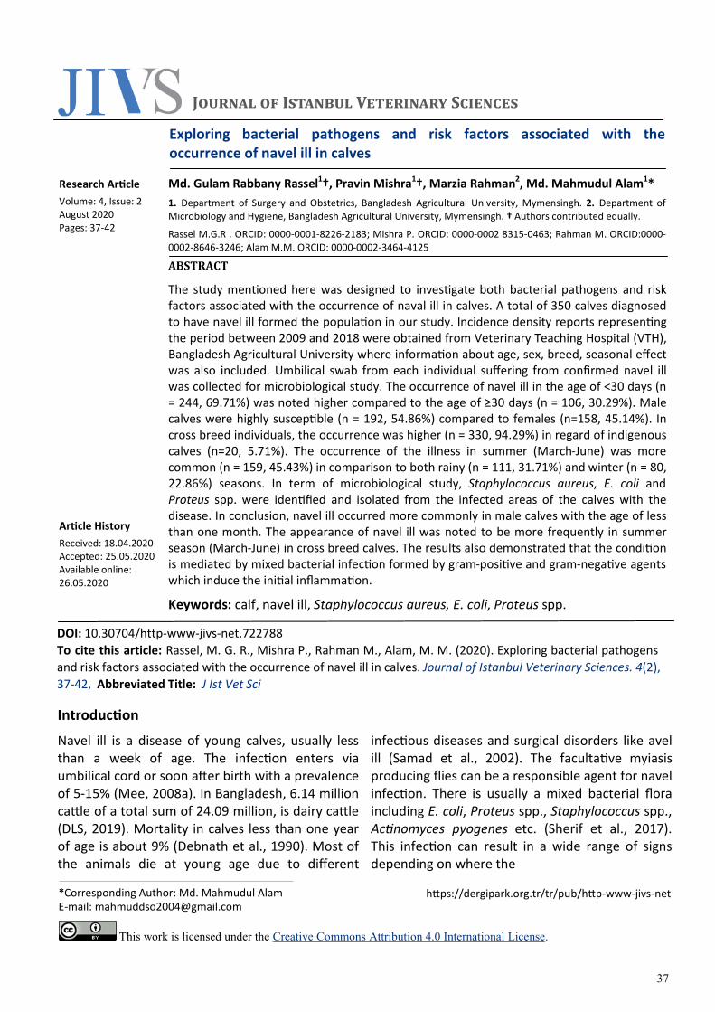
Effects of different variables on the occurrence of navel ill: A total amount of 350 calves diagnosed with naval ill were recorded from 2009 to 2018 at VTH, BAU, Mymensingh. Different attributes on which the occurrence of the condition was based on are shown below. Occurrence of navel ill based on age: The effect of age on navel ill in calves is presented in Figure 1. Occurrence was higher (n = 244, 69.71%) at the age of less than 30 days in comparison with the occurrence at the age higher than or equal to 30 days (n = 106, 30.29%).
Rassel, et al. 2020 / Journal of Istanbul Veterinary Sciences. Volume 4, Issue 2, pp: 37-42
39
Figure 1. Occurrence of navel ill in calves based on age.
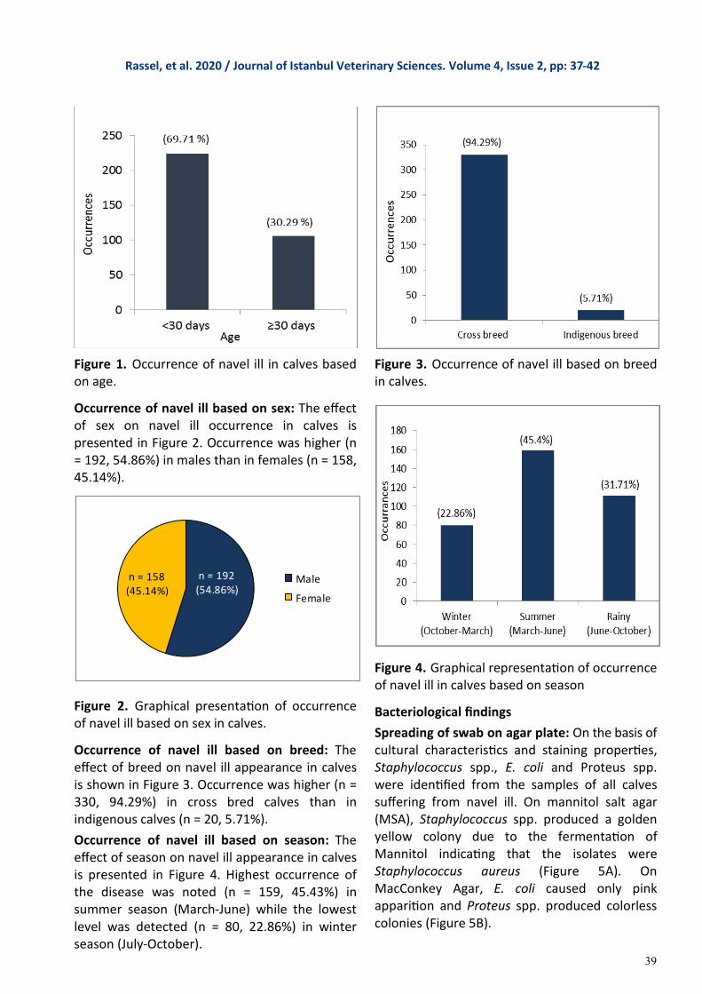
Occurrence of navel ill based on sex: The effect of sex on navel ill occurrence in calves is presented in Figure 2. Occurrence was higher (n = 192, 54.86%) in males than in females (n = 158, 45.14%).
Figure 2. Graphical presentation of occurrence of navel ill based on sex in calves.
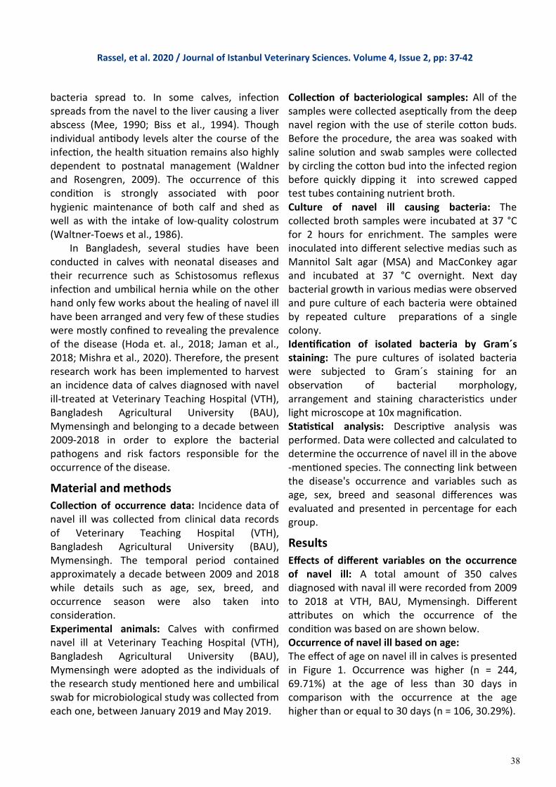
Occurrence of navel ill based on breed: The effect of breed on navel ill appearance in calves is shown in Figure 3. Occurrence was higher (n = 330, 94.29%) in cross bred calves than in indigenous calves (n = 20, 5.71%).
Occurrence of navel ill based on season: The effect of season on navel ill appearance in calves is presented in Figure 4. Highest occurrence of the disease was noted (n = 159, 45.43%) in summer season (March-June) while the lowest level was detected (n = 80, 22.86%) in winter season (July-October).
Figure 3. Occurrence of navel ill based on breed in calves.
Figure 4. Graphical representation of occurrence of navel ill in calves based on season
Bacteriological findings
Spreading of swab on agar plate: On the basis of cultural characteristics and staining properties, Staphylococcus spp., E. coli and Proteus spp. were identified from the samples of all calves suffering from navel ill. On mannitol salt agar (MSA), Staphylococcus spp. produced a golden yellow colony due to the fermentation of Mannitol indicating that the isolates were Staphylococcus aureus (Figure 5A). On MacConkey Agar, E. coli caused only pink apparition and Proteus spp. produced colorless colonies (Figure 5B).
(94.29%)
(5.71%)
Rassel, et al. 2020 / Journal of Istanbul Veterinary Sciences. Volume 4, Issue 2, pp: 37-42
n = 192(54.86%)
n = 158(45.14%)
Male
Female
40
Pure culture finding: A single colony from each plate were spread on the same agar plate respectively. On MSA, a golden yellow colony were found; which was suspected to be Staphylococcus aureus (Figure 6A) and on
Figure 6. Colony characteristics of bacteria. (A) A Golden yellow color colony was found which was the characteristics of Staphylococcus aureus, (B1) A characteristics pinkish colony with metallic sheen was detected and the colony was evaluated as E. coli and (B2) a pale color colony was found which are usually recognized as the colony characteristics of Proteus spp.
MacConkey Agar, a pinkish colony was found with metallic sheen, which was thought to be E. coli (Figure 6B1) and a colony with lose color were found, which was suspected to be Proteus spp. (Figure 6B2).
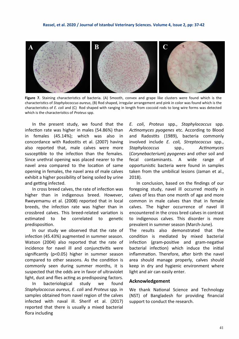
Characteristics of isolated bacteria: On Gram´s staining, The Staphylococcus aureus showed Gram positive, spherical shaped in cluster form (Figure 7A), the E. coli showed Gram negative rod-shaped organisms arranged in single or short chain like (Figure 7B) and the Proteus spp. showed Gram negative short rod arranged with periodic cycles of migration producing concentric zones
or spread in a uniform film (Figure 7C).
Discussion
During the study period it was found that among 350 of infected calves, 244 were brought to VTH earlier than thirty days and 106 calves were of ≥30 days of age. Samad et al. (2001) reported that the infection rate was higher (73.03%) in those aged between 0-30 days compared to individuals aged between 31-90 days (24.72%) while the same rate was measured very low (2.25%) for those aged >90 days. Blowey and Weaver (2011) also reported that navel ill usually occurs in calves younger than one weeks of age, as a result of inflammation, due to umbilical tissue infection following parturition in insanitary environment; which was compatible with our study.
Figure 5. Isolated bacteria from navel ill patient. (A) Growth of Staphylococcus aureus in MSA (B) Growth of suspected (1) E. coli and (2) Proteus spp. on MacConkey Agar.
Rassel, et al. 2020 / Journal of Istanbul Veterinary Sciences. Volume 4, Issue 2, pp: 37-42
41
In the present study, we found that the infection rate was higher in males (54.86%) than in females (45.14%); which was also in concordance with Radostits et al. (2007) having also reported that, male calves were more susceptible to the infection than the females. Since urethral opening was placed nearer to the navel area compared to the location of same opening in females, the navel area of male calves exhibit a higher possibility of being soiled by urine and getting infected. In cross breed calves, the rate of infection was higher than in indigenous breed. However, Rweyemamu et al. (2008) reported that in local breeds, the infection rate was higher than in crossbred calves. This breed-related variation is estimated to be correlated to genetic predisposition. In our study we observed that the rate of infection (45.43%) augmented in summer season. Watson (2004) also reported that the rate of incidence for navel ill and conjunctivitis were significantly (p<0.05) higher in summer season compared to other seasons. As the condition is commonly seen during summer months, it is suspected that the odds are in favor of ultraviolet light, dust and flies acting as predisposing factors. In bacteriological study we found Staphylococcus aureus, E. coli and Proteus spp. in samples obtained from navel region of the calves infected with naval ill. Sherif et al. (2017) reported that there is usually a mixed bacterial flora including
E. coli, Proteus spp., Staphylococcus spp. Actinomyces pyogenes etc. According to Blood and Radostits (1989), bacteria commonly involved include E. coli, Streptococcus spp., Staphylococcus spp., Actinomyces (Corynebacterium) pyogenes and other soil and fecal contaminants. A wide range of opportunistic bacteria were found in samples taken from the umbilical lesions (Jaman et al., 2018). In conclusion, based on the findings of our foregoing study, navel ill occurred mostly in calves of less than one month of age and more common in male calves than that in female calves. The higher occurrence of navel ill encountered in the cross bred calves in contrast to indigenous calves. This disorder is more prevalent in summer season (March-June). The results also demonstrated that the condition is mediated by mixed bacterial infection (gram-positive and gram-negative bacterial infection) which induce the initial inflammation. Therefore, after birth the navel area should manage properly, calves should keep in dry and hygienic environment where light and air can easily enter.
Acknowledgement
We thank National Science and Technology (NST) of Bangladesh for providing financial support to conduct the research.
Rassel, et al. 2020 / Journal of Istanbul Veterinary Sciences. Volume 4, Issue 2, pp: 37-42
Figure 7. Staining characteristics of bacteria. (A) Smooth, convex and grape like clusters were found which is the characteristics of Staphylococcus aureus, (B) Rod shaped, irregular arrangement and pink in color was found which is the characteristics of E. coli and (C) Rod shaped with ranging in length from coccoid rods to long wire forms was detected which is the characteristics of Proteus spp.
42
Biss, M. E., Hathaway, S. C. & Johnstone, A. C. (1994). Evaluation of the risk of potential bacteraemia in carcasses from very young slaughter calves with localized navel ill. British Veterinary Journal, 150(4), 377-384.
Blood, D. C. Radostits, O. M. (1989). Omphalitis, omphalophlebitis and urachitis in newborn farm animals (“navel ill“). In veterinary medicine. London, UK: Balliere.
Blowey, R. & Weaver, AD. (2011). Urinogenital disorders In Color atlas of diseases and disorders of cattle. (pp. 173-201). Missouri, US: Mosby.
Debnath, N. C., Sil, B. K., Selim, S. A., Prodhan, M. A. M. &Howlader, M. M. R. (1990). A retrospective study of calf mortality and morbidity on smallholder traditional farms in Bangladesh. Preventive Veterinary Medicine, 9(1), 1-7.
Department of Livestock Service (DLS). (2019 July 10). Livestock Economy at a Glance 2017-18. Retrieved from http://www.dls.gov.bd/site/page/22b1143b-9323-44f8-bfd8-647087828c9b/Livestock-Economy.
Hoda, N., Karim, M. R., Mishra, P., Shihab, M. M., Jaman, M. M., Alam, M. R. & Alam, M. M. (2018). Occurrence of Schistosomus reflexus in neonatal bovine calves in certain areas of Bangladesh: a retrospective study. Bangladesh Veterinary Journal, 52(1-4), 39-45.
Jaman, M. M., Mishra, P., Rahman, M. & Alam, M. M. (2018). Clinical and laboratory investigation on the recurrence of the umbilical hernia after herniorrhaphy in bovine calves. Journal of the Bangladesh Agricultural University, 16(3), 464-470.
Mee, J. F. (1990). Navel ill. The Veterinary Record, 126(14), 341.
Mee, J. F. (2008a). Prevalence and risk factors for dystocia in dairy cattle: A review. The Veterinary Journal, 176(1), 93-101.
Mee, J. F. (2008b). Newborn dairy calf management. Veterinary Clinics of North
America: Food Animal Practice, 24(1), 1-17.
Mishra, P., Mahmud, M. M., Yadav, V. K. & Hasan, M. (2020). Umbilical Hernia with Extensive Adhesion and Evisceration in a Bovine Calf. Iranian Journal of Veterinary Surgery, 15(1), 92-95.
Radostits, O. M., Gay, C. C. & Hinchcliff, K. W. (2007). A textbook of the diseases of cattle, horses, sheep, pigs and goats. In Veterinary Medicine 10th ed.(pp. 007; 345-674). London, UK: Saunders.
Rweyemamu, M., Roeder, P., Mackay, D., Sumption, K., Brownlie, J., Leforban, Y., Valarcher, J. F., Knowles, N. J. & Saraiva, V. (2008). Epidemiological patterns of foot‐and‐mouth disease worldwide. Transboundary and emerging diseases, 55(1), 57-72.
Samad, M. A. (2001). Animal husbandry and medicine. 2nd ed. Mymensingh, Bangladesh: LEP Publication.
Samad, M. A., Islam, M. A. & Hossain, A. (2002). Patterns of occurrence of calf diseases in the district of Mymensingh in Bangladesh. Bangladesh Veterinary Journal, 36(1-2),01-05.
Sherif, H. H., Khalil, S. K., Hegazi, A. G., Khalil, W. A. & Moharram, M. A. (2017). Factors affecting the antibacterial activity of chitosan-silver nanocomposite. IET Nanobiotechnology 11(6), 731-737.
Waldner, C. L. & Rosengren, L. B. (2009). Factors associated with serum immunoglobulin levels in beef calves from Alberta and Saskatchewan and association between passive transfer and health outcomes. The Canadian Veterinary Journal, 50(3), 275.
Waltner-Toews, D. M. S. W. M. A. H., Martin, S. W. & Meek, A. H. (1986). Dairy calf management, morbidity and mortality in Ontario Holstein herds. III. Association of management with morbidity. Preventive Veterinary Medicine, 4(2), 137-158.
Watson, C. L. (2004). Eye disease in the growing animal-Can we prevent it? Cattle Practice, 12, 213-218.
References
Rassel, et al. 2020 / Journal of Istanbul Veterinary Sciences. Volume 4, Issue 2, pp: 37-42