Journal of Pharmacological and Toxicological Methods xxx (2016) xxx–xxx
JPM-06339; No of Pages 12
Contents lists available at ScienceDirect
Journal of Pharmacological and Toxicological Methods
j ourna l homepage: www.e lsev ie r .com/ locate / jpharmtox
An evaluation of 30 clinical drugs against the comprehensive in vitroproarrhythmia assay (CiPA) proposed ion channel panel
William J. Crumb Jr. a,⁎, Jose Vicente b, Lars Johannesen b, David G. Strauss b
a Cytocentrics, New Orleans, LA, United Statesb Office of Clinical Pharmacology, Office of Translational Sciences, Center for Drug Evaluation and Research, U.S. Food and Drug Association, Silver Spring, MD, United States
⁎ Corresponding author at: Cytocentrics Inc., 1441 CanUnited States
Please cite this article as: Crumb, W.J., et al.,proposed ion channel panel, Journal of Pharm
a b s t r a c t
a r t i c l e i n f o
Article history:Received 30 January 2016Received in revised form 28 March 2016Accepted 30 March 2016Available online xxxx
Introduction: The Comprehensive in vitro Proarrhythmia Assay (CiPA) is intended to address the misidentifica-tion of drug-associated torsade de pointes risk based solely on hERG and QT data. This new paradigmwill consistof four interrelated components, one of which is a panel consisting of six ion channelswhose currents are impor-tant in both depolarization and repolarization of the cardiac action potential. This study examined the effects of30 clinical drugs on these ion channels.Methods: Ion currentswere evaluated in expression systems using themanualwhole cell patch clamp technique.Currents were elicited using either a ventricular action potential waveform or step-ramp voltage protocols.Results:Of the seven ion currents studied, hERGwas themost often blocked current followed byNav1.5-late, andCav1.2. Using a 20% reduction in current amplitude as an arbitrarymaker, at a free plasma Cmax concentration, nodrug tested blockedNav1.5-peak, KvLQT1/mink, Kir2.1 and Kv4.3 by that amount. At a 3x free plasma Cmax, everycurrent except Kir2.1 had at least one drug reduce current amplitude by at least 20%.Discussion: This is the first study of its kind to examine the effects of 30 clinical drugs against the seven ioncurrents currently proposed to makeup the CiPA ion channel panel. The results indicate the importance ofdrug-induced block of hERG, Nav1.5-late and Cav1.2 at clinically relevant concentrations, with low risk torsadedrugs having equal or greater Nav1.5-late or Cav1.2 block compared to hERG block. In addition, the results ofthis study provide data which can be used to test the ability of various in silico models to predict drug-inducedarrhythmias.
Keywords:CiPAElectrophysiologyIon channelsManual patch clampTorsade de pointes
1. Introduction
The Comprehensive in vitro Proarrhythmia Assay (CiPA) is intendedto address the misidentification of drug-associated torsade de pointesrisk based solely on hERG and QT data (Sager, Gintant, Turner, et al.,2014). This new paradigmwill consist of four interrelated components:an ion channel panel, in silico action potential reconstructions of the ionchannel panel activity, and verification of results in stem cell derivedhuman cardiomyocytes and in human phase 1 ECGs. To this end, theSafety Pharmacology Society organized the Ion Channel WorkingGroup (ICWG) consisting of representatives from the pharmaceuticalindustry, regulatory agencies, contract research organizations, and aca-demia. The ICWGwas tasked with selecting the ion channels to be test-ed (Fermini, Hancox, Abi-Gerges, et al., 2016). The ion channel paneldecided upon consists of six ion channels whose currents are importantin both depolarization and repolarization of the cardiac action potential(IKr, INa, ICa, IKs, Ito and IK1) (Fermini et al., 2016).
al St., New Orleans, LA 70112,
b).
An evaluation of 30 clinical dacological and Toxicological M
These currents contribute to all components of the cardiac action po-tential (Grant, 2009; Nerbonne & Kass, 2005). Starting from a restingmembrane potential, determined largely by the inwardly rectifying po-tassium current (IK1), the upstroke or Vmax of the cardiac action poten-tial is due to an influx of Na+ ions through the Na channel (INa-peak).This is followed by a rapid phase of repolarization or phase 1 due tothe efflux of K+ ions through the transient outward potassium current(Ito). Following this is the plateau of the cardiac action potential carriedby an influx of Ca++ ions through the L-type Ca channel (ICa) and to asmaller extent through an influx of Na+ ions through the Na channel.This Na current flowing during the plateau period is commonly referredto as the late Na current (INa-late). Blockade of both the L-type Ca cur-rent and INa-late have been associatedwith a reduction inQTc prolonga-tion and torsade even in the presence of hERG block (Belardinelli et al.,2013; Gintant, Su, Martin, & Cox, 2006; Johannesen et al., 2014). The ac-tion potential returns to a resting state via an efflux of K+ ions throughboth the rapid and slow components of the delayed rectifier potassiumcurrent (IKr and IKs, respectively). For ease of use across industry and ac-ademia, these currents are most often recorded in expression systemsand not primary cardiac myocytes. Therefore, IK1, INa (peak and late),Ito, ICa, IKr, and IKs are routinely recorded using cell lines expressingKir2.1, Nav1.5, Kv4.3, Cav1.2, hERG, and KvLQT1/mink, respectively.
rugs against the comprehensive in vitro proarrhythmia assay (CiPA)ethods (2016), http://dx.doi.org/10.1016/j.vascn.2016.03.009
NS, not solubleAdditional concentrationswere added for some of the drugs and some currents as indicat-ed in the Tables in Supplement 1.
2 W.J. Crumb Jr. et al. / Journal of Pharmacological and Toxicological Methods xxx (2016) xxx–xxx
The purpose of the present study was to evaluate the ion currentblocking profile of thirty clinical drugs against these expressed ion chan-nels using themanual patch clamp technique and to provide insight intowhich ion currents are more frequently blocked at concentrations cor-responding to free plasma Cmax and multiples thereof. This could havepreliminary implications for whether all 7 ion channel currents shouldbe assessed for every drug under CiPA. In addition, the ion channelblocking patterns of drugs that have high, intermediate and low torsadede pointes risk is compared. Furthermore, the results presented in thisstudy can be used to evaluate various in silico action potential modelsfor their ability to define and categorize the arrthymogenic risk of thedrugs tested here.
2. Methods
The cloned equivalent of the human IKr (hERG-HEK), IKs (KvLQT1/mink-HEK), INa (Nav1.5-HEK), Ito (Kv4.3-HEK), IK1 (Kir2.1-CHO), andthe L-type ICa (Cav1.2-CHO)were used in this study. Cellswere obtainedfrom Cytocentrics Bioscience GmbH (Joachim-Jungius-Straße 9, 18059Rostock, Germany).
2.1. Internal and external recording solutions
The external (bath) solution for hERG, KvLQT1/minK, Kir2.1, Kv4.3had a composition of in mM: NaCl 137, KCl 4, MgCl2 1, CaCl2 1.8,HEPES 10, dextrose 11 with a pH of 7.4 (NaOH). The internal (pipette)solution had a composition of (in mM): KCl 130, MgCl2 1, NaCl 7,HEPES 5, EGTA 5 with a pH of 7.2 (KOH). The external solution forCav1.2 and Nav1.5 had a composition of in mM: NaCl 137, KCl 4,MgCl2 1, CaCl2 1.8, HEPES 10, dextrose 11 with a pH of 7.4 (NaOH).The internal solution was (in mM): CsCl 130, MgCl2 1, NaCl 7, HEPES5, EGTA 5 with a pH of 7.2 (CsOH). In Cav1.2 experiments, 1.8 mMCaCl2 was replaced with 4mM BaCl2. Chemicals were obtained fromeither Sigma-Aldrich (MA) or Fisher Scientific (PA).
2.2. Test compounds
Test compounds were obtained from either Tocris Biosciences (MN)(dofetilide, quinidine, ranolazine, verapamil, mexiletine, lidocaine, dilti-azem) or Sigma-Aldrich (MA) (all others). Compounds were dissolvedin either de-ionized H2O or DMSO to create master stock solutions.This stock was subdivided into aliquots which were stored frozenuntil use. Dilutions of themaster stock to create the final test concentra-tions were performed on the day of experimentation. Compounds weretested at four different concentrations (free plasmaCmax and 3multiplesof Cmax (see Table 1).
2.3. Data acquisition and analysis
Experiments (n = 3–6) were performed at 36 ± 1 °C with the ex-ception of Kv4.3 which was performed at 22 ± 1 °C. This was done be-cause separation of the Kv4.3 ionic current from the capacitive currentcould not be done at 36 °C,making it impossible tomeasure current am-plitude accurately. Currents were measured using the whole-cell vari-ant of the patch clamp method as previously described (Crumb, Pigott,& Clarkson, 1995). Glass pipettes were pulled from borosilicate glassby a horizontal puller (Sutter Instruments, USA). Pipette tip resistancewas approximately 1 to 2MΩwhen filledwith internal solutions. Seriesresistance was compensated electronically by approximately 60–80%.An Axopatch 1-B amplifier (Axon Instruments, Foster City, CA) wasused for whole-cell voltage clamping. Creation of voltage clamp pulsesand data acquisition was controlled by a computer running pClampsoftware (ver 9.2 Axon Instruments). After rupture of the cell mem-brane (entering whole-cell mode), current kinetics and amplitudeswere allowed to stabilize (typically 3–5 min) as the cell was dialyzedwith internal solution and the voltage protocol was applied every 10 s
Please cite this article as: Crumb, W.J., et al., An evaluation of 30 clinical dproposed ion channel panel, Journal of Pharmacological and Toxicological M
(applied at 0.1 Hz). For Cav1.2 and KvLQT1/mink, currents which tendto exhibit more current rundown, the reduction in current amplitudetypically occurred within the first minutes after rupture of the cellmembrane. If present (N20% current reduction), these cells werediscarded. Only cells with a stable ionic current and stable holdingcurrent were used. The voltage protocols used to record the variouscurrents are given in Panel A of Figs. 1–7. A ventricular action poten-tial waveform was used to elicit hERG, Cav1.2, and KvLQT1/mink(kindly provided by Dr. Gail Robertson, University of Wisconsin).For Nav1.5 (peak and late), Kv4.3 and Kir2.1, the voltage protocolsused were those proposed by the ICWG. For measurement ofNav1.5-late, current was elicited by the addition of 50 μMveratridineto the external solution. Drug was applied to the bath solution via arapid perfusion apparatus. Not all four concentrations were exam-ined in every cell. Some currents were also examined for drug blockingeffects by applying the pulse protocol every 1 s (applied at 1 Hz). Thedrugs examined (quinidine, moxifloxacin, dofetilide, ranolazine, diltia-zem, and verapamil) were those evaluated in recent clinical studies(Johannesen, Vicente, Mason, et al., 2014, 2016). Not all of these drugswere examined for every current at 1 Hz.
Time-control experiments were performed for each current inwhich cells were exposed to non-drug containing external solutionspiked with 0.1% DMSO for 5–8 min (the time period of a typicalexperiment).
Data are presented as % reduction of current amplitude. This wasmeasured as current reduction after an apparent steady-state effecthas been reached in the presence of drug (or vehicle) relative to currentamplitude before drug (or vehicle) was introduced (control). Each cellserved as its own control. When possible (greater than 50% block inany cell was observed), a nonlinear curve-fitting routine was utilizedto fit a three-parameter Hill equation to the results using R version
rugs against the comprehensive in vitro proarrhythmia assay (CiPA)ethods (2016), http://dx.doi.org/10.1016/j.vascn.2016.03.009
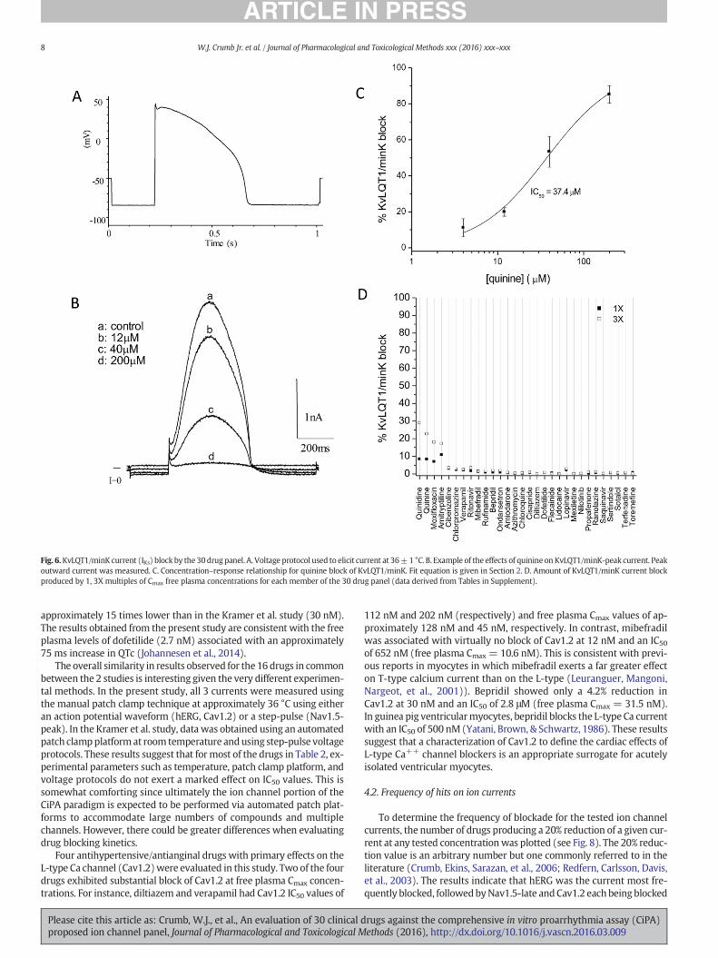
Fig. 1. hERG current (IKr) block by the 30 drug panel. A. Action potential waveform used to elicit hERG current at 36 ± 1 °C. B. Example of the effects of sotalol on hERG current. I = 0represents zero current. Peak outward current was measured. C. Concentration–response relationship for sotalol block of hERG. Fit equation is given in Section 2. D. Amount of hERGcurrent block produced by 1, 3X multiples of Cmax free plasma concentrations for each member of the 30 drug panel (data derived from Tables in Supplement).
3W.J. Crumb Jr. et al. / Journal of Pharmacological and Toxicological Methods xxx (2016) xxx–xxx
3.2.2 (R Foundation for Statistical Computing, Vienna, Austria). Theequation is:
y ¼ V maxxn
kn þ xn
where Vmax = 100, k, and n are unconstrained variables. Datatables with individual values for each drug are given in Supplement 1.
3. Results
3.1. hERG (IKr)
The hERG current is an important current in repolarization of themyocardium and the most common target for QT prolonging drugs.The effect of drugs on the amplitude of the hERG current was assessedusing the protocol shown in Fig. 1A. This voltage protocol elicited acurrent that peaked at voltages corresponding to the repolarizationphase of the action potential (Fig. 1B). As an example, addition of QTprolonging drug sotalol produced a concentration-dependent reductionof hERG current with an IC50 value of 86.4 μM (Fig. 1C). Fig. 1D plots thepercent hERG block associatedwith all 30 drugs at the free plasma Cmax.As indicated, drugs commonly associatedwithQT prolongation producea marked blockade of hERG current at therapeutic concentrations (e.g.quinidine, dofetilide). To assess the rate-dependence of hERG block, asubset of 6 drugswere evaluated at near IC50 concentrations by applying
Please cite this article as: Crumb, W.J., et al., An evaluation of 30 clinical dproposed ion channel panel, Journal of Pharmacological and Toxicological M
the voltage protocol every second. The block produced by quinidine(0.3 μM), moxifloxacin (70 μM), dofetilide (2 nM), ranolazine(6.9 μM), diltiazem (12.5 μM), and verapamil (0.5 μM) at 1 Hz wasless than 6% greater than that observed at 0.1 Hz, suggesting a lack ofrate-dependent block of hERG current by these tested drugs at theserates. This however does not exclude the possibility that rate-dependent changes may be observed with other drugs. Time-controlexperiments indicate that over the time course of a typical experiment(5–6 min) hERG current amplitude was reduced by 2.5 ± 1.9% (datanot shown; n = 6).
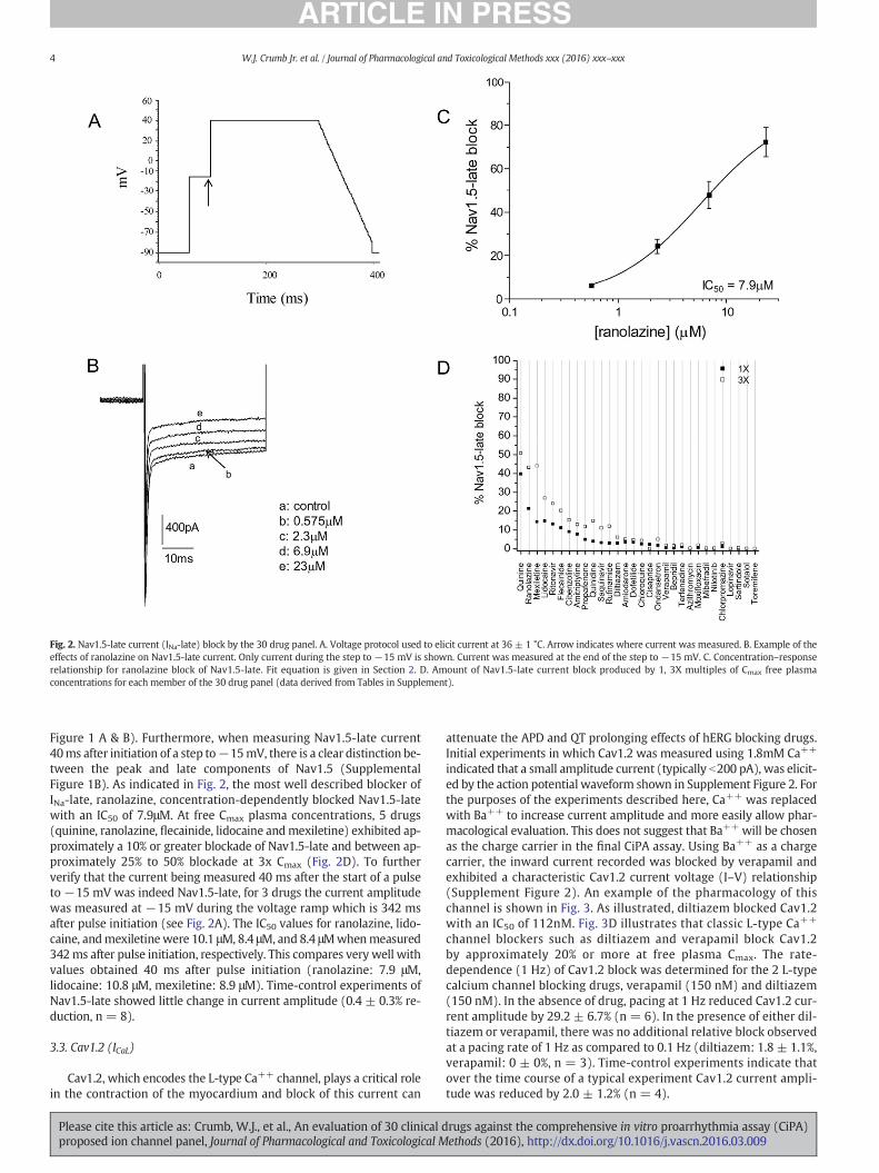
3.2. Nav1.5-late (INa-late)
TheNa current flowing during the plateau of the action potential hasbeen shown to contribute to action potential duration (APD). This cur-rent has been designated INa-late. Blockade of INa-late can shorten APDand QT, attenuate prolongations resulting fromhERG block and preventearly after depolarizations. At 37 °C, Nav1.5 normally decays rapidly andwithin approximately 3 ms there is little measurable current remaining(Supplemental Figure 1A). In order to observe Nav1.5-late, the normalprocess of channel inactivation must be altered. This is typically donewith a toxin such as ATX-II or veratridine (Belardinelli et al., 2013;Chevalier, Amuzescu, Gawali, et al., 2014). In the present experiments,Nav1.5-late was elicited by the application of 50 μM veratridine to thebath solution. In the presence of veratridine, INa decays much moreslowly and a measurable late current is easily observed (Supplemental
rugs against the comprehensive in vitro proarrhythmia assay (CiPA)ethods (2016), http://dx.doi.org/10.1016/j.vascn.2016.03.009
Fig. 2. Nav1.5-late current (INa-late) block by the 30 drug panel. A. Voltage protocol used to elicit current at 36 ± 1 °C. Arrow indicates where current was measured. B. Example of theeffects of ranolazine on Nav1.5-late current. Only current during the step to −15 mV is shown. Current was measured at the end of the step to −15 mV. C. Concentration–responserelationship for ranolazine block of Nav1.5-late. Fit equation is given in Section 2. D. Amount of Nav1.5-late current block produced by 1, 3X multiples of Cmax free plasmaconcentrations for each member of the 30 drug panel (data derived from Tables in Supplement).
4 W.J. Crumb Jr. et al. / Journal of Pharmacological and Toxicological Methods xxx (2016) xxx–xxx
Figure 1 A & B). Furthermore, when measuring Nav1.5-late current40ms after initiation of a step to−15mV, there is a clear distinction be-tween the peak and late components of Nav1.5 (SupplementalFigure 1B). As indicated in Fig. 2, the most well described blocker ofINa-late, ranolazine, concentration-dependently blocked Nav1.5-latewith an IC50 of 7.9μM. At free Cmax plasma concentrations, 5 drugs(quinine, ranolazine, flecainide, lidocaine andmexiletine) exhibited ap-proximately a 10% or greater blockade of Nav1.5-late and between ap-proximately 25% to 50% blockade at 3x Cmax (Fig. 2D). To furtherverify that the current being measured 40 ms after the start of a pulseto −15 mV was indeed Nav1.5-late, for 3 drugs the current amplitudewas measured at −15 mV during the voltage ramp which is 342 msafter pulse initiation (see Fig. 2A). The IC50 values for ranolazine, lido-caine, andmexiletinewere 10.1 μM, 8.4 μM, and 8.4 μMwhenmeasured342ms after pulse initiation, respectively. This compares very well withvalues obtained 40 ms after pulse initiation (ranolazine: 7.9 μM,lidocaine: 10.8 μM, mexiletine: 8.9 μM). Time-control experiments ofNav1.5-late showed little change in current amplitude (0.4 ± 0.3% re-duction, n = 8).
3.3. Cav1.2 (ICaL)
Cav1.2, which encodes the L-type Ca++ channel, plays a critical rolein the contraction of the myocardium and block of this current can
Please cite this article as: Crumb, W.J., et al., An evaluation of 30 clinical dproposed ion channel panel, Journal of Pharmacological and Toxicological M
attenuate the APD and QT prolonging effects of hERG blocking drugs.Initial experiments in which Cav1.2 was measured using 1.8mM Ca++
indicated that a small amplitude current (typically b200 pA), was elicit-ed by the action potential waveform shown in Supplement Figure 2. Forthe purposes of the experiments described here, Ca++ was replacedwith Ba++ to increase current amplitude and more easily allow phar-macological evaluation. This does not suggest that Ba++ will be chosenas the charge carrier in the final CiPA assay. Using Ba++ as a chargecarrier, the inward current recorded was blocked by verapamil andexhibited a characteristic Cav1.2 current voltage (I–V) relationship(Supplement Figure 2). An example of the pharmacology of thischannel is shown in Fig. 3. As illustrated, diltiazem blocked Cav1.2with an IC50 of 112nM. Fig. 3D illustrates that classic L-type Ca++
channel blockers such as diltiazem and verapamil block Cav1.2by approximately 20% or more at free plasma Cmax. The rate-dependence (1 Hz) of Cav1.2 block was determined for the 2 L-typecalcium channel blocking drugs, verapamil (150 nM) and diltiazem(150 nM). In the absence of drug, pacing at 1 Hz reduced Cav1.2 cur-rent amplitude by 29.2 ± 6.7% (n = 6). In the presence of either dil-tiazem or verapamil, there was no additional relative block observedat a pacing rate of 1 Hz as compared to 0.1 Hz (diltiazem: 1.8 ± 1.1%,verapamil: 0 ± 0%, n = 3). Time-control experiments indicate thatover the time course of a typical experiment Cav1.2 current ampli-tude was reduced by 2.0 ± 1.2% (n = 4).
rugs against the comprehensive in vitro proarrhythmia assay (CiPA)ethods (2016), http://dx.doi.org/10.1016/j.vascn.2016.03.009
5W.J. Crumb Jr. et al. / Journal of Pharmacological and Toxicological Methods xxx (2016) xxx–xxx
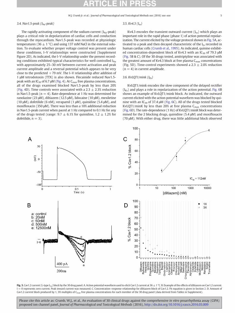
3.4. Nav1.5-peak (INa-peak)
The rapidly activating component of the sodium current (INa-peak)plays a critical role in depolarization of cardiac cells and conductionthrough the myocardium. Nav1.5-peak was recorded at physiologictemperatures (36 ± 1 °C) and using 137 mMNaCl in the external solu-tion. To evaluate whether proper voltage control was present underthese conditions, I–V relationships were constructed (SupplementFigure 2D). As indicated, the I–V relationship under the present record-ing conditions exhibited typical characteristics for well controlled INawith approximately 25–30 mV between current activation and peakcurrent amplitude and a reversal potential which appears to be veryclose to the predicted +79 mV. The I–V relationship after addition of1 μM tetrodotoxin (TTX) is also shown. Flecainide reduced Nav1.5-peak with an IC50 of 6.7 μM (Fig. 4). At Cmax free plasma concentrations,all of the drugs examined blocked Nav1.5-peak by less than 20%(Fig. 4D). Time controls were associated with a 2.3 ± 2.3% reductionin Nav1.5-peak (n = 4). Rate-dependence at 1 Hz was determined forranolazine (23 μM), diltiazem (12.5 μM), lidocaine (10 μM), mexiletine(10 μM), dofetilide (6 nM), verapamil (1 μM), quinidine (5.4 μM), andmoxifloxacin (350 μM). There was less than a 10% additional reductionin Nav1.5-peak current when paced at 1 Hz compared to 0.1 Hz for anyof the drugs tested (range: 9.7 ± 6.1% for quinidine, 1.2 ± 1.2% fordofetilide, n = 3).
Fig. 3.Cav1.2 current (L-type ICa) block by the 30drugpanel. A. Action potentialwaveformusedI = 0 represents zero current. Peak inward current was measured. C. Concentration–responseCav1.2 current block produced by 1, 3X multiples of Cmax free plasma concentrations for each
Please cite this article as: Crumb, W.J., et al., An evaluation of 30 clinical dproposed ion channel panel, Journal of Pharmacological and Toxicological M
3.5. Kv4.3 (Ito)
Kv4.3 encodes the transient outward current (Ito) which plays animportant role in the rapid phase (phase 1) of action potential repolar-ization. The current elicited by the voltage protocol shown in Fig. 5A, ac-tivated to a peak and then decayed characteristic of the Ito recorded inhuman cardiac cells (Crumb et al., 1995). As indicated, quinine exhibit-ed concentration-dependent block of Kv4.3 with an IC50 of 79.3 μM(Fig. 5B & C). Of the 30 drugs tested, amitriptyline was associated withthe greatest amount of Kv4.3 block at free plasma Cmax concentrations(Fig. 5D). Time-control experiments showed a 2.3 ± 2.0% reduction(n = 4) in current amplitude.
3.6. KvLQT1/mink (IKs)
KvLQT1/mink encodes the slow component of the delayed rectifier(IKs) and plays a role in repolarization of the action potential. Fig. 6Bshows an example of KvLQT1/mink block. As indicated, the outwardcurrent elicitedwith the action potential waveformwas blocked by qui-nine with an IC50 of 37.4 μM (Fig. 6C). All of the drugs tested blockedKvLQT1/minK by less than 20% at free plasma Cmax concentrations(Fig. 6D). The rate-dependence (1Hz) of KvLQT1/minK blockwas deter-mined for the 2 blocking drugs, quinidine (5.4 μM) and moxifloxacin(70 μM). With either drug, there was little additional block observed
to elicit Cav1.2 current at 36±1 °C. B. Example of the effects of diltiazemonCav1.2 current.relationship for diltiazem block of Cav1.2. Fit equation is given in Section 2. D. Amount ofmember of the 30 drug panel (data derived from Tables in Supplement).
rugs against the comprehensive in vitro proarrhythmia assay (CiPA)ethods (2016), http://dx.doi.org/10.1016/j.vascn.2016.03.009
Fig. 4.Nav1.5-peak current (INa-peak) block by the 30 drug panel. A. Voltage protocol used to elicit current at 36± 1 °C. Arrow indicates where current was measured. B. Example of theeffects of flecainide on Nav1.5-peak current. Only current during the step to −15 mV is shown. Peak inward current at the beginning of the step to −15 mV was measured. C.Concentration–response relationship for flecainide block of Nav1.5-peak. Fit equation is given in Section 2. D. Amount of Nav1.5-peak current block produced by 1, 3X multiples ofCmax free plasma concentrations for each member of the 30 drug panel (data derived from Tables in Supplement).
6 W.J. Crumb Jr. et al. / Journal of Pharmacological and Toxicological Methods xxx (2016) xxx–xxx
at a pacing rate of 1 Hz as compared to 0.1 Hz (quinidine: 3.8 ± 2.2%,moxifloxacin: 2.6 ± 2.6%, n = 3). Time-control experiments showedan 8.2 ± 0.4% reduction (n = 4) in current amplitude.
3.7. Kir2.1 (IK1)
Kir2.1 encodes the inwardly rectifying potassium current (IK1)which helps to set the resting membrane potential in cardiac cells andalso contributes to the terminal phase of repolarization of the action po-tential. Using the voltage protocol shown in Fig. 7A, an inward current isactivated that is blocked by chlorpromazine with an IC50 of 9.3 μM. Asillustrated, none of the drugs tested reached a 10% mean blockade ofKir2.1 at free plasma Cmax concentrations (Fig. 7D).
3.8. Relative incidence of channel block
It is important to gain insight into the relative frequencywith whichthe ion currents which have been suggested to be included in routinescreening are affected by these 30 drugs. Taking a 20% reduction in cur-rent amplitude as an arbitrary marker, the relative frequency withwhich the tested ion currents were blocked at any concentration, 1xtherapeutic free plasma Cmax, and 3x Cmax is shown in Fig. 8. As expect-ed, hERG is themost frequently blocked current. Nav1.5-late and Cav1.2
Please cite this article as: Crumb, W.J., et al., An evaluation of 30 clinical dproposed ion channel panel, Journal of Pharmacological and Toxicological M
were the next most frequently blocked currents with approximately 4times fewer drugs blocking by 20% or more at 1x and 3x free plasmaCmax. In contrast, at 1x therapeutic Cmax, the other currents testedwere not blocked by 20% or more by any of the drugs tested. At a 3xmultiple of free plasma Cmax, Kv4.3 was blocked approximately as fre-quently as Nav1.5-late and Cav1.2, while Nav1.5-peak and KvLQT1/mink were less frequently blocked. Kir2.1 was not blocked by anydrug tested by 20% or more at a 3x multiple of free plasma Cmax.
3.9. Relationship between channel block and arrhythmic risk
The relationship between ion channel block and proarrhythmic riskwas examined for a subset of 12 drugs that were rated by the CiPA clin-ical working group to have low, intermediate or high risk of torsade depointes. As indicated in Figs. 9 and 10, at both a 1X and a 3X free plasmaCmax concentration, drugs which are classified as possessing a high riskfor pro-arrhythmic events are associated with a large and rather selec-tive block of hERG. In contrast, drugs in the low risk category block ei-ther Cav1.2 or Nav1.5-late more so or to a similar degree as hERG atCmax. Drugs in the intermediate category block hERG more than othercurrents but on average are associated with less hERG block at Cmax
than members of the high risk category. Of note, the 40% hERG blockshown in Fig. 9 for terfenadine (intermediate risk drug) is associated
rugs against the comprehensive in vitro proarrhythmia assay (CiPA)ethods (2016), http://dx.doi.org/10.1016/j.vascn.2016.03.009
Fig. 5. Kv4.3 current (Ito) block by the 30 drug panel. A. Voltage protocol used to elicit current at 22 ± 1 °C. Arrow indicates where current was measured. B. Example of the effects ofquinine on Kv4.3-peak current. Peak outward current was measured. C. Concentration–response relationship for quinine block of Kv4.3. Fit equation is given in Section 2. D. Amount ofKv4.3 current block produced by 1, 3X multiples of Cmax free plasma concentrations for each member of the 30 drug panel (data derived from Tables in Supplement).
7W.J. Crumb Jr. et al. / Journal of Pharmacological and Toxicological Methods xxx (2016) xxx–xxx
with a Cmax of 9 nM, a concentration only achieved after metabolic inhi-bition. However, a more standard Cmax of 0.3 nM is only associated with8% hERG block, which could contribute to it being an intermediate ver-sus high risk drug. Other factors such as the kinetics of drug binding tothe hERG channel may also be important for differentiating intermedi-ate from high risk drugs.
4. Discussion
For more than a decade, the cardiac potassium channel hERG hasbeen the focus of both regulatory agencies and drug developers. Whilethis attention has been warranted since drug induced blockade of thischannel has been associated with QT prolongation and in rare cases tor-sade de pointes, the singular focus on this channel has likely led to un-warranted drug attrition and labeling (Stockbridge, Morganroth, Shah,et al., 2013). In virtually all cases of drug-associated torsade de pointes,the offending drug has been shown to selectively block hERG at relevantplasma concentrations. However, there are drugswhichblock hERG anddespite this activity are associated with little or no QT prolongation andminimal torsade de pointes risk, sometimes due to block of the L-typeICa (e.g. verapamil) or INa-late (e.g. ranolazine). In the present study,we observed that the most frequently blocked channels at clinical freeCmax concentrations were hERG, Nav1.5-late and Cav1.2. While the
Please cite this article as: Crumb, W.J., et al., An evaluation of 30 clinical dproposed ion channel panel, Journal of Pharmacological and Toxicological M
substantial hit rate for hERG is expected, results from the presentstudy using the manual patch clamp technique under physiologic con-ditions highlight for the first time that a large number of drugs alsoblock Nav1.5-late and/or Cav1.2 currents, which can reduce the risk oftorsade de pointes caused by hERG block (Antoons, Oros, Beelman,et al., 2010; Antzelevitch, Belardinelli, Zygmunt, et al., 2004; Fauchier,Babuty, Autret, et al., 1999; Gintant et al., 2006; January & Riddle,1989). This is consistent with the four CiPA low risk drugs in thisstudy having equal or greater Nav1.5-late block (ranolazine andmexiletine) or Cav1.2 block (verapamil, diltiazem) than hERG block,while the intermediate and high risk drugs had greater hERG blockthan other channels.
4.1. Comparison to published data
The results obtained in the present study were compared to previ-ously published results (Kramer, Obejero-Paz, Myatt, et al., 2013).While many of the drugs evaluated in the present study have publisheddata for various ion channels, the Kramer et al. dataset has many of thedrugs tested here and contains data on hERG, Nav1.5-peak, and Cav1.2.As indicated in Table 2, with few exceptions, the IC50 values from the 2studies are very similar. One notable exception is dofetilide, which hasan IC50 value for block of hERG in the present study (2 nM) which is
rugs against the comprehensive in vitro proarrhythmia assay (CiPA)ethods (2016), http://dx.doi.org/10.1016/j.vascn.2016.03.009
Fig. 6.KvLQT1/minK current (IKs) block by the 30drug panel. A. Voltage protocol used to elicit current at 36±1 °C. B. Example of the effects of quinine on KvLQT1/minK-peak current. Peakoutward current was measured. C. Concentration–response relationship for quinine block of KvLQT1/minK. Fit equation is given in Section 2. D. Amount of KvLQT1/minK current blockproduced by 1, 3X multiples of Cmax free plasma concentrations for each member of the 30 drug panel (data derived from Tables in Supplement).
8 W.J. Crumb Jr. et al. / Journal of Pharmacological and Toxicological Methods xxx (2016) xxx–xxx
approximately 15 times lower than in the Kramer et al. study (30 nM).The results obtained from the present study are consistent with the freeplasma levels of dofetilide (2.7 nM) associated with an approximately75 ms increase in QTc (Johannesen et al., 2014).
The overall similarity in results observed for the 16 drugs in commonbetween the 2 studies is interesting given the very different experimen-tal methods. In the present study, all 3 currents were measured usingthe manual patch clamp technique at approximately 36 °C using eitheran action potential waveform (hERG, Cav1.2) or a step-pulse (Nav1.5-peak). In the Kramer et al. study, data was obtained using an automatedpatch clampplatformat room temperature andusing step-pulse voltageprotocols. These results suggest that formost of the drugs in Table 2, ex-perimental parameters such as temperature, patch clamp platform, andvoltage protocols do not exert a marked effect on IC50 values. This issomewhat comforting since ultimately the ion channel portion of theCiPA paradigm is expected to be performed via automated patch plat-forms to accommodate large numbers of compounds and multiplechannels. However, there could be greater differences when evaluatingdrug blocking kinetics.
Four antihypertensive/antianginal drugs with primary effects on theL-type Ca channel (Cav1.2)were evaluated in this study. Two of the fourdrugs exhibited substantial block of Cav1.2 at free plasma Cmax concen-trations. For instance, diltiazem and verapamil had Cav1.2 IC50 values of
Please cite this article as: Crumb, W.J., et al., An evaluation of 30 clinical dproposed ion channel panel, Journal of Pharmacological and Toxicological M
112 nM and 202 nM (respectively) and free plasma Cmax values of ap-proximately 128 nM and 45 nM, respectively. In contrast, mibefradilwas associated with virtually no block of Cav1.2 at 12 nM and an IC50of 652 nM (free plasma Cmax = 10.6 nM). This is consistent with previ-ous reports in myocytes in which mibefradil exerts a far greater effecton T-type calcium current than on the L-type (Leuranguer, Mangoni,Nargeot, et al., 2001)). Bepridil showed only a 4.2% reduction inCav1.2 at 30 nM and an IC50 of 2.8 μM (free plasma Cmax = 31.5 nM).In guinea pig ventricularmyocytes, bepridil blocks the L-type Ca currentwith an IC50 of 500 nM (Yatani, Brown, & Schwartz, 1986). These resultssuggest that a characterization of Cav1.2 to define the cardiac effects ofL-type Ca++ channel blockers is an appropriate surrogate for acutelyisolated ventricular myocytes.
4.2. Frequency of hits on ion currents
To determine the frequency of blockade for the tested ion channelcurrents, the number of drugs producing a 20% reduction of a given cur-rent at any tested concentrationwas plotted (see Fig. 8). The 20% reduc-tion value is an arbitrary number but one commonly referred to in theliterature (Crumb, Ekins, Sarazan, et al., 2006; Redfern, Carlsson, Davis,et al., 2003). The results indicate that hERG was the current most fre-quently blocked, followedbyNav1.5-late andCav1.2 each beingblocked
rugs against the comprehensive in vitro proarrhythmia assay (CiPA)ethods (2016), http://dx.doi.org/10.1016/j.vascn.2016.03.009
Fig. 7. Kir2.1 current (IK1) block by the 30 drug panel. A. Voltage protocol used to elicit current at 36 ± 1 °C. Arrow indicates where current was measured. B. Example of the effects ofchlorpromazine on Kir2.1 current. Trace is truncated to show current during ramp to −120 mV. Peak inward current was measured at −120 mV at the end of the ramp. C.Concentration–response relationship for chlorpromazine block of Kir2.1. Fit equation is given in Section 2. D. Amount of Kir2.1 current block produced by 1, 3X multiples of Cmax freeplasma concentrations for each member of the 30 drug panel (data derived from Tables in Supplement).
9W.J. Crumb Jr. et al. / Journal of Pharmacological and Toxicological Methods xxx (2016) xxx–xxx
by greater than 60% of the drugs tested. Kir2.1 was the least frequentlyblocked current. In addition, the frequency of block at 1x and 3x the freeplasma Cmaxwas plotted. This showed a very different profilewith hERGbeing blocked by 20% ormore by 37% of the drugs and all other currentsbeing blocked by less than 10% of the drugs tested. No drugs blocked
Fig. 8. Relative ion current panel activity. Plot of the % of drugs out of 30 which blocked the inplasma Cmax (B), and at 3X the free plasma Cmax (C). Reference data was obtained from tables
Please cite this article as: Crumb, W.J., et al., An evaluation of 30 clinical dproposed ion channel panel, Journal of Pharmacological and Toxicological M
Nav1.5-peak, Kv4.3, LvLQT1/mink, or Kir2.1 by 20% or more at a 1xCmax concentration. At a 3x Cmax concentration, hERG was blocked by70% of the drugs tested whereas all other currents were blocked by20% or less of the drugs tested. None of the drugs tested blockedKir2.1 by 20% or more at a 3x Cmax concentration.
dicated ion currents by at least 20% at any of the concentrations tested (A), at 1X the freein the Supplement.
rugs against the comprehensive in vitro proarrhythmia assay (CiPA)ethods (2016), http://dx.doi.org/10.1016/j.vascn.2016.03.009
Fig. 9. Plot of the ion channel blocking profile ofmembers of the high (top row), intermediate (middle row), and low (bottom row) CiPA proarrhythmic risk categories. % block representsblock at 1X free plasma Cmax. Data was derived from Tables in the Supplement. The Cmax chosen for terfenadine was 9 nM, a concentration achieved after metabolic inhibition. A morestandard Cmax of 0.3 nM is only associated with 8% hERG block.
Fig. 10. Plot of the ion channel blocking profile ofmembers of the high (top row), intermediate (middle row), and low (bottom row) CiPA proarrhythmic risk categories. % block representsblock at 3X free plasma Cmax. Data was derived from Tables in the Supplement.
10 W.J. Crumb Jr. et al. / Journal of Pharmacological and Toxicological Methods xxx (2016) xxx–xxx
Please cite this article as: Crumb, W.J., et al., An evaluation of 30 clinical drugs against the comprehensive in vitro proarrhythmia assay (CiPA)proposed ion channel panel, Journal of Pharmacological and Toxicological Methods (2016), http://dx.doi.org/10.1016/j.vascn.2016.03.009
All values are IC50 values or the highest concentration tested (in μM). First value in eachpair is from the present study, second value from Kramer et al., 2013. In some cases inthe present study and IC50 valuewas not reached. In those cases, the IC50 value is designat-ed as being N than the highest concentration tested.
11W.J. Crumb Jr. et al. / Journal of Pharmacological and Toxicological Methods xxx (2016) xxx–xxx
4.3. Stratification of safety risk
Included in the drugs tested were 4 members from each of the CiPArisk categories (high, intermediate and low). These categories werebased upon the risk of drug-associated torsade de pointes. We com-pared ion channel block against all tested ion currents at free plasmaCmax values of 1x and 3x. This method has been used before on hERG,Cav1.2, and peak Nav1.5 using relative IC50 values. As indicated inFigs. 9 & 10, members of the high risk category blocked hERG at thera-peutic free plasma Cmax either exclusively or to a much greater extentthan any other current examined. In the intermediate category, drugblock of hERG was on average less than in the high risk category,supporting a lower risk of torsade de pointes. In the low risk category,the members were associated with a greater or equal block of eitherCav1.2 or Nav1.5-late when compared to block of hERG. These resultsclearly indicate the need for testing drug candidates against a panel ofion channels.
As indicated in Figs. 9 and 10, the ion current blocking profile forsome members of the intermediate torsade de pointes risk category issimilar to that observed with some members of the high risk category.There are severalpossible reasons for this including(1)pharmacokineticor drug interaction propertieswhichmay lead to higher plasma concen-trations some in patients, (2) differences in risk factors in patient popu-lations receiving each drug, (3) the assignment of drugs into high andintermediate risk categories may be imperfect, and (4) the kinetics ofthe drug block, particularly for hERG, may make some drugs moreproarrhythmic than others. The kinetics of drug binding to the hERGchannel and its ability to improve risk stratification is currently being in-vestigated further by the CiPA ion channel and in silico working groups(Fermini et al., 2016).
In a recent clinical study of quinidine and dofetilide, the free plasmaconcentrations associated with the approximately 70% and 55% reduc-tion in hERG current amplitude, respectively (see Fig. 9) was associatedwith a79ms and 74ms increase in QTc (Johannesen et al., 2014) and T-wave morphology changes (Vicente, Johannesen, Mason, et al., 2015).Furthermore, in the same study, ranolazine was associated with a12 ms increase in QTc and verapamil was associated with no increasein QTc. The ion channel profile for verapamil supports the clinical resultsin that there is more block of Cav1.2 than hERG at therapeutic concen-trations. The observation of a modest increase in QTc upon exposureto therapeutic concentrations of ranolazine suggests that the hERGblock induced QTc prolongation is not completely counteracted byblock of Nav1.5-late, but is enough to counteract torsade de pointes
Please cite this article as: Crumb, W.J., et al., An evaluation of 30 clinical dproposed ion channel panel, Journal of Pharmacological and Toxicological M
risk. However, the QTc prolongation associated with ranolazine has aspecific ‘signature’ that involves Tpeak-Tend prolongation without pro-longation of the heart rate corrected J-Tpeak (J-Tpeakc) interval(Johannesen et al., 2014). Additional recent clinical study suggests thatthe J-Tpeakc interval is a better biomarker (than QTc) in assessing thebalance of inward and outward current block and predictor of low tor-sade risk drugs (Johannesen et al., 2014). The CiPA clinical phase 1ECG working group is expanding this analysis to additional drugs andwill propose a framework for potential implementation of this new ap-proach at an April 2016 Cardiac Safety Research Consortium meeting.
4.4. Limitations
The following are limitations of the present study. 1). The action po-tential waveform used was recorded from rabbit, and not human, ven-tricle. While this waveform has been used previously to characterizehERG (Zhou, Gong, Ye, et al., 1998), we are cognizant that the datamay be more relevant if a human ventricular AP was used instead. 2).Kv4.3 current was measured at room temperature and not physiologictemperature, aswith the other ion currents. Thiswas done to allow sep-aration of the capacitive transient from peak Kv4.3 current. It is notknown if the Kv4.3-drug interaction, for the drugs tested here, is tem-perature sensitive. 3). The drug effects on ion currents were tested at apacing rate of 0.1 Hz. This pacing rate was chosen because it is oftenused in the literature and it represents a commonpacing rate for all cur-rents tested that was not associated with significant rundown. Howev-er, currents were evaluated against some drugs at a pacing rate of 1 Hz,which showed limited differences from the 0.1 Hz data. 4). Nav1.5-latewas elicited by the use of veratridine. Although sodium channel inacti-vation modifiers such as veratridine and ATX-II are commonly used toincrease the amplitude of late-INa, it is not known if they may inter-fere/alter the effects of late INa blockers. 5). In the present study, drug-effects were not “washed out” to provide insight into whether the re-duction in current amplitude was due to actual drug block or currentrundown. However, time controls suggest that any reduction in currentamplitudewas due primarily to drug-related ion current block. Further-more, not all drug blocking effects, particularly at higher concentrations,are able to be “washed out”. 6). The Ca++ andMg++ in the external so-lution can have blocking effects on hERG current (Ho, Kim, Lee, & Earm,1998; Po, Wang, Yang, et al., 1999). This may make it difficult in calcu-lating the actual drug-hERG current blocking effects. However, the ionconcentrations were physiologic and it is unlikely that Ca++ andMg++ will be absent in the plasma of individuals taking these drugs.
The in silico and ion channel working group for the CiPA initiativeare currently studying voltage clamp protocols that model dynamicdrug–ion channel interactions for hERG, and potentially for other chan-nels (Fermini et al., 2016). Thus, final CiPA voltage clamp protocols forhERG, and potentially other channels, may differ substantially fromthose used in this study. However, a critical first step is to identify IC50values or the maximal percent block at clinically relevant drug concen-trations to determine which channels are the most important channelsto be assessed under CiPA.While this ion channel data could also be en-tered into in silico models, such as the O'Hara-Rudy model (O'Hara,Virág, Varró, & Rudy, 2011), we elected to not do that here because itis beyond the scope of this manuscript and because the in silicoproarrhythmia metrics have not been finalized. Rapidly publishing thisdata will allow any investigators to use it for in silico modeling. Finally,drugs were selected with a stratified risk of torsade de pointes, existingclinical ECG data, and with a range of ion channel effects. However, thehit rates for block of the different channels are only reflective of the 30drugs in this study.
4.5. Conclusion
This is the first study of its kind to examine the effects of 30 clinicaldrugs against the 7 ion channel currents currently proposed to makeup
rugs against the comprehensive in vitro proarrhythmia assay (CiPA)ethods (2016), http://dx.doi.org/10.1016/j.vascn.2016.03.009
12 W.J. Crumb Jr. et al. / Journal of Pharmacological and Toxicological Methods xxx (2016) xxx–xxx
the CiPA ion channel panel. The results of the present study highlightthe importance of drug-induced block of hERG, Nav1.5-late andCav1.2 at clinically relevant concentrations. These were the most fre-quently blocked currents and are likely the most important in terms ofincreased risk for torsade de pointes (blocking hERG) vs. decreasingrisk (blocking Nav1.5-late and/or Cav1.2). This was consistent withthe 12 CiPA drugs, where high and intermediate risk drugs had greaterhERG block than other channels, while low risk drugs had equivalent orgreater Nav1.5 block (ranolazine and mexiletine) or Cav1.2 block(verapamil, diltiazem) than hERG block. Nav1.5-peak block was thenext most commonly blocked channel, and its importance in conduc-tion and potential for use-dependent block that was not thoroughlyassessed here, suggest that it also is important to be assessed underCiPA. Based upon the 30 drugs tested in this study, the less frequenthit rates for Kv4.3, KvLQT1/mink and Kir2.1 suggest that these channelsmay be less critical for assessment of all drugs under CiPA. This shouldbe further evaluated with consideration of potential false positivehit rates along with the impact of block of these channels has onproarrhythmic risk in the CiPA in silico action potential model.
Conflict of interest statement
This work was funded by the US Food and Drug AdministrationCritical Path Initiative.
Acknowledgments
The step ramp protocols used in this study were developed by theCiPA ion channel working group. The authors would like to thank Drs.Bernard Fermini, Najah Abi Gerges, Jules Hancox, Adam Hill, JamieVandenberg and Norman Stockbridge for their expert opinions and re-view of the manuscript. This project was supported by the FDA CriticalPath Initiative and appointments to the Research Participation Pro-grams at the Oak Ridge Institute for Science and Education through aninteragency agreement between the Department of Energy and FDA.
Appendix A. Supplementary data
Supplementary data to this article can be found online at http://dx.doi.org/10.1016/j.vascn.2016.03.009.
References
Antoons, G., Oros, A., Beelman, J. D. M., et al. (2010). late na+ current inhibition byranolazine reduces torsades de pointes in the chronic atrioventricular block dogmodel. Journal of the American College of Cardiology, 55, 801–809.
Antzelevitch, C., Belardinelli, L., Zygmunt, A. C., et al. (2004). Electrophysiological effects ofranolazine, a novel antianginal agent with antiarrhythmic properties. Circulation, 110,904–910.
Please cite this article as: Crumb, W.J., et al., An evaluation of 30 clinical dproposed ion channel panel, Journal of Pharmacological and Toxicological M
Belardinelli, L., et al. (2013). A novel, potent, and selective inhibitor of cardiac late sodiumcurrent suppresses experimental arrhythmias. Journal of Pharmacology andExperimental Therapeutics, 344, 23–32.
Chevalier, M., Amuzescu, B., Gawali, V., et al. (2014). Late cardiac sodium current can beassessed using automated patch-clamp. F1000Research, 3. (pp. 245) doi:10.12688/f1000research.5544.1.
Crumb, W. J., Ekins, S., Sarazan, R. D., et al. (2006). Effects of antipsychotic drugs on I to, INa, I sus, I K1, and hERG: QT prolongation, structure activity relationship, and networkanalysis. Pharmaceutical Research, 23, 1133–1143.
Crumb,W. J., Jr., Pigott, J. D., & Clarkson, C.W. (1995). Description of a nonselective cationcurrent in human atrium. Circulation Research, 77, 950–956.
Fauchier, L., Babuty, D., Autret, M. L., et al. (1999). Effect of verapamil on QT intervaldynamicity. American Journal of Cardiology, 83, 807–808.
Fermini, B., Hancox, J. C., Abi-Gerges, N., et al. (2016). A new perspective in the field of car-diac safety testing through the comprehensive in vitro proarrhythmia assay para-digm. Journal of Biomolecular Screening, 21, 1–11.
Gintant, G., Su, Z., Martin, R. L., & Cox, B. F. (2006). Utility of hERG assays as surrogatemarkers of delayed cardiac repolarization and QT safety. Toxicologic Pathology, 34,81–90.
Grant, A. O. (2009). Basic science for the clinical electrophysiologist. Cardiac ion channels.Circulation. Arrhythmia and Electrophysiology, 2, 185–194.
Ho, W. K., Kim, I., Lee, C. O., & Earm, Y. E. (1998). Voltage-dependent blockade of HERGchannels expressed in Xenopus oocytes by external Ca2+ and Mg2+. The Journalof Physiology, 507(Pt 3), 631–638.
January, C. T., & Riddle, J. M. (1989). Early afterdepolarizations: mechanism of inductionand block. A role for L-type Ca2+ current. Circulation Research, 64, 977–990.
Johannesen, L., Vicente, J., Mason, J. W., et al. (2014). Differentiating drug-induced multi-channel block on the electrocardiogram: randomized study of dofetilide, quinidine,ranolazine, and verapamil. Clinical Pharmacology and Therapeutics, 96, 549–558.
Johannesen, L., Vicente, J., Mason, J. W., et al. (2016). Late sodium current block for drug-induced long QT syndrome: Results from a prospective clinical trial. ClinicalPharmacology and Therapeutics, 99, 214–223.
Kramer, J., Obejero-Paz, C. A., Myatt, G., et al. (2013). MICE models: Superior to the HERGmodel in predicting torsade de pointes. Science Reports, 3, 2100.
Leuranguer, V., Mangoni, M. E., Nargeot, J., et al. (2001). Inhibition of T-type and L-typecalcium channels bymibefradil: Physiologic and pharmacologic bases of cardiovascu-lar effects. Journal of Cardiovascular Pharmacology, 37, 649–661.
Nerbonne, J. M., & Kass, R. S. (2005). Molecular physiology of cardiac repolarization.Physiological Reviews, 85, 1205–1253.
O'Hara, T., Virág, L., Varró, A., & Rudy, Y. (2011). Simulation of the undiseased human car-diac ventricular action potential: Model formulation and experimental validation.PLoS Computational Biology doi: 10.1371/journal.pcbi.1002061.
Po, S. S., Wang, D.W., Yang, I. C., et al. (1999). Modulation of HERG potassium channels byextracellular magnesium and quinidine. Journal of Cardiovascular Pharmacology, 33,181–185.
Redfern, W. S., Carlsson, L., Davis, A. S., et al. (2003). Relationships between preclinicalcardiac electrophysiology, clinical QT interval prolongation and torsade de pointesfor a broad range of drugs: Evidence for a provisional safety margin in drug develop-ment. Cardiovascular Research, 58, 32–45.
Sager, P. T., Gintant, G., Turner, J. R., et al. (2014). Rechanneling the cardiac proarrhythmiasafety paradigm: A meeting report from the Cardiac Safety Research Consortium.American Heart Journal, 167, 292–300.
Stockbridge, N., Morganroth, J., Shah, R. R., et al. (2013). Dealingwith global safety issues :Was the response to QT-liability of non-cardiac drugs well coordinated? Drug Safety,36, 167–182.
Vicente, J., Johannesen, L., Mason, J. W., et al. (2015). Comprehensive T wave morphologyassessment in a randomized clinical study of dofetilide, quinidine, ranolazine, and ve-rapamil. J. Am. Heart Assoc., 4, e001615 doi: 10.1161.
Yatani, A., Brown, A. M., & Schwartz, A. (1986). Bepridil block of cardiac calcium and so-dium channels. Journal of Pharmacology and Experimental Therapeutics, 237, 9–17.
Zhou, Z., Gong, Q., Ye, B., et al. (1998). Properties of HERG channels stably expressed inHEK 293 cells studied at physiological temperature. Biophysical Journal, 74, 230–241.
rugs against the comprehensive in vitro proarrhythmia assay (CiPA)ethods (2016), http://dx.doi.org/10.1016/j.vascn.2016.03.009