40
December 2014 Journal of the American Association for Emergency Psychiatry Seth M. Powsner, M.D., Editor

December 2014
Journal of the American
Association for
Emergency Psychiatry
Seth M. Powsner, M.D., Editor

2
TABLE OF CONTENTS
Information for Contributors...………………………….…………………………………… 3
Manuscripts:
Editor’s Note ……………………………………………………………………...………………………. 4
Revival of an Antidote: Bedside Experience with Physostigmine ………………………. 5
Assessment of Suicide Risk in Psychiatric Patients Using a Brief Screener:
Validation of the SIS-MAP-scn ……………………………………………………………………… 25
Letter to the Editor:
Malingering and Homelessness ……………………………………………………………………. 34
News of the AAEP.………………………………………………………………………………….. 36
American Association
for Emergency Psychiatry
One Regency Drive
P.O. Box 30
Bloomfield, CT 06002
Phone: 888-945-5430
Fax: 860-286-0787
www.emergencypsychiatry.org

3
Emergency Psychiatry
Journal of the American Association for Emergency Psychiatry
We invite all members and colleagues in the field of Emergency Psychiatry to submit a
manuscript or book review for publication.
Information for Contributors
Emergency Psychiatry is intended to be a forum for the exchange of multidisciplinary ideas. Manuscripts
that deal with the interfaces of emergency psychiatry are welcome. This includes psychiatric evaluation of in-
dividuals in the emergency room setting, education and training in the field and research into causes, and
treatment of behavioral problems. Manuscripts are evaluated for style, clarity, consistency, and suitability.
All articles represent the opinions of the authors and not those of the Association. Except where specifically
stated, published articles are not official statements of the American Association for Emergency Psychiatry.
Manuscripts are considered for publication with the understanding that their essential substance has not
been published previously and has not been submitted simultaneously to other publications.
Manuscript Form: Only electronic transmissions of manuscripts will be accepted. Manuscripts can be
submitted directly to the Office at [email protected]. Submission of manuscripts that are
longer than 15 manuscript pages, including references, is discouraged.
Tables and Illustrations: Tables, charts, and photographs should be submitted on disk or by e-mail. Pho-
tographs should be submitted in JPEG format. Tables and charts should be executed in Excel or PowerPoint.
References: List references in alphabetical order. Each listed reference should be cited in text, and each
text citation should be listed in the References section.
Include the address, telephone number, and email address for the corresponding author on all manuscripts.
Submit manuscripts or queries to: Marie L. Westlake, Executive Assistant,
Email address: [email protected]
The mailing address of the Journal is:
Journal of the American Association for Emergency Psychiatry
One Regency Drive, P.O. Box 30, Bloomfield, CT 06002
Phone: 888-945-5430; Fax: 860-286-0787

4
President
Kimberly D. Nordstrom, M.D.
President-Elect
Leslie S. Zun, M.D., M.B.A.
Immediate Past President
Seth Powsner, M.D.
Board of Directors
Daryl K. Knox, M.D.
Jagoda Pasic, M.D., Ph.D.
Jack S. Rozel, M.D., M.S.L.
Director of Emergency Psychiatry
Research
Michael P. Wilson, M.D., Ph.D.
Social Work Liaison
Janet S. Richmond, M.S.W.
Past Presidents
Scott L. Zeller, M.D.
Anthony T. Ng, M.D.
Avrim B. Fishkind, M.D.
Jon S. Berlin, M.D.
Glenn W. Currier, M.D., M.P.H.
Rachel L. Glick, M.D.
Joseph J. Zealberg, M.D.
Michael H. Allen, M.D.
Douglas H. Hughes, M.D.
Peter L. Forster, M.D.
Ricardo Mendoza, M.D.
AAEP Executive Office Staff
Executive Director
Jacquelyn T. Coleman, ACE
Executive Assistant
Marie L. Westlake
Editor's Note:
In the manuscript entitled Revival of an Antidote: Bedside Experience with Physostigmin beginning on the next page, Dr Rasimas and colleagues remind us of two well known facts:
-- atropinic substances and drugs with atropinic side effects can cause serious delirium aka atropinic psy-chosis; and
-- physostigmine can reverse many of effects of atro-pinic toxicity. This observational study confirms old, clinical lore that physostigmine can be very helpful in the treatment of atropinic toxicity, with relatively little risk. This should cause us to question why physostig-mine is used so little in most medical center.
Poll your local emergency medicine physicians or internists for their opinion on physostigmine as an acute treatment. You may find some in favor. You will likely find most agree it works. However, you may find that many are not convinced it saves much time or morbidity when compared with simple, supportive care. And, even if they believe it might save time, your own colleagues may not believe it is worth the possible complications, even though infrequent and rarely seri-ous.
Whatever you decide, we hope this article will stimulate your thinking and some conversations with your colleagues.
---Seth Powsner, MD
www.EmergencyPsychiatry.org

5
Abstract
Anticholinergic activity is pharmacologically rele-
vant for many medicinal and natural toxins. Delirium is a
common consequence of toxicity. Although a direct anti-
dote, physostigmine is available, it fell out of use after
case reports of suspected cardiotoxicity were propagated
through the literature. Physostigmine was previously
used with high frequency and to good effect to reverse
anticholinergic delirium from a variety of compounds. It
was also employed in cases of delirium with other, some-
times unknown etiologies, without serious adverse
events.
At one toxicology center, physostigmine contin-
ues to be employed in the emergency department and
acute hospital setting. It is given 0.02 mg/kg IV at a rate
of 0.5 mg/min with repeat doses q1-2h PRN. The follow-
ing reports a six-year retrospective review of the practice
and a detailed prospective one-year observational study
of bedside use of the antidote.
1197 patients were treated with physostigmine.
The overall positive response rate was nearly 80%. The
rate of arrhythmias was 0.17%, and each event was minor
and self-limited. The rate of seizures was 0.75%; none
resulted in clinically significant sequelae or morbidity.
Cholinergic signs occurred in 6.4% of patients in pro-
spective study; diaphoresis, nausea, emesis, and incon-
tinence were all short-lived and manageable. There
were no cases of bronchorrhea or respiratory distress.
In patients with toxicity from tricyclic antidepressants,
responses were positive in 94.5% of cases. The only
documented adverse events in these cases were two
episodes of diaphoresis out of 315 patients treated.
Electrocardiographic abnormalities were not consid-
ered contraindications to physostigmine therapy, and
there were no serious adverse events regardless of QRS
duration (72 to 168 msec) or QTc interval (341 to 662
msec).
Physostigmine is a safe diagnostic and poten-
tially therapeutic antidote for cases of suspected toxic
delirium. Concerns about cardiotoxicity are unfounded.
Seizures are rare, and can be prevented with benzodi-
azepine pretreatment in cases involving suspicion of
highly epileptogenic toxins.
____________________________________
Keywords: physostigmine, antidote, anticholinergic,
overdose, delirium, tricyclic antidepressant (TCA)
Revival of an Antidote: Bedside Experience with Physostigmine
J.J. Rasimas, M.D., Ph.D.1,2,3; Kamal K. Sachdeva, M.D.1; and J. Ward Donovan, M.D.1,2
1 PinnacleHealth Toxicology Center, Harrisburg, PA; 2 Penn State College of Medicine, Department of Emergency Medicine, Hershey, PA; 3 Penn State College of Medicine, Department of Psychiatry, Hershey, PA
Corresponding Author: J.J. Rasimas, M.D., Ph.D., HealthPartners/Regions Hospital, 640 Jackson Street, MS12002A, St. Paul, MN 55101; Phone: (651) 254-1892; Fax: (651) 254-2410; Email: [email protected]
No grant funding supported this work. The authors report no conflicts of interest.
Acknowledgements: The authors thank Erica E. Smolcic, M.D. and Amanda Cresswell R.N., M.S.N., C.M.S.R.N. for assistance with chart review. We are also grateful to Kara Gemberling and Patti Metherell for arranging data extraction from the electronic medical and pharmacy records of PinnacleHealth. In addition, Jeremiah Escajeda, M.D. provided invaluable assistance with background research for the preparation of this manuscript.
MANUSCRIPTS

6
Introduction
Delirium is a syndromic presentation of seri-
ous underlying medical conditions. It is a harbinger of
increased risk of morbidity and mortality and compli-
cates care because of behavioral disturbances and im-
pediments to communication.1 Anticholinergic activi-
ty is a key factor in many delirious states. Pharmaco-
logically, it has been identified in a host of natural
sources and mediates the effects of a vast array of
medications, including anti-depressants, antihista-
mines, antiparkinsonian drugs, antipsychotics and
muscle relaxants. The widespread availability of these
medications has made them common intoxicants in
both accidental and intentional overdoses.2
The anticholinergic ingestion often presents
with a toxidrome, which includes tachycardia, mydri-
asis, dry skin and mucosae, urinary retention, ileus
and most importantly, neuropsychatric disturbance.
However, the non-polar chemistry of many causative
agents partitions them to fatty tissues including the
CNS, thereby producing delirium without consistently
yielding peripheral anticholinergic symptoms. Physo-
stigmine reverses central nervous system (CNS) ef-
fects of anticholinergic poisoning. It was widely used
in psychiatry and anesthesiology in the 1960s and
1970s.3 It is a tertiary aminocarbamate that reversibly
binds to and inhibits the action of acetyl cholinester-
ase. The result is an increase in acetylcholine at the
muscarinic synapse and competitive reversal of neu-
rotransmission blockade. The drug has been shown to
be safe and effective in reversing delirium and associ-
ated neuropsychiatric unrest in multiple studies, with
only minor adverse reactions such as emesis, saliva-
tion, and diaphoresis described in most patients.4,5
More severe reactions, however, have been reported,
including seizures and lethal arrhythmias.6,7
The majority of isolated, severe adverse events
associated with physostigmine administration report-
ed in the literature, occur in patients with tricyclic an-
tidepressant (TCA) overdoses. These patients suffer
an array toxic effects of the ingested drug in addition
to its anticholinergic activity, including γ-
aminobutyric acid (GABA) inhibition, adrenergic vol-
atility, and both sodium and potassium channel
blockade. Electrocardiographic changes are common-
ly seen in severe TCA overdoses and have often been
cited as a contraindication for physostigmine use in
TCA toxic patients.6,7 However, other authors have
suggested that the severe adverse events, such as asys-
tole and seizures are the result of TCA toxicity, itself,
and not physostigmine therapy.8 Others have pro-
posed that fast drug administrations may have con-
tributed to the severity of the reactions observed in
some of the most often cited reports of harm.9
While the safety of physostigmine has been
debated in the literature for over 25 years, many phy-
sicians err on the side of caution, and as a result, phy-
sostigmine continues to be an underutilized antidote
in even the most obvious cases of antimuscarinic poi-
soning.10 With this study combining both retrospec-
tive review and prospective observation, we present
seven years of physostigmine experience in a diverse
patient population. Our work supports the safety and
efficacy of physostigmine use as both a diagnostic and
therapeutic modality in the delirious patient.
Methods
Clinical Use of Physostigmine
Clinical use of physostigmine based upon its
observed safety and efficacy in routine acute toxico-
logic care serves as the foundation for this study. The
setting is the highest volume acute care toxicology
practice in North America, whose senior medical toxi-
cologists (including JWD) have over three decades of
experience with the antidote. PinnacleHealth Hospi-
tals serve a diverse urban, suburban, and rural popu-
lation. The toxicology service at Harrisburg Hospital
is a regional center in the downtown city, which cares
for patients throughout central and eastern Pennsyl-
vania by direct acute presentation and by referral
from hospitals across the state. Its sister campus,
Community General Hospital, is located on the subur-
ban outskirts of Harrisburg, Pennsylvania, where the
catchment is more locally limited. Patients are either
referred to the toxicology service through the emer-
gency departments of PinnacleHealth Hospitals or
transferred directly to the intensive care of the service
from one of over fifty referring hospitals in central
and northern Pennsylvania. Initial history is gathered
from first responders, other providers, family mem-
bers, and the patient to the extent that it is possible.
Direct bedside care is provided by rotating emergency
medicine and internal medicine residents, medical
toxicology fellows, and/or toxicology attending physi-
cians.

7
A general outline for the use of physostigmine
by this service in this setting over the course of the
study period is presented in Figure 1. Physostigmine
is considered potentially beneficial for symptoms of
delirium or coma. There is typically a suspicion of ac-
cess to xenobiotics with anticholinergic properties,
though the ubiquity of such compounds trumps any
demand for confirmatory history. Rapid assessment
including physical examination with attention to auto-
nomic and neurologic status is performed. Typically,
but not always, patients who represent good candi-
dates for a diagnostic and therapeutic trial of physo-
stigmine have normal to exaggerated deep tendon re-
flexes and an increase in heart rate with mild stimula-
tion. The antidote is not given to patients who are pro-
fusely diaphoretic—a sign of cholinergic excess that
suggests mental status abnormalities are unlikely to
be antimuscarinic in nature. Mild sweating does not
rule out central anticholinergic toxicity.
Continuous cardiac monitoring is attempted
during antidote delivery, but sometimes is rendered
impractical in patients with delirious agitation prior
to treatment. Electrocardiography is performed dur-
ing initial assessment when it is feasible, as well. A
widened QRS complex, elevation of the terminal R-
wave in lead aVR, and/or deep, widened S-waves in
limb leads are deemed potential signs of sodium chan-
nel blockade.11,12 As these cardiac finding serve as a
marker of analogous activity in the CNS with accom-
panying risk of seizures,11,13 benzodiazepines are given
as a prophylactic pretreatment to the use of physostig-
mine in such cases. Sodium bicarbonate is typically
administered, as well, if the QRS duration exceeds 115
msec, but this intervention is not required prior to
physostigmine.
The physostigmine is then given by slow intra-
venous infusion of 0.5 mg/min at a dose of 2 mg in
adults, weight-based in younger patients of smaller
size. Slow infusion permits the tertiary amino com-
pound to partition to its site of preferential effect in
the brain, while minimizing peripheral impact on the
cardiopulmonary system. As the effects of this indirect
-acting agent are delayed until cholinesterase inhibi-
tion is achieved, assessment for effect is performed
approximately 15 minutes after infusion. A positive
response produces improved wakefulness, cleared
cognition, and/or decreased agitation. The Riker Se-
dation-Agitation Scale is typically used to simply and
effectively describe the psychobehavioral status of pa-
tients, though they are not routinely recorded in the
medical record, and were not for this study.14 In terms
of this scale, patients with a positive response to anti-
dote would be described as having a Riker score that
moves from their pretreatment state nearer to 4 (the
score associated with calm, cooperative wakefulness).
In the event of such a response, repeat doses of physo-
stigmine are given every 1-2 hours as needed to treat
delirium and allow patients to participate in their own
care so they do not require other sedative agents and
accompanying interventions such as urinary catheter-
ization and endotracheal intubation. In the event of
cholinergic side effects like profuse diaphoresis, nau-
sea, emesis, and incontinence of urine or feces, physo-
stigmine is discontinued. Patients are treated and
monitored with the torso elevated whenever possible
to minimize sequelae of emesis, which will abate in
most cases within minutes. The routine use of physo-
stigmine in this general fashion underlies the system-
atic study of its safety and efficacy as outlined below.
Retrospective Study
For the retrospective portion of this work, the
authors reviewed the electronic medical records in the
health system, first identifying all patients under the
care of the toxicology service attending physicians
from June 2003 to June 2009. The patients included
in the study were seen as emergency department con-
sultations, inpatient consultations and/or cared for
primarily by the toxicology service in the medical toxi-
cology or intensive care units. Within this group, pa-
tients whose treatment included physostigmine were
identified through a keyword search of the electronic
medication reconciliation database. Patient demo-
graphic information, comorbidities and toxicologic
diagnoses were obtained by a review of the medical
records associated with each case. In addition, for
each identified patient, a separate, written database
record, kept by toxicology service attending physi-
cians during patient care, was consulted for support-
ing information about the patient’s presentation,
treatment course, treatment outcome and adverse
events. In accordance with clinical practice as out-
lined above, treatment outcome was recorded as posi-
tive on the basis of increased responsiveness, cleared
cognition, and/or decreased agitation. A negative re-
sponse to antidote implies no change in a patient’s
level of consciousness or symptomology. Adverse
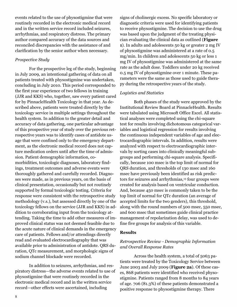
8
events related to the use of physostigmine that were
routinely recorded in the electronic medical record
and in the written service record included seizures,
arrhythmias, and respiratory distress. The primary
author compared accuracy of the data sources and
reconciled discrepancies with the assistance of and
clarification by the senior author when necessary.
Prospective Study
For the prospective leg of the study, beginning
in July 2009, an intentional gathering of data on all
patients treated with physostigmine was undertaken,
concluding in July 2010. This period corresponded to
the first year experience of two fellows in training
(JJR and KKS) who, together, saw every patient cared
for by PinnacleHealth Toxicology in that year. As de-
scribed above, patients were treated directly by the
toxicology service in multiple settings throughout the
health system. In addition to the greater detail and
accuracy of data gathering, one particular advantage
of this prospective year of study over the previous ret-
rospective years was to identify cases of antidote us-
age that were confined only to the emergency depart-
ment, as the electronic medical record does not cap-
ture medication orders until after the time of admis-
sion. Patient demographic information, co-
morbidities, toxicologic diagnoses, laboratory find-
ings, treatment outcomes and adverse events were
thoroughly gathered and carefully recorded. Diagno-
ses were made, as in previous years, on the basis of
clinical presentation, occasionally but not routinely
supported by formal toxicologic testing. Criteria for
response were consistent with the retrospective study
methodology (v.s.), but assessed directly by one of the
toxicology fellows on the service (JJR and KKS) in ad-
dition to corroborating input from the toxicology at-
tending. Taking the time to add other measures of im-
proved clinical status was not deemed feasible due to
the acute nature of clinical demands in the emergency
care of patients. Fellows and/or attendings directly
read and evaluated electrocardiography that was
available prior to administration of antidote. QRS du-
ration, QTc measurement, and morphologic signs of
sodium channel blockade were recorded.
In addition to seizures, arrhythmias, and res-
piratory distress—the adverse events related to use of
physostigmine that were routinely recorded in the
electronic medical record and in the written service
record—other effects were ascertained, including
signs of cholinergic excess. No specific laboratory or
diagnostic criteria were used for identifying patients
to receive physostigmine. The decision to use the drug
was based upon the judgment of the treating physi-
cian evaluating the clinical data as outlined (Figure
1). In adults and adolescents 50 kg or greater 2 mg IV
of physostigmine was administered at a rate of 0.5
mg/min. In children and adolescents 50 kg or less 1
mg IV of physostigmine was administered at the same
rate as the adult dose. Toddlers under 20 kg received
0.5 mg IV of physostigmine over 1 minute. These pa-
rameters were the same as those used to guide thera-
py during the retrospective years of the study.
Logistics and Statistics
Both phases of the study were approved by the
Institutional Review Board at PinnacleHealth. Results
were tabulated using Microsoft Office Excel. All statis-
tical analyses were completed using the chi-square
test for results involving dichotomous categorical var-
iables and logistical regression for results involving
the continuous independent variables of age and elec-
trocardiographic intervals. In addition, results were
analyzed with respect to electrocardiographic inter-
vals by sorting cases into clinically meaningful sub-
groups and performing chi-square analysis. Specifi-
cally, because 100 msec is the top limit of normal for
QRS duration, and thresholds of 130 msec and 160
msec have previously been identified as risk predic-
tors for seizures and arrhythmias,13 four groups were
created for analysis based on ventricular conduction.
And, because 450 msec is commonly taken to be the
top limit of normal for QTc duration (an average of
accepted limits for the two genders), this threshold,
along with the round numbers of 500 msec, 550 msec,
and 600 msec that sometimes guide clinical practice
management of repolarization delay, was used to de-
fine five groups for analysis of this variable.
Results
Retrospective Review - Demographic Information
and Overall Response Rates
Across the health system, a total of 5063 pa-
tients were treated by the Toxicology Service between
June 2003 and July 2009 (Figure 2a). Of these cas-
es, 868 patients were identified who received physo-
stigmine. Patients ranged from 8 months to 84 years
of age. 706 (81.3%) of these patients demonstrated a
positive response to physostigmine therapy. There

9
were just 8 documented adverse events attributable to
the antidote. Figure 2b demonstrates the annual use
of physostigmine. There was a general trend toward
increased use over time during the study period.
Retrospective Review – Physostigmine Use, Re-
sponse Rates, and Adverse Events by Comorbid Di-
agnoses
TCA overdoses, seizure disorders, and EKG
abnormalities are often cited as contraindications to
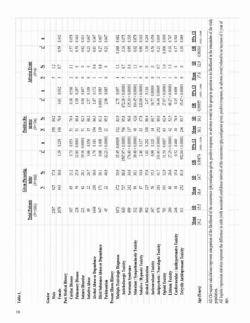
physostigmine administration. Table 1 demonstrates
the number of patients treated with multiple toxico-
logic diagnoses, TCA toxicity, sedative abuse, alcohol
abuse, and comorbidities such as heart and lung dis-
ease and seizure disorders between 2003-2009. The
category of Parkinsonism includes patients with Park-
inson’s Disease along with individuals who had Par-
kinsonian symptoms from medications and other
causes. Retrospective review of the record was not
sufficient to distinguish the specific etiologies. How-
ever, patients with Parkinsonism were more likely to
receive the antidote and have a positive response to it
than patients without the movement condition; and
they were no more likely to have a documented ad-
verse event.
The number of patients treated with physo-
stigmine in each sub-population is reported in Table
1. Individuals with multiple toxicologic diagnoses
were statistically more likely to receive the antidote—a
finding consistent with the described clinical practice
of using physostigmine diagnostically and therapeuti-
cally in cases of undifferentiated poisoning. Specific
diagnoses with phenomenologic overlap with anticho-
linergic toxicity also predicted a higher rate of anti-
dote use. These include serotonin syndrome, stimu-
lant toxicity, and lithium toxicity. A large percentage
of patients with neuroleptic toxicity received the anti-
dote, as well. And the overwhelming majority of pa-
tients with TCA poisoning were treated with physo-
stigmine.
A positive response rate to physostigmine
ranging from 64-86% was observed across the various
demographic and comorbidity subgroups. Male pa-
tients were somewhat more likely to have a docu-
mented positive response to the antidote; this signifi-
cant difference disappeared when controlling for toxi-
cologic diagnosis, as male patients were also more
likely to have been exposed to an anticholinergic
agent. There was a trend toward patients with an alco-
hol abuse or dependence history being slightly less
likely to receive physostigmine, but discounting those
whose toxicologic diagnosis was alcohol withdrawal,
those with an alcohol history were more likely to re-
ceive the antidote.
Positive response rates to physostigmine on
the basis of toxicologic diagnosis varied from 38-96%,
with patients having multiple diagnoses responding at
rate of 83.9%—similar to the overall response rate of
81.3%. Patients with TCA toxicity were statistically
more likely to respond favorably to therapy. Response
rates were lower in patients with serotonin syndrome,
opioid toxicity, anticonvulsant toxicity, and lithium
toxicity, but adverse events were still very uncommon
regardless of diagnosis.
Eight adverse events were documented
in the retrospective analysis—one arrhythmia and sev-
en seizures. The rhythm disturbance involved a 25-
year-old female patient with diphenhydramine toxici-
ty and a history of mitral valve prolapse who devel-
oped premature ventricular contractions for less than
30 seconds, approximately 25 minutes after physo-
stigmine administration (Table 2). The event was
captured on cardiac monitoring, but documentation
indicates that the patient remained asymptomatic for
its duration, and no further arrhythmias were record-
ed; subsequent doses of physostigmine were given
during her hospital course. All of the cases of seizures
were described as generalized tonic-clonic. None re-
sulted in further complications or morbidity. In each
instance, the ictal events were self-limited and non-
recurrent; both medical and pharmacy records cor-
roborate that seizures stopped on their own before
benzodiazepines were given. In 5 of the 7 cases, the
patients also had a positive response to physostig-
mine, and in 4 cases the antidote was administered
again, after lorazepam treatment, with no further sei-
zures. Intoxicants in these cases included stimulants,
SSRI and SNRI antidepressants, antipsychotics, lithi-
um, and tramadol. None of the recorded medical
comorbidities or toxicologic conditions (including
TCA toxicity or having multiple toxicologic diagnoses)
was associated with a statistically greater likelihood of
adverse events. There were no documented cases of
respiratory distress or extra pyramidal reactions to
physostigmine.
10

11
12
13

14
Prospective Study – Physostigmine Use, Response
Rates and Adverse Events
A total of 1026 patients between the ages of 2
and 89 years were treated by the Toxicology Service
between July 2009 and July 2010, and each case was
included in the prospective arm of this study. Of these
cases, 329 received physostigmine. Patients given the
antidote ranged in age from 3 to 89 years, with 154
males and 175 females. 243 (73.9%) of these patients
demonstrated a clinically meaningful positive re-
sponse. Table 2 reports the number of patients treat-
ed for multiple toxicologic diagnoses, TCA toxicity,
sedative abuse, alcohol abuse, and comorbidities such
as heart and lung disease and seizure disorders be-
tween July 2009 and July 2010. More details were
captured with prospective study, and some differences
in patient characteristics are reported, as a result.
The prospective study identified a significantly
greater proportion of patients with seizure disorders,
alcohol abuse, and abuse of sedative medications and
illicit substances than the retrospective design. The
category of Parkinsonism, again, includes patients
with Parkinson’s Disease along with individuals who
had Parkinsonian symptoms from medications and
other causes. The prospective study methodology
made it possible to distinguish the specific etiologies
for each case—9 with medication induced Parkinson-
ism and 2 with Parkinson’s Disease. As in the retro-
spective review, patients with Parkinsonism were
again more likely to receive the antidote than patients
without the movement condition; the positive re-
sponse rate was high in these patients, and they were
no more likely to have a documented adverse event.
None had an exacerbation of movement disorder
symptoms.
Fifteen patients with cardiac disease were giv-
en physostigmine, including 4 with documented
rhythm disorders. Six patients presenting with TCA
toxicity also had a history of coronary artery disease—
3 with prior myocardial infarction. None of these car-
diac patients had an arrhythmogenic event with anti-
dote. The one adverse event reported in this group
was a case of mild nausea and diaphoresis. Pulmonary
conditions represented in the cohort exposed to phy-
sostigmine include asthma, chronic obstructive pul-
monary disease, sarcoidosis, recent pulmonary embo-
lism, and lung cancer. Most patients with seizure dis-
orders were prescribed medications to control the
condition; adherence to treatment was generally low.
Abuse disorders involved a variety of substances, in-
cluding alcohol, heroin, cocaine, cannabis, phencycli-
dine, opioid analgesics, benzodiazepines, synthetic
cannabinoids, amphetamines, synthetic cathinones,
and hydrocarbon inhalants. Greater than 25% of pa-
tients had nicotine dependence, as well. None of these
conditions conferred an increased risk of adverse
events with physostigmine.
The number of patients treated prospectively
with physostigmine in each toxicologic diagnostic
group is demonstrated in Table 2. As in the retro-
spective study, individuals with multiple toxicologic
diagnoses were statistically more likely to receive the
antidote—a finding consistent with the described clin-
ical practice of using physostigmine diagnostically and
therapeutically in cases of undifferentiated poisoning.
Serotonin syndrome and stimulant toxicity again pre-
dicted a higher rate of antidote use, as well as a lower
rate of positive response. Patients with neuroleptic
toxicity were more likely to receive the antidote, as
well, and have a positive response. Opioid toxicity and
sedative toxicity predicted a lower likelihood of ad-
ministration and response, though nearly half of these
patients still did show clinically positive responses.
The majority of patients with TCA poisoning were
treated with physostigmine, and all but one had a pos-
itive response. The only patient with a negative re-
sponse coingested oxycodone and presented late, with
evidence of damage to multiple organs due to shock.
None of the TCA patients suffered serious adverse ef-
fects from physostigmine; two manifested diaphoresis
late in their hospital course after multiple doses of an-
tidote had been administered previously with positive
responses.
In 329 patients given physostigmine during
the prospective year, twenty-four total adverse events
were observed, the majority of which were minor cho-
linergic signs (Table 2). Thirteen patients experi-
enced diaphoresis and 8 patients had gastrointestinal
effects in the form of nausea, vomiting or stool incon-
tinence. None of these effects led to clinically signifi-
cant sequelae. The most common adverse event was
transient diaphoresis. No adverse effects involving
extra pyramidal reactions or respiratory distress
(secondary to bronchospasm or pulmonary conges-
tion) were observed during the prospective year.

15
Three adverse events were noted similar to those doc-
umented from the retrospective review.
Two patients developed seizures approximate-
ly 12 minutes after antidote administration. One oc-
curred in a 62-year-old patient with intellectual disa-
bility and epilepsy whose laboratory studies later con-
firmed sub-therapeutic serum concentrations of both
of his 2 antiepileptic medications. He presented with
quetiapine toxicity, and the associated delirium re-
sponded well to physostigmine, despite the isolated
20-second ictus. So, he was treated with lorazepam
prior to subsequent doses of the antidote and had no
further seizures. The other seizure arose in a 39-year-
old man with schizophrenia who had taken a purpose-
ful overdose of clozapine and trifluphenazine. He also
had a positive response to physostigmine first, and
then a tonic-clonic seizure began just shortly after de-
lirium cleared and lasted approximately 25 seconds.
Lorazepam had not been given in accordance with
standard protocol for epileptogenic toxins, but it was
given prior to future doses of antidote, which yielded
positive responses and no further adverse events. The
same patient also accounts for one of the cases of phy-
sostigmine-induced seizure activity in the retrospec-
tive study period.
Physostigmine administration was associated
with one arrhythmia during the prospective period, as
well. A 45-year-old man presented with quetiapine
toxicity and alcohol intoxication in a state of delirium.
In violation of standard protocol, the patient was giv-
en 2 doses of 2 mg physostigmine 14 minutes apart,
the second via rapid IV push. His heart rate dropped
to 40 bpm, and he felt subjectively-light headed.
Within minutes, his rhythm converted spontaneously
to atrial fibrillation. The patient felt subjectively well
again, and the new electrocardiographic pattern con-
verted spontaneously back to sinus rhythm within 75
minutes. Subsequent doses of physostigmine were
delivered in accordance with the established dosing
protocol (Figure 1), and the patient suffered no fur-
ther side effects. None of the patients with these reac-
tions encountered complications or lasting sequelae
that could be attributed to the dosing of antidote.
Prospective Study – Electrocardiographic Character-
istics of Physostigmine Treated Patients
The majority of acute toxicology patients in
our practice have an electrocardiogram (EKG) per-
formed early in their assessment. Of the 329 patients
treated with physostigmine during the prospective
year, 312 had an EKG before receiving the first dose of
antidote, and 245 of those were performed within 30
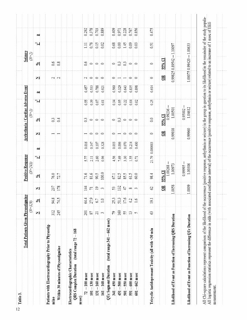
minutes of physostigmine infusion. Table 3 describes
the EKG characteristics of patients given physostig-
mine during the prospective study year. Over half of
patients had at least one abnormal interval measure-
ment, with the most common being mild QTc prolon-
gation. More than 20% of patients, however, had
more significant repolarization delay, and over one
third of patients had impaired ventricular conduction
as evidenced by QRS duration greater than 100 msec.
Those patients with an abnormally wide QRS
complex were statistically more likely to have a posi-
tive response to physostigmine than those with nor-
mal QRS duration. Logistic regression indicated a
roughly 0.6% greater probability of positive response
for each 1 msec above 100 in QRS measurement. This
trend correlated with a larger proportion of patients
with a diagnosis of TCA toxicity. A QTc interval be-
tween 450 and 550 msec was associated with greater
likelihood of positive response to antidote than meas-
urements outside this range. As noted above, the
number of serious side effects was very low; none of
the EKG findings predicted physostigmine-related
adverse events. The patient who experienced arrhyth-
mia secondary to physostigmine overdosing had a
QRS duration of 96 msec and a QTc interval of 484
msec. Patients with seizures both had QRS measure-
ments of 92 msec; the QTc for one was 477 msec and
the other 536 msec. All patients with TCA toxicity had
an EKG within 30 minutes of receiving physostig-
mine, and none had a seizure or arrhythmia.
Discussion
Anticholinergic toxicity is a relatively com-
mon, but often unrecognized direct precipitant of de-
lirium. As delirium, regardless of cause, confers an
increased risk of morbidity and mortality, its prompt
identification and treatment is a central concern in
acute medical practice. Although debate continues
regarding the most effective management strategies
for the symptoms of the syndrome, it has been well
established that the most important intervention for
any delirious state is treatment of the underlying
cause. For anticholinergic delirium, there is a direct
16
antidote available that addresses the neurochemical
etiology, but it has been vastly underutilized for dec-
ades due to concerns about adverse effects. The clini-
cal data outlined above should largely dispel any myth
that physostigmine is an unsafe antidote for suspected
central anticholinergic toxicity.
Combining both the retrospective review and
prospective study we present 7 years of experience
with physostigmine at PinnacleHealth, where bedside
use of the antidote increased over that time period on
the basis of its observed utility and safety profile. 1197
patients treated with physostigmine for suspected an-
ticholinergic toxicity showed a positive response rate
of nearly 80% and experienced a total of just 11 signif-
icant adverse reactions, none of which resulted in
complications or lasting sequelae. Mild peripheral
cholinergic signs were observed during the detailed
prospective study year, but each occurrence was self-
limited with the dosing algorithm we have adopted.
The antidote is therefore used frequently for diagnos-
tic purposes in cases of undifferentiated poisoning
with delirium and then continued as targeted treat-
ment when anticholinergic toxicity is identified by the
favorable response.
Based upon the treatment practice outlined
above (Figure 1), the only patients who would receive
a diagnosis of anticholinergic toxicity and not receive
physostigmine would be those with very mild central
effects or just peripheral symptoms. Routine practice
has been to assign a diagnosis of anticholinergic tox-
icity to all patients who have a demonstrable positive
response to physostigmine. One could contend with
this practice on the basis of the notion that physostig-
mine might function as an analeptic even in cases in
which compounds with anticholinergic activity are not
present and responsible for altered mentation and
behavior. There have been cases reporting positive
response to physostigmine in reversing general anes-
thesia and in cases of toxicity involving other agents
like opioids and benzodiazepines;15,16,17 though one
could also argue that impaired cholinergic neuro-
transmission is an accompanying effect of such
agents.18,19,20 Nevertheless, we do appreciate that the
assignment of the diagnosis of anticholinergic toxicity
in this clinical sample may be viewed as dependent
upon a degree of circular logic, so the report of all the
positive responses in this diagnostic subgroup may
not be statistically meaningful. However, the safety
(and, in our estimation, utility) of the antidote cannot
be denied, especially noting that 38% of the patients
studied had multiple toxicologic diagnoses, and before
confirmation of those toxicities, physostigmine was
used in a significantly higher proportion of that com-
plex subgroup with a response rate exceeding 80%.
Therefore, the oft quoted physostigmine contraindica-
tion of “undifferentiated poisoning” is not supported
by bedside experience.9,10 In addition, virtually every
patient with TCA toxicity will have anticholinergic de-
lirium, and none of those patients in this large study
suffered a significant adverse reaction to physostig-
mine, while there was a very high response rate.
Although we appreciate that a medication with
cholinergic activity has the potential to impact cardiac
function, there has never been a solid physiochemical
rationale for fearing the use of physostigmine in TCA
patients. The mechanisms involved in cardiotoxicity
from TCAs are based upon altered cation flow—both
sodium channel blockade and potassium efflux inhibi-
tion increase the risk of a lethal arrhythmia. The for-
mer also markedly impairs cardiac inotropic function
with the potential for hypotension and shock. Physo-
stigmine, however, reduces chronotropy via enhanced
vagal activity, and does not impact myocardial con-
duction or the flow of ions that modulate it. The major
documented risk factor in cases of morbid and mortal
outcomes of TCA patients given physostigmine that
have been propagated through the medical literature
is the presence of TCA toxicity itself.8,10 Severe, pro-
gressive cardiotoxicity with sodium channel blockade
devolves to shock and arrest on its own, as evidenced
by many cases of overdose death involving these com-
pounds. It has merely been post hoc ergo propter hoc
reasoning that lays blame with the cholinesterase in-
hibitor antidote. As long as it is delivered by slow in-
fusion, thereby allowing the lipophilic tertiary ami-
nocarbamate to partition to the CNS without being
circulated quickly and undiluted to the pulmonary
and coronary vasculature, concerns about toxicity are
unfounded.
This assertion is supported by the electrocardi-
ographic data presented in Table 3. Patients with a
wide variety of conduction variability, both in width of
the QRS complex and duration of the QT segment
were given physostigmine without suffering arrhyth-
mias. Longer QRS duration was actually associated
with a statistically greater likelihood of positive re-
17
sponse to the antidote. This effect appears to be driv-
en by the fact that TCAs almost invariably produce an
anticholinergic delirium in toxic exposure that will
respond to physostigmine, and they frequently cause
sodium channel blockade that widens the QRS com-
plex, as well. Most TCA patients in our study had QTc
intervals ranging between 450 and 550 msec—also the
range that predicted a somewhat greater likelihood of
positive response to physostigmine. Many patients
with delirium from other anticholinergic agents also
had this degree of QT prolongation. Otherwise, the
EKG abnormalities caused by scores of different tox-
ins, including TCAs, had no significant impact on
safety or response rates. The single case of arrhythmia
in our prospective study involved a dosing error, in
which an adult patient with anticholinergic delirium
from quetiapine toxicity was given 4 mg of physostig-
mine within 14 minutes. Absent such misuse, even the
most severely TCA poisoned patients in our critical
care toxicology practice received physostigmine with-
out cardiac side effects.
The question of seizures does require some
discussion. Many anticholinergic toxins, including
TCAs, have other pharmacologic properties that in-
crease the risk of seizures.2 Physostigmine is an ana-
leptic, and as a stimulating antidote, can increase neu-
ral activity in such a way that augments this risk and
precipitates an ictus. Seizures and the resulting acido-
sis can complicate the care of toxicologic patients, so
individuals whose suspected ingestion would yield a
significant seizure risk should receive a dose of benzo-
diazepines prior to intervention with physostigmine.
This is obviously the case for TCA patients with any
significant toxicity, since the noradrenergic surge and
GABA inhibition have robust synergistic epileptogen-
icity; attention to this key aspect of TCA poisoning
management explains the absence of physostigmine
induced seizures in our clinical study. As a result, we
find the antidote a safe and useful adjunctive treat-
ment for TCA patients, reducing the need for re-
straint, oversedation, mechanical ventilation, and
bladder catheterization with a very high response rate
with toxins that produce anticholinergic delirium.
Clearing of delirium also reduced the need for further
workup and expensive testing, including computed
tomography scanning of the head.
The overall rate of positive response to physo-
stigmine was higher for men than for women in both
the retrospective and prospective arms of this study.
However, the significant difference between the gen-
ders does not persist when controlling for toxicologic
diagnosis; more men in our practice had exposure to
anticholinergic agents, including TCAs. Although
there may be neuropsychiatric differences in the cho-
linergic system between men and women,21 we know
of no physiologic reason for there to be differential
response to antidote. Therefore we do not favor any
modification of the use of the antidote on the basis of
gender.
We suspect that the majority of differences in
patient characteristics between the prospective and
retrospective arms of the study relate to our ability to
gather more complete clinical datasets during the pro-
spective year. It is unlikely, for instance, that rates of
alcohol use and seizure disorders would have in-
creased significantly in one year’s time without chang-
es in practice or referral patterns, which were not ob-
served. The only notable exception was a rise in the
use of synthetic cannabinoids and cathinones whose
clinical presentations prompted the use of physostig-
mine in many cases without positive response; this
trend may explain some of both the greater use of and
lower response rate to physostigmine in substance
using patients in the prospective year.
In accordance with the study design, we also
suspect that overall use of physostigmine was not
markedly higher in the prospective year as compared
to the final year of the retrospective study as it ap-
pears. Rather, the prospective study captured cases in
which the antidote was used in the emergency depart-
ment but then not used again after admission. This
interpretation is consistent with the overall lower rate
of positive response in the prospective year, as a lack
of improvement with a single dose of physostigmine
in the emergency setting would be documented as a
negative response, and repeat doses would not be giv-
en later in the hospital course after admission. The
antidote would have served a diagnostic purpose to
rule out central anticholinergic toxicity, but would not
be of further therapeutic value to warrant subsequent
use. Such was likely the case in our practice from
2003-2009, as well, but simply not captured by the
retrospective methodology. Single use cases also oc-
curred with positive results, as well though, in expo-
sures involving short acting toxins like doxylamine
and diphenhydramine. The use of physostigmine in

18
these cases would have been captured in the prospec-
tive year, but not the retrospective review.
With respect to side effects, in the prospective
study year the majority of those reported pertain to
the first dose of antidote given to any particular pa-
tient. In the event of an adverse reaction in the ab-
sence of positive response, no further doses of physo-
stigmine would be administered in accordance with
clinical practice (Figure 1). The two patients who
both responded favorably to the antidote and devel-
oped seizures, however, were treated with benzodiaze-
pines and then responded to subsequent doses of phy-
sostigmine for management of their delirium during
the remainder of hospitalization. Occasionally pa-
tients had positive responses to a series of doses and
then, with clearance of toxins, would develop cholin-
ergic signs when given physostigmine later in their
course. Even these adverse events are reported in the
data (Table 3), and yet the total incidence of side ef-
fects using the dosing regimen for physostigmine as
outlined remained low. Only major adverse reactions
were documented in the medical record for the years
reviewed prior to prospective study; all 8 correspond
to the first dose of physostigmine in a given episode of
care, at the point of diagnostic assessment.
Serotonin syndrome is the most common toxi-
cologic differential diagnosis in patients with anticho-
linergic syndrome. Although textbook descriptions of
the two would suggest being able to differentiate on
the basis of reflexes, lower extremity tone, bowel mo-
tility, and/or level of secretions and sweating, the bed-
side reality is more complicated.22 Furthermore, as
many of the compounds involved in such cases are
highly lipophilic and preferentially partition to the
central compartment, effects of serotonergic agents
and anticholinergic agents often will cause similar de-
liria while yielding non-distinct peripheral manifesta-
tions. This biochemical partitioning with correspond-
ing clinical predominance of CNS effects supports the
use of the centrally acting physostigmine without con-
cern for peripheral signs such as pupil size, bowel
sounds, or heart rate. Only profuse sweating is a con-
traindication with our clinical protocol (Figure 1),
therefore many patients with serotonin syndrome re-
ceive a dose of physostigmine in our practice to differ-
entiate the two conditions and potentially provide
benefit in treating toxic delirium. A negative response
in such circumstances lends more support for a diag-
nosis of serotonin syndrome with corresponding
abandonment of physostigmine in favor of benzodiaz-
epines and/or other sedative treatments.
Comorbid alcohol intoxication was a common
finding in our patient population. No more adverse
events attributable to physostigmine were observed in
this subgroup, either retrospectively or prospectively,
and response rates were similarly robust. Positive re-
sponse rates were lower in patients with a diagnosis of
anticonvulsant toxicity. However, carbamazepine has
significant anticholinergic activity, so the response
rate in cases involving this particular agent was actu-
ally higher than average—over 90%. Similarly, a num-
ber of antipsychotic medications are anticholinergic.
Thus a larger proportion of patients with toxic expo-
sures to neuroleptics was given physostigmine and,
correspondingly, displayed a significantly higher re-
sponse rate. A fraction of the patients listed in this
category had initial presentations involving dystonic
reactions or other extra pyramidal manifestations
without delirium, and were therefore obviously not
given physostigmine, but treated with anticholinergic
medications, instead.
Although rates of positive response were lower
in patients with opioid and lithium toxicity diagnoses,
major adverse events were no more likely. There were
no adverse events in patients with toxicity from cardi-
ovascular medications apart from one case of nausea,
and the likelihood of positive response to physostig-
mine (due to coingestion of anticholinergic com-
pounds) was comparable to the overall response rate.
We reported data for this subgroup of patients to fur-
ther highlight the cardiac safety profile of the anti-
dote. In general, the clinical experience underlying
these studies indicates that physostigmine can be
used safely and effectively in patients with multiple
toxicities to diagnose and treat the neurobehavioral
impairments associated with anticholinergic burden,
even if that is only one of several manifestations of
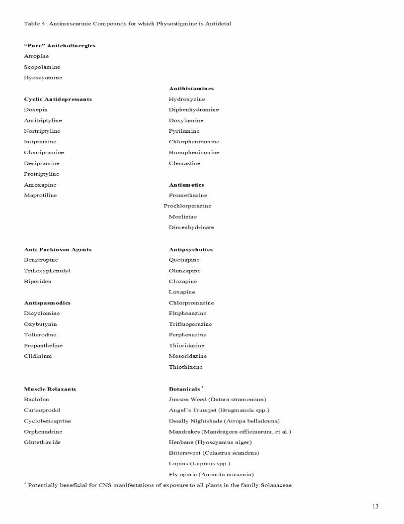
acute poisoning. A list of substances with anticholin-
ergic activity (representing a variety of chemical and
medicinal classes) for which we have found physostig-
mine efficacious is listed in Table 4.
Attempts to be thorough and unbiased were
pursued throughout the course of this research, but
the limitations of the work must be noted. The retro-
spective design of the first leg of this study resulted in
a cohort of patients with an occasionally-incomplete

19
medical record and reliance on a secondary written
record. Some adverse events may have been missed,
because they may not have been recorded in the medi-
cal record. Such is not the case, however, for the pro-
spective year of study. As already noted, the lack of an
electronic medical record for the emergency depart-
ment limited full evaluation of some cases and inevi-
tably resulted in undercounting of cases of antidote
use during the retrospective period, some of which
potentially involved adverse reactions, as well.
The possibility of this research, however, de-
pended upon the invaluable resource of the toxicology
practice database—a systematic documentation rou-
tine established by the medical director of the service
(JWD) at its inception in June 2003 and consistently
maintained for the study period. Although potentially
biased by the practitioner’s perspective on the course
and care of patients, the detailed notes represent ex-
tensive bedside experience in toxicology and the use
of antidotes based not upon offsite case consultative
input, but upon direct physician assessment and in-
tervention. This system served as the model and start-
ing point for designing the prospective arm of the
study. During that study year, both junior fellows
(JJR and KKS) gathered data for the study, and these
records were reconciled with each other and with the
attending’s notes (per JWD’s established routine).
Most patients were seen by both fellows during their
care by the toxicology service—discrepancies were ra-
re, and reconciled by the primary author with the doc-
umentation and guidance of the senior author. We
acknowledge the potential for biased reporting in this
process. Furthermore, not every administered dose of
antidote was observed directly by the authors (or even
a rotating trainee physician). The use of physostig-
mine as outlined (Figure 1) is so routine in our prac-
tice, that nursing staff follow PRN orders as long as
patients show improvements in mental status; a phy-
sician is contacted if cholinergic signs are observed, so
although these reactions were recorded by study phy-
sicians, personnel may have gone to the bedside after
they occurred, and would obviously not administer
physostigmine again merely to reproduce the side ef-
fect for confirmation.
The subjective nature of the clinical assess-
ment of outcomes may have also contributed to re-
corder bias with respect to antidotal response. And
although responses to antidote were recorded as
simply positive or negative, the reality of bedside
treatment is that some patients experienced much
more complete restoration of neuropsychiatric func-
tion than others. The design of this study did not ac-
count for that variability, but instead ascribed a posi-
tive response to all patients who displayed at least
some clinically relevant improvement in wakefulness,
cognition, or behavior. A portion of patients still re-
quired adjunctive pharmacologic and non-
pharmacologic interventions for management of neu-
robehavioral manifestations of toxicity, even when
physostigmine was deemed effective.
With respect to efficacy, this study does not
directly address that question. Despite its size and the
report of regular clinical use based on this extensive
experience, the present study does not definitively in-
dicate whether physostigmine use in toxicologic prac-
tice is responsible for better clinical outcomes. Such a
claim would require a placebo-controlled trial of the
antidote with the clinical methodology suggested
(Figure 1). Our service has previously reported short-
ening the length of stay in Datura poisoned patients
who received physostigmine rather than placebo as
part of their care.23 Since that time, the clinical experi-
ence documented for a wide variety of toxic patients
in the retrospective portion of this study has con-
vinced our service and numerous trainees rotating
through it that physostigmine is safe and effective.
Scores of patients who would have been intubated and
sedated, thereby increasing their risk of nosocomial
infection and other complications from greater instru-
mentation and increased lengths of stay serve as testa-
ment to this perspective. Positive antidotal response
has also reduced utilization of computed tomography
and allowed more thorough assessment of patients
through productive interviewing and detailed physical
examination that revealed different acute care needs
that might otherwise have been missed. It is experi-
ence with those patients that convinced the authors
the prospective year would be more ethically conduct-
ed as an observational study instead of a randomized
trial that would deny a large number of patients an
efficacious treatment. We appreciate, however, that a
placebo-controlled experiment has not been conduct-
ed to measure lengths of stay, complication rates, and
long-term outcomes to support this claim; our report
of positive results in this study, despite its size, lacks a
comparison group.

20

21
Figure 2: Retrospective Study Overview
a.
5063 Total Cases
2387 Men (47.1%), 2676 Women (52.9%)
Age range 8 months to 94 years
Mean age 39.2 years
868 treated with physostigmine
Age range 8 months to 84 years
706 positive responses (81.3%)
Adverse events:
7 seizures, 1 arrhythmia, 0 respiratory distress
b.
22
It is worth noting, however, that clinical sci-
ence does already support use of physostigmine in the
manner described. As noted above, the best treatment
for any delirial state is the one aimed at the underly-
ing cause. Physostigmine has the potential to address
a toxic etiology directly and diminish or even reverse
CNS dysfunction—a more targeted and definitive
treatment for the syndrome than more commonly em-
ployed agents for managing agitation that typically
produce sedation but leave patients impaired. Such
impairments demand a higher rate of catheterization,
intubation, and restraint. And those instruments, in
turn, come with increased risk of complications. So
although we do not have direct data on the ability of
physostigmine to decrease complication rates and
lengths of stay, logic and circumstantial evidence
point in that direction. More definitive data, however,
speak to implications that last long after patients first
arrive in the emergency department with an overdose
or toxic misadventure.
Some physicians choose not to treat delirial
symptoms unless they appear to be a threat to life and
limb as a result of agitation, or at the very least, pose
challenges to nursing care. However, recent studies
indicate that any untreated delirium increases the risk
of long-term poor health. Post-traumatic stress disor-
der (PTSD) as a consequence of medical trauma is
much more likely in patients who suffer delirium re-
gardless of the cause, and the more severe and longer
lasting the delirium, the greater that risk.24 Further-
more, other independent predictors of PTSD one and
two years after discharge include amnesia for the early
portion of hospitalization, youth, female gender, low
education level, trait anxiety, and lack of social sup-
port24—all characteristics that are more common in
the acute toxicology patient than those hospitalized
for other reasons.
The risk of functionally meaningful depression
during the years following hospitalization also in-
creases greatly as a function of delirium and its severi-
ty. Depression prevalence following a hospital stay
with delirium is 31%,25 an estimated two-fold increase
in mood disorder diagnosis as compared to those
whose hospitalizations proceed without similar CNS
insult. As with PTSD, common characteristics of toxi-
cology patients—female gender and lack of social sup-
port—increase this depression risk.26 Additionally, the
cumulative dose of benzodiazepines during an inten-
sive care unit stay is positively correlated with depres-
sion rates in the years following discharge,27 and this
is the class of medications most commonly employed
to manage agitation and maintain sedation when the
direct therapy, physostigmine, could reduce their use
in many of cases.5 So although not routinely employed
in most emergency settings, physostigmine may be
useful not only as a short term diagnostic tool and
treatment for agitation in toxicology patients, but also
helpful in taking the long view of patients’ physical
and mental resilience against the threat of delirium.
As supported by the largest study to date, phy-
sostigmine is a safe and potentially effective medica-
tion in the undifferentiated patient with delirium and
a differential diagnostic list that includes toxic etiolo-
gies for the alterations in mental status. Even in the
setting of TCA and mixed drug ingestions and in pa-
tients with a variety of medical comorbidities, the an-
tidote produces few side effects when properly dosed.
The key to its safe use is clinical assessment of vital
signs, cognition, neuromuscular activity, and secreto-
ry status followed, where indicated, by slow weight-
based infusion and reassessment of neurobehavioral
status after 15 minutes. With attention to the other
demands of toxicologic care, the incidence of seizures
is low, and cardiotoxic sequelae are essentially absent.
Cholinergic adverse effects are generally mild and self
-limited; maintaining an erect posture of the torso
prevents complications from emesis. Based on the
ubiquity of anticholinergic activity in widely used me-
dicinal and abusable substances, the potential to diag-
nose toxicity, clear cognition rapidly, and allow more
patients to participate in their own care is great. On
the basis of this study, we advocate for the expanded
use of physostigmine in cases of altered mental status
in emergency and hospital medicine.
References
1. Clary GL, Krishnan KR. Delirium: diagnosis,
neuropathogenesis, and treatment.
J Psychiatr Pract. 2001;7:310-323.
2. Smith SW. Drugs and pharmaceuticals: man-
agement of intoxication and antidotes. EXS.
2010;100:397-460.
3. Heiser JF, Gillin JC. The reversal of anticho-
linergic drug-induced delirium and coma with

23
physostigmine. Am J Psychiatry.
1971;127:1050-1054.
4. Schneir AB, Offerman SR, Ly BT, Davis JM,
Baldwin RT, Williams SR, Clark RF. Complica-
tions of diagnostic physostigmine administra-
tion to emergency department patients. Ann
Emerg Med. 2003;42:14-19.
5. Burns MJ, Linden CH, Graudins A, Brown
RM, Fletcher KE. A comparison of physostig-
mine and benzodiazepines for the treatment of
anticholinergic poisoning. Ann Emerg Med.
2000;35:374-381.
6. Knudsen K, Heath A. Effects of self poisoning
with maprotiline. Br Med J (Clin Res Ed).
1984;288:601-603.
7. Pentel P, Peterson CD. Asystole complicating
physostigmine treatment of tricyclic antide-
pressant overdose. Ann Emerg Med.
1980;9:588-590.
8. Kulig K, Rumack BH. Physostigmine and asys-
tole. Ann Emerg Med. 1981;10:228-229.
9. Shannon M. Toxicology reviews: physostig-
mine. Pediatr Emerg Care. 1998;14:224-226.
10. Suchard JR. Assessing physostigmine's contra-
indication in cyclic antidepressant ingestions.
J Emerg Med. 2003;25:185-191.
11. Liebelt EL, Francis PD, Woolf AD. ECG lead
aVR versus QRS interval in
predicting seizures and arrhythmias in acute
tricyclic antidepressant toxicity.
Ann Emerg Med. 1995;26:195-201.
12. Liebelt EL, Ulrich A, Francis PD, Woolf A. Se
rial electrocardiogram changes in
acute tricyclic antidepressant overdoses. Crit
Care Med. 1997;25:1721-1726.
13. Boehnert MT, Lovejoy FH Jr. Value of the QRS
duration versus the serum drug level in pre-
dicting seizures and ventricular arrhythmias
after an acute overdose of tricyclic antidepres-
sants. NEJM. 1985; 313:474-479.
14. Brandl KM, Langley KA, Riker RR, Dork LA,
Quails CR, Levy H. Confirming the reliability
of the sedation-agitation scale in ICU nurses
without prior experience in its use. Pharma-
cotherapy 2001; 21:431-436.
15. Shulman MS, Sandler A, Brebner J. The rever-
sal of epidural morphine induced
somnolence with physostigmine. Can Anaesth
Soc J. 1984;31:678-680.
16. Rupreht J. Physostigmine reversal of diaze-
pam. Anesthesiology. 1980;53:180-181.
17. Ongini E, Parravicini L, Bamonte F. Effects of
physostigmine on benzodiazepine
toxicity. Arch Int Pharmacodyn Ther.
1981;253:164-176.
18. Eisendrath SJ, Goldman B, Douglas J, Di-
matteo L, Van Dyke C. Meperidine-induced
delirium. Am J Psychiatry. 1987;144:1062-
1065.
19. Tune LE. Serum anticholinergic activity levels
and delirium in the elderly. Semin Clin Neuro-
psychiatry. 2000;5:149-153.
20. Tune LE. Anticholinergic effects of medication
in elderly patients. J Clin Psychiatry. 2001;62
Suppl 21:11-14.
21. Furey ML, Khanna A, Hoffman EM, Drevets
WC. Scopolamine produces larger antidepres-
sant and antianxiety effects in women than in
men. Neuropsychopharmacology.
2010;35:2479-2488.
22. Boyer EW, Shannon M. The serotonin syn-
drome. N Engl J Med. 2005;352:1112-1120.
23. Burkhart KK, Magalski AE, Donovan JW. A
retrospective review of the use of activated
charcoal and physostigmine in the treatment
of jimson weed poisoning. J Toxicol Clin Toxi-
col. 1999;37:389.
24. Granja C, Gomes E, Amaro A, Ribeiro O, Jones
C, Carneiro A, Costa-Pereira A, JMIP Study
Group. Understanding posttraumatic stress
disorder-related symptoms after critical care:

24
the early illness amnesia hypothesis. Crit Care
Med. 2008;36:2801-2809.
25. Davydow DS. Symptoms of depression and
anxiety after delirium. Psychosomatics.
2009;50:309-316.
26. Myhren H, Ekeberg O, Tøien K, Karlsson S,
Stokland O. Posttraumatic stress, anxiety and
depression symptoms in patients during the
first year post intensive care unit discharge.
Crit Care. 2010;14:R14.
27. Davydow DS, Gifford JM, Desai SV, Needham
DM, Bienvenu OJ. Posttraumatic stress disor-
der in general intensive care unit survivors: a
systematic review. Gen Hosp Psychiatry.
2008;30:421-434.

25
Abstract
Objectives: To assess the validity of the brief version
of the SIS-MAP, we compared brief version scores for
psychiatric inpatients against scores for psychiatric
outpatients. A secondary goal was determining clini-
cal cut-offs to inform level of care decisions. Meth-
ods: In addition to clinical and demographic data, 35
patients were assessed on depressive symptoms, psy-
chosis, and suicide risk. Results: SIS-MAP-scn
scores were associated with known risk factors for sui-
cide, psychotic and depressive symptoms, and reasons
for admission. Scores on the SIS-MAP-scn were also
significantly correlated with patients’ outpatient/
inpatient status, suggesting its utility for assisting lev-
el of care decisions. Conclusion: The results suggest
the potential utility of the SIS-MAP-scn, and provide
preliminary clinical cut-offs for admission.
Suicide risk presents a challenging issue in
clinical psychiatry. Currently there are a number of
assessment tools available to gauge suicide risk (see
Cochrane-Brink, Lofchy, & Sakinofsky, 2000); howev-
er, to inform decisions around the level of psychiatric
care required, instruments must be developed that can ef-
fectively identify individuals who are at serious risk
for attempting suicide and those who are not
(Kreitman & Foster, 1991; Pompili, Amador, Girardi et
al., 2007). Clinical judgment is the rule of the thumb
and risk assessment tools are used only when clinical
decision making is difficult in a given situation (e.g.,
Thienhaus & Piasecki, 1997; Bryan & Rudd, 2006).
We developed a scale for clinical settings that
objectively assesses multi-factorial risk: the Scale for
Impact of Suicidality - Management, Assessment and
Planning of Care (SIS-MAP; Nelson, Johnston, &
Shrivastava, 2010). Our purpose was to provide quan-
tification for care and decision-making. The SIS-MAP
was designed to assess the various risk and protective
factors, both internal and external to the individual,
known to contribute to suicidal risk in order to numer-
ically guide level of care decisions.
Assessment of Suicide Risk in Psychiatric Patients Using a Brief Screener: Validation of the SIS-MAP-scn
Megan E. Johnston, PhD, Amresh Shrivastava, MD MRCPsych FRCPC, Robbie Campbell, FRCPC, Miky Kaushal, MD, Charles Nelson, PhD CPsych
Corresponding Author: Dr. Megan E. Johnston, Dept. of Medicine, University of Otago, Christchurch Private Bag 4710, Christchurch Hospital, Christchurch, New Zealand 8140 Phone: 64 3 378 6487; Fax 64 3 364 0935; Email: [email protected]

26
SIS-MAP-scn
A brief screener (SIS-MAP-scn) comprised of
selected items from the full SIS-MAP was developed
in order to further facilitate and expedite the suicide
risk assessment process (Johnston, Nelson, &
Shrivastava, 2013). In contrast to the full SIS-MAP
clinical interview, the screener allows for a prompt
initial evaluation providing an indication of whether
an individual should undergo further assessment.
In a preliminary investigation of the validity of
the SIS-MAP-scn, Johnston et al. (2013) found that
scores on the 23-item abbreviated scale were signifi-
cantly predictive of total SIS-MAP scores, F (1, 48) =
168.77, p < .001. The screener was shown to account
for 77.90% of the variance in total suicide risk scores
obtained using the full SIS-MAP.
The objective of the study was to assess the
validity of the brief version of the SIS-MAP. A second-
ary goal was to determine clinical cut-offs on the SIS-
MAP-scn to inform level of care decisions (e.g., admit-
tance to psychiatric inpatient care and to inform deci-
sions to discharge in some unique situations).
Method
Information was collected from 35 patients’
clinical assessments as part of an ongoing study of re-
hospitalization. Consecutive patients reporting to cri-
sis services at a psychiatric hospital were recruited. In
addition to clinical and demographic data, patients
were assessed on depressive symptoms, psychosis,
and suicide risk. Informed consent was obtained from
all patients who participated in this study.
Measures
Demographic and clinical data. Infor-
mation was collected on patients’ marital status (1 =
married; 2 = single; 3 = divorced/separated; 4 = wid-
owed) and number of children. The presence or ab-
sence of drug abuse (0 = no, 1 = yes) and alcoholism
(0 = no, 1 = yes) was recorded. Data was also collected
on patients’ duration of illness (1 = less than a month;
2 = less than 6 months; 3 = more than 6 months),
number of previous hospitalizations, and the reason
for their admission (0 = out-patient; 1 = suicide at-
tempt; 2 = homicide attempt; 3 = gravely disabled; 4
= diagnosis of severe mental illness; 5 = other). Final-
ly, psychiatric diagnosis was assessed according to the
DSM-IV (APA, 1994).
Psychiatric symptoms. Psychiatric symp-
toms were measured with the Brief Psychiatric Rating
Scale (BPRS; Ventura, Lukoff, Nuechterlein et al.,
1993). This scale is comprised of 24 types of psychiat-
ric symptoms (e.g., hallucinations, emotional with-
drawal, somatic concern) that clinicians rate from 1
(absence of symptom) to 7 (extremely severe symp-
tom). Scores are based on a combination of patients’

27
self-reports and clinicians’ observations of patient be-
havior and speech.
Depression. Depressive symptoms were
measured with the Hamilton Depression Rating Scale
(HDRS; Hamilton, 1960). On this 17-item scale, each
item is scored on a range of either 0 – 4 or 0 – 2. Ad-
ministered by clinicians, the items assess symptoms of
depression experienced over the past week, with an
emphasis on the melancholic and physical symptoms
of depression.
Suicidality. Suicide risk was assessed with
the SIS-MAP-scn (Johnston et al., 2013). The SIS-
MAP-scn is a clinician-administered screener for sui-
cide risk comprised of 23 items assessing 7 domains of
risk: demographic, psychological, comorbidities, fami-
ly history, protective factors, clinical, and psychoso-
cial. All items are scored as yes or no (for all but one
item, score of 1 given for yes and 0 for no). Final
scores are calculated as the sum of all items except for
the two protective items, with these protective items
then subtracted from the sum.
Analyses
Data was analyzed using the Statistical Pack-
age for Social Sciences (SPSS). Pearson correlations
and one-way analyses of variance (ANOVAs) were
used to determine any significant differences in SIS-
MAP-scn scores based on the clinical and demograph-
ic data, psychiatric symptoms, or depressive symp-
toms. When ANOVAs were significant, Tukey’s post-
hoc tests were used to determine those groups that
differed significantly from one another. Post-hoc tests
were used only when more than two levels of the vari-
able were present and only for groups that had two or
more cases.
Next, discriminant function analysis was used
to classify patients into outpatient versus inpatient
status based on their SIS-MAP-scn scores, allowing
for comparison to actual patient status. The purpose
of this analysis was to evaluate the ability of the SIS-
MAP-scn to inform level of care decisions and to de-
termine clinical cut-offs.
Results
Sample Characteristics
The age of the 35 patients (18 females) ranged
from 19 to 80 years (M = 38.26, SD = 14.78). On aver-
age they had 1.37 children (SD = 1.88; range = 0 – 9)
and only 7 were married (19 were single, 8 were di-
vorced/separated, and 1 was widowed). The average
number of previous hospitalizations for the sample
was 2.03 (SD = 1.56; range = 0 – 6). All patients had a
duration of illness of at least one month, 11.4% had an
illness duration of less than 6 months and 88.6% of
patients had an illness duration of more than 6
months. Seven of the patients suffered from alcohol-
28
ism and 11 from drug abuse. Frequency statistics for
diagnoses on the DSM-IV are presented in Table 1.
SIS-MAP-scn Scores
SIS-MAP-scn risk index scores ranged from 1
to 15 (M = 6.8, SD = 3.60). Correlations and ANOVA
tests are presented in Tables 2 and 3, respectively.
Risk index scores did not differ by gender (F (1,33) =
0.25, p = .62), age (r = .004, p = .98), or marital status
(F (2,31) = 1.44, p = .252). Number of children was
also not significantly correlated with SIS-MAP-scn
score (r = .71, p = .62).
SIS-MAP-scn scores were significantly associ-
ated with patients’ duration of illness (F (1,33) = 4.16,
p = .05) and marginally related to the number of pre-
vious hospitalizations (r = .31, p = .07). Patients with
a duration of illness longer than 6 months (M = 7.23,
SD = 3.59) scored higher on the SIS-MAP-scn than
patients with a duration of illness less than 6 months
(M = 3.50, SD = 1.00).
There was also a trend towards higher SIS-
MAP-scn scores in patients with alcoholism (M =
8.86, SD = 2.85) than patients without alcoholism (M
= 6.29, SD = 3.62; F (1,33) = 3.04, p = .09). However,
there were no significant differences in SIS-MAP risk
scores based on the presence (M = 7.73, SD = 3.38)
versus absence (M = 6.38, SD = 3.68) of drug abuse (F
(1,33) = 1.07, p = .31).
As seen in Table 2, SIS-MAP-scn scores dif-
fered based on Axis I diagnosis (F (3,31) = 3.83, p
= .02) but not diagnosis on any of the other 3 axes.
Tukey’s post-hoc tests indicated that patients diag-
nosed with depressive disorders (M = 10.13, SD =
2.64) scored significantly higher in SIS-MAP-scn risk
scores than patients diagnosed with schizophrenia
and psychotic disorders (M = 6.00, SD = 3.68) and
patients diagnosed with bipolar disorders (M = 5.63,
SD = 2.45). As only one patient was diagnosed with a
cognitive disorder, this diagnosis category was not
included in the post-hoc testing.
SIS-MAP-scn risk scores were significantly
associated with the patients’ reason for admission (F
(4,30) = 4.24, p = .008). Those admitted for a suicide
attempt scored higher on the SIS-MAP-scn (M =
10.71, SD = 2.81) than those who were not admitted
(M = 5.41, SD = 2.40) or those who were admitted
based on a diagnosis of severe mental illness (M =
5.33, SD = 3.44). Individuals admitted based on at-
tempted homicide did not differ significantly from any
other group in their SIS-MAP-scn risk scores (M =
8.00, SD = 5.22).
SIS-MAP-scn risk scores were significantly
positively correlated with HDRS scores (r = .62, p
< .001) and with BPRS scores (r = .41, p = .014). In
other words, patients scoring highly on the SIS-MAP-
scn were more likely to show high levels of depressive

29
symptoms and psychotic symptoms than patients
scoring low on the SIS-MAP-scn.
Determining Level of Care
The present sample consisted of an approxi-
mately equal number of outpatients (n = 17) and inpa-
tients (n = 18). A canonical discriminant function
analysis was used in order to evaluate whether SIS-
MAP-scn scores were predictive of whether individu-
als were outpatients or inpatients. The analysis result-
ed in a total 68.6% of original grouped cases being
correctly classified (Wilks λ = .86, p = .02). The
specificity of the scale (correctly identifying individu-
als who did not require admission) was 76.5%, while
the sensitivity of the scale (correctly identifying indi-
viduals who required admission) was 61.1%. The false
positive rate was 23.5%, and 38.9% of cases resulted
in a false negative.
Discussion
The present results provide preliminary evi-
dence of the validity of the SIS-MAP-scn, a shortened
version of the full SIS-MAP clinical interview. The re-
sults of the present study show that SIS-MAP-scn
scores are associated in the predicted direction with
known risk factors for suicide, psychiatric and depres-
sive symptoms, and reasons for admission. Scores on
the SIS-MAP-scn were also significantly associated
with patients’ status as an outpatient or inpatient.
Suicide Risk Factors
Patients with an illness duration of more than
6 months had higher SIS-MAP-scn scores than pa-
tients with an illness duration of less than 6 months.
Similarly, the number of previous hospitalizations was
marginally positively associated with higher SIS-MAP
-scn risk scores. Although previous research shows a
link between suicide risk and abuse of alcohol and
drugs (Murphy & Wetzel, 1990) the present findings
show only a marginal, non-significant trend towards
alcoholics scoring higher than non-alcoholics on the
SIS-MAP-scn. This non-significant association may
reflect the small sample size and the fact that only 7
patients in the sample suffered from alcoholism.
There was no difference in SIS-MAP-scn risk scores
between patients with and without drug abuse, how-
ever, despite a moderately high prevalence of drug
abuse in the sample (31.4% of the patients). Although
some research has found links between suicide risk
and drug abuse, other studies have shown that this as-
sociation is complex and may depend on the particular
substance abused (Petronis, Samuels, Moscicki et al.,
1990).
Scores on the SIS-MAP-scn were associated
with psychiatric symptoms, as assessed with the
BPRS, and depressive symptoms as assessed by the
HDRS, providing evidence that SIS-MAP-scn scores

30
correlate in the expected direction with indicators of
psychopathology. Previous research has identified de-
pressive symptoms as the most important psychiatric
symptoms associated with suicide risk (Hall, Platt, &
Hall, 1999). The results of the present research are
consistent with this premise, as SIS-MAP-scn scores
were significantly higher in patients diagnosed with
depression than patients diagnosed with either a bipo-
lar mood disorder or schizophrenia/psychotic disor-
ders.
The validity of the SIS-MAP-scn was demon-
strated further by higher scores obtained from those
patients admitted based on a suicide attempt as com-
pared to outpatients or those admitted based on the
diagnosis of a severe mental illness. SIS-MAP-scn
scores did not differ between those admitted as a re-
sult of a suicide attempt and those admitted as a re-
sult of a homicide attempt.
Level of Care
A discriminant function analysis was used to
determine whether patients could be accurately classi-
fied as outpatients and inpatients based on their SIS-
MAP-scn scores. Almost 70% of the sample was cor-
rectly classified based on this brief 23-item scale. A
similar analysis conducted using the full SIS-MAP
clinical interview in a different sample found 74% of
patients could be correctly classified, indicating that
SIS-MAP-scn scores approximate the level of accuracy
of the full length SIS-MAP. The specificity and sensi-
tivity of the SIS-MAP-scn were found to be on par
with other instruments in the field, including the
higher specificity than sensitivity that is typical for
suicide risk assessment scales (Cochrane-Brink et al.,
2000).
Based on the SIS-MAP-scn mean scores in the
groups of outpatients and inpatients, preliminary clin-
ical cut-off scores were determined. Scores below 4
are a strong indication for outpatient status, while
scores above 9 suggest the need for inpatient care.
Scores of 5 through 8 require clinical judgment to de-
termine whether individuals should be admitted.
However, it should be noted that the SIS-MAP-scn is
primarily intended as a screener in order to determine
whether further assessment of risk is required. De-
spite the fact that clinical cut-offs are presented in this
paper, scores of 5 and above suggest that a more thor-
ough assessment of suicide risk is required. Scores on
the SIS-MAP-scn should be predictive of total risk in-
dex scores on the SIS-MAP clinical interview and are
thusly indicative of the level of care likely needed. It is
advisable whenever possible to conduct the full clini-
cal interview following a screener score suggesting
admittance to psychiatric care. Finally, SIS-MAP-scn
scores may be used to indicate the likelihood of a sui-
cide attempt following hospital discharge, along with

31
clinical judgment. Scores of 4 and below would pro-
vide an indication that patients admitted with high
suicide risk can now be considered for discharge.
Limitations and Future Directions
The present results are promising, however
they represent only a preliminary evaluation of this
new measure. Further research is needed to strength-
en the validity of the scale and to determine the relia-
bility of these results. The present research was lim-
ited by a small sample size, so replications are re-
quired to confirm these results as well as to examine
the cross-cultural validity of the scale. Follow-up stud-
ies are essential for evaluating the ability of the SIS-
MAP-scn to prevent suicide and to effectively guide
care planning. Overall, however, the results of this
research using the SIS-MAP-scn suggest the potential
utility of this screener, and its use should be explored
across healthcare settings (including those outside
psychiatric care).
Table 1. Diagnosis Frequency
NOTE: some patients had multiple diagnoses on Axes
III and IV
Axis Diagnosis Frequency
I Schizophrenia + Psychotic Disor- 18
Depressive Disorders 8
Bipolar Disorders 8
Cognitive Disorders 1
II Personality Disorder 3
Mental Retardation 0
No Axis II diagnosis 32
III Diabetes Mellitus 4
Hypertension 3
Hypercholesterolemia 5
Hyperthyroidism/Hypothyroidism 3
Other 9
No Axis III diagnosis 20
IV Problems with Primary Support 16
Problems Related to Social Envi- 14
Educational Problems 1
Occupational Problems 9
Housing Problems 3
Economic Problems 7
Interaction with the Legal System 1
No Axis IV diagnosis 2
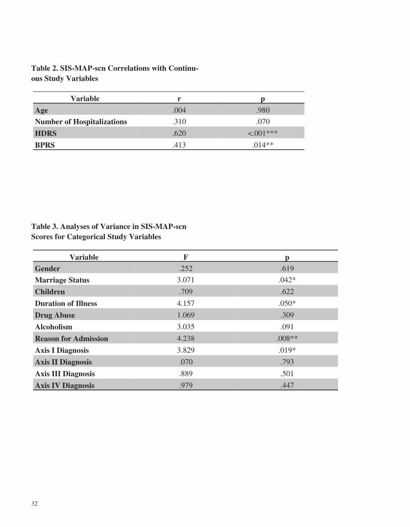
32
Table 2. SIS-MAP-scn Correlations with Continu-ous Study Variables
Table 3. Analyses of Variance in SIS-MAP-scn Scores for Categorical Study Variables
chiatry, 23, 56–62.
Variable r p
Age .004 .980
Number of Hospitalizations .310 .070
HDRS .620 <.001***
BPRS .413 .014**
Variable F p
Gender .252 .619
Marriage Status 3.071 .042*
Children .709 .622
Duration of Illness 4.157 .050*
Drug Abuse 1.069 .309
Alcoholism 3.035 .091
Reason for Admission 4.238 .008**
Axis I Diagnosis 3.829 .019*
Axis II Diagnosis .070 .793
Axis III Diagnosis .889 .501
Axis IV Diagnosis .979 .447
33
17, 212-222. doi: 10.1080/13811118.2013.80
Kreitman, N., & Foster, J. (1991). The construction
and selection of predictive scales, with spe-
cial reference to parasuicide. The British
Journal of Psychiatry, 159, 185 –192.
Murphy, G. E., & Wetzel, R. D. (1990). The lifetime
risk of suicide in alcoholism. Archives of
General Psychiatry, 47, 383-392.
Nelson, C., Johnston, M., & Shrivastava, A. (2010).
Improving risk assessment with suicidal
patients: A preliminary evaluation of the
clinical utility of The Scale for Impact of
Suicidality - Management, Assessment and
Planning of Care (SIS-MAP). Crisis: The
Journal of Crisis Intervention and Suicide
Prevention, 31, 231-237.
Petronis, K.R., Samuels, J.F., Moscicki, E.K., et al.
(1990). An epidemiologic investigation of
potential risk factors for suicide attempts.
Social Psychiatry and Psychiatric Epide-
miology, 25, 193-199.
Johnston, M.E., Nelson, C., & Shrivastava, A. (2013).
Dimensions of suicidality: Analyzing the do-
mains of the SIS-MAP suicide risk assess-
ment instrument and the development of a
brief screener. Archives of Suicide Research,
5639
Hall, R.C., Platt, D.E., & Hall, R.C. (1999). Suicide risk
assessment: a review of risk factors for sui-
cide in 100 patients who made severe suicide
attempts. Evaluation of suicide risk in a time
of managed care. Psychosomatics, 40, 18 -27.
Hamilton, M. (1960). A rating scale for depression.
Journal of Neurology, Neurosurgery, & Psy-
chiatry.
Cochrane-Brink, K.A., Lofchy, J.S., & Sakinofsky, I.
(2000). Clinical rating scales in suicide risk
assessment. General Hospital Psychiatry,
22, 445-451.
References
American Psychiatric Association. (1994). Diagnostic
and statistical manual of mental disorders,
4th edition. Washington, DC: American Psy-
chiatric Association.
Bryan, C.J., & Rudd, M.D. (2006). Advances in the
assessment of suicide risk. Journal of Clini-
cal Psychology: In Session, 62, 185-200.
Pompili, M., Amador, X. F., Girardi, P., et al.
(2007). Suicide risk in schizophrenia:
learning from the past to change the fu-
ture. Annals of General Psychiatry, 6. doi:
10.1186=1744-859X-6-10
34
Thienhaus, O.J., & Piasecki, M. (1997). Assessment
of suicide risk. Emergency Psychiatry, 48,
293-294.
Ventura, J., Lukoff, D., Nuechterlein, K.H., et al.
(1993). Appendix 1: Brief Psychiatric Rat-
ing Scale BPRS. Expanded Version
4.0.scales, anchor points and administra-
tion manual. International Journal of
Methods in Psychiatric Research, 3, 227–
243.

35
Malingering and Homelessness
To the Editor:
Identification and management of malingering, factitious disorder or person feigning
illness, is a difficult problem in emergency psychiatry (Brady et al., 2013). As many as 0.14 –
5% of psychiatric admissions (Bhugra 1988; Popli et al., 1992; Nicholson and Roberts 1994)
and as many as 13% of emergency patients (Yates and Nordquist 1996), may be factitious dis-
orders. Homelessness has been previously reported as one of the potential causes of malinger-
ing (i.e., feigning illness to avoid homelessness). However, there are no data available regard-
ing how common this is as a motivating factor. We examined all of the charts of individuals
discharged from a dedicated emergency psychiatric service with the diagnosis of malingering
over one year and collected information regarding homelessness status. Among the 6000 pa-
tient visits in 2013, 47 (0.8%) were discharged with a diagnosis of malingering. The charts
were available for review in 30 patients. The notes did not identify the secondary gain goals of
these patients. Among these subjects, information about homelessness status was not availa-
ble in one. Of the remaining 29 subjects, 14 (48.3%) were homeless, and another 7 (24%) were
staying with friends or non-custodial family members (non-first degree relatives). Among 6
for whom information was available, the duration of homelessness was over 2 years in one and
an average of 24 days in the remaining 5 subjects. The high rates of homelessness among pa-
tients identified as malingering suggests that this is either an important factor in feigning ill-
ness in emergency psychiatric settings or an important factor leading clinicians to diagnose
feigned illness.

36
References
Bhugra D. Psychiatric Munchausen’s syndrome: literature review with case reports. Acta Psychiatr
Scand 1988;77:497-503.
Brady MC, Scher LM, Newman W. “I just saw Big Bird. He was 100 feet tall!” Malingering in the
emergency room. Current Psychiatry 2013;12(10):33-38.
Nicholson SD, Roberts GA. Patients who (need to) tell stories. Br J Hosp Med 1994;51:546-549
Popli AP, Masand PS, Dewan MJ. Factitious disorders with psychological symptoms. J Clin Psychi-
atry 1992;53:315-318.
Yates DB, Nordquist CR, Schultz-Ross RA. Feigned psychiatric symptoms in the emergency room.
Psychiatr Serv 1996;47(9):998-1000.
Latif Saiyad, MD
Andrew Vaughn
Cassie Conrad
Anne Buck
Rif S. El-Mallakh, MD
Department of Psychiatry
University of Louisville School of Medicine
Louisville, Kentucky

37
News of the
American Association for Emergency Psychiatry
MANUSCRIPTS AND BOOK REVIEWS WELCOME!
The AAEP would like to invite all members and colleagues in the field of
Emergency Psychiatry to submit a manuscript or book review for publication in
the Journal of the American Association for Emergency Psychiatry.
This Journal is intended to be a forum for the exchange of multidisciplinary ide-
as. Manuscripts that deal with the interfaces of emergency psychiatry are wel-
come. This includes psychiatric evaluation of individuals in the emergency room
setting, education and training in the field and research into causes, and treat-
ment of behavioral problems. Manuscripts are evaluated for style, clarity,
consistency, and suitability.
Submit manuscripts or queries electronically to: Marie Westlake, at:
[email protected]. Please include the address, telephone num-
ber, and email address for the corresponding author on all manuscripts.
AAEP MEMBER NEWS AND ANNOUNCEMENTS
What’s going on with your Emergency Psychiatry facility, with you and/or your staff? We’d like to know and share it with other AAEP members in our newsletter. Did you or one of your colleagues publish an article related to Emergency Psychiatry? Have
you, your staff, or your program recently been honored? Is there a new educational or training process you are using that you believe could help your peers?
We welcome you to share any news relevant to Emergency Psychiatry with your fellow members. Please send your announcements to us by email to:
[email protected]. ___________________________________________________________
AAEP MEMBERS-ONLY LISTSERV
Check out the AAEP members-only listserv discussion group that is available via invi-tation to members in good standing only. This is a great opportunity to discuss press-ing issues, diagnostic dilemmas and treatment approaches in Emergency Psychiatry,
and to obtain consultation from your fellow experts on difficult cases.
If you have misplaced or did not receive your invitation email, please contact Marie Westlake at: [email protected]

38
Katherine A. Backes MD (Chicago, IL) Michael A. Bohrn MD (Wyomissing, PA) Kathleen M. Boudreaux (Lafayette, LA) Hetal Brahmbhatt (Johnson City, TN) Millicent A. Branch (Franklin, TN) Karen Budde RN (Denver, CO) Brian Buggie MD (New York, NY) Lisa Carchedi (Cedar Park, TX) Mary R. Carlson RN (Denver, CO) Julien J. Cavanagh de Carvalho MD (New York, NY) John Ceilley MD (Denver, CO) Erick H. Cheung MD (Los Angeles, CA) Larry Countryman RN (Denver, CO) Kurt Cousins (Houston, TX) Rachel Davis MD (Denver, CO) Barbara Dawson (Houston, TX) Maria De Pena-Nowak (White Plains, NY Carol L. DiGiuseppe (Westland, MI) Audrey Dumas MD (Denver, CO) Thomas Dunn PhD (Denver, CO) Su Duong (Houston, TX) Martha Edelman MD (Manhasset, NY) Sara Flores (Houston, TX) Brenda C. Fore (Asheville, NC) Anique K. Forrester MD (Baltimore, MD) John C. Furman MD (San Francisco, CA) Sandra Grimes (Houston, TX) Allison Hadley MD (San Diego, CA) Susie Harris MD (Denver, CO) Penny Hipp (Houston, TX) Kelly R. Hobgood MD (Charlotte, NC) Diana Ingram MD (Victoria, Canada) Rachael Anne Jasperson (Fruit Heights, UT) Christopher Johnson (Houston, TX) Madhuri Kamble (Houston, TX) Raheel A. Khan (West Sacramento, CA) Marylou Killian (LaGrangeville NY) Stephanie R. Knight MD (Baltimore, MD) Charles Kopecky (Houston, TX) Stuart Kretchmar (Wilmette, IL) Nithin Krishna MD (Baltimore, MD) Vernon R. Kubiak (Pocatello, ID) Rhonda R. Lampen MD (Marlton, NJ Philip Lavine MD (Burlington, NC) Kappy Madenwald MSW (Columbus, OH) Charles Marsh PharmD (Fort Smith, AR) Eva M. Mathews (Baton Rouge, LA)
Chanda Mayers-Elders (Houston, TX) Julia M. McLaughlin (San Francisco, CA) Eli F. Merritt MD (San Francisco, CA) Christopher W. Miller MD (Baltimore, MD) Barry Mills (Austin, TX) Alainia N. Morgan-James MD (Baltimore, MD) David W. Morris PhD (Dallas, TX) Karen Murrell (Sacramento, CA) Mai Nguyen (Houston, TX) Ian O. Nnatu (London, England) Karen O’Flynn, M.D. (Torrington, CT) Sadat Olatunbosun (Houston, TX) Jacob O’Meilia MD (Fort Worth, TX) Maryanne Pappas (Wethersfield, CT) Victoria Pappas-Villafane (Tintion Falls, NJ) Amber Pastusek (Houston, TX) Marla Pidgeon RN (Denver, CO) Sheryl Porter LCSW (Denver, CO) Kristine Powell (Bartonville, TX) Rhian E. Purdy (Augusta, GA) Benjamin W. Raatjes MD (New York, NY Lydia Reich (Houston, TX) Beth M. Reiley (York, PA) Shirley M. Repta PhD (Shreveport, LA) Mark A. Rodriguez (Phoenix, AZ) John Samson (Minneapolis, MN) Alexander Sanchez MD (West New York, NJ) Dan Severn MD (Denver, CO) Ravi N. Shah (New York, NY) Nima Sharif (Philadelphia, PA) Kevin Simmons (Houston, TX) Robert C. Strayhan (Texarkana, TX) Sujath Syed MD (Houston, TX) David E. Telford (Eagle River, AK) Anthony Thomas (Brooklyn, NY Susan Thompson (Norman, OK) Donald L. Thompson MD (Baltimore, MD) Kathryn C. Trosky MD (Austin, TX) Veronica T. Tucci PhD (Pearland, TX) Penny Vigneau (Newark, NJ) Diana Villareal (Houston, TX) Christian Williams (San Francisco, CA) Deloran Wilson MD (Baltimore MD) Jennifer Wren (Houston, TX) Adel S. Zaraa MD (Akron, OH) Emily M. Zarse (Indianapolis, IN) Bonnie Ziegler (Downington, PA)
Welcome to New AAEP Members in 2014 !

39
Watch your email and the AAEP
website for details of AAEP Activities during
the American Psychiatric
Association Annual Meeting
May 16—May 20, 2015
in Toronto!
www.EmergencyPsychiatry.org

40


