Journal of the Egyptian Society of Cardio-thoracic Surgery EDITOR-IN-CHIEF Ezzeldin A. Mostafa, MD PAST EDITORS Hassouna M. El-sabea, FRCS (1995-1996) Mohamed S. El-fiky, MD (1997-2004) CO-EDITOR Yasser M. Hegazy, FRCS, MD STATISTICS EDITOR Ahmed A. Hassouna, MD ETHICS EDITOR M. Anwar Balbaa, MD ASSOCIATE (SECTION) EDITORS Ahmed M. Deebis, MD Ibrahim M. Abdel Meguid, MD Mohamed A. Nasser, MD Samir A. Hassan, MD Samir A. Keshk, MD Website & Managing Editor Mohamed A. Othman, MS Submit Manuscripts: Editorial office Journal of the Egyptian Society of Cardio-Thoracic Surgery 330 El Sudan Street, Embaba , Egypt Tel. (+ 202) 3303 8054 Website: www.arabmedics.com/jescts.html Email : [email protected]
Transcript
Journal of the
Egyptian Society of
Cardio-thoracic SurgeryEDITOR-IN-CHIEF
Ezzeldin A. Mostafa, MD
PAST EDITORSHassouna M. El-sabea, FRCS (1995-1996)
Mohamed S. El-fiky, MD (1997-2004)
CO-EDITORYasser M. Hegazy, FRCS, MD
STATISTICS EDITOR Ahmed A. Hassouna, MD
ETHICS EDITORM. Anwar Balbaa, MD
ASSOCIATE (SECTION) EDITORSAhmed M. Deebis, MD
Ibrahim M. Abdel Meguid, MDMohamed A. Nasser, MD
Samir A. Hassan, MDSamir A. Keshk, MD
Website & Managing EditorMohamed A. Othman, MS
Submit Manuscripts: Editorial office Journal of the Egyptian Society of Cardio-Thoracic Surgery
cularization in patients on Clopidogrel Mohammed Abdel-Aal ,MD.,Bakir M Bakir ,MD.,Osama Abbas ,MD.,Mustafa Sabban ,MD.,Ahmad A. Alshaer ,MD.,Nazeh El-Fakarany,MRCP(UK),Ihab Yehya ,MD.
22 Perioperative Intra Aortic Balloon Pump Sup--port For Coronary Artery Bypass Surgery. Five Years Experience. Amr Mohamed Rushdi, MD., Tarek Hussein El-Taweel, MD. Mohamed Helmi, MD. , Saed Ab--delaziz Badr, MD. , Ahmed Helmi, MD.
27 Coronary revascularization using bilateral inter--nal mammary artery grafting in insulin-treated diabetics Early resultsMarwan Mohamed ,MD., Diaa El-Din A.Seoud ,MD., Yasser Menaissy , MD.
33 Implications of Valve Prosthesis-patient Mis--match after Aortic Valve Replacement with Small Sized Mechanical Prosthesis Mostafa Abd El Sattar MD.,Mohamed Essa MD., Ayman Gabal MD., Ahmed Abd El Aziz MD.
39 Assessment of Different Techniques of Aortic Valve
Journal of The Egyptian Society of Cardio-Thoracic Surgery
Volume 15 Mar-Jun 2007 Number 1,2 ISSN 1110-578X
Replacement in Patients with Small Aortic AnnulusHany A. El Maboud MD., Ayman Amar MD., Mohsin Abdel-Karim MD., Walid Ismail MRCS
46 Risks and Complications of Resternotomy in Adult’s Cardiac OperationsIhab M.Yehya MOURSI MD., Mohamed M. Abdel Aal MD.
51 New Management Technique for Deep Sternal Surgical Site InfectionMS. AbdAllah MD.,Hasan Abady MD., Ahmed Ab--del Aziz MD., Mounir Osman MD.,Mohamed Abdel Hady MD.,
57 Early Post Operative Results of on Pump Versus off Pump Coronary Artery Bypass Surgery in High Risk PatientsM . s e w i e l a m , M D . , T. S . A b d a l l a h , M D . , O .Abulkasem,MD. , M.abuldahab,MD. , A.osama,MD.
THORACIC66 The Benefit of Surgical Lung Biopsy in Diagnosis
and Prognosis of Diffuse Infiltrative Lung DiseaseTarek A Mohsen MD., FRCS, Mohamed M Kamel, MD., Amany A Abou Zeid MD., MRCP, FCCS, Medhat Abdel Khalek Soliman MD., FCCP.
71 A Prospective Randomized Trial for thoracoscopic Talc Poudrage Versus Povidone-iodine for Pleurod--esis for Effusion Due to Metastatic Breast CancerTarek A Mohsen MD., FRCS, Mohamed M Kamel, MD., Amany A Abou Zeid MD., MRCP, FCCS, Medhat Abdel Khalek Soliman MD., FCCP.
83 Surgical Treatment of Bronchiectasis in ChildrenAbd El Ghaffar El-zaanin MD.,Spiro Al-taweel FRCS , Hussein Al-attar (MSc), Mohammed Abu assan MSc , Doran Al-Hatto MSc
86 Minimal-access for Thymectomy is Preferable for the Treatment of Myasthenia GravisMohamed M. Abd Alaal, MD., Ahmed Alshaer, MD.
THE WAY I DO IT92 Esophageal Perforation: Emphasis on Manage--
ment Kamal A. Mansour, MD. ,Bradley L. Bufkin, MD.,Joseph I. Miller, Jr, MD., Ezzeldin A. Mostafa, MD.
Journal of The Egyptian Society of Cardio-Thoracic Surgery
Volume 15 Sep-Dec 2006 Number 3,4 ISSN 1110-578X
A9The Journal of Egyptian Society of Cardiothoracic Surgery ● Volum 15, Number (1-2)
Editorial Office Please address all correspondence to:Ezzeldin A. Mostafa, MD, Editor, In-chiefJournal of the Egyptian Society of Cardio-thoracic Surgery 330 El-Sudan St., Imbaba, Cairo, Egypt.Telephone: (+202) 303 6634Fax: (+202) 303 8054E-Mail: [email protected]
The Journal of the Egyptian Society of Cardio-Thoracic Surgery [ISSN 1110-578 X] is the official publication of the Egyptian Society of Cardio-thoracic Surgery. The journal is published every three months .
General Instructions
Every submission must include: Cover letter, indicating the category of article , the Complete manuscript, including title page, abstract, text, tables, ac--knowledgments ,references and illustrations .
Required Disclosures;
A. Conditions for Publication Form which includes dis--closures regarding freedom of investigation and conflicts of interest, signed by all authors. In single Author publication an additional Senior Consultant Signature is required.B. Written permission from the publisher (copyright holder) is required to reproduce any previously published table(s), illustration(s) or photograph(s) in both print and electronic media. C. Written permission from unmasked patients appearing in photographs is also required.
Revised_Manuscripts:Revised manuscripts must be submitted in three parts as Microsoft word-processing files : (1) cover letter with responses to reviewers’ comments (2) revised, marked manuscript showing additions and deletions; (3) revised, un--marked manuscript.
General Information Three copies of the Manuscripts should be sent preferably
prepared in Microsoft Word , typed double-spaced throughout (including title page, abstract, text, references, tables and legends) with one (1) inch (2.5 cm) margins all around. Place Author name and page number in the upper right corner of each page. Manuscripts written in 12 point Arial or Times New Roman fonts are preferred (Note: Do not submit your manuscript in PDF format it causes problems in processing your submis--sion.)Arrange manuscript as follows: (1) title page, (2) abstract, (3) text, (4) acknowledgments, (5) disclosures if required, (6) references, (7) tables and (8) legends. Number pages consecu--tively, beginning with the title page as page 1 and ending with the legend page.If your manuscript contains illustrations, in addition to submit--ting them online, you must send two sets of original illustra--tions to the editorial office labeled with manuscript number, first author, and figure number on back. Tables and figures should be provided separate from the text while there position in the text should be marked on the manu--script.
Word Limits by Category of Manuscript
Original articles should not exceed 4500 words including title page, abstract of 150-200 words, text, figure legends and refer--ences. The combined total of illustrations and tables should not exceed 10 and the number of references should not exceed 40.
Case reports and “The way I do it” articles are limited to a total of 1500 words including title page, abstract, text, refer--ences and figure legends. For each illustration subtract 100 words and for each table subtract 300 words from the word limit. References are limited to eight. A “how to do it” article should be a description of a useful surgical technique and con--tain descriptive, illustrative material.
Images in cardiothoracic surgery are limited to 350 words including title and text and to two, possibly three figures. The entire contribution must fit on one printed page .
Review articles are limited to 6500 words including title page, abstract, text, figure legends and all references. The total number of references should not exceed 80. Subtract 100
Guidelines for Authors
Journal of The Egyptian Society of Cardio-Thoracic Surgery (J. Egypt. Soc. Cardiothorac. Surg.)
A10 The Journal of Egyptian Society of Cardiothoracic Surgery ● Mar-Jun 2007
words for each illustration and 300 words for each table.
Our surgical heritage articles are limited to 2500 words in--cluding title page, abstract, text, figure legends and references. Subtract 100 words for each illustration and 300 words for each table.
Correspondence (Letters to the Editor) and commentaries are limited to 500 words. Subtract 100 words for each illustration and 300 words for each table.
Editorials are limited to 2500 words including references. Subtract 100 words for each illustration and 300 words for each table.
Manuscript Preparation
Title Page (first page)
The title is limited to 100 characters and spaces for original manuscripts and to 80 characters and spaces for all other cat--egories of manuscripts. The title may not contain acronyms or abbreviations. All submissions, must have a title.
Running Head. Supply a short title of 40 characters and spaces.
Authors. List all authors by first name, all initials, family name and highest academic degree using “MD, PhD” for hold--ers of both degrees ( if more then 7 Authors justifie).
Institution and Affiliations. List the name and full address of all institutions where the work was done. List departmental affiliations of each author affiliated with that institution after each institutional address.
Meeting Presentation. If the paper has been or is to be pre--sented at the annual meeting of The Society, provide the name, location and dates of the meeting.
Keywords. Provide up to 5 keywords selected from the ap--pended list to describe the manuscript. Do not use any key--words that are not on the list.
Word Count. Provide the electronic total word count of the entire manuscript including title page, abstract,text,figure leg--ends and entire reference list.
Corresponding Author. Provide the name, exact postal ad--dress with postal code, telephone number, fax number and e-mail address of the author to whom communications, proofs and requests for reprints should be sent.
Abstract Page (Second page)
Original articlesProvide a structured Abstract, no longer than 250 words, di--vided into four sections: Background or Objective, Methods, Results, Conclusions. Avoid abbreviations and acronyms. In--
dicate the abstract word count below the abstract. Case reports, “the way i do it” articles, review articles and our surgical heritage articles. Provide an unstructured abstract of 100 words. Images, correspondence, commentaries, editorials and up--dates. No abstract is required.
Text Text should be organized as follows: Introduction, Mate--rial (or Patients) and Methods, Results, and Comment.Cite references,illustrations and tables in numeric order by order of mention in the text.
Avoid abbreviations. Consult the American Medical Associa--tion Manual of Style, 9th edition, for recommended abbrevia--tions. Define abbreviations at first appearance in the text. If 8 or more abbreviations or acronyms are used, provide a separate table of abbreviations and acronyms.
Measurements and weights should be given in standard metric units.Statistical nomenclature and data analysis. Fol--low the “Guidelines for Data Reporting and Nomenclature” published in The Annals of Thoracic Surgery (1988;46:260-1). Footnotes. Type footnotes at the bottom of the manuscript page on which they are cited. Suppliers of drugs, equipment and other brand mentioned in the article within parentheses , giving company name, city and country .
AcknowledgmentsGrants, financial support and technical or other assistance must be acknowledged at the end of the text before the references.
ReferencesIdentify references in the text using Arabic numerals in brack--ets on the line.Type references double-spaced after text or acknowl--edgments beginning on a separate sheet. Number con--secutively in the order in which they appear in the text. Journal references should provide inclusive page num--bers; book references should cite specific page numbers. Journal abbreviations should conform to those used in Index Medicus. follow the formats outlined below: Journal ArticleJones DR, Stiles BM, Denlinger CE, Antie P . Pulmonary segmentectomy: results and complications. Ann Thorac Surg 2000;76:343-9.(List all authors if 6 or fewer; otherwise list first 3 and add “et al.”)
Chapter in Book12. Vinten-Johansen J, Zhao Z-Q, Guyton RA. Cardiac surgi--cal physiology. In: Cohn LH, Edmunds LH Jr, eds. Cardiac Surgery in the Adult. 2nd ed. New York, NY: McGraw-Hill; 2003:53-84.
A11The Journal of Egyptian Society of Cardiothoracic Surgery ● Volum 15, Number (1-2)
Internet Address3. 1996 NRC Guide for the Care and Use of Laboratory Ani--mals. Available at: http://www.nap.edu/readingroom/books/labrats/contents.html. Accessed October 20, 2003.
Tables :Tables should be typewritten double-spaced on separate sheets (one to each page). Do not use vertical lines. Each table should be numbered (Arabic) and have a title above. Legends and explanatory notes should be placed below the table. Abbrevia--tions used in the table follow the legend in alphabetic order. Lower case letter superscripts beginning with “a” and follow--ing in alphabetic order are used for notations of within-group and between-group statistical probabilities. FigureLegends :Figure Legends should be numbered (Arabic) and typed double-spaced in order of appearance beginning on a sepa--rate sheet. Identify (in alphabetical order) all abbreviations appearing in the illustrations at the end of each legend. Cite the source of previously published material in the legend and indicate permission has been obtained. Proof of permis--sion must be surface mailed or faxed to the editor .
Illustrations :You must send two sets of original illustrations to the editorial office labeled with manuscript number, first author, and figure number on back.
Images or figures are submitted online as one or more separate files that may contain one or more images. Within each file containing images, use the figure number (eg, Figure 1A) as the image filename. The system accepts Powerpoint (.ppt) files Most illustrations will be reproduced at a width of one column (8.25 cm; 3 1/4 inches). Black, white and widely crosshatched bars are preferable; do not use stippling, gray fill or thin lines.
Instructions :Identify print proofs of figures on the back with figure number and name of the first author; when necessary, indicate the top with an up arrow For figures submitted in electronic format, all images should be at least 5 inches wide. Graphics software such as Photoshop and Illustrator, should be used to create art. Color images need to be at least 300 dpi.Gray scale images should be at least 300 dpi .Line art should be at least 1200 DPI .
Cover letter :Include with the manuscript a cover letter that provides 1) the category of manuscript (e.g., original research, Brief Commu--nication, Letter to the Editor); 2) statement that the material
has not been previously published or submitted elsewhere for publication; 3) information about any personal conflicts of interest of any of the authors; and 4) names of sources of out--side support for research, including funding, equipment, and drugs .You may also submit the name of one reviewer of your choice. You should include that individual’s mailing address, telephone, fax and e-mail address. Editorial Policies Scientific Responsibility StatementBefore publication of an accepted manuscript, each author is required to certify by signing the Conditions for Publication Form that he or she has participated sufficiently in the work and approved the final version of the manuscript to be pub--lished. Exclusive Publication StatementEach author must certify that none of the material in this manuscript has been published previously in either print or electronic form, and that none of this material is currently under consideration for publication elsewhere. This includes symposia and preliminary publications of any kind except an abstract of 400 words or fewer.
Conflict of Interest :Authors should disclose any conflict of interests. Authors who have a financial relationship with one or more companies whose products are featured in an article will disclose the ex--istence of this relationship in a box at the bottom of the first page of the published article.
Consultant Statistician and Statistical Methods : All manuscripts with statistical analysis are required to undergo biostatistical review .The most appropriate way is to involve a biostatistician consultant or coauthor from the investigators’ home institution . Manuscripts may undergo further biostatistical review by the Journal after submission. Additional information on statistical methods can be found in “Uniform Requirements for Manuscripts Submitted to Biomedical Journals”(www.acponline.org/journals/resource/unifreqr.htm).
Copyright :Authors of articles submitted to The J. Egypt. Soc. Cardiotho--rac. Surg. must transfer copyright to The Egyptian Society of Cardio-Thoracic Surgery by signing the “Conditions for Publi--cation Form.” This transfer becomes binding upon acceptance of the article for publication. No part of the published material may be reproduced elsewhere without written permission.Date of Receipt: The “received for publication” date is the date when the editorial office receives the manuscript, the cover let--ter, and the Copyright Transfer and Author Declaration State--ment, signed by all authors. For Date of acceptance : letter is provided from the editor.
A12 The Journal of Egyptian Society of Cardiothoracic Surgery ● Mar-Jun 2007
Checklist
A] Cover Letter □ Letter to the Editor □ Manuscript category designation .□ Single-journal submission affirmation .□ Conflict of interest statement (if appropriate). □ Sources of outside funding. □ Signed Statistical Collaboration .
B] Complete Manuscript□ Title page .□ Title of article□ Full name(s), academic degrees, and affiliation(s) of authors.□ Corresponding author .□ Telephones, fax, and e-mail address□ Abstract (250 words; double-spaced) .□ Ultramini-abstract (50 words) .□ Text (double-spaced). □ References (double-spaced; separate pages). □ Tables (double-spaced; separate pages). □ Figures (separate files; on hardcopy; properly identified), □ Figure legends (double-spaced; separate pages) .□ Word count.
C] Required Disclosures □ Conditions for Publication Form signed by all authors. Which transfers copyright to The
Egyptian Society of Cardio-Thoracic Surgery□ Written permission from the publisher to reproduce any previously published material .□ Written permission from unmasked patients .
A13The Journal of Egyptian Society of Cardiothoracic Surgery ● Volum 15, Number (1-2)
A15The Journal of Egyptian Society of Cardiothoracic Surgery ● Volum 15, Number (1-2)
This form MUST be completed, signed by ALL authors, and returned to the Editorial Office before your manuscript can be accepted for publication.
Scientific Responsibility Statement:Each author must sign this form to certify that he or she
has participated sufficiently in the work to take responsibility for a meaningful share of the content of the manuscript, and that this participation included: (a) conception or design of the experiment(s), or collection and analysis or interpretation of data; (b) drafting the manuscript or revising its intellectual content; and (c) approval of the final version of the manuscript to be published. In addition, each author must indicate whether or not he or she has had full freedom of investigation; defined as freedom from outside interests in controlling the design of the study, collection, analysis, and interpretation of data, and having freedom to full disclose all results.
Exclusive Publication Statement:Each author must sign this form to certify that none of the
material in this manuscript has been published previously in either print or electronic form, and that none of this material is currently under consideration for publication elsewhere. This includes symposia, transactions, books, articles published by invitation and preliminary publications of any kind except an abstract of 400 words or fewer.
Copyright Transfer Agreement:Each author must sign this form to certify that, if the manu--
script is accepted for publication in the Journal of the Egyptian Society of Cardio-Thoracic Surgery ( JESCTS), copyright (including the right to obtain copyright registration, whether separately or as part of a journal issue .) in and to the above article transfers throughout the world and for the full term and all extensions and renewals thereof to: THE EGYPTIAN SO--CIETY OF CARDIO-THORACIC SURGERY
This transfer includes the right to adapt the article for use in conjunction with computer systems and programs, includ--ing reproductions or publication and incorporation in retrieval systems.
Rights of authors:The ESCTS hereby licenses the following rights back to
the author(s): A. Patent and trademark rights to any process or procedure
described in the article. B. The right to photocopy or make single electronic copies of
the article for their own personal use, including for their
Conditions for Publication Form
own classroom use, or for the personal use of colleagues, provided the copies are not offered for sale .
C.The right, subsequent to publication, to use the article or any part thereof free of charge in a printed compilation of works of their own, such as collected writings or lecture notes.
Note: All copies, paper or electronic, or other use of the informa--
tion must include an indication of The ESCTS copyright and a full citation of the journal source.
Authorship: If copyright is held by the employer, the employer or an
authorized representative of the employer must sign in addi--tion to the author(s).
Warranties: The author(s) warrant that the article is the author’s origi--
nal work and has not been published before. The author(s) war--rant that the article does not infringe on the rights of others. If excerpts from copyrighted works are included, the author(s) has (have) obtained written permission from the copyright owners and will credit the sources in the article.
Preprints: The author(s) warrant(s) that if a prior version of this work
(normally a preprint) has been posted to an electronic server, such version was accessible to only a small group of individu--als and the author(s) will cause its prompt removal from such server.
Conflict of Interest Disclosure Statements: Each author must indicate below that either (a) no financial
conflict of interest exists with any commercial entity whose products are described, reviewed, evaluated or compared in the manuscript, except for that disclosed under “Acknowledgements” or (b) a potential conflict of interest exists with one or more commercial entities whose products are described, reviewed, evaluated or compared in the manuscript through the existence of one or more of the following relationships: the author is a full or part-time employee of a company; has an existing or optional equity interest in a company; owns or partly owns patents licensed to a company; has an ongoing retainer relationship (consultantship, speaker, etc.) with a company for which he/she receives financial remuneration; or has received financial compensation for this publication. If Yes is checked, a box on the first page of the published article will read: ?Dr. X discloses that he/she has a financial relationship with company Y.?
A16 The Journal of Egyptian Society of Cardiothoracic Surgery ● Mar-Jun 2007
Author: Manuscript Title:
I agree with the preceding conditions and provide the appropriate signatures and information below accordingly:
A 16
If there are additional authors on the article, please photocopy this form and attach additional sheets as need be with appropri-ate information and signatures affixed .
Author:Manuscript Title:
I agree with the preceding conditions and provide the appropriate signatures and information below accordingly:
Author’s Name:_____________________________________________________________________________________Signature:______________________________________________ Date:_______________________________________Author’s employer’s signature, if appropriate: ___________________________________________________________Conflict of interest: Yes ___ No ___ If yes, with which entity: _______________________________________________Did you have freedom of investigation in all aspects of this work?: Yes ___ No ___
Author’s Name:_____________________________________________________________________________________Signature:______________________________________________ Date:_______________________________________Author’s employer’s signature, if appropriate: ___________________________________________________________Conflict of interest: Yes ___ No ___ If yes, with which entity: _______________________________________________Did you have freedom of investigation in all aspects of this work?: Yes ___ No ___
Author’s Name:_____________________________________________________________________________________Signature:______________________________________________ Date:_______________________________________Author’s employer’s signature, if appropriate: ___________________________________________________________Conflict of interest: Yes ___ No ___ If yes, with which entity: _______________________________________________Did you have freedom of investigation in all aspects of this work?: Yes ___ No ___
Author’s Name:_____________________________________________________________________________________Signature:______________________________________________ Date:_______________________________________Author’s employer’s signature, if appropriate: ___________________________________________________________Conflict of interest: Yes ___ No ___ If yes, with which entity: _______________________________________________Did you have freedom of investigation in all aspects of this work?: Yes ___ No ___
Author’s Name:_____________________________________________________________________________________Signature:______________________________________________ Date:_______________________________________Author’s employer’s signature, if appropriate: ___________________________________________________________Conflict of interest: Yes ___ No ___ If yes, with which entity: _______________________________________________Did you have freedom of investigation in all aspects of this work?: Yes ___ No ___
Author’s Name:_____________________________________________________________________________________Signature:______________________________________________ Date:_______________________________________Author’s employer’s signature, if appropriate: ___________________________________________________________Conflict of interest: Yes ___ No ___ If yes, with which entity: _______________________________________________Did you have freedom of investigation in all aspects of this work?: Yes ___ No ___
Author’s Name:_____________________________________________________________________________________Signature:______________________________________________ Date:_______________________________________Author’s employer’s signature, if appropriate: ___________________________________________________________Conflict of interest: Yes ___ No ___ If yes, with which entity: _______________________________________________Did you have freedom of investigation in all aspects of this work?: Yes ___ No ___
The Journal of Egyptian Society of Cardiothoracic Surgery ● Sep-Dec 2006
Author’s Name:_____________________________________________________________________________________Signature:______________________________________________ Date:_______________________________________Author’s employer’s signature, if appropriate: ___________________________________________________________Conflict of interest: Yes ___ No ___ If yes, with which entity: _______________________________________________Did you have freedom of investigation in all aspects of this work?: Yes ___ No ___
Author’s Name:_____________________________________________________________________________________Signature:______________________________________________ Date:_______________________________________Author’s employer’s signature, if appropriate: ___________________________________________________________Conflict of interest: Yes ___ No ___ If yes, with which entity: _______________________________________________Did you have freedom of investigation in all aspects of this work?: Yes ___ No ___
Author’s Name:_____________________________________________________________________________________Signature:______________________________________________ Date:_______________________________________Author’s employer’s signature, if appropriate: ___________________________________________________________Conflict of interest: Yes ___ No ___ If yes, with which entity: _______________________________________________Did you have freedom of investigation in all aspects of this work?: Yes ___ No ___
Author’s Name:_____________________________________________________________________________________Signature:______________________________________________ Date:_______________________________________Author’s employer’s signature, if appropriate: ___________________________________________________________Conflict of interest: Yes ___ No ___ If yes, with which entity: _______________________________________________Did you have freedom of investigation in all aspects of this work?: Yes ___ No ___
Author’s Name:_____________________________________________________________________________________Signature:______________________________________________ Date:_______________________________________Author’s employer’s signature, if appropriate: ___________________________________________________________Conflict of interest: Yes ___ No ___ If yes, with which entity: _______________________________________________Did you have freedom of investigation in all aspects of this work?: Yes ___ No ___
Author’s Name:_____________________________________________________________________________________Signature:______________________________________________ Date:_______________________________________Author’s employer’s signature, if appropriate: ___________________________________________________________Conflict of interest: Yes ___ No ___ If yes, with which entity: _______________________________________________Did you have freedom of investigation in all aspects of this work?: Yes ___ No ___
Author’s Name:_____________________________________________________________________________________Signature:______________________________________________ Date:_______________________________________Author’s employer’s signature, if appropriate: ___________________________________________________________Conflict of interest: Yes ___ No ___ If yes, with which entity: _______________________________________________Did you have freedom of investigation in all aspects of this work?: Yes ___ No ___
If there are additional authors on the article, please photocopy this form and attach additional sheets as need be with appropriate information and signatures affixed .
A 16
If there are additional authors on the article, please photocopy this form and attach additional sheets as need be with appropri-ate information and signatures affixed .
Author:Manuscript Title:
I agree with the preceding conditions and provide the appropriate signatures and information below accordingly:
Author’s Name:_____________________________________________________________________________________Signature:______________________________________________ Date:_______________________________________Author’s employer’s signature, if appropriate: ___________________________________________________________Conflict of interest: Yes ___ No ___ If yes, with which entity: _______________________________________________Did you have freedom of investigation in all aspects of this work?: Yes ___ No ___
Author’s Name:_____________________________________________________________________________________Signature:______________________________________________ Date:_______________________________________Author’s employer’s signature, if appropriate: ___________________________________________________________Conflict of interest: Yes ___ No ___ If yes, with which entity: _______________________________________________Did you have freedom of investigation in all aspects of this work?: Yes ___ No ___
Author’s Name:_____________________________________________________________________________________Signature:______________________________________________ Date:_______________________________________Author’s employer’s signature, if appropriate: ___________________________________________________________Conflict of interest: Yes ___ No ___ If yes, with which entity: _______________________________________________Did you have freedom of investigation in all aspects of this work?: Yes ___ No ___
Author’s Name:_____________________________________________________________________________________Signature:______________________________________________ Date:_______________________________________Author’s employer’s signature, if appropriate: ___________________________________________________________Conflict of interest: Yes ___ No ___ If yes, with which entity: _______________________________________________Did you have freedom of investigation in all aspects of this work?: Yes ___ No ___
Author’s Name:_____________________________________________________________________________________Signature:______________________________________________ Date:_______________________________________Author’s employer’s signature, if appropriate: ___________________________________________________________Conflict of interest: Yes ___ No ___ If yes, with which entity: _______________________________________________Did you have freedom of investigation in all aspects of this work?: Yes ___ No ___
Author’s Name:_____________________________________________________________________________________Signature:______________________________________________ Date:_______________________________________Author’s employer’s signature, if appropriate: ___________________________________________________________Conflict of interest: Yes ___ No ___ If yes, with which entity: _______________________________________________Did you have freedom of investigation in all aspects of this work?: Yes ___ No ___
Author’s Name:_____________________________________________________________________________________Signature:______________________________________________ Date:_______________________________________Author’s employer’s signature, if appropriate: ___________________________________________________________Conflict of interest: Yes ___ No ___ If yes, with which entity: _______________________________________________Did you have freedom of investigation in all aspects of this work?: Yes ___ No ___
The Journal of Egyptian Society of Cardiothoracic Surgery ● Sep-Dec 2006
A17The Journal of Egyptian Society of Cardiothoracic Surgery ● Volum 15, Number (1-2)
Purpose of Peer ReviewOne is to evaluate objectively the science of the submitted
paper and the other is to provide a constructive critique indicat--ing how the paper could be or could have been improved by the authors. Reviewers should avoid unpleasant comments.
Acceptance of a Manuscript for ReviewReviewers should accept assignments to review manu--
scripts that are within their sphere of expertise, which they plan to review within the 21 day deadline. Reviewers should decline assignments for which a conflict exists between the reviewer and authors or between the reviewer and commercial products that are integral to the content of the article.
Category of the Manuscript The broad categories of papers for which peer review is
undertaken are original scientific articles; new technology papers; case reports, the way i do it articles , images; and re--view articles. The editor and/or associate editors review corre--spondence, invited commentaries, editorials, surgical heritage , ethical and statistical papers.
General Requirements for PublicationThe paper should conform to the format and restrictions
for the category to which it belongs and be written in good, readable English. The paper should address an important or in--teresting subject and provide new and original information. Il--lustrative material should be well chosen and of good quality.
Original Scientific ArticleThe reviewer should assess the articles’ interest to readers;
strengths and weaknesses; originality; clarity of text, tables, illustrations and figure legends; presentation; analysis of re--sults; credibility of results; importance of the findings; depth of scholarship and relationship of the results to the existing lit--erature Ethical issues, such as prior publication of all or part of the data; plagiarism; transgression of human or animal rights; or dishonesty should be noted, if detected.
The following topics are offered to help guide the review--
er’s assessment of an original scientific article. • ‘Title’ should reflect the content of the article and be concise
and clear• ‘Abstract’ should indicate the purpose of the study, subjects
and methods used, most important results and the main con--clusions supported by results.
• ‘Introduction’ should indicate the rationale and focus of the study and state the purpose or hypothesis.
• ‘Methods’ should present the design of the study, fully de--scribe the number and subjects and exclusion and inclusion criteria; whether subjects were enrolled consecutively; meth--ods used to gather data, including follow-up data; methods
Guidelines for Reviewersby which control and experimental groups were assembled; the primary outcome variable; secondary outcome variables; how outcome measurements were made and validated; the statistical design of the study; and the statistical methods used to analyze the study.
• ‘Results’ should concisely present the most important find--ings in text . Data should be reported as means or medians with appropriate indicators of variance and exact p values in tables and text. Figures should be well selected to high--light important findings . Survival and event curves should indicate specified confidence limits or subjects at risk. Re--gression diagrams should include the regression equations, regression coefficient and exact p value in the figure legend. Figure legends should adequately and clearly describe the important information illustrated.
• ‘Comment’ should not repeat results, but should point out the significance and conclusions of the new data, integrate the authors’ new data with that in the prior literature, draw inferences and conclusions regarding the question or purpose addressed by the study and point out the limitations of the study. The ‘Comment’ section should not be a review of the literature.
• References should be properly cited, reasonably current, ac--curateand in proper format.
New TechnologyArticles describing new technology are necessarily de--
scriptive and do not pose or test a hypothesis. These articles evaluate new devices, systems, monitors, implantable mate--rial and similar technology designed for improving patient care and outcomes. The reviewer is asked to evaluate the efficacy, safety and indications of the new technology .
The reviewer needs to inspect the ‘Disclosure statement’ after the text, before References. This statement should dis--close the source of funds used for the evaluation study and whether or not the product was purchased, borrowed or do--nated by the manufacturer or inventor. Conflicts of interest statements for authors are managed by the editorial staff.
Case Reports, The Way I Do It, ImagesCase reports describe interesting presentations of disease
and innovative management of the patient’s or patients’ problem. How to Do It articles emphasize innovations in the operative management of technical challenges and new ways of doing things. Images, which must fit on one printed page, are graphics of interesting presentations of disease within the chest.
Reviewers should evaluate the clarity and completeness of the case or procedure descriptions and the selection and quality of the illustrative material. Reviewers should also note whether or not the paper adheres to the format restrictions enumerated in “Information for Authors”. The reference list
A18 The Journal of Egyptian Society of Cardiothoracic Surgery ● Mar-Jun 2007
should be selective rather than inclusive.
Review ArticleReviewers should assess the importance of the subject
matter, need for the review and probable interest to readers. Reviews of very rare and unusual diseases are discouraged . Reviewers should note if authors have respected the format and restrictions of this category as stated in “Information for Authors”.
The ‘Introduction’ should provide the rationale for re--viewing the subject matter and provide the outlines of what is included and not included in the review. In the ‘Methods’ section reviewers should assess the methods used to search for articles, including search words and databases probed. The body of the review should be well organized with well chosen topical headings arranged in logical order. Within each topi--
cal heading the material should be presented in an integrated, comprehensive, objective manner. Statements should be refer--enced accurately. Reviewers should look for a “summing up” of the topical content .
The review should provide a general overview of the subject matter assessing progress, pointing out deficiencies in present management and indicating opportunities and di--rections of future work. The reviewer should also assess the selection of references .
FootnoteThe reviewer remains anonymous . The reviewer should
direct his or her critique to the authors in the style and format that suits them best. The recommendation to the editor is made separately with or without additio
A19The Journal of Egyptian Society of Cardiothoracic Surgery ● Volum 15, Number (1-2)
19 - 23 SEPTEMBER 2007 KANSAS CITY, MO UNITED STATES AmSECT>s 15th Annual Symposium on New Advances in Blood ManagementWestin Crown CenterAbstract submission deadline: 31 July 2007For information, contact:Donna Pendarvis2209 Dickens Rd, Richmond, VA 23230-2005Phone: 804 565-6363Fax: 804 282-0090Email: [email protected] information: http://www.amsect.org
20 - 21 SEPTEMBER 2007 2007 Annual Education Conference, Developing Careers in Surgical Education conferenceAdditional information: http://www.rcseng.ac.uk/education/courses/annual_education_2007.html 20 - 21 SEPTEMBER 2007 BLACKPOOL UNITED KINGDOM David Sharpe Memorial Symposium Blackpool Interactive Mitral Valve WorkshopBlackpool Victoria HospitalFor information, contact:Lorraine Richardson58 Kiln Close, Calvert Green, Buckingham, MK18 2PDPhone: +44 (0)1296 733 823Email: [email protected] information: http://www.bfwhospitals.nhs.uk/dsms/
20 - 21 SEPTEMBER 2007 LONDON UNITED KINGDOM Careers in Surgical Education: Management, Training and RsearchRoyal College of Surgeons of EnglandFor information, contact:Raven Department of Education, Royal College of Surgeons of England35-43 LIncoln>s Inn Fields, London,, WC2A 3PEPhone: +44 (0)20 7869 6350Email: [email protected] information: http://www.rcseng.ac.uk/education/courses/annual_education_2007.html/
21 SEPTEMBER 2007 BAIONA, PONTEVEDRA SPAIN The II Symposium on Dissection and Aortic Root SurgeryParador de BaionaFor information, contact:Congrega, S.L., Technical SecretariatRosalía de Castro, 13 - 1ºIzq., 15004 A Coruña (Spain)Phone: 34-981-216 416Fax: 34-981-217 542Email: [email protected] information: http://www.raizaortica.org
25 - 28 SEPTEMBER 2007 LAS VEGAS, NV UNITED STATES VIVA 07 - Vascular Interventional Advances - The National Education Course For Peripheral Vascular InterventionsMandalay Bay ResortFor information, contact:MMC6133 N. River Road, Rosemont, Illinois 60018Phone: 1 866-511-VIVAFax: 1 847 292-5801Email: [email protected] information: http://vivapvd.com/index.cfm
26 - 27 SEPTEMBER 2007 CESKE BUDEJOVICETEL CZECH REPUBLIC East European Heart Valve Postgraduate Course (EEHVPG)Hotel GOMELFor information, contact:Mokracek Ales, M.D.Hospital Ceske Budejovice, Department of Cardiothoracic Surgery, B.Nemcove 54, 370 87 Ceske Budejovice, Czech
The 15th Annual Conference of the Egyptian Society of Cardiothoracic Surgery Cairo sheraton Hotel
27 - 30 SEPTEMBER 2007 BIRMINGHAM UNITED KINGDOM Birmingham Review Course in Cardiothoracic Surgery. Ap--proved by EACTS.Education Centre, Birmingham Heartlands HospitalFor information, contact:Lorraine Richardson, L.R. Associates58, Kiln Close, Calvert Green, Buckingham, MK18 2FD, United KindgomPhone: +44 (0)1296 733 823
27 - 30 SEPTEMBER 2007 BIRMINGHAM UNITED KINGDOM Birmingham Review CourseEducation Centre, Birmingham Heartlands HospitalFor information, contact:Ms L Richardson58 Kiln Close, Calvert Green, Buckingham, MK18 2PDPhone: 01296 733 823Fax: 01296 733 823Email: [email protected]
27 - 30 SEPTEMBER 2007 OPATIJA CROATIA (HRVATSKA) 19th Annual Meeting of the Mediterranean Association of Cardiology and Cardiac SurgeryAmbasador HotelFor information, contact:MACCS 2007 International SecretariatPhone: +39 040 7600101Fax: +39 040 7600123Email: [email protected] information: http://www.maccs2007.org
1 - 2 OCTOBER 2007 LEEDS UNITED KINGDOM Leeds Perioperative Echocardiography two day lecture courseDepartment of Anaesthesia, Leeds General Infirmary, Great George Street, Leeds LS1 3EXFor information, contact:Jennie Smith
Department of Anaesthesia, Leeds General Infirmary, Great George Street, Leeds LS1 3EXPhone: +44 (0)113 392 6672Fax: +44 (0)113 392 2645Email: [email protected]
1 - 3 OCTOBER 2007 CLEVELAND, OH UNITED STATES Cleveland Clinic Innovations 2007 Medical Innovation Sum--mit State of the Heart: Cardiovascular TechnologiesInterContinental Hotel & Bank of America Conference CenterFor information, contact:Phone: 1 800-884-9951Additional information: http://www.clevelandclinic.org/in--novations/summit/
3 - 5 OCTOBER 2007 LUND SWEDEN Bleeding Complications in the treatment of Acute Coronary SyndromeLund University HospitalAbstract submission deadline: 1 June 2007For information, contact:Lotta AhlbertzMalmo Kongressbyra AB, Norra Vallgatan 16, SE-211 25 Malmo, SwedenPhone: +46 40 25 85 50Fax: +46 40 25 85 59Email: [email protected] information: http://www.malmokongressbyra.se
3 - 5 OCTOBER 2007 LEEDS UNITED KINGDOM Leeds Perioperative Echocardiography three day <hands-on> courseLeeds General InfirmaryFor information, contact:Jennie SmithDepartment of Anaesthesia,Phone: +44 (0)113 392 6672Email: +44 (0)113 392 2645
3 - 5 OCTOBER 2007 BUENOS AIRES ARGENTINA XVI Argentine Congress of Cardiovascular and Endovascular Surgeons, II Congress of the Latin American Society of Car--diovascular and Thoracic SurgerySheraton Hotel & TowersAbstract submission deadline: 15 July 2007For information, contact:Pilar Ponce de LeónTinogasta 4151 (C1417EIQ), Ciudad de Buenos Aires, Ar--gentinaPhone: 54 911 4422-4410Fax: 54 11 4567-4481Email: [email protected] information: http://www.caccv.org
4 - 7 OCTOBER 2007 KONSTANZ GERMANY
A21The Journal of Egyptian Society of Cardiothoracic Surgery ● Volum 15, Number (1-2)
Joint Meeting of the German Society for Thoracic Surgery, the Swiss Society for Thoracic Surgery and the Austrian Soci--ety for Thoracic and Cardiovascular SurgeryKonzil KonstanzAbstract submission deadline: 30 April 2007For information, contact:Medizinische Congress-organisation NürnbergZerzabelhofstr. 29, 90478 Nürnberg, GermanyPhone: 49 (0) 911 39 31 60Fax: 49 (0) 911 33 12 04Email: [email protected] information: http://www.tc2007.de/
4 - 5 OCTOBER 2007 BEVERLY HILLS, CA UNITED STATES Controversies and Advances in the Treatment of Cardiovascu--lar Disease: The Seventh in the SeriesThe Beverly Hills HotelAbstract submission deadline: 17 August 2007For information, contact:Laurel SteigerwaldPromedica International CME, a California Corporation, 2333 State Street, Suite 203, Carlsbad, CA 92008Phone: 1 760 720-2263Fax: 1 760 720-6263Email: [email protected] information: http://www.promedicacme.com
4 - 5 OCTOBER 2007 BRUSSELS BELGIUM Pediatric Cardiology and Cardiac Surgery Meeting: Scientific Future in Pediatric Cardiology and Cardiac SurgeryBest Western - SodehotelFor information, contact:
4 - 6 OCTOBER 2007 MONTREAL, PQ CANADA Best Practices in PerfusionLe Centre SheratonFor information, contact:AmSECT National Headquarters22209 Dickens Road, Richmond, VA 23230-2005Phone: ++(804)565-6363Fax: ++(804)282-0090Email: [email protected] information: http://www.amsect.org
4 OCTOBER 2007 LONDON UNITED KINGDOM A Practical Guide to 360 Degree Appraisal76 Portland PlaceFor information, contact:Hannah ParkerPhone: +44 (0)20 8541 1399Email: [email protected] information: http://www.healthcare-events.co.uk
4 OCTOBER 2007 CHICAGO, IL UNITED STATES «Translational Research in Thoracic Malignancies: How Can--
cer Management Will Change in the Next Five Years» (Ap--proved for 4 category 1 CME credits)The University of Chicago Medical Center - 2 PM until 6 PMFor information, contact:Mark K. Ferguson, M.D., Professor of SurgeryThe University of Chicago, 5841 S. Maryland Avenue MC5035, Chicago, IL 60637Phone: 1 773 702-3551Fax: 1 773 702-2642Email: [email protected]
5 - 7 OCTOBER 2007 BELO HORIZONTE, MINAS GERAIS - MG BRAZIL International Congress On Cardiovascular SciencesHotel MercureFor information, contact:Elton GomesRua Jose Do Patrocinio, 522 - Belo Horizonte - MG Brasil, CEP 31.530-000Phone: 5531-3452 7143Fax: 5531-3452 7143Email: [email protected] information: http://www.isciforum.com
5 - 6 OCTOBER 2007 EINDHOVEN NETHERLANDS <>4th International Symposium on Peri-Operative Blood Management>>Catharina HospitalAdditional information: http://www.feret.nl
5 - 6 OCTOBER 2007 SAN DIEGO, CA UNITED STATES Evolving Concepts in Management of Complex Congenital Heart DiseaseOmni San Diego HotelFor information, contact:Rady Children>s Hospital-San DiegoPhone: 1 888 892-9249Fax: 1 858966-8587Email: [email protected] information: http://www.rchsd.org/cme
5 OCTOBER 2007 LEEDS UNITED KINGDOM Yorkshire Chest Imaging CourseRadiology Academy, Leeds General InfirmaryFor information, contact:Email: [email protected] information: http://www.thesrt.org.uk/frame.html?http://www.thesrt.org.uk/forum/messages/482/55220.html?1184612793
5 OCTOBER 2007 LEEDS UNITED KINGDOM Yorkshire Chest Imaging CourseRadiology Academy, Leeds General Infirmary, Leeds LS1 3EXFor information, contact:Radiology Academy
A22 The Journal of Egyptian Society of Cardiothoracic Surgery ● Mar-Jun 2007
5 OCTOBER 2007 LONDON UNITED KINGDOM Independent Practitioner: Developing Private PracticeChurch House, Conference CentreFor information, contact:Hannah ParkerPhone: +44 (0)20 8541 1399Email: [email protected] information: http://www.healthcare-events.co.uk
6 - 10 OCTOBER 2007 NEW YORK, NY UNITED STATES The 3rd Annual New York (ACE) Advance In Cardiac En--dovascular Theraphy MeetingGrand Hyatt New YorkFor information, contact:TotalCME1313 Lord Sterling Road Washington Crossing, PA 18977Phone: 1 267 395-0001Fax: 1 267 395-0002New Techniques and Technologies in the Management of Heart DiseaseParque MiramonFor information, contact:Dr Ernesto Greco MDPoliclinica Gipuzkoa, Paseo Miramón 174, ES 20009 San Sebastian, SpainPhone: +34 943 002772Fax: +34 943 002771Email: [email protected]
16 - 17 NOVEMBER 2007 NEW YORK, NY UNITED STATES 12th Annual Perspectives in Thoracic OncologyWestin New York Times SquareFor information, contact:Organizer: Imedex4325 Alexander Drive, Alpharetta, Georgia 30022-3740Phone: 1 770 751-7332Fax: 1 770 751-7334Email: [email protected] information: http://www.imedex.com/announce--ments/291.asp
17 NOVEMBER 2007 WOLUWE, BRUSSELS BELGIUM 12th Congress on Cardio-Thoracic SurgerySodehotelAbstract submission deadline: 1 August 2007For information, contact:Dr. A. PonceletChairman Scientific Committee BACTSCardio-Thoracic & Vascular UnitCliniques Universitaires Saint-LucAvenue Hippocrate 101200 BrusselsEmail: [email protected] information: http://www.bacts.org
17 NOVEMBER 2007 BERGAMO ITALY The Transfer of Knowledge and Skills to the Young Cardio-Thoracic SurgeonsCentro Congressi Giovanni XXIIIFor information, contact:Organising Secretariat: Barbara Del MaggioCentro Congressi Giovanni XXIII, V.le Papa Giovanni XXI--II° 106, 24121 BergamoPhone: +39 035 236435Fax: +39 035 236474Email: [email protected] information: http://www.gavazzeni.it/default.php?idref=395&aid=1480
19 - 20 NOVEMBER 2007 BOLOGNA ITALY Surgery of the Thoracic Aorta, Fourth Postgraduate Course. Approved by EACTSBoscolo Hotel TowesFor information, contact:Noema SrlVia Orefici, 4, 40124 Bologna, ItalyPhone: +39 051 230385Fax: +39 051 221894Email: [email protected] information: http://www.noemacongressi.it
22 - 23 NOVEMBER 2007 KUALA LUMPUR MALAYSIA IJN - International Heart Failure SymposiumInstitut Jantung Negara (National Heart Institute)For information, contact:Ms. Hasma AbudllahPhone: + 603 2617 8631Fax: + 603 2692 0336Email: [email protected] information: http://www.ijn.com.my/cms/index.asp
22 - 25 NOVEMBER 2007 SUN CITY, NORTH WEST PROVINCE SOUTH AFRICA Heart 2 Heart/Africa Congress 2007Abstract submission deadline: 28 September 2007For information, contact:Sue McGuinnessCommunications & Event Management, PO Box 782243, Sandton 2146, Johannesburg, South AfricaPhone: +27 (0) 11 447 3876Fax: +27 (0) 11 442 8094Email: [email protected] information: http://www.heart2heart.co.za/
23 - 25 NOVEMBER 2007 KOLKATA INDIA Second Eastern India Conclave of Cardiac SciencesSwabhumi Heritage ParkAbstract submission deadline: 15 October 2007For information, contact:Kunal Sarkar, M.D.
A23The Journal of Egyptian Society of Cardiothoracic Surgery ● Volum 15, Number (1-2)
Rabindranath Tagore International Institute of Cardiac Sci--ences, 125, Mukundapur, Kolkata 700099 IndiaPhone: +91 332 4364000Fax: +91 332 4361267Email: [email protected] information: http://www.rtiics.org
23 - 25 NOVEMBER 2007 KOLKATA, WEST BENGAL INDIA The 9th Annual Conference of The Pediatric Cardiac Society Of IndiaSwabhumi,The Heritage PlazaAbstract submission deadline: 31 August 2007For information, contact:Dr.Biswajit BandyopadhyayRabindranath Tagore International Institute of Cardiac Sci--ences, 124,Mukundapur,Off E M Bypass, Kolkata-94 India
23 - 24 NOVEMBER 2007 HONG KONG CHINA 2nd Asian Cardiothoracic Surgery Specialty Update CourseEsther Lee Building, The Chinese University of Hong Kong, ShatinFor information, contact:2nd ACSSUC Secretariat Officec/o Conference Team, Department of Surgery, The Chinese University of Hong Kong, 4/F, Clinical Sciences Building, Prince of Wales Hospital, Shatin, NT, Hong Kong, ChinaPhone: +852 2632 2951Fax: +852 2647 3074
21The Journal of Egyptian Society of Cardiothoracic Surgery ● Volume 15, Number (1-2)
Master Classes in Cardiovascular &Thoracic Surgery Fall 2007
Date: Sat 3rd November 2007 Time: 9.00 am sharp
Venue: Main Auditorium, National Heart Institute (NHI)
09.00 09.30 FUND1 TEE for Cardiac Surgeons Ghada Elshahed
09.45 10.15 FUND2 Imaging in CTS: MSCT Ahmed Khashba
1The Journal of Egyptian Society of Cardiothoracic Surgery ● Volume 15, Number (1-2)
Dear Colleagues
It is my honor to address members of the ESCTS for our new objectives which we hope to achieve to meet the international standards of practice in our society , to improve the results , and to compete the challenges fac--ing our specialty.
As a member of the ESCTS, you have already expe--rienced the value of membership. Obvious tangible ben--efits include a subscription to The Egyptian Journal of Cardio Thoracic Surgery which I deeply thank Prof. Ezz Eldin Mostafa and Prof. Yasser Hegazy for their strenu--ous effort to produce it in this new respected shape and content. Also, participation in the ESCTS National Da--tabase, and attendance at the ESCTS Annual Meetings and postgraduate courses provide superior educational programming that contributes to better trained surgeons providing better patient care.
The ESCTS is exploring new ways to offer CME credit (e.g., e-learning) that will allow our members to meet the increased demands on MOC(Maintenance of Certification ) which is planned for by ministry of Health and population. Our next annual meeting will be CME accredited.
Important scientific research (stem cell in cardiac surgery) is disseminated through collaboration of many centers including Ain Shams university, National Heart Institute, and Army Hospitals .
Presidential Address
The ESCTS National Database project sets the gold standard for health care practice nationwide so, we need active participation of all the centers in this national project to improve the service.
For the ESCTS to continue and expand these efforts, we need the support of all CT surgeons. Our mission is “to help cardiothoracic surgeons serve patients better.” The ESCTS is doing important work in this regard, but we can do even more if we work together. We welcome all of your suggestions.
Finally, we invite all the members to actively par--ticipate with quality scientific papers in our next annual meeting that will be held from 12 – 14 March ,2008 in Cairo Sheraton Hotel.
Wishing the best for all of you, please accept my regards.
Prof. Magdy Mostafa ESCTS President
Statistics Ahmed M Deebis
Stat
istic
s
2 The Journal of Egyptian Society of Cardiothoracic Surgery ● Mar-Jun 2007
Notes in Medical Statistics (2) Risks and Odds
There is some confusion about the use of the odds ratio versus the relative risk. We will try to explain the difference between these two numbers.
• A ratio is the value obtained by dividing one number by an--other. If the number is included in the denomenator, it is a rate or proportion.
A rate measures a frequency of an event in the population and must be included in the denomenator. Furthermore rates indicate the time during which the outcome has occurred and it is usually multiplied by a multiplier, a base of ten, to yield whole numbers.
Proportion differ from rate in that it does not have a time component. Since the numerator (top) and denomenator (bottom) have the same units, these di--vide out, leaving a dimensionless quantity, a number without units. Thus it is usually a percentage.
Binary response variables, where each individual has one of two possible outcomes are usually presented as risks or odds.
The risk is the proportion of the group under study who develop the out--come of interest, i.e. number of subjects in a group who have an event divided by total number of subjects in the group.It is the probability of (proportion) having an event in that group ( p ).Example (1), if 2500 primigravid women are followed throughout their pregnancy and 50 of them develop gestational diabetes, the risk of devloping gestational diabetes in this group( P ) is 50/2500 = 0.02 .
The odds is the ratio of the number of a group who develop the outcome of interest to the number who do not.Using the example ( 1 ), the odds of devlop--ing gestational diabetes are 50/(2500 - 50) = 50 / 2450 = 0.0204
Also, odds can be calculated as P / (1-P) = 0.02 / ( 1- 0.02) = 0.02 / 0.98 =0.0204,where (P) is the risk ( probability of having an event in that group). As, odds = probability / (1 - probability) therefore odds can take on any value between 0 and infinity whereas probability may vary only between 0 and 1. Odds and log odds are therefore better suited than probability to some types of calculation.
Other example( 2 ) , on average 51 boys are born in every 100 births, so the odds of any randomly chosen delivery being that of a boy is: number of boys 51 / number of girls 49, or about 1.04 Equivalently we could have calculated the same answer as the risk (or prob--ability) of having a boy is simply 51/100, or 0.51. and the risk (or probability)
Ahmed M Deebis,MD
Ahmed M Deebis Statistics
Stat
istic
s
3The Journal of Egyptian Society of Cardiothoracic Surgery ● Volume 15, Number (1-2)
it not being a boy (0.49). If the odds of an event are greater than one the event is more likely to happen than not (the odds of an event that is certain to happen are infinite); if the odds are less than one the chances are that the event won’t happen (the odds of an impossible event are zero).
The most common use for odds or risk is in com--parisons of two groups; a ratio of the odds ( the odds ratio - OR ) or risk ( relative risk - RR ) between the two groups is calculated and this gives a measure of the dif--ference between the groups. Confidence intervals can be calculated for the OR or RR and results are commonly presented in this way.
Relative risk ( RR) is the ratio of risk in exposed group to risk in not exposed group( control group ) ( P1 / P2 ).
Example (3),Type of Vaccine Got Influenza Avoided Influenza Total
I 90 460 550
II (control) 123 387 510
Risk of vaccine in group I = 90 / 550 = 0.164Risk of vaccine in group II = 123 / 510 = 0.241Relative Risk ( Risk Ratio ) = 0.164 /0.241 = 0.68
Odds ratio( OR ) is calculated by dividing the odds in the treated or exposed group by the odds in the control group.
Also, odds ratio = { P1 / (1-P1)} / { P2 / (1-P2)}, where (P) is the risk (probability of having an event in that group)
Using the example ( 3 ),Odds of disease in vaccine group I = 90 / 460 = 0.196Odds of disease in vaccine group II = 123 / 387 = 0.318Odds ratio of getting disease in group I relative to group
II = 0.196 / 0.318= 0.62Also, odds ratio = { P1 / (1-P1)} / { P2 / (1-P2)} =
It is clear that when the prevalence of the event is low, the RR is a good approximation of the OR, and there is a marked difference when the prevalence of event is large.
Why use an Odds ratio( OR ) rather than Relative risk ( RR)?
In recent years, odds ratios have become widely used in medical reports. There are three reasons for this. First--ly, they provide an estimate (with confidence interval) for the relationship between two binary (“yes or no”) variables, and it submits to a superior method of confi--dence interval construction, Secondly, they enable us to examine the effects of other variables on that relation--ship, using logistic regression. Thirdly, they have a spe--cial and very convenient interpretation in case-control studies.
References
1-Al-Badawy A. Tests of significance, measurement of out--comes. In : Research methodology, 2004; 42-63.
2-Armitage P, Berry G. Statistical methods in epidemiology. In Statistical methods in medical research. third edition, Lon--don – Edinburgh – Boston, 1994; 507-534.
3- Bland JM, Altman DG. Statistics Notes, The odds ratio. BMJ 2000;320:1468 .
4-Bland M. The analysis of cross-tabulation. In An introduc--tion to medical statistics second edition, Oxford – New York – Tokyo, 1996; 225-252.
5-Brocklehurst P, Gates S. Statistics. In , O΄Brien P M S, Pip--kin F B editors: Introduction to research and methodology for specialists and trainees, London, 1999;147-160.
6- Grunkemeier GL, Wu YX. What are the odds? Ann Thorac Surg 2007;83:1240-1244.
4 The Journal of Egyptian Society of Cardiothoracic Surgery ● Mar-Jun 2007
Statistics
CONSULTANT CREDIT SYSTEM
This is a suggested system of points scoring trying to encourage surgical Consultants to provide a good quality service for all pa--tients without depriving high risk cases from the surgical option .This system pushs the consultants to adopt structured training Programs for the junior doctors and to run regular research activ--
ity in their practice (1); without being unfairly judged . This system which will encompass all the above points crediting the sur--
geon positively or negatively according to his performance without omitting any of the above cornerstones of the surgical practice. This will enable us to overview the whole spectrum of individual performance comparing it with ac--ceptable national and international standards (2).
The system is a scoring system divided into 4 sectors , each taking a per--
centage translated to scoring points (units) either positively or negatively ac--cording to certain definite aspects;
First ;Mortality Mortality will score negative points according to the predicted risk % cal--
culated for each case based on the risk scoring systems (whether European or American) as the Euro Score or the Society of Thoracic Surgeons risk Algo--rithm (3).
This system will encourage surgeons to operate upon high risk cases as they will gain positive points ( according to a set equation) if the cases survives , in addition to increasing the Surgeons Experience and self confidence (4).
The surgeon as well will not be unfairly penalized for the mortality and morbidity of such high risk patients preserving for them the surgical option even if slim .
Second;MorbidityThe sstem scores negative points for the resulting morbidity or each post--
operative complication according to the predicted risk % calculated for each case based on the previously recognized risk scoring systems (5).
Third TrainingWe have to create a systems which comply with the ever changing pressures
without affecting training . This can be achieved if the goal of the practice is to assume that young Doctors have to progress and not just to assist (6).
This system scores positive points or units for training the junior staff ;so if the consultant is assisting a junior surgeon he will gain points credited for the
5The Journal of Egyptian Society of Cardiothoracic Surgery ● Volume 15, Number (1-2)
Statistics
operation plus the training points in that case.
This of course will encourage senior consultants to give more surgical work to the junior surgeons & auto--matically will improve the training quality and the surgi--cal standard of the trainee which by the end will produce better trained surgeons giving better quality service for the patients.
Fourth Scientific Research Scientific Research is a corner stone in any surgi--
cal practice. Every surgeon has to put an honest opin--ion in his practice; either to convince other surgeons to pursue certain technique or surgical management or to alarm them from certain areas ; this will help in the de--velopment & progression of the individual and general surgical practice . The Doctor who is omitting research attitude is limiting his experience only to the number of patients he is dealing with; while from his research can benefit his colleagues and their patients ,may be all over the world.
(n. b: research scores positive points for any patient operated upon; enlisted in an approved research program by nationally recognized hospitals).
Surgical Consultant credit cycle
50%
Mortality
Research 10% 30% Morbidity
Training
10%
fig[1]Each operation will be represented by 100 credit unit =100% calculated either positively or negatively .
MortalityIf the patient survives, the surgeon will be credited
positive units equivalent to the mortality risk percentage (calculated based on the recognized risk scoring sys--tems); representing 50% of the credit cycle .
Survival positive credit units = mortality risk% X 50 ------------------------------ 100
If the patient dies the surgeon will be credited nega--
tive units equivalent to 100 minus the mortality risk per--centage ;representing 50% of the credit cycle.
Mortality negative credit units = (100-mortality risk%) X 50 -------------------------------------
100
Example 1 : If in Coronary Artery Bypass Grafting--Mortality rate for straight forward cases is 2%
If the patient survives he will be credited positively ; as the chance of surviving was 98% so the surgeon over--came 2% of mortality and according to the credit circle in which mortality represents 50%Survival positive credit units =
mortality risk% X 50 / 100(2x50)/100 = + 1 U
If patient dies credit will be in negative units ; as the chance of surviving was 98% which the surgeon lost
and according to the credit circle in which mortality represents 50%Mortality negative credit units =
(100-mortality risk%) X 50 / 100 (100-2)X50/100 (98x50)/100 = - 49 U Example 2: in ischemic VSD repair Mortality
risk rate is 34%if patient survives credit will be positive
(34x50)/100 = + 17 U
if patient dies credit will be negative (100-34)x50/100 = - 33 U Morbidity Surgeon Scores negative points only ,in case there is
any postoperative complication ,Based on international recognized surgical risks percentage of different compli--cations occurrence;
according to the credit circle in which morbidity represents 30%
Morbidity negative credit units = (100-morbidity risk%) X 30
--------------------------------------- 100 Example1 ; CNS stroke rate in CABG is arround 2%
Yasser M W Hegazy
Stat
istic
s
6 The Journal of Egyptian Society of Cardiothoracic Surgery ● Mar-Jun 2007
Statistics
if patient strokes credit will be negative (100-2)x30/100 = -29.4U
Example 2 ; if Reopening for Bleeding rate is ar--round 5% if patient bleeds credit will be negative
(100-5)x30/100 = -28U
Training If the consultant assists a junior doctor he will score 10 positive points
Training positive credit units = 100 X 10
--------------------------- = + 10 U 100
ResearchThe consultant scores 10 positive points for any patient operated upon enlisted in a research program approved by the hospital scientific X ethial committes .
Research positive credit units =
100 X 10 --------------------------- = + 10 U 100
Sointotal if a CABG case is operated upon by a ju--nior staff assisted by a senior consultant and enrolled in a research protocol and the patient strokes :The senior consultant will score ;
+10 for the training +10 for research+1for survival – 29.4 for morbidityTotal = - 8.4 negative units
by the end of each year the total score of the consul--tant as additive score for all the patients under his surgi--cal responsibility will be calculated resulting in a credit number whether positive or negative which will be com--pared with the national and international average
Therefor the consultant practice will be evaluated by the governing bodying accordingly which we consider more or less fair way
promoting the practice ,training and research.
n. b: Mortality is defined arbitrarily as death within 30 days of surgery
or as death in the base-hospital on the same admis--sion as surgery
n. b: U = unitAccording to the Society of Cardio thoracic Sur--
geons In Great Britain and Ireland , the rate of mortality in the cardiac surgical register (4) For the year 1998 was
For isolated Coronary Surgery : 2.2%Valve Surgery only : 5.5%CABG + Valve surgery : 7.8%Other operations for IHD : 17.1%Congenital : 4.2%Miscellaneous : 14.9%
In Conclusion our aim is to record exactly the na--ture and magnitude of the evolving surgical practice with highlighting areas of weakness or deficiency to im--prove and areas of strength to build upon .It will help in a way the intended appraisal and revalidation system (8) . The provided data through this system will also enrich the national and international statistical analysis of the surgical work; pushing forwards the scientific research, educational and training programs(9) . Most probably it will eliminate the public misconception and increase trust on more honest basis in the practicing surgical per--sonnel. By the end this system will set a standard of per--formance in order to help any substandard practice to rectify itself through auditing and retraining programs (10). In contrast to the Report Card used in some states in the USA for individual Surgeons which has greater potential for promoting unintended negative behavior such as high-risk case avoidance (11) this in contrary with our system where high risk cases give the patient higher credit points promoting their credibility and com--mitment to quality which will proactively support data collection and outcome analysis.
Yasser M W Hegazy
Stat
istic
s
7The Journal of Egyptian Society of Cardiothoracic Surgery ● Volume 15, Number (1-2)
Statistics
References
(1) Chris Munsh .Update on modernizing Cardiothoracic Training.The Society of Cardiothoracic Surgeons of Great Britain and Ireland;The Bulletin.December 2004 :5.
(2) Bruce E K ,Kinsman R . National Adult Cardiac Surgical Database Report 1998 . May 1999; appendix 7: 57-58.
(3) Johan N, Lars A , Peter H, Carsten L & Johan B . Early mortality in Coronary By pass Surgery: The Euroscore ver--sus the Society of Thoracic Surgeons risk Algorithm: Ann Thorac Surg 2004;77:1235-1239.
(4) Bruce E K ,Kinsman R . National Adult Cardiac Surgical Database Report 1998 . May 1999; appendix 6: 57-58.
(5) A Laurie W S , Laura P C ,Eric D P, Mary C E, Elizabeth R D, Anita C , T Bruce F , Frederick LG,Fred H E. The Soci--ety of Thoracic Surgeons : 30- day operative mortality and morbidity risk models: Ann Thorac Surg 2003;75:1856-1865.
(6) Simon Kendall. Staffing and Training in the Future Car--diothoracic Unit – The Middlesbrough Model. The Bulle--tin , The Society For Cardiothoracic Surgery . December
2005;10-12.
(7) The United Kingdom Cardiac Surgical Register – Annual Report 1999-2000.
(8) Appraisal and Revalidation;GMC report. June 2003: 1- 4.
(9) Hegazy Y M W . Project of National Adult Cardiac Registry ;Egyptian Society of Cardio-Thoracic Surgery. J. of Egypt. Society of Cardiothorac. Surg. 2004 ; Vol XII No.(1):5-14.
(10) Reviews; Report published on medical professional--ism GMC Today February/March 2006;06:14.
(11) Shahian DM, Torchiana DF , Normand SL-T et al . Implementation of a Cardiac Surgery Report Card: Les--sons from the Massachusttes Experience . Ann Thorac Surg 2005;80: 1145-50.
Cardiovascular Elsayed M. Elmistekawy et al
8
Car
diov
ascu
lar
The Journal of Egyptian Society of Cardiothoracic Surgery ● Mar-Jun 2007
PREDICTORS of PACKED RED BLOOD CELL TRANSFUSION after ISOLATED PRIMARY CORONARY ARTERY BYPASS
GRAFTING: the EXPERIENCE of A LOCAL CENTER
Background: Preoperative patients’ characteristics can predict the need for perioperative blood component transfusion in cardiac surgical opera--tions. Determining predictors of blood component use in cardiac surgical patients can improve utilization of blood bank resources and allow for the employment of costly blood conservation strategies in patients at high risk of transfusion.Objective: The aim of this prospective observational study is identify peri--operative patient characteristics predicting the need for allogeneic packed red blood cell (PRBC) transfusion in isolated primary CABG operations.Methods: 105 patients undergoing isolated, first-time CABG (both elec--tive and emergency, on-pump and off-pump) during the period from May 2003 to April 2005 were reviewed for their preoperative (demographic, comorbidity, surrogate cardiac disease severity, and laboratory) variables and followed perioperatively for intraoperative and postoperative data collection including transfusion of PRBCs. Patients were 97 males and 8 females, with mean age 58.28 ± 10.97 years. Regression logistic analysis was used for identifying the strongest perioperative predictors of PRBC transfusion.Results: PRBC transfusion was used in 71 patients (67.6%); 35 patients (33.3%) needed > 2 units and 14 (13.3%) of these needed > 4 units. Uni--variate analysis identified female gender, age > 65 years, body weight ≤ 70 Kg, BSA ≤ 1.75 m2, BMI ≤ 25, preoperative hemoglobin ≤ 13 gm/dL, pre--operative hematocrit ≤ 40%, serum creatinine > 100 µmol/L, Euro SCORE (standard / logistic) > 2, use of CPB, radial artery use, higher number of distal anastomoses, and postoperative chest tube drainage > 1000 mL as significant predictors. The strongest predictors using multivariate analy--sis were CPB use, hematocrit, body weight, and serum creatinine.Conclusion: PRBC use perioperatively in CABG patients can be pre--dicted from preoperative characteristics. The strongest predictors are use of CPB, hematocrit ≤ 40%, weight ≤ 70 Kg, and serum creatinine > 100 µmol/L. Female gender, older age, higher Euro SCORE, and number of distal anastomoses are also significant. Using such predictors to identify CABG patients who will need blood component transfusion periopera--tively would lead to better utilization of blood bank resources and cost-ef--ficient targeted use of expensive blood conservation modalities.
Blood component transfusion has been an important part of coro--nary artery bypass graft surgery (CABG) since its inception (1). Transfusion rates in cardiac surgery remain high despite major advances in perioperative blood conservation and institutions continue to vary significantly in their transfusion practices for
CABG surgery (2,3,4,5,6,7). The mean number of packed red blood cells
Accepted for publication May3, 2007
Address reprint request to : Hosam F.
Fawzy Department of Cardiovascular
surg .st michael’s hospital bond street -
Toronto Email : hosamfawzy@hotmail.
com
Codex : 04 / 36 / cord / 0703
Elsayed M. Elmistekawy, MDAshraf Ragab Khalil ,MDHosam F. Fawzy, MD, Abd El-Mohsen M. Hammad, MD Hassan Darwish, MDAitizaz Uddin, MD Arto Nemlander, MD
Elsayed M. Elmistekawy et al Cardiovascular
Car
diov
ascu
lar
9The Journal of Egyptian Society of Cardiothoracic Surgery ● Volume 15, Number (1-2)
(PRBCs) transfused in CABG ranges from 0 to 6.3 units per patient, and the frequency of transfusion ranges from 16% to 100% (1). The National Blood Service for Eng--land issues approximately 2.2 million units of blood a year, of which 10% are used in cardiac surgical units (8,9). Nearly 20% of all blood transfusions in the United States are associated with cardiac surgery (7,10).
In the early days of CABG, almost all patients re--ceived blood components or whole blood. Despite cur--rent reductions in transfusion requirements for patients undergoing CABG, many patients continue to require transfusion (1). Not all patients are at the same risk for transfusion requirement. Some patient variables can be used to predict the risk for perioperative transfusion (11).
Although often life-saving, blood transfusions are associated with significant risk to the patient and esca--lating costs to the blood bank system and hospital (12). The risks associated with the use of allogeneic blood product transfusion include ABO/Rh incompatibility, sepsis, febrile reactions, immunosuppression, and vi--ral transmission (10,13). Transmission of hepatitis B and C and HIV by transfusion occurs in 1 in 300 000 cases, despite screening programs. Non-fatal but seri--ous transfusion errors occur in 1 in 16 000 transfusions (14). Blood transfusions have been linked to increased morbidity and mortality (15). Homologous transfusions are immunosuppressive and associated with a higher risk of postoperative infection (16). In addition, blood transfusion during or after coronary artery bypass opera--tions were associated with increased length of intensive care unit and hospital stay (17) and long-term mortality (15,17,18).
A blood transfusion is a costly transplantation of tis--sue that may endanger the health for the recipient (19). The average cost for a 2 units transfusion in Sweden in 2005 was found to be 702 Euro for filtered allogeneic RBCs (20). The aggregate mean societal unit cost of RBCs transfused on an inpatient basis in Canada in 2002 was 264.81 US dollars. The societal unit cost of PRBC transfusion has doubled since 1994. Further increases in unit costs would be expected as additional safety mea--sures are introduced (21). More recently, the introduc--tion of leukodepleted blood has led to an increased cost pressure on health resources (9,22).
Donated blood is a limited resource (23). The large demand for blood products places significant pressure on the national blood supply, resulting in frequent short--ages (10). Risk in the future lies primarily in the increas--ing demand for RBCs and further shrinkage of the sup--ply-and-demand margin (24). It is likely that blood will become scarcer with increasing population age and the
increased number of donors excluded (22).Blood cross-matched for patients undergoing cardiac
surgery is used infrequently and represents a significant cost (25). For that reason, transfusion practices in many institutions require that only 2 units of PRBCs be cross-matched for CABG-only procedures. However, the need for more than 2 units of PRBCs would require the blood bank to perform additional cross-matches during a short period of time and would, therefore, require increased resources (1). The ability to predict the use of blood components during surgery will improve the blood bank’s ability to provide efficient service and make bet--ter utilization of blood resources (1,26).
A number of pharmacologic and non-pharmacologic (mechanical) methods for reducing transfusion require--ments are currently used (1). Pharmacologic products to decrease blood use include 1-deamino-8-D-arginine vasopressin (DDAVP), tranexamic acid, epsilon-amino--caproic acid, and aprotinin (13). The foremost mechani--cal methods of perioperative conservation of red blood cells are autologous blood donation, acute perioperative normovolemic hemodilution and intraoperative blood salvage (9,27,28). The adoption of available blood con--servation techniques, either alone or in combination, in patients undergoing cardiac surgery, could result in an estimated 75% reduction of unnecessary transfusions (7). Both pharmacological and mechanical blood conser--vation methods have cost implications (9). Preoperative identification of those at high risk for requiring blood will allow for the cost-effective use of blood conserva--tion modalities (11,29).
The aim of this prospective observational study is identify preoperative (demographic, clinical, laboratory), intraoperative and postoperative patient characteristics predicting the need for allogeneic packed red blood cell (PRBC) transfusion in isolated, primary coronary artery bypass grafting in our local cardiac surgical service.
Patients and Methods:After hospital’s ethics committee approval, this pro--
spective observational study included all patients who underwent first-time isolated coronary artery bypass grafting during the period from May 2003 to April 2005 (n = 105). Patients were reviewed for their preoperative demographic, clinical (surrogate coronary artery disease severity and comorbidity) and laboratory variables and then followed till discharge to record their intraoperative data and postoperative outcomes (table 1).
All the patients included in the study were managed according to the hospital’s current policies regarding preoperative preparation, intraoperative surgical and an--esthetic management, and postoperative care.
Cardiovascular Elsayed M. Elmistekawy et al
10
Car
diov
ascu
lar
The Journal of Egyptian Society of Cardiothoracic Surgery ● Mar-Jun 2007
Surgical TechniqueAll the patients included in this study were operated
upon by one surgical team. Operations were done either on-pump or off-pump. Routinely, left internal mammary artery was used as in-situ pedicled graft for the left ante--rior descending coronary artery. Arterial conduits (right internal mammary artery and radial artery conduit) were used whenever possible according to patient’s character--istics and saphenous vein grafts were used for comple--tion of revascularization.
Preoperative Preparation:All cardiac medications were continued till the day of
surgery except for antiplatelet drugs which were stopped 5 days prior to surgery in elective patients except when the expected risk of acute coronary events was higher than risk of bleeding. Risk stratification of patients was done by means of Euro SCORE (standard / logistic) (30). In the current study, emergency CABG was de--fined as surgical intervention indicated within 72 hours from coronary angiographic diagnosis or acute coronary event to prevent death or major morbidities.
Anesthetic TechniqueAnesthetic technique was standardized in all pa--
tients. Patients were premedicated with midazolam 7.5 mg orally the night before surgery and morphine 10 mg intramuscularly 30 minutes before sending the patient to theater. A 16-G peripheral venous cannula and 20-G radial artery catheter were inserted under local infiltra--tion anesthesia. Anesthesia was induced with midazolam 0.05 – 0.1 mg/kg, fentanyl 5 – 10 µg/kg, and propofol 1 – 2 mg/kg. Pancuronium 0.15 mg/kg was used to facili--tate endotracheal intubation and mechanical ventilation. Central venous catheter and Swan-Ganz catheter were inserted after induction of anesthesia. Patients were me--chanically ventilated to keep EtCO2 between 30 – 35 mmHg. Anesthesia was maintained with sevoflurane 1 – 1.5 MAC and N2O 50% in oxygen (Drager Primus, Drager Medical AG & Co., Lübeck, Germany). Fentanyl and pancuronium supplements were administered as re--quired.
Conventional on-pump CABGOn-pump CABG operations were done via median
sternotomy. After harvesting conduits, cardiopulmonary bypass (CPB) was established with an ascending aor--
• Gender • Age • Body weight• Height• Body surface area (BSA)• Body mass index (BMI)• Current smoking• History of DM• History of hypertension• History of cerebrovascular disease• History of COPD• History of renal impairment• History of peripheral arterial disease • Preoperative hemoglobin (Hb)• Preoperative hematocrit (Hct)• Preoperative platelet count• Preoperative PT• Preoperative PTT• Preoperative creatinine• Preoperative albumin• Angina functional class• Previous myocardial infarction < 90 days• Preoperative use of β-blocker • Preoperative use of nitrates• Preoperative use of ACE inhibitors • Preoperative use of lipid lowering agents• Preoperative use of other cardiac medications• Preoperative use of antiplatelet drugs• Preoperative use of heparin• Preoperative heart rate (bpm)• Preoperative systolic blood pressure (SBP)• Preoperative diastolic blood pressure (DBP)
• Left ventricular ejection fraction (LVEF)• EURO score (standard)• EURO score (logistic)• Number of diseased vessels• Type of intervention: elective / emergency• Operative technique: on-pump / off-pump• Number of distal anastomoses• Use of LIMA• Use of RIMA• Use of radial artery• Use of saphenous vein grafts• Aortic cross clamp time• CPB time• Periopeative use of IABP• Postoperative chest tube drainage • CK-MB• CK-MB%• Duration of intubation• Length of ICU stay• Reintubation• ICU readmission• Perioperative myocardial infarction• Postoperative renal failure / dialysis• Postoperative neurologic dysfunction• Postoperative stroke• Postoperative chest infection• Postoperative urinary tract infection• Postoperative wound infection• Postoperative mediastinitis• Length of hospital stay• Discharge postoperative day (POD)• Hospital mortality
Table (1): Patients’ characteristics involved in the study.
Elsayed M. Elmistekawy et al Cardiovascular
Car
diov
ascu
lar
11The Journal of Egyptian Society of Cardiothoracic Surgery ● Volume 15, Number (1-2)
Variable Total Transfusion No Transfusion P Value
Gender MaleFemale
97 (92.4%)8 (7.6%)
63 (88.7%)8 (11.3%)
34 (100%)0 (0%)
0.1002
Age (years) Mean ± SD 58.28 ± 10.97 60.46 ± 10.69 53.73 ± 10.36 0.0028
Albumin (gm/L) Mean ± SD 41.66 ± 5.79 41.70 ± 6.10 41.56 ± 5.17 0.8992
≤ 35> 35
13 (12.4%)58 (87.6%)
10 (14.1%)61 (85.9%)
3 (7.8%)31 (92.2%)
0.4435
Table (2): Preoperative patients’ characteristics (demographic, comorbidity, laboratory).Data are presented as mean ± SD or No. (%).
Cardiovascular Elsayed M. Elmistekawy et al
12
Car
diov
ascu
lar
The Journal of Egyptian Society of Cardiothoracic Surgery ● Mar-Jun 2007
tic cannula, single two-stage right atrial cannula, and ascending aortic cardioplegia cannula and vent line. Heparin was given in a dose of 300 IU/kg to reach target activated clotting time ≥ 480 seconds. CPB technique in--cluded roller, non-pulsatile pump (Cobe Century, Cobe Cardiovascular, Inc., Arvada, CO, USA) with a flow of 2.4 L/min/m2, membrane oxygenator (Capiox SX Oxy--genator, Terumo Cardiovascular Systems, Corp., Tokyo, Japan), and moderate hypothermia (28 - 32 oC). The prime volume of the circuit consisted of 1,500 mL of Plasmalyte-A and 25 gm of mannitol.
Blood was added to keep hematocrit on the CPB around 20 - 22%. Cold antegrade multidose blood car--dioplegia (Modified St. Thomas cardioplegic solution) was used to induce and maintain diastolic cardiac arrest. Mean arterial blood pressure was kept at 50 – 70 mmHg. Cold saline was applied topically to aid myocardial pres--ervation. After the conclusion of all anastomoses and termination of CPB, intravenous protamine was used to neutralize circulating heparin.
off- pump CABGOff-pump patients were operated upon through me--
dian sternotomy. After harvesting conduits and making pericardial sling, heparin 150 IU/kg was administered, and supplemental doses were added, as needed, to main--tain activated clotting time (ACT) between 200 - 250 seconds. At first, left internal mammary artery (LIMA) was anastomosed to left anterior descending artery (LAD), then the distal anastomoses were done followed by the proximal ones.
Each coronary artery was stabilized in turn using the Medtronic Octopus III Tissue Stabilization System (Medtronic, Inc., Minneapolis, MN, USA). Starfish Re--positioner (Medtronic, Inc., Minneapolis, MN, USA) or deep pericardial stitch was used for positioning and ex--posure of the heart.
Intracoronary shunts (Clearview Arteriotomy Shunts, Medtronic, Inc., Minneapolis, MN, USA) were used in all patients to maintain distal coronary perfu--sion during the distal anastomosis of the grafts and to aid visualization during performing anastomosis. After completing of the anastomoses, the total dose of heparin was reversed with protamine (1:1 ratio).
ICU Management:Patients were transferred to ICU intubated and were
mechanically ventilated until they were ready for wean--ing. Monitoring, sedation, analgesia, and inotropic and vasoactive drug administration were managed according to ICU protocols.
Blood Conservation Techniques:For on-pump operations, moderate hemodilution was
used during CPB. All blood remaining in the circuit of CPB was retransfused to the patient at the end of the pro--cedure. In off-pump cases, cell saver (Brat 2 Autologous Blood Recovery System, Cobe Cardiovascular, Inc., Ar--vada, CO, USA) was routinely used except for cases that needed only one anastomosis (LIMA-to-LAD grafting). No prophylactic antifibrinolytic medications were used during the entire period of the study.
Transfusion Guidelines:Blood was transfused during CPB if hematocrit val--
ue was less than 20% and/ or hemoglobin was less than 7 gm. Postoperatively, blood transfusion was started if he--moglobin was less than 8 gm and/or hematocrit was less than 25% according to the departmental protocol unless the patient was showing signs of hemodynamic insta--bility or ongoing bleeding. Usually two units of packed RBCs were used in a single transfusion setting.
Study Design:The study endpoint was packed red blood cell (PRBC)
transfusion. Data were reported both categorically (i.e. whether PRBCs were used or not) and quantitatively (number of units transfused). Clinical events such as re--exploration for bleeding, morbidity and mortality were reported. Multivariate logistic regression models were built to assess the predictors of packed red blood cell transfusion. The transfusion of fresh frozen plasma and platelets was not included in this analysis.
Statistical Methods:SPSS for Windows, Release 7.5.1, statistical pack--
age (SPSS, Inc., Chicago, IL, USA) was used for statis--tical analysis. Patients preoperative, intraoperative and postoperative variables and PRBC transfusion data were presented as mean ± SD or number (%) as appropriate. Student’s t-test or Mann-Whitney U test were used to compare numerical variables and Pearson chi-square test, Yates’ correction, or Fisher’s exact test were used to compare ordinal variables between transfused and non-transfused patient population. Then, logistic regression analysis was performed to select the best predictors of PRBC transfusion. Univariate analysis was done first to detect significant predictors, followed by multivariate regression analysis models. Continuous variables were analyzed twice, first as continuous and then as dichoto--mous variables. Cut points were derived from mean, me--dian, high or low normal, or previously defined values. A P value < 0.05 was considered significant.
Elsayed M. Elmistekawy et al Cardiovascular
Car
diov
ascu
lar
13The Journal of Egyptian Society of Cardiothoracic Surgery ● Volume 15, Number (1-2)
Results:This prospective observational study comprised 105
patients presenting for first-time, isolated, coronary ar--tery bypass grafting from May 2003 to April 2005. Pa--tient population consisted of 97 males (92.4%) and 8 females (7.6%), with mean age 58.28 ± 10.97 (range, 31 – 90) years. CABG operations were either elective (82 patients, 78.1%) or emergency (23 patients, 21.9%). On-pump operations were 48 (45.7%) and the remaining 57 (54.3%) were done off-pump. The number of distal anastomoses ranged from 1 – 5, with a mean of 3.02 ± 0.88. In patients who underwent on-pump CABG, mean CPB time was 99.53 ± 24.13 minutes and mean aortic cross-clamp time was 62.85 ± 21.06 minutes.
Of the 105 patients included, packed red blood cell (PRBC) transfusion was utilized in 71 patients (67.6%). Thirty-five patients (33.3%) received more than 2 units of PRBCs; of these, 14 patients (13.3%) received more than 4 units. A total of 227 units of PRBCs were used, with a mean of 2.16 ± 2.25 units per patient. The mean number PRBC units received was 1.21 ± 1.58 in off-pump patients and 3.27 ± 2.45 in on-pump patients (P = 0.026) (figure 1).
≤ 2 units, 36 patients, 34.3%
3 - 4 units, 21 patients, 20%
> 4 units, 14 patients, 13.3%
Transfused, 71 patients, 67.6%
Not Transfused, 34 patients, 32.4%
Figure (1): Packed red blood cell transfusion data; 71 patttients (67.6%) were transfused, 35 patients (33.3%) received > 2 units, 14 patients (13.3%) received > 4 units.
All the eight female patients included in the study (8/8, 100%) received PRBC transfusion compared with 63 male patients (63/97, 64.9%). Patients who received PRBC transfusions had significantly older age and lower body weight, body surface area (BSA), body mass index (BMI), and preoperative hemoglobin (Hb) and hemato--crit (Hct). Transfused and non-transfused patient popula--tions didn’t differ significantly as regards height, platelet count, PT, PTT, serum creatinine, and serum albumin. Associated comorbidity percentages were comparable in transfused and non-transfused groups, including current smoking, diabetes mellitus, hypertension, cerebrovacsu--lar disease, chronic obstructive pulmonary disease, renal impairment, and peripheral vascular disease (table 2).
Also, transfused patients had significantly higher Euro SCORE (logistic and standard) compared with non-transfused patients. Left ventricular ejection fraction (LVEF) was comparable in transfused and non-trans--fused patients. Number of patients who were receiving heparin in the preoperative period and those in whom antiplatelet therapy was continued in the last 5 preopera--tive days were comparable in transfused and non-trans--fused populations. Surgical intervention was elective in 76.1% (54/71) and emergency in 23.9% (17/71) of trans--fused patients while in non-transfused patients surgery was elective in 82.4% (28/34) and elective in 17.6% (6/34) (P = 0.4653). CPB was used in 57.7% (41/71) of transfused patients and in only 20.6% (7/34) of non-transfused patients (P = 0.0003). Patients who received perioperative PRBC transfusions had more distal anas--tomoses than patients who were not transfused (3.14 ± 0.88 vs. 2.76 ± 0.82, respectively, P = 0.0390). Use of variable types of conduits for grafting was comparable in transfused and non-transfused patients, except for the radial artery (used in 74.6% of transfused and 50% of non-transfused patients, P = 0.0121). In on-pump pa--tients, CPB time and aortic cross clamp time showed no significant differences between transfused and non-transfusion patients (table 3).
Postoperatively, patients who received PRBC trans--fusions had significantly higher chest tube drainage than those who were not transfused (1005 ± 727 vs. 883.59 ± 632 mL). PRBC transfusion was significantly associ--ated with prolonged intubation time and hospital stay, but not ICU stay. Perioperative myocardial infarction, renal impairment, neurologic dysfunction, pneumonia, wound infection, and mediastinitis didn’t hold a signifi--cant association with transfusion. In-hospital mortality was recorded in 4 patients (3.8%), all of them received PRBC transfusion (P > 0.05) (table 4).
Using derived cut points, transfused patient popula--tion, compared to non-transfused, included significantly more patients > 65 years old (33.8% vs. 14.7%), ≤ 70 kg body weight (60.6% vs. 23.5%), ≤ 1.75 m2 BSA (60.6% vs. 35.3%), ≤ 25 BMI (43.7% vs. 11.8%), ≤ 13 gm/dL Hb (43.7% vs. 23.5%), ≤ 40% Hct (59.2% vs. 29.4%), > 100 µmol/L creatinine (38% vs. 11.8%), > 2 Euro Score (standard, 57.7% vs. 32.4%; logistic, 53.5% vs. 32.4%), ≥ 1000 mL postoperative chest tube drainage (35.2% vs. 5.9%).
Univariate analysis showed that significant preopera--tive predictors of perioperative PRBC transfusion were: female gender, age > 65 years, body weight ≤ 70 Kg, BSA ≤ 1.75 m2, BMI ≤ 25, preoperative hemoglobin ≤ 13 gm/L, hematocrit ≤ 40, serum creatinine > 100 µmol/L, and Euro SCORE (logistic / standard) > 2. Significant
Cardiovascular Elsayed M. Elmistekawy et al
14
Car
diov
ascu
lar
The Journal of Egyptian Society of Cardiothoracic Surgery ● Mar-Jun 2007
intraoperative and postoperative predictors were use of cardiopulmonary bypass, higher number of distal anas--tomoses, radial artery use, and postoperative chest tube drainage > 1000 mL (table 5).
By multivariate regression analysis, the strongest predictors of blood transfusion were the use of CPB (P = 0.0001), preoperative hematocrit ≤ 40 (P = 0.0021), body weight ≤ 70 kg (P = 0.0105), and serum creatinine > 100 (P = 0.0142) (table 5).
Discussion:The use of allogeneic blood transfusion after coro--
nary artery surgery is still high despite published transfu--sion guidelines and costly blood conservation strategies (31,32). Readily available patient variables can predict
patients at risk for transfusion (33). The classification of coronary artery bypass graft patients on the basis of attributes known preoperatively and by conduits used yields subsets of patients with distinctly different trans--fusion requirements and in-hospital outcomes (34).
Prediction models based on preoperative variables may facilitate blood component management and im--prove the efficiency of ordering blood before operations for patients undergoing CABG surgeries in order to as--sist blood banks in improving responsiveness to clinical needs (1,35).
Two patterns are usually prevailing for preoperative blood component stocking and preparation in cardiac surgical patients. One practice is the cross-matching of a large number (4 or more) of PRBC units that are usually
Variable Total Transfusion No Transfusion P Value
Previous MI < 90 days No. (%) 41 / 105 (39%) 29 / 71 (40.8%) 12 / 34 (35.3%) 0.5851
Antiplatelet use in the last 5 days before surgery No. (%)
Ao X time (min) 62.85 ± 21.06 64.74 ± 19.87 51.85 ± 25.86 0.1347
Table (3): Preoperative details of cardiac disease and operative surgical data.Data are presented as mean ± SD or No. (%).
Elsayed M. Elmistekawy et al Cardiovascular
Car
diov
ascu
lar
15The Journal of Egyptian Society of Cardiothoracic Surgery ● Volume 15, Number (1-2)
at least in part - not utilized, leading to wasting blood bank efforts and resources, including time, space, and reagents. The other is cross-matching a limited number of units (usually 2 units of PRBCs) and typing an ex--tra 2 units to be cross-matched at need. This may lead to a blood bank emergency when a patient is in urgent need for blood component transfusion, putting a consid--erable stress and time load on the blood bank team. A model to predict patients at risk of requiring more than the standard number of blood components would allevi--ate plenty of blood bank stress, save time and resources, and allow better utilization of available space and blood resources (1).
Blood conservation has become one of the most im--portant issues in cardiac surgery (33,36). Some of blood conservation strategies are cost-efficient and simple to utilize and can be employed in nearly all cardiac surgi--cal patients without adding further risk to the patient or
effort to the operating room team, including non-hemic prime of cardiopulmonary bypass machine, salvage of blood from surgical field using cardiotomy suction, he--modilution during CPB, retransfusion of all contents of oxygenator at the end of CPB, and use of ultrafiltration and modified ultrafiltration during and after CPB. Use of other modalities, such as antifibrinolytic therapy, pre--operative autologous blood donation, use of cell saving devices, and autotransfusion of shed mediastinal chest tube drainage is still limited owing to doubts about their effectiveness and inappropriateness for use in many pa--tients (8,37,38,39,40). In addition, the routine use of such expensive and sophisticated techniques in all car--diac surgical patients causes higher cost exceeding the benefit obtained. So, these strategies should be utilized more critically (41).
The use of predictors of allogeneic PRBC use in CABG patients allows for preoperative risk stratifica--tion and may allow for more rational resource allocation of costly blood conservation strategies (32). Identifying high-risk patients for transfusion would alter periopera--tive patient management and allow the employment of a multimodality approach to blood conservation resulting in a lower transfusion rate at a reasonable cost-efficiency (26).
In our study, 67.6% (71 patients) of all patients un--dergoing primary isolated CABG operations either on pump or off-pump received PRBC transfusion, 33.3% of the total number (35 patients) needed more than 2 units, and only 13.3% (14 patients) needed more than 4 units of PRBCs. The mean number of PRBC units received was 2.16 ± 2.25 unit per patient.
The strongest predictors of PRBC use in our study were the use of CPB (P = 0.0001), preoperative hemato--crit ≤ 40 (P = 0.0021), body weight ≤ 70 kg (P = 0.0105), serum creatinine > 100 µmol/L (P = 0.0142). Other pre--dictors included female gender, age > 65 years, BSA ≤ 1.75 m2, BMI ≤ 25, hemoglobin ≤ 13 gm/dL, Euro SCORE > 2, radial artery use, and higher number of dis--tal anastomoses (a cut point could not be derived).
Current smoking; associated comorbidities (includ--ing diabetes mellitus, hypertension, cerebrovascular dis--ease, COPD, renal impairment, and peripheral vascular disease); preoperative platelet count, PT, PTT, and se--rum albumin; left ventricular EF; type of intervention (elective / emergency); type of conduits used (apart from radial artery); and duration of CPB and aortic cross-clamping were not significant predictors for PRBC trans--fusion in our patients. Also, preoperative use of heparin or continuation of antiplatelet treatment beyond the last 5 preoperative days didn’t prove as significant predictors of PRBC transfusion.
Table (4): Postoperative outcome data.
Variables Total Transfusion No Transfu--sion
P value
Postoperative blood lossMean ± SD
883.59 ± 632 1005 ± 727 632 ± 207 0.0043
≤ 1000 mL> 1000 mL
78 (74.3%)27 (25.7%)
46 (64.8%)25 (35.2%)
32 (94.1%)2 (5.9%)
0.0012
Intubation time (hours)
29.24 ± 78.06
37.11 ± 94.28
13.03 ± 5.680
0.03674
CSU stay (days)
7.31 ± 15.40 7.54 ± 16.25 6.85 ± 13.66 0.8227
Hospital stay (days)
9.19 ± 6.82 9.90 ± 8.08 7.67 ± 1.77 0.0294
Discharge POD (days)
8.03 ± 6.91 8.93 ± 8.20 6.09 ± 0.94 0.0052
Reintubation (no. %)
1 / 105 1 / 71 0 / 34 P > 0.05
Readmission to CSU (no. %)
1 / 105 1 / 71 0 / 34 P > 0.05
Perioperative MI (no. %)
3 / 105 2 / 71 1 / 34 P > 0.05
Renal failure (no. %)
3 / 105 2 / 71 1 / 34 P > 0.05
Dialysis (no. %)
2 / 105 2 / 71 0 / 34 P > 0.05
Neurologic dysfunction (no. %)
6 / 105 6 / 71 0 / 34 P > 0.05
Pneumonia (no. %)
1 / 105 1 / 71 0 / 34 P > 0.05
UTI (no. %) 1 / 105 1 / 71 0 / 34 P > 0.05
Wound infec--tion (no. %)
4 / 105 4 / 71 0 / 34 P > 0.05
Mediastinitis (no. %)
1 / 105 0 / 71 1 / 34 P > 0.05
Death (no. %) 4 / 105 4 / 71 0 / 34 P > 0.05
Cardiovascular Elsayed M. Elmistekawy et al
16
Car
diov
ascu
lar
The Journal of Egyptian Society of Cardiothoracic Surgery ● Mar-Jun 2007
Clinical studies conducted to identify perioperative risk factors and predictors of blood component use in cardiac surgical procedures varied widely regarding study designs, surgical procedure characteristics, study period, and study end points.
Cosgrove et al. (1985) (35) conducted a study to de--termine the predictors of blood utilization during myo--cardial revascularization. Out of 441 consecutive pa--tients undergoing primary myocardial revascularization, 44 patients (10%) received blood during hospitalization with a mean transfusion of 0.3 ± 1.4 units per patient. Age, sex, weight, body surface area, preoperative hema--tocrit, blood volume, and red blood cell volume demon--strated a statistically significant trend for both need and amount of blood transfusion. Neither number of grafts nor duration of cardiopulmonary bypass demonstrated statistically significant trends. The best predictors of the need for transfusion was red cell volume followed by
age. Ferraris and Gildengorin (1989) (42) studied 159
consecutive CABG patients to determine clinical and laboratory predictors of excessive postoperative PRBC transfusion (> 5 units). Bleeding time and red blood cell volume were the best predictors of excessive postopera--tive PRBC use. Variables that did not achieve statistical significance were PTT, PT, platelet count, preoperative hematocrit level, urgency of operation, recent intake of aspirin, and recent administration of heparin.
In 1994, Yamamuro et al. (43) also carried out a study to determine the factors which influence the ne--cessity for homologous blood transfusion as well as au--tologous blood transfusion. Best predictors of homolo--gous blood transfusion were age, body weight, number of autologous blood donations, amount of donated au--tologous blood, preoperative Hb and Hct, aortic cross clamping time, CPB time, amount of transfused autolo--
VariableTransfusion vs. No Transfusion Transfusion Risk Univariate Analysis
Multivariate Analysist-test X2 OR CI (95%) Numerical Categorical
Gender – 0.1002 – – – 0.014
Age 0.0028 0.0405 2.962 1.017 – 8.625 0.0050 0.0335
S. creatinine 0.9780 0.0005 4.602 1.460– 14.506 0.9778 0.0092 0.0142
Table (5): Transfusion risk and regression logistic analysis of perioperative variables associated with blood transfusion.
Elsayed M. Elmistekawy et al Cardiovascular
Car
diov
ascu
lar
17The Journal of Egyptian Society of Cardiothoracic Surgery ● Volume 15, Number (1-2)
gous blood, and amount of drainage. Total amount of drainage proved to be the best contributor of autologous blood transfusion, followed by preoperative Hb, body weight, amount of concentrated blood from CPB circuit, and amount of donated autologous blood.
Surgenor et al. (1996) (34) studied 2476 consecutive patients undergoing CABG in five teaching hospitals over 18 months. Red cell transfusion was significantly associated with admission hematocrit, age, gender, pre--vious CABG, active tobacco use, catheterization during the same admission, coagulation defects, insulin-depen--dent diabetes, recent myocardial infarction, and severe clinical complications.
Magovern et al. (1996) (33) conducted a prospec--tive study to determine if the need of blood transfusion could be predicted from preoperative patient variables. Out of 2,033 patients studied, 1,245 patients (61%) re--ceived transfusion during hospitalization. Preoperative predictors of transfusion were emergency operation, urgent operation, cardiogenic shock, catheterization-induced coronary occlusion, low body mass index, left ventricular ejection fraction lower than 0.30, age greater than 74 years, female sex, low red cell mass, peripheral vascular disease, insulin-dependent diabetes, creatinine level greater than 1.8 mg/dL, albumin value lower than 4 gm/dL, and redo operations.
Hardy et al. (2000) (44) studied 230 patients included in an autologous blood donation program before cardiac surgical procedures with CPB to verify if predictors of allogeneic RBC transfusion also predicts the need for allogeneic transfusion with autologous blood predona--tion. Patients with complex / reoperative surgical pro--cedures and those over age 64 years with low red cell volume were more likely to require allogeneic red cells. Younger patients with a low red cell volume undergoing simple procedures carried an intermediate risk. Alloge--neic transfusion was avoided in 95% of patients under--going simple procedures when red cell volume was ≥ 2070 mL.
In a Japanese study, Isomatsu et al. (2001) (36) stud--ied 89 patients undergoing isolated CABG surgery over 2 years, from 1997 to 1999, to determine preoperative predictors of the need for blood transfusions during CABG surgery. Of these, 66 patients (74%) received transfusion during hospitalization. Independent predic--tors were emergency surgery, lower hematocrit, older age, and presence of peripheral vascular disease. Opti--mal cutoff of hematocrit was 39% and age 64 years.
Karkouti et al. (2001) (29) studied all patients under--going elective first-time CABG surgery prospectively from January 1997 to September 1998 in Ontario, Cana--da. Transfusion rate was 29.4 percent. Predictors includ--
ed preoperative hemoglobin, weight, age, and sex. In a retrospective analysis of 400 consecutive patients
undergoing CABG, including emergency and re-opera--tions, Litmathe et al. (2003) (11) found that 132 patients (33%) received PRBC transfusion during hospitaliza--tion. On average, 2.2 ± 0.68 units of red cell concentrate were transfused per patient. Predictive parameters were age > 70 years, preoperative hemoglobin < 11 gm/dL, reoperation, and ejection fraction < 0.35. The authors could not find any significantly increased red cell con--centrate transfusion in female cases, insulin dependent diabetes mellitus, or impaired renal function.
In a prospective observational study, Parr et al. (2003) (45) studied patients undergoing cardiac surgery with CPB over a 7-month period. Increased age, high preop--erative creatinine, low body surface area, preoperative hematocrit, nonelective surgery, lower temperature on bypass, and duration of bypass were associated with an increased risk of transfusion of > 2 units of PRBCs. On the other hand, gender, preoperative INR, preoperative antiplatelet medications, and preoperative intravenous heparin were not associated with transfusion of more than 2 units of PRBCs.
Covin et al. (2003) (1) evaluated 83 risk variables in 3,034 patients undergoing elective CABG-only pro--cedures; 1033 patients received a blood component transfusion during CABG. Previous heart surgery and decreased ejection fraction were significant predictors of transfusion for all blood components. Baseline hemo--globin was a predictive factor for transfusion of more than 2 units of RBCs.
Scott et al. (2003) (46) studied impact of CPB, he--matocrit, gender, age, and body weight on blood use in 1235 consecutive patients undergoing primary CABG over a period of 2 years under on-pump or off-pump technique. PRBC transfusion was used in 72.5% of on-pump patients compared with 45.7% of off-pump patients. A lower percentage of males (52.6%) than fe--males (79.4%) received transfusion. Use of CPB, pre--operative hematocrit < 35%, female gender, increasing age (≥ 65 years), and decreased body weight (≤ 83 Kg) were significant predictors of transfusion. The strongest predictors of PRBC transfusion were preoperative Hct < 35% and use of CPB.
Arora et al., in 2004 (32), studied 3,046 consecutive, isolated CABG patients over 3 years to identify inde--pendent predictors of allogeneic blood product transfu--sion. Allogeneic blood was used in 23% of all patients and 16.9% of isolated, elective, first-time CABG cases. Independent predictors of blood product usage were pre--operative hemoglobin 12 gm/dL or less, emergent opera--tion, renal failure, female sex, age 70 years or older, left
Cardiovascular Elsayed M. Elmistekawy et al
18
Car
diov
ascu
lar
The Journal of Egyptian Society of Cardiothoracic Surgery ● Mar-Jun 2007
ventricular ejection fraction 0.40 or less, redo procedure, and low body surface area. The authors validated this model on 2,117 consecutive isolated CABG patients.
In a prospective study on 307 consecutive patients undergoing various cardiac surgical procedures requir--ing the use of CPB in a center employing a multimodality approach to blood conservation, Moskowitz et al. (2004) (26) reported intraoperative or postoperative allogeneic transfusion in 11% of their patients. Independent predic--tors for transfusions included RBC mass, type of opera--tion, urgency of operation, number of diseased vessels, serum creatinine 1.3 mg/dL or more, preoperative PT, CPB time, 3 or fewer bypass grafts, lesser volume of acute normovolemic hemodilution removed, and total crystalloid infusion of at least 2,500 mL.
Al-Shammari et al. (2005) (47) reviewed the medi--cal records of 159 consecutive primary CABG patients retrospectively to determine the perioperative factors as--sociated with intraoperative blood transfusion. Overall, 128 (80.5%) patients received blood product transfusion intraoperatively, 113 (70.5%) of them received PRBCs and the remaining received fresh frozen plasma and platelets. Moreover, 23 patients (12.6%) received more than two PRBC units intraoperatively. Totally, 159 pa--tients consumed 342 units of PRBCs at an average of 2.1 unit per patient. Significant factors associated with intra--operative RBC transfusion were: age > 60 years, female gender, preoperative hemoglobin < 12 gm/dL, and 3 or more coronary bypass grafts constructed.
McDonald and McMillan, in 2005 (25), utilized the product of BSA (m2) and preoperative hemoglobin (gm/L) as an index for intraoperative blood transfusion, the Transfusion Predictor Product (TPP). For patients with TPP less than 211.7 units, 46% received blood transfu--sion intraoperatively. At a TPP greater than 211.7 units, 6% of patients had intraoperative blood transfusion. They suggested that patients with a TPP > 211.7 do not require routine cross-matching of blood.
The role of increased postoperative chest tube drain--age should be considered as the cause of lowering post--operative hemoglobin / hematocrit to the level neces--sitating PRBC transfusion according to the transfusion guidelines employed and not as a predictor.
Preoperative coagulation parameters (platelet count, PT, PTT) couldn’t prove to be significant predictors for PRBC use in our study. This can be explained by the very small number of patients in our study with subnor--mal platelet count or prolonged PT and PTT due to our center’s adherence to a strict policy regarding preopera--tive coagulation status. In elective cases, antiplatelets are stopped for at least 5 days preoperatively except if strongly indicated, highest PT and PTT allowed for
CABG surgery is 15 seconds and 44 seconds, respec--tively, and the lowest platelet count accepted is 150 x103/L. In emergency cases, patients not fulfilling these criteria are usually postponed for few hours up to 72 hours for control of their coagulation parameters unless earlier / immediate surgical intervention is indicated. This accounts to the narrow range reported in this study for PT (9 – 15 seconds) and PTT (28 – 46 seconds), thus ameliorating the effect of these otherwise important pa--rameters as predictors of bleeding / transfusion.
Although use of CPB and number of distal anasto--moses are intraoperative events, rather than preopera--tive; yet, in the vast majority of cases, both are usu--ally planned from preoperative parameters (including patient’s general condition and demographic variables, left ventricular function, and coronary angiography data about number and sites of diseased coronary vessels). Changes from off-pump to on-pump and omitting an anastomoses based on intraoperative findings and/or events are usually kept within a narrow range. So, use of CPB and number of distal anastomoses still maintain their validity as preoperative predictors for blood prepa--ration and use of blood conservation modalities.
Radial artery use was found statistically significant as a predictor for PRBC use in the perioperative set--ting. This could not be explained in light of previous re--searches or literature. We couldn’t determine if this was a mere coincidential association or due to the associa--tion of radial artery use with other verified predictors for transfusion, e.g. number of distal anastomoses and use of CPB.
We chose body weight (and not BSA or BMI) for analysis in most models owing to the fact that univariate analysis and multivariate analysis proved that weight is a stronger predictor for PRBC transfusion than BSA and BMI. Also, Hct was used in most multivariate analysis models instead of Hb as it was a stronger predictor for transfusion than Hb by univariate analysis.
The strongest predictor of PRBC use in our study was the use of CPB. Of 48 patients in whom CPB was used, 41 (85.4%) received PRBC transfusion compared with 30 patients (52.6%) of 57 off-pump patients. Off-pump CABG eliminates the risks of cardiopulmonary bypass and the systemic inflammatory response it elicits (48).
A retrospective review of 744 patients undergoing multivessel CABG either on-pump (n = 609) or off-pump (n = 135) was carried out by Kshettry et al. (2000) (49). Postoperative blood loss and use of blood transfu--sions were significantly less in patients operated upon off-pump.
In a retrospective study, Nuttal et al. (2003) (50) stud--ied charts of 200 adult patients who underwent CABG
Elsayed M. Elmistekawy et al Cardiovascular
Car
diov
ascu
lar
19The Journal of Egyptian Society of Cardiothoracic Surgery ● Volume 15, Number (1-2)
either on-pump or off-pump. Although heparin was not reversed at the end of OPCAB patients, OPCAB surgery was associated with an overall reduction in allogeneic transfusion requirements.
In a large series, Frankel et al. (2005) (48) compared 3646 off-pump CABG patients with a contemporaneous control group of 5197 on-pump CABG patients. Off-pump CABG was associated with a lower need for sin--gle and multiple unit postoperative blood transfusions compared to on-pump CABG patients.
On the contrary, Gerola et al. (2004) (51), in a mul--ticenter randomized study on 160 selected low-risk pa--tients undergoing CABG on-pump (80 patients) or off-pump (80 patients), did not find any statistical difference in blood component use between on-pump and off-pump patients; 43.7% of on-pump patients and 43% of off-pump patients received blood component transfusion. Number of blood units used in on-pump patients was 2.9 ± 1.8 unit per patient and 2.2 ± 1.3 unit per patient in off-pump patients.
In our study, out of 620 units of PRBCs cross-matched for 105 patients undergoing first-time isolated CABG (4 units for patients) according to our hospital’s policy, only 192 units were transfused (30.9%) and an--other 35 units were cross-matched and transfused in the 14 patients who received > 4 units of PRBCs. This reflects the necessity of developing a blood reservation policy considering patients individually based on their predicted transfusion risks. Such a policy would have saved > 50% of blood bank’s efforts and resources.
Among the limitations of this study is the small num--ber of patients included due to the small size of our cen--ter’s target population. Another limitation is restricting the study to PRBC transfusion predictors only. There is a strong need in our center for determining the predictors of fresh frozen plasma and platelet transfusion in cardiac surgical procedures. The cost-efficiency of application of a blood conservation strategy targeting patients at risk of transfusion needs verification through prospec--tive clinical studies.
Conclusion:
Need for PRBC transfusion in isolated, first-time CABG patients can be predicted preoperatively. The strongest predictors are use of CPB, hematocrit ≤ 40%, body weight ≤ 70 Kg, and serum creatinine > 100 µmol/L. Female gender, older age, higher Euro SCORE, and number of distal anastomoses are also significant predic--tors. Ability to identify patients at risk of PRBC transfu--sion would save blood bank efforts and resources and allow the employment of a targeted blood conservation policy in CABG patients.
Acknowledgement: The authors are thankful to Mr. Felix Mayao,
statistical supervisor, TQM Department for his valuable assistance in the statistical analysis.
Walczak S, Reiquam W, Rajagopalan C, Shroyer L. Factors affecting transfusion of fresh frozen plasma, platelets, and red blood cells during elective coronary artery bypass graft surgery. Arch. Pathol. Lab. Med.; 127 : 415 – 423, 2003.
2. Surgenor DM, Wallace EL, Churchill WH, Hao SH, Chap--man RH, Collins JJ Jr. Red cell transfusions in coronary artery bypass surgery (DRGs 106 and 107). Transfusion; 32 : 458 – 464, 1992.
3. Goodnough LT, Johnston MF, Toy PT. The variability of transfusion practice in coronary artery bypass surgery. Tansfusion Medicine Academic Award Group. JAMA; 265 : 86 – 90, 1991.
4. Hasley PB, Lave JR, Hanusa BH, Arena VC, Ramsey G, Kapoor WN, Fine MJ. Variation in the use of red blood cell transfusions. A study of four common medical and surgical conditions. Med. Care; 33 : 1145 – 1160, 1995.
5. Kytola L, Nuutinen L, Myllyla G. Transfusion policies in coronary artery bypass – a nationwide survey in Finland. Acta Anaesthesiol. Scand.; 42 : 178 – 183, 1998.
6. Stover EP, Siegel LC, Parks R, Levin J, Body SC, Maddi R, D’Ambra MN, Mangano DT, Spiess BD. Variability in transfusion practice for coronary artery bypass surgery per--sists despite national consensus guidelines: a 24-institution study. Instutions of the Multicenter Study of Perioperative Ischemia Research Group. Anesthesiology; 88 : 327 – 333, 1998.
7. Shander A, Moskowitz D, Rijhwani TS. The safety and ef--ficacy of “bloodless” cardiac surgery. Semin. Cardiothorac. Vasc. Anesth.; 9 : 53 – 63, 2005.
9. McGill N, O’Shaughnessy D, Pickering R, Herbertson M, Gill R. Mechanical methods of reducing blood transfusion in cardiac surgery: randomised controlled trial. Br. Med. J.; 324 : 1299 – 1301, 2002.
10.Shander A, Rijhwani TS. Clinical outcomes in cardiac sur--gery: conventional surgery versus bloodless surgery. Anes--thesiol. Clin. North America; 23 : 327 – 345, 2005.
11. Litmathe J, Boeken U, Feindt P, Gams E. Predictors of ho--mologous blood transfusion for patients undergoing open heart surgery. Thorac. Cardiovasc. Surg.; 51 : 17 – 21, 2003.
12.Freedman J, Luke K, Monga N, Lincoln S, Koen R, Esco--bar M, Chiavetta J. A provincial program of blood conser--vation: The Ontario Transfusion Coordinators (ONTraC). Transfus. Apher. Sci.; 33 : 343 – 349, 2005.
The Journal of Egyptian Society of Cardiothoracic Surgery ● Mar-Jun 2007
rhage, coagulation, and transfusion: a risk-benefit analysis. J. Cardiothorac. Vasc. Anesth.; 8 (1 Suppl. 1) : 19 – 22, 1994.
14.Berger A. Science commentary: Why is it important to reduce the need for blood transfusion, and how can it be done? Br. Med. J.; 324 : 1302, 2002.
15.Engoren MC, Habib RH, Zacharias A, Schwann TA, Rior--dan CJ, Durham SJ. Effect of blood transfusion on long-term survival after cardiac operation. Ann. Thorac. Surg.; 74 : 1180 – 1186, 2002.
16.Murphy PJ, Connery C, Hicks GL Jr, Blumberg N. Homol--ogous blood transfusion as a risk factor for postoperative infection after coronary artery bypass graft operations. J. Thorac. Cardiovasc. Surg.; 104 : 1092 – 1099, 1992.
17. Taylor RW, Manganaro L, O’Brien J, Trottier SJ, Parkar N, Veremakis C. Impact of allogenic packed red blood cell transfusion on nosocomial infection rates in the critically ill patient. Crit. Care Med.; 30 : 2249 – 2254, 2002.
18.Kuduvalli M, Oo AY, Newall N, Grayson AD, Jackson M, Desmond MJ, Fabri BM, Rashid A. Effect of peri-operative red blood cell transfusion on 30-day and 1-year mortality following coronary artery bypass surgery. Eur. J. Cardio--thorac. Surg.; 27 : 592 – 598, 2005.
19.Johansson T, Pettersson LG, Lisander B. Tranexamic acid in total hip arthroplasty saves blood and money: a random--ized, double-blind study in 100 patients. Acta Orthop.; 76 : 314 – 319, 2005.
20.Glenngard AH, Persson U, Soderman C. Costs associated with blood transfusions in Sweden – the societal cost of autologous, allogeneic and perioperative RBC transfusion. Transfus. Med.; 15 : 295 – 306, 2005.
21. Amin M, Fergusson D, Wilson K, Tinmouth A, Aziz A, Coyle D, Hebert P. The societal unit cost of allogenic red blood cells and red blood cell transfusion in Canada. Trans--fusion; 44 : 1479 – 1486, 2004.
22.Sanders G, Mellor N, Rickards K, Rushton A, Christie I, Nicholl J, Copplestone A, Hosie K. Prospective random--ized controlled trial of acute normovolaemic haemodilution in major gastrointestinal surgery. Br. J. Anaesth.; 93 : 775 – 781, 2004.
23.Niraj G, Puri D, Arun D, Chakravarty V, Aveek J, Chari P. Assessment of intraoperative blood transfusion practice during elective non-cardiac surgery in an Indian tertiary care hospital. Br. J. Anaesth.; 91 : 586 – 589, 2003.
24.Sullivan MT, Wallace EL. Blood collection and transfusion in the United States in 1999. Transfusion; 45 : 141 – 148, 2005.
25.McDonald MB, McMillan J. Predicting blood usage in car--diac surgery – the transfusion predictor product. J. Extra. Corpor. Technol.; 37 : 157 – 160, 2005.
26.Moskowitz DM, Klein JJ, Shander A, Cousineau KM, Goldweit RS, Bodian C, Perelman SI, Kang H, Fink DA, Rothman HC, Ergin MA. Predictors of transfusion require--ments for cardiac surgical procedures at a blood conserva--tion center. Ann. Thorac. Surg.; 77 : 626 – 634, 2004.
27.Dietrich W, Thuermel K, Heyde S, Busley R, Berger K. Autologous blood donation in cardiac surgery: reduction of allogeneic blood transfusion and cost-effectiveness. J. Car--
wair H, Cohen A, Caputo M, Angelini GD. Safety, efficacy, and cost of intraoperative cell salvage and autotransfusion after off-pump coronary artery bypass surgery: a random--ized trial. J. Thorac. Cardiovasc. Surg.; 130 : 20 – 28, 2005.
29.Karkouti K, Cohen MM, McCluskey SA, Sher GD. A multi--variable model for predicting the need for blood transfusion in patients undergoing first-time elective coronary bypass graft surgery. Transfusion; 41 : 1193 – 1203, 2001.
30.Nashef SA, Roques F, Michel P, Gauducheau E, Lemeshow S, Salamon R. European system for cardiac operative risk evaluation (EuroSCORE). European J. Cardiothorac. Sur--gery; 16 : 9 – 13, 1999.
31.LoCicero J 3rd, Massad M, Gandy K, Sanders JH Jr, Hartz RS, Frederiksen JW, Michaelis LL. Aggressive blood con--servation in coronary artery surgery: impact on patient care. J. Cardiovasc. Surg. (Torino); 31 : 559 – 563, 1990.
32. Arora RC, Legare JF, Buth KJ, Sullivan JA, Hirsch GM. Identifying patients at risk of intraoperative and postopera--tive transfusion in isolated CABG: toward selective con--servation strategies. Ann. Thorac. Surg.; 78 : 1547 – 1554, 2004.
33.Magovern JA, Sakert T, Benckart DH, Burkholder JA, Li--ebler GA, Magovern GJ Sr, Magovern GJ, Jr. A model for predicting transfusion after coronary artery bypass grafting. Ann. Thorac. Surg.; 61 : 27 – 32, 1996.
34.Surgenor DM, Churchill WH, Wallace EL, Rizzo RJ, Chap--man RH, McGurk S, Bertholf MF, Goodnough LT, Kao KJ, Koerner TA, Olson JD, Woodson RD. Determinants of red cell, platelet, plasma, and cryoprecipitate transfusions dur--ing coronary artery bypass graft surgery: the Collaborative Hospital Transfusion Study. Transfusion; 36 ; 521 – 532, 1996.
35.Cosgrove DM, Loop FD, Lytle BW, Gill CC, Golding LR, Taylor PC, Forsythe SB. Determinants of blood utilization during myocardial revascularization. Ann. Thorac. Surg.; 40 : 380 – 384, 1985.
36.Isomatsu Y, Tsukui H, Hoshino S, Nishiya Y. Predicting blood transfusion factors in coronary artery bypass surgery. Jpn. J. Thorac. Cardiovasc. Surg.; 49 : 438 – 442, 2001.
38. Winton TL, Charrette EJ, Salerno TA. The cell saver during cardiac surgery: does it save? Ann. Thorac. Surg.; 33 : 379 – 381, 1982.
39.Leyh RG, Bartels C, Joubert-Hubner E, Bechtel JF, Siev--ers HH. Influence of modified ultrafiltration on coagulation, fibrinolysis and blood loss in adult cardiac surgery. Eur. J. Cardiothorac. Surg.; 19 : 145 – 151, 2001.
40.Murphy GJ, Allen SM, Unsworth-White J, Lewis CT, Dal--rymple-Hay MJ. Safety and efficacy of perioperative cell salvage and autotransfusion after coronary artery bypass grafting: a randomized trial. Ann. Thorac. Surg.; 77 : 1553 – 1559, 2004.
41. Willburger RE, Ruberg K, Knorth H, Casagranda O, Laubenthal H, Wiese M. Medical and cost efficiency of au--
Elsayed M. Elmistekawy et al Cardiovascular
Car
diov
ascu
lar
21The Journal of Egyptian Society of Cardiothoracic Surgery ● Volume 15, Number (1-2)
tologous blood donation in total hip or knee replacement. Z. Orthop. Ihre. Grenzgeb.; 143 : 360 – 364, 2005.
42.Ferraris VA, Gildengorin V. Predictors of excessive blood use after coronary artery bypass grafting. A multivari--ate analysis. J. Thorac. Cardiovasc. Surg.; 98 : 492 – 497, 1989.
43. Yamamuro M, Kudou K, Hosoda Y, Nukariya M, Sasaguri S, Watanabe M, Yuasa S. Determinants of homologous blood utilization in addition to autologous blood transfu--sion – a multivariate study. Nippon Kyobu Geka Gakkai Zasshi; 42 : 1123 – 1131, 1994.
44.Hardy JF, Harel F, Belisle S. Transfusions in patients un--dergoing cardiac surgery with autologous blood. Can. J. Anaesth.; 47 : 705 – 711, 2000.
45.Parr KG, Patel MA, Decker R, Levin R, Glynn R, Avorn J, Morse DS. Multivariate predictors of blood product use in cardiac surgery. J. Cardiothorac. Vasc. Anesth.; 17 : 176 – 181, 2003.
46.Scott BH, Seifert FC, Glass PS, Grimson R. Blood use in patients undergoing coronary artery bypass surgery: impact of cardiopulmonary bypass pump, hematocrit, gender, age, and body weight. Anesth. Analg.; 97 : 958 – 963, 2003.
47. Al-Shammari F, Al-Duaij A, Al-Fadhli J, Al-Sahwaf E, Tarazi R. Blood component transfusion in primary coro--
nary artery bypass surgery in Kuwait. Med. Princ. Pract.; 14 : 83 – 86, 2005.
48.Frankel TL, Stamou SC, Lowery RC, Kapetanakis EI, Hill PC, Haile E, Corso PJ. Risk factors for hemorrhage-related reexploration and blood transfusion after conventional ver--sus coronary revascularization without cardiopulmonary bypass. Eur. J. Cardiothorac. Surg.; 27 : 494 – 500, 2005.
50.Nuttall GA, Erchul DT, Haight TJ, Ringhofer SN, Miller TL, Oliver WC Jr, Zehr KJ, Schroeder DR. A comparison of bleeding and transfusion in patients who undergo coro--nary artery bypass grafting via sternotomy with and without cardiopulmonary bypass. J. Cardiothorac. Vasc. Anesth.; 17 : 447 – 451, 2003.
51.Gerola LR, Buffolo E, Jasbik W, Botelho B, Bosco J, Brasil LA, Branco JN. Off-pump versus on-pump myocardial re--vascularization in low-risk patients with one or two vessel disease: perioperative results in a multicenter randomized controlled trial. Ann. Thorac. Surg.; 77 : 569 – 573, 2004.
Cardiovascular Osama AbouelKasem et al
22 The Journal of Egyptian Society of Cardiothoracic Surgery ● Mar-Jun 2007
Car
diov
ascu
lar
Redo mitral valve replacement for prosthetic mechanical valve dysfunction : risk factors and hospital mortality.
Background: Prosthetic mechanical valve malfunction in mitral position is a well known complication that may occur in some patients at sometime following mitral valve replacement. The main aim of this study was to determine the risk factors associated with hospital mortality in this group of patients from our experience in Kasr El-Ainy hospital. Methods: Fifty patients undergoing mitral valve re-replacement for pros--thetic mechanical valve dysfunction (PMVD) were prospectively studied in the departments of cardio-thoracic surgery, Kasr El-Ainy hospital, Fac--ulty of Medicine, Cairo University over the period from February 2004 to March 2007. Statistical analysis was used to analyze preoperative as well as intraoperative risk factors for hospital mortality. Results: The overall hospital mortality in our study was 14% (7 patients). The main risk factors for hospital mortality were hemodynamic instabil--ity which was reflected by the functional class of the patient (FC) and this was related to the prosthetic valve pathology, pulmonary hyperten--sion and the creatinine level, other risk factors were reported as history of shock and operative time but all were not statistically significant. Conclusion: Functional class, pulmonary artery pressure and creatinine level were among the most important factors that contribute to the high hospital mortality in patients submitted for redo prosthetic mitral valve replacement.
With the increasing number of patients performing mitral valve replacement for many pathologies such as rheumatic or degenerative valve, reoperation for prosthetic mechani--cal valve dysfunction became more frequent. Many fac--tors contribute to prosthetic mechanical valve dysfunction:
thrombosis, pannus formation, paravlvular leakage and prosthetic endocarditis remain among the most important factors that contribute to prosthetic valve dysfunction [1].
It is well known that the ideal prosthetic valve do not yet exist, conse--quently, with time many valve related complications develop in some patients that may necessitate re-operation for re-replacement of that prosthetic valve [2]. Because of increasing surgical experience and the understanding of the possible causes of different types of prosthetic valve dysfunction as well as the improvements that occurred in methods of myocardial protections and anes--thetic techniques, the results of redo surgery for prosthetic valve dysfunction has improved considerably. However many factors still contribute to higher hospital mortality and still need further study as cardiac function at the time of surgery, renal condition, neurological status and the pathology of the prosthetic valve [3].
Valve replacement for the different types of pathologies can be performed using different varieties of prostheses. Bioprostheitc or mechanical valve can
Accepted for publication May28 , 2007 Address reprint request to : Dr Osama AbouelKasemDepartment of Cardio-thoracic Surgery, Kasr El-Ainy Hospital,Faculty of Medicine, Cairo University.Email : [email protected] : 04 / 37 / cord / 0705
23The Journal of Egyptian Society of Cardiothoracic Surgery ● Volume 15, Number (1-2)
Car
diov
ascu
lar
be used, the selection of the type of prostheses is mainly patient related. Patient and physician have to choose one of these option, many factors affect the decision to use either of these valves as the age of patient and any his--tory of bleeding. Mechanical valve demand long term anticoagulation while bioprosthetic valve risk degenera--tion, so the benefit of bioprostheses have to be weighted against the durability of mechanical valve and the risk of redo surgery [4].
To improve the survival rate, we have revised the data of our patients who are undergoing redo mitral valve replacement for prosthetic mechanical valve dys--function to determine the most important risk factors that contribute to hospital mortality.
Aim of Work: This research is a participation to the process of evaluating the risk factors that is related to the high hospital mortality in patients undergoing redo mi--tral valve replacement for prosthetic mechanical valve dysfunction.
Patients and Methods:Study design:
This prospective study for 50 patients was carried out in the department of cardio-thoracic surgery, Kasr El-Ainy hospital, Faculty of Medicine, Cairo University in the period from February 2004 to March 2007.
Patient charts were reviewed for pre, intra and post-operative data.
Inclusion criteria:This include patients having a confirmed diagnosis
of prosthetic mechanical valve dysfunction (PMVD) in mitral position, documented by clinical examination and special investigations.
Exclusion criteria:Pregnant females presented with prosthetic mitral
valve dysfunction were excluded, as well as patients with other associated surgical procedure rather than prosthetic mitral valve re-replacement.
Patient characteristics:All patients were investigated preoperatively with
chest X-ray, E.C.G., echocardiography and routine labo--ratory investigation including assessment of renal and liver function, bleeding profile and full blood picture. Blood culture was also done in suspected cases of en--docarditis.
Operative technique :After induction of general anaesthesia and being
intubated with an endobroncheal tube, aprotinine (tra--sylol) was administered in all the patients to help in hemostasis. All operations were carried out through median sternotomy and cardiopulmonary bypass perfu--sion was established through aorto-bicaval cannulation. The heart was mobilized by gentle dissection over the right atrium, leaving the left side undissected in most of the cases to avoid bleeding form exposed raw surface. Aorta was cross clamped and myocardial protection was done using cold blood cardoiplegia solution infused through cannula inserted in the ascending aorta and sys--temic hypothermia to 28c was also initiated. Approach to the mitral valve was done through transeptal approach in 27 patients (54 %) and direct left atrial approach in 23 patients (46 %). The malfunction prosthetic mitral valve was gently removed, together with any thrombus or pannus, the native ring was debrided from any previ--ous stitches or fibrous tissue and the new prosthesis was inserted using 12 to 14 transverse mattress suture of 2/0 ethibond. Closure of atrium using 3/0 polyproplene su--ture, rewarming, debubbling, cross-clamp removed and cardiopulmonary bypass discontinued. Haemostasis en--sured and closure over two mediastinal tube.
Post-operative care :Patients were transferred to I.C.U. on inotropic sup--
port adrenaline as well as tridil (nitroglycerine). Pa--tients were monitored continuously for arterial pressure through invasive arterial cannula and central venous pressure through venous line inserted in the internal jug--ular vein. Chest tube drainage is connected to low suc--tion and the amount of drainage was monitored hourly and patients were ventilated postoperatively until they become fully conscious and hemodynamically stable.
Operative mortality was defined as death that occur within 30 days of operation or before hospital discharge. Mitral valve dysfunction was defined as a change in valve function causing significant stenosis or regurgita--tion. Emergency operation was defined as surgery that is carried out because of cardiovascular instability endan--gering patient life.
Statistical analysis method :Data were statistically described in terms frequen--
cies (number of cases) and relative frequencies (percent--ages). Comparison of the study variables was done using Yates corrected Chi square (χ2) test. A probability value (p value) less than 0.05 was considered statistically sig--nificant. All statistical calculation were done using com--puter programs Microsoft Excel 7 (Microsoft Corpora--tion, NY, USA) and SPSS
(statistical package for the science; SPSS Inc; Chi--
Cardiovascular Osama AbouelKasem et al
24 The Journal of Egyptian Society of Cardiothoracic Surgery ● Mar-Jun 2007
Car
diov
ascu
lar
cago, IL, USA) statistical program.
Results :Fifty patients were studied prospectively having
PMVD operated upon in the department of cardio-tho--racic surgery, Kasr El-Ainy hospital, Faculty of Medi--cine, Cairo University, in the period from February 2004 to March 2007. There were 36 males (72 %) and 14 females (28 %), the age ranged between 23 and 65 years with a mean age of 43.59 years. No statistical sig--nificance was found in hospital mortality related to the sex or age of the patients. (p : NS).
The total number of hospital mortality was 7 cases (14%), the main cause of death was low cardiac output, 2 patients died intraoperatively and 5 cases in the I.C.U., 3 because of heart failure and 2 because of multiorgan failure. Risk factors related to hospital mortality was F.C. of the patients reflecting the pathology of the stuck valve (p < .05)., the presence of pulmonary hyperten--sion more than 60 mmHg and high creatinine level more than 1.8 mg%.
Table 1 display the details of preoperative data, the functional class of the patients was mostly of F.C. III, 27 patients (54%). There was 8 patients (16%) presenting with FC II and 15 patients (30%) with FC IV. 2 patients (4%) were found to have disturbed conscious level at the time of surgery, this was due to severe hemodynamic instability, both these patient died intraoperatively.
The creatinine level was elevated >1.8 mg% in 5 of the mortality cases contributing to 26.32% mortality as shown in table 3. The total number of patients having diabetes was 12 patients (24%) and 38 non diabeteic pa--tient (76%) with no proved effect on the hospital mor--tality.
Most of the patients were in atrial fibrillation rhythm, 39 patients (78%). Echocardiography findings were very important guide to plane for the surgery. Table 2 shows echocardiography findings of patients. 5 patients (10%) were found to have L.V.E.D.D. more than 5.3cm. and 4 patients having L.V.E.S.D. more than 3.9cm.. The ejec--tion fraction was not related directly to the hospital mor--tality, but what was of significant effect is the pulmonary hypertension, all cases died was found to have pulmo--nary pressure more than 60 mmHg.
The main indications for surgical interference were prosthetic valve thrombosis in 18 patients (36%), pan--nus formation in 11 patients (22%), paravalvular leakage in 7 patients (14%) and prosthetic valve endocarditis in 14 patient (28 %). These pathologies were confirmed in--traoperatively and shown in table 4.
The number of patients that were operated upon ur--gently was 29 patients (58%) while 21 patients (42%)
were operated upon electivly. 31 patients (62%) were found to have previously implanted prosthetic valve of tiliting disc type. The operative time was prolonged in 12 cases including 6 of the mortality cases, this may be related to time needed to provide circulatory support be--fore trial of weaning off cardiopulmonary bypass, also 8 patients were found to have cross clamp time more than 90 min including 4 of the mortality cases as seen in table 6 & 7.
Total cases Deaths P value
No. of cases
Percent No. of cases
Percent
Males 36 72.00 5 13.89 0.676
Females 14 28.00 2 14.29
TotalNumber cases 50Range age 23 – 65Mean age 43.59SD 10.26
50 100.00 7 14.00
NYHA class I 0 0.00 0 0.00 0.031*II 8 16.00 0 0.00
III 27 54.00 2 7.41
IV 15 30.00 5 33.33
Total 50 100.00 7 14.00Cerebrovascular accidents Yes
Cross clamp Total cases DeathsP valuetime No. of cases Percent No. of cases Percent
≤ 90 min 42 84.00 3 7.140.058
> 90 min 8 16.00 4 50.00Total 50 100.00 7 14.00
Table 7
Discussion :Redo prosthetic valve replacement is associated with
much higher hospital mortality than the primary valve replacement, this may be contributed to many factors some may be related to surgical technique used and oth--er is related to the pathology of the prosthetic valve that is malfunctioning. This study shows that the risk factors for the high mortality in this series of 50 patients with mechanical valve dysfunction in mitral position include preoperative functional class, pulmonary artery pressure & renal function. The overall hospital mortality in our study was 7 patients (14%). Brandao et al reported a hospital mortality of 10.9% in his study in both mitral and aortic position [5]. Autunes in similar reports dem--onstrated a mortality rates on reoperation on 249 me--chanical prosthesis of 12% in the mitral position which was comparable to our results [6]. McGrath et al in his study in patients with failed mechanical and bioproth--esis valves showed 13.2% operative mortality for bio--prothesis and 12.4% for mechanical valve [3].
Functional class (FC) is one of the most important predictors of hospital mortality. Husebye et al identified FC as the main predictors of hospital mortality [7]. The pathology of the malfunction prosthesis was also an im--portant predictor for the hospital mortality, the high cre--atinine level cannot be passed without emphasize. The creatinine level was elevated >1.8 mg% in 5 of the mor--tality cases contributing to 26.32% mortality, the cause of this renal impairment is attributed to marked hae--modynamic instability or due to renal microemblisation associated with endocarditis cases. Biglioli et al showed similar results as well as, Akin et al who demonstrate renal function as an independent risk for hospital mor--tality [8,2].
Long operative time was also another important risk factor that has been associated with a high mortality, but not without statistical significant, the cause of this pro--longed time was in most cases due to extra time needed for circulatory support due to associated left ventricular dysfunction. Prolonged cross clamp time was also as--sociated with higher hospital mortality but also without statistical significant, unlike reports by Biglioli et al that demonstrated a highly significant values [7].
The sex and age of patients in our study were similar to those described by cohn et al, he showed that sex and age did not affect the hospital mortality [9]. The same was detected by Lytle et al; he found that women have an increased risk of death than men in redo surgery in the aortic position, but not in mitral position [10]. Indica--tion for re-operation in our study was thrombus forma--tion over the prosthetic valve, prosthetic endocarditis, pannus formation and paravalvular leakage.The highest
Cardiovascular Osama AbouelKasem et al
26 The Journal of Egyptian Society of Cardiothoracic Surgery ● Mar-Jun 2007
Car
diov
ascu
lar
mortality was among patients having valve thrombosis 4 patients (22%) followed by those with prosthetic en--docarditis 2 patients (14%). The study which was con--duced by Lytle demonstrates high hospital mortality in those presented with prosthetic endocarditis which was also a cause of early requirement for surgery following valve replacement [10].
The type of prosthesis requiring re-intervention significantly influence hospital mortality, as number of deaths were 6 cases having mitral valve replacement using monoleaflet mitral valve prosthesis (tilting disc). Bortolotti et al identified the type of prosthesis as a risk factor for hospital mortality [11], Brandao et al show that the type of valve is not significant in his study [5].
Diabetes and neurological condition in term of pre--vious stroke, are risk factors but was not of statistical significant in our study, unlike other studies as that con--ducted by Lytle et al and Jamieson et al in which pa--tients presented with neurological problems was a high risk for hospital mortality [10,4]. In our study non of our patients presented with stroke, but 2 of our patients have disturbed conscious level due to hemodynamic in--stability that needed intubations and ventilatory support before surgery.
Atrial fibrillation was observed in 39 patients (78%), this may be attributed to the fact that the main cause of the primary surgery for valve replacement was due to rheumatic affection but its effect on hospital mortality was not significant. Although that the urgency of surgery is an important risk factor in our study it was of no sta--tistical significant. The reports conducted by Pansis et al showed that the emergency of surgery was an indepen--dent risk for mortality [1].
Our study suggests that left ventricular dysfunction due to stuck valve, was associated with higher hospital mortality but didn’t reach statistical significance. Bor--tolotti et al in his series showed that E.F. significantly influence hospital mortality rate, in our study not only ejection fraction but also L.V.E.D.D. and L.V.E.S.D. affect the mortality rate, unlike Bortolotti that demon--strate no influence of ventricular dimension [11]. Pul--monary artery pressure was another risk factor in our study, patients having higher pulmonary artery pressure showed higher hospital mortality rate with statistical significant, unlike the study that was done by Biglioli that showed that R.V.S.P. reflecting the pulmonary artery pressure did not influence operative mortality[8]. Akin et al showed no effect of systolic pulmonary pressure on hospital mortality, but again according to Carabello et al the only independent echocardiographic predictor for poor outcome in valve surgery was systolic volume index [2, 12].
hypertension and high creatinine level are the most im--portant risk factors for hospital mortality in patient un--dergoing mitral valve redo operation for prosthetic me--chanical valve dysfunction.References1-Pansisi S , Ottino G, Forsennati PG , Serpieri G , Zattera G ,
Casabona R , Di Summa M , Villani M , Poletti GA , Morea M .Reoperation on Heart Valve Prostheses : An Analysis of Operative Risks and Late Results. Ann Thorac Surg 1990;50:590–596.
2-Akin CW, Bucklyley MJ, Daggett WM, Hilgenberg AD, Vla--hakes GJ, Torchiana DF , Madsen JC . Risk Of Reoperative Valve Replacment For F ailed Mitral And Aortic Biopros--theses Ann Thorac Surg 1998; 65 : 1545- 1557.
3-McGrath LB , Fernandez J, Laub GW , Anderson WA , Bai--ley BM , ChenC , .Perioperative Events in Patients With Failed Mechanical and Bioprosthetic Valve .Ann Thorac Surg. 1995;60:s475–s478.
4-Jamieson WR , Rosado LJ , Munro AI , Gerein AN , Burr LH , Miyagishima RT. Carpentier-Edwards Standard Por--cine Bioprosthesis : Primary Tissue Failure ( structural Valve Deterioration ) By Age Groups . Ann Thorac Surg 1988;46:155–162.
5-Brandao Almeida CM , Alberto Ponerantezff PM , Rapold Souza L , Tarasoutchi F , Franchini Ramires JA , Almeida De Oliveria S . Multivariate Analysis of Risk Factors For Hospital Mortality in Valvular Reoperation For Prosthet--ic Valve Dysfunction., European Journal of Cardiothorc Surg.2002;22:9220–926.
7-Husebye DG , Pluth JR , Piehler JM , Schaff HV , Orszu--lak TA , Puga FJ, Danielson GK. Reoperation on Prosthetic Heart Valve an Analysis of Risk Factors in 552 Patients . J Thorac Cardiovasc Surg 1983;86:543–552.
8-Biglioli P , Di Matteo S , Antona C , Arena V , Sala A . Re--operative Cardiac Valve Surgery : A Multivariable Analysis Of Risk Factors. Cardiovasc Surg 1994; 2: 216-222
9-Cohn LH , Aranki SF , Rizzo RJ. Decrease in Oprerative Risk of Reoperative Valve Surgery. Ann Thorac Surg. 1993;56:15–21.
10-Lytle BW , Cosgrove DM, Taylor PC , Gill CC , Goormastic M , Golding LR , Stewart RW , Lopp FD.Reoperation For Vlave Surgery: Perioperative Mortality and Determinants Of Risk of 1000 Patients . Ann Thorac Surg.1986;42:632–643
11-Bortolotti U , Milano A , Mossuto E.Early and L ate Out--come After Reoperation For Prosthetic Dysfunction Analy--sis of 549 Patients During a26 Year Period.J Heart Valve Disease 1994;3:81–87.
12-Carobello BA , Williams H , Gash AK , Kent R , Belber D , M aurer A , Siegel J , Balsius K , Spann JF Hemodynamic Predictors of Outcomen Patients Undergoing Valve Replace--ment.Circulation 1986 ;74:1309–1316.
Osama A. Abbas et al Cardiovascular
27The Journal of Egyptian Society of Cardiothoracic Surgery ● Volume 15, Number (1-2)
Car
diov
ascu
lar
OPCAB, on-Pump beating CAB, and CCABG A comparative study as regarding incidence of post-operative AF.
Background: Atrial fibrillation (AF) is the most common electrical dis--turbance in patients undergoing coronary artery bypass grafting, with a significant increase postoperative morbidity, prolonged hospital stay, and excessive resource utilization. (1)Objective: To study the differences in the incidence of atrial fibrillation (AF) following coronary artery bypass (CAB) using conventional, on-pump beating and off-pump techniques.Method: A retrospective analysis of all isolated coronary bypass operations (n=230) performed in King Fahad Cardiac Center, Riyadh over a period of two years starting from January 2005, for evaluation of post-opera--tive atrial fibrillation. Out of 230 patients, 138 underwent conventional CAB graft (CCABG), 60 had on-pump beating heart coronary bypass, and 32 had off pump coronary bypass grafting (OPCAB). All patients had prophylactic b-blocker till the day of operation, and restarted early after extubation. All patients with pre-operative AF were excluded from this study. Results: Preoperative characteristics of patients in all 3 groups were simi--lar. The bypass times in the conventional CABG group were significantly longer than the on-pump beating group. The maximum number of grafts was in the on-pump beating group, followed by the conventional CABG, and the least in the OPCAB group. Six out of 138 cases (4.3%) in the conventional bypass group, three out of 60 patients (5%) in the on-pump beating group, and two in 32 patients (6.2%) operated by the OPCAB technique developed post-operative atrial fibrillation. Age above 65 years, emergency surgery, low ejection fraction, preoperative congestive cardiac failure, and/or cardiogenic shock, were found to be significant risk factors independent of the bypass technique.Conclusions: This study shows no significant difference in the incidence of post-operative atrial fibrillation (AF) among different techniques of bypass surgery in our institution. However, prophylactic b-blocker usage decreases the incidence of AF after the bypass surgery regardless the tech--nique used.
The most important complication after coronary artery bypass graft--ing is supraventricular arrhythmias, particularly atrial fibrillation (2,12,13). The reported incidence of postoperative atrial fibrilla--tion varies between 5 and 50% during the first postoperative week (1,17,19 ). These variations in the incidence of AF depend on the
patient populations studied, type of cardiac surgery, the definition of arrhyth--mia and the duration of observation period (9,12).
This clinical problem continues to remain a significant source of morbidity (13,15,17). Increased risk of stroke, length of stay in the intensive care unite, total hospital stay, need of antiarrythmics and permanent pacemakers, as well
Osama A. Abbas, MD Bakir M. Bakir, MDMohammed M. Abdal-Aal, MD Mohammed M. Mahdy, MD Mostafa A. Sabban, MD Ahmad A. Alshaer, MDIbrahim A. Nasr,MD Nazeh El-Fakarany, MRCP.
Cardiovascular Osama A. Abbas et al
28 The Journal of Egyptian Society of Cardiothoracic Surgery ● Mar-Jun 2007
Car
diov
ascu
lar
as total cost of patient care have all been attributed to post-operative AF (9,17,23). A common suggestion has been that AF after cardiac operations is related to the extracorporeal circulation (ECC) (6,7,14,21,26), and it has been suggested that changes in the cannulation tech--nique, the performance of ECC, or the substitutes of the cardioplegia might affect the incidence of AF after car--diac operation(9,11,13,14).
The aim of this study is to compare the incidence of post-operative atrial fibrillation (AF) in patient who underwent coronary revascularization using conven--tional coronary artery bypass (CCABG), on-pump beat--ing coronary artery bypass (on-pump beating CAB), and off-pump coronary artery bypass (OPCAB).
Methods:A retrospective study of all patients who underwent
isolated coronary bypass grafting in, King Fahad Car--diac Center, Riyadh, Saudi Arabia for two years, starting from January 2005. These operations were performed by 4 experienced consultant cardiac surgeons. The to--tal number of patients was 230. Out of these, 138 pa--tients had conventional CABG, 60 underwent on-pump beating coronary artery bypass, and 32 had OPCAB. The choice between these 3 techniques depended on the surgeon’s assessment, with preference of on- pump beating technique in patients with low ejection fraction (EF). All patients had preoperative coronary angiog--raphy, echocardiography, electrocardiography (ECG), routine laboratory tests, pulmonary function test, and carotid duplex scanning as a routine. AF was defined as a supra-ventricular arrhythmia characterized by uncoor--dinated atrial activation with consequent deterioration of atrial mechanical function. On the electrocardiogram (ECG), AF is manifested by the replacement of consis--tent p waves by rapid oscillations or fibrillatory waves that vary in size, shape and timing, associated with an irregular, frequently rapid ventricular response when atrioventricular (AV) conduction is intact, and requiring pharmacological intervention or electrical cardioversion, or both. Fast AF was defined as AF where the ventricu--lar response rate is over 100 bpm. Preoperative cata--strophic state was defined as any of the following events necessitating emergency surgery: aborted sudden death, ventricular tachycardia, ventricular fibrillations, patient requiring cardiac massage for severe hemodynamic compromise, and dependence on mechanical ventila--tion or inotropic support before arriving at the operating room. Recent myocardial infarction (MI) was defined as one occurring within 8 weeks of operation. Preopera--tive renal dysfunction was defined as a serum creatinine >200 micro mol/l. Operative mortality was defined as
death occurring within 30 days of operation. All patients were continuously monitored postoperatively during the duration of ICU stay. In the ward, routine clinical ob--servation was performed every hour and 12 lead ECG was accomplished once a day. Continuous monitoring restarted in the case of any arrhythmia with re-admission to the ICU. Postoperatively all patients were kept within the normal range for plasma magnesium and potassium levels, which were replaced as needed and all patients had prophylactic b-blocker till the day of operation, and restarted early after extubation. All patients with pre-op--erative AF were excluded from this study.
The anesthetic technique was standardized in the 3 groups; patients were premedicated with lorazepam 2 mg orally the night before surgery and morphine 0.1 mg/kg intramuscular one hour before operation. Anes--thesia was induced with sufentanil 1-1.5 μg/kg, mid--azolam 0.05-0.1 mg/kg and rocuronium 0.9 mg/kg, and was maintained with infusion of sufentanil 0.2 μg/kg/hr, midazolam 1.5 ug/kg/hr, and rocuronium 0.5 mg/kg/hr. It was further supplemented with sevoflurane as required. Supplementation of further induction as well as anes--thetic maintenance doses was guided by signs of lack of analgesia correlated with hemodynamic changes. All pa--tients in the conventional CABG groups underwent car--diopulmonary bypass (CPB) with a single venous and ascending aortic cannula using a membrane oxygenator equipped with an arterial filter and under hypothermia (28-30C). The CPB circuit was primed with a crystal--loid solution with or without colloid solution (albumin). Myocardial protection was achieved with ante-grade sanguineous cardioplegic solution and local hypother--mia. The heparin dose was 300 U/Kg in the CPB groups and 100 U/Kg in the OPCAB group. In the on-pump beating heart group, the cardiopulmonary bypass was established similar to the conventional group, however, the patients were kept normo-thermic and the aorta was not clamped. The coronary artery immobilization was achieved with mechanical stabilizers, namely, Octopus and Urchin devices. Vessel occlusion was achieved by external encircling with silicone rubber bands. Intra-coronary shunts were used in all vessels where stenoses were non-critical. For the OPCAB group, the technique of stabilization was the same as the on pump beating group. However, the CPB was not used. The patients’ data was acquired from electronic database (Cascade Cardiac Surgery 2005, Cascade Databases, Lahore) and exported to Excel spread sheet.Data were analyzed us--ing a statistical software package (Graph Pad In Stat® version 3.00 for Windows, Graph Pad Software Inc., San Diego, California, USA) and presented as mean (SD), number (percentage), or ratio as appropriate. Groups
Osama A. Abbas et al Cardiovascular
29The Journal of Egyptian Society of Cardiothoracic Surgery ● Volume 15, Number (1-2)
Car
diov
ascu
lar
were compared using the parametric or the nonparamet--ric versions of analysis of variance (ANOVA) followed by the appropriate post-hoc analysis if significance was detected. Nominal data were compared using the Chi-square test or alternatively by Fisher’s exact test, as ap--propriate. P values < 0.05 were considered significant.
Results:The preoperative characteristics of the 3 groups are
presented in Tables 1 & 2. These tables indicate that the risk profiles of all 3 groups were very similar. So, as re--garding the preoperative predictive factors of AF, there was no significant difference except for two factors, Ejec--tion Fraction (<0.0181) and Diabetes Mellitus(<0.0118), which had a statistical significant difference between on-pump beating coronary bypass group and the other two groups.
This difference because the majority of cases with poor left ventricular function and severely diseased coronary arteries underwent CABG using the on-pump beating technique.
ConventionalCABG n= 138 { 60 % }
OPCABn = 32 {13.91% }
On-pump beating CABG n = 60 { 26.09 % }
PValue
Age ( Years )
61.637 ± 10.261
56.937 ± 11.101
60.823 ± 11.421
NS
CCS
2.384 ± 0.969
2.218 ± 0.870
2.066 ± 0.971
NS
NYHA class
2.586 ± 0.742
2.375 ± 0.941
2.433 ± 0.789
NS
BMI 27.804 ± 5.804
30.311 ± 6.582
28.666 ± 5.942
NS
EF ( % ) 47.533 ± 10.705
47.618 ± 12.286
41.732 ± 11.908
<0.0181
Adda--tive-Uroscore
3.282 ± 2.996
2.218 ± 2.599
3.583 ± 2.836
NS
Logistic- Uroscore
5.718 ± 7.819
4.188 ± 3.434
6.222 ± 7.907
NS
Table 1 t Preoperative patient characteristics: continuous variables.
CABG t coronary artery bypass graft, OPCAB t Offtpump coronary bypass graft, CCS t Canadian Cardiac Society, NYHA t New York Heart Association, BMI t Body Mass Index, EF t Ejection Fraction.
Conven--tionalCABGn = 138{ 60 % }
OPCABn = 32{ 13.91%}
On-pump beating CABGn = 60{ 26.09% }
P Value
M ( 187)Gender F (43)0 F
81.159 % 81.25 % 81.66 % NS
26.138 % 18.75 % 18.33 % NS
CHF 0 % 0% 1.66 % NS
Hypertension 61.594 % 59.375 % 65 % NS
Pulmonary hypertension 8.797 % 9.375 % 8.333 % NS
DM 69.562 % 50 % 80 % 0.0118
Smoking 28.268 % 34.375 % 23.33 % NS
Beta-Blocker usage 70.28 % 84.375 % 78.33 % NS
Emergency surgery 22.46 % 21.875 % 18 % NS
Ventricular arrythmia 5.79 % 6.375 % 5 % NS
Table 2 t Preoperative patient characteristics: categoric varittables.Mt Male, Ft Female, CHFt Congestive Heart Failure, DMt Diabetes Mellitus
The relation between the age as a preoperative pre--disposing factor and occurrence of postoperative AF is outlined in (Figure 1); there was a statistical significant difference (< 0.002) as regarding the mean age between patients with AF (73.1819±8.25), and patients with out AF (60.671±10.697). With regard to operative charac--teristics, the patients in the conventional CABG group had a significantly longer duration of CPB than the on-pump beating group, namely, (109 ± 40.56) minutes ver--sus (81.78 ± 33.47) minutes (p<0.001). This difference is because in the conventional CABG group, the opera--tions were carried out on moderate hypothermia requir--ing longer duration on pump for re-warming.
Relation Between AF and Age (As a Pre-operative Risk Factor)
Series1
Series1
0
10
20
30
40
50
60
70
80
AF No AF
Age
( y
ears
)
AFNo AF
73.1819 ± 8.25 60.671±10.697
Figure 1: Relation between AF and Age (as a pretoperative Risk Factor)
Cardiovascular Osama A. Abbas et al
30 The Journal of Egyptian Society of Cardiothoracic Surgery ● Mar-Jun 2007
Car
diov
ascu
lar
On the contrary, patients in the on-pump beating group were kept normo-thermic during surgery. The mean number of distal anastomoses was maximum in the on-pump beating group (3.87 ± 1.34), followed by the conventional CABG group (2.98 ± 0.56), and the least in the OPCAB group (2 ± 0.77). This difference was statistically significant (p<0.001). Postoperative charac--teristics of the patients in all groups were summarized in Table 3. The postoperative AF was observed in total of 11 patients, 7 males (63.6%), and 4 females (36.3%).The distribution of patients with AF among the 3 groups was, 6 patients (4.34 %) in Conventional CABG group, 2 patients (5 %) in On-pump beating CABG group, and 3 patients (6.25 %) in OPCAB group. There was no sig--nificant difference among all groups when considering the incidence of postoperative AF. Also there was no statistical significant difference among the three groups as regarding the post-operative amount of chest tubes drainage, duration of Ventilation, ICU stay, and hospital stay. Patients in the OPCAB group received inotropic support for significantly shorter duration (p<0.0086) compared to the conventional CABG group and the on-pump beating group. We had a total of 11(4.78%) mor--talities, seven patients (5.07%) in conventional CABG group, one was having AF, but death was not a compli--cation of AF, four patients (6.6%) in On-pump beating CABG group, and there were no mortalities in OPCAB group. There was no significant difference (p<0.353) in hospital mortality among all groups
Table 4 shows the impact of Atrial Fibrillation, pa--tients who developed Atrial Fibrillation had significant--ly longer duration of mechanical ventilation, ICU stay, dependence on inotropic agents, longer overall hospital stay.
Table 3 t Postoperative characteris ICU t intensive care unit
AF Mean ± SD P Value
Inotropic support dura--tion ( Hours )
NO
Yes
20.34 ± 25.722
132.67 ± 92.170 < 0.001
Ventilation time ( Hours )
NO
Yes
14.26 ± 15.865
181.822 ± 54.67 < 0.001
ICU stay ( Hours )
NO
Yes
58.17 ± 40.590
266.091 ± 95.723 < 0.001
Hospital stay ( Days )
NO
Yes
8.78 ± 5.821
74.44 ± 115.275 < 0.001
Table 4 t Impact of Atrial Fibrillation.
AFtAtrial Fibrillation, ICU t intensive care unit
Discussion:Atrial fibrillation (AF) is the most common complica--
tion following coronary artery bypass grafting (CABG), with an incidence ranging from 5% to 50% [1,19]. Al--though post-CABG AF is not associated with a signif--icant increase in mortality, it has been noted to result in increased morbidity related to postoperative stroke, hemodynamic compromise, ventricular dysrhythmias, and iatrogenic complications associated with treatment [15, 17]. Patients who suffer AF post-operatively also have greater length of stay and, from the patient’s per--spective; the episode is usually symptomatic with pal--pitations, nausea and malaise [3, 22]. In addition, these patients require more resources, including monitoring, higher dependency care and increased nursing support [8, 18].
The pathophysiological mechanisms of postopera--tive AF have been the subject of speculations for de--cades despite extensive investigations focusing on the identification of risk factors for the initiation of AF [10]. There are several pre-operative conditions that perse could predispose to the development of AF. Older age is one of the most important preoperative risk factor and has consistently predicted a higher incidence of AF af--ter CABG [9, 13]. The incidence of AF is increased by at least 50% per decade of older age [12]. This higher incidence is possibly due to increased atrial fibrosis and dilatation by aging [10]. In our study, the probability of AF occurrence after myocardial revascularization also increased significantly as the patient age increased over 65 years of age without regarding the method of myo--cardial revascularization.
Sex is the second important preoperative predictive factor and male gender is more prone to develop post- CABG AF due to sex differences in ion-channel expres--
Osama A. Abbas et al Cardiovascular
31The Journal of Egyptian Society of Cardiothoracic Surgery ● Volume 15, Number (1-2)
Car
diov
ascu
lar
sion and hormonal effects on autonomic tone [9,10]. In our study, the rate of AF was significantly higher in men after all techniques of myocardial revascularization in our institution (P<0.05).
Hypertension was reported as another important pre--operative predictive factor for AF as in general popula--tion [6, 10, 14], since it causes fibrosis and dispersion of atrial refractoriness. In our study, hypertension was not significantly affecting occurrence of post CABG AF, in all groups. As the patients with previous AF were ex--cluded from this present study, we could not analyze the affect of previous AF on postoperative AF rates in all groups.
The usage of pre-operative b-blocking agent is a way for preventing postoperative AF [20, 24]. Moreover, us--age of prophylactic b-blocking agent is the only pre-op--erative factor that can be managed by the physician [9]. As b-blocking agents are frequently used in treatment of coronary artery disease, many patients continue their or--dinary medication until the operation [9,10]. Reinstitu--tion of b-blocking agent decreases the incidence of post--operative supraventricular tachycardia (SVT) [9, 12]. The hypersensitivity in the atria that was induced by the adrenergic stimulation after b-blocker withdrawal may induce postopeartive SVT [16, 24]. This rebound effect is more pronounced after 24–60 h, when postoperative AF is most likely to occur [2]. Although b-blocker may cause a reduced incidence of postoperative SVT, there is no defined subset of patients who would benefit from such prophylactic therapy [16]. Numerous randomized, controlled trials have been demonstrated the benefit of prophylactic use of b-blocking agent in patients under--going cardiac surgery [17–19]. The meta-analysis dem--onstrated that therapy with a b-blocking agent decreased the incidence of post-CABG AF by 77% [22, 24].
In this present study, when the patients had been re--ceiving preoperative b-blocker, the regimen was restart--ed immediately after the patients were extubated. The total incidence of AF was, 11 patients from 230 under--went CABG procedure (4.78%), distributed among the 3 groups as 6 patients (4.34 %) in Conventional CABG group, 2 patients (5 %) in On-pump beating CABG group, and 3 patients (6.25 %) in OPCAB group, had post-operative AF. There were also several intraopera--tive factors for developing postoperative AF. Several reports have targeted the use of CPB as a significant fac--tor. Reston and Colleagues; 2003, Puskas and associates 2003, Cheng and co-workers; 2005, concluded in three big meta-analyses of randomized trials, that off-pump coronary artery bypass surgery significantly decreased the incidence atrial fibrillation. Atrial manipulation and cannulation, cardioplegic arrest, prolonged aortic cross-
clamp times, poor preservation of atrial tissue, and re--lease of systemic mediators have also been implicated [1, 4, 6, 7, 25]. We agreed with Sabban et al; 2007, that the development of drug eluting stents has made a strong impact on the practice of coronary artery bypass surgery. Many surgeons have noticed an up to 20% decrease in the referral of cases for CABG. Moreover, especially in the Asian region, coronary bypass surgery has become much more challenging as the cases referred for sur--gery are generally elderly with diffuse coronary disease and many co-morbid conditions. These challenges have pushed us to modify our methodology of CABG surgery. On-pump beating heart surgery is one of the examples of these modifications, which we found very useful in sick hearts. As this study was designed to determine if there is a difference in the incidence of postoperative atrial fibrillation among CCABG group, On-pump beat--ing CABG group, and OPCAB group, we agreed with Athanasiou and Colleagues;2004, Bainbridge and asso--ciates; 2005, Enc and Colleagues;2004, Legare and co-workers;2004, that there was no significant difference in the incidence of postoperative atrial fibrillation, regard--less the technique of CABG.
Our study has 3 limitations: first, this is a retrospec--tive analysis and therefore all the limitations of a retro--spective analysis apply (for example, no randomization), second, there was the inherent selection bias among surgeons and the third, is the small total number of pa--tients, which is probably the reason why the traditionally known risk factors like, diabetes mellitus smoking, mor--bid obesity, and hypertension failed to appear significant in this analysis. The study is however, unique because it has also included the method of on-pump beating coro--nary bypass, which is a relatively new technique and many surgeons are not convinced of its role.
In conclusion, this study shows no significant differ--ence in the incidence of postoperative atrial fibrillation (AF) among different techniques of coronary bypass surgery in our institution. Age above 65 years was found to be significant risk factor, irrelevant of the method used for CABG. Prophylactic b-blocker usage decreases the incidence of AF after the coronary bypass surgery regardless the technique used.
References:1. Alex J, Guvendik L. Evaluation of ventral cardiac denerva--
tion as a prophylaxis against AF after coronary artery by--pass grafting. Ann Thorac Surg 2005; 79:517—20.
2. Alghamdi AA, Al-Radi OO, Latter DA. Intravenous mag--nesium for prevention of atrial fibrillation after coronary ar--tery bypass surgery: a systematic review and meta-analysis. J Card Surg 2005;20:293—9.
Cardiovascular Osama A. Abbas et al
32 The Journal of Egyptian Society of Cardiothoracic Surgery ● Mar-Jun 2007
Car
diov
ascu
lar
3. Al-Ruzzeh S, Nakamura K, Athanasiou T, et al. Does off-pump coronary artery bypass (OPCAB) surgery improve the outcome in high-risk patients? A comparative study of 1398 high-risk patients. Eur J Cardiothorac Surg 2003; 23:50–5.
4. Ascione R, Caputo M, Angelini GD: Off-pump coronary artery bypass grafting: not a flash in the pan. Ann Thorac Surg 2003; 75:306–13
5. Athanasiou T, Aziz O, Mangoush O, et al. Do off-pump techniques reduce the incidence of post-operative atrial fibrillation in elderly patients undergoing coronary artery bypass grafting? Ann Thorac Surg 2004; 77:1567–74.
6. Athanasiou T, Aziz O, Mangoush O, et al. Does off-pump coronary artery bypass reduce the incidence of post-opera--tive atrial fibrillation? A question revisited. Eur J Cardio--thorac Surg 2004; 26:701–10.
7. Bainbridge D, Martin J, Cheng D: Is off-pump bypass sur--gery truly superior to conventional coronary artery bypass grafting? Ann Thorac Surg 2005; 79:
8. Cheng D. C., Bainbridge D., Martin J. E., et al. Does Off-pump Coronary Artery Bypass Reduce Mortality, Morbid--ity, and Resource Utilization When Compared with Con--ventional Coronary Artery Bypass? A Meta-analysis of Randomized Trials. Anesthesiology 2005; 102:188–203.
9. Dunning J., Treasure T., Versteegh M., et al. Guidelines on the prevention and management of de novo atrial fibrilla--tion after cardiac and thoracic surgery. Eur J Cardiothorac Surg 2006; 30:852-872.
10.Enc Y., Ketenci B., Ozsoy D., et al. Atrial fibrillation af--ter surgical revascularization: is there any difference be--tween on-pump and off-pump? Eur J Cardiothorac Surg 2004;26:1129-1133.
11. Hazama S, Eishi K, Yamachika S, et al. Inflammatory re--sponse after coronary revascularization: Off-pump versus on-pump (heparin-coated circuits and poly2methoxyeth--ylacrylate-coated circuits). Ann Thorac Cardiovasc Surg 2004; 10:90–6
12.Hazelrigg SR, Boley TM, Cetindag IB, et al. The efficacy of supplemental magnesium in reducing atrial fibrillation after coronary artery bypass grafting. Ann Thorac Surg 2004;77:824—30.
13.Holzhey D. M., Jacobs S., Mochalski M., et al. Seven-year follow-up after minimally invasive direct coronary artery bypass: experience with more than 1300 patients. Ann. Thorac. Surg., January 1, 2007; 83(1): 108 - 114.
14.Karthik S, Musleh G, Grayson AD, et al. Effect of avoiding cardiopulmonary bypass in non-elective coronary artery bypass surgery: a propensity score analysis. Eur J Cardio--thorac Surg 2003; 24(1):66–71.
15.Keizer AM, Hijman R, van Dijk D, et al. Cognitive self as--
sessment one year after on-pump and off-pump coronary artery bypass grafting. Ann Thorac Surg 2003; 75:835–8
16.Kerstein J, Soodan A, Qamar M, et al. Giving intravenous and oral amiodarone perioperatively for the prevention of postoperative atrial fibrillation in patients undergoing coro--nary artery bypass surgery: the GAP study. Chest 2004;126: 716—9.
17.Kollar A., S. Lick D., Vasquez K. N., et al. Relationship of Atrial Fibrillation and Stroke after Coronary Artery Bypass Graft Surgery: When is Anticoagulation Indicated? Ann. Thorac. Surg., August 1, 2006; 82(2): 515 - 523.
18.Legare JF, Buth KJ, King S, et al. Coronary bypass surgery performed off pump does not result in lower in-hospital morbidity than coronary artery bypass grafting performed on pump. Circulation 2004; 109:887–92
19.Melo J, Voigt P, Sonmez B, et al. Ventral cardiac dener--vation reduces the incidence of atrial fibrillation after coronary artery bypass grafting. J Thorac Cardiovasc Surg 2004; 127: 511–5.
20. Parolari A, Alamanni F, Cannata A, et al. Off-pump versus on-pump coronary artery bypass: Metaanalysis of currently available randomized trials. Ann Thorac Surg 2003; 76:37–40
21.Puskas JD, Williams WH, Duke PG, et al. Off-pump coro--nary artery bypass grafting provides complete revascu--larization with reduced myocardial injury, transfusion re--quirements, and length of stay: A prospective randomized comparison of two hundred unselected patients undergoing off-pump versus conventional coronary artery bypass graft--ing. J Thorac Cardiovasc Surg 2003; 125:797–808
22.Reston JT, Tregear SJ, Turkelson CM: Meta-analysis of short-term and mid-term outcomes following off-pump coronary artery bypass grafting. Ann Thorac Surg 2003; 76:1510–5
23.Sabban M. A., , Jalal A., Bakir B. M., et al. Comparison of neurological outcomes in patients undergoing conventional coronary artery bypass grafting, on-pump beating heart coronary bypass, and off-pump coronary bypass. Neurosci--ences 2007; Vol. 12 (1): 35-41.
24.Sanjuan R, Blasco M, Carbonell N, et al. Preoperative use of sotalol versus atenolol for atrial fibrillation after cardiac surgery. Ann Thorac Surg 2004; 77:838—43.
25.Straka Z, Widimsky P, Jirasek K, et al. Off-pump versus on-pump coronary surgery: Final results from a prospec--tive randomized study PRAGUE-4. Ann Thorac Surg 2004; 77:789–93
26. Van der Heijden GJMG, Nathoe HM, Jansen EWL, et al. Meta analysis on the effect of off-pump coronary bypass surgery. Eur J Cardiothorac Surg 2004;26:81-4.
Bakir M Bakir et al Cardiovascular
33The Journal of Egyptian Society of Cardiothoracic Surgery ● Volume 15, Number (1-2)
Car
diov
ascu
lar
Is EuroSCORE a good predictor of postoperative mortality in our practice of cardiac surgery?
Objective: To study the use of the additive and logistic European System For Cardiac Operative Risk Evaluation (EuroSCORE) to predict mortality and hospital stay following adult cardiac surgery. The EuroSCORE(ES). Risk model is a very successful and widely used cardiac surgery model and it comes in both an additive and a full logistic version. Methods: retrospective analysis of prospectively collected data. 313 con--secutive patients undergoing cardiac surgery July 2005 to December 2006. According to the factors utilized by standard EuroSCORE formula, patients were divided to group1 (N=105) in which EuroSCORE was 0-2, group2 (N=110) EuroSCORE was 3-5 and group3 (N=98) EuroSCORE > 5. Observed in hospital mortality was compared to predicted mortality as determined by both additive and logistic EuroSCORE. The performance of the Euro SCORE was examined in all adults’ cardiac surgery. Results : 313 patients underwent surgery. The predictive mortality in high risk group was 17.63% by logistic and 7.48% by the additive Euroscore while the observed mortality for this group was 17.35%. The overall pre--dicted mortality for all patients was 8.3% by logistic and 4.5% by additive whereas the observed mortality was 7.3%. Conclusion: The logistic EuroSCORE is more accurate at predicting mor--tality in our practice of cardiac surgery, while additive EuroSCORE sig--nificantly under-predicts in high risk group.
The European System for Cardiac Operative Risk Evaluation (Euro-SCORE) identifies a number of risk factors which help to predict mortality from cardiac surgery. (1)EuroScore comes in both ad--ditive and logistic versions. Additive EuroScore is simple, easy to apply and widely used for risk prediction. Each factor is given
a weight or a number of points which when added provide an estimate of the percent predicted operative mortality. (2)
Logistic regression utilizes the same preoperative risk factors as additive EuroScore however, it is more realistic and it yields an equation that provides each patient with an individualized prediction of his or her operative risk. It is claimed to be a better predictor of operative risk than the additive model (3).
It is important to assess cardiac surgical results as precisely as possible. Risk stratification models are increasingly used in cardiac surgery to inves--tigate patient outcomes in relation to patient and pre-operative disease char--acteristics. This aims to get proper informed consent and is a prerequisite for effective comparisons between hospitals and individual clinicians. (4)
The aim of this work was to study the use of the additive and logistic Euro--pean System For Cardiac Operative Risk Evaluation (EuroSCORE) to predict mortality following adult cardiac surgery.
Bakir M Bakir, MD Mohammed Abdel-Aal, MD Osama Abbas, MDNazeh El-FakaranyMRCP(UK), Mustafa Sabban, MDMohamed Mahdy, MDAnjum Jalal FRCS-CTh,MDAhmed Al-Saddique FACS, Khalid Abdullah, MD Mohammed Fouda FRCS.
Cardiovascular Bakir M Bakir et al
34 The Journal of Egyptian Society of Cardiothoracic Surgery ● Mar-Jun 2007
Car
diov
ascu
lar
MethodsFrom July 2005 to December 2006, 313 patients un--
derwent cardiac surgery at our center. Data were collect--ed prospectively on all patients undergoing adult cardiac surgery as part of routine clinical practice and entered into our data base system.
Patients were divided according to the additive Eu--roScore into 3 groups: group1 (N=105) in which ad--ditive EuroSCORE was 0-2, group2 (N=110) additive EuroSCORE was 3-5 and group3 (N=98) additive Eu--roSCORE > 5. Observed in hospital mortality was com--pared to predicted mortality as determined by both addi--tive and logistic EuroSCORE ,as well as the Parsonnet system.
Statistical analysisAll data were analyzed with SAS. Categorical data
are shown as a percentage. Predicted mortality was calculated for each patient by using the logistic EuroS--CORE formula.
Results:For the whole group, there were 23 deaths yielding
an actual mortality of 7.3%.The overall predicted mor--tality for all patients was 8.3% by logistic and 4.5% by additive whereas the observed mortality was 7.3%. The predictive mortality in high risk group was 17.63% by logistic and 7.48% by the additive Euroscore while the observed mortality for this group was 17.35%.while by Parsonnet, it was 13.23%.
Table (1 a, b and c) summarizes patients and disease characteristics according to the 17 factors utilized by standard Euro score formula.
Table (2) displays cardiac surgical procedures where isolated coronary artery bypass grafting (CABG) was performed in 60.3% of patients; valve procedures alone were performed in 31.15% of patients, of which 14.05% were mitral valve surgery, 8.4% were aortic valve sur--gery, 5.7% were double valve surgery and mitral and tri--cuspid was in 2.5% of patients. Valve surgery was com--bined with CABG in 26 patients (8.3%). In two patients (0.6%), surgery was performed as combined CABG and carotid endarterectomy. Surgery on the thoracic aorta ac--counted for 0.6%.
Table (3) summarizes P- value for observed versus predicted mortality as regards risk category. The risk category has been defined for each risk scoring system. The figures showed that the observed mortality was sig--nificantly higher than predicted by additive in the high risk group while it was in parallel with that determined by the logistic Euroscore and less than that determined by the Parsonnet score system. In other risk categories
there was no significant difference.This data was presented in figure 1 where the ob--
served and predicted mortalities by EuroScore were il--lustrated in a graphic form. Figure 2 and 3 showed the total ICU and hospital stay in the 3 risk groups. It is clear that the ICU and hospital stay has a linear relation with risk score. Figure 4 showed the cumulative predicted and observed mortalities in the whole series of patients. It is clear that the cumulative mortality curve of actual mortality is in parallel with the logistic EuroScore curve whereas the cumulative mortality predicted by Parson--net score was higher than actual mortality while that pre--dicted by additive EuroScore is below the actual mortal--ity curve.
Variable EuroScore0-2(N=105)
Euroscore3-5(N=110
Euroscore>5(N=98)
Age 55 ± 16.3(14-87)
58 ± 13.9(14-81)
59 ± 13.6(25-85)
Sex (F/M) 26/79 28/82 23/76Chronic pulmonary disease
2 2 12
Extra-cardiac arte--riopathy
1 2 5
Neurological dys--function
0 3 3
Previous cardiac surgery
0 2 10
Serum creatinine >200mmol/l
0 1 3
Active endocarditis 0 0 10
Critical preopera--tive state
0 2 26
Table (1 a) displays pretoperative patients related factors utilized by EuroScore.
Variable EuroScore0-2(N=105)
Euroscore3-5(N=110
Euroscore>5(N=98)
Ejection fraction>50%
59 57 52
Ejection fraction 30-50%
29 34 42
Ejection fraction<30%
0 4 13
Unstable angina 0 2 26
Recent MI 0 6 8
Pulmonary hy--pertension
0 0 8
Table (1 b) displays pretoperative cardiac related factors utilized by EuroScore
Bakir M Bakir et al Cardiovascular
35The Journal of Egyptian Society of Cardiothoracic Surgery ● Volume 15, Number (1-2)
Car
diov
ascu
lar
Variable EuroScore0-2(N=105)
Euroscore3-5(N=110
Euroscore>5(N=98)
Emergency 0 1 5
Combined proce--dure
4 6 14
Thoracic aortic surgery
0 0 2
Post infarctionseptal
0 0 0
Table (1 c) displays operation related factors utilized by EuttroScore
Cardiac procedure No. of patients % Isolated CABG 189 60.3
Figure (4) displays cummulative observed mortality vs cumttmulative predicted mortality.Discussion:
Appropriate risk assessment in cardiac surgery is very important as the patient population may differ among institutions; especially in this era when cardiolo--gists have become very aggressive in the interventional procedures and the patients referred for cardiac sur--gery have worsen risk profile than before. Therefore the risk assessment has gained more importance in obtain--ing a properly informed consent and at the same time to monitor the quality of surgeons and institutions which is meaningless without risk adjustments.(5)
Without risk stratification, surgeons and hospitals treating high-risk patients will appear to have worse results than others. This may affect referral patterns, lead to a reluctance to operate on high risk patients. This is undesirable in cardiac surgery because it is precisely this group of patients which stands to gain most from surgical treatment, in spite of the increased risk. (6,7, ) This will help to eliminate the bias against high-risk patients.
However cardiologists and cardiac surgeons should bear in mind that when using predictive models at bed--side to provide the patient with an estimate of surgical risk, they assign a reliable probability of death of a pop--ulation and not for the actual patient. (8)
Out of many models developed for risk stratification, the EuroScore system has been developed and came out in both additive and logistic versions.( 9) While it is de--sirable to have a simple, bed-side test to calculate the risk, however, this should not be at the cost of accuracy especially after the wide spread availability of informa--tion technology systems.
Although the standard EuroScore is a simple and eas--ily applied system of risk assessment, however the com--bined impact of two or more factors on operative mortal--ity is more than the simple sum of their parts especially when each factor has an impact on the outcome.(10)
The concept of high risk patients is differently de--fined in the literature. An expected risk of 6 or higher seemed reasonable to select patients with an objectively increased mortality risk (11,12). We studied the relation of EuroScore system with operative mortality in a group of patients mostly of Arab-Asian nationalities.
Of the 313 patients who underwent different adult cardiac surgical procedures in our center between Janu--ary 2005 and December 2006, we have shown that in the high risk patients the additive EuroSCORE significantly under-predicted the risk compared to the observed mor--tality (7.48% vs. 17.35%). However, the logistic Eu--roSCORE proved to be more accurate in this group of patients predicting a mean mortality of 17.63 %. Also, in a recent study by Michel et al.2003 (12) it is concluded that while the additive EuroSCORE continues to be a simple and accessible ‘gold standard’ of risk assessment, it is inaccurate in high-risk cases and significantly un--der-predicts the risk.
Toumpoulis and colleagues (13) mentioned that Eu--roSCORE can be used to predict not only in hospital mortality, for which it was originally designed, but also the 3 months mortality, prolonged length of stay and spe--cific postoperative complications as renal and respira--tory failure. In study by Kurki and colleagues (14) both ICU and hospital stay increased with patients in higher risk groups, which matches the results obtained from our study. Recently, Zingone etal ;( 15) 2004 and Karthik etal; 2004 (5), in their series, have compared the additive and logistic EuroScORE models, both studies showed that the additive version underestimated risk, especially in higher risk patients.
In the future we need to study a larger sample size, get more institutions involved and study the relationship of EuroScore to postoperative complications
In conclusion, while the additive EuroSCORE is a simple and easily applicable risk assessment tool, we have shown that it is inaccurate in high risk patients, while the logistic EuroSCORE is a better and more ac--curate method of risk assessment and can be applied to our patient population.
References: 1. Kawachi Y., Nakashima A., Toshima Y., Arinaga K., Kawa--
no H. Evaluation of the quality of cardiovascular surgery care using risk stratification analysis according to the Eu--roSCORE additive model. Circ J 2002;66(2):145-148.
2. Jin R., Grunkemeier, G. L. Additive vs. logistic risk mod--els for cardiac surgery mortality Eur J Cardiothorac Surg 2005;28:240-243
3. Roques F, Michel P, Goldstone AR, Nashef SA. The logistic EuroSCORE. Eur Heart J 2003;24:881-882.
4. Bhatti, A D Grayson, G Grotte, B M Fabri, J Au, M Jones,
Bakir M Bakir et al Cardiovascular
37The Journal of Egyptian Society of Cardiothoracic Surgery ● Volume 15, Number (1-2)
Car
diov
ascu
lar
The logistic EuroSCORE in cardiac surgery: how well does it predict operative risk? Heart, December 1, 2006; 92(12): 1817 - 1820.
5. Karthik S, Srinivasan AK, Grayson AD, Jackson M, Sharpe DA, Keenan DJ, Bridgewater B, Fabri BM. Limitations of additive EuroSCORE for measuring risk stratified mortality in combined coronary and valve surgery. Eur J Cardiotho--rac Surg 2004;26:318-322.
6. Gogbashian A, Sedrakyan A, Treasure T. EuroSCORE: a systematic review of international performance. Eur J Car--diothorac Surg 2004;25:695-700.
7. Shanmugan G., West M., Berg G. additive and logistic eu--roscore performance in high risk patients.interactive car--diovascular and thoracic Surgery 4 (2005 )299-303.
8. Jin R. and Grunkemeier G. L. Does the logistic EuroSCORE offer an advantage over the additive model? Interactive CardioVascular and Thoracic Surgery, February 1, 2006; 5(1): 15 - 17.
9. Nashef S.A.M., Roques F., Hammill B.G., Oeterson E.D., Michel P., Grover F.L., Wyse R.K., Ferguson T.B. Valida--tion of European System for Cardiac Operative Risk Evalu--ation (EuroSCORE) in North American cardiac surgery.
Eur J Cardiothorac Surg 2002;22(1):101-105.10.Roques F, Michel P, Goldstone AR, Nashef SA. The logistic
EuroSCORE. Eur Heart J 2003;24:881-882.11. Bernstein AD, Parsonnet V. Bedside estimation of risk as
an aid for decision-making in cardiac surgery. Ann Thorac Surg 2000;69:823-828.
12.Michel P, Roques F, Nashef SA. Logistic or additive Eu--roSCORE for high-risk patients?. Eur J Cardiothorac Surg 2003;23:684-687.
13. Toumpoulis IK, Anagnostopoulos CE, DeRose JJ, Swistel DG. European system for cardiac operative risk evaluation predicts long-term survival in patients with coronary artery bypass grafting. Eur J Cardiothorac Surg 2004;25:51-58
14.Kurki TS, Hakkinen U, Lauharanta J, et al. Evaluation of the relationship between preoperative risk scores, postoper--ative and total length of stays and hospital costs in coronary bypass surgery. Eur J Cardiothorac Surg2001;20:1183–7.
15.Zingone B, Pappalardo A, Dreas L. Logistic versus additive EuroSCORE. A comparative assessment of the two models in an independent population sample. Eur J Cardiothorac Surg 2004;26:1134-1140
Cardiovascular Hany A. El Maboud et al
38 The Journal of Egyptian Society of Cardiothoracic Surgery ● Mar-Jun 2007
Car
diov
ascu
lar
Does the anesthetic technique affect the immune response in patients undergoing coronary artery bypass grafting in both
Surgery and anesthesia for coronary artery bypass grafting (CABG) can compromise the specific limb of the immune response resulting in a de--crease in lymphocytes, antibody production and an inhibition of cell medi--ated toxicity. (Tashiro et al., 1999)General anesthesia interferes with immune cell number and immune cell response. This may explain the clinically well-recognized disturbance of human immunity after surgery and general anesthesia. . (Brand et al., 1998)There is an ability of some anesthetic agents (Propofol, Midazolam, Ket--amine and Thiopental) to induce the release of cytokines by human mono--cytes and lymphocytes in vitro. The greatest Interleukin -1 alpha (IL-1 al--pha) production increase was induced by Propofol. (Rossano et al., 1992)Surgery and anesthesia affect the immune defense mechanisms, but it has been shown that anesthesia per se has either none, or only minor immuno--logical effects. In contrast, surgical trauma can cause immunosuppression of clinical importance (Rossano et al., 1992)Depression of all components of the immune system have been described following cardiopulmonary by-pass (CPB) whereas the serum level of im--munoglobulins and complements were diminished as well as their func--tional capacity as defense mechanism. (Roth et al., 2000)Coronary by-pass on a beating heart may provide a safer form of surgical revascularization by avoiding the well-documented side effects of cardio--pulmonary by-pass. (Davit et al., 2002)The aim of this study is to investigate the immune-inflammatory response of the CPB by comparing this effect in patients undergoing CABG opera--tions performed by beating heart or extracorporeal circulation technique. And to compare the immuno-inflammatory response of inhalational anes--thesia versus total intravenous anesthesia in patients undergoing CABG operations.
This study was conducted at the Cardiothoracic Surgery Depart--ment of Ain Shams University Hospitals, after approval by the institutional review board of our department. The study was con--ducted on sixty (60) adult patients . They underwent elective heart surgery for coronary arteries revascularization and were allocated
to two equal groups according to the anesthetic technique, and then each group is subdivided into two subgroups according to the utilization of the CPB.Con--sent of the patient was taken in addition to hospital committee approval.
Anesthetic Procedures:Standard premedication consisting of 3 mg Lexotanil oral at the night of
surgery and 0.15 mg/Kg morphine IM was given 60-90 minutes preoperatively in the ward.
Hany A. El Maboud ,MDKhaled Hassen Saad ,MDAhmed samy, MDMohamed A EL Fatah ,MDSaeid refaat elassi ,MD Ashraf El Sebaee, MDMohamed safwan, MD Ayman amar, MD Yasser el nahas, MD
Hany A. El Maboud et al Cardiovascular
39The Journal of Egyptian Society of Cardiothoracic Surgery ● Volume 15, Number (1-2)
Car
diov
ascu
lar
On arrival to OR, basic monitors were installed, installed,including lead II and V5 ECG with continous ST-segment trend analysis, oximetry, capnography and noninvasive blood pressure.The radial artery of the non-dominant hand ,good peripheral vein and the internal jagular vein were all cannulated using 2% Lignocaine as local anaesthetic and increments of Midazolam (1-3 mg) intravenous sedation.
A pulmonary artery catheter (Swan-Ganz) was in--serted in some critical cases.
Induction of anaesthesia was achieved using Thio--pental Sodium (1-3 mg/Kg), Fentanyl (5-10 mcg/Kg) and Pancuronium Bromide (0.15 mg/Kg) to facilitate endotracheal intubation.
Controlled ventilation with a fresh gas flow=or>2L/min was initiated in both groups and adjusted to main--tain end-tidal carbon dioxide at 35-40 mmHg
According the maintenance of anesthesia the patients were grouped into:•Group 1: Propofol-Fentanyl group:
In this group (30 patients) anesthesia was maintained by continuous infusion of propofol using syringe pump in a dose of 8mg/kg/hr.the infusion was started after the induction of anesthesia then reduced to 6mg/kg/hr after 10 min, then reduced to 4mg/kg/hr after another 10 min, then the infusion is continued at a rate of 3mg/kg/hr. the rate of infusion was manipulated according to the blood pressure.
•Group 2: Isoflurane-fentanyl group:In this group (30 patients) anesthesia was maintained
by inhalation of isoflurane in concentrations of 0.5% to 2% in 100% O2 .the inhaled concentration was manipu--lated according to the blood pressure.
In both groups fentanyl 2µg/kg and Pancuronium Bromide 0.05 mg/Kg was given before sternotomy and during CPB according to the needs.
Each group was randomly divided into 2 equal subgroups:1.Group A: On Pump Group:
In this group (15 patients) the patient underwent the operation under the use of the CPB machine.2.Group B: Off Pump Group:
In this group (15 patients) the patient underwent the operation under the use of the beating heart (Off-Pump) technique.
Median sternotomy and harvesting of the left inernal mammary artery as the conduit of choice simultaneously with harvesting of the left radial artery and saphenous
vein graft.Heparin (3mg/Kg) was given at the end of left in--
ternal mammary artery harvesting and checking be--fore cannulation aiming for activated clotting time (ACT)=or>400seconds.
For the conventional operations cardiopulmonary by--pass was performed with a membrane oxygenator (Jostra Quadrox hollow fiber membrane oxygenator,Germany), haemodilution down to a haematocrit value between 20-30% with optimum 25% and systemic normothermia.For myocardial protection antegrade intermittent warm cardioplegia (Calafiori technique) was used.
For the Off-Pump cases the Octupus Stabilizer (4 )Medtronic has been used ,the heparin dose was (2mg/kg)
Following cardiopulmonary, protamine sulphate 3-4.5 mg/Kg was given to antagonize heparin aiming to bring activated clotting time down to control level.
Sampling:From all groups 5 ml venous blood samples were
withdrawn under complete aseptic conditions at the fol--lowing times:
1. On Bypass groups sampling times:•Before induction of anesthesia•Before CPB•After weaning from CPB•24 hours postoperative
2. Off Bypass groups sampling times:•Before induction of anesthesia•Before coronary revascularization•After coronary revascularization•24 hours postoperative
Each sample was divided into 2 ml put on EDTA tubes for
•Total and differential leucocytic count measure--ment.
And the other 3 ml were collected on plain tubes, for separation of serum. Serum was then kept at -70 c till assay time to measure the following:
•Serum level of IgG•Serum level of C3•Serum level of Interleukin-6
Methodology:•Estimation of IL-6 in serum using BioSource IL-6
EASIA kit (supplied by BioSource Europe S.A., Bel--gium)
•Estimation of serum IgG and C3 using BINDARID/NANORID radial immunodiffusion kit(supplied by the
Cardiovascular Hany A. El Maboud et al
40 The Journal of Egyptian Society of Cardiothoracic Surgery ● Mar-Jun 2007
Car
diov
ascu
lar
Binding site Ltd., Birmingham, UK)
RESULTS:This study was done on sixty (60) healthy adult
patients of both sexes . They underwent elective heart surgery for coronary arteries revascularization and were allocated to two equal groups according to the anesthetic technique, and then each group is subdivided into two subgroups according to the utilization of the CPB.
TIVA INHON PUMPN=15
OFF PUMPN=15
ON PUMPN=15
OFF PUMPN=15
Age 53±2 51±6 53±4 54±2
Gender (M/F) 15/0 15/0 15/0 12/3Table(1). Demographic data of different patients groups
There is insignificant difference (p<0.05) between groups as regard the age and gender.
TIVA INH P3Mean (SD) P1 Mean
(SD)P2
1 7.52(1.61) 6.12(1.42)
2 7.46(2.16) >0.05 6.24(1.48) >0.05 >0.05
3 7.26(1.68) >0.05 7.26(0.8)* <0.05 >0.05
4 9.02(1.29)* <0.05 9.4(0.68)* <0.05 >0.05Table(2). Changes in Total leucocytic count (cell/mm3×10t3) in off pump group with different anesthetic techniques.* Significant value
in the mean value of TLC before and after coronary revascularization while there was significant increase (P1<0.05) 24 hrs post-operatively compared to the pre--operative value.
INH group:The table shows insignificant increase (P2>0.05) in
the mean value of TLC before coronary revasculariza--tion while there was significant increase (P2<0.05) after coronary revascularization and 24 hrs post- operatively compared to the preoperative value.
Comparing TIVA and INH groups:There was insignificant difference (P3 > 0.05) be--
tween both groups in the mean value of TLC in all stud--ied times.
TIVA INHP3Mean (SD) P1 Mean (SD) P2
1 7.29(2.35) 8.92(2.43)
2 7.62(2.6) * <0.05 8.4(2.09) * <0.05 >0.05
3 5.58(1.7) ** <0.01 6.0(1.95) ** <0.01 >0.05
4 9.86(3.0) * <0.05 10.9(0.9) * <0.05 >0.05
Table(3). Changes in Total leucocytic count (cell/mm3×10t3) in ON pump group with different anesthetic techniques.* significant value **Highly significant value
TIVA group:The table shows significant increase (P1<0.05) in the
mean value of TLC before CPB while there was highly significant decrease (P1 <0.01) after CPB then signifi--cant increase (P1<0.05) 24 hrs post- operatively com--pared to the preoperative value.
INH group:The table shows significant increase (P2>0.05) in the
mean value of TLC before CPB while there was highly significant decrease (P2 <0.01) after CPB then signifi--cant increase (P2>0.05) 24 hrs post- operatively com--pared to the preoperative value.
Comparing TIVA and INH groups:There was insignificant difference (P3 > 0.05) be--
tween both groups in the mean value of TLC in all stud--ied times.
Table (4) Changes in Total Lymphocytic Count (cell/mm3×10t3) in Off pump group with different anesthetic techniques.* significant value **Highly significant value
TIVA group:The table shows insignificant decrease(P1>0.05) in
the mean value of Total Lymphocytic Count before and after coronary revascularization while there was highly significant decrease (P1<0.01) 24 hrs post-operatively compared to the preoperative value.
in the mean value of Total Lymphocytic Count before coronary revascularization while there was significant
Hany A. El Maboud et al Cardiovascular
41The Journal of Egyptian Society of Cardiothoracic Surgery ● Volume 15, Number (1-2)
Car
diov
ascu
lar
increase (P2<0.05) after coronary revascularization then highly significant decrease (P1<0.01) 24 hrs post- op--eratively compared to the preoperative value.
Comparing TIVA and INH groups:There was insignificant difference (P3 > 0.05) be--
tween both groups in the mean value of Total Lympho--cytic Count in all studied times.
0
0.5
1
1.5
2
2.5
1st reading 2nd reading 3rd reading 4th reading
TIVA INH
Figure (1): Changes in Total Lymphocytic Count (cell/mm3×10t3) in off pump group with different anesthetic techttniques
4 0.8(0.18) <0.01 0.88(0.17) <0.01 >0.05Table(5). Changes in Total Lymphocytic Count (cell/mm3×10t3) in ON pump group with different anesthetic techniques.* significant value**Highly significant value
TIVA group:The table shows significant decrease (P1<0.05) in
the mean value of Total Lymphocytic Count before CPB and after CPB while there was highly significant de--crease (P1 <0.01) 24 hrs post- operatively compared to the preoperative value.
INH group:The table shows significant decrease (P2<0.05) in
the mean value of Total Lymphocytic Count before CPB while there was insignificant decrease (P2 >0.05) after CPB then highly significant decrease (P2<0.01) 24 hrs post- operatively compared to the preoperative value.
Comparing TIVA and INH groups:There was insignificant difference (P3 > 0.05) be--
tween both groups in the mean value of Total Lympho--
cytic Count in all studied times.
0
0.5
1
1.5
2
1st reading 2nd reading 3rd reading 4th reading
TIVA INH
Figure (2): Changes in Total Lymphocytic Count (cell/mm3×10t3) in ON pump group with different anesthetic techniques
Table (6) Changes in Serum Interleukint6 (Pg/ml) in Off pump group with different anesthetic techniques.* significant value
TIVA group:The table shows significant increase(P1<0.05) in
the mean value of Serum Interleukin-6 before and after coronary revascularization and 24 hrs post-operatively compared to the preoperative value.
INH group:The table shows significant increase (P2<0.05) in
the mean value of Serum Interleukin-6 before and after coronary revascularization and 24 hrs post- operatively compared to the preoperative value.
Comparing TIVA and INH groups:There was significant difference (P3 < 0.05) between
both groups in the mean value of Serum Interleukin-6 before coronary revascularization being higher in TIVA group than INH group while there was insignificant dif--ference between groups in the remaining studied times (P3 > 0.05) .
Cardiovascular Hany A. El Maboud et al
42 The Journal of Egyptian Society of Cardiothoracic Surgery ● Mar-Jun 2007
Car
diov
ascu
lar
0200400600800
100012001400
1st reading 2nd reading 3rd reading 4th reading
TIVA INH
Figure (3): Changes in Serum Interleukint6 (Pg/ml) in off pump group with different anesthetic techniques
Table(7). Changes in Serum Interleukint6 (Pg/ml) in ON pump group with different anesthetic techniques.* significant value **Highly significant value
TIVA group:The table shows significant increase (P1<0.05) in the
mean value of Serum Interleukin-6 before CPB while there was highly significant increase (P1 <0.01) after CPB and 24 hrs post- operatively compared to the pre--operative value.
INH group:The table shows significant increase (P2<0.05) in the
mean value of Serum Interleukin-6 before CPB while there was highly significant increase (P2<0.01) after CPB and 24 hrs post- operatively compared to the pre--operative value.
Comparing TIVA and INH groups:There was insignificant difference (P3 > 0.05) be--
tween both groups in the mean value of Serum Interleu--kin-6 in all studied times.
0
500
1000
1500
2000
2500
3000
1st reading 2nd reading 3rd reading 4th reading
TIVA INH
Figure (4): Changes in Serum Interleukint6 (Pg/ml) in ON pump group with different anesthetic techniques
43The Journal of Egyptian Society of Cardiothoracic Surgery ● Volume 15, Number (1-2)
Car
diov
ascu
lar
TIVA group:The table shows significant decrease (P1<0.05) in
the mean value of Serum IgG before CPB while there was highly significant decrease (P1 <0.01) after CPB and 24 hrs post- operatively compared to the preopera--tive value.INH group:
The table shows significant decrease (P2<0.05) in the mean value of Serum Interleukin-6 before CPB while there was highly significant decrease (P2<0.01) after CPB and 24 hrs post- operatively compared to the preoperative value.Comparing TIVA and INH groups:
There was significant difference (P3 < 0.05) between both groups in the mean value of Serum IgG being lower in INH group than TIVA group in all studied times.
0
5000
10000
15000
20000
1st reading 2nd reading 3rd reading 4th reading
TIVA INH
Figure (5): Changes in Serum IgG (mg/L) in ON pump group with different anesthetic techniques
Table (10) Changes in Serum C3 level (mg/L) in Off pump group with different anesthetic techniques.
TIVA INH P3Mean (SD) P1 Mean (SD) P2
1 1362(173.1) 1702(95.6)
2 1009.6(161.1) * <0.05 1064(207) * <0.05 >0.05
3 1048(130.1) * <0.05 1248(464.4) * <0.05 <0.05*
4 993.6(107.7) * <0.05 1068(183.2) * <0.05 <0.05*
* significant value TIVA group:
The table shows significant decrease (P1<0.05) in the mean value of Serum C3 before and after coronary revascularization and 24 hrs post-operatively compared to the preoperative value.INH group:
The table shows significant decrease (P2<0.05) in the mean value of Serum C3 before and after coronary revascularization and 24 hrs post- operatively compared
to the preoperative value.Comparing TIVA and INH groups:
There was insignificant difference (P3 > 0.05) be--tween both groups in the mean value of C3 before coro--nary revascularization while there was significant differ--ence (P3 < 0.05) between both groups in the mean value of Serum C3 after coronary revascularization and 24 hrs post- operatively being lower in TIVA group than INH group.
0
500
1000
1500
2000
1st reading 2nd reading 3rd reading 4th reading
TIVA INH
Figure (6): Changes in Serum C3 level (mg/L) in off pump group with different anesthetic techniques
TIVA INH P3
Mean (SD) P1 Mean (SD) P2
1 1484(236) 1260(2686)
2 1206(73.3) * <0.05 1150(182.2) * <0.05 >0.05
3 923.4(137.3) ** <0.01 831(72.2) ** <0.01 >0.05
4 879.2(103.1) ** <0.01 841.2(198) ** <0.01 >0.05
Table(11). Changes in Serum C3 (mg/L) in ON pump group with different anesthetic techniques.
* significant value **Highly significant valueTIVA group:
The table shows significant decrease (P1<0.05) in the mean value of Serum C3 before CPB while there was highly significant decrease (P1 <0.01) after CPB and 24 hrs post- operatively compared to the preopera--tive value.INH group:
The table shows significant decrease (P2<0.05) in the mean value of Serum C3 before CPB while there was highly significant decrease (P2<0.01) after CPB and 24 hrs post- operatively compared to the preopera--tive value.
Cardiovascular Hany A. El Maboud et al
44 The Journal of Egyptian Society of Cardiothoracic Surgery ● Mar-Jun 2007
Car
diov
ascu
lar
Comparing TIVA and INH groups:There was insignificant difference (P3 > 0.05) be--
tween both groups in the mean value of C3 in all studied times.
0200400600800
1000120014001600
1st reading 2nd reading 3rd reading 4th reading
TIVA INH
Figure (6): Changes in Serum C3 (mg/L) in ON pump group with different anesthetic techniques
Discussion:It is well established that the combination of anes--
thesia and major surgery as cardiac surgery with the use of CPB is associated with a temporary perioperative immunological alternation such as the decrease in lym--phocytes, antibody production and an inhibition of cell mediated toxicity.
Anesthetic agents are believed to have an adverse ef--fect on human immune defense mechanisms. Anesthesia by itself plays an important role in altering immune re--sponse. Opiate drugs and intravenous and inhaled anes--thetics have been shown to contribute to postoperative immunosuppression, especially when given in higher doses or for long periods of time. A number of mech--anisms have been suggested to account for this sup--pression including stress hormones release, inhibition of leucocyte activities, decreased lymphocyte count and proliferation in addition to the release of different pro and anti inflammatory cytokines and complement cas--cades activation. (Jorg et al., 1997)
Depression of all components of the immune system have been described following cardiopulmonary by-pass (CPB) whereas the serum level of immunoglobulins and complements were diminished as well as their functional capacity as defense mechanism.(Roth et al., 2000)
In vitro studies have shown that granulocytes and monocytes obtained from patients undergoing CPB are hyperactivated and dysfunctional in the respect that they do not respond to chemotaxic agents. This dysfunction--al state was induced by CPB-plasma containing high
amount of cytokines.Cytokines are a large and rapidly expanding group of
polypeptides produced by many different cell types and necessary for optimal function of the immune system. Recently, considerable interest has been focused on the systemic inflammatory response syndrome (SIRS) and the involvement of cytokines during and after cardiac surgery. (El Azab et al., 2002)
The success of catheter-based techniques for treat--ing ischemic coronary syndromes, combined with the shift towards less invasive approaches by other surgical specialties, has renewed interest in minimally invasive approaches for cardiac surgery, including beating heart surgery. (Reichenspurner et al., 1999).
Recognition of improved outcomes in selected pa--tients with the elimination of cardiopulmonary bypass serves as the impetus to develop off-pump coronary artery bypass (OPCAB) as a treatment option for multi--vessel coronary artery disease. (Puskas et al., 1998).
.This study is done to evaluate the immuno-inflamma--
tory response of two anesthetic approaches (inhalational versus total intravenous anesthesia) in CABG operations performed by conventional extracorporeal circulation or beating heart techniques.
Concerning the effect of different anesthetic tech--niques and different surgical techniques on that immuno inflammatory response during coronary artery revascu--larization surgeries the results revealed that:
The TLC, neutrophils and total lymphocytic count show slight changes after induction of anesthesia and start of surgery while maximum changes occurred 24 hrs postoperatively. The changes in count did not differ according to technique of anesthesia used denoting the indifferent effect of anesthetic technique and that mean alternation in immune response were similar in both an--esthetic groups. This supports the concept that surgical trauma and neuro-endocrine response greatly contribute to the observed changes.
As regards the effect of CPB was obviously noted as marked suppression of immune system is present re--gardless of the technique of anesthesia in all on pump patient showing significant or highly significant decrease in TLC, neutrophils and total lymphatic count after the use of CPB.
The results revealed an obvious stimulation of cyto--kine production as the IL-6 level increased after anes--thesia induction with little difference between the two anesthetic techniques used. The isoflurane group shows less level of IL-6 than propofol group after induction of anesthesia but there was no significant difference in IL-6 levels after the surgery and up to 24 hrs postoperatively.
Hany A. El Maboud et al Cardiovascular
45The Journal of Egyptian Society of Cardiothoracic Surgery ● Volume 15, Number (1-2)
Car
diov
ascu
lar
The effect of CPB on IL-6 production is clear as there was highly significant increase in IL-6 levels in patient underwent CABG under on pump technique with no significant difference between groups according to anesthesia technique.
The results show immunopressive effect of anesthe--sia and surgery presented as significant decrease in IgG and C3 after induction of anesthesia and continued till 24 hrs after surgery while the maximum depression of immune response occurred when CPB machine is used leading to highly significant decrease in IgG and C3 af--ter its use, denoting the obvious immunosuppressive ef--fect of the CPB.
There was significant difference between groups ac--cording to anesthesia techniques showing more decrease in IgG level in INH group than TIVA group in all studied times regardless the surgical technique which denotes the more immunosuppressive effect of isoflorane than pro--pofol while the same effect did not occur in C3 results as there was insignificant difference between groups with different anesthetic techniques .
These results coincides with that of Markewitz et al (1993), who found that the total leucocytic count and lymphocytic count were significantly lowered intra and postoperatively in comparison with preoperative value and he attributed this to the immunosuppressive effect of the CPB.
Also in agreement with Eskola (1984) et al that there was obvious reduction in lymphocyte transformation at the end of surgery while he stated that anesthesia itself did not affect the lymphocytic count which is different with our results showing little changes in counts being decreased in some patients and increased in other pa--tients which could be explained by the variable effect of stress hormone produced in response to anesthesia and surgery .He stated that the effect of the different anes--thetic techniques were similar which coincides with our results .
The results coincides with Tajima et al(1993) who stated that there was significant decrease in total-lym--phocytic count in first day the post operative while the count is not decreased during the CPB which is against our results that show highly significant decreas in total lymphocytic count in response to the use of CPB. This difference between the two results may be due to the ef--fect of blood dilution as the results of Tajima shows an increase in the level of suppressor/cytotoxic T-cells dur--ing CPB, this increase was so obvious due to correcting the effect of blood dilution.
Brand et al (1997) results showed a none significant decrease in total lymphocytic count after induction of anesthesia followed by a significant increases after skin
incision which coincides with our results.Leaver et al (2000) stated that the lymphocyte fall
during and after surgery while the immunosuppressive effect was much more in open compared to be minimally invasive thoracic surgery, this finding go in hand with the finding of our work .
Our results go in hand with Pirttikangas et al (1995) in the reduction of the total lymphocytic count with no statistically significant overall difference between groups immediately after surgery , The results also co--incides with Pirttikangas et al as regard that there was increase in neutrophils immediately postoperatively and at end of surgery with insignificant difference between the two anesthetic techniques.
Kustal et al (1989) results showed a significant de--crease in the leucocytic count of the blood taken from the left atrium 5 minutes after resumption of partial by--pass and this agree with our results and confirm the ef--fect of the CPB on the TLC.
The results go in hand with Franke et al (2002), who stated that there was an increase in the pro- inflamma--tory as well as anti-inflammatory cytokines immediately after cardiac operations; also he added that the increase in these cytokines represents primarily a nonspecific re--sponse of the innate immune system.
El Azab et al (2002) stated that there was an insig--nificant increase in IL-6 after induction of anesthesia and skin incision before the start of CPB .This differs than, our results showing significant increase in IL-6 af--ter the induction of anesthesia and Skin incision, and this may be explained by the effect of propofol, midazolam and sufentanil on the monocytes, which are the main source of IL-6 production. He stated that the level of IL-6 started to increase significantly after aortic declamping and peaked two hours after the skin closure without dif--ferences between groups receiving different anesthetic techniques, this coincides with our results.
Brix-Christensen and colleagues (1998) showed that the anesthetic drug or technique does not modify the cytokine response to CPB which implies that the pro-inflammatory cytokine responses during cardiac surgery seems to be largely determined by an ischemia-reperfu--sion phenomenon or the effect of CPB itself, this result differs than ours as there was an obvious changes in the cytokine response related to the anesthesia technique, and this difference may be due to the different opioid doses that may affect the perioperative cytokine response to surgery due to its effect on the opioid receptors of the monocytes. as in Brix-Christensen and colleagues high doses of fentanyl were used compared to our result.
Parawis et al (2001), studying the inflammatory responses of lungs during CABG with CPB showing
Cardiovascular Hany A. El Maboud et al
46 The Journal of Egyptian Society of Cardiothoracic Surgery ● Mar-Jun 2007
Car
diov
ascu
lar
considerable increase in IL-6 after re-perfusion in com--parison to pre by-pass levels that coincides with our re--sults in confirming the effects of CPB on inflammatory response.
Gilliand et al (1997) results show no increase in IL-6 level until four hours post operative and continued for the 12 To 24 hrs post operatively with no significant difference between groups receiving different anesthetic technique which is against our results showing early rise of IL-6 levels after induction off anesthesia with continuous rising throughout surgery and 24 hours post operative ,this difference in the results may be explained by the complex effects of propofol and isoflurane on calcium channels which plays an important role in the regulation of cytokines production, it is possible that the difference in cytokine production between results may be related to quantitative and temporal changes in in--tracellular calcium levels. Alternatively, the difference between results may reflect a decrease in the number of cytokine-producing cells.
Steinberg et al (1993) results stated that IL-6 in--creased after CPB and reached It’s maximum three hours after CPB And still elevated 24 hours after CPB This coincides with our results.
Our results differs than Jorg et al (1997) who stated that there was no statistically significant alternation in IL-6 secretion for up to 50 minutes after induction of anesthesia and start of surgery.
Brix-Christensen and colleagues et al (1998) stated that surgery and CPB elicited a marked and transient simultaneous pro and anti inflammatory cytokines re--sponses with no difference between groups of different anesthetic techniques , this coincides with our results .
Pirttikagnas et al (1995) results go in hand with our results In that IL-6 was elevated in both groups after surgery and found no difference in IL-6 levels between anesthetic groups .
Kustal et al (1989) results showed complement ac--tivation partially by anesthesia and partially by tissue damage and theses activations were aggravated when plasma contacted the pump oxygenator system, these results coincides with our results.
Chenowth et al (1981) observed significant elevation of the C3 cleavage product C3a within ten minutes af--ter contact between blood and pump up oxygenator, and this coincides with our results and confirming the effect of CPB on complement activation.
Collet et al (1984) found that the concentration of C3 and C4 were not significantly affected by anesthesia sternotomy and heparinization and complement activa--tion occurred during CPB, this is against our results that shows significant complement activation under the ef--
fect of anesthesia & surgery while their results coincides with ours in that complement activation occurred during CPB.
In agreement with our results Steinberg et al (1993) results confirm the widely known fact that complement is activated in patients undergoing CPB the increased levels of C3, C4 occurred during the period of the CPB, but their results differ than ours in that the levels of C3 & C4 did not increase after induction of anesthesia or after heparin administration.
Eskola et al (1984) stated that immunoglobulin con--centration are decreased after surgery denoting The de--pressed immune function occurred after heart surgery .This go in hand with our results while Eskola et al re--sults differ in that there was no changes occurred in im--munoglobulin concentrations secreted during anesthesia in either groups of different anesthetic techniques as our results revealed a significant difference in the mean val--ue of serum IgG being lower in inhalational group than TIVA group denoting that the immunosuppressive effect of isoflurane is more than that of propofol and this could be explained by the attenuation of the cortisol response to surgery by the use of propofol compared with isoflu--rane anesthesia. Conclusion
Anesthesia and surgery for coronary artery bypass grafting (CABG) can compromise the specific limb of the immune response resulting in a decrease in lym--phocytes, antibody production and an inhibition of cell mediated toxicity. General anesthesia interferes with im--mune cell number and immune cell response. This may explain the clinically well-recognized disturbance of hu--man immunity after surgery and general anesthesia.
Depression of all components of the immune system have been described following cardiopulmonary by-pass (CPB) whereas the serum level of immunoglobulins and complements were diminished as well as their functional capacity as defense mechanism.
The effect of anesthesia on the immuno-inflamma--tory response is obvious, including the changes in TLC , lymphocytic count, Neutrophils, and the changes in IL-6, IgG ,C3 which occurred after induction of anesthesia, but is obvious that these changes slightly differs accord--ing the technique of anesthesia used either the TIVA technique or the inhalational anesthesia technique. As the isoflurane has more immunosuppressant effect than propofol although this difference does not affect all im--mune system components and affects some components in a non-significant way.
On the other hand the effect of CPB on the immuno-inflammatory response is clear and affects all immune system components. This has been revealed by compar--
Hany A. El Maboud et al Cardiovascular
47The Journal of Egyptian Society of Cardiothoracic Surgery ● Volume 15, Number (1-2)
Car
diov
ascu
lar
ing the changes in these parameters during and after the coronary revascularization either by conventional meth--od using the CPB and the new technique of Off-pump coronary artery bypass grafting (OPCAP).
It is anticipated that the number of coronary artery bypass procedures performed without the aid of cardio--pulmonary bypass will continue to increase in the near future. The impetus to change established practice pat--terns rests upon the scientific validation of the clinical benefits of off-pump surgery. Large multicenter studies are needed to produce confirmation of the clinical ben--efits seen in smaller observational reports.References• Brand JM., Kirchner H., Poppe C., Schmucker P.: Cyto--
kine release and changes in mononuclear cells in peripheral blood under the influence of general anesthesia. Anesthetist 1998 May; 47(5): 379-86.
• Brix-Christensen V.: The systemic inflammatory response after cardiac surgery with cardiopulmonary bypass in chil--dren. Acta Anaesthesiol Scand 2001; 45:671 -9.
• Brix-Christensen V, Tonnesen E, Sorensen IJ, Bilfinger TV, Sanchez RG and Stefano GB: Effects of anesthesia based on high versus low doses of opioids on the cytokine and acute-phase protein responses in patients undergoing car--diac surgery. Acta Anaesth Scand 1998; 42: 63-70.
• Chenoweth DE, Cooper SW Hughi TE, et al: Complement activation during cardiopulmonary bypass; evidence for generation of C3a and C5a anaphylatoxins. N Engl J med 1981; 304:497-503.
• Collett B, Alhaq A, Abdullah NG, et al: Pathways to com--plement activation during cardiopulmonary bypass. Br Med J 1984; 289:1251-1254.
• Davit S., Senkaya I., Ercan AK., Kan II. and Ozkan H. : Is 100% beating heart coronary by-pass justified? Cardiovasc Surg. 2002; 10(6): 579-85.
• Demirbilek S, Ganidagli S, Aksoy N, Becerik C, Baysal Z.: The effects of remifentanil and alfentanil-based total intra--venous anesthesia (TIVA) on the endocrine response to ab--dominal hysterectomy. J Clin Anesth. 2004 Aug; 16(5):358-63.
• El Azab SR, Rossel PMJ, De Lange JJ, Van Wijk EM, Strik Van R, Scheffer GJ: Effect of VIMA with sevoflurane ver--sus TIVA with propofol or midazolam-sufentanil on the cy--tokine response during CABG surgey. Euro J of Anaesth. 2002; 19: 276-282
• Eskola J, Salo M, Viljanen MK and Ruuskanen O: Impaired B lymphocyte function during open-heart surgery. Br J An--aesth. 1984; 56:333-338.
• Gilliand HE, Armstrong MA, Carabine U and McMurray TJ: The Choice of anesthetic maintenance technique influ--ences the anti-inflammatory cytokine response to abdomi--nal surgery. Anesth Analg 1997; 85: 1394-8.
• Kustal A, Ersoy U, Yeniag I, Bozer A: Complement activa--tion during CPB. J Cardio-Vasc. Surg Torino. 1989; 30 (3): 359-363.
• Leaver HA, Craig SR, and Walker WS: Lymphocyte re--sponse following open and minimally invasive thoracic surgery. Eur J Clin. Invest 2000; 30 (3): 230-238.
• Markewitz A, Lang S, Andres S, Fuchs D, Reichart B and Norman ES: Successful restoration of cell-mediated im--mune response after cardiopulmonary bypass by immuno--modulation. J Thorac Cardiovasc Surg 1993; 105: 15-24.
• Parwis Massoudy, Stefen Zahler Bernhard F Becker, Sieg--mund L Braun, Andreas Barankay and Hans Meisner:Evi--dence for Inflammtory responses of the lungs during coro--nary artery bypass grafting with cardiopulmonary bypass. CHEST 2001 ; 119: 31-36
• Pirttikangas CO, Perttila J, Salo M, Vailo O, Liukko-Sipi S: Propofol infusion anesthesia and immune response in mi--nor surgery. Anesthesia 1994; 49:13-16.
• Pirttikangas CO, Salo M, Mansikka M, Gronroos J, Pulk--ki K and Peltola O: The influence of anesthetic technique upon the immune response to hysterectomy. Anaesthesia. 1995; 50: 1056-1061.
• Puskas JD, Wright CE, Ronson RS, et al: Off-pump multi--vessel coronary bypass via sternotomy is safe and effective. Ann Thorac Surg 1998; 66:1068
• Quigley RL, Weiss SJ, Pym J, et al: Creative arterial bypass grafting can be performed on the beating heart. Ann Thorac Surg 2001; 72:793.
• Reichenspurner H, Boehm DH, Welz A, et al: Minimally invasive coronary artery bypass grafting: port-access ap--proach versus off-pump techniques. Ann Thorac Surg 1999; 66:1036.
• Rossano F., Tufano R., Cipollaro de L'Ero G., Servillo G., Baroni A. and Tufano MA: Anesthetic agents induce hu--man mononuclear leukocytes to release cytokines. Immu--nopharmacol Immunotoxicol 1992; 14(3): 439-50
• Roth I., Dibbelt L. and Schumucker P.: Blood levels of corticosteroids in patients undergoing coronary artery by--pass grafting with cardiopulmonary bypass. Steroids 2000; 65:513-520.
• Steinberg JB, Kapelanski DP, Olson JD, and Weiler JM: Cytokine and complement levels in patients undergoing cardiopulmonary bypass. J Thorac Cardiovasc Surg 1993; 106: 1008-16.
• Tajima K, Fumo Y, Ohei K, Izumi N, Hiroshi S, Toshio A and Yasunaru K: CPB and cellular immunity : Changes in lymphocyte subsets and natural killer cell activity. Ann Thorac Surg 1993; 55:625-30.
• Tashiro T., Yamamori H., Takagi K. and Hayashi N. Chang--es in immune function following surgery; Nutrition. 1999; 15(10): 760-766.
Cardiovascular M Shaaban Ali et al
48 The Journal of Egyptian Society of Cardiothoracic Surgery ● Mar-Jun 2007
Car
diov
ascu
lar
EFFECT OF POST OPERATIVE POSITIVE END EXPIRATORY PRESSURE (PEEP) ON LUNG ATELECTASIS IN PATIENTS
UNDERGOING OPEN HEART SURGERY
Background: Pulmonary dysfunction is a significant cause of postopera--tive morbidity following open-hear surgery. Atelectasis and hypoxemia are the main clinical findings of pulmonary dysfunction. Atelectasis seems to be caused by reduced lung volume and small airway collapse. The aim of this study was to study the effect of postoperative 10 cm H2O PEEP on lung atelectasis in patients undergoing valve surgery.Methods: During the postoperative period in ICU, 24 patients were ran--domly allocated into two equal groups; Group Ι (control group): in which patients were mechanically ventilated with 0 cm H2O end-expiratory pressure, and group ΙΙ (treatment group): in which patients received 10 cm H2O positive end-expiratory pressure as part of their postoperative ventilatory strategy. In all patients, Thoracic Computed Tomography (TCT) was made preoperatively and 24-30 hours postoperatively. Results: Both groups were comparable as regard age, bypass and isch--emic times and the duration of mechanical ventilation. Also, no significant differences in blood gas parameters either during surgery or in the post operative period between both groups. No significant difference in the at--electatic areas between the PEEP and non PEEP groups. Conclusion; The use postoperative 10 cm H2O PEEP failed to offer any advantage in reducing lung atelectasis in patients undergoing valve sur--gery.
Pulmonary dysfunction is a significant cause of postoperative mor--bidity following open heart surgery. The underlying causes of pul--monary dysfunction seems to be multifactorial, including effects of anaesthesia and muscle paralysis, sternotomy, inflammatory reac--tions due to extracorporeal circulation, increase in extravascular
lung water, alveolar collapse and altered chest wall mechanics [1].Pulmonary dysfunction prolongs mechanical ventilation, length of stay in
the intensive care unit and in the hospital, and increases treatment costs [2].Atelectasis and hypoxaemia are the main findings. Atelectasis seems to be
caused by reduced lung volume and small airway collapse. Hypoxemia may reflect increased intrapulmonary shunt due to collapsed lung areas and/or al--tered ventilation perfusion ratio [1].
Early in the postoperative period, 80% of patients undergoing on pump surgery experience atelectasis which causes arterial hypoxemia and increase intrapulmonary shunt fraction [2].
Computed tomography scans made on the first postoperative day showed bilateral dependent densities in all patients who have had cardiac surgery [3]. The amount of non aerated lung tissue correlated highly with venous admix--ture [4].
Early impairment of gas exchange that occurs postoperatively in cardiac
M Shaaban Ali ,MD H A Ismaeil ,MD S Sayed ,MD A M Saleem MD, G Morsy, MD M S Raheem, MD H I Kotb, MD
M Shaaban Ali et al Cardiovascular
49The Journal of Egyptian Society of Cardiothoracic Surgery ● Volume 15, Number (1-2)
Car
diov
ascu
lar
surgery can be reversed by different ventilatory tech--niques e.g. lung recruitment maneuver (LRM) and/or positive end expiratory pressure (PEEP) [5].
Dyhr and others [5] reported that in ventilated patients after cardiac surgery PEEP increases lung volumes, but not PaO2, whereas lung recruitment maneuver (LRM) without subsequent PEEP had no sustained effect. Com--bination of LRM and PEEP were needed to increase and maintain the increased lung volume and PaO2.
Whether the PEEP induced improvement in gas ex--change and lung mechanics [1] is coupled with decrease in the degree of post-operative atelectasis diagnosed by CT remains unanswered question especially in patients undergoing valve surgery.
Aim of the workThis study was designed to quantify the degree of
postoperative atelectasis in patients undergoing valve replacement when PEEP is used or not as a part of post operative ventilatory strategy.
MethodsAfter obtaining the local research ethical commit--
tee approval and written informed consent twenty four patients were randomly allocated into two groups; post operative 10 cmH2O PEEP group (n = 12) and no post--operative PEEP group (n = 12). Patients scheduled for single valve replacement surgery either mitral or aortic valve replacement surgery were involved in the study.
Patients with a history of pre-existing lung disease, or considerable preoperative lung atelectasis by CT scan were excluded from the study. Also, patients who devel--oped postoperative hypoxemia necessitating changing their ventilatory strategy or application of higher levels of PEEP were excluded from the study.
Anaesthetic techniqueAll patients were premedicated with oral diazepam
10 mg 60-90 minutes before surgery. Anaesthesia was induced by propofol (2-3 mg kg-1), fentanyl (5-10µg kg-1) and pancuronium (0.1mg kg-1) was given to fa--cilitate endotracheal intubation. Anaesthesia was main--tained with supplemental isoflurane (0.5%–1.0%) in O2/air, propofol (2-5 mg kg-1 h-1), fentanyl (1-2 µg kg-1 h-1), and incremental doses of pancuronium was given as required.
Cardiopulmonary Bypass A non-pulsatile heart-lung machine was used
(COBE), (Cardiovascular, Inc. ARAVADA, Co. 80004–3599 USA) with a flow rate of 2.4 l.min-1.m-2 body
surface area and a membrane oxygenator with no arte--rial-line filter. The pump was primed with crystalloid, non- glucose containing solution (Ringers’ solution). Alpha-stat blood gas strategy was used; PaCO2 was maintained between 35 and 45mmHg and moderate hy--pothermia (28ºC-30ºC) was used. Cold crystalloid car--dioplegia was used and the solution was administered in the aortic root. The cardioplegia solution was given by the anaesthetist at a dose of 10-20 ml.kg-1. Further half-doses were given every 20-30 min as required. Continuous infusion of fentanyl (1-2µg kg-1 h-1) and propofol (2-5 mg kg-1 h-1) was given to maintain anaes--thesia during CPB. Also, the mean arterial blood pres--sure (MAP) was kept between 50 and 70mmHg by using either glyceryl trinitrate (GTN) or incremental doses of ephedrine, as appropriate.
Continuous monitoring of mean arterial pressure (MAP), right atrial pressure, electrocardiogram, oxygen saturation, and intermittent monitoring of blood gases, electrolytes and clotting analysis were done.
Blood gases were taken after induction, 10 min on CPB, before and after end of CPB. Also, in the ICU sam--ples were drawn at the start of mechanical ventilation, before and after weaning patients from ventilation, and every 6h until 24h.
Postoperative ventilation of patients was carried out using the protocol adopted in the postoperative cardiac ICU. Patients were mechanically ventilated using FiO2 of 0.6, tidal volume (Vt) of 6-8 ml.kg-1 and respiratory frequency of 10-12cycles.min-1. In the postoperative period patients were either received 10 cm.H2O positive end expiratory pressure (PEEP) in the studied group or no PEEP in the control group as part of their mechanical ventilation.
In all patients, Thoracic Computed Tomography (TCT) with both pulmonary and mediastinal windows was made preoperatively and 24-30 hours postopera--tively. CT scanning was performed with patients in the supine position. A frontal scout view covering the chest was obtained at the end of expiration. Subsequently, CT scans were obtained at the end of expiration: (1) 2 cm above the right costophrenic angle; (2) at the tracheal bifurcation; and (3) at two levels spaced equidistant be--tween the first two scans.
Intrapulmonary shunt in the postoperative period was calculated according to atelectatic area detected by TCT as follows:
Shunt = 1.7 + (1.6 × atelectatic area cm2) [4].Where the atelectatic area = the maximum transverse
diameter × the maximum anteroposterior diameter of the atelectatic area in cm2.
Postoperative analgesia was given in the form of
Cardiovascular M Shaaban Ali et al
50 The Journal of Egyptian Society of Cardiothoracic Surgery ● Mar-Jun 2007
Car
diov
ascu
lar
0.5-1μg.kg-1.h-1 and 1gm rectal paracetamol every 6h in both groups.
Statistical analysisAll data were analyzed with SPSS version 11 for
Windows (SPSS Inc., Chicago, IL). Data was presented as mean (SD) or median and interquartile range unless otherwise stated. Comparison between both groups was done by using Mann-Whitney U test, while within groups’ analysis was done using Wilcoxon Signed Ranks test compared with baseline values. P < 0.05 was consid--ered significant.
Results Both groups were matched in age, type of valve le--
sion, ejection fraction, ischemic and bypass times and operative time. Also, no significant deference was ob--served between groups in the duration of mechanical ventilation or ICU days (Table 1).
PaCO2 (mmHg)
No PEEP P E E P ( 1 0 c m H2O)
After induction 35.58 (4.87) 31.91 (3.42)
10 min on CPB 38.41 (7.97) 37.0 (9.71)
Before end of CPB 31.33 (5.33) 30.83 (6.8)
After end of CPB 40.0 (12.46) 37.83 (7.49)
Start of ventilation in the ICU
40.66 (7.34) 34.5 (5.8)
Before weaning from ventilation
38.66 (4.99) 37.33 (6.1)
Directly after wean--ing from ventilation
38.0 (4.7) 34.83 (6.97)
6h after weaning from ventilation
37.58 (5.41) 34.41 (5.5)
12h after weaning from ventilation
36.25 (5.6) 35.5 (5.12)
18h after weaning from ventilation
37.5 (5.8) 33.0 (4.15)
24h after weaning from ventilation
38.58 (3.23) 34.41 (5.97)
Table 1: Patients characteristics, and operative and postoptterative variables of both groups
No significant difference in the level of PaCO2 be--tween both groups at any of the studied periods [Table 2]. Mean arterial oxygen tension (PaO2) was significant--ly increased in both groups only from the start of CPB until the onset of mechanical ventilation compared with baseline values. However, there was no significant dif--ference between both groups [Fig 1].
None of the patients had areas of atelectasis in the Thoracic Computed Tomography (TCT) in the preop--
erative period. Areas of atelectasis in the right lung ap--peared in 10 of 12 and 7 of 12 in patients’ with no PEEP and with PEEP respectively in the postoperative period. Also, 10 patients out of 12 in each group had areas of atelectasis in the left lung.
0
50
100
150
200
250
300
350
400
450
500
Afterinduction
10 min onCPB
Before endof CPB
After endof CPB
Start ofventilationin the ICU
Beforeweaning
fromventilation
Directlyafter
weaningfrom
ventilation
6h afterweaning
fromventilation
12h afterweaning
fromventilation
18h afterweaning
fromventilation
24h afterweaning
fromventilation
No PEEPPEEP(10 cm H2O)
* *
**
** *
*
Fig1 Mean (SD) of arterial oxygen tension (PaO2 ) during sur--gery and in the postoperative period*P < 0.05 using Wilcoxon Signed Ranks Test compared with baseline values. No significant difference between groupsNo significant difference between both groups
No PEEP PEEP (10 cm H2O)
Age (years) 26.83(10.89) 26.66 (8.94)
Weight 55.75 (13.73) 54.73 (15.57)
Male/Female 7/5 8/4Ejection fraction (%)
59.66 (8.40) 67.00 (9.13)
Valve lesion-Mitral stenosis-Aortic stenosi
66
75
Operative time (min)
2 9 1 . 6 6 (56.86)
293.75 (71)
Duration of CPB (min)
126.25 (37) 123.75 (31.34)
Ischemic time 97.91 (33.19) 95.83 (30.06)
Duration of Me--chanical ventilation (min)
320 (60.50) 312 (65)
ICU days 4.83 (0.83) 4.33 (1.49)
Table 2: Mean (SD) of arterial carbon dioxide tension during surgery and in the postoperative periodNo significant difference between both groups
In addition, no significant difference was observed in the total atlectatic areas or between areas of atelectasis in the right and left lung between groups [Fig 2, 3]. Also,
M Shaaban Ali et al Cardiovascular
51The Journal of Egyptian Society of Cardiothoracic Surgery ● Volume 15, Number (1-2)
Car
diov
ascu
lar
the median and interquartile range of the estimated shunt from the TCT was comparable between both groups [Fig 2].
1212 1212N =
GROUP
10 cm H2O PEEPNo PEEP
80
60
40
20
0
-20
Total atelectatic ar
eas
CT.SHUNT
24
24
Fig 2 Median and interquartile range of total atelectatic artteas and the calculated shunt from CTNo significant difference between groups
1212 1212N =
GROUP
10 cm H2O PEEPNo PEEP
30
20
10
0
-10
RT.POST
LT.POST
24
87
11
Fig 3 Median and interquartile range of Right and left atelttectatic areas in both groupsNo significant difference between groups
Fig 4 shows TCT scan in the preoperative period with no areas of atelectasis, and in the postoperative periods with areas of atelectasis in both groups (Fig 5).
DiscussionPulmonary gas exchange is regularly impaired during
general anaesthesia with mechanical ventilation. This re--sults in decreased oxygenation of blood. A major cause is collapse of lung tissue (atelectasis) that produces a shunt of blood flow through non-ventilated lung tissue. Atelectasis is present in 90 % of all subjects, both dur--
ing spontaneous breathing and after muscle paralysis, and whether intravenous or inhalational anaesthetics are used [6-10].
The pathophysiology of postoperative pulmonary dysfunction after cardiac surgery is complex and re--flects the combined effects of general anaesthesia, sur--gical injury, median sternotomy and CPB to produce hypoxia, atelectasis, pleural effusion and dysfunction of the diaphragm [11]. However, CPB induced activation of complement, neutrophils, monocytes, macrophages, platelets, and endothelial cells leads to “pump lung” [12] or “post pump syndrome” [13] which is independent of extra-CPB factors (i.e., general anaesthesia, sternotomy, and breach of the pleura).
Atelectasis is more prominent after cardiac surgery with cardiopulmonary bypass (CPB) than after other forms of surgery. Using a porcine model, Magnusson et al. found that atelectasis is produced to a much larger extent after CPB than after anesthesia alone or with ster--notomy [6].
In this study we have found that the use of 10 cm H2O did not reduce the occurrence of atelectasis as--sessed by TCT in the post operative period in adult pa--tients’ undergoing valve surgery.
In the ventilated patients after cardiac surgery PEEP increases lung volumes, but not PaO2, whereas lung re--cruitment maneuver (LRM) without subsequent PEEP had no sustained effect. Combination of LRM and PEEP were needed to increase and maintain the increased lung volume and PaO2. However, the authors did not mea--sure the atelectatic areas by CT [5].
Fig 4 Pretoperative CT axial scan (pulmonary window) at the level of the lower lobes reveals no evidence of atelectas
Cardiovascular M Shaaban Ali et al
52 The Journal of Egyptian Society of Cardiothoracic Surgery ● Mar-Jun 2007
Car
diov
ascu
lar
PEEP
NO PEEP
Fig 5 Posttoperative CT axial scan (pulmonary window) at the level of the lower lobes reveals bilateral atelectasis affecttting the posterior segment of each lower lobe more on the left side in one patient from each group of the PEEP and non PEEP
Also, positive end-expiratory pressure was thought to minimize development of atelectasis. However, it was subsequently shown that atelectasis nonetheless devel--ops as soon as treatment is discontinued [14, 15].
The application of 10 cm H2O PEEP has been tested in several studies and consistently re-opened collapsed lung tissue. This is more likely an effect of increased
inspiratory airway pressure than of PEEP per se [16]. However, some atelectasis persists in most patients and PEEP appears not to reduce atelectasis. This could be
attributed to; First, shunt is not reduced proportionately and arterial oxygenation may not improve significantly. This may be explained by a redistribution of blood flow towards more dependent parts of the lungs when intra-thoracic pressure is increased by PEEP. Under such cir--cumstances, any persisting atelectasis in the bottom of the lung receives a larger share of the pulmonary blood flow than without PEEP [17]. Second, an increased in--tra-thoracic pressure will impede venous return and de--crease cardiac output. Third, the lung re-collapses rapid--ly after discontinuation of PEEP and within one minute after cessation of PEEP the collapse is as large as it was before the application of PEEP [14].
Therefore, if sustained reopening of atelectasis and reduction of pulmonary shunt is the main goal of a thera--peutic measure, a vital capacity manoeuvre may be more appropriate than PEEP, although direct comparison of these two procedures are lacking. However, if used to--gether with a vital capacity manoeuvre, 10 cm H2O of PEEP significantly reduces the rate of renewed lung col--lapse [5, 18].
Conclusion: It appears from this study that the use postopera--
tive 10 cm H2O PEEP failed to offer any advantage in reducing lung atelectasis in patients undergoing valve surgery.
bouisson LM. The effects of positive end-expiratory pres--sure on respiratory system mechanics and hemodynamics in postoperative cardiac surgery patients. Braz J Med Biol Res 2000; 33:31-42.
2- Andrejaitiene J, Sirvinskas E, Bolys R. The influence of cardiopulmonary bypass on respiratory dysfunction in early postoperative period. Medicina (Kaunas) 2004; 40 Suppl 1:7-12.
3- Tenling A, Hachenberg T, Tyden H, Wegenius G, Hedensti--erna G. Atelectasis and gas exchange after cardiac surgery. Anesthesiology 1998;89:371-8.
4- Hachenberg T, Brussel T, Roos N, Lenzen H, Mollhoff T, Gockel B, Konertz W, Wendt M. Gas exchange impairment and pulmonary densities after cardiac surgery. Acta Anaes--thesiol Scand 1992; 36:800-5.
5- Dyhr T, Nygard E, Laursen N, Larsson A. Both lung re--cruitment maneuver and PEEP are needed to increase oxygenation and lung volume after cardiac surgery. Acta Anaesthesiol Scand 2004;48:187-97.
6- Duggan M, Kavanagh B. Pulmonary Atelectasis; A Patho--genic Perioperative Entity. Anesthesiology 2005; 102:838–54.
7- Magnusson L, Zemgulis V, Wicky S, Tyden H, Thelin S, Hedenstierna G: Atelectasis is a major cause of hypoxemia
M Shaaban Ali et al Cardiovascular
53The Journal of Egyptian Society of Cardiothoracic Surgery ● Volume 15, Number (1-2)
Car
diov
ascu
lar
and shunt after cardiopulmonary bypass: An experimental study. Anesthesiology 1997; 87:1153–63.
8- Lundquist H, Hedenstierna G, Strandberg A, Tokics L, Bris--mar B: CT assessment of dependent lung densities in man during general anaesthesia. Acta Radiol 1995; 36:626–32.
9- Nunn JF, Payne JP: Hypoxaemia after general anaesthesia. Lancet 1962; 2:631–2
10- Bendixen HH, Hedley-Whyte J, Chir B, Laver MB. Im--paired oxygenation in surgical patients during general an--esthesia with controlled ventilation. N Engl J Med 1963; 269:991–6.
11-Taggart DP. Respiratory dysfunction after cardiac surgery: effects of avoiding cardiopulmonary bypass and the use of bilateral internal mammary arteries. Eur J Cardiothorac Surg 2000; 18: 31-37.
14-Brismar B, Hedenstierna G, Lundquist H, Strandberg A,
Svensson L, Tokics L. Pulmonary densities during anes--thesia with muscular relaxation: a proposal of atelectasis. Anesthesiology 1985; 62:422-8.
15-Hedenstierna G. Atelectasis formation and gas exchange impairment during anaesthesia. Monaldi Arch Chest Dis 1994;49: 315-22.
16-Tokics L, Hedenstierna G, Strandberg A, Brismar B, Lun--dquist H. Lung collapse and gas exchange during general anesthesia: effects of spontaneous breathing, muscle paral--ysis, and positive end-expiratory pressure. Anesthesiology 1987; 66: 157-67.
17-West JB, Dollery CT, Naimark A. Distribution of blood flow in isolated lung:relations to vascular and alveolar pres--sure. J Appl Physiol 1964; 19: 13-24.
18-Neumann P, Rothen HU, Berglund JE, Valtysson J, Mag--nusson A, Hedenstierna G. Positive end-expiratory pressure prevents atelectasis during general anaesthesia even in the presence of a high inspired oxygen concentration. Acta An--aesthiol Scand 1999; 43: 295-301.
Cardiovascular Amr Rushdi et al
54 The Journal of Egyptian Society of Cardiothoracic Surgery ● Mar-Jun 2007
Car
diov
ascu
lar
PROXIMAL VERSUS DISTAL RADIAL ARTERY COMPOSITE GRAFTS
Background: Despite the variance of the histological architecture of the radial artery along its course, no clinical data are available concerning the usage of proximal versus distal parts of radial artery. Methods: A total of 56 patients who had isolated coronary artery bypass grafting (CABG) between January 2004, and December 2006 were eval--uated. They were divided into two groups; group A (27 patients) where proximal radial artery was used and group B (29 patients) where distal radial was used. Preoperative patient characteristics, operative and post--operative data were evaluated. Patients were followed up at six months intervals. Assessment of the symptoms, ECG, thallium scintigraphy and coronary angiography were done. Results: The incidence of radial artery Spasm was significantly lower in group (A) “ p = 0.0431”. There was no statistically significant difference between the two groups as regards the incidence of wound complications, low cardiac output, arrhythmias and systemic complications. The perfect patency rate of distal radial artery grafts was lower than that of the proxi--mal RA (5/9 versus 5/8) Conclusion:The use of proximal Radial artery in CABG is preferable than its distal segments.
The propensity of the radial artery to spasm represents a major concern about its use as a coronary artery bypass graft conduit. The thick muscular wall of the radial artery (RA) varies along its course being more profound in the distal part1. Mario Gaudino and colleagues found that the distal radial artery grafts have an
enhanced vasospastic tendency and lower perfect patency rates at midterm fol--low up2. The purpose of this study was to evaluate using proximal versus distal parts of the radial artery on the composite left internal mammary artery (LIMA)-RA grafts.
MethodsBetween January 2004, and December 2006, 56 patients underwent iso--
lated coronary artery bypass grafting (CABG) in Kasr Al-Aini Hospitals using composite-Y- grafts made of LIMA and either proximal or distal parts of the RA. Patients were divided randomly in to two groups:
GROUP (A): 27 patients were submitted to CABG using the proximal portion of the radial artery. GROUP (B): 29 patients were submitted to CABG using the distal portion of the radial artery. Demographic information, clinical characteristics, operative data and surgical outcomes were collected prospec--tively.
egy in patients younger than 60 years, who have more than 70% proximal target vessel stenosis. Adequate col--lateral circulation of the hand was assessed by the modi--fied Allen test. Capillary refill within 10 seconds was considered acceptable.
Radial artery harvesting and preparation:Incisions were different when harvesting proximal
and distal parts of the radial artery. For the proximal part, the skin incision was confined to the proximal half of the forearm just immediately medial to the biceps tendon till the mid half of the brachioradialis tendon(fig.1). For the distal part, the skin was incised curvilinear although the forearm from the biceps tendon to the midpoint of the line between the radial styloid process and the tendon of the flexor digtorum superficialis. The radial artery and its venae committee were dissected sharply as a pedicle. The RA was stored in a warm solution composed of Ringer solution (300ml), nitroglycerine (2.5 mg), vera--pamil (5 mg), Na bicarbonate (0.3 mEq), and of heparin sulphate (0.2mg).
Fig.1: Harvesting the proximal portion of the radial artery
monary bypass and warm cardioplegic cardiac arrest were used for all patients. The radial artery was anasto--mosed distally to target vessels either separately or se--quentially using 8/0 polypropylene.
Prophylaxis of conduit spasm:
Topical papaverine 1% was used to guard against mammary artery spasm. On the other hand, verapamil hydrochloride systemic infusion at a rate of 0.5mg/hour was started at the begging of the radial artery harvest. This was continued all through the operation and the early postoperative period until oral intake was started. Then it was replaced by oral verapamil 40 mg t.d.s. for six months. Infusion of nitroglycerine (0.5 to 2 µ/kg/min) was commenced on release of aortic cross clamp and continued routinely for 24 hours. After bypass, the mean pressure was kept higher than 80 mm Hg by judi--cious volume loading and, if required, adrenaline infu--sion.
Follow-up:At 6 months intervals all patients were followed by
telephone and/or clinical interviews. All patients were subjected to Nuclear Myocardial stress perfusion imag--ing. Coronary angiography was done for proven isch--emic patients.
STATISTICAL ANALYSIS:Quantitative variables were expressed using mean
and standard deviation, they me compared using t- stu--dent test. Qualitative variables were compared using chi-square test was of Fischer’s exact test when appro--
Ejection fraction (%) 53.107 ±7.909 52.448 ± 10.565 NSAngina Class III, IV (CCS)
1 (3.7%) 1 (03.45%) NS
Left main disease 1 (3.704%) 2 (6.897%) NSCOPD 1 (3.704%) 2 (6.897%) NSTable (1) : Patients’ demographic data and risk factors.NS = Insignificant difference (p≥0.05).CCS = Canadian Cardiovascular Society.COPD = Chronic obstructive pulmonary disease.
Fig.1: Harvesting the proximal portion of the radial artery
Grafting strategyThe left internal mammary artery (LIMA) was rou--
tinely used for the LAD. The RA was used for the diago--nal, and the circumflex arteries. Rarely, additional sa--phenous vein graft (SVG) was used because of geometry or limited conduit length. SVGs were used to revascu--larize the right coronary artery (RCA).
Surgical techniqueThe RA was anastomosed proximally end to side to
the LIMA using running 8/0 polypropylene, at the level at which the LIMA enters the pericardium. Cardiopul--
Cardiovascular Amr Rushdi et al
56 The Journal of Egyptian Society of Cardiothoracic Surgery ● Mar-Jun 2007
Car
diov
ascu
lar
priate, in all tests; p value was considered significant when p<0.05 and highly significant when p<0.01.
ResultsThere was no mortality in both groups. Preoperative
patient’s characteristics were enlisted in table (1). Hy--perlipidemia was the only statistically significant param--eter among all. Table (1): Patients’ demographic data and risk facto--Operative data:
There was no statistically different data in compari--Group (A) (n= 27)
Group B (n=29)
LIMA to LAD 27(100%) 29 (100%)
RA to Diagonal (D) 3(11%) 4(13.8%)
RA to obtuse marginal (OM) 3(11%) 3(10.3%)
Sequential RA to OM & D 16(59.3%) 17(58.6%)
Sequential RA to OM1 & OM2 3(11%) 5(17.2%)
SVG to D 2(7.4%) 1(3.4%)
SVG to RCA 15(55.5%) 16(55.2%)
Table 2: distribution of grafts
son. Among the proximal group, the mean operative time was 198.7 ± 21.7, mean bypass time was 77.53 ± 24.5 minutes and the mean ischemic time was 51.23 ± 18.84 minutes. Among the distal group, the mean opera--tive time was 223.2 ± 12.1 minutes, mean bypass time was 89.8 ± 46.5 minutes and mean ischemic time was 56.4 ± 28.6 minutes. 97 distal anastomoses were done in the distal group compared to 88 distal anastomoses in the proximal group. The distribution of grafts was en--listed in table 2.
Table 2: distribution of grafts
ICU and hospital data RA spasm was followed intimately by frequent
monitoring of ECG changes and cardiac enzymes. ST segment elevation in the related leads and/ or persistent ventricular arrhythmias was considered as positive signs of RA spasm. This event was statistically significant as 5 patients (17.2%) of the distal group experienced RA spasm versus a single patient (3.7%) of the proximal group. (P = 0.0431). ICU data were enlisted in table 3. ICU and hospital data were enlisted in table 3.
Table 3: ICU and hospital data
Follow up data The mean follow up time was 17.1 months for both
groups (range 6-30 months). Three patients (10.34%) of the distal group developed postoperative angina versus
one patient of the proximal group (3.70%) with no sta--tistical significance.
Stress myocardial perfusion scanning One patient in the proximal group showed evidence
of a new anterolateral scar. This was correlated, with ECG findings of anterolateral infarction. This was an insulin dependent diabetic patient who had been dis--charged with no ECG changes. The sustained infarction was silent and the patient was symptomatic for dyspnea.. New or residual ischemia data were enlisted in table 4.
Table (4): Results of stress myocardial perfusion scanning
Postoperative angiography
Group (A) Group (B) P valueICU stay (hours) 49.655 ± 15.621 41.778 ± 11.550 0.037
Need for Inotropes 3/27 (11.111%) 11/29 (37.931%) 0.020.
LCO** 1/27(3.7%) 3/29(10.3%) NS
Bleeding amount (cc) 423.448 ± 194.023
308.148 ± 207.791
NS
Superficial sternal infection
3/27 (11.1%) 4/29 (13.8%) NS
Forearm complications3/27 (11.1%) 4/29 (13.8%) NS
Table 3: ICU and hospital dataNS = Insignificant difference (p≥0.05).* Postoperative hospital stays including ICU stay.** Low cardiac output necessitating the use if intrataortic balloon.
Group (A) proximal RA
Group (B) Distal RA p*
New scar 1/ 27 (3.7%) 0 NS
New /residual ischemia 4/27(14.8%) 6/29 (20.6%) NS
LAD and D territory 1 0 NS
OM territory 2 6 0.004
RCA territory 2 3 NS
Table (4): Results of stress myocardial perfusion scanning
Amr Rushdi et al Cardiovascular
57The Journal of Egyptian Society of Cardiothoracic Surgery ● Volume 15, Number (1-2)
Car
diov
ascu
lar
A total of fourteen patients had angiographic follow-up studies either for check-up or because of positive non-invasive studies of ischemia. They were 6 patients of group (A) and 8 patients of group (B). In group (A) one patient had a 40% new stenosis of the diagonal ar--tery. One patient had 30% narrowing of a RA supplying OM and D. No string sign was encountered. Two svgs to RCA were found occluded.
In group (B) (distal group), 2 patients had 50% RA stenosis to the D1 and OM2 respectively and 1 patients had extensive radial narrowing (string sign).[Fig. 2] Three svgs to RCA were found occluded.
the number of RA distal anastomoses9. Supplying most of the coronary circulation through
a single source of inflow may be questionable and con--cerns about this technique center on the possible inef--ficiency of the LIMA to fully respond to the coronary system flow demand, particularly at short term after the operation. It has been shown however that soon after the operation the left ITA used as a Y-graft with the RA can efficiently adapt to an increase in flow demand, keep--ing normal the O2 supply-to demand ratio9. Moreover, there is evidence that the flow reserve of the left ITA used as a composite graft increases after 6 months from the operation10.
Among arterial conduits the RA may be particularly sensitive to competitive flow given its propensity for graft spasm11. Several investigators have demonstrated an association between grafting moderately stenosed vessels and a subsequent increase in the incidence of arterial graft occlusion or an angiographic ‘string sign’. The composite graft technique should be reserved for patients with severe (70% or more) left anterior descend--ing and circumflex arterial stenosis12, 13.
Moreover, the histological architecture of the RA varies along its course, as the internal elastic lamina in the media decreases distally giving way to increased smooth muscles. This explains the increased tendency towards intimal hyperplasia and enhanced vasospastic tendency of the distal portion of the RA2, 1.
In our study, the two groups were homogenous re--garding the preoperative characteristics, the number and distribution of grafts. The only difference was the segment of RA used. 17.2% of patients experienced RA Spasm in the distal group compared to 3.7% of patients in the proximal group. (P = 0.0431). Consequently pa--tients of the distal groups experienced more ventricu--lar arrhythmias and needed more inotropic support, and intra-aortic balloons, and suffered more postoperative angina.
The perfect patency rate of distal radial artery grafts was lower than that of the proximal RA (5/9 versus 5/8). In a similar study done by Mario Gaudino and associates2 Radial artery grafts obtained from the distal portion of the artery have a higher vasospastic tendency, greater incidence of string sign( 9/29 versus 0/29), and lower midterm perfect patency rate(19/29 versus 27/29) than graft taken from the more proximal part of the ar--tery.
ConclusionThe proximal part of the RA should be preferred for
use as a conduit for surgical myocardial revasculariza--tion. Avoiding the use of the more distal portion of the
DiscussionThe kind of graft used in CABG determines the fate
of patients. Longterm survival with both IMAs is better than that with a single IMA3.4. Surgeons seldom use Bi--lateral IMA because of the increased operative times, the potentially increased morbidity rates and the technical complexity of the operation in comparison to RA5, 6.
Myocardial revascularization using RA as a second arterial graft is receiving increasing acceptance because the RA is easy to use, can extend the benefits of mul--tiple arterial grafting to those patients who are usually excluded from bilateral IMAs7 and achieves both excel--lent early clinical results and short-term patency8. Anas--tomosing the RA to the LIMA can bring the RA as much as 10 cm more closer to the coronary arteries, allowing it to reach most targets and extend as much as possible
Cardiovascular Amr Rushdi et al
58 The Journal of Egyptian Society of Cardiothoracic Surgery ● Mar-Jun 2007
Car
diov
ascu
lar
radial artery yields a better outcome at midterm.
References1-Van Son J AM, Smedts F, Josef G, et al.: Comparative ana--
tomic studies of various arterial conduits for myocardial re--vascularization. Journal Thoracic Cardiovascular surgery 1990; 99: 703-707.
2-Gaudino M, Giuseppe N, Carlo C, et al: Midterm Angio--graphic patency and vasoreactive profile of proximal ver--sus distal radial artery grafts. Ann thoracic surgery 2005; 79: 1987-1990.
4- Buxton BF, Komeda M, Fuller JA, Gordon I. Bilateral inter--nal thoracic artery grafting may improve outcome of coro--nary artery surgery. Circulation 1998;98:1–6.
5- Loop FD. Coronary artery surgery: the end of the begin--ning. Eur J Cardiothorac Surg 1998;14:554–71.
6-Tatoulis J, Buxton BF, Fuller JA. Bilateral radial artery grafts in coronary reconstruction: techniques and early results in 261 patients. Ann thoracic surgery 1998; 66: 714-720.
7- Lemma M, Gelpi G, Mangini A, et al. Myocardial revascu--larization with multiple arterial grafts: comparison between the radial artery and the right internal thoracic artery. Ann Thorac Surg 2001;71:1969–1973.
9- Lemma M, Mangini A, Gelpi G, et al.Is it better to use the radial artery as a composite graft?Clinical and angiographic results of aorto-coronary versus Y-graft. European Journal of Cardio-thoracic Surgery 26 (2004) 110–117
10- Wendler O, Hennen B, Markwirth T, et al. T grafts with the right internal thoracic artery to left internal thoracic artery versus the left internal thoracic artery and the radial artery: flow dynamics in the internal thoracic artery main stem. J Thorac Cardiovasc Surg 1999;118: 841–848.
11- Chardigny C, Jebara VA, Acar C, et al. Vasoreactivity of the radial artery. Comparison with internal mammary and gastroepiploic arteries with implication for coronary artery surgery. Circulation 1993;88(Suppl 2):115-127.
12- Hashimoto H, Isshiki T, Ikari Y,et al. Effects of competi--tive blood flow on arterial graft patency and diameter. Me--dium-term postoperative follow-up. J Thorac Cardiovasc Surg 1996;111:399–407
13- Pevni D, Hertz I, Medalion B, et al. Angiographic evi--dence for reduced graft patency due to competitive flow in composite arterial T-grafts. J Thorac Cardiovasc Surg 2007;133:1220-1225
Hosam Fawzy et al Cardiovascular
59The Journal of Egyptian Society of Cardiothoracic Surgery ● Volume 15, Number (1-2)
Car
diov
ascu
lar
IS OFF-PUMP CORONARY ARTERY BYPASS GRAFTING WORTH IT?
Introduction: Off- Pump Coronary Artery bypass surgery has recently become a popular technique, despite limited data from randomized trials regarding graft patency and clinical outcome.The primary aim of this prospective randomized single center study was to compare angiographic patency of bypass grafts done Off-pump with those done classically using On-pump. Methods: A total of 158 consecutive patients (mean age of 62 years) sched--uled for elective isolated coronary revascularization were randomized into two groups: group A (On-Pump, n = 75) and group B (Off-Pump, n = 83). Short term follow up by angiography was done 3-6 months post-opera--tively. Operative and post-operative data were analyzed using chi-square, t-test as appropriate; p <0.05 was considered significant.Results: The pre-op demographic data and risk factors were comparable between both groups. The graft patency was 93% for On-pump Internal Mammary Artery Grafts and 96% done Off-pump (p=NS). Radial artery graft patency was 82% in the On-pump and 75% in the Off-pump (p=NS). Saphenous vein graft patency was significantly higher done On-pump (92%) than Off-pump (82%) (p= 0.031). The post-op allogenic transfusion rate was significantly lower in the Off-pump group than in the On-pump group (p=0.003). No significant difference was found between both groups in post-op adverse event rates. There were no deaths in either group.Conclusion: The short term patency of arterial conduits done On-pump and Off-pump is similar. However, in this study the patency of vein grafts done Off-pump was lower than On-pump. One benefit of Off-pump sur--gery was a significantly lower use of blood products post-operatively. Post-operative adverse event rates were comparable in both groups.John Gibbon introduced the Cardiopulmonary bypass (CPB) to clinical practice in 1954. (1)
His contribution has allowed surgeons to approach and repair car--diac disease that would have otherwise progressed untreated. In particular, CPB has facilitated surgery on the coronary arteries and, as a result millions of patients with coronary artery disease have led healthier with longer lives. In a motionless and blood--
less field, surgeons can construct an optimal anastomosis between the bypass conduit and the native coronary artery. However, CBP has become recognized as a potentially avoidable hazard. (2)
The development of cardiac stabilizers in the late 1990s allowed wide--spread application of alternative techniques of coronary revascularization that do not require CPB. Off-pump coronary artery bypass graft (OPCAB) was purported to reduce perioperative mortality and morbidity compared with on-pump coronary artery bypass graft surgery (ONCAB). (3) The first experience with this technique was limited to single-vessel disease and for young patients
Hosam Fawzy ,MDMary Keith, MDDavid Mazer, MDDavid Latter, MD Daniel Bonneau, MD Lee Errett ,MD
Cardiovascular Hosam Fawzy et al
60 The Journal of Egyptian Society of Cardiothoracic Surgery ● Mar-Jun 2007
Car
diov
ascu
lar
with a good left ventricular function.(4) With time the indications of OPCAB extended to include high risk pa--tients (5), patients with chronic pulmonary disease (6), advanced age (7 & 8), and severe left ventricular dys--function (9). Nevertheless, the tendency of indications for beating heart surgery considers the clinical status of the patient rather than the anatomy of his coronary disease. (10-12) However, the quality and durability of OPCAB revascularization remain poorly defined.
Compared with coronary revascularization on the ar--rested heart, surgery on the beating heart is technically more challenging, but whether this affects graft patency remains to be elucidated. Some randomized controlled trials have been conducted, but these studies have been small and produced equivocal results. However, short and long-term outcomes of OPCAB compared with con--ventional coronary artery bypass grafting remain poorly defined. (2)
To address this issue, we have evaluated the short-term outcomes of patients who underwent CABG in our institution from 2001-2004 to compare the rate of graft patency outcomes between a group of ONCAB patients undergoing traditional cardiopulmonary bypass versus those undergoing off-pump coronary artery bypass sur--gery.
Patients and Methods:Patient selection:
This is a prospective randomized clinical study con--ducted on all patients who were accepted for primary, isolated and elective CABG and in whom complete re--vascularization can be obtained with either procedure. The exclusion criteria included: Patients with pre-op coagulation disorders; heparin incompatibility; ejection fraction of < 30% by angiogram or echocardiogram (LV grade III & IV); small diffuse coronary vessels (diam--eter < 1.5 mm); intramyocardial LAD; > 80% stenosis of the left main coronary artery; severely calcific coronary arteries; atherosclerosis of ascending aorta; undergoing previous CABG; a history of alcohol abuse (>14 drinks/week); a history of neurological disease; medications that may significantly affect the central or peripheral nervous system; pulmonary disease (FEV1 < 1.0); his--tory of severe CPOD; uncontrolled hypertension; valve replacement or repair; atrial fibrillation; 3+ mitral regur--gitation; limited English reading ability; unable to give informed consent or participating in other research stud--ies within 30 days.
On the basis of indications for surgical myocardial revascularization and after the informed consent with en--rollment in the study had been signed, patients were ran--domizly divided into two groups: Group (A) included 75
patients (47.5%) underwent on-pump coronary bypass (ONCAB) procedure and group (B) included 83 (52.5%) off-pump coronary artery bypass (OPCAB) procedure. The envelope method with random numbers was used. Pre-operative demographic data is shown in table 1. The two groups did not show any statistically significant dif--ference in the preoperative variables. The surgeon could change the technique at his discretion any time before or during the course of surgery for the best interest of the patient. Twenty three patients were converted to the other surgical procedure because of different reasons as shown in appendix A. Therefore, the number of the pa--tients changed in both groups to be 96 patients in group (A) and 62 patients in group (B). All patients continued their preoperative drugs until the morning of the opera--tion. Anti-platelets drugs were ceased 5 days prior to the scheduled date of surgery.
61The Journal of Egyptian Society of Cardiothoracic Surgery ● Volume 15, Number (1-2)
Car
diov
ascu
lar
Propofol 1-3 mg/Kg, Fentanyl 10-15 mic/Kg and Pan--curonium 0.1 mg/kg and maintained with a Propofol in--fusion 5-15mg/Kg/hr and Isoflurane inhalation 0.1-1%. In the OPCAB group, anesthesia induction was obtained with Midazolam 0.1-0.2 mg/Kg, Fentanyl 15-20 mic/Kg and Norcuron 0.1mg/Kg and was maintained by a Mid--azolam infusion 0.1-0.2 mg/Kg.
Surgical Technique:In all patients a median sternotomy was performed.
Arterial and venous conduits were harvested simultane--ously.
Group A- On- Pump: Cardiopulmonary bypass was established by standard ascending aortic cannulation and two-stage venous cannulation of the right atrium. Heparin was given at a dosage of 3 mg /Kg with a tar--get activated clotting time >420s. Pump flow was main--tained at 2.4 L/min/m2. Minimal central temperature was 34˚C. A cold intermittent antegrade 4:1 blood car--dioplegic solution was routinely administered for myo--cardial protection according to the Buckberg’s protocol. Warm induction was only used in patients with unstable angina, ejection fraction < 40 %, cardiogenic shock and acute myocardial infarction. On completion of all anas--tomoses, Protamine was given to reverse the effect of Heparin in the dose of 1:1 mg/mg Heparin.
Group B- Off-Pump: Heparin was administered at a dosage of 1.5mg/Kg. The target activated clotting time was > 250s. The exposure of lateral and right coronary branches was achieved by a sling fixed with a tourniquet in the pericardium between the left inferior pulmonary vein and inferior vena cava. Mechanical stability of cor--onary arteries was achieved with Octopus (Medtronic Inc., Minneapolis, MN, USA). Trendelenburg position was used to gain a good lateral and inferior left ventricu--lar wall exposure, maintaining a stable hemodynamic.
Proximal occlusion of the target vessel was per--formed with an encircling 3-0 polypropylene suture with pledget and tourniquet. Whenever necessary a flow-shunt was introduced in the coronary artery pro--viding a better visualization at the anastomotic site. Left anterior descending coronary artery was revascularized first to minimize the effect of heart manipulation. Distal coronary anastomosis was performed using 7-0 polypro--pylene suture. Proximal vein anastomosis was carried out with a 6-0 polyprolene suture under partial aortic oc--clusion clamp. The proximal anastomoses were always performed before the distal anastomoses. Heparinization was reversed with Protamine in the dose of 1:1 mg/mg Heparin after all anastomoses were performed.
Both groups of patients received aspirin from the first postoperative day. All operations were performed
by four cardiac surgeons experienced in both on-pump and off-pump techniques.
Clinical data collection, monitoring and definitions:After surgery the following data were measured:
Serum Creatine kinase (CK) MB Isoenzyme peak level within the first 24 hours (Creatine Kinase Liquid, Roche/Hitachi), Troponin I level, inotropic drug requirement for a period > 12 hours, balloon pump support, incidence of atrial fibrillation and other dysrhythmia, time to extuba--tion, intensive care unit stay and hospitalization length.
Postoperative blood loss was defined as total chest tube drainage during the first 24 hours.
We also studied the incidence of post-operative com--plications which include incidence of cerebrovascular accidents and acute myocardial infarction occurring dur--ing the period of hospital stay.
Postoperative renal function was assessed by cre--atinine peak release. Complications included acute renal failure, defined as an elevated creatinine level (>2mg/dl) or the requirement of hemodialysis. Neurological com--plications included permanent and transient strokes.
Hospital mortality included all in-hospital deaths among patients who were not discharged after surgery.
Graft patency:Bypass graft patency was evaluated using the stan--
dardized angiographic techniques 3-6 months after the surgical procedure. The patency of each graft was as--sessed using the FitzGibbon classification: the proximal and distal anastomoses as well as the trunk were assessed separately and assigned a latter: A= excellent, B= fair and O= occluded. Graft patency ratings were combined into two categories: patent (A+B) or occluded (O) for statistical analysis.
Statistical analysis:Preoperative, operative and postoperative clinical
data were reported as mean ±SD. The Chi-square test was used to compare categorical variables. Unpaired Student’s t-test was used for continuous variables be--tween the two groups. A P value < 0.05 was considered statistically significant. Statistical analysis was per--formed using SPSS software for Windows.
Results:Hospital Course:
We attempted comparison between group A and B without exclusion of the patients converted preopera--tively or perioperatively to the other technique, i.e. we compared the patients who had the final surgery either ONCAB or OPCAB. Detailed data on postoperative
Cardiovascular Hosam Fawzy et al
62 The Journal of Egyptian Society of Cardiothoracic Surgery ● Mar-Jun 2007
Car
diov
ascu
lar
period is presented in Tables 2 &3. No statistically sig--nificant difference was found as regard ventilation time (15.4 + 31.8 hours in On-pump vs. 11.9 + 9.4 hours in Off-pump, P =0.12), also the length of intensive care unit stay showed no statistical difference (29.9 + 37.8 hours vs. 27.9 + 15.1 hours respectively, P = 0.27). Similarly, no statistical difference was found in the length of hos--pital stay between the two groups (6.9 + 3.3 days vs. 5.9 + 2.6 days respectively, P = 0.21). There was no hospital mortality in either group.
Hospital Morbidity:Post-operative atrial fibrillation rate that required
treatment was 24% in On-Pump and 14.5% in Off-pump, p=014.There was no statistically significant difference in post-operative arrhythemia between both groups. Only one patient (1%) in group A and no patients in group B (p= 1.0) required chest reopening due to bleeding. Com--parable results were found also for prolonged use of ino--tropes; 14 patients (14.5%) in group A and 10 patients (16%) in group B (p= 0.82). Similarly, no statistical dif--ference was found between both groups in post-operative use of IABP (one patient in each group, P=1.0). Post-op--erative incidence of myocardial infarction was found in 2 patients (2%) in group A and no patients in group B (P= 0.52). No patients in both groups developed post-op cerebrovascular accidents. Post-op renal insufficiency
with high Creatinine developed in 3 patients (3.1%) in ONCAB vs. 2 patients (3.2%) in OPCAB, P= 1.0.
(Creatinine was 93.7 + 21 Ummol/L in group A vs. 88.8 + 24.4 Ummol/L in group B, P= 0.96)
Superficial sternal wound infection was significantly higher in ONCAB (6 patients 6.3% in ONCAB vs. no patients in OPCAB, P= 0.04). There were no statistically significant differences in overall post-op complications between both groups. (Figure No.1)Cardiac Enzymes:
OPCAB was associated with significant lower serum CK levels (417.8 + 179.3 in ONCAB vs.220.3 + 136.5 U/ml in OPCAB, P= 0.006); CK-MB peak release (38.7 + 2.5 U/ml in ONCAB vs.19.5 + 1.5 U/ml in OPCAB, P< 0.001) and Troponin I release (4.4 + 6.3 mic/L in ONCAB vs.1.4 + 1.7 mic/L in OPCAB, P= 0.002). (Ta--ble No. 4).
Prolonged use of Inotropes 14 (14.5%) 10 (16%) 0.82
IABP 1 (1.0%) 1 (1.6%) 1.0
Post-op MI 2 (2.0%) 0 0.52
New Stroke 0 0 0
Renal Insufficiency 3 (3.1%) 2 (3.2%) 1.0
Superficial Sternal Wound Infection
6 (6.3%) 0 0.04*
Table No. 3: Posttoperative Complications.
Item On-Pump(n=96)
Off-Pump(n=62)
P-Value
CK (U/mL) 417.8 + 179.3 220.3 + 136.5 0.006*
CK-MB (U/mL) 38.7 + 2.5 19.5 + 1.5 < 0.001*
Troponin I (ug/L) 4.4 + 6.3 1.4 + 1.7 0.002*
Creatinine (Ummol/L) 93.7 + 21 88.8 + 24.4 0.96
Table No. 4: Posttop Cardiac Enzymes and Renal Function.
Post-operative Bleeding and Blood Transfusion:There was no statistical difference found as for bleed--
ing between both groups (960 + 555 ml in ONCAB vs. 1114 + 615 in OPCAB, P = 0.31). Total packed RBCs transfusion was significantly lower in OPCAB group (1.09 + 2.2 units in ONCAB vs. 0.93 + 1.93 OPCAB, P = 0.003). The same result was found when comparing the total units of Platelets transfused (0.94 + 2.44 units in ONCAB vs. 0.46 + 1.69 OPCAB, P= 0.007). There was no difference in the total number of plasma units transfused in both groups (Table No. 5).
63The Journal of Egyptian Society of Cardiothoracic Surgery ● Volume 15, Number (1-2)
Car
diov
ascu
lar
Clinical Outcomes:The mean preoperative Canadian Cardiovascular
Society (CCS) class was 2.71 in ONCAB versus 2.66 in OPCAB (P=NS). This classification profoundly de--creased postoperatively to 0.7 in both groups (P=NS). While NYHA class dropped from 1.5 pre-operatively to 1.1 post-operatively in both groups with no statistical difference.
Graft Patency:Although the OPCAB group has less number of
grafts, there was no statistical significant difference be--tween both groups as regards the number of distal anas--tomosis /conduit nor the total number of grafts ( Tables no. 6 & 7)
0
1
2
3
4
5
Hypote
nsion
ST elev
ation
Ventric
ular T
achy
1
Asysto
leCHF
Tampo
nade
Perica
rditis P.E
.
Resp F
.
Asthma
Pl effu
sion
Pneum
othora
x
Post-op Complications.
No.
of P
atie
nts.
ONCABOPCAB
Figure No.1: Posttoperative Complications:According to the postoperative coronary angiogram,
the 3-6 months’ patency rates of arterial and saphenous grafts in both groups are shown in Table No. 8 & 9 and Figures No. 2,3&4.These results showed that left inter--nal mammary artery (LIMA) graft patency remains high with either surgical technique ( 93.3% in On-pump Vs. 96.3%, P= NS). Radial Artery graft patency was sur--prisingly low in both techniques with a trend toward better results in the On-pump group with no statistical difference between both groups (80.8% vs. 75%, P = NS, respectively). Saphenous vein graft patency was
C o n --duit
ON – Pump(n=96)
Off-Pump(n=62)
P-val--ue
LAD L C X /OM
RCA LAD L C X /OM
RCA
LIMA 75 0 0 54 0 0 NS
RIMA 1 2 3 0 0 1 NS
Radial 6 35 11 3 15 10 NS
Veins 19 37 42 9 18 30 NS
To t a l (%)
101 (43.8%)
74 (32%)
56 (24.2%)
66 (47.1%)
33 (23.6%)
41 (29.3%)
NS
Table No.6: Sites of Distal Anastomoses /Conduit:
Item On-Pump(n=96)
Off-Pump(n=62)
P-Value
Total No. of arterial grafts/ patient.
2.04 + 1.12 1.55 + 0.92 0.274
Total No. of venous grafts/ patient.
1.27 + 1.1 1.29 + 1.03 0.872
Total No. of grafted vessels/ patient.
3.25 + 0.92 2.86 + 0.89 0.598
Table No. 7: Total Number of Grafts.
Vessel On-Pump (n=96) Off-Pump (n=62) P-Value
LIMA 93.3% 96.3% 0.65
Radial 80.8% 75% 0.50
RIMA 100% 100% 1.0
Vein 91.8% 82.5% 0.031*
Table No. 8: Overall Graft Patency.
significantly lower in off- pump (91.8% in On-pump vs. 82.5% in Off-pump, P = 0.031). This unexpected low patency in radial artery grafts in both groups and sig--nificant low patency rate in vein grafts in Off-pump was found inspite of routine use of antithrombotic therapy post-operatively. Detailed analysis did not show any dif--ference in antithrombotic therapy between both groups. The influence of the coronary artery to which the graft was anastomosed revealed the highest patency rates for grafts anastomosed to The LAD. There was no statis--tically significant difference in patency among grafts anastomosed to various coronary arteries.Discussion:
Off – pump coronary artery bypass grafting has pro--gressively become an alternative to the conventional standard procedure for coronary revascularization. Beat--ing heart surgery without the use of cardiopulmonary bypass should reduce operative trauma, the rate of post-operative complications, length of rehabilitation, and in--tensive care unit and total hospital length of stay. On one hand, this approach is characterized by achievement of comparable early graft patency, lower blood loss, fewer blood transfusions, less intoropic agents, shorter hospi--tal length of stay, better neurological and neurocogni--tive outcomes, and also reduced mortality in some sub--groups of patients.(13) On the other hand, it required an adequately skilled team of cardiac surgeons to perform it, and it is possibly associated with incomplete revas--cularization and the presence of intraoperative ischemia with the risk of perioperative myocardial infarction and arrhythemias. Questions have also raised about the long - term patency of anastomoses. (14)
In a randomized study, Khan et al. (15) showed that
Cardiovascular Hosam Fawzy et al
64 The Journal of Egyptian Society of Cardiothoracic Surgery ● Mar-Jun 2007
OPCAB caused less myocardial damage and was as safe as ONCAB, but it resulted in lower graft-patency rates after 3 months, which may influence long-term out--comes. In another randomized single-center trial, carried out on 300 patients requiring CABG, Legare et al. (16) were unable to demonstrate any advantage with OPCAB in terms of patient morbidity, transfusion, perioperative myocardial infarction, stoke, new atrial fibrillation, ster--nal wound infection, or length of hospitalization. Simi--lar conclusions were obtained by Gerola et al. (17)
0%
20%
40%
60%
80%
100%
On Pump OPCAB
perc
enta
ge (%
)
A B O Unknown
Figure No. 2: LIMA Patency (All to LAD/ Diag)
A recent meta- analysis found a potential clini--cal benefit of OPCAB, indicating that the avoidance
of extracorporeal circulation might result in improved clinical outcome. (18) Two randomized controlled trials (BHACAS 1 &2) also showed that Off-pump coronary surgery significantly lowers in-hospital morbidity with--out compromising outcome compared with conventional On-pump coronary surgery. (19) We could not find any advantageous benefit in the clinical outcomes for OP--CAB over the ONCAB in our series. In agreement with us, the Octopus multi center trial (20) failed to show any difference in clinical outcomes (i.e. mortality and rates of serious complications) within 30 postoperative days between On-pump and Off-pump groups of patients.
0%
20%
40%
60%
80%
100%
CX/OMON
CX/OMOff
RCAON
RCAOff
LADON
LADOff
Perc
ent (
%)
A B O Unknown
Figure No. 3: Radial Artery Graft by Territory.Hospital stay varies quiet significantly between re--
ports and may to some extent be related to the local set
Hosam Fawzy et al Cardiovascular
65The Journal of Egyptian Society of Cardiothoracic Surgery ● Volume 15, Number (1-2)
Car
diov
ascu
lar
–up of health services. We discharge patients when they are able to take care of themselves in their own homes. The average length of hospital stay was 7 days for both of our study groups, whereas Van Dijk et al., (20) re--ported a statistically significant shorter hospital stay in the Off-pump group than in the On-pump group.
0%10%20%30%40%50%60%70%80%90%
100%
CXOM-0n CX/OM- Off RCA- ON RCA-Off LAD- ON LAD-Off
perc
ent (
%)
A B O unknown
Figure No. 4: Vein Graft by Territory.
We observed a lower total blood loss (not associated with postoperative administration of blood products) in OPCAB group. We only had one chest reopening for bleeding in On-pump group. Lund et al, (21) had more frequent re-sternotomy for bleeding or sternal dehis--cence/infection in OPCAB than in ONCAB. They ex--plained that by significantly more bilateral IMA harvests and obesity in the OPCAB groups, which together with insulin treated diabetes mellitus, as in numerous other studies, constituted the independent risk factors.
We do not have any patient who developed stroke or TIA after surgery in either group. These incidences were higher in Lund study, (21) as they had 1.1% in ONCAB and 0.6% in OPCAB with significant difference.
We did not have any significant difference in the de--velopment of post-op atrial fibrillation in both groups. Others have indicated that Off-pump coronary bypass is less likely followed by atrial fibrillation than On-pump surgery (22).
In our study, there were no differences between the two groups in terms of incidence of hospital mortality as we have zero mortality in both groups. Lund et al, (21) had a mortality of 1.2% after Off-pump and 1.4% after On-pump. Kshettry et al, (23) and Arom et al, (24) both had also higher hospital mortality in both Off-pump and On-pump groups.
Our study did document a significantly lower CK-MB release in OPCAB group although there was no difference in the prevalence of myocardial infarction in both groups. The reduction in postoperative CK-MB re--lease suggests avoiding CPB can reduce the degree of myocardial necrosis. The entity of local ischemia during coronary occlusion that occurs in beating heart surgery is less harmful than myocardial ischemia during cardio--
plegic arrest. This reduction in CK-MB release confirms the results reported by other studies (25 & 26) that inves--tigated the level of markers of myocardial ischemia and the role of “minor myocardial damage” related to the use of CPB. Ischemic myocardial arrest also leads to a postoperative increase in Troponin I in patients operated On -pump, even if myocardial protection technique is optimal, although the clinical relevance of these findings is still unclear.
These results suggest that OPCAB technique could be better for very unstable ischemic patients who need a better myocardial preservation. The higher CK-MB did not increase intensive care unit stay, ventilation time and total complications for the ONCAB group. The frequen--cy of increased CKMB was lower after Off-pump than On-pump surgery as in other studies. (21, 24 & 27)
In agreement with most studies, we confirmed a re--duced number of bypass grafts in OPCAB group. The lower rate of bypass per patient in our study seems to be related to the arterial size and quality rather than lo--cation. In the presence of a calcified coronary artery of insufficient diameter, the cardiac surgeon might be ex--pected to refuse to make an anastomosis more frequently rather than to convert to CPB. Our early results did not show any impact of this fact on the clinical outcome. The clinical impact of lower numbers of grafts per patient will become apparent at longer-term follow-up. In spite of that, we had a high conversion rate from OPCAB to ONCAB. Straka et al, (28) had similar high conversion rate like ours. They converted twenty patients to ON--CAB in the course of surgery because of the presence of small or intramuscular arteries and heavy calcification while six patients were converted due to heamodynamic instability. They also converted two patients to OPCAB because of previously unsuspected arteriosclerosis of the ascending aorta.
Our study was designed to eliminate the possible ef--fect of the cardiac surgeon on randomization and to limit the exclusion criteria. The patients were randomized by a cardiologist, and the cardiac surgeon was allowed to change the operative technique at any time, but was not allowed to influence the decision as who could be randomized. Thus, this study presents truly unselected surgical patients, including those with acute coronary syndromes.
A prerequisite of Off-pump surgery is that anastomo--sis quality does not suffer. Our graft patency of LIMA was 96.3% in OPCAB group that is similar to that re--ported by others like Lund (21) and better than Gerola et al (29) and Royse et al (30) who published an 80% and 82% patency rate respectively. Competing flow from a well –functioning LIMA on LAD via septal collaterals
Cardiovascular Hosam Fawzy et al
66 The Journal of Egyptian Society of Cardiothoracic Surgery ● Mar-Jun 2007
Car
diov
ascu
lar
may be part of explanation of such results. It seems that graft material and strategy rather than CPB are the rel--evant determinants of anastomosis quality.
Data on graft patency after OPCAB usually shows equal patency of arterial grafts (compared with On-pump) and lower patency of saphenous grafts by the Off-pump method. (31&32)
We also have the same results as our graft patency for veins was significantly lower in OPCAB (82.5%). The early patency rates of Widimsky (33) group in Off-pump were 96% for arterial grafts and 86% for saphe--nous vein grafts.
The most surprising finding in our study was low saphenous graft patency in both study groups. This can be explained by the fact that patients with good qual--ity vessels (i.e. vessels › 2mm, without heavy diffuse calcifications and without chronic total occlusions) are treated by percutanous coronary interventions. Thus, the current cardiac surgery patient population consists of pa--tients not suitable for angioplasty i.e. the most advanced coronary artery disease cases.
Several causal mechanisms may be suspected for the explanation of graft stenosis: It can be hypothesized that the stenosis at the suture line of the anastomosis or on the graft itself was caused by edema, in the latter case caused by the vessel occluder. In those cases where ste--nosis was observed in the LAD distal to the anastomotic site, hemorrhage caused by the occluding suture may be suspected (34) as a result, we prefer if possible to avoid the distal occlusion suture and use intracoronary shunts instead. Spasm is another mechanism of action, perhaps more easily provoked in the early postoperative period.
Conclusion:Short term graft patency rate appears similar in both
On-pump and Off-pump except for vein grafts that are significantly less in Off-pump. Benefit of Off-pump is on less use of blood products post-operatively. The low--er CK-MB release in OPCAB suggests that avoidance of CPB could be better in terms of myocardial preser--vation. Therefore, high-risk patients could benefit more than others from off-pump revascularization.
In conclusion, we did not find any significant differ--ence between the two techniques in terms of early clini--cal outcome. Long term follow-up is now necessary to assess the clinical relevance of that opinion.
References:1- Gibbon J. Application of a mechanical heart and lung appa--
ratus to cardiac surgery. Minn Med. 1954; 37:171.2- Williams M; Muhlbaier L; Schroder J; Hata J; Peterson E;
Smith P; Landolfo K; Messier R; Davis D; Milano C. Risk-
Adjusted Short and Long-Term Outcomes for On-Pump Versus Off-pump Coronary Artery Bypass Surgery. Circu--lation. 2005; 1129-:1366-1370.
3- Jin R; Hiratzka L; Grunkemeier G; Krause A; Page S. Aborted Off-Pump Coronary Artery Bypass Patients Have Much Worse Outcomes than On-Pump or Successful Off-Pump Patients. Circulation. 2005; 1129-:1332-1337.
4- Cartier R. Systematic off-pump coronary artery revascular--ization: experience of 275 cases. Ann Thorac Surg 1999; 68:1494-7.
5- Yokoyama T, Baumgartner FJ, Gheissari A, Capuya ER, Panagiotides GP, Declusin RJ. Off-pump versus on-pump coronary bypass in high-risk subgroups. Ann Thorac Surg 2000; 70:1546-50.
6- Guller M, Kirali K, Toker ME, et al. Different CABG meth--ods in patients with chronic obstructive pulmonary disease. Ann Thorac Surg 2001; 71: 152-7.
7- Bull DA, Neumayer LA, Stringham JC, Meldrum P, Affleck DG, Karwande SV. Coronary artery bypass grafting with cardiopulmonary bypass versus off-pump coronary artery bypass grafting: does eliminating the pump reduce morbid--ity and cost? Ann Thorac Surg 2001; 71: 170-5.
8- Ricci M, Karamanoukian HL, Abraham R, et al. Stoke in octogenarians undergoing coronary artery surgery with and without cardiopulmonary bypass. Ann Thorac Surg 2000; 69: 1471-5.
9- Arom KV, Flavin TF, Emery RW, Kshettry VR, Petersen R, Janey PA. Is low ejection fraction safe for off-pump coro--nary bypass operation? Ann Thorac Surg 2000; 70: 1021-5.
10- Buffolo E, Andreade JC, Branco JN, Teles CA, Aguilar LF, Gomes WJ. Coronary artery bypass grafting without car--diopulmonary bypass. Ann Thorac Surg 1996; 61: 63-6.
11- Omeroglu SN, Kirali K, Guler M, Toker ME. Midterm an--giographic assessment of coronary artery bypass grafting without cardiopulmonary bypass. Ann Thorac Surg 2000; 70:844-50.
12- Koutlas TC, Elbeery JR, Williams JM, Moran JF, Fran--calancia NA, Chitwood RJ. Myocardial revascularization in the elderly using beating heart coronary artery surgery with or without cardiopulmonary bypass. Ann Thorac Surg 2000; 69: 1471-5.
13- Mack MJ. Pro: beating- heart surgery for coronary revas--cularization: is it the most important development since the introduction of the heart- lung machine? Ann Thorac Surg
2000; 70:1774-8.14- Cooley DA. Con: beating- heart surgery for coronary re--
vascularization: is it the most important development since the introduction of the heart- lung machine? Ann Thorac Surg 2000; 70:1779-81.
15- Khan NE, De Souza A, Mister R et al. A randomized com--parison of off-pump and on-pump multivessel coronary ar--tery bypass surgery. N Engl J Med 2004; 350: 21-8.
16- Legare JF, Buth KJ, King S et al. Coronary bypass surgery performed off pump does not result in lower in-hospital morbidity than coronary artery bypass grafting on pump. Circulation 2004; 109:887-92.
17- Gerola LR, Buffolo E, Jasbik W, et al. Off-pump versus
Hosam Fawzy et al Cardiovascular
67The Journal of Egyptian Society of Cardiothoracic Surgery ● Volume 15, Number (1-2)
Car
diov
ascu
lar
on-pump myocardial revascularization in low-risk patients with one or two vessel disease: perioperative results in a multicenter randomized controlled trial. Ann Thorac Surg 2004; 77: 569-73.
18- Parolari A, Alamanni F, Cannata A, Naliato M, Bonati L, Rubini P, Veglia F, Tremoli E, Biglioli P. Off-pump ver--sus on-pump coronary artery bypass: meta-analysis of cur--rently available randomized trials. Ann Thorac Surg. 2003; 76: 37-40.
19- Angelini GD, Taylor FC, Reeves BC, Ascione R. Early and midterm outcome after off-pump and on-pump surgery in beating heart against cardioplegic arrest studies BHACAS 1and 2-: a pooled analysis of two randomized controlled trials. Lancet 2002; 359: 1194-99.
20- Van Dijk D, Nierich AP, Jansen EWL, et al. Early outcome after off-pump versus on-pump coronary bypass surgery: results from a randomized study. Circulation 2001; 104: 1761-6.
21- Lund O, Christensen J, Holme S, Fruergaard K, Olesen A, Kassis E and Abildgaard U.On-pump versus off-pump coronary artery bypass: independent risk factors and off-pumpgraft patency. Eur J Cardiothorac Surg 2001; 20: 901-907.
22- Ascione R, LIoyd CT, Gomes WJ, Caputo M, Bryan AJ, Angelini GD. Beating versus arrested heart revasculariza--tion: evaluation of myocardial function in a prospective--randomized study. Eur J Cardiothorac Surg 1999; 15: 685-690.
24- Arom K, Flavin TF, Emery RW, Kshettry VR, Janey PA, Petersen RJ. Safety and efficacy of Off-pump coronary ar--tery bypass grafting. Ann Thorac Surg 2000; 69:704-710.
25- Ascione R, LIoyd CT, Gomes WJ, et al. Beating versus arrested heart revascularization: evaluation of myocardial function in a prospective randomized study. Eur J Cardio-thorac Surg 1999; 15: 685-90.
26- Kilger E, Pichler B, Weis F, et al. Markers of myocardial ischemia after minimally invasive and conventional coro--nary operation. Ann Thorac Surg 2000; 70: 2023-8.
27- Wan S, Izzat MB, Lee TW, Wan IYP, Tang NLS, Yim APC. Avoiding cardiopulmonary bypass in multivessel CABG reduces cytokine response and myocardial injury. Ann Tho--rac Surg 1999;68: 52-57.
28- Straka Z, Widimsky P, Jirasek K, Stors P, Votava J, Vanek T, Brucek P, Kolesar M and Spacek R. Off-pump versus On-pump coronary surgery: Final results from a prospec--tive
randomized study PRAGUE-4. Ann Thorac Surg 2004; 77: 789-93.
GP, Souto RCM, Oppi EC, Souza AHS. Right internal tho--racic artery through the transverse sinus in myocardial re--vasculariztion. Ann Thorac Surg 1996; 61:1708-1713.
30- Royse AG, Royse CF, Tatoulis J, Grigg LE, Shah P, Hunt D, Better N, Marasco SF. Postoperative radial artery angi--ography for coronary artery bypass surgery. Eur J Cardio--thorac Surg 2000; 17:294-304.
31- Mack MJ, Osborne JA, Shennib H. Arterial graft patency in coronary artery bypass grafting: what do we really know? Ann Thorac Surg. 1998;66: 1055-1059.
33- Widimsky P, Straka Z, Stros P, Jirasek K, Dvorak J, Vo--tava J, Lisa L, Budesinsky T, Kolesar M, Vanek T, Brucek P. One-year coronary bypass graft patency: A randomized comparison between Off-pump and On-pump surgery angi--ographic results of the PRAGUE- 4 trial. Circulation 2004; 119022-: 3418-23.
34- Goldstein JA, Safian RD, Aliabadi D, O’Neill WW, Shan--non FL, Bassett J, Sakwa M. Intraoperative angiography to assess graft patency after minimally invasive coronary bypass. Ann Thorac Surg 1998; 66:1978-82.
Appendix A: Crossover between the two groups.
1- Crossover from Off-pump to On-pump (22 cases):- Too fat: one case.- Unsuitable coronaries: one case.- Intramyocardial LAD diagnosed intraoperatively: 10 cases.- Hemodynamic instability: 2 cases.- Extensive scar tissue due to pericarditis: one case.- Too large heart: one case.- RCA endarterectomy: one case.- Poor exposure: one case.- Aortic stenosis causing hemodynamic instability: one case.- Surgeon decision: one case.- Both On and Off pump: two cases,
* One case due crashing after doing two Vessels
Off-pump then the last vessel was done On-pump. * One case done Off-Pump and because of poor
graft flow, all grafts were redone On-pump.
2- Crossover from On-pump to Off-pump (one case): - That was due to surgeon’s error.
Cardiovascular Abdel-Fatah et al
68 The Journal of Egyptian Society of Cardiothoracic Surgery ● Mar-Jun 2007
Car
diov
ascu
lar
Combining Tissue Doppler Imaging with Dobutamine Stress Echocardiography in Assessing Regional Left Ventricular Function
before and after Coronary Revascularisation
Objective: Dobutamine stress echocardiography (DES) is an established technique for the detection of myocardial ischemia with a better sensitiv--ity. Doppler tissue imaging (DTI) is a new imaging technology developed for the evaluation of myocardial motion. Tissue Doppler assessment of myocardial Doppler velocity is a quantitative method that could be ap--plied to the interpretation of stress echocardiography. This study aimed to assess the effect of revascularization processes on regional left ventricular function detected by tissue Doppler imaging combined with Dobutamine stress echocardiography. methods: Sixty patients with coronary artery disease admitted to Cardiol--ogy department (Tanta University) and cardiac surgery department (Ain Shams university) scheduled for coronary revascularization evaluated be--fore and one month after revascularization by Dobutamine stress echocar--diography and tissue Doppler imaging.Results: The sensitivity for the diagnosis of the significant stenosis for the single vessel disease during peak stress before coronary revascularization was, 92% for LAD, 83% for CX, and 89% for RCA with DTI versus 92%, 75%, and 78% with 2D-DSE respectively. The specificity for the diagnosis of single vessel disease was, 90%, 68%, and 72% with DTI versus 100%, 100%, and 88% with 2D-DSE respectively.Conclusion: We found that a combination of both TDI and 2D DSE gave a higher sensitivity and specificity for the diagnosis of significant coronary vessels disease than using either of them alone. About the effect of revas--cularisation, we found there was a significant improvement of ischemic segments by 2D-DSE after coronary revascularisation during peak stress. There was a highly significant improvement of the number of ischemic segments by tissue Doppler imaging parameters after coronary revascu--larisation.
The sequence of events during myocardial ischemia begins with maldistribution of the coronary blood flow between territories supplied by stenotic and non stenotic coronary arteries. This is followed by left ventricular (LV) diastolic dysfunction and re--gional wall motion abnormalities (1, 2). Several studies (3, 4)
suggested that the impairment of LV diastolic function precedes the develop--ment of systolic dysfunction and that changes in diastolic parameters may be considered the earliest indicators of myocardial ischemia. Because of these ob--servations there is a clinical interest in investigating regional as well as global LV performance and to consider both systolic and diastolic function. Dobuta--mine Stress Echocardiography is an established technique for the detection of myocardial ischemia with better sensitivity compared to exercise ECG. It is a useful, feasible and safe exercise independent stress modality for assessing
Abdel-Fatah Alasfar, MDAhmed Z. Darwish ,MDFathia A. El.Sheshtawy ,MDMohamed H. Badr ,MDWalid Shaker ,MDMohamed Abdelfattah, MDSaeed M.R. Elassy ,MD
Abdel-Fatah et al Cardiovascular
69The Journal of Egyptian Society of Cardiothoracic Surgery ● Volume 15, Number (1-2)
Car
diov
ascu
lar
the presence, localisation and extent of CAD (5).Dop--pler tissue imaging (DTI) is a new imaging technology developed for the evaluation of myocardial motion (6).Tissue Doppler assessment of myocardial Doppler velocity is a quantitative method that could be applied to the interpretation of stress echocardiography (7). In--tramyocardial wall velocities generated by cardiac con--traction may be measured by tissue Doppler imaging. (8). Also a velocity gradient can be detected across the myocardium. No previous study has directly compared the effect of revascularisation on left ventricular func--tion by tissue Doppler versus dobutamine Stress Echo--cardiography. Aim of the work: Is to assess the effect of revascularisation processes on regional left ventricular function detected by tissue Doppler imaging compared with dobutamine stress echocardiography.
Patients and Methods: 60 patients with CAD (single vessel, two vessel and
3 vessel disease) admitted in both departments, who were scheduled for coronary revascularisation in the pe--riod from July 2004 to March 2006.
Exclusion criteria:
1. Patients with recent myocardial infarction (<4W) 2. Uncontrolled hypertension 3. Severe valvular heart disease 4. Hypertrophic cardiomyopathy 5. Poor Echocardiographic imaging qualities 6. Contraindication to atropine (9) The first examination was done 1-2 days before
coronary revascularisation and the second examination was done one month after revascularisation to allow for recovery of viable segments.
All these patients underwent: 1- Clinical history taking, 2- Physical examination, 3- ECG (Resting), 4-Transthoracic Echocardiography,5- Dobutamine stress Echocardiography
(DSE) protocol:The DSE was performed according to the standard
protocol (10). According to this protocol, a rest electro--cardiogram (ECG) and two-dimensional echocardio--gram are acquired, intravenous access is secured, and Dobutamine is then administrated intravenously by an infusion pump starting at 5 or 10 µg/kg per min for 3 min, increasing by 10µg/kg/min every 3 min up to a maximum of 40 µg/kg/min.
Echocardiographic Interpretation For purpose of analysis, the left ventricle is usually
divided into 16-segment model recommended by the American Society of Echocardiography (11,12). Based on the known distribution of the segments according to vascular territory (13).The left anterior descending vas--cular territory (LAD), consisted of the basal and mid-an--terior, basal and mid-anteroseptal, mid-inferoseptal, and apical segments (anterior, lateral, inferior, and septal) (a total of 9 segments). The left circumflex vascular terri--tory (CX), consisted of the basal-inferolateral, mid-in--ferolateral, basal-anterolateral and mid-anterolateral (4 segments).The right coronary vascular territory (RCA), consisted of the basal-inferior, mid-inferior and basal-inferoseptal (3 segments).
A positive test of DSE is denoted by: (1)New inducible wall motion abnormalities (worse
wall motion in at least 2 segments at peak dose of Dobu--tamine compared with rest or low dose).
(2)Worsening of the regional resting wall motion ab--normalities.
(3)Biphasic response (i.e., initial improvement of the resting wall motion abnormalities at low dose of dobu--tamine followed by worsening of the resting wall motion abnormalities at high dose of dobutamine) . (14,15)
6- Tissue Doppler Imaging ( TDI): With the above steps TDI was also be recorded in
each segment step by step with dobutamine infusion by applying modified filter settings in the conventional pulsed Doppler technique, Doppler myocardial imaging selectively detects low range Doppler shifts in tissue sig--nals and, thus, measures regional myocardial velocities. Pulsed Doppler myocardial mapping traces were record--ed at rest, at 10, 20, 30, and 40 µg/kg/min dobutamine.
Three sampling sites were used (if possible) within each of the six myocardial walls, and the sampling gate consecutively moved to the clearest spectral trace: 1) in the basal third, 2) in the middle third and 3) in the apical third of the wall. The resulting 16 LV segments were combined to reflect the territories of the coronary arter--ies as mentioned before in DSE (16-18).
A. Systolic (S) wave: Usually positive wave coin--cide with the T wave of the ECG and corresponding to rapid and reduced ejection phase of the cardiac cycle (250).
B. Early diastolic wave (E): Usually negative wave coinciding with early electrical diastole of the ECG and corresponding to early relaxation and rapid filling phase of the cardiac cycle (250).
C. Late diastolic wave (A): Usually negative wave, following the P wave of the ECG and corresponds to the
Cardiovascular Abdel-Fatah et al
70 The Journal of Egyptian Society of Cardiothoracic Surgery ● Mar-Jun 2007
Car
diov
ascu
lar
atrial contraction and late diastolic filling. D. E/A ratio: can be used as an index of ventricular
diastolic function. The normal adult population values ranged from 1.9±0.9 to 3.0±1.4 and segmental E/A ratio was found to correlates directly with global E/A ratio. (16-18)
7- Coronary Angiography and revascularisation: There were patients with single vessel, two vessel and three vessel disease. Dobutamine stress echocardiogra--phy (DSE) and tissue Doppler imaging (TDI) were done with each other before and after coronary revascularisa--tion which was either with PCI or CABG.
Statistical analysis:The continuous data were presented as mean±SD,
whereas the categorical data were analysed using Chi-square test. Variables were analysed using paired t-test and the predictors of single coronary artery disease were found using multivariate binary regression analysis. The differences were considered as a significant by error probability p < 0.05.
Results:The study was performed for 60 patients admitted in Cardiology and cardiac surgery departments. 34 of them had single vessel disease (13 pt. with left anterior de--scending artery stenosis, 12 pt. with circumflex artery stenosis and 9 pt. with right coronary artery stenosis), 16 had 2 vessel disease and 10 had 3 vessel disease. Per--cutaneous Transluminal Coronary Angioplasty (PTCA) was done for 52 patients and coronary artery bypass grafting (CABG) was done for 8 patients.
All 60 patients underwent analysis 1 day before cor--onary revascularisation and 32±4 days after coronary re--vascularisation (ranged from 23 days to 47 days). 54 out of 60 patients showed a positive DSE test during peak stress before coronary revascularisation with overall sensitivity 90%. 28 out of 34 patients with single ves--sel disease showed a positive test with DSE with 82% sensitivity.
All the 16 patients with 2 vessel disease showed a positive DSE with 100% sensitivity.
All the 10 patients with 3 vessel disease showed a positive DSE test with 100% sensitivity. 12 out of 13 patients with single LAD significant stenosis showed a positive DSE test during peak stress before coronary re--vascularisation with 92% sensitivity and 100% specific--ity. 9 out of 12 patients with single CX significant steno--sis showed a positive DSE test with 75% sensitivity and 100% specificity. 7 out of 9 patients with single RCA
significant stenosis showed a positive DSE test with 78% sensitivity and 88% specificity.
(Table 1): The predictive accuracy of dobutamine stress echocardiography (DSE) for detection of significant corottnary artery stenosis during peak stress before coronary rettvascularisation:
Medial anterior segment is the most predictive for left anterior descending artery (LAD) significant steno--sis with a positive predictive value (PPV) 100%, and 72% negative predictive value (NPV)
Basal-anterolateral segment is the most predictive for circumflex artery (CX) significant stenosis with a positive predictive value (PPV) 100%, and 73%negative predictive value (NPV).Basal inferior segment is the most predictive for right coronary artery (RCA) sig--nificant stenosis with a positive predictive value (PPV) 90%, and 90% negative predictive value (NPV).
Comparison of dobutamine stress echocardiog--raphy (DSE) assessed ischemic segments before and after coronary revascularization:
During peak stress, the number of ischemic segments assessed by dobutamine stress echocardiography before coronary revascularisation was 253 segments, mean = 4.22±2.21 compared with 69 ischemic segments mean = 1.15±1.22 during peak stress after coronary revasculari--sation (P < 0.0001) which means a significant improve--ment after coronary revascularisation. During rest no significant changes for the number of ischemic segments before and after coronary revascularization.
Tissue Doppler Imaging Studies
All the 60 patients underwent analysis with tissue Doppler imaging(TDI) at the same time with 2-D echo--cardiography during rest and peak stress one day before coronary revascularisation and 32±4 days after coronary revascularisation.
Abdel-Fatah et al Cardiovascular
71The Journal of Egyptian Society of Cardiothoracic Surgery ● Volume 15, Number (1-2)
Car
diov
ascu
lar
-The differences of TDI parameters before (BCR) and after (ACR) coronary revascularisation:
S wave velocity: There was a significant increase of the mean “S” velocity during rest from 6.84±0.81 cm/sec. before coronary revascularisation to 7.23±1.15 cm/sec. after coronary revascularization which indicate a significant improvement of ischemic segments during rest (p < 0.001).
There was a significant increase of the mean “S” ve--locity during peak dobutamine stress from 12.72±1.99 cm/sec. before coronary revascularization to 14.16±2.47 cm/sec. after coronary revascularization, which indicate a significant improvement of ischemic segments during peak (p < 0.001).
E wave velocity: There was a significant increase of the mean “E” velocity during rest from 10.99±1.18 cm/sec. before coronary revascularisation to 12.07±1.07 cm/sec. after coronary revascularization, which indicate a significant improvement of ischemic segments during rest (p < 0.001). There was a significant increase of the mean “E” velocity during peak dobutamine stress from 11.08±1.80 cm/sec. before coronary revascularisation to 13.37±1.31 cm/sec. after coronary revascularization, which indicate a significant improvement of ischemic segments during peak. (P < 0.001)
A wave velocity: There was a non significant decrease of the mean “A” velocity during rest from 8.04±0.90 cm/sec. before coronary revascularisation to 7.97±0.57 cm/sec. after coronary revascularization, which indicate a non significant improvement of ischemic segments dur--ing rest. (p>0.05).There was a significant decrease of the mean “A” velocity during peak dobutamine stress from 11.30±1.00 cm/sec. before coronary revascularisation to 10.26±0.69 cm/sec. after coronary revascularization, which indicate a significant improvement of ischemic segments during peak. (P < 0.001)
E/A ratio: The mean resting E/A ratio was 1.55±0.22 cm/sec. before coronary revascularisation and 1.56±0.13 cm/sec. after coronary revascularisation with non signif--icant differences (p > 0. 05).
The mean peak E/A ratio was 1.06±0.22 cm/sec. be--fore coronary revascularisation and 1.35±0.11 cm/sec. after coronary revascularisation with a significant differ--ence (p< 0.001), which indicate a highly improvement of ischemic segments after coronary revascularisation. Fifty six out of 60 patients showed a positive TDI test during peak stress before coronary revascularisation with overall sensitivity 93%.
Thirty out of 34 patients with single vessel disease showed a positive test with TDI 88% sensitivity. All the sixteen patients with 2 vessel disease showed a positive TDI with 100% sensitivity. All the ten patients with 3
vessel disease showed a positive TDI test with 100% sensitivity. Using multivariate binary regression analy--sis, the average number of ischemic segments by tissue Doppler imaging, that detected a significant single ves--sel disease was 6 segments and above 9 segments for two and three vessel disease.
Comparison of tissue Doppler assessed ischemic segments before and after coronary revascularization:
The number of ischemic segments assessed by TDI during peak stress before coronary revascularisation was 445 segments, mean =7.4± 3.52 compared to 167 segments, mean = 2.78±1.81 during peak stress after coronary revascularisation. (P < 0.0001) which indicate a highly significant improvement of the ischemic seg--ments after coronary revascularisation.
The sensitivity of the combined test (dobutamine stress echocardiography and tissue Doppler imaging) for diagnosis of significant coronary artery stenosis during peak stress before coronary revascularization : 59 out of 60 patients showed a positive test during peak do--butamine stress (when at least 2 ischemic segments are present in DSE and/or TDI) before coronary revasculari--sation with overall sensitivity 98%. 31 out of 34 patients with single vessel disease showed a positive test with 97% sensitivity.
All the 16 patients with 2 vessel disease showed a positive test with 100% sensitivity. All the 10 patients with 3 vessel disease showed a positive test with 100% sensitivity.
The sensitivity of the combined test (dobutamine stress echocardiography and tissue Doppler imaging) for diagnosis of significant (= or > 70% stenosis) single vessel disease:
There was a significant improvement for a signifi--cant left anterior descending coronary artery (LAD) stenosis from 86% stenosis before coronary revasculari--sation to 12% stenosis after coronary revascularisation. There was a significant improvement for a significant left circumflex coronary artery (CX) stenosis from 87% stenosis before coronary revascularisation to 19% steno--sis after coronary revascularisation.
The was a significant improvement for a significant right coronary artery (RCA) stenosis from 87% stenosis before coronary revascularisation to 14% stenosis after coronary revascularisation. There was a significant im--provement for a significant all coronary artery stenosis from 87% stenosis before coronary revascularisation to 14% stenosis after coronary revascularisation.
The above results of coronary artery revascularisa--tion indicate that, there was a successful coronary revas--cularisation.
Cardiovascular Abdel-Fatah et al
72 The Journal of Egyptian Society of Cardiothoracic Surgery ● Mar-Jun 2007
Car
diov
ascu
lar
TDI Data BCR ACR P
Resting TDI “S” velocity ( cm/sec)
6.84±0.81 7.23±1.15 <0.001
Peak TDI “S”velocity ( cm/sec)
12.72±1.99 14.16±2.47 <0.001
Resting TDI “E”velocity ( cm/sec)
10.99±1.18 12.07±1.07 <0.001
Resting TDI “A" velocity ( cm/sec)
8.04±0.90 7.97±0.57 NS
Resting TDI “E/A” ratio ( cm/sec)
1.55±0.22 1.56±0.13 NS
Peak TDI “E” velocity ( cm/sec)
11.08±1.80 13.37±1.31 <0.001
Peak TDI “A” velocity ( cm/sec)
11.30±1.00 10.26±0.69 <0.001
Peak TDI “E/A” ratio ( cm/sec)
1.06±0.22 1.35±0.11 <0.001
Table (2): The differences of TDI parameters before and after PTCA
Coronary revas--cularization
Stage of Dobutamine
Number of ischemic segments with DSE
Number of ischemic segments with TDI
P value
Before coronaryRevascularisa--tion
Rest 15 169 Sig.
Peak 253 445 Sig.
After coronary revascularization
Rest 15 85 Sig.
Peak 69 167 Sig.
Table (3): Comparison between dobutamine stress echocarttdiography and tissue Doppler echocardiography to diagnose number of ischemic segments before and after coronary revascularisation
DiscussionThe present study proved that, the overall sensitiv--
ity for diagnosis of significant coronary artery stenosis was 90% during peak stress with dobutamine stress echocardiography (DSE) before coronary revascularisa--tion. Also it proved that the overall sensitivity for di--agnosis of significant coronary artery disease with tis--sue Doppler imaging (TDI) during peak stress was 93% before coronary revascularisation which is higher than the sensitivity with DSE 90%. This was explained by the fact that, TDI is a quantitative method, depends on measurements which overcome the disadvantages of 2-dimensional DSE as a visual estimation. The sensitivity with the combination of both DSE and TDI together for
the diagnosis of significant coronary disease was 98%, which is higher than with each test alone. TDI alone has a higher sensitivity than DSE but has some limitations as there were some segments that were not good visual--ized due to difficulty of quantification of the apical seg--ments. So, the combination of both tests (DSE and TDI) helped to overcome the disadvantages of each test alone and gave a higher sensitivity for diagnosis of significant coronary artery stenosis. Since the first report by Berthe et al. in 1986 (19), a number of other investigators have also studied the value of dobutamine echocardiography in detecting coronary artery disease. Berthe et al.(19) conducted a study on 30 patients with a history of MI( myocardial infarction) or echocardiographic signs of wall motion abnormalities at rest with a maximum dose of dobutamine(40 µg/kg/min) and made a comparison between dobutamine stress echocardiography and dobu--tamine ECG. They found that, the sensitivity of dobu--tamine to induce ischemia with echocardiography was 85% and with ECG 77%, which is lower than the sensi--tivity in our study. Also the number of patients is lower than in the present study.
A similar study was conducted by Marcowitz et al (20) on 141 patients. The maximum dose of dobutamine was (30 µg/kg/min). They found that, the sensitivity of dobutamine to induce ischemia with echocardiography was 96% (87% without MI). With ECG the sensitivity was 17%, which was higher than our results as regard to either dobutamine stress echocardiography or tissue Doppler imaging tests alone, but lower than the sensitiv--ity of our study when we made a combination of both tests together. In our study, the sensitivity for diagno--sis of 3-vessel disease was 100% with DSE and 100% with TDI which was so considered a highly diagnostic test. Roxy et al, (21) evaluated the ability of dobutamine stress echocardiography to identify multivessel CAD and ascertain its incremental value when combined with clinical and exercise test variables. 121 consecutive pa--tients referred for coronary arteriography on the basis of symptoms and exercise electrocardiography in their study underwent dobutamine stress echocardiography. They found that, dobutamine echocardiography signifi--cantly enhanced the prediction of multivessel disease when combined with clinical and exercise test variables (p = 0.001). They concluded that, Dobutamine Stress echocardiography adds independent and incremental in--formation to clinical and exercise test variables for iden--tifying multivessel CAD. (21)
The effect of coronary revascularisation To our knowledge, this is the first study to assess
the effect of coronary revascularisation on regional left
Abdel-Fatah et al Cardiovascular
73The Journal of Egyptian Society of Cardiothoracic Surgery ● Volume 15, Number (1-2)
Car
diov
ascu
lar
ventricular function by dobutamine stress echocardiog--raphy (DSE) compared and combined with tissue Dop--pler imaging(TDI). The mean time to dobutamine stress test was considered 31± 4 days after coronary revascu--larisation which was expected to be enough to complete functional recovery because early (< 1 month) tests lack for specificity. In our study, we found a reduction in the frequency of patients with anginal pain from 48 patients (80%) before coronary revascularisation to 5 patients (8%) after coronary revascularisation which indicates a successful revascularisation and low probability of re--stenosis at this time of stress test, as regard to the clinical data. We also found that, there was a significant reduc--tion in the frequency of development of new wall mo--tion abnormalities from a total of 253 segments, mean = 4.22±2.21 segments before coronary revascularisation compared to a total of 69 segments, mean = 1.15±1.23 segments after coronary revascularisation with dobuta--mine stress echocardiography. < 0.0001)
The above mentioned results indicated a significant improvement after coronary revascularisation. Also, we found a large number of improved segments after coro--nary revascularisation by TDI, a total of 278 segments compared to a total of 184 segments before coronary revascularisation with DSE during peak stress. During rest, there was no significant improvement of ischemic segments after coronary revascularisation with dobuta--mine stress echocardiography (15 ischemic segments before and 15 ischemic segments after coronary revas--cularisation).
However, there was improvement of ischemic seg--ments after coronary revascularisation from a total of 169 ischemic segments with TDI before and a total of 85 ischemic segments after coronary revascularisation. From the above mentioned results, we found more de--crease in inducible RWMA with TDI more than with DSE which indicated that, TDI is highly predictable for functional recovery than DSE but both of them together were significantly improved.
Conclusion:
We found that a combination of both TDI and 2D DSE gave a higher sensitivity and specificity for the diagnosis of significant coronary vessels disease than using either of them alone. About the effect of revas--cularisation, we found there was a significant improve--ment of ischemic segments by 2D-DSE after coronary revascularisation during peak stress. There was a highly significant improvement of the number of ischemic seg--ments by tissue Doppler imaging parameters after coro--nary revascularisation Thus in summary, a combination of both tests (TDI and 2D DSE) is recommended.
References1- Labovitz AJ et al, Evaluation of left ventricular systolic and
diastolic dysfunction during transient myocardial ischemia produced by angioplasty. J Am Coll Cardiol 1987; 10:748-755.
2- UptonMT, et al, Detecting abnormalities in left ventricular function during exercise before angina and ST-segment de--pression. Circulation 1980; 62:341-349.
3- Aroesty JM, et al, Simultaneous assessment of left ven--tricular systolic and diastolic dysfunction during pacing-in--duced ischemia. Circulation 1985; 71:889-900.
4- Mahmarian JJ, et al. Silent myocardial ischemia in patients with coronary artery disease possible links with diastolic left ventricular dysfunction. Circulation 1990; 81:33-40.
5- A.Salustri, et al. Dobutamine stress echocardiography: its role in the diagnosis of coronary artery disease. European Heart Journal 1992; 13:70-77.
6- Mckdicken Wn., et al. Color Doppler imaging of the myo--cardium. Ultrasound med. Biology. 1992;18:651-4
7- Peter L., et al: Application of Tissue Doppler to interpre--tation of Dobutamine Echocardiography and comparison with quantitative coronary angiography. Am J Cardiol. 2001 March 1;87:525-31
8- Veyrat C., et al. Doppler Tissue Imaging of pre-ejection left ventricular wall dynamics in normal subjects .Archmal--coeur Vaiss.1998 Jan;9(2):29-38
9- Carroll JD, Lang RM, Neumann AL, Borow KM, Rajfer SI. The differential effects of positive inotropic and vasodilator therapy on diastolic properties in patients with congestive cardiomyopathy. Circulation 1986; 74:815-825.
10- Horst m, Sawada, Stephen G, Thomas R, Douglas S, Segar, et al. Symptoms, adverse effects and complications associ--ated with dobutamine stress echocardiography, experience in 1118 patients. Circulation 1993; 88:15-19.
11- Marcel L, et al. Methodology, feasibility, safety and diag--nostic accuracy of Dobutamine stress echocardiography. J Am Coll Cardiol 1997; 30:595-606.
12- George P. Rodgers, et al. ACC/AHA. Clinical competence statement on stress testing. Jacc 2000; 36:1441-53
13- American Society of Echocardiography Committee on Standards (Subcommittee on Quantitation of Two-Dimen--sional echocardiograms). Recommendations of quantifica--tions of the left ventricle by two-dimensional echocardiog--raphy. J Am Soc Echocardiogr 1989; 2:358-67.
14- Rallidis L, et al. Comparison of dobutamine and treadmill exercise echocardiography in inducing ischemia in patients with coronary artery disease. J Am Coll Cardiol 1997; 30:16660-8
15- Peter D, et al. Dobutamine stress echocardiography pre--dicts left ventricular remodeling after acute myocardial in--farction. J Am Soc Echocardiography 1999; 12:777-84.
16- Pai RG, et al. Amplitudes, durations and timing of apically directed left ventricular myocardial velocities: I. their nor--mal pattern and coupling to ventricular filling and ejection. J Am Soc Echocardiogr 1998;11:105-11
17- Garcia-Fern adet MA, et al. Quantitative analysis of seg--
Cardiovascular Abdel-Fatah et al
74 The Journal of Egyptian Society of Cardiothoracic Surgery ● Mar-Jun 2007
Car
diov
ascu
lar
mental left ventricular wall diastolic dysfunction by pulsed Doppler tissue imaging. A new insight into diastolic perfor--mance. Eur Heart J 1995; 16(suppl) 451:2641
18- Sohn DW, et al. Assessment of mitral annulus velocity by Doppler tissue imaging in the evaluation of left ventricular diastolic function. J Am Coll Cardiol. 1997; 30:474-480
19 - Berthe C, et al. Predicting the extent and location of coro--nary artery disease in acute myocardial infarction by echo--
cardiography during dobutamine infusion. Am J Cardiol 1986; 58:1167-72.268?
20 - Marcovitz P, et al. Accuracy of Dobutamine stress echo--cardiography in detecting coronary artery disease. Am J Cardiol 1992; 69:1269-73?
21- Roxy S, et al. Value of Dobutamine stress echocardiogra--phy for the detection of multivessel coronary artery disease. Am J Cardiol 2002; 75:25D-34D.
Marwan Mostafa et al Cardiovascular
75The Journal of Egyptian Society of Cardiothoracic Surgery ● Volume 15, Number (1-2)
Car
diov
ascu
lar
Risk factors for cerebrovascular stroke after coronary artery bypass grafting
Background: Neurological complications are second only to heart failure in accounting for morbidity and mortality following cardiac surgery. Stroke is one of the most devastating complications of coronary artery bypass operations with 25%-50% mortality rates. It is also reported that mean postoperative hospital stay following stroke is 25 days among survivors. This study was designed to assess the various risk factors for developing stroke after CABG.Methods and Results: A prospective case control study, data were obtained from 183 patients undergoing coronary artery bypass graft (isolated CABG or combined procedure). All Patients underwent preoperative ca--rotid artery ultrasound scanning. New strokes were considered as a single end point and were categorized with respect to whether they were detect--ed immediately after surgery (early stroke) or after an initial, uneventful neurological recovery from surgery (delayed stroke). Strokes occurred in 4 patients (2.2%); 3 of them (75%) were early strokes. Prior neurological event, aortic atherosclerosis, and carotid artery disease were independent--ly associated with postoperative strokeConclusions: in the modern era of CABG there are several factors that might have an independent role in determining stroke. These include pre--vious stroke, ascending atherosclerosis of the aorta and carotid artery dis--ease. These factors should be taken into consideration when informing each patient on the possible risk of stroke, and in the decision-making process on the surgical strategy and postoperative management.
Coronary artery bypass grafting (CABG) is the most commonly done cardiac operation worldwide1 and neurological complica--tions after cardiac surgery are now recognized as a serious and costly healthcare problem.2-6 Thanks to many technological ad--vances over the past four decades, there has been a steady fall in
both the mortality and morbidity associated with these procedures.7 Neurolog--ical complications are second only to heart failure in accounting for morbidity and mortality following cardiac surgery.2,8,9 Stroke is one of the most devas--tating complications of coronary bypass operations with 25%-50% mortality rates. It is also reported that mean postoperative hospital stay following stroke is 25 days among survivors.10
The overall frequency of this complication is reported to be as high as 5% in patients undergoing CABG, it reaches up to 9% in patients 75 years of age undergoing CABG, and nearly 16% in patients undergoing concomitant valve surgery or those with preexisting cerebrovascular disease.9
Many risk factors have been implicated as contributors to a higher occur--rence of perioperative neurological dysfunction. Investigators have implicated hypotension, hypo-perfusion, air embolism and intracranial and extracranial
76 The Journal of Egyptian Society of Cardiothoracic Surgery ● Mar-Jun 2007
Car
diov
ascu
lar
vascular diseases as the causative factors. Other inves--tigators have identified groups of patients at high risk for the perioperative stroke, for example patients with carotid artery stenosis, patients with mural thrombi, and patients with previous transient ischemic attacks (TIAs).11
Advanced age, the time of aortic cross-clamping, the number of bypasses, depressed left ventricular function with an ejection fraction < 35%, anemia, and insulin-de--pendent diabetes mellitus correlated with the incidence of peri-operative neurological complications.12
Atherosclerosis of the ascending aorta has emerged as one of the most important risk factors for postopera--tive complications in cardiac surgery, particularly in on-pump CABG when the diseased aorta is manipulated by cannulation and clamping. These manoeuvres can be as--sociated with intraoperative atheromatous embolization into the cerebral circulation, resulting in persistent cog--nitive deficit or postoperative stroke, a serious complica--tion with a considerable mortality of up to 21%.8
This study was designed to assess the various risk factors for developing stroke after CABG.
MethodsPatient population
A case control study was carried out using data col--lected from 183 consecutive patients undergoing first-time CABG by a single surgeon. All data were collected prospectively at the time of operation. Cases were de--fined as patients who had suffered a stroke and controls were defined as all other patients in the cohort.Preoperative Data
Preoperatively at the time of recruitment, all patients were subjected to the following:
• Full neurological assessment with special emphasis on information on previous neurological events, such as stroke.13-17 Documentation of a prior stroke required review of medical records, and review of results of CT and/or MRI when available. A detailed neurological ex--amination was carried on the day before the surgery by the same neurologist.
• Full cardiac assessment with special emphasis on symptoms of cardiac rhythm disturbances like: palpita--tions, dizziness and syncope. Thorough clinical exami--nation for: the presence of clinical heart failure and the presence of carotid bruit.
• 12 lead surface electrocardiograms to diagnosis and localize myocardial infarction if present and to docu--ment any arrhythmias.
• Trans-thoracic echocardiography M-mode; 2D; and color Doppler flow imaging with pulsed and continuous wave spectral analysis of transvalvular flow were done
to all patients to assess segmental wall motion abnor--malities,
• left ventricular systolic function to detect patients with impaired left ventricular function (EF < 35% using the modified Simpson’s rule algorithm in the apical 4 chamber view) and presence of mural thrombi: location, type, and size.18
• Carotid artery duplex scanning for all patients. Ca--rotid artery stenosis was graded as follows: insignificant or no disease (luminal narrowing ≤ 50%) and significant (narrowing > 50 up to complete occlusion).19
Risk factors evaluated were gender, age, history of previous stroke or prior transient ischemic attacks (TIAs), diabetes mellitus, hypertension and smoking. Other preoperative variables included carotid artery dis--ease on Doppler scan and presence of intramural throm--bus on echocardiography, and left ventricular function.
Operative dataGeneral anesthesia was induced in all patients of the
study by Midazolam 2-5 mg, Fentanyl 150-250 mcg and propofol 30-50 and maintained by Sevofluarane inhala--tional anesthetic. Cistracrium was used as skeletal mus--cle relaxant in all patients given at top-up dose every 45 minutes in a dose of 0.3 mg/kg.
Standard surgical technique was used in all patients. Normothermic cardiopulmonary bypass (CPB) was instituted and maintained using ascending aortic can--nulation and two-stage venous cannulation of the right atrium after palpation of the ascending aorta to avoid the calcified segments. Myocardial protection was achieved with intermittent antegrade hyperkalemic warm blood cardioplegia.
During cardiopulmonary bypass, pump flow was maintained between 2.0 and 2.5 L/minute per square meter and mean arterial pressure between 50 and 60 mm Hg in patients without significant carotid disease and be--tween 60 and 70 mm Hg in those with greater than 50% stenosis. norepinephrine was used as needed to maintain the mean arterial pressure at the above mentioned val--ues. Proximal vein anastomoses have been performed after removal of the aortic cross clamp using side biting clamp over the ascending aorta. During the operation the presence of significant ascending aortic calcification or atherosclerotic plaque was assessed by palpation and visual inspection at the site of cannulation and proximal vein anastomoses. Obvious areas of disease were avoid--ed by choosing alternative sites for aortic cannulation (i.e., under surface of arch) and if necessary perform--ing a single proximal anastomosis to the ascending aorta and anastomosing the other vein grafts as vein to vein anastomosis. In patients with extensive atherosclerosis
Marwan Mostafa et al Cardiovascular
77The Journal of Egyptian Society of Cardiothoracic Surgery ● Volume 15, Number (1-2)
Car
diov
ascu
lar
of the ascending aorta, single aortic cross clamp tech--nique was used in the form of performing both the distal and proximal anastomoses before removing the aortic cross clamp.
Operative considerations evaluated were the type of surgery (isolated CABG or combined procedure), total bypass time, total aortic cross clamp time, the presence of aortic disease and presence of intracavitary thrombi.
Postoperative care and outcomeAll patients were neurologically evaluated on the
first postoperative day and reevaluated upon consulta--tion from the attending physician in case of suspicion of any neurological insult discovered during routine follow up or noticed by the patient or his relatives during his hospital stay.
Postoperative stroke was defined as a new, perma--nent, global or focal neurological deficit. Operative mortality was defined as death within 30 days of the op--eration or during the hospital stay. Patients who died in hospital were included in case or control groups on the same basis as patients who survived, i.e., according to whether or not they showed signs of having experienced a stroke.
Strokes were diagnosed by the neurologist, and were confirmed by CT brain scan. The temporal onset of the deficits was classified as either an early stroke, if the neurological deficit was present after emergence from anesthesia, or a delayed stroke, if the patient developed the neurological deficit after first awaking from surgery without a neurological deficit.
Statistical analysisStatistical analysis was carried out with SPSS 10.0
software. Continuous variables are presented as median (minimum-maximum). Univariate comparisons be--tween subjects with and without stroke were performed with Chi-square tests for non-parametric variables. Cat--egoric variables are given as percentages. Continuous data were evaluated by Student’s t test and ANOVA test. Statistical significance was associated with a p value of less than 0.05.
ResultsIn this study we investigated 183 consecutive patients
undergoing first-time CABG. One hundred and sixty one patients had isolated CABG and 22 had combined sur--gery (2 had CABG and aortic valve replacement, 4 had CABG and mitral valve repair, 1 had CABG and mitral valve replacement, 15 had CABG and left ventricular restoration). Of the 183 patients, 168 (91.8%) were male and mean age was 54.54 ± 9.06 years (range 37 to 76
years). Risk factors for coronary artery disease included diabetes in 104 (56.8%), hypertension in 128 (69.9%), smoking history in 103 (56.3%). Four (2.2%) had his--tory of previous stroke, 17 (9.3%) had significant carotid artery disease. The mean preoperative ejection fraction was 48.8 ± 13 (range 15 to 70).
The prevalence of postoperative stoke was 2.2 % (4/183), 3 early and 1 late. Table 1 compares preopera--tive variables in patients with and without stroke.
The mean cardiopulmonary bypass time was 103.69 ± 37.4 minutes (range 20 to 227) and the mean aortic cross clamp time was 59.82 ± 24.09 minutes (range 10 to 150). Four patients (2.2%) had atheromatous plaques in the ascending aorta, 20 patients (10.9%) had intracar--diac thrombi. Table 2 compares operative variables in patients with and without stroke.
Operative variables No stroke Stroke p ValueMean CBP time 104.11 ± 37.67 84.75 ± 12.42 0.307Mean ACC time 60.03 ± 24.24 50.50 ± 15.02 0.435Aortic plaques 2 (1.1%) 2 (50%) 0.000Intracardiac thrombi 19 (10.6%) 1 (25%) 0.362Table 2: operative variables in patients with and without stroke
Overall in-hospital mortality was 4.9 % (9 /183), 1.1 % (2/183) died due to the stroke while 3.8 % patients (7/183) died due to low cardiac output and were not re--lated to stroke. Fifty percent (2/4) of the stroke group died due to the stroke while 3.9 % (7/179) of the non-stroke group died from other causes (p value 0.000).
DiscussionChanges in the demographics of the cardiac surgi--
cal population have been observed over the last decade, mostly among patients undergoing coronary revascular--ization with a trend to operate on older/sicker patients. Consequently, major neurological complications con--stitute a growing percentage of serious post-operative morbidity. They are reported to occur in 0.9% to 16%
Cardiovascular Marwan Mostafa et al
78 The Journal of Egyptian Society of Cardiothoracic Surgery ● Mar-Jun 2007
Car
diov
ascu
lar
of patients, and are responsible for a 21% post-CABG mortality rate, an average stay of 11 days in the intensive care unit, 25 days in hospital, and a cost of 5 to 10 times the in-hospital charge for rehabilitative and outpatient support.20-23
In our study, the incidence of post-operative stroke was 2.2%. The potential causes of stroke are multifac--torial. Results of our study confirm that post-operative stroke is associated with a significantly higher in-hospi--tal mortality, regardless of when the event occurs. In this study, we tried to evaluate the risk factors for post-oper--ative stroke. We found that, in contrary to some studies, the majority of strokes were discovered upon recovery from anaesthesia. A history of previous stroke, ascend--ing aorta atherosclerosis and carotid artery disease were independent predictors of early but not delayed stroke.
The stroke rate observed (2.2%) is lower than that reported from other studies (3% to 5.6%).2,9,13,15,24,25,26,27,28,29 The percentage of delayed strokes that occurred in our study (25% of strokes) is in contrary to that previously reported in other studies that late strokes are more prevalent than early strokes.30 An understand--ing of the mechanisms for early and delayed stroke and whether they differ has important implications for po--tential preventive strategies and thus requires further investigation. Moreover, these data suggest that future clinical trials should consider temporal onset of stroke to accurately judge the efficacy of strategies aimed at spe--cifically preventing early and/or delayed strokes.13-17
Prior stroke and carotid artery stenosis have been found in many studies to increase susceptibility to post-operative stroke, possibly by identifying individu--als with widespread cerebrovascular disease, impaired cerebral blood flow, and/or increased susceptibility to atheroembolism or thromboembolism. 2,9,13,15,24,25,26,27,28,29 Carotid disease has been associated with major neurological events following CABG.31 Strokes caused by carotid disease are particularly devastating, since they often occur on the 2nd to 9th postoperative day in the midst of an apparently smooth recovery.32 Previous stroke and/or presence of carotid artery stenosis were independent predictors of stroke in our study. The importance of these risk factors for stroke in our study reflects the benefits of detecting atherosclerosis of the carotid arteries by performing routine pre-operative ca--rotid scanning for all patients undergoing CABG. These findings support the notion that many of these previously identified risk factors for stroke may represent important markers for risk factors not previously evaluated, such as atherosclerosis of the ascending aorta.
It is debatable whether carotid screening and com--bined carotid endarterectomy decrease the incidence
of stroke, severity of stroke, or mortality at the time of CABG? Mickleborough et al.33 in their study supported the concept that risk of stroke in patients with asymp--tomatic carotid stenosis is relatively low and in these pa--tients a combined procedure is not likely to decrease risk of stroke at the time of CABG. Clinical data strongly suggest that an unoperated carotid stenosis greater than 70% (symptomatic or asymptomatic) is associated with a 5-year stroke rate of 20% to 30%.34 Some have argued that a combined procedure in these patients should be undertaken to most efficiently deal with both problems. However, this approach can only be recommended if the combined procedure can be accomplished without added risk. To date, most series of combined proce--dures, in symptom- free patients, suggest that the risk exceeds that achieved with staged procedures.35 It is not clear whether combined endarterectomy and CABG decreases the risk or severity of perioperative stroke in patients with symptomatic carotid stenosis or bilateral disease. Reports in the literature do not answer this ques--tion because of small series size, inconsistent degree of stenosis considered to be significant (50% to 80%), un--known prevalence of other risk factors for stroke, and the variable interpretation of what is a significant neu--rologic event. Reported stroke rates after a combined procedure have varied from 0% to 20% with a mortality of 5.7%. 35-37 Some authors have advocated perform--ing the carotid endarterectomy during cardiopulmonary bypass at varying degrees of hypothermia to protect the brain.38,39 A combined procedure in the study done by Mickleborough et al.33 was performed only in patients with symptomatic or bilateral disease as identified on carotid screening. They reported that the incidence of stroke in this group was 14.3% and all strokes occurred in patients with bilateral disease. However, the severity of stroke in this group was low and there were no deaths, suggesting that the combined approach may favorably alter these two outcomes.33 In our study due to the fact that none of our patients with significant carotid stenosis were symptomatic nor had bilateral significant carotid stenosis, we did not perform any combined CABG and endarterectomy.
Diabetes mellitus and advanced age are known risk factors for stroke in the general population.40 There are controversial results regarding diabetes as a risk factor of post-operative stroke in patients undergoing CABG, with studies that found diabetes as a risk factor,41,42 and studies that did not.43 However, in our study we found no significantly increased stroke rate in patients with diabetes mellitus. Also, the lack of an independent association between age and post-operative stroke in our study suggests that the relationship between these vari--
Marwan Mostafa et al Cardiovascular
79The Journal of Egyptian Society of Cardiothoracic Surgery ● Volume 15, Number (1-2)
Car
diov
ascu
lar
ables may be associated with age-related risk factors and not age per se. Actually, we also did not find an associa--tion between gender and postoperative stroke rate. Gen--der as a risk factor for stroke varies in the literature. On one hand, male gender is a known risk factor of stroke in the general population.40 On the other hand, Hogue and coworkers30 found female gender to be a risk factor for post-operative stroke in patients undergoing cardiac surgery. No statistical association was found between stroke and mural thrombi in our study. This has been also reported by Mickleborough et al.33 however, Lynn et al.36 found this association to be significant. The inci--dence of stroke was not related to the aortic crossclamp or pump time, as has been suggested by others.26,33,37
Indeed, the identification of an atherosclerotic as--cending aorta has been reported as the single, most sig--nificant marker for an adverse cerebral outcome after CABG,31 reflecting the role of aortic atheroembolism as the main cause of ischemic stroke.44 Ascending aortic atherosclerosis was an independent predictor of stroke in our study. Hogue and his colleagues30 reported that ascending aorta atherosclerosis was an independent pre--dictor of delayed strokes suggesting that risk of stroke associated with this condition may result from mecha--nisms other than direct atheroembolism. In addition to being a potential cause of cerebral embolism, ascending aorta atherosclerosis may be a marker of widespread ath--erosclerosis of the aortic arch and cerebral vessels.13-17 In other series where on and off pump techniques were used, the disease was not associated with neurologic events,45 possibly demonstrating a preventive effect of the surgical modifications used.
It has also been repeatedly reported that the palpation of the aorta is inadequate to detect significant athero--sclerosis of the aorta, particularily soft cheesy plaques at the sites of aortic instrumentation.15,17 Many reports point to a clear relationship between aortic atherosclero--sis and postoperative stroke, and use of epiaortic ultra--sound scanning to avoid the atheromatous plaques has been estimated to lower the postoperative stroke risk by upto 50%.15,17,46 Trehan et al.47 reported their expe--rience in CABG patients by using epiaortic ultrasound scanning to evaluate aortic atherosclerosis, and by using selective techniques (changing the canulation site, the grafting technique, switching to OPCAB) to avoid ma--nipulation of severely atherosclerotic aorta. Their study indicates that it is possible to significantly lower the risk of stroke by application of selective techniques. In our study, due to the unavailability of the epiaortic ultra--sound, we depended on palpation of the ascending aorta to avoid manipulating the atherosclerotic areas.
Single aortic crossclamping has repeatedly been
demonstrated to be a method that can prevent strokes in CABG.48,49 In 2 cases that had extensive atherosclero--sis of the ascending aorta, we used single aortic cross--clamp technique to avoid applying side biting clamp over the atherosclerotic aorta, and none of these patients had stroke. However, complete prevention of strokes, was not observed with this technique in the study con--ducted by Hangler et al.45
Limitations of the StudyWe detected aortic atherosclerosis by surgical palpa--
tion. Recent studies have shown that ultrasonography is superior to palpation in detecting aortic atheroma, but our study was not designed in such an aspect. Second, due to the small size of the cohort, the study could only identify factors that conferred a substantial increase in risk because of the rarity of stroke. Other factors that were not found to be significantly associated with stroke from a statistical perspective may nevertheless confer a clinically important increase in risk. These potential predictors of stroke need to be investigated further in larger cohorts.
ConclusionThis study shows relatively low incidence of post--
operative stroke and that most strokes after cardiac sur--gery occur early before recovery from surgery. It also highlights that in the modern era of coronary surgery there are several factors that might have an independent role in determining stroke. We found several factors to be important in predicting the occurrence of stroke after CABG. These included previous stroke, ascending aorta atherosclerosis and carotid artery disease. These factors should be taken into consideration when informing each patient on the possible risk of stroke, and in the decision-making process on the surgical strategy and postopera--tive management.
Long term outcome when major complications follow coronary artery bypass graft surgery. Recoveries after complicated coronary artery bypass graft surgery. Chest 1999;110(6): 1394-1398.
2. Gardner TJ, Horneffer PJ, Manolio TA, Pearson TA, Gott VL, Baumbartner WA, Borkon AM, Watkins L, Reitz BA.: Stroke following coronary artery bypass grafting: A ten-year study. Ann Thorac Surg. 1985; 40: 574–581.
3. Libman RB, Wirkowski E, Neystat M, Barr W, Gelb S, Graver M.: Stroke associated with cardiac surgery. Deter--minants, timing, and stroke subtypes. Arch Neurol. 1997; 54: 83- 87.
80 The Journal of Egyptian Society of Cardiothoracic Surgery ● Mar-Jun 2007
Car
diov
ascu
lar
gery and the brain. NEJM. 2001; 344: 451- 452. 5. Newman MF, Kirchner JL, Phillips-Bute BS, Gaver V, Gro--
cott H, Jones RH, Mark DB, Reves JG, Blumenthal JA.: Longitudinal assessment of neurocognitive function after coronary artery bypass surgery. NEJM. 2001; 344: 395–402.
6. McKhann GM, Goldsborough MA, Borowicz LM, Selnes OA, Mellitis ED, Enger C, Quaskey SA, Baumgartner WA, Cameron DE, Stuart RS, Gardner TJ.: Predictors of stroke risk in coronary artery bypass patients. Ann Thorac Surg. 1997; 63: 516- 521.
7. Newman MF: Neurological complications after cardiac sur--gery in adults. From 2nd International Symposium on the Pathophysiology of Cardiopulmonary Bypass. Neurologi--cal complications after surgery Aachen, Germany. Critical Care 2000, 4(Suppl B): L4
8. Roach GW, Kanchuger M, Mora-Mangano C, Newman M, Nussmeier N, Wolman R, Aggarwal A, Marschall K, Gra--ham SH, Ley C, Ozanne G, Mangano DT.: Adverse cerebral outcomes after coronary bypass surgery. Multicenter Study of Perioperative Ischemia Research Group and the Ischemia Research and Education Foundation Investigators. NEJM. 1996; 335: 1857- 1863.
9. Boyd, WC, Hartman, GS.: Neurologic dysfunction in car--diac surgery. New Horizons 1999; 7: 504- 510.
10. John R, Choudhary A, Weinberg A, Ting W, Rose E, Smith C, Mehmet C.: Multicenter review of preoperative risk fac--tors for stroke after coronary artery bypass grafting. Ann Thorac Surg. 2000; 69: 30- 35.
15. Kouchoukos NT, Wareing TH, Daily BB, Murphy SF.: Management of the severely atherosclerotic aorta during cardiac operations. J Card Surg. 1994; 9: 490- 494.
16. Dávila-Román VG, Phillips KJ, Daily BB, Dávila RM, Kouchoukos NT, Barzilai B.: Intraoperative transesopha--geal echocardiography and epiaortic ultrasound for assess--ment of atherosclerosis of the thoracic aorta. J Am Coll Cardiol.. 1996; 28: 942–947.
17. Dávila-Román VG, Barzilai B, Wareing TH, Murphy SF, Schechtman KB, Kouchoukos NT.: Atherosclerosis of the ascending aorta: prevalence and role as an independent pre--dictor of cerebrovascular events in cardiac patients. Stroke.
GA, Bierman FZ, Davis JL, Douglas PS, Faxon DP, Gillam LD, Kimball TR, Kussmaul WG, Pearlman AS, Philbrick JT, Rakowski H, Thys DM, Antman EM, Smith SC Jr., Alp--ert JS, Gregoratos G, Anderson JL, Hiratzka LF, Faxon DP, Hunt SA, Fuster V, Jacobs AK, Gibbons RJ, Russell RO.: Guideline update for the clinical application of echo--cardiography: a report of the American College of Cardi--ology/American Heart Association Task Force on Practice Guidelines (ACC/AHA/ASE Committee to Update the 1997 Guidelines for the Clinical Application of Echocar--diography). J Am Coll Cardiol. 2003; 42: 954- 960.
20. Tardiff BE, Newman MF, Saunders AM, Strittmatter WJ, Blumenthal JA, White WD, Croughwell ND, Davis RD, Roses AD, Reves JG.: Preliminary report of a genetic basis for cognitive decline after cardiac operations. Ann Thorac Surg. 1997; 64: 715- 720.
22. Benedict RH.: Cognitive function after open-heart surgery: are postoperative neuropsychological deficits caused by CPB? Neuropsychol Rev. 1994; 4: 223- 255.
23. Shaw PJ, Bates D, Cartlidge NE.: Early intellectual dys--function following coronary bypass surgery. Q J Med 1986; 58: 59- 68.
24. Reed GL III, Singer DE, Picard EH, DeSanctis RW.: Stroke following coronary artery bypass surgery: a case-control estimate of the risk from carotid bruit. NEJM. 1988; 319: 1246- 1250.
25. Frye RL, Kronmal R, Schaff HV, Myers WO, Gersh BJ.: Stroke in coronary artery bypass graft surgery: an analysis of the CASS experience. Int J Cardiol. 1992; 36: 213- 221.
26. Tuman KJ, McCarthy RJ, Najafi H, Ivankovich AD.: Dif--ferential effects of advanced age on neurologic and cardiac risks of coronary artery operations. J Thorac Cardiovasc Surg. 1992; 104: 1510- 1517.
27. Ricotta JJ, Faggioli GL, Castilone A, Hassett JM.: Risk fac--tors for stroke after cardiac surgery. J Vasc Surg. 1995; 21: 359- 364.
28. Beall AC, Jones JW, Guinn GA, Svensson LG, Nahas C.: Cardiopulmonary bypass in patients with previously com--pleted stroke. Ann Thorac Surg. 1993; 55: 1383 -1385.
29. Craver JM, Weintraub WS, Jones EL, Guyton RA, Hatcher CR.: Predictors of mortality, complications and length of stay in aortic valve replacement for aortic surgery. Circula--tion. 1988; 78 (suppl I): I-85–II-90.
30. Hogue CW, Jr, Murphy SF, Schechtman KB, Dávila-Román VG.: Risk Factors for Early or Delayed Stroke After Cardiac Surgery. Circulation. 1999; 100: 642- 647.
31. Edmunds LH.: Cardiac surgery in the adult. New York: McGraw-Hill, 1997.
32. Harrison MJG.: Neurologic complications of coronary ar--tery bypass grafting: diffuse or focal ischemia? Ann Thorac
Marwan Mostafa et al Cardiovascular
81The Journal of Egyptian Society of Cardiothoracic Surgery ● Volume 15, Number (1-2)
Car
diov
ascu
lar
Surg. 1995; 59: 1356- 1358.33. Mickleborough LL, Walker PM, Takagi Y, Ohashi M, Iva--
nov J, Tamariz M.: Risk factors for stroke in patients under--going coronary artery bypass grafting. J Thorac Cardiovasc Surg. 1996; 112: 1250- 1259.
34. Moneta GL, Taylor DC, Nicholls SC, Bergelin RO, Zierler RE, Kazmers A, Clowes AW, Strandness DE Jr.: Operative versus nonoperative management of asymptomatic high grade internal carotid artery stenosis: improved results with endarterectomy. Stroke. 1987; 18: 1005- 1010.
35. Brenner B J, Brief DK, Alpert J, Goldenkranz R J, Par--sonnet V.: The risk of stroke in patients with asymptomatic carotid stenosis undergoing cardiac surgery: a follow-up study. J Vasc Surg. 1987; 5: 269- 279.
36. Lynn GM, Stefanko K, Reed JF, Gee W, Nicholas G.: Risk factors for stroke after coronary artery bypass. J Thorac Cardiovasc Surg. 1992; 104: 1518- 1523.
37. Rizzo RJ, Whittemore AD, Couper GS, Donaldson MC, Aranki SF, Collins JJ, Mannick JA, Cohn LH.: Combined carotid and coronary revascularization: the preferred ap--proach to the severe vasculopathy. Ann Thorac Surg. 1992; 54: 1099- 1109.
38. Kouchoukos NT, Daily BB, Wareing TH, Murphy SF.: Hy--pothermic circulatory arrest for cerebral protection during combined carotid and cardiac surgery in patients with bilat--eral carotid artery disease. Ann Surg. 1994; 219: 69- 76.
39. Saccani S, Beghi C, Fragnito C, Barboso G, Fesani F.: Carotid endarterectomy under hypothermic extracorporeal circulation: a method of brain protection for special pa--tients. J Cardiovasc Surg. 1992; 33: 311- 314.
40. Goldstein LB, Adams R, Becker K, Furberg CD, Gorelick PB, Hademenos G, Hill M, Howard G, Howard VJ, Jacobs B, Levine SR, Mosca L, Sacco RL, Sherman DG, Wolf PA, del Zoppo GJ.: Primary prevention of ischemic stroke. Stroke. 2001; 32: 280- 299.
41. D’Ancona G, Ibarra JI, Baillot R, Mathieu P, Doyle D, Metras J, Desaulniers D, Dagenais F.: Determinants of
42. Stamou SC, Hill PC, Dangas G, Pfister AJ, Boyce SW, Dul--lum MK, Bafi AS, Corso PJ, Silver B: Stroke after coronary artery bypass: incidence, predictors, and clinical outcome. Stroke. 2001; 32: 1508- 1513.
43. Ascione R, Reeves BC, Chamberlain MH, Ghosh AK, Lim KH, Angelini GD.: Predictors of stroke in the modern era of coronary artery bypass grafting: a case control study. Ann Thorac Surg. 2002; 74: 474- 480.
44. Hammon JW, Stump DA, Kon ND, Cordell AR, Hudspeth AS, Oaks TE, Brooker RF, Rogers AT, Hilbawi R, Coker LH, Troost BT.: Risk factors and solutions for the devel--opment of neurobehavioral changes after coronary artery bypass grafting. Ann Thorac Surg. 1997; 63: 1613- 1618.
45. Hangler HB, Nagele G, Danzmayr M, Mueller L, Rutt--mann E, Laufer G, Bonatti J.: Modification of surgical tech--nique for ascending aortic atherosclerosis: Impact on stroke reduction in coronary artery bypass grafting. J Thorac Car--diovasc Surg. 2003; 126: 391- 400.
47. Trehan N, Mishra M, Kasliwal RR, Mishra A.: Surgical strategies in patients at risk for stroke undergoing coronary artery bypass grafting. Ann Thorac Surg. 2000; 70: 1037-1045.
48. Aranki SF, Rizzo RJ, Adams DH, Couper GS, Kinchla NM, Gildea JS, Cohn LH.: Single-clamp technique: an important adjunct to myocardial and cerebral protection in coronary operations. Ann Thorac Surg. 1994; 58: 296- 303.
49. Aranki SF, Sullivan TE, Cohn LH.: The effect of the single aortic cross-clamp technique on cardiac and cerebral com--plications during coronary bypass surgery. J Card Surg. 1995; 10: 498- 502.
Cardiovascular Yasser Farag et al
82 The Journal of Egyptian Society of Cardiothoracic Surgery ● Mar-Jun 2007
Car
diov
ascu
lar
Neuropsychiatric complications after open heart surgery
Objective: The aim of this study is to determine the incidence of neuro--psychiatric complications following open-heart operations; and to identify the possible risk factors of statistical significance that may correlate with such complications, hoping to minimize their incidence in future.Patients and Methods: This is a prospective study on 95 consecutive pa--tients who had performed different open heart operations in the Cardio--thoracic Surgery Department, Mansoura University Hospitals, for differ--ent indications: rheumatic heart in 77 patients, congenital heart in 12, and ischemic heart diseases in 6 patients. The patients were divided into two groups: Group A (32 patients) who developed neuropsychological complications and Group B (63 patients) as a control group. Neurologi--cal evaluation and psychiatric evaluation with application of Middle Sex Questionnaire and Minimental Status Examinations were performed as a base line preoperatively. For all patients the following data were collected intraoperatively: vital signs, blood gases, electrolytes, aortic cross clamp time, bypass time and any event of importance. The need for postopera--tive inotropic support or IABP was also recorded. Results: The mean age of these patients was 35.6± 10.9 years, 52 (54%) were males. Higher age (43.9±7.7), associated hypertension (15.6%), and associated diabetes mellitus (15.6%) were identified as preoperative risk factors for the development of neuropsychiatric complications.Type of surgical procedures such as double valve replacement alone (31.3%) or associated with tricuspid valve repair (19%), mitral valve replacement with tricuspid valve repair (15.6%), acidosis during CPB (28.1%) and during aortic cross clamp (34.4%), Hyperkalemia during CPB (40.6%) and during aortic cross clamp15 (46.9%), prolonged CPB time more than 141.8 minutes, prolonged aortic cross clamp time more than 99.6 minutes were all identified as important risk factors by univari--ate analysis. Postoperative bleeding (21.9%), low cardiac output (37.5%), arrhythmia (31.3%), and renal impairment (46.9%) were also identified as postoperative risk factors for developing neuropsychiatric complica--tions.Conclusion: Development of neuropsychiatric complications after open cardiac surgery is the result of multiple factors. Associated medical con--ditions, operative technique, cardiopulmonary bypass-related factors and proper postoperative care are the major items to be considered to minimize the risk of neuropsychiatric problems after open heart surgery.
Since Gibbon’s pioneering work of the introduction of the cardio--pulmonary bypass technique in clinical use at 1953, the field of open cardiac surgery has very much expanded. Many complica--tions started to appear and threaten the outcome of such operations (1). Out of the big list of complications that may follow open-heart
Yasser F. AL-Ghoneimy, M.DMohammed F. Ismail ,MdReda E. AL-Refaie ,M.DSalah A. Khalaf ,M.DAbed A. Mowafy ,M.D Mohammed AL-Hussieni, M.D
Yasser Farag et al Cardiovascular
83The Journal of Egyptian Society of Cardiothoracic Surgery ● Volume 15, Number (1-2)
Car
diov
ascu
lar
operations, the neurological complications stand as the most grave of all (2). Despite improved myocardial pro--tection and better surgical outcome since the start of open-heart surgery, yet throughout the 1980s rates of cognitive dysfunction after cardiac operations have not improved (3).
Morbidity resulting from neurological and neuropsy--chological complications has been relatively high (4), exceeding 60% in some reports (5).
With introduction of better myocardial protection techniques the reported incidence of major neurologi--cal complications such as stroke, seizures and diffuse encephalopathy varies between 2% - 5% (6). The inci--dence of minor neurological symptoms such as nystag--mus, decreased coordination, hyperreflexia and abnormal sensation may occur in 20%-25% of the patients (7). The mechanisms of these neurological and neuropsychologi--cal complications are not well understood (8). It may be due to cerebral ischemia associated with hypotension or embolism. Macro embolism accounts for most periopera--tive strokes, and micro embolism can cause focal prob--lems and be responsible for diffuse changes as seen in the neuropsychologic sequelae of open heart surgery (9).
Low mean arterial pressure, patient age, previous neurological or psychiatric disease, operation time, car--diopulmonary bypass time, type of oxygenator, type of flow (pulsatile or non pulsatile) are also contributing factors for these complications (10). It was also found that both the preoperative physical and cardiac status of the patients, and measures of the surgical process itself, are significantly associated with early neuropsychologi--cal morbidity (11).
Patients and MethodsThis study involved 95 consecutive patients for
whom open heart operations were performed for differ--ent types of heart diseases, at Cardiothoracic Surgery Department, Mansoura University Hospitals.
According to the presence or absence of postopera--tive neuropsychiatric complication, the patients were divided into 2 groups: A) patients who developed any postoperative neuropsychiatric complications (32 pa--tients) and group B) patients free of any neuropsychiat--ric problems (63 patients) as a control group.
All patients subjected to complete pre-, intra- and post-operative evaluation in order to correlate any pos--sible incidence of neurological and/or psychological complication to pre, intra, or post-operative factors, and to study their statistical significance.
Inclusion CriteriaThe study included patients who underwent open
heart surgery valvular, congenital, and ischemic heart diseases; and redo open heart surgery, on elective bases.
Exclusion Criteria The following patients were excluded from the study:
age more than 60 years, patients who had a major psy--chiatric illness before surgery, patients with preoperative stroke, and patients with emergency operation.
Preoperative evaluationFull medical history with complete epidemiologic
data, clinical examination and thorough preoperative in--vestigations were done.
Neurological evaluationIt included assessment of intellectual functions,
speech, articulation, cranial nerve examination, clinical examination of the motor system, reflexes, trophic and vasomotor changes, vegetative and endocrine disorders, and spine and cranium.
Psychiatric evaluationMiddle sex examination was performed using the
scale of Crown and Crisp, 1966 (12) “the Middlesex Hospital Questionnaire (MHQ)”. The recording of that test was based on calculation of points for each ques--tionnaire of that schedule. For (Yes), 1 points were counted, and 0 for (No). In cases of three items test (No) item took 0, (Sometimes) took 1 and (Frequently) took 2 points. These data were right for the entire questionnaire except patients number (5& 34) as Yes = 0 and No = 1. The questionnaire dealt with six different abnormalities; anxiety, phobia, obsession, somatic, depression, and hysteria. Each abnormality was represented with each 7th number of the questionnaire.
The total score was 16 points for each abnormality. The patient was considered free if the score was less than 8. There was susceptibility for the disease if the score ranged between 8 to 12. The patient was consid--ered diseased if the score exceeded 12.
Minimental status examination: The questionnaire of minimental status examination was taken as that of Fol--stein and colleagues, 1975 (13). The total score was 30. The patient was considered as positive for the test if his score was less than 24.
Intra-operative evaluation:Anesthesia was induced in all patients using sodium
thiopental in a dose of 4mg/kg I.V. The muscle relax--ant used was pancuronium in a dose of 0.1mg/kg I.V. Analgesia was insured by fentanyl, a dose of 10 ug/kg I.V. Patients were ventilated with a tidal volume of 10c.c/kg/min. Heparinization was done using heparin in a
Cardiovascular Yasser Farag et al
84 The Journal of Egyptian Society of Cardiothoracic Surgery ● Mar-Jun 2007
Car
diov
ascu
lar
dose of 3mg/kg, 5 minutes before attempting cannula--tion. Activated clotting time (ACT) was checked and heparin was given accordingly keeping an ACT level of <500 seconds during bypass.
Myocardial protection:In this study methods of myocardial protection were:
1) Cold blood cardioplegia in all patients. 2) Moderate systemic hypothermia of (28-30 ºC) in all patients. 3) Topical cooling on the surface of the heart using iced saline.
The cold blood cardioplegia solution, that was used, (St. Thomas solution number 2) was made of a mixture of blood from the aortic root and crystalloid in a ratio of 4:1 with the following additives (Na Cl 110 mm/L, KCl 16 mm/L, MgCl2 16 mm/L, CaCl2 1.2 mm/L Na HCO3 10 mm/L) to maintain pH around 7.8 and osmolarity of 300-320 m osm.
Cardiopulmonary bypass was established, then the ascending aorta was cross-clamped and the heart was emptied. The cold blood cardioplegic solution was pumped through the cardioplegia needle in the aortic root. The blood cardioplegic solution was usually at a temperature of 8-10 ºC.
Subsequent doses were given every 20 to 30 minute or sooner if the heart demonstrated any electrical activ--ity.
De-airing was done with LV canuula inserted at the left atrium, and aortic root canuula inserted at the aortic root with the patient in Trendlenberg’s position. Wean--ing of CPB with or without inotropic support (minimal doses of inotropes were considered without support) was also recorded.
The need for postoperative inotropic support or IABP was also recorded.
Post-operative evaluation:
In the cardiothoracic surgery intensive care unit as--sisted ventilation with a volume cycled, pressure limited mechanical ventilators for at least 6 hours after opera--tion was done. Some patients with preoperative respira--tory problems or long cross clamp time required ventila--tion for prolonged periods. Extubation was done only when the patient was stable hemodynamically and fully conscious and the other parameters were quite good as arterial blood gases, urine output, adequate peripheral perfusion, and no mediastinal bleeding.
Hemodynamic parameters: blood pressure, ECG, oxygen saturation, and temperature were routinely mon--itored.
The need for inotropic support, vasodilators or other cardiovascular drugs as anti-arrhythmic therapy was
used as indicated. Laboratory investigations including serial blood gases, hematocrite, serum Na+ and K+, co--agulation status and ACT were done on need.
In case of valve surgery anticoagulation was started when the patient was stable with no evidence of bleed--ing. Heparin was started and adjusted to maintain the clotting time at 12-20 minute. Oral anticoagulation was started in the second postoperative day and the dose was adjusted to achieve a prothrombin time 2-2.5 the control value or the INR between 2.5-3.5.
All patients were submitted to pre-discharge com--plete clinical examination, routine laboratory investiga--tions, ECG, and chest X-rays. Clinical examination and control of anticoagulation was checked on the subse--quent visits monthly.
Neurological evaluation:The time of recovery from anesthesia, the mode of
regain of consciousness, calm, alert, or irritable, and any abnormal mode of recovery denoted presence of neuro--logical affection were recorded
Neurological examination included the (same as pre--operative) assessment of intellectual functions, speech, articulation, cranial nerve examination, clinical exami--nation of the motor system, reflexes, trophic and vaso--motor changes, vegetative and endocrine disorders and spine and cranium. It was done routinely at the second postoperative day, if there was no contraindications like delayed regain of consciousness, unweaned from me--chanical ventilation or in need for aggressive critical care, where it was delayed till correction of these fac--tors.
C.T brain was requested if neurological deficit ap--peared, or delayed regain of consciousness. This was performed for 5 patients.
Psychiatric evaluation:This evaluation was routinely done at the 7th post-
operative day same as pre-operative. For patients who already developed psychological
complications in the form of abnormal response to the previous questionnaire tests were repeated monthly for a period of three months.
Electroencephalographic (EEG) recording was done for all surviving patients. Fifty-six patients did their EEG using Alvar 8- channels EEG machine while 39 patients did their EEG using Nihon kohden 18- chan--nels machine. Electrodes were arranged according to 10-20 international system of electrode placement using mono and bipolar montages. Hyperventilation for 3 min--utes were done for all patients to provoke any existing abnormality, the EEG tracings were carefully analyzed as regard frequency, amplitude and asymmetry of the
Yasser Farag et al Cardiovascular
85The Journal of Egyptian Society of Cardiothoracic Surgery ● Volume 15, Number (1-2)
Car
diov
ascu
lar
background activity as well as the presence of any ab--normality.
According to the International Federation of Soci--eties for Electroencephalography and Clinical Neuro--physiology (IFSECN) definition, the alpha rhythm has a frequency of 8 to 13 Hz and was present over the poste--rior head regions in a state of relaxed wakefulness. Beta activity constituted waves of 30 to 40 msec duration, which equates to rhythms of 25 to 33 Hz. Delta activi--ties comprised both rhythmic activity with a frequency less than 4 Hz and individual waves that would produce a rhythm of less than 4 Hz if they occurred in succes--sion. Theta activity (a ciganek rhythm’s frequency) was almost always between 5 and 7 Hz and most often was 6 Hz (14).
An epileptiform activity was defined as the occur--rence of abnormal waveforms due to abnormal synchro--nous cerebral neural activity. Such abnormal discharges may be in the form of spikes, sharp waves or slow waves (15)
Spike –”A transient clearly distinguished from the background activity, with a pointed peak at conventional paper speeds and a duration from 20 to less than 70 msec. Sharp wave –”A transient, clearly distinguished from the background activity, with a pointed peak at conventional paper speeds and duration of 70-200 msec. Slow wave –” wave with duration longer than alpha waves”, waves that are not pointed and have duration longer than 125 msec are considered slow waves (16).
The EEG findings were described as normal or ab--normal. The abnormalities were described as focal, gen--eralized or focal with secondary generalization.
ResultsAccording to neurological and psychological assess--
ment performed for all our patients, the total number of patients who developed neuropsychological com--plications was 32 (33.7%) patients; where 22 (23.1%) patients developed pure psychological complications, 4 (4.2%) patients developed neurological complications and 6 (6.3%) patients developed both psychological and neurological complications.
Our study revealed several pre – intra- and post-op--erative risk factors were significantly related to neuro--psychological complications that developed after sur--gery.
In the pre-CPB period, age more than 43.9 years and associated hypertension, or DM are associated with high risk of neuropsychological complications after open heart surgery where there were statistically highly significant difference between both groups regarding the age of the patients where the mean age for group
A was 43.9±7.8 and 31.1±9.6 for group B (P<. 001). Also there was 5(15.6%) hypertensive and diabetic pa--tients in group A with only one (1.6%) patients in group B and this was statistically significant (P< .05). (Table 1) Intra-operative and pre CPB hypotension, bleeding, arrhythmia (6 patients in group A Vs nil in group B, P<. 05), hyperkalemia (11 patients in group B Vs 5 patients in group B, P<. 05), acidosis (11 patients in group A Vs 1 patients in group B, P<.001) and duration pre CPB more than 70 minutes, all increases the risk of neuropsycho--logical complications after open heart surgery (Table 2)
Group A (32) Group B (63)P value
No (%) No (%)
Age (years±SD) 43.93±7.79 31.09±9.65 <.001
Associated hypertention 5 (15.6%) 1 (1.6%) .008
Associated DM 5 (15.6%) 1 (1.6%) .008
Redo surgery 3 (9.4%) 3 (3.2%) .382
Table (1): Pre operative risk factors for neuropsychiatric in group (A) compared with the control group (B).
During CPB, acidosis, hyperkalemia, CPB time more than 141.8 minutes, prolonged aortic cross clamp time more than 99.6 minutes, temperature more than 36.8 ºC, and blood pressure less than 22.8 mmHg were associ--ated with high incidence of neuropsychological compli--cations where there was statistically highly significant difference between the 2 groups regarding these factors. (Table 2)
The risk increased also with presence of valve calci--fication, left atrial thrombus, post CPB bleeding, hypo--tension, and the need for inotropes for weaning of CPB and in the post operative period where there was a sta--tistical significance between both groups regarding these aforementioned factors. The type of surgical procedure influenced the risk of neuropsychological complications where the incidence was high in double valve replace--ment alone or associated with tricuspid valve repair, fol--lowed by mitral valve replacement with tricuspid valve repair. (Table 2)
In the postoperative period, there was statistical sig--nificant difference between both group regarding systolic blood pressure less than 84.4 mmHg, or more than 170 mmHg, diastolic blood pressure less than 47.6 mmHg or more than 100.6 mmHg, bleeding, low COP, arrhythmia, acidosis, hyperkalemia, renal impairment, temperature more than 39.1 ºC, and prolonged time for regain of con--sciousness more than 247.5 minutes. This reflected in higher incidence of neuropsychological problems with these factors. (Table2).
Cardiovascular Yasser Farag et al
86 The Journal of Egyptian Society of Cardiothoracic Surgery ● Mar-Jun 2007
Acidosis during aortic cross clamp 11 (34.4%) 1 (1.6%) <.001
Hyperkalemia during CPB 13 (40.6%) 1 (7.9%) <.001
Hyperkalemia during aortic cross clamp 15 (46.9%) 1 (1.6%) <.001
Difficult Weaning of CPB 22 (68.8%) 3 (4.8%) <.001
Valve calcification 11 (34.4%) 0 (0%) <.001
Thrombus 4 (12.5%) 1 (1.6%) .024
CPB time (minutes ±SD) 141.84±47.63 83.74±20.39 <.001
Aortic cross clamp time (minutes ±SD) 99.65±29.3 61.41±14.31 <.001
Lower blood pressure during CPB) 22.78±5.54 27.62±3.77 <.001
Bleeding 7 (21.9%) 1 (1.6%) .001
Low COP 12 (37.5%) 1 (1.6%) <.001
Arrhythmia 10 (31.3%) 0 (0%) <.001
Acidosis 11 (34.4%) 1 (1.6%) <.001
Hyperkalemia 16 (50%) 1 (1.6%) <.001
Renal impairment 15 (46.9%) 0 (0%) <.001
Table (2): Operative procedures as a risk factor for neurottpsychiatric in group (A) compared with the control group (B).
Discussion There is general agreement in the observational lit--
erature that increasing age, the presence of arterioscle--rosis, and in some studies, the presence of diabetes, are associated with increased risk of perioperative neurolog--ical complications as stated by Craven and associates, 1990 (17).
In this study, we found that age is a significant risk factor for either neurological or psychiatric complica--tions. For group A age was 43.9±7.8 years with P value of 0.001, while in group B it was 31.1±9.6 years with P value < 0.001. Smith, 1995 also emphasized that the correlation of age with the incidence of neuropsychiat--ric complications following open heart surgery may be related to the more incidence of associated general dis--eases (e.g.: diabetes mellitus, hypertension, peripheral vascular diseases... etc) with their known impact on the central nervous system (18). Also, in old ages, the dura--tion of heart disease with the resultant effect on myocar--dial function is longer with an indirect effect on nervous system.
Gender in our study was found to be insignificant with P value of 0.722 and this matched with Edwards and colleagues, 1998 who found no effect of gender on the incidence of neuropsychiatric complications (19). Tuman and associates, 1992 reported that women had a higher incidence of new postoperative neurological events and higher 30-day mortality when they suffered neurological complications (20). They found that most of them were operated upon at older age, which increas--es the incidence of neurological morbidities.
Diabetes mellitus was found to be independent pre--dictor of neurological complications in our study (P< 0.008). Shaw and colleagues, 1985 also correlated neu--ropsychiatric complications with preoperative diabetes mellitus, but such correlation was interpreted with cau--tion due to small number of diabetics in their study.
We found that patients with hypertension were at in--creased risk for stroke (P< 0.008). These results were correlated to other studies by Almassi and associates, 1999, Wolman and associates, 1999, and Davis and asso--ciates, 1998 (21, 22, 23). Cautious management of blood pressure in severely hypertensive patients is important because of the risk of precipitating ischemic stroke dur--ing aggressive blood pressure reduction as stated by Strandgaard, 1996 (24).
Previous cardiac surgery was not statistically signifi--cant as a risk factor for neurological complications (P= .382). However, Bucerius and associates, 2003 found different results when they operated for redo- cardiac surgery (25). This may be probably due to extended du--ration of CPB and elevated risk of particulate emboliza--
Yasser Farag et al Cardiovascular
87The Journal of Egyptian Society of Cardiothoracic Surgery ● Volume 15, Number (1-2)
Car
diov
ascu
lar
tion. Also, such patients are at higher risk of injury of the heart or great vessels during surgical dissection resulting in severe hemorrhage and prolonged hypotension.
In our study, the incidence of postoperative neuro--logical and psychiatric complications was highly de--tected in patients undergoing valve surgery than in pa--tients having coronary bypass procedures. Kuroda and colleagues 1993 reported a higher incidence of neuro--logical and psychiatric complications in patients having CABG (11%, 71/638) than valve surgery (7%, 24/345). They related this to the fact that patients having CABG have higher mean age values, more incidences of hy--pertension, and diabetes mellitus and more prolonged bypass times (26). Our results regarding that point can be explained by the low number of CABG patients as compared to valve replacement patients, as well as their relative younger age as compared to other studies.
Shaw and colleagues, 1989 correlated CNS dysfunc--tion after surgery with” unexpected intraoperative dif--ficulties” but did not differentiate between events before or after commencing cardiopulmonary bypass (27). We had pre and post cardiopulmonary bypass bleeding to be significantly recorded as P value was found to be (0.001) with either psychiatric or neurological complication in comparison to the free group.
Arrhythmias were significantly associated with either neurological or psychiatric complications (P = 0.001). These arrhythmias were ranged from atrial fibrillation to premature ventricular ectopics. These arrhythmias especially atrial fibrillation may be associated with left atrial thrombus formation which increase the incidence of shower emboli. It may also be associated with hemo--dynamic instability.
In our study, metabolic acidosis before, during and after cardiopulmonary bypass was found to be highly significant for causing a neuropsychiatric complication (P < .001). John and colleagues 2000 had similar results in their study (28). We think that the abnormal acid-base status before bypass is a result rather than a cause. They are a result of decreased perfusion in states of hemody--namic insult.
In our study, we noticed a high correlation between hyperkalemia that occured before, during and after car--diopulmonary bypass and the presence of postoperative neuropsychiatric complications (P < 0.001). We think that hyperkalemia may affect central nervous system either directly or indirectly by inducing acidosis. This entity was not discussed in any of available literature.
The duration of cardiopulmonary bypass has been re--ported as a risk factor for the development of neurologi--cal complications. Clearly, prolonged CPB is often as--sociated with more difficult cases and advanced athero--
sclerotic disease or severe valvular calcification. Mills and colleagues, 1995 stated that this interrelationship of risk factors makes identification of the relative contribu--tions of each difficult (29). On the other hand Van Wer--meskerken and colleagues, 2000 found no association between prolonged cardiopulmonary bypass time and the presence of neuropsychiatric complications by either univariate or multivariate analysis (30). D’Ancona and colleagues at 2003 agreed with that opinion and reported that CPB time longer than 120 minutes did not impact on incidence of cerebrovascular accident (31).
In the present study ischemic time was found to be highly significant for causing neuropsychiatric compli--cations (P < 0.001). Many investigators had the same results that correlate with our data, with the associa--tion of other predisposing factors, as Ahlgren and Aren, 1998 who reported the association of ischemic time with prolonged cardiopulmonary bypass time and associated atherosclerosis to the development of neuropsychiatric complications (32). On the other hand D’Ancona and colleagues, 2003 reported that ischemic time even lon--ger than 90 minutes did not independently impact on cerebrovascular accident rate (31).
Intraoperative hypotension and cerebral hypoperfu--sion have been cited as potential sources of postopera--tive neurological impairment since the classic descrip--tion by Gilman, 1965 of neurological dysfunction after cardiac operations (33). Also early studies by Russell and Bharucha, 1978, Stockard and colleagues, 1973 and Tufo and colleagues, 1970) supported hypotension with global hypoperfusion as the cause of cerebrovascular ac--cidents after cardiac surgical procedures (34, 35, 36).
Lee and colleagues, 1971 and Smith, 1995 correlated high incidence of neuropsychiatric complications with mean pressure of 50 mmHg and less during bypass (37, 18). They suggested that operative hypotension plays a contributing and aggravating role, independent of the primary factor (or factors) inherent in the extracorpo--real circulation situation. However, Ellis and colleagues, 1980 mentioned that low flow rates and low mean ar--terial pressure levels could be safely employed during bypass, and found no evidence of long-term cerebral dysfunction after operations in which these methods are used. In our study lower mean blood pressure during cardiopulmonary bypass was found significant for caus--ing neuropsychiatric complications (p < 0.001) for mean pressure of 22.78 mmHg and less (38).
Difficulty of weaning from the heart-lung machine was found significant for causing neuropsychiatric complications (P < 0.001). Shaw and colleagues, 1985 proved similar significance for inducing an abnormal depression of the level of consciousness postoperatively
Cardiovascular Yasser Farag et al
88 The Journal of Egyptian Society of Cardiothoracic Surgery ● Mar-Jun 2007
Car
diov
ascu
lar
(p = 0.01). On the other hand, weaning off cardiopulmo--nary bypass with IABP was recorded in only one case after CABG with no significance. This is explained by its occurrence in only one case, and that if a statistically significant number of patients was included, the results might be changed.
The presence of valve calcifications was found sig--nificant in the present study (P < 0.001). Nussmeir and associates, 1986 in a prospective study of patients under--going open heart surgery, noted that neurological com--plications were significantly related to calcification of replaced valves (39). Sakakibara and colleagues, 1991 have correlated the presence of a calcified ascending aorta with the postoperative incidence of neurological complications (P< 0.001) (40). It is obvious that calci--fications either valve or in the ascending aorta can lead to showers of calcific emboli to the brain or other distal organs; this can be aided with the lengthy operative time and the poor patient condition.
The presence of intracardiac thrombus was statisti--cally significant with the occurrence of neuropsychi--atric complications (P < 0.024). Clark and colleagues, 1995 found that approximately 20% of their patients with thrombus developed neurological complications (41). Supportive of this finding are the results of several smaller studies suggesting that left atrial or ventricular clot or thrombus is associated with cerebral injury as stated by Bull and colleagues, 1993, and van der Lin--den and Casimir-Ahn, 1991 (42, 43). They emphasized that finding to be due to substantial manipulation of the heart, particularly to achieve surgical exposure in these patients.
Sakakibara and colleagues, 1991 have found that the presence of left atrial thrombus correlates with the inci--dence of postoperative neurological dysfunction. They gave a reasonable explanation “ in spite of proper evacu--ation and wash of the thrombus, with plication of left atrial appendage, still this factor was significant which may be related to the fact that the rough surface of the left atrial wall after removal of the thrombus has the po--tential to be a focus of fresh thrombus formation, and thus careful timing for starting anticoagulant therapy is necessary for such cases “ (40).
In the present study postoperative need for inotro--pic agents was found statistically significant for induc--ing postoperative neuropsychqatric complications (P < 0.001). This fact is agreed upon also by Breuer and col--leagues, 1983 who found that the postoperative use of vasopressor agents or of an intra-aortic balloon pump (both markers for hypotensive and severely ill patients) correlated well with the development of prolonged post--operative encephalopathy (44).
In the present study, significant postoperative events were found significant for causing postoperative neuro--psychiatric complications. These factors included bleed--ing (P =0.001), and arrhythmias (P < 0.001). A similar result has been obtained by Sakakibara and colleagues, 1991 who correlated postoperative exploration for bleeding with postoperative neurological dysfunction (p < 0.05) (40).
Postoperative abnormal acid-base changes were found in our study to be significant with neuropsychiat--ric complications (p < 0.001). This is a result rather than a cause. It usually follows conditions of hemodynamic instability with decreased perfusion. The resultant aci--dosis harms the brain more in normothermia.
Renal impairment was highly significant and asso--ciated with neuropsychiatric complications (P<0.001). Charlesworth and colleagues, 2003 found the same find--ing in their study as they stated that, the regression mod--el significantly predicted the occurrence of stroke (45).
Postoperative low cardiac output syndrome was re--corded statistically significant in our study (P < 0.001). Blossom and colleagues, 1992 found that patients with low cardiac output syndrome had nearly a threefold in--creased risk of neuropsychiatric complications (OR = 3.1, p < 0.001), whereas patients needed prolonged ino--tropic agents had a fivefold increase in risk (OR = 5.0, p < 0.001) (46).
ConclusionNeuropsychiatric complications after open cardiac sur--gery are the result of multiple factors. Most of these fac--tors are avoidable and correctable. Patient’s selection and understanding of the predisposing factors is asso--ciated with lower incidence of these complications and better clinical outcome.
References: 1. Blossom GB, Fietsam R Jr, Bassett JS, Glover JL, Bendick PJ. Characteristics of cerebrovascular acci--dents after coronary artery bypass grafting. Am Surg. 1992 Sep; 58(9): 584-9; discussion 589. 2.Baker RA, Andrew MJ, Knight JL. Evaluation of neu--rological assessment and outcomes in cardiac surgical patients. Semin Thorac Cardiovasc Surg. 2001 Apr; 13(2): 149-57.3. Treasure T, Smith PL, Newman S, Schneidau A, Jo--seph P, Ell P, Harrison MJ. Impairment of cerebral func--tion following cardiac and other major surgery. Eur J Cardiothorac Surg. 1989; 3(3): 216-21. 4. Mahanna EP, Blumenthal JA, White WD, Croughwell ND, Clancy CP, Smith LR, Newman MF. Defining neu--ropsychological dysfunction after coronary artery bypass
Yasser Farag et al Cardiovascular
89The Journal of Egyptian Society of Cardiothoracic Surgery ● Volume 15, Number (1-2)
Car
diov
ascu
lar
grafting. Ann Thorac Surg. 1996 May; 61(5): 1342-7. 5.Townes BD, Bashein G, Hornbein TF, Coppel DB, Goldstein DE, Davis KB, Nessly ML, Bledsoe SW, Veith RC, Ivey TD, et al. Neurobehavioral outcomes in cardiac operations. A prospective controlled study. J Thorac Cardiovasc Surg. 1989 Nov; 98(5 Pt 1): 774-82.6. Shaw PJ, Bates D, Cartlidge NE, French JM, Heavi--side D, Julian DG, Shaw DA. Long-term intellectual dysfunction following coronary artery bypass graft sur--gery: a six month follow-up study. Q J Med. 1987 Mar; 62(239): 259-68. 7. Hammeke TA, Hastings JE. Neuropsychologic altera--tions after cardiac operation. J Thorac Cardiovasc Surg. 1988 Aug; 96(2): 326-31. 8. Degirmenci B, Durak H, Hazan E, Karabay O, Dere--bek E, Yilmaz M, Ozbilek E, Oto O. The effect of coro--nary artery bypass surgery on brain perfusion. J Nucl Med. 1998 Apr; 39(4): 587-91.9. Stephan H, Weyland A, Kazmaier S, Henze T, Menck S, Sonntag H. Acid-base management during hypother--mic cardiopulmonary bypass does not affect cerebral metabolism but does affect blood flow and neurological outcome. Br J Anaesth. 1992 Jul; 69(1): 51-7. 10. Michael J. G. H. Neurologic complication of coro--nary artery bypass grafting: Diffuse or focal ischaemia? Ann Thorac Surg 1995; May: 59:1356-1358.11. Patel RL, Turtle MR, Chambers DJ, Newman S, Venn GE. Hyperperfusion and cerebral dysfunction. Effect of differing acid-base management during cardiopulmo--nary bypass. Eur J Cardiothorac Surg. 1993;7(9):457-63; discussion 464.12. Vingerhoets G, Van Nooten G, Vermassen F, De Soete G, Jannes C. Short-term and long-term neuropsy--chological consequences of cardiac surgery with extra--corporeal circulation. Eur J Cardiothorac Surg. 1997 Mar; 11(3): 424-31.13. Crown S, Crisp AH. A short clinical diagnostic self-rating scale for psychoneurotic patients. The Middlesex Hospital Questionnaire (M.H.Q.). Br J Psychiatry. 1966 Sep; 112(490): 917-23. 14. Folstein MF, Folstein SE, McHugh PR. “Mini-men--tal state”. A practical method for grading the cognitive state of patients for the clinician. J Psychiatr Res. 1975 Nov;12(3):189-98.15. Niedermeyer E. Epileptic seizure disorders, In: Niedermeyer E, and Lopes da Silva F (eds). Electro--enchephalography, basic principles, clinical applica--tions, and related fields. Baltimore: Williams& Wilkins 1999; p 476-585.16. Niedermeyer E. The normal EEG of the walking adult, In: Niedermeyer E, and Lopes da Silva F (eds). Electroenchephalography, basic principles, clinical ap--
plications, and related fields. Baltimore: Williams& Wilkins 1999; p 149-173. (B)17. Stern J, and Engel J: Atlas of EEG Patterns. Lippin--cott: Williams& Wilkins 2005; p 27-54. 18. Craven TE, Ryu JE, Espeland MA, Kahl FR, McKin--ney WM, Toole JF, McMahan MR, Thompson CJ, Heiss G, Crouse JR 3rd. Evaluation of the associations be--tween carotid artery atherosclerosis and coronary artery stenosis. A case-control study. Circulation. 1990 Oct; 82(4): 1230-42.19. Smith PL. Cerebral dysfunction after cardiac sur--gery: closing address. Ann Thorac Surg. 1995 May; 59(5): 1359-62. 20. Edwards FH, Carey JS, Grover FL, Bero JW, Hartz RS. Impact of gender on coronary bypass operative mor--tality. Ann Thorac Surg. 1998 Jul; 66(1): 125-31.21. Tuman KJ, McCarthy RJ, Najafi H, Ivankovich AD. Differential effects of advanced age on neurologic and cardiac risks of coronary artery operations. J Thorac Cardiovasc Surg. 1992 Dec; 104(6): 1510-7.22. Almassi GH, Sommers T, Moritz TE, Shroyer AL, London MJ, Henderson WG, Sethi GK, Grover FL, Hammermeister KE. Stroke in cardiac surgical patients: determinants and outcome. Ann Thorac Surg. 1999 Aug; 68(2): 391-7; discussion 397-8.23. Wolman RL, Nussmeier NA, Aggarwal A, Kan--chuger MS, Roach GW, Newman MF, Mangano CM, Marschall KE, Ley C, Boisvert DM, Ozanne M, Her--skowitz A, Graham SH, Mangano DT. Cerebral injury after cardiac surgery: identification of a group at extraor--dinary risk. Multicenter Study of Perioperative Ischemia Research Group (McSPI) and the Ischemia Research Education Foundation (IREF) Investigators. Stroke. 1999 Mar; 30(3): 514-22.24. Davis BR, Vogt T, Frost PH, Burlando A, Cohen J, Wilson A, Brass M, Frishman W, Price T, Stamler J. Risk factors for stroke and type of stroke in persons with iso--lated systolic hypertension. Systolic Hypertension in the Elderly Program Cooperative Research Group. Stroke. 1998 Jul; 29(7): 1333-40.25. Strandgaard S. Hypertension and stroke. J Hypertens Suppl. 1996 Oct; 14(3):S23-7.26. Bucerius J, Gummert JF, Borger MA, Walther T, Doll N, Onnasch JF, Metz S, Falk V, Mohr FW. Stroke after cardiac surgery: a risk factor analysis of 16,184 consecutive adult patients. Ann Thorac Surg. 2003 Feb; 75(2): 472-8. 27. Kuroda Y, Uchimoto R, Kaieda R, Shinkura R, Shi--nohara K, Miyamoto S, Oshita S, Takeshita H. Central nervous system complications after cardiac surgery: a comparison between coronary artery bypass grafting and valve surgery. Anesth Analg. 1993 Feb; 76(2): 222-7.
Cardiovascular Yasser Farag et al
90 The Journal of Egyptian Society of Cardiothoracic Surgery ● Mar-Jun 2007
Car
diov
ascu
lar
28. Shaw PJ, Bates D, Cartlidge NE, French JM, Heavi--side D, Julian DG, Shaw DA. An analysis of factors pre--disposing to neurological injury in patients undergoing coronary bypass operations. Q J Med. 1989 Jul; 72(267): 633-46. 29. John R, Choudhri AF, Weinberg AD, Ting W, Rose EA, Smith CR, Oz MC. Multicenter review of preopera--tive risk factors for stroke after coronary artery bypass grafting. Ann Thorac Surg. 2000 Jan; 69(1): 30-5; dis--cussion 35-6.30. Mills SA. Risk factors for cerebral injury and cardiac surgery. Ann Thorac Surg. 1995 May; 59(5): 1296-9.31. van Wermeskerken GK, Lardenoye JW, Hill SE, Grocott HP, Phillips-Bute B, Smith PK, Reves JG, New--man MF. Intraoperative physiologic variables and out--come in cardiac surgery: Part II. Neurologic outcome. Ann Thorac Surg. 2000 Apr; 69(4): 1077-83.32. D’Ancona G, Saez de Ibarra JI, Baillot R, Mathieu P, Doyle D, Metras J, Desaulniers D, Dagenais F: Determi--nants of stroke after coronary artery bypass grafting. Eur J Cardiothorac Surg. 2003 Oct; 24(4): 552-6.33. Ahlgren E, Aren C. Cerebral complications after coronary artery bypass and heart valve surgery: risk fac--tors and onset of symptoms. J Cardiothorac Vasc Anesth. 1998 Jun; 12(3): 270-3.34. GILMAN S. Cerebral disorders after open-heart op--erations. N Engl J Med. 1965 Mar 11; 272:489-98. 35. Russell RW, Bharucha N. The recognition and pre--vention of border zone cerebral ischaemia during car--diac surgery. Q J Med. 1978 Jul; 47(187): 303-23.36. Stockard JJ, Bickford RG, Schauble JF. Pressure-dependent cerebral ischemia during cardiopulmonary bypass. Neurology. 1973 May; 23(5): 521-9.37. Tufo HM, Ostfeld AM, Shekelle R. Central ner--vous system dysfunction following open-heart surgery. JAMA. 1970 May 25; 212(8): 1333-40. 38. Lee WH Jr, Brady MP, Rowe JM, Miller WC Jr. Ef--fects of extracorporeal circulation upon behavior, per--sonality, and brain function. II. Hemodynamic, metabol--ic, and psychometric correlations. Ann Surg. 1971 Jun;
173(6): 1013-23.39. Ellis RJ, Wisniewski A, Potts R, Calhoun C, Loucks P, Wells MR. Reduction of flow rate and arterial pressure at moderate hypothermia does not result in cerebral dys--function. J Thorac Cardiovasc Surg. 1980 Feb; 79(2): 173-80.40. Nussmeir NA, Arlund C, and Slogoff S. Neuropsy--chiatric complications after cardiopulmonary bypass: cerebral protection by a barbiturate. Anesthesiology 1986; 64:165–170.41. Sakakibara Y, Shiihara H, Terada Y, Ino T, Wanibu--chi Y, Furuta S. Central nervous system damage follow--ing surgery using cardiopulmonary bypass--a retrospec--tive analysis of 1386 cases. Jpn J Surg. 1991 Jan; 21(1): 25-31.42. Clark RE, Brillman J, Davis DA, Lovell MR, Price TR, Magovern GJ. Microemboli during coronary artery bypass grafting. Genesis and effect on outcome. J Tho--rac Cardiovasc Surg. 1995 Feb; 109(2): 249-57; discus--sion 257-8.43. Bull DA, Neumayer LA, Hunter GC, Keksz J, Sethi GK, McIntyre KE, Bernhard VM. Risk factors for stroke in patients undergoing coronary artery bypass grafting. Cardiovasc Surg. 1993 Apr; 1(2): 182-5.44. van der Linden J, Casimir-Ahn H. When do cerebral emboli appear during open heart operations? A transcra--nial Doppler study. Ann Thorac Surg. 1991 Feb; 51(2): 237-41.45. Breuer AC, Furlan AJ, Hanson MR, Lederman RJ, Loop FD, Cosgrove DM, Greenstreet RL, Estafanous FG. Central nervous system complications of coronary artery bypass graft surgery: prospective analysis of 421 patients. Stroke. 1983 Sep-Oct; 14(5): 682-7.46. Charlesworth DC, Likosky DS, Marrin CA, Maloney CT, Quinton HB, Morton JR, Leavitt BJ, Clough RA, O’Connor GT; Northern New England Cardiovascular Disease Study Group. Development and validation of a prediction model for strokes after coronary artery bypass grafting. Ann Thorac Surg. 2003 Aug; 76(2): 436-43.
A. El-Minshawy et al
91The Journal of Egyptian Society of Cardiothoracic Surgery ● Volume 15, Number (1-2)
Car
diov
ascu
lar
Cardiovascular
Additional Procedures But Not Unusual Coronary Patterns Increase Morbidity Following The Arterial Switch Procedure
OBJECTIVE: Unusual coronary artery patterns in transposition of the great arteries (TGA) are recognized as a risk factor for mortality and morbidity following arterial switch operation(ASO). We re-evaluated the influence of abnormal coronary artery patterns and complex anatomy in the modern era. METHODS: Between May 1997 and December 2001, 57 patients suitable for biventricular repair underwent ASO by the same surgeon. Thirty-six with simple TGA, and 21 with complex anatomy. Preoperative status, op--erative details and postoperative course were studied.RESULTS: Median age at operation was 8 days and median weight 3.5kg. 32/57 patients presented with the usual coronary artery pattern (Group A). Unusual coronary artery patterns were found in 25/57 patients (Group B). These included 12 cases with the circumflex artery from the RCA in si--nus 2, 7 cases with a single coronary (one with intramural course), 3 other cases of intramural course, 2 cases with inverted origins and 1 case with LAD+RCA from sinus 2 and LCx from sinus 1. Unusual coronary pat--terns were not associated with increased risk of early death (p=0.21), or perioperative morbidity. Complex anatomy increased cardiopulmonary bypass time (187.5 vs. 134.7 min., p=0.0001) and cross clamp time (104.8 vs. 76.4 min., p=0.002). There was a trend towards increased mortality (p=0.06) and morbidity with complex anatomy. The 2 deaths (3.5 %) oc--curred in patients with complex disease and usual coronary pattern. CONCLUSION: Improvement in surgical management of abnormal cor--onary patterns has reduced the risk of morbidity and mortality in the ASO. The most complex procedures can be performed with acceptable but higher risk.
The arterial switch operation (ASO) has become the procedure of choice for surgical correction of neonatal transposition of the great arteries (TGA), after its introduction by Jatene and associ--ates in 1975 (1) and its modification by Lecompte and associates in 1981(2). Variations in the connection of the coronary arteries
to the ostia within the sinuses of Valsalva and distribution of the coronary arteries are more prevalent in TGA than in normal hearts. Although surgeons consider most coronary anatomic variants in TGA to be compatible with an ad--equate arterial switch repair, coronary complications remain a frequent cause of morbidity and mortality immediately or late after surgery (3-6). Many stud--ies found that complex coronary artery patterns were associated with increased mortality (4, 7, 8). An intramural passage of the coronary arteries has been identified as being independently associated with an increased risk of mortal--ity (7). However, Quaegebeur et al. (9) found no effect of coronary anatomy pattern on early death in an early series of 66 patients. Although the results of the study of Blume et al. (10) showed that the impact of coronary artery anato--
92 The Journal of Egyptian Society of Cardiothoracic Surgery ● Mar-Jun 2007
Car
diov
ascu
lar
Cardiovascular
my has diminished with surgical experience, they found that there is an impact on intraoperative variables and postoperative morbidity. Patients with inverted coronary artery origins and those with a single right coronary ar--tery pattern had longer duration of cardiopulmonary by--pass, a higher incidence of reintervention at the coronary anastomosis site and an increased risk of delayed sternal closure. Postoperatively, patients with inverted coronary anatomy had a longer duration of mechanical ventilation and longer postoperative hospital stay (10). In this study we re-evaluate the influence of unusual coronary artery patterns on the early outcome after the arterial switch operation.
Patients and MethodsBetween May 1997 and December 2001, 57 patients who were suitable for biventricular repair and had the ASO for TGA or DORV performed by the same surgeon, were included in the study. During the period of study, no patient was refused the ASO because of any unusual coronary artery anatomy.
Exclusion criteria Exclusion criteria included segmental anatomy of {S, L, L} (i.e., solitus atria, L-ventricular loop and L-transpo--sition) prior atrial repair (Mustard or Senning) and con--comitant atrial and arterial switch operations (‘‘double switch’’). Patients with single ventricles and TGA were also excluded from this study.
Coronary nomenclatureCoronary nomenclature is as previously described (3, 8). The leftward, more anterior ‘ facing ’ sinus was alterna--tively designated as sinus 1 and the rightward, more pos--terior ‘ facing ’ sinus as sinus 2 as previously described by Gittenberger-de Groot (11).
Patients groupsThe patients were divided into 2 groups according to the coronary artery pattern. The usual coronary artery pattern was encountered in 32/57 patients (Group A). Unusual coronary patterns were found in 25/57 patients (Group B). These included 12 cases with the circumflex artery from the RCA in sinus 2, 7 cases with a single cor--onary artery (one with intramural course), 3 other cases of intramural course, and 2 cases with inverted origins and 1 case with LAD+RCA from sinus 2 and LCx from sinus 1.
Simple TGA was found in 36/57 patients and includ--ed isolated TGA with intact ventricular septum (IVS). Complex anatomy was encountered in 21 patients and in--cluded; 8 TGA+VSD, 2 TGA+VSD+Aortic coarctation
Preoperative variablesPatient variables included demographic information, prematurity (gestational age less than 37 weeks), age and weight at time of surgery. Preoperative variables also included balloon atrial septostomy and prior cardiac surgical procedures.
Operative techniques and variablesStandard surgical techniques were used. The following surgical details were considered. Each coronary ostium was harvested with a generous U-shaped aortic cuff and dissected carefully away from the sinus of Valsalva. We managed intramural coronary course by modifications of the Aubert technique (12). Another modification by Day and colleagues (6) was also used, where a large coronary button that contains the intramural segment can be left facing anteriorly and can be patched to the new aorta with a pericardial conduit. When separate coronary branches originate in close proximity from the same sinus, a large single button that encircles both ostia was also performed as reported by Day and col--leagues (6). The Lecompte manoeuvre was routinely used with the exception of 2 cases (3.5%). One of them had TGA+DORV+PS with a single CA from non-facing sinus and it was felt that Lecompte manoeuvre would compress the constructed coronary tube, which was ly--ing anterior to the aorta. The second case had TGA with single CA from sinus 1, intramural left system giving rise to RCA and LAD with aberrant Cx posterior to pul--monary artery. The aorta and pulmonary artery were ly--ing side by side and a Lecompte manoeuvre was clearly going to compress any patch used to lead blood supply to the coronary button. The pulmonary artery recon--struction was always performed with a single pantaloon-shaped autologous pericardial patch except in cases with single coronary artery pattern. The VSD was closed in all patients (21/57) by means of a bovine pericardium (n = 12) or gortex (n = 9). According to the location and anatomy of the VSD, several different approaches were used. Transatrial closure was done in 10/21 cases (47.6%), transaortic in 3/21 cases (14.3%), and right ventriculotomy in 8/21 patients (38.1%). In all patients with VSD, a continuous Polypropylene suture technique was used. In any form of left or right ventricular outflow tracts obstruction, after transection of the great arteries, resection of obstructive myocardium or fibrous tissue was performed either through the aorta, pulmonary ar--
A. El-Minshawy et al
93The Journal of Egyptian Society of Cardiothoracic Surgery ● Volume 15, Number (1-2)
Car
diov
ascu
lar
Cardiovascular
tery, right ventriculotomy and/or transatrially. Operative data included cross-clamp times, total cardiopulmonary bypass (CPB) support times, circulatory arrest times, and operative blood needs.
Postoperative care and variablesPostoperative care was undertaken in the paediatric intensive care unit. Sedation was routinely used for at least the first twelve hours after surgery with Fentanyl and/or Midazolam. Monitoring of the patient included continuous electrocardiogram, pulse oxymetry, arte--rial line, central venous line, intraoperatively placed left atrial line and central as well as peripheral temperature probes. Two atrial and two ventricular temporary pacing wires were routinely placed intraoperatively. Inotropic support was given in the form of Dopamine or Dobuta--mine as appropriate. Afterload reduction was obtained with the use of phosphodiesterase inhibitor (Milrinone), which had the advantage of inotropic support as well as afterload reduction. Nitric oxide inhalation was used to prevent postoperative reactive pulmonary hypertension as well as pulmonary hypertensive crisis especially in patients with TGA-VSD. The strategy of postoperative management was based on haemodynamic parameters as well as regularly performed echocardiographic ex--aminations.Postoperative variables included length of mechanical support and inotropic support, postopera--tive blood requirements and cardiac intensive care unit length of stay. Early postoperative morbidity and mor--tality were also recorded.
DefinitionsThe Taussig-Bing anomaly, included in the study, was defined as a double outlet right ventricle with a subpul--monary ventricular septal defect, mitral-to-pulmonary valve fibrous discontinuity and anteroposterior or side-by-side great vessels.
Early reoperation was defined as reoperation before hospital discharge or within 30 days of ASO.
Early mortality was defined as death before hospital discharge or within 30 days of ASO.
Statistical analysisContinuous variables are presented as means±standard error of mean (SEM) except age and weight as median (range). Categorical variables are presented as percent--ages. Levene’s test was used to test for equality of the variances, the unequal variance being selected if the F statistic for the equality of the variances was associated with a p value of 0.05 or less. Means were compared with independent sample t-test and proportions with Chi-squared test or Fisher’s exact test as appropriate.
The prediction of freedom from reoperation and death (± standard error of the mean) was estimated with the Kaplan-Meier product limit method and the resulting curves compared with the log-rank test. The level of statistical significance was set at a P value of less than 0.05. Analysis was done using the statistical package SPSS PC (version 10.0) (SPSS INC., 444 N. Michigan Avenue, Chicago, IL 60611, USA).
ResultsPatients characteristics and preoperative variablesA total of 57 patients underwent the ASO with the fol--lowing characteristics summarised in table 1.Median age at surgery (range) aMedian weight at surgery (range) b
Premature patientsGroup A (usual coronary artery (CA) pat--tern)Group B (unusual CA patterns)TGA with intact ventricular septum (IVS)Complex anatomy
TGA+VSDTGA+VSD+CoATaussig Bing+CoATaussig Bing+IAA cTGA+DORVTGA+DORV+PSTGA+VSD+Dextrocardia+Persistent left SVCTGA+VSD+Criss cross heart
Previous palliative proceduresModified Blalock-Taussig shunt d Modified Blalock-Taussig shunt and PA banding eCoarctation repair and pulmonary artery banding f
Pre-ASO balloon atrial septostomy
8 days (range 1-969 days)3.5 Kg (range 1.65-15 Kg)5 (8.7 %)32 (56.1 %)
25 (43.9 %)36 (63.2 %)21 (36.8 %)8231321
18 (14.0 %)21
5
39 (68.4 %)
Table 1 [Patients characteristics]
a Fifty-one patients were neonates at the time of ASO, 40 of them were 2 weeks or less.
b Twenty–seven cases were 3 Kg or less, 5 of them were 2 Kg or less at the time of surgery.
c Patient with associated interrupted aortic arch un--derwent repair at the same time of ASO.
d Palliative modified Blalock-Taussig shunt was done in 2 patients with TGA+DORV+PS.
e Palliative modified B-T shunt and PA banding were done in a patient with TGA+VSD+Criss cross heart.
f Coarctation repair and pulmonary artery banding were done in 5 patients, two with TGA+VSD+CoA and 3 with Taussig Bing+CoA.
Coronary artery patternsThe details of coronary artery patterns and their distri--
A. El-Minshawy et al
94 The Journal of Egyptian Society of Cardiothoracic Surgery ● Mar-Jun 2007
Car
diov
ascu
lar
Cardiovascular
bution within the patients of the study are presented in tables 2 and 3.
Patterns Number of cases (%)
Usual pattern (group A)Unusual patterns (group B)
LCx from RCASingle CA from sinus 2 aSingle CA from sinus 1 b Single CA from non-facing sinus cInverted originsAortic intramural course dLAD+RCA from sinus 2 and LCx from sinus 1
32 (56.1%)25 (43.9%)1251123*1
Table 2 [Coronary artery patterns]a Two cases with vestigial conal branch from sinus 1,
one with vestigial conal branch from non-facing sinus, one with collateral branch from circumflex to the LAD, and another one with double orifice.
b Single CA from sinus 1, intramural left system giv--ing rise to RCA and LAD and aberrant circumflex poste--rior to pulmonary artery.
c Single CA from non-facing sinus giving off both a right and a left system. No obvious LAD vessel but the proximal RCA gave off several right ventricular branches, which traversed the conal septum and entered the interventricular groove.
d Two cases with LCA and RCA from sinus 1 (intra--mural), and one case with RCA and LCA from sinus 2 (intramural).
Three cases in addition to one case, which belongs to single CA from sinus 1 with intramural left system.
Coronary artery patterns Simple TGA Complex anatomy Total
UsualUnusual
2115
1110
3225
Total 36 21 57
Table 3 [Coronary artery patterns in all patients, subdivided into those with simple TGA, and with complex anatomy]
Operative and postoperative variables, Those vari--ables are summarised in tables 4 and 5.
Table 4 presented comparison between group A and B.While table 5 showed comparison between simple TGA and complex anatomy.
Early postoperative morbidityThe early postoperative morbidity is represented, as a comparison between group A and group B in table 6, and between simple TGA and complex anatomy in table 7.
Variables Group AN=32
Group BN=25
P value
OperativeCross clamp time (min)Total cardiopulmonary bypass time (min)Operative blood requirements (mls/Kg)Postoperative (ICU)Inotropic support (hours)Mechanical ventilation (hours)Postoperative blood require--ments (mls/Kg)Postoperative ICU stay (hours)
86.47±5.71156.81±8.62
15.54±7.61
51.75±6.1652.77±6.7714.55±2.05
77.30±8.56
87.48±4.02150.8±6.51
8.72±3.19
43.24±6.0537.32±5.1216.02±2.44
69.00±7.71
0.890.59
0.45
0.330.070.64
0.48
Table 4 [Operative and postoperative variables in group A and B]
Data represented as mean±standard error of mean (SEM).
All p values were non significant.
Variables Simple TGAN=36
Complex anatomy
N=21
P value
OperativeCross clamp time (min)Total cardiopulmonary bypass time (min)Operative blood requirements (mls/Kg)Postoperative (ICU)Inotropic support (hours)Mechanical ventilation (hours)Postoperative blood require--ments (mls/Kg)Postoperative ICU stay (hours)
76.44±2.27134.72±2.61
9.72±2.43
43.56±4.9843.02±4.9713.69±1.46
68.75±6.46
104.86±7.70187.52±11.34
17.40±11.55
55.65±8.0851.08±8.8017.78±3.41
82.09±11.37
0.0020.001
0.52
0.180.390.28
0.27
Table 5 [Operative and postoperative variables in simple TGA and complex anatomy] Data represented as mean±SEM. p= significant level.
Table 6 [Early postoperative morbidity in group A and B] p = non significant level between groups in all morbidities.Complete heart block was temporary in two patients, one in each group.
A. El-Minshawy et al
95The Journal of Egyptian Society of Cardiothoracic Surgery ● Volume 15, Number (1-2)
Table 7 [Early postoperative morbidity in simple TGA and complex anatomy]p= non significant level between groups in all morbidities. Complete heart block was temporary in two patients. One patient in simple TGA with unusual CA pattern needed imttplantation of a permanent pacemaker.
Early reoperationEarly reoperation for postoperative bleeding, revision of the coronary anastomosis, or obstruction of either outflow tracts, was not needed in any patient in both groups. Four patients (4/57, 7.01%) needed early re--operation. One patient with simple TGA (1/36, 2.8%), while in complex TGA 3/21 (14.2%). The difference in incidence of early reoperation was statistically insignifi--cant (p=0.10).
The first patient with simple TGA and unusual coro--nary artery pattern needed reoperation for implantation of a permanent pacemaker due to permanent heart block. The second patient with complex TGA (Taussig-Bing anomaly and coarctation of the aorta), needed early re--operation for residual VSD closure (2 weeks after ASO). The third patient with complex TGA needed reopera--tion for residual large VSD closure. The fourth one with complex TGA was reoperated to plicate the paralysed left hemidiaphragm.
Early postoperative mortalityThe early postoperative mortality occurred in 2/57 (3.5%).One of them with complex disease and usual coronary pattern. That patient had a diagnosis of TGA+DORV+PS and a previous right modified Blalock-Taussig shunt. He had severe perioperative coagulopathy and died of acute lung injury 7 days post surgery.
An echocardiography performed 4 hours before ar--rest showed good biventricular functions and good hae--modynamics.
The other patient was operated at the age of 5 days and 2.8 Kg with a diagnosis of complex TGA, VSD with usual coronary artery pattern, Dextrocardia, LSVC, Si--tus solitus, Digeorge syndrome, Cleft palate and hemi--vertebra in thoracic spine. He had a residual large VSD and needed reoperation for VSD closure and died in the 6th postoperative day after sudden cardiac arrest and could not be resuscitated.
The unusual coronary artery pattern had no statisti--cally significant effect on the incidence of early post--operative mortality (p=0.49). However, there was no statistically significant difference (with a trend toward significance) in the incidence of mortality in complex anatomy (9.5%) 2/21 patients versus simple TGA (0%) 0/36, (p= 0.06).
Follow upAfter discharge from hospital, all the surviving patients were seen in the outpatient clinics at regular intervals by the paediatric cardiologists. Two-dimensional echocar--diography and Doppler studies were routinely performed before discharge from the hospital and during each out--patient visit. The mean±SEM follow up was 80.27±2.29 months, range (57.10 – 110.37 months).
Late survival and freedom from reoperationKaplan-Meier 120-months survival for all patients was 96.49 ± 0.02% (fig.1). Kaplan-Meier, 120-months sur--vival was (100%) for simple TGA and (90.91 ± 0.06%) for complex anatomy (p= 0.07) (fig. 2). While 120-months freedom from reoperation was (97.14 ± 0.02%) for simple TGA and (85.71 ± 0.07%) for complex anat--omy (p= 0.10) (fig. 3).
Fig., (1) 120tmonths survival after the ASO in all patients
A. El-Minshawy et al
96 The Journal of Egyptian Society of Cardiothoracic Surgery ● Mar-Jun 2007
Car
diov
ascu
lar
Cardiovascular
Fig., (2) 120tmonths survival in simple and complex TGA patients.
Fig., (3) 120tmonths freedom from reoperation in simple TGA and complex anatomy.
DiscussionAge at repair
The results of our study indicated that the ASO can be performed particularly safely in the first two weeks of life (Median age at ASO = 8 days, 51/57 patients were neonates at the time of ASO and 40 of them were 2 weeks or less). We agree with the study of Kirklin et al. (4) that early repair is important because of the lethal--ity of the malformations, particularly in patients with simple TGA, among whom 5-10% are dead within 30 days of birth when untreated surgically and in spite of balloon atrial septostomy. Elective repair was performed in the first 1 or 2 weeks of life whenever possible. Al--though earlier in the study period, 4 patients (with com--plex anatomy) underwent elective repair in later infancy, at the current time, the ASO is recommended during the same admission as the diagnosis is made, even in the
presence of a large VSD with adequate systemic oxygen saturation. The newborn infant with TGA-IVS should be repaired within the first week of life while the left ventricle is still capable of systemic work (8). Early neonatal repair is also performed for the patient with TGA-VSD. In TGA-VSD, early repair is recommended to minimise the adverse systemic effects of prolonged cyanosis or congestive heart failure, limit the likelihood of spontaneous closure of VSDs (which would result in a low-pressure left ventricle that may be unprepared for systemic work), and to decrease the likelihood of pul--monary vascular disease. Earlier date of operation has been a risk factor in most studies (4, 7, 8). This might result from a leaning curve of the operative team (3). With respect to operative management, our study group is quite homogeneous during the study period, with only one surgeon being involved in the ASO together with a standardised cardiopulmonary bypass (CPB) and anaes--thetic management. Additional factors are improvement in CPB management and equipment as well as changes in anaesthesia and intensive care (13). All of these fac--tors allowed us to operate 51 patients in the neonatal period with 5 of them premature infants weighing 2 kg or less. Only single mortality occurred in one patient op--erated in the neonatal period and no mortality occurred in any premature or low birth weight infants. This is in contrast to other studies where lower birth weight in--fants continue to be at increased risk for both mortality and morbidity after CPB and complex congenital car--diac surgery (4, 7, 8).
Pre-ASO balloon atrial septostomyPre-ASO balloon atrial septostomy was done in 39 pa--tients (68.4%) (All patients with simple TGA, in addition to those with inadequate intercirculatory mixing). We, like others (8), recommend balloon atrial septostomy in all patients without a large, native atrial septal defect. An adequate atrial communication allows for [1] improved intercirculatory mixing and oxygen delivery before the operation, [2] the ability (in most cases) to discontinue prostaglandin infusion, thereby limiting total body oede--ma and maintaining tissue integrity, [3] more ‘‘elective’’ timing of the arterial switch, during which time nutrition may be instituted and other potential neonatal problems (e.g., sepsis) may be ruled out, and [4] adequate decom--pression of the left atrium during CPB via a single ve--nous cannula placed in the right atrium (8).
Coronary artery anatomyThe ASO was performed in all patients who were sched--uled for this procedure in our centre, independent of cor--onary artery anatomy. No patient was refused the ASO
A. El-Minshawy et al
97The Journal of Egyptian Society of Cardiothoracic Surgery ● Volume 15, Number (1-2)
Car
diov
ascu
lar
Cardiovascular
because of any unusual coronary artery pattern. In most of the patients, the coronary artery anatomy was deter--mined by preoperative echocardiography. In our study, 39/57 patients (68.4%) underwent cardiac catheterisa--tion to perform balloon atrial septostomy. Currently, in our institution, indication for cardiac catheterisation, in addition to perform balloon atrial septostomy, is restrict--ed to complex TGA, excluding those with a single VSD, and to patients with TGA-IVS beyond the neonatal peri--od to assess intracardiac pressures. This is in agreement with other centres (13, 14).
The primary technical limitation of the ASO most commonly relates to the precise transfer of the coro--nary arteries without undue tension, torsion, or kinking. Particularly susceptible to the latter is the origin of the coronary arteries from a single aortic sinus (15). The op--eration can be performed in the presence of any coro--nary artery branching pattern, as was demonstrated by Quaegebeur and colleagues (9), and even when the aorta is posterior (16). However, a few institutions elected not to perform the ASO in the presence of certain coronary artery patterns.
Over the past decade, many institutions reported their experience with different varieties of CA patterns and outcome of the ASO for TGA (3, 7, 9, 17). Despite an increased mortality for some coronary patterns, many reports suggest that no CA pattern is a contraindication for anatomic correction. This is true in our study and no patient was refused the ASO because of any unusual CA pattern. Quaegebeur and associates (9) found no cor--relation of mortality with the coronary anatomy; how--ever, the epicardial course of the coronary branches was not defined in their report. Similarly, mortality was not increased in patients with atypical coronary patterns in the study of Kurosawa et al. (17). In these two series, however, 9% to 13% of patients with the usual coronary pattern died, and the cause of death was not explained. In contrast to that, in our study, only (2/57) patients died with the usual coronary pattern, but one of them with complex anatomy (TGA+DORV+PS and a previous modified Blalock-Taussig shunt) and in both of them the cause of death was not related to the coronary artery pat--tern. The first patient had severe perioperative coagulop--athy and died of acute lung injury 7 days post surgery. An echocardiography performed 4 hours before arrest showed good biventricular functions and good haemo--dynamics. So, the cause of death was definitely not re--lated to myocardial ischemia or any problem in coronary transfer. The other patient was operated at the age of 5 days and 2.8 Kg with a diagnosis of TGA, Dextrocardia, LSVC, Situs solitus, Digeorge syndrome, Cleft palate and hemivertebra in thoracic spine. He had a residual
large ASD and needed reoperation for ASD closure and died in the 6th postoperative day after sudden cardiac arrest and could not be resuscitated and no definite cause of death could be clearly defined.
Coronary patterns with commissural origins or an intramural segment between the great arteries increased the risk of the arterial switch operation in a multicen--tre study reported by Yamaguchi and co-workers (7). In a neonatal study, Planche and colleagues (18) also re--ported an increased mortality associated with commis--sural coronary origins between the great arteries. A large series of 290 patients, who had the ASO, was reported by Mayer and colleagues (3). They found an increased risk for the arterial switch operation when the entire left coronary circulation passed posterior to the pulmonary artery, whether all vessels originated from sinus 2 as a single coronary trunk or whether the right coronary orig--inated separately from sinus 1 with an epicardial course anterior to the aorta. In our study, the unusual coronary artery patterns (25/57 patients) were not associated with increased risk of early mortality (p=0.49), or morbidity (table 6). The increased morbidity and mortality associ--ated with some coronary patterns, in other series (3, 4, 7, 8), may reflect distinct learning curves for these pat--terns and the need for improved techniques to prevent coronary occlusion. We successfully managed intramu--ral coronary course by modifications of the Aubert tech--nique (12). Another modification by Day and colleagues (6) was also used, where a large coronary button that contains the intramural segment can be left facing ante--riorly and can be patched to the new aorta with a pericar--dial conduit. When separate coronary branches originate in close proximity from the same sinus, a large single button that encircles both ostia was also performed as reported by Day and colleagues (6). In our group, only 4 (7.01%) of the patients had an intramural course and none of them died. This (7.01%) incidence is higher than the reported incidence by Sim and associates (19), who described an intramural course of a coronary in 0.8% of 255 autopsy specimens. Other groups reported an inci--dence of 5% in large series (3, 14). While Daebritz and colleagues (13) reported an intramural course in 4/312 patients (1.3%). These variations in incidence might re--sult from a different definition of an intramural course. We agree with the definition of Daebritz and co-workers (13) who defined an intramural course if the coronary artery was crossing a commissure and the orifice of the coronary artery did not have a cone.
In a recent study by Brown et al. (20), the early mor--tality of ASO of 4.8% for TGA-IVS and the 10.0% mor--tality for TGA-VSD in their second phase of study were also reported by others in the recent literature (3, 4, 8,
A. El-Minshawy et al
98 The Journal of Egyptian Society of Cardiothoracic Surgery ● Mar-Jun 2007
Car
diov
ascu
lar
Cardiovascular
21). However, in our study, the overall early mortality was 3.5% (2/57 patients) with no reported late mortality (fig. 1.). The reasons for most of the early deaths in other studies (3, 4, 8, 20, 21) were related to acute myocardial failure due to imperfect coronary transfer or persistent pulmonary hypertension. Imperfect coronary transfer ceased to be a significant risk factor for death in phase 2 of the analysis done by Brown and colleagues (20). While in our study, no patient died because of acute myocar--dial failure due to imperfect coronary transfer as well as no patient needed revision of the coronary anastomosis site. The problem of persistent pulmonary hypertension was overcome in our patients in the postoperative period with the use of nitric oxide inhalation, a potent pulmo--nary vasodilator, which can reverse reactive pulmonary hypertension and pulmonary hypertensive crisis.
Although many pervious studies found that complex coronary artery patterns were associated with increased mortality (3, 4, 7, 8), the results of the study of Blume and colleagues (10), showed that the impact of coronary artery anatomy has diminished with surgical experience. However, Blume et al. (10), found that there was an im--pact of coronary pattern on intraoperative variables and postoperative morbidity. They found that patients with inverted coronary origins and those with a single right coronary artery pattern had longer duration of cardio--pulmonary bypass, a higher incidence of reintervention at the coronary anastomosis site and an increased risk of delayed sternal closure. Postoperatively, their patients with inverted coronary anatomy had longer duration of mechanical ventilation and a trend toward longer post--operative hospital stay. Their findings likely reflect an evolution in progress of surgical management of com--plex coronary patterns. Although the techniques used to transfer these coronaries have evolved to such a degree that they were not associated with excess mortality, the time required by them to perform these procedures was still longer compared with uncomplicated coronary pat--terns. The situation in our study was different in that all of the unusual CA patterns were not associated with sig--nificant increase in any of the intraoperative variables (Table 4). Also in the postoperative period, no signifi--cant increase in the duration of mechanical ventilation or postoperative intensive care length of stay was observed in patients with the unusual CA patterns when compared with the usual CA pattern patients (Tables 4 and 6).
Simple TGA and complex anatomyThe definition of ‘complex’ TGA usually included TGA and VSD patients (22). In our study, complex TGA in--cluded all patients with TGA-VSD in addition to all other patients of TGA associated with other major anomalies
e.g., DORV or Taussig-Bing anomaly, who were liable for biventricular repair. The combination of TGA and VSD resulted in a higher rate of mortality in the major--ity of published series of ASO for TGA (8, 14, 21). In a recent study by Brown et al. (20), the early mortality of ASO of 4.8% for TGA-IVS and the 10.0% mortality for TGA-VSD in their second phase of study were also re--ported by others in the recent literature (3, 4, 8, 21). The increased risk was believed to be due to the increased CPB time, aortic cross-clamp time and duration of the procedure in general as well as the more complicated anatomic features, namely the size and/or the develop--ment of pulmonary vascular obstructive disease. Our ex--perience with a good number of patients with complex anatomy showed that the mortality rates for simple TGA was 0/36 (0%) and complex TGA was 2/21 patients (9.5%) and the difference in incidence was statistically insignificant (p=0.06) but with a trend toward increased mortality. Therefore, we disagree with the assumptions made in the literature to date (8, 14, 21). However, our results are not away from the results of Wetter et al. (22) who had a mortality of 5/105 (4.7%) in patients with complex TGA.
The operative and postoperative variables in pa--tients with simple TGA and complex anatomy (tables 5 &7) showed no statistically significant differences be--tween the variables except in aortic cross-clamp time (p=0.002), and total CPB time (p=0.001). Also the rate of early reoperation was statistically insignificant being (p=0.10) with a higher incidence of early reoperation in complex TGA (7.01%) than simple TGA (2.8%). How--ever, our results of surgery were not influenced by the length of the aortic cross-clamp time and the CPB time, as reported by Wetter and colleagues (22).
The closure of VSD was performed through the right atrium in 47.6% of our patients (10/21). In our experi--ence, and in others (22), this technique had the advantage of a high degree of safety, while an adequate exposure of the VSD could be achieved with 2 stay sutures placed through the tricuspid annulus for retraction and also with the aid of a radial incision to the tricuspid septal leaflet. We always tend to cannulate both caval veins in order to avoid total circulatory arrest during VSD closure.
Associated aortic arch obstruction was present in 6/21 of our patients with complex anatomy (28.57%). Two patients with TGA + VSD + CoA, three with Tauss--ig-Bing + CoA and one patient with Taussig-Bing + IAA. Several centres reported the feasibility of a single-stage surgical repair with a low risk (10, 23). Planche et al. (23) reported a mortality rate of 14% with primary repair compared with 30.7% mortality with two-stage repair. These results, however, are difficult to interpret, since
A. El-Minshawy et al
99The Journal of Egyptian Society of Cardiothoracic Surgery ● Volume 15, Number (1-2)
Car
diov
ascu
lar
Cardiovascular
the two series were from different time periods. Some centres have advocated a staged approach, with early re--pair of the aortic arch (either coarctation or interrupted arch repair) with or without placement of a PA band to restrict pulmonary blood flow, followed by ASO and PA-plasty at a later date. Although this approach affords a low initial mortality and a left thoracotomy access for the aortic arch, the potential disadvantage include branch pulmonary stenosis secondary to PA band manipulation, neoaortic valve insufficiency and adhesions as a result of prior surgical intervention (10). We performed two-stage approach with coarctation repair and pulmonary artery banding in 5 patients, two of them with TGA + VSD + CoA and 3 with Taussig Bing + CoA, followed by ASO later on. While one-stage primary repair was undertaken in one patient with Taussig-Bing + IAA. None of the 6 patients died, either early or late, as well as none of them needed early or late reoperation for recurrent coarcta--tion.
The procedure of choice for patients with Taussig-Bing type DORV remains controversial. Mavroudis et al. (24) compared the results of ASO (n = 16) with those of the Kawashima intraventricular repair (n = 4) in pa--tients with Taussig-Bing anomaly and side-by-side great vessels. None of the 4 patients who underwent the Ka--washima operation had died. While they found a mortal--ity in the ASO population of approximately 7%, which is similar to the findings of Blume et al. (10). There is also evidence that the ASO can also be used successfully for Taussig-Bing anomaly with anterior-posterior great vessel anatomy (8, 24, 25). In our study we encountered four patients with Taussig-Bing anomaly. Three of them with associated CoA and side-by-side great vessels while the other with IAA and anterior-posterior great vessel anatomy. The ASO was performed for all of them, in a two-stage repair in patients with CoA, and in a one-stage repair in the patient with interrupted aortic arch. None of them died or needed early or late reoperation except one of them with (Taussig-Bing anomaly and coarcta--tion of the aorta), needed early reoperation for residual VSD closure (2 weeks after ASO). Therefore, the ASO continues to be the procedure of choice at our institution for patients with the Taussig-Bing type DORV in whom a biventricular repair can be performed in accordance with Blume et al. (10).
The postoperative morbidity in simple TGA and complex anatomy was not statistically different in all morbidities (table 7). However, the incidence of early reoperation in complex TGA was higher (7.01%) than simple TGA (2.8%) but with no statistical significance (p=0.10). Also the long term follow-up showed that, the 120-months freedom from reoperation in simple TGA
and complex anatomy was not statistically different (p=0.10) (fig. 3.). We can agree with Wetter et al. (22) that the ASO for complex TGA can be performed with a low mortality [< 9.5% in our study versus <5% in Wetter et al. study (22)], a low risk of reintervention [< 14.2% in our study versus <15% in Wetter et al. study (22)] and promising long-term outcome (fig. 1, 2, and 3).
We believe that our good results may be due to im--portant changes in many factors. We found that these changes occurred in three areas: [1] surgical technique (with increasing experience in coronary translocation in particular and the new techniques that were reported by many authors) (6, 18), [2] age at operation (elective repair at younger age in particular), [3] perioperative care.
ConclusionImprovement in surgical management of abnormal coro--nary artery patterns, in the current era, has reduced the risk of morbidity and mortality in the ASO. Improved perioperative management ensures that complex and simple procedures follow an equally satisfactory post-operative course and the most complex procedures can be performed with acceptable but higher risk.
tomic correction of transposition of the great vessels. A pre--liminary report. Arq Bras Cardiol 1975; 28(4):461-64.
2. Lecompte Y, Zannini L, Hazan E, et al. Anatomic correc--tion of transposition of the great arteries. J Thorac Cardio--vasc Surg 1981;82(4):629-31.
3. Mayer JE, Jr., Sanders SP, Jonas RA, Castaneda AR, Wer--novsky G. Coronary artery pattern and outcome of arterial switch operation for transposition of the great arteries. Cir--culation 1990;82(5 Suppl):IV139-45.
4. Kirklin JW, Blackstone EH, Tchervenkov CI, Castaneda AR. Clinical outcomes after the arterial switch operation for transposition. Patient, support, procedural, and institu--tional risk factors. Congenital Heart Surgeons Society. Cir--culation 1992;86(5):1501-15.
5. Yasui H, Yonenaga K, Kado H, et al. Arterial switch op--eration for transposition of the great arteries: surgical tech--niques to avoid complications. J Cardiovasc Surg (Torino) 1992;33(4):511-7.
6. Day RW, Laks H, Drinkwater DC. The influence of coro--nary anatomy on the arterial switch operation in neonates. J Thorac Cardiovasc Surg 1992;104(3):706-12.
7. Yamaguchi M, Hosokawa Y, Imai Y, et al. Early and mid--term results of the arterial switch operation for transposi--tion of the great arteries in Japan. J Thorac Cardiovasc Surg 1990;100(2):261-9.
8. Wernovsky G, Mayer JE, Jr., Jonas RA, et al. Factors influ--encing early and late outcome of the arterial switch opera--tion for transposition of the great arteries. J Thorac Cardio--
A. El-Minshawy et al
100The Journal of Egyptian Society of Cardiothoracic Surgery ● Mar-Jun 2007
Car
diov
ascu
lar
Cardiovascular
vasc Surg 1995;109(2):289-301; discussion 301-2.9. Quaegebeur JM, Rohmer J, Ottenkamp J, et al. The arterial
10. Blume ED, Altmann K, Mayer JE, Colan SD, Gauvreau K, Geva T. Evolution of risk factors influencing early mor--tality of the arterial switch operation. J Am Coll Cardiol 1999;33(6):1702-9.
11. Gittenberger-deGroot AC, Sauer U, Oppenheimer-Dekker A, Quaegebeur J. Coronary arterial anatomy in transposi--tion of the great arteries: a morphologic study. Ped Cardiol 1983;4(Suppl 1):15-24.
12. Aubert J, Pannetier A, Couvelly JP, Unal D, Rouault F, De--larue A. Transposition of the great arteries: new tehnique for anatomical correction. Br Heart J 1978;40:204-8.
13. Daebritz SH, Nollert G, Sachweh JS, Englehardt W, Ber--nuth G, Messmer BJ. Anatomical risk factors for mortality and cardiac morbidity after arterial switch operation. Ann Thorac Surg 2000;69:1880-6.
15. Shukla V, Freedom RM, Black MD. Single coronary artery and complete transposition of the great arteries: A technical challenge resolved? Ann Thorac Surg 2000;69:568-71.
16. Tam S, Murphy JD, Norwood WI. Transposition of the great arteries with posterior aorta: Anatomic repair. J Tho--rac Cardiovasc Surg 1990;100:441-444.
17. Kurosawa H, Imai Y, Kawada M. Coronary arterial anatomy in regard to the arterial switch procedure. Cardiol Young 1991;1:54-62.
18. Planche C, Bruniaux J, Lacour-Gayet F. Switch operation for transposition of the great arteries in neonates: a study of 120 patients. J Thorac Cardiovasc Surg 1988;96:345-63.
19. Sim EK, Van Son JAM, Edwards WD, Julsrud PR, Puga FJ. Coronary artery anatomy in complete transposition of the great arteries. Ann Thorac Surg 1994;57:890-4.
20. Brown JW, Park HJ, Turrentine MW. Arterial switch op--eration: Factors impacting survival in the current era. Ann Thorac Surg 2001;71:1978-84.
21. Haas F, Wotthe M, Halger P, Meisner H. Long term survival and functional follow-up in patients after the arterial switch operation. Ann Thorac Surg 1999;68:1692-7.
22. Wetter J, Belli E, Nicodeme S, Blaschzok HC, Brecher AM, Urban AE. Transposition of the great arteries associ--ated with ventricular septal defect: surgical results and long term outcome. European Journal of Cardio-thoracic Sur--gery 2001;20:816-823.
23. Planche C, Serraf A, Comas JV, Lacour-Gayet F, Bruniaux J, Touchot A. Anatomic repair of transposition of grat arter--ies with ventricular septal defect and aortic arch obstruc--tion: One-stage versus two-stage procedure. J Thorac Car--diovasc Surg 1993;105:925-33.
25. Serraf A, Nakamura T, Lacour-Gayet F, al. e. Surgical ap--proaches for double outlet right ventricle or transposition of the great arteries associated with straddling atrioventricular valves. J Thorac Cardiovasc Surg 1996;111:527-35.
Abla Saab et al
101The Journal of Egyptian Society of Cardiothoracic Surgery ● Volume 15, Number (1-2)
Car
diov
ascu
lar
Cardiovascular
ON-PUMP VERSUS OFF-PUMP CORONARY ARTERY BYPASS GRAFT SURGERY IN FEMALES: COMPARATIVE STUDY
Background: It has been well documented that females have high--er morbidity and mortality rates following coronary artery bypass graft (CABG) surgery compared to males. In view of the above evidence, this study aims to define the possible advantages of Off-Pump CABG surgery (OPCAB) in female patients compared to On-Pump CABG (ON-CABG).
Methods: This study prospectively, included 52 female patients un--dergoing two-vessel CABG and randomized into two equal groups. Twen--ty six patients underwent ON-CABG and 26 underwent OPCAB surgery at Ain Shams University hospitals during the period from June 2004 to December 2006.
Results: No statistically significant difference between OPCAB and CABG with cardiopulmonary bypass (CPB) as regards age, hypertension, DM, hypercholesterolemia was found. There was also no statistically signif--icant difference as regards postoperative mediastinitis, or need for IABP use .There was statistically significant difference between both groups as regards postoperative arrhythmia, duration of intubation and duration of hospitalization (p≤0.05).
Conclusion: Evidence suggests that OPCAB surgery may be better for women than ON-CABG surgery because it appears to reduce mortality and respiratory complications, and to shorten lengths-of-stay.
The trend towards less invasive surgical techniques aimed at reduc--tion of perioperative morbidity and mortality. In cardiac surgery, techniques of cardiopulmonary bypass (CPB) have been refined for decades; however, the morbidity associated with CPB remains significant (1). It has also been well established that females, when
compared with males, have higher morbidity and mortality rates following coronary artery bypass graft (CABG) surgery (2, 3). In view of these higher rates in women, the aim of this study is to define the possible advantages of OPCAB in females compared to ON-CABG.
Although OPCAB surgery is increasing in popularity, little comprehensive data are available on the results in females with OPCAB versus ON-CABG. A number of earlier reports suggested that the female sex was an indepen--dent risk factor for higher mortality and morbidity after CABG surgery, but that long-term survival and functional recovery were similar to those in men undergoing CABG surgery (4-8). More recent studies have suggested that on average, women have a disadvantageous preoperative clinical profile that may account for much of this observed difference. These findings are not universal, as significant differences exist in clinical practice between institutions (9).
The potential benefits of OPCAB are significant. Cardiopulmonary bypass
Abla Saab MD,Mohamed Abdelfattah MD, Saeed M.R. Elassy MD, Waleed H. Shaker MD,Hany Abdelmaaboud MD,Ashraf elsebaie MD,Ahmed Samy, MD
102The Journal of Egyptian Society of Cardiothoracic Surgery ● Mar-Jun 2007
Car
diov
ascu
lar
Cardiovascular
has been associated with a 1% to 5% incidence of stroke as well as other complications including postoperative low cardiac output syndrome, adult respiratory distress syndrome, bleeding, and renal insufficiency (10). Pa--tients undergoing OPCAB have exhibited reduced mor--tality and complication rates, reduced lengths of hospital stay, and cost reduction in selected subgroups (11).
A decrease in mortality and morbidity in women un--dergoing CABG without cardiopulmonary bypass has been suggested. In a recent study of 16,871 consecutive women undergoing OPCAB and ON-CABG surgery at 78 hospitals between January 1998 and June 2001, off-pump CABG in women was associated with a reduction in mortality and morbidity (12).
Patients and methodsWe prospectively studied 52 female patients with one or two vessels disease , they were assigned to two groups, in the first group coronary artery bypass grafting surgery were done using cardiopulmonary bypass (ON-CABG) (n =26) , in the other group cardiopulmonary bypass was not used (OP-CABG) (n =26).
The mean age was 70.69 ± 6.938 in the on pump group and 68.96 ± 8.229 in the off pump group with no statistical difference between the two groups. The over--all mean age was 69.83 ± 1.22.
We excluded patients with severe left ventricular dysfunction (ejection fraction <0.3), renal impairment (serum creatinine ≥ 180 µmol/l or active renal replace--ment therapy), those who needed concomitant cardiac surgery using cardiac valve surgery, previous heart sur--gery, those who needed emergency surgery after angio--graphic intervention and patients requiring more than 2 grafts.
Anesthetic technique was conducted similarly in both groups. All preoperative cardiac medications were continued until the morning of surgery. IV midazolam 0.05mg/kg was given for premeditation on the morning of surgery. In all patients, analgesia was achieved with fentanyl by a loading dose 5µg/kg followed by continu--ous infusion (1µg/kg /min) until the end of surgery. For induction of anesthesia, all patients received an IV bolus of thiopental (1-2mg/kg) followed by pancuronium bro--mide (0.1mg/kg) to facilitate tracheal intubation. After tracheal intubation and for the whole duration of the pro--cedure, anesthesia was maintained with 0.4 -0.8 vol % end-tidal isoflurane. Patients received routine monitor--ing for coronary artery surgery (five-lead electrocardio--gram, arterial and central venous lines, pulse oximetry, capnography and temperature).
Surgical techniqueOff-pump technique
The heart was stabilized using the suction tissue stabi--lization system Octopus 2 (Medtronic, Minneapolis, USA). Pericardial retraction sutures helped positioning the heart for grafting. The coronary artery was occluded proximally and distally using silastic sutures or polypro--line sutures with Teflon pledgets if the silastic sutures are not available.
Anticoagulation was achieved with 150 U/kg of hep--arin. If required, heparin was supplemented to maintain the activated clotting time above 250 seconds and was reversed by protamine at the end of the procedure.
Blood pressure was continually optimized during the procedure, and the mean arterial pressure was main--tained above 50 mm Hg by repositioning the heart, intra--venous fluids or selective use of vasoconstrictors.
Cardiopulmonary bypass techniqueCardiopulmonary bypass was instituted with a single right atrial two stage cannula and an ascending aorta perfusion cannula. Standard bypass management in--cluded membrane oxygenators, systemic hypothermia down to 32°C, and non-pulsatile flow of 2.4 l/min/m2, with a mean arterial pressure greater than 50 mm Hg. The myocardium was protected by using intermittent antegrade blood enriched crystalloid cardioplegia. Anti--coagulation was achieved using 300 U/kg of heparin. If required, heparin was supplemented to maintain the acti--vated clotting time above 480 seconds and was reversed by protamine at the end of the procedure.
Statistical analysisData was collected, tabulated, coded then analyzed us--ing SPSS® version 12.0 and MedCalc® version 7.4.4.1 computer software. Firstly, numerical variables were examined for normality (by D’Agostino–Pearson test of skewness) then were presented as mean and standard deviation (SD), on the other hand categorical variables were presented as number of cases and percent. Error bars represent 95% confidence interval (95% CI).
Un-paired Student’s “t” test was used for between–groups comparison of numerical variables if it was nor--mally distributed; otherwise Mann–Whitney test was used.
Chi–square test or Fisher’s exact test were used whenever appropriate, for comparison between groups as regard categorical variables.
A difference between variables was considered sta--tistically significant if the “p” value < 0.05.
Abla Saab et al
103The Journal of Egyptian Society of Cardiothoracic Surgery ● Volume 15, Number (1-2)
Car
diov
ascu
lar
Cardiovascular
Results:No statistically significant difference between OPCAB and ON-CABG was found as regards preoperative vari--ables that were positive in both groups. (Table 1, 2 & 3).
Number percent Statistical significance
Hypertension Group I 16 61.5%P =0.555Group II 19 73.1%
Total 35 67.3 %Hypercholesterolemia Group I 17 65.4%
P=1.000Group II 16 61.5%Total 33 63.45 %
Previous cardiologicalintervention
Group I 2 7.7 %P=1.000Group II 1 3.8%
Total 3Elevated liver enzymes Group I 0 0 %
P= 0.85Group II 1 3.8 %Total 1 1.9 %
Peripheral vascular diseases (D.V.T or Periph--eral vascular occlusion)
Group I 2 7.7 %P=1.000Group II 3 11.5 %
Total 5 9.6 %Table (1): Preoperative variables and their statistical signifittcance.
DM Total
negative NIDDM IDDM
Groups onpump
Count 16 7 3 26
% within Groups 61.5% 26.9% 11.5% 100.0%
off pump
Count 14 4 8 26
% within Groups 53.8% 15.4% 30.8% 100.0%
Total Count 30 11 11 52
% within Groups 57.7% 21.2% 21.2% 100.0%
p value 00.199
Table (2): Comparison between groups concerning diabetes mellitus (DM)
The average number of grafts in the on pump group was 1.6 ± 0.496 versus 1.4 ± 0.50 in the off pump group with no statistical difference between the two groups. In the on pump group the average cross clamp time was 26.26923 ± 12.1542 and the average bypass time 41.54 ± 13.88.
There was one inhospital mortality in the on pump group due to coagulopathy which required massive transfusion and reexploration twice and finally died from multiorgan failure 5th postoperative day. There was no mortality in the off pump group.
Regarding postoperative morbidity, there was no sta--tistically significant difference as regards postoperative mediastinitis (table 3), renal complication, neurological, blood transfusion or need for IABP use (table 3).
There was only one event of delayed recovery in the on pump group. This temporary neurological complica--tion according to the STS database definition used in this paper was resolved completely within 72 hrs of the operation.
Two patients in the on pump group had elevated se--rum creatinine level postoperatively and decreased urine output to less than 0.2 ml/ kg. It resolved spontaneously without the need for renal replacement therapy.
Number percent Statistical significance
Intraaortic balloon usageGroup I 2 7.7 %
P=1.000Group II 2 7.7 %Total 4 7.7 %
MediatinitisGroup I 2 7.7 %
P=1.000Group II 0 0 %Total 2 3.8 %
Neurological complicationGroup I 1 7.7 %
P= 0.85Group II 0 3.8%Total 3
Renal complicationGroup I 2 0 %
P= 0.85Group II 0 3.8 %1 1.9 %
Table (3): Perioperative variables and their statistical sigttnificance.
There was no statistical difference in the amount of blood transfusion between the two groups (table 4).
Groups N Mean Std.Deviation
Std Error Mean P value
Blood transfusion
on pump 26 0.577 L 744.208 145.9510.144
off pump 26 0.308 L 549.125 107.692
Table (4): Comparison between groups as regards amount of blood transfusion.
There was statistically significant difference between both groups as regards, duration of intubation , duration of hospitalization (p≤0.05) (table 5, fig. 1), postopera--tive arrhythmia (table 6, fig. 2)
Groups N Mean Std.Deviation
Std. Error Mean P value
Duration of Intubation (hrs)
on pump 26 23.73 15.117 2.965
0.042off
pump 26 16.88 6.569 1.288
Duration of Hospitalization (dys)
on pump 26 8.69 3.415 .670
0.019off
pump 26 6.81 2.000 .392
Table (5): Comparison between groups as regards duration of intubation & duration of hospitalization.
Abla Saab et al
104The Journal of Egyptian Society of Cardiothoracic Surgery ● Mar-Jun 2007
Car
diov
ascu
lar
Cardiovascular
on pump off pump
Groups
10
15
20
25
30
Dura
tion
of In
tuba
tion
�
�
�
�
Fig (1): Duration of intubation in the two groups.
GroupsTotal on pump off pump
Arrythmias
negativeCount 14 22 36
% within Groups 53.8% 84.6% 69.2%
positiveCount 12 4 16
% within Groups 46.2% 15.4% 30.8%
p value 0.034Table (6): Comparison between groups as regards postoperatttive arrhythmias.
on pump off pump
Groups
0
20
40
60
80
Perc
ent
Arrythmiasnegativepositive
Fig (2): postoperative arrhythmias in the two groups.
DiscussionCoronary artery disease is the leading cause of mortality in women, with 250 000 deaths annually, easily surpass--
ing breast cancer as the leading cause (13). Furthermore, more women than men die each year of heart disease. Numerous studies have shown that women have a 1.5- to 2-times higher mortality rate after coronary artery bypass grafting (CABG) than men (14, 15). In the So--ciety of Thoracic Surgeons National Cardiac Database, since 1994, women who comprised 28% of the entries had a significantly higher operative mortality than men (4.5% versus 2.6%, P<0.001) (16). Possible reasons for this increased mortality include a higher preoperative risk profile, including later clinical presentation (17), more acute presentation (18), older age (19). , higher incidence of diabetes mellitus(20) , higher incidence of left ventricular hypertrophy and hypertensive heart dis--ease(21), and smaller coronary arteries(22, 23). In terms of intra- and perioperative complications, several studies have demonstrated a higher incidence of stroke, postop--erative haemorrhage, prolonged mechanical ventilation and heart failure in females (24).
More recent studies have shown that in-hospital mortality in women undergoing CABG surgery is de--creasing. Nevertheless, women remain at higher risk compared to their male counterparts. Vaccarino et al. showed that women younger than 50 years of age who undergo CABG surgery were three times more likely to die than men (3.4% v 1.1%) and women 50–59 years of age were 2.4 times more likely to die than men (2.6% v 1.1%) (25). Ninety seven per cent of the excess mortali--ty in women was due to diabetes or urgent or emergency presentation.
Off-pump CABG (OPCAB) has been introduced in the mid 1990s as a technique to lessen overall opera--tive mortality and morbidity. Numerous series indicate that there may be some overall benefit to off-pump surgery, (26, 27), as well as a select benefit in certain high-risk subgroups ((28, 29). Two retrospective analy--ses have shown a decreased, but not statistically signifi--cant, mortality in women undergoing OPCAB compared with ON-CABG (30, 31). Although the use of off-pump CABG, in terms of graft patency, remains controversial (32, 33), the potential benefit of this form of surgery has been recently investigated in women.
The results showed that there was statistically signif--icant difference between both groups as regards postop--erative arrhythmia (table 6, fig 2), duration of intubation & duration of hospitalization (fig1).
The results of this study agrees with a study involv--ing in 16,871 consecutive women undergoing OPCAB and ON-CABG surgery which has shown that women undergoing OPCAB surgery had reduced mortality, re--spiratory complications and length of hospital stay(34).. A more recent study investigated a total of 7376 women
Abla Saab et al
105The Journal of Egyptian Society of Cardiothoracic Surgery ● Volume 15, Number (1-2)
Car
diov
ascu
lar
Cardiovascular
undergoing CABG surgery(35) Compared to a propen--sity-matched sample of women who underwent ON-CABG, women who underwent OPCAB surgery had a 32.6% lower mortality rate, a 35.1% lower complication rate due to bleeding, a 18.6% lower rate of neurologi--cal complications, and a 49.3% lower rate of respiratory complication. (35). The above results are also in agree--ment with the results of this study, although the statisti--cal insignificance of the lower mortality, lower renal and neurological complication may be related to the smaller substrate of this study.
Again, our results coincide with the results of the study done by Bucerius et al. in which data were pro--spectively gathered on 2,182 consecutive female pa--tients undergoing CABG either with or without car--diopulmonary bypass from 1996 to 2001 One hundred fifty-two (7.0%) female patients underwent OPCAB surgery during the study period. Surgery using OPCAB was associated with shorter hospital stays, less bleeding, less transfusion requirements, and lower mortality rates compared to ON-CABG. Furthermore, OPCAB surgery was independently associated with a lower prevalence of postoperative respiratory insufficiency, postoperative renal insufficiency, and dialysis. (36).
Similar to our results, in a study conducted by Al-Ruzzeh et al, patients in the ON-CABG group required significantly longer mechanical ventilation than patients in the OPCAB (7.7 hours v 3.9 hours, P = 0.03), signifi--cantly more blood transfusions (1.7 units v 1.0 unit, P = 0.02), and longer stay in hospital (10.8 days v 8.9 days, P = 0.03) (37).
By elimination of cardiopulmonary bypass and the associated systemic inflammatory response associated, it was hoped that early adverse outcomes could be de--creased without compromising long-term outcomes. However, 3 published randomized series of OPCAB grafting versus ON-CABG have shown no clear benefit from the standpoint of operative mortality and neuro--logical complication, they are all underpowered, even in a meta-analysis, to demonstrate benefit (38-40). Signifi--cant benefit has been shown, however, in some other end points, such as postoperative atrial fibrillation and need for transfusion. Retrospective analysis of large databases has shown a statistically significant benefit in operative mortality with the use of OPCAB (16, 41, 42).
There have been 2 previous studies that have specifi--cally examined the possible benefit of OPCAB in wom--en. An analysis of the female subgroup by Petro et al. showed that there was lower, but insignificant, mortal--ity in OPCAB group compared with ON-CABG surgery (2.3% versus 4.1%, p=0.12). In an earlier analysis by Brown et al. also showed an insignificantly lower mor--
tality in females undergoing OPCAB compared with ON-CABG (3.12% versus 3.9%, p=0.052) (43, 44).
In a multivariate comparison which analyzed 14 out--comes for 14,240 consecutive women who underwent ON-CABG with 2,631 consecutive women who under--went OPCAB surgery in 78 hospitals over a 3.5-year period, four results were concluded: women undergoing OPCAB had lower mortality rates, fewer major compli--cations (shock/hemorrhage, cardiac, neurological, acute renal failure), lower rates than the nine other complica--tions (respiratory complications, renal complications, adult respiratory distress syndrome, mechanical compli--cations, postoperative infection, septicemia, pneumonia, and peripheral vascular complications) than women undergoing ON-CABG surgery. Three of eight compli--cations reflected a trend revealing better outcomes for women undergoing OPCAB surgery than for women undergoing ON-CABG surgery but only one complica--tion—respiratory—was statistically significant. Respi--ratory complications were 42% higher for women un--dergoing ON-CABG surgery than women undergoing OPCAB (p < 0.0285). Women having off-pump CABG surgery would have shorter lengths of hospital stay than women having on-pump CABG surgery. (34).
However, in a multicenter prospective randomized study, one hundred and sixty selected low-risk patients were enrolled; 80 patients were operated ON-CABG( group I) and 80 patients were operated OPCAB, (group II), .no statistical difference in hospital mortality and morbidity using on-pump or off-pump techniques for low-risk patients. (45) One hundred and five were male and ages ranged from 39 to 70 years old& low risk pa--tients. This may explain why these results are different from ours.
Surprisingly, in a meta-analysis of 37 randomized controlled trials (RCTs) (n=3,449) and 22 risk-adjusted (logistic regression or propensity-score) observational studies (n=293,617) randomized controlled trials did not find, aside from atrial fibrillation, the significant reductions in short-term mortality and morbidity dem--onstrated by observational studies. These discrepancies are explained, in part, by differing patient selection and study methodology. (46)
The finding of a significantly shorter period of me--chanical ventilation in OPCAB group concurs with some randomized trials (40, 47, 48), but not all (49-52). This could be due to several reasons, including statis--tical methods used for evaluation, the variability in lo--cal practice and protocols between centers and teams, and even differences in the type of patients randomized. Despite this contradiction, recent evidence suggests that this difference in ventilation requirements could be more
Abla Saab et al
106The Journal of Egyptian Society of Cardiothoracic Surgery ● Mar-Jun 2007
Car
diov
ascu
lar
Cardiovascular
related to myocardial damage (53), that is, some degree of pump failure, than to lung damage caused by cardio--pulmonary bypass (54). The difference in ventilation time was not associated with a difference in stay in the intensive therapy unit. This could be because clinical de--cisions on discharge were made once or twice daily.
Conclusions: OPCAB in female patients undergoing coronary artery bypass surgery is safe and seems to be beneficial with regard to perioperative outcome as com--pared with ON-CABG. For that reason, off-pump sur--gery may be an effective method of lowering morbidity in these relatively high-risk patients.
Chenoweth D.E., Pacifico A.D. Complement and damaging effects of cardiopulmonary bypass. J Thorac Cardiovasc Surg 1983; 86:845-857.
2.Edwards F.H., Carey J.S., Grover F.L., Bero J.W., Hartz R.S. Impact of gender on coronary bypass operation mortality. Am Thorac Surg 1998; 66:125-131.
3.O’Connor G.T., Morton J.R., Diehl M.J., et al. Difference be--tween men and women in hospital mortality associated with coronary artery bypass surgery. Circulation 1993;88(Part I):2104-2110.
4.Brandup-Wognsen G., Berggren H., Hartford M., et al. Fe--male sex is associated with increased mortality and morbid--ity early, but not late, after coronary artery bypass grafting. Eur Heart J 1996;17:1426-1431
5.Risum O., Abdelnoor M., Nitter-Hauge S., Levorstad K., Svennevig J.L. Coronary artery bypass surgery in women and in men; early and long-term results: a study of the Nor--wegian population adjusted by age and sex. Eur J Cardio--thorac Surg 1997; 11:539-546.
6.Hockman J.S, McCabe C.H., Stone P.H., et al. Outcome and profile of women and men presenting with acute coronary syndromes: a report from TIMI IIIB: TIMI Investigators: thrombolysis in myocardial infarction. J Am Coll Cardiol 1997; 30:141-148.
7.Hammar N., Sandberg E., Larsen F.F., Ivert T. Comparisons of early and late mortality in men and women after isolated coronary artery bypass graft surgery in Stockholm, Swe--den, 1980–1989. J Am Coll Cardiol 1997; 29:659-664.
8.Findlay I.N. Coronary bypasses surgery in women. Curr Opin Cardiol 1994; 9:650-657.
9.King K.B., Clark P.C., Hicks G.L., Jr Patterns of referral and recovery in women and men undergoing coronary artery bypass grafting. Am J Cardiol 1992;69:179-182
10.Edmund L.H. Cardiopulmonary bypass for open heart sur--gery. In: Baue A.E., ed. Glenn’s throacic and cardiovas--cular surgery, 6th ed. Norwalk, CT: Appleton & Lange, 1996:1631-1652.
11. King R.C., Reece T.B., Hurst J.L., et al. Minimally invasive coronary bypass grafting decreases hospital stay and cost. Ann Surg 1997; 225:805-811.
12.Brown PP, Mack MJ, Simon AW, et al. Outcomes expe--
rience with off-pump coronary artery bypass surgery in women. Ann Thorac Surg. 2002; 74: 2113–2119.
13. Wenger NK. Is what’s good for the gander good for the goose? J Thorac Cardiovasc Surg. 2003; 126: 929–931.
14.Edwards FH, Carey JS, Grover FL, et al. Impact of gender on coronary bypass operative mortality. Ann Thorac Surg. 1998; 66: 125–131.
15.Lawton JS, Brister SJ, Petro KR, et al. Surgical revascu--larization in women: Unique intraoperative factors and considerations. J Thorac and Cardiovasc Surg. 2003; 126: 936–938.
16.Society of Thoracic Surgeons. STS National Database Ex--ecutive Summary. Available at http://ctsnet.org/file/stsna--tionaldatabasefall2002executive summary. PDF 2003.
17.O’Connor GT, Morton JR, Olmstead EM, et al. For the Northern New England CV Study Group. Excess mortal--ity among women undergoing coronary artery bypass graft surgery [abstract]. Circulation. 2002; 106 (suppl1): II-552
18.O’Rourke DJ, Malenka DJ, Olmstead EM, et al. for the Northern New England Cardiovascular Disease Study Group. Improved in-hospital mortality in women under--going coronary artery bypass grafting. Ann Thorac Surg. 2001; 71: 507–
19.Koch CG, Mangano CM, Schwann N, et al. Is it gender, methodology, or something else? J Thorac and Cardiovasc Surg. 2003; 126 (4): 932–935.
20. Vaccarino V, Lin ZQ, Kasl SV, et al. Gender differences in recovery after coronary artery bypass surgery. J Am Coll Cardiol. 2003; 41: 307–314.
21. Mickelborough LL, Takagi Y, Mariyama H, et al. Is sex a factor in determining operative risk for aortocoronary by--pass surgery? Circulation. 1995; 92 (suppl 1): II80–II84.
22.Tyras DH, Barner HB, Kaiser GC, et al. Myocardial re--vascularization in women. Ann Thorac Surg. 1978; 25: 449–453.
23.Mickleborough LL, Carson S, Ivanov J. Gender differences in quality of distal vessels: effect on results of coronary artery bypasses grafting. J Thorac Cardiovasc Surg. 2003; 126: 950–958.
24.Szabo Z, Hakanson E, Svedjeholm R. Early postoperative outcome and medium-term survival in 540 diabetic and 2239 nondiabetic patients undergoing coronary artery by--pass grafting. Ann Thorac Surg. 2002; 74: 712–719
25. Vaccarino V, Abramson JL, Veledar E, et al. Sex differences in hospital mortality after coronary artery bypass surgery. Evidence for a higher mortality in younger women. Circu--lation. 2002; 105: 1176–1181.
26.Mack MJ. Current Status and Outcomes of Coronary Re--vascularization 1999–2002: 154,701 Consecutive Surgi--cal and Percutaneous Coronary Interventions. Ann Thorac Surg. 2004; 77: 761–768.
27.Mack MJ, Pfister A, Bachand D, et al. Comparison of coro--nary bypass surgery with and without cardiopulmonary by--pass in patients with multivessel disease. J Thorac Cardio--vasc Surg. 2004; 127: 167–173.
28.Puskas JD, Williams WH, Duke PG, et al. Off-pump coro--nary artery bypass grafting provides complete revascu--larization with reduced myocardial injury, transfusion re--
Abla Saab et al
107The Journal of Egyptian Society of Cardiothoracic Surgery ● Volume 15, Number (1-2)
Car
diov
ascu
lar
Cardiovascular
quirements, and length of stay: A prospective randomized comparison of two hundred unselected patients undergoing off-pump versus conventional coronary artery bypass graft--ing. J Thorac Cardiovasc Surg. 2003; 125: 797–808
29.Stamou SC, Corso PJ. Coronary revascularization without cardiopulmonary bypass in high-risk patients: a route to the future. Ann Thorac Surg. 2001; 71: 1056–1061.
30. Ascione R, Narayan P, Rogers CA, et al. Early and midterm clinical outcome in patients with severe left ventricular dys--function undergoing coronary artery surgery. Ann Thorac Surg. 2003; 76: 793–799.
31.Petro KR, Dullum MK, Garcia JM, et al. Minimally inva--sive coronary revascularization in women: a safe approach for a high-risk group. Heart Surg Forum. 2000; 3: 41–46.
32.Khan NE, De Souza A, Mister R, et al. A randomized com--parison of off-pump and on-pump multivessel coronary-ar--tery bypass surgery. N Engl J Med 2004; 350:21–8.
33. Widimsky P, Straka Z, Stros P, et al. One-year coronary bypass graft patency. A randomized comparison between off-pump and on-pump surgery. Angiographic results of the PRAGUE-4 trial. Circulation 2004; 110:3418–23.
34.Brown PP, Mack MJ, Simon AW, et al. Outcomes expe--rience with off-pump coronary artery bypass surgery in women. Ann Thorac Surg 2002; 74:2113–20
35.Mack MJ, Brown P, Houser F, et al. On-pump versus off-pump coronary artery bypass surgery in a matched sample of women. A comparison of outcomes. Circulation 2004; 110 (suppl II):II1–6.
36.Bucerius J, Jan F, Walther T, Michael A, et al. Impact of Off-Pump Coronary Bypass Grafting on the Prevalence of Adverse Perioperative Outcome in Women Undergo--ing Coronary Artery Bypass Grafting Surgery .Ann Thorac Surg 2005; 79:807-812
37. Al-Ruzzeh S, George S, Mahmoud B, Wray J, Ilsley C, et al.Effect of coronary artery bypass surgery on clinical, angi--ographic, neurocognitive, and quality of life outcomes: ran--domized controlled trial. BMJ 2006; 332:1365 (10 June)
38.Puskas JD, Williams WH, Duke PG, et al. Off-pump coro--nary artery bypass grafting provides complete revascu--larization with reduced myocardial injury, transfusion re--quirements, and length of stay: A prospective randomized comparison of two hundred unselected patients undergoing off-pump versus conventional coronary artery bypass graft--ing. J Thorac Cardiovasc Surg. 2003; 125: 797–808.
39. Van Dijk D, Nierich AP, Jansen EW, et al. Early outcome after off-pump versus on-pump coronary bypass surgery: results from a randomized study. Circulation. 2001; 104: 1761–1766.
40. Angelini GD, Taylor FC, Reeves BC, et al. Early and mid--term outcome after off-pump and on-pump surgery in Beat--ing Heart Against Cardioplegic Arrest Studies (BHACAS 1 and 2): a pooled analysis of two randomized controlled trials. Lancet. 2002; 359: 1194–1199.
41.Mack MJ. Current Status and Outcomes of Coronary Re--vascularization 1999–2002: 154,701 Consecutive Surgi--cal and Percutaneous Coronary Interventions. Ann Thorac Surg. 2004; 77: 761–768.
42.Mack MJ, Pfister A, Bachand D, et al. Comparison of coro--nary bypass surgery with and without cardiopulmonary by--pass in patients with multivessel disease. J Thorac Cardio--vasc Surg. 2004; 127: 167–173.
43.Petro KR, Dullum MK, Garcia JM, et al. Minimally inva--sive coronary revascularization in women: a safe approach for a high-risk group. Heart Surg Forum. 2000; 3: 41–46.
44.Brown PP, Mack MJ, Simon AW, et al. Outcomes expe--rience with off-pump coronary artery bypass surgery in women. Ann Thorac Surg. 2002; 74: 2113–2119.
45.Roberto L, Buffolo E, Jasbik W, Botelho B, et al, Off-pump versus on-pump myocardial revascularization in low-risk patients with one or two vessel disease: perioperative re--sults in a multicenter randomized controlled trial .Ann Tho--rac Surg 2004; 77:569-573
46. Wijeysundera D, Beattie S, Djaiani G, Rao V, Borger M, Karkouti K. Off-pump coronary artery surgery for reducing mortality and morbidity. Meta-analysis of randomized and observational studies. J Am Coll Cardiol 2005; 46: 872-82.
47. Van Dijk D, Nierich AP, Jansen EW, Nathoe HM, Suyker WJ, Diephuis JC, et al. Early outcome after off-pump ver--sus on-pump coronary bypass surgery: results from a ran--domized study. Circulation 2001; 104: 1761-6.
48.Puskas J, Williams W, Duke P, Staple JR, Glas KE, Marshall JJ, et al. Off-pump coronary artery bypass grafting provides complete revascularization with reduced myocardial injury, transfusion requirements and length of stay: a prospective randomized comparison of two hundred unselected patients undergoing off-pump versus conventional coronary artery bypass grafting. J Thorac Cardiovasc Surg 2003; 125: 797-808
49.Khan NE, De Souza A, Mister R, Flather M, Clague J, Da--vies S, et al. A randomised comparison of off-pump and on-pump multivessel coronary artery bypasses surgery. N Engl J Med 2004; 350: 21-8.
50.Legare JF, Buth KJ, King S, Wood J, Sullivan JA, Friesen CH, et al. Coronary bypass surgery performed off pump does not result in lower in-hospital morbidity than coro--nary artery bypass grafting performed on pump. Circulation 2004;109: 887-92
51.Straka Z, Widimsky P, Jirasek K, Stros P, Votava J, Vanek T, et al. Off-pump versus on-pump coronary surgery: final results from a prospective randomized study PRAGUE-4. Ann Thorac Surg 2004; 77: 789-93.
52.Gerola L, Buffolo E, Jasbik W, Botelho B, Bosco J, Brasil LA, et al. Off-pump versus on-pump myocardial revascu--larization in low-risk patients with one or two vessel dis--ease: perioperative results in a multicenter randomized con--trolled trial. Ann Thorac Surg 2004; 77: 569-73.
53.Onorati F, De Feo M, Mastroroberto P, Cristodoro L, Pezzo F, Renzulli A, et al. Determinants and prognosis of myo--cardial damage after coronary artery bypass grafting. Ann Thorac Surg 2005; 79: 837-45.
54.Cox C, Ascione R, Cohen A, Davies I, Ryder I, and An--gelini G. Effect of cardiopulmonary bypass on pulmonary gas exchange: a prospective randomized study. Ann Thorac Surg 2000; 69: 140-5.
Ahmad Al-Khaddour et al
108The Journal of Egyptian Society of Cardiothoracic Surgery ● Mar-Jun 2007
Car
diov
ascu
lar
Cardiovascular
HAEMODYNAMIC ADVANTAGES OF RIGHT HEART DECOMPRESSION DURING CARDIAC VERTICALIZATION
IN BEATING HEART SURGERY
Objective:Cardiac manoeuvring in off-pump coronary revascularization (OPCAB) can compress the right ventricle causing temporary dysfunc--tion and haemodynamic instability. We investigated the haemodynamic impact of a decompressing technique comprising right pleurotomy and pericardial release during cardiac verticalisation in OPCAB. Methods: Twelve consecutive patients with normal ventricular function undergoing OPCAB by a single surgeon underwent intraoperative contin--uous real-time monitoring of their cardiac index (CI) and stroke volume index (SVI) using the PulseCO™ system. A pulmonary artery catheter was used to monitor the pulmonary artery pressures (PAP) and systemic venous oxygen saturation (SVO2). Haemodynamic changes during car--diac verticalisation were measured before and after performing a right pleurotomy and pericardial release.Results: Inotropic support remained unchanged throughout the study. Following right heart decompression SVI, CI, mean arterial blood pres--sure (ABP) and SVO2 were significantly better preserved during cardiac verticalisation. The influence of the decompressive manoeuvre on the cen--tral venous pressure (CVP) and the mean PAP during cardiac verticalisa--tion were not statistically significant.
Exposure of the posterior or lateral coronary vessels during off-pump coronary artery bypass (OPCAB) surgery requires signifi--cant cardiac displacement, which results in transient haemody--namic impairment and may occasionally prevent the completion of the operation with the OPCAB approach. Previous studies have
shown that the haemodynamic impairment during cardiac verticalization is pri--marily due to right ventricular dysfunction, as a result of direct mechanical compression of the right ventricle between the interventricular septum and the surrounding right fibrous pericardium and pleura (1, 2). Therefore, some cen--tres have explored the use of right ventricular assist devices as an adjunct to haemodynamic stability during OPCAB surgery (1, 3). Trendelenburg position has been shown to partly restore right ventricular dimensions during cardiac verticalisation by augmenting the preload and filling pressures (4, 5).
As an adjunct to haemodynamic stability we have used a decompressing technique comprising right pleurotomy and pericardial release during cardiac verticalization in OPCAB. This aims to decompress the right cardiac cham--bers and improve haemodynamic stability during cardiac manipulation. In this study we evaluated the haemodynamic impact of this manoeuvre during car--diac verticalization in OPCAB in patients with normal ventricular function.
MATERIALS AND METHODSTwelve consecutive patients awaiting primary elective OPCAB who fulfilled the study criteria (Table 1) were recruited into the study after obtaining in--
Ahmad Al-Khaddour MD,Theodore Velissaris MD,Augustine T Tang MD,Ahmed El-Minshawy MD,Robert G Stuklis MD,David A Hett MD,Max M Jonas MD,Sunil K Ohri MD.
109The Journal of Egyptian Society of Cardiothoracic Surgery ● Volume 15, Number (1-2)
Car
diov
ascu
lar
Cardiovascular
formed consent. These were essentially low-risk sub--jects with normal preoperative cardiac function. All pa--tients were operated by the same surgeon (SKO). The study was approved by the Southampton and South West Local Research Ethics Committee.
Surgical techniqueThe heart was approached through a median sternotomy and pericardiotomy. Vertical displacement of the heart was facilitated using the “single suture” technique as previously described (6, 7). Trendelenburg posture was employed throughout the period of performing the distal anastomoses. The Octopus®3 suction -based mechani--cal stabilizer (Medtronic Ltd., Watford, UK) was used to immobilize the target coronary arteries prior to arteriot--omy. An intraluminal coronary shunt (Flo-Thru, Biovas--cular Inc., Minnesota, USA) was used to maintain distal myocardial perfusion during distal anastomoses.
The right decompressing manoeuvre comprises the application of no traction to the incised right pericardial edge and right pleurotomy. The latter is performed by the surgeon’s assistant using diathermy or scissors. The incision runs laterally to the incised right pericardial edge starting at the level of the superior vena cava where it joins the right atrium and continuing down to the level of the diaphragm, where it extends vertically towards the inferior vena cava in an L-shaped fashion. The inci--sion stops just anterior to the inferior vena cava. Care is taken to avoid injury to the right phrenic nerve. During cardiac verticalization the lack of traction to the right pericardium combined with the right pleurotomy ensure that there is adequate space in the right hemithorax to accommodate the right cardiac chambers, which would otherwise get compressed against the fibrous pericar--dium and pleura.
Anaesthetic management A standard protocol was followed in which fentanyl based anaesthesia was used in combination with benzo--diazepine and vecuronium as a muscle relaxant. Aspi--rin was discontinued 7 days prior to surgery. All other antianginal, antihypertensive or antiarrhythmic medica--tion was continued up to the morning of the operation. Haemodynamic stability (target mean arterial pressure of 60mmHg and cardiac index of 2 L/min/m2) was achieved primarily with preload management (intrave--nous fluid administration and Trendelenburg posture) and temporary inotropic support if necessary.
Haemodynamic monitoringThe LiDCO/PulseCO system was used to monitor the haemodynamic changes during cardiac verticalization.
The use of the LiDCO/PulseCO system to provide con--tinuous, real-time measurement of the Cardiac Output (CO) has been previously described (8, 9). Briefly, lith--ium injection dye dilution is used to measure the CO and the measured value is used to calibrate the PulseCO system that calculates the CO continuously by analysing the arterial blood pressure waveform.
Shortly after anaesthetic induction the LiDCO sys--tem was used to measure the CO; isotonic lithium chlo--ride (150mM) was injected as a bolus (0.33 mmol) via the central venous line and a concentration-time curve was generated by a lithium-selective electrode situated in a flow-through cell and attached to the arterial line manometer system. Indicator dilution curves recorded in arterial blood consist of primary and secondary curves due to the initial circulation and then recirculation of the indicator. The cardiac output is calculated from the lithi--um dose and the area under the concentration time curve prior to recirculation (10) using the following equation:
Lithium dose (mmol) X 60Area X (1-PCV)Cardiac output (L/min) = ,
Where area is the integral of the primary curve and PCV is the packed cell volume (the correction for PCV is needed because lithium is distributed in the plasma).
The CO measurement obtained with the lithium dilu--tion technique was used to calibrate the PulseCO system, which allows continuous, real-time measurement of the CO by analysis of the arterial blood pressure trace, using transformation of the arterial pressure into a volume-time waveform and the mathematical technique of autocorre--lation. The following parameters were monitored: Car--diac Index (CI), Stroke Volume Index (SVI), Systemic Vascular Resistance Index (SVRI), mean arterial blood pressure (MAP), and heart rate (HR). Central venous pressure (CVP) measurements were also recorded.
A Swan-Ganz catheter (Edwards Lifesciences, Ir--vine, USA) was also inserted through the right internal jugular vein shortly after anaesthetic induction, to moni--tor changes in mean pulmonary arterial pressure (mPAP) and mixed venous blood oxygen saturation (SVO2).
Assessment of haemodynamic effect of pleuroperi--cardial release
Revascularization of the left anterior descending cor--onary artery was performed first using the left internal mammary artery. Baseline haemodynamic values with the heart in anatomical position were obtained (position 1), then the heart was elevated using the single suture technique in a position that was deemed suitable for grafting the posterior descending coronary artery (posi--
Ahmad Al-Khaddour et al
110The Journal of Egyptian Society of Cardiothoracic Surgery ● Mar-Jun 2007
Car
diov
ascu
lar
Cardiovascular
tion 2) and haemodynamic parameters were recorded. During this initial cardiac verticalization traction was applied to the incised right pericardial edge using two pericardial sutures. The heart was then returned to ana--tomical position, the right pericardial traction was re--leased and a right pleurotomy performed and haemo--dynamic values recorded once more (position 3). The heart was then verticalized for a second time to expose the posterior descending artery and final haemodynamic recordings obtained (position 4). Table 2 summarizes the positions in which the haemodynamic parameters were measured. Percentage changes of the haemodynamic pa--rameters during cardiac verticalization were calculated according to the formulae: (value in position 2 - value in position 1) value in posi--tion 1 % change before decompression = X 100%(value in position 4 - value in position 3) value in posi--tion 3 % change after decompression = X 100%
No changes were made to the fluid volume or ino--tropic support during the study period. Trendelenburg posture was employed throughout the manoeuvres.
Statistical analysisResults were expressed as mean ± standard deviation. Percentage changes before and after decompression were compared using the paired-samples t-test. A paired t-test was also used to compare percentage changes in haemo--dynamic parameters during cardiac verticalization with the pleura either open or closed. Statistical significance was set at the p<0.05 level. The Statistical Package for Social Sciences (SPSS) version 10.1 software was used for the analysis.
RESULTSAll patients completed the study protocol and no patient was excluded from the study. The patient characteristics are summarized in Table 3. No patient required the use of cardiopulmonary bypass. The mean (SD) number of grafts performed was 2.8 (0.9). During the cardiac ma--nipulations required by the study protocol one patient received infusion of Dopamine for cardiac function support. The infusion rate remained unchanged during the study at 4.45 µg/kg/min. No other patient required any inotropic or chronotropic support during the study. There was no mortality or major morbidity and in par--ticular there were no complications related to the right pleurotomy, such as pneumothorax or pleural effusion.
The haemodynamic values recorded at the four study positions are summarized in Table 4. There were significant haemodynamic changes during cardiac ver--
ticalization before and after pleuropericardial release. Before the manoeuvre, there were significant drops in CI, SVI, MAP and SVO2 during cardiac verticalization. With right decompression there were less marked drops in the above parameters during cardiac elevation; only the SVI fall was statistically significant in that phase of the study. With the right pleura either open or closed we observed significant elevations in CVP during cardiac verticalization. The effect of cardiac verticalization on the HR, mPAP and SVRI was not significant.
Table 5 summarizes the percentage changes in hae--modynamic values during cardiac verticalization before and after a right decompression manoeuvre. Following right heart decompression the CI, SVI, mean ABP and SVO2 were significantly better preserved during cardiac verticalization.
DISCUSSIONOptimizing haemodynamic stability during distal anas--tomoses in OPCAB is of paramount importance. Despite the increasing popularity of the technique, there is grow--ing awareness that it is associated with significant tran--sient haemodynamic impairment (11, 12) that can lead to end-organ injury. Several studies have documented perioperative injury with OPCAB to organs such as the brain, the lungs and the kidneys, with the extent of in--jury in some studies comparable to that sustained with the use of CPB (13-15). It would therefore appear that OPCAB may cause ischaemic organ injury despite the transient nature of haemodynamic changes. Moreover, if severe severe haemodynamic instability is encountered, the use of CPB and cardioplegic arrest may be required, which may have an adverse effect on outcome (16).
Several studies have documented the presence of significant haemodynamic alterations during the distal anastomotic phase of OPCAB (4, 11, 12, 17). Cardiac manipulation, in particular the apex-to-ceiling manoeu--vre that is required for exposure of the less accessible targets, the temporary occlusion of the target coronary arteries and the regional effects of a pressure or suc--tion-based mechanical myocardial tissue stabilizer are the main factors responsible for these changes. What be--comes readily apparent from these studies is that cardiac output changes can be significant despite only modest arterial blood pressure changes due to compensatory vasoconstriction. Therefore, haemodynamic monitor--ing using systemic pressure monitoring alone should not be a cause for complacency; every effort to optimize haemodynamic performance during OPCAB should be made.Haemodynamic impairment during cardiac
Ahmad Al-Khaddour et al
111The Journal of Egyptian Society of Cardiothoracic Surgery ● Volume 15, Number (1-2)
Car
diov
ascu
lar
Cardiovascular
verticalization is primarily due to a reduction in right ventricular preload and mechanical dysfunction of the right ventricle (1, 2, 11). Cardiac elevation results in an anti-clockwise rotation of the heart and compression of the right ventricle against the surrounding tissues. Echo--cardiographic investigations from previous studies have shown that during cardiac elevation the right ventricle is compressed between the left ventricle and the surround--ing right fibrous pericardium and pleura, to the extent that part of the right ventricular free wall is pressed against the interventricular septum during the entire car--diac cycle (1). This explains the role of Trendelenburg posture as an adjunct to haemodynamic stability during cardiac manipulation; the manoeuvre exerts its benefi--cial effects by augmenting the preload and filling pres--sures and restoring right ventricular dimensions (18). More evidence for the central role of right ventricular dysfunction comes from studies that have explored the role of ventricular assistance in OPCAB (2, 3). Right ventricular assist devices were found to significantly en--hance haemodynamic stability during cardiac manipula--tion in OPCAB, while left ventricular assistance does not have a beneficial effect (1).
The technique we investigated in this study has been previously proposed as a manoeuvre that can facilitate exposure of target coronary arteries during OPCAB (16). In this study we demonstrated the beneficial haemody--namic impact of the manoeuvre during cardiac elevation in OPCAB. Haemodynamic changes were observed dur--ing cardiac verticalization before and after performing a right pleuropericardial release. Before the manoeuvre, there were significant falls in CI, SVI, MAP and SVO2 during cardiac verticalization. After opening the right pleura and releasing the right fibrous pericardium the CI, SVI, MAP and SVO2 were significantly better preserved during cardiac elevation. The superior haemodynamic stability is due to the fact that the technique decompress--es the right ventricle by allowing it to herniate into the right hemithorax as the heart is lifted. It should be noted that we did not observe any significant changes in the HR and SVRI during cardiac elevation, although other studies have reported a compensatory increase in these parameters during distal anastomoses in OPCAB, to pre--serve the MAP (3, 12). We think this may be due to the fact that there was not enough time for these compensa--tory changes to take place, as we recorded the haemody--namic changes immediately once the heart was elevated to a position that was deemed adequate for exposure of the posterior descending coronary artery.
To avoid several confounding factors the study was
designed so that every patient was his own control and haemodynamic changes were recorded before and after performing the decompressing manoeuvre. Real-time cardiac output monitoring allowed us to perform all ma--nipulations and recordings required for the study in a short period of time, which ensured that no change in inotropic support or fluid status would occur during the study. Because of the nature of the study the sequence of cardiac verticalizations had to be the same on all patients: the first one was the control, the second one was after a right pleuropericardial release. There is no evidence to suggest that cardiac verticalization “precon--ditions” the heart to tolerate better a subsequent repeat verticalization, therefore we do not think that this would affect the results of this study.
There are many factors that may have influenced the haemodynamic picture during the study manipulations. The most important are left ventricular (LV) function, anaesthetic management, revascularization sequence and body positioning. All our patients had normal LV function, and whether the right decompression manoeu--vre may have a similar beneficial effect in patients with a poorly functioning or dilated LV needs to be inves--tigated. Our patients were in Trendelenburg posture throughout the study and they all had the LAD artery re--vascularized first to improve myocardial perfusion prior to cardiac verticalization.
A limitation of this study is that we did not assess the haemodynamic impact of pleuropericardial release during a distal anastomosis, when other factors such as the application of the mechanical stabilizer, temporary coronary occlusion or administration of vasoconstrictors and inotropes may significantly affect haemodynamics. Our study was specifically designed to assess the effect of right pleuropericardial release on haemodynamic sta--bility during cardiac elevation. This study design had the advantages that several confounding factors were avoid--ed and every patient was his own control. However, the extent to which this manoeuvre may be haemodynami--cally beneficial during a distal anastomosis remains un--known.
In conclusion this study provides evidence that right pleuropericardial release is an adjunct to haemodynamic stability during OPCAB, when cardiac elevation is re--quired to expose the less accessible coronary targets. The technique is easy to perform, it is safe and can be used in conjunction with other manoeuvres, such as Trendelenburg position, to optimize haemodynamic sta--bility during OPCAB.
Ahmad Al-Khaddour et al
112The Journal of Egyptian Society of Cardiothoracic Surgery ● Mar-Jun 2007
CM, Jansen EW. Exposure of circumflex branches in the tilted, beating porcine heart: echocardiographic evidence of right ventricular deformation and the effect of right or left heart bypass. The Journal of Thoracic and Cardiovascular Surgery 1999;118(2):316-23.
2. Porat E, Sharony R, Ivry S, Ozaki S, Meyns BP, Flameng WJ, et al. Hemodynamic changes and right heart support during vertical displacement of the beating heart. Ann Tho--rac Surg 2000;69(4):1188-91.
3. Mathison M, Buffolo E, Jatene AD, Jatene FB, Reichenspurn er H, Matheny RG, et al. Right heart circulatory support facilitates coronary artery bypass without cardiopulmonary bypass. The Annals of Thoracic Surgery 2000;70(3):1083-5.
4. Grundeman PF, Borst C, van_Herwaarden JA, Mans--velt_Beck HJ, Jansen EW. Hemodynamic changes during displacement of the beating heart by the Utrecht Octopus method. The Annals of Thoracic Surgery 1997;63(6 Suppl):S88-92.
5. Grundeman PF, Borst C, van Herwaarden JA, Verlaan CW, Jansen EW. Vertical displacement of the beating heart by the octopus tissue stabilizer: influence on coronary flow. Ann Thorac Surg 1998;65(5):1348-52.
6. Bergsland J, Karamanoukian HL, Soltoski PR, Salerno TA. “Single suture” for circumflex exposure in off-pump coronary artery bypass grafting. Ann Thorac Surg 1999;68(4):1428-30.
7. Ricci M, Karamanoukian HL, D’Ancona G, Bergsland J, Salerno TA. Exposure and mechanical stabilization in off-pump coronary artery bypass grafting via sternotomy. Ann Thorac Surg 2000;70(5):1736-40.
8. Linton RA, Band DM, Haire KM. A new method of mea--suring cardiac output in man using lithium dilution. Br J Anaesth 1993;71(2):262-6.
9. Linton NWF, Linton RAF. Estimation of changes in cardiac output from the arterial blood pressure waveform in the up--
per limb. Br J Anaesth 2001; 86(4):486-96.10. Band DM, Linton RA, O’Brien TK, Jonas MM, Linton
NW. The shape of indicator dilution curves used for car--diac output measurement in man. J Physiol 1997; 498(Pt 1):2259.
11. Mathison M, Edgerton JR, Horswell JL, Akin JJ, Mack MJ. Analysis of hemodynamic changes during beating heart sur--gical procedures. Ann Thorac Surg 2000; 70(4):1355-61.
12. Watters MPR, Ascione R, Ryder IG, Ciulli F, Pitsis AA, Angelini GD. Haemodynamic changes during beating heart coronary surgery with the Bristol Technique. European Journal of Cardio-thoracic Surgery 2001;19(1):34-40.
13. Lloyd CT, Ascione R, Underwood MJ, Gardner F, Black A, Angelini GD. Serum S-100 protein release and neuropsy--chologic outcome during coronary revascularization on the beating heart: a prospective randomized study. The Journal of Thoracic and Cardiovascular Surgery 2000;119(1):148-54.
14. Cox CM, Ascione R, Cohen AM, Davies IM, Ryder IG, Angelini GD. Effect of cardiopulmonary bypass on pulmo--nary gas exchange: a prospective randomized study. The Annals of Thoracic Surgery 2000;69(1):140-5.
15. Tang AT, Knott J, Nanson J, Hsu J, Haw MP, Ohri SK. A prospective randomized study to evaluate the renoprotec--tive action of beating heart coronary surgery in low risk patients. European Journal of Cardio-Thoracic Surgery : Official Journal of the European Association For Cardio-Thoracic Surgery 2002;22(1):118-23.
16. Soltoski P, Salerno T, Levinsky L, Schmid S, Hasnain S, Diesfeld T, et al. Conversion to cardiopulmonary bypass in off-pump coronary artery bypass grafting: its effect on out--come. Journal of Cardiac Surgery ;13(5):328-34.
17. Nierich AP, Diephuis J, Jansen EW, Borst C, Knape JT. Heart displacement during off-pump CABG: how well is it tolerated? Ann Thorac Surg 2000;70(2):466-72.
18. Grundeman PF. Vertical displacement of the beating heart by the Utrecht Octopus tissue stabilizer: effects on haemo--dynamics and coronary flow. Perfusion 1998;13(4):229-30.
Wael A.Aziz Cardiovascular
113The Journal of Egyptian Society of Cardiothoracic Surgery ● Volume 15, Number (1-2)
Car
diov
ascu
lar
TOTAL REPAIR OF TETRALOGY OF FALLOT IN CHILDREN AND ADULTS
Objective: To evaluate the preoperative and technical factors influencing the postoperative outcome after repair of tetralogy of fallot in patients aged 2 years or more.Methods: Between May 2000 and march 2006, 79 cases who underwent repair of tetralogy of fallot were studied. There were 21 females (26.6%) and 58 males (73.4%), with mean age of 9.7±7.6 years. The study included 7 patients (8.8%) with previous palliative shunt. Associated anomalies in--clude PDA in 7 cases (8.8%), and ASD in 28 cases (35.4%). Preoperative assessment of pathology of the anomaly was based on ecchodoppler study in 70 cases (88.6%). In 30 cases (38%), resection of infundibular bands, pulmonary valvuloplasty and VSD repair was the technique adopted, with transatrial and transpulmonary approach in 10 cases (12.7%) and longitudinal right ventriculotomy in 20 cases (25.3%). In the remaining 49 cases (62%), the technique included longitudinal infundibulotomy, re--section of obstructing bands and the valve, and transannular pericardial patch that extended beyond pulmonary bifurcation, this was done due to small pulmonary annulus.Results: Cases with an intact pulmonary valves needed shorter postop--erative ventilation time (mean 8±2 hours). On the other hand cases with transannular patch needed longer ventilatory support (18±12 hours) (p-value <0.001). Two of these cases (2.53%) needed peritoneal dialysis, and needed postoperative ventilatory support for 76 hours and 240 hours, respectively. Early postoperative mortalities occurred in 7 cases (8.8%). Early postoperative morbidities occurred in 5 cases (6.3%). All survivors were in NYHA class I and II on hospital discharge and throughout the post--operative follow up period that extended for a mean period of 46.6±12.2 months. Only 4 patients (5.06%) (15 years and older) required long term medical therapy. Late mortality observed in one patient (1.26%). Early pre-discharge postoperative and early follow up recordings of echodop--pler studies revealed a significant increase in RVOT gradient in cases without transannular patch (p-value <0.001). Some of these declined on subsequent follow up echodoppler assessment.
Congenital heart disease comprises the most common group of congenital malformations, and the term “congenital heart dis--ease” is an umbrella for a heterogeneous group of lesions with varying hemodynamic consequences. These range from minimal circulatory disturbances to cyanosis and/or heart failure; thus,
there are different requirements for intervention [1]. Tetralogy of Fallot (TOF) is the most common form of cyanotic congenital heart disease presenting in infancy, and intervention is almost invariably needed [2]. TOF was defined as anterocephalad deviation of the insertion of the outlet septum, producing an interventricular septal defect and causing right ventricular outflow tract ob--
Accepted for publication June 2 , 2007
Address reprint request to : Dr Wael ab--
del Aziz
Cardiothoracic Surgery Department,
Faculty of Medicine - Mansoura Uni--
versity.
Email : . Wael abdel aziza @hotmail.
com
Codex : 04 / cong / 46 / 0706
Wael A.Aziz, M.D
Cardiovascular Wael A.Aziz
114The Journal of Egyptian Society of Cardiothoracic Surgery ● Mar-Jun 2007
Car
diov
ascu
lar
struction and right ventricular hypertrophy [3].
Since the first successful repair by Lillehei et al. in 1955 [4], the surgical approach for correction of TOF has changed considerably. Primary repair is the treat--ment of choice in patients with TOF. The timing of re--pair, however, remains controversial, and an initial pal--liative procedure might be considered a valuable option in the early management of symptomatic young infants and in those with either unfavourable anatomy, major associated lesions or chromosomal abnormalities with a poor life expectancy [5].
Right ventricular (RV) dysfunction is a significant cause of morbidity and mortality after surgical correc--tion of tetralogy of Fallot (TOF). Transatrial/transpul--monary repair avoids a ventriculotomy (in contrast to the transventricular approach) emphasizing maximal preservation of RV structure and function [6]. Some au--thors stated that the use of a transannular patch was as--sociated with increased morbidity [7]. However, others reported that transannular patching does not appear to be a significant risk factor for right ventricular failure at long-term follow-up [8]. The purpose of this study was to evaluate the preoperative and technical factors influ--encing the postoperative outcome after repair of tetral--ogy of fallot in patients aged 2 years or more.
PATIENTS AND METHODS:Patient selection:Between May 2000 and march 2006, 79 cases under--went repair of tetralogy of fallot were studied. There were 21 females (26.6%) and 58 males (73.4%), with mean age of 9.7±7.6 years. The age of the studied cases was 2-5 years in 51 cases (64.5%), over 5-10 years in 15 cases (19%), and over 15 years in the remaining 13 cases (16.5%). The study included 7 patients (8.8%) with pre--vious palliative shunt: 2 cases with previous right modi--fied Blaolock Taussig shunt (M.B.T.S) their age was 5 and 7 years old, 4 cases with previous left M.B.T.S (one case 10 years old, 2 cases 2-5 years old, and one case 16 years old), and one case with previous classic B.T.S (33 years old). Associated anomalies include PDA in 7 cases (8.8%), and ASD in 28 cases (35.4%), Table (1).
Preoperative assessment of pathology of the anoma--ly was based on echodoppler study in 70 cases (88.6%). Only 9 cases (11.4%) underwent cardiac catheterization before they were referred to us for surgery.
Surgical technique:In 30 cases (38%), including 13 cases over 15 years old, resection of infundibular bands, pulmonary valvuloplasty and VSD repair was the tech--
nique adopted. In 10 cases (12.7%) this could be done with transatrial and transpulmonary approach without the need for ventriculotomy. In the rest of these cases (20 cases, 25.3%), longitudinal right ventriculotomy was done through which resection of infundibular bands was performed and then closed using pericardial patch.
Variable No. %Age:
2-5 years 51 64.5Over 5-10 years 15 19Over 15 years 13 16.5
In the remaining 49 cases (62%), the technique in--cluded longitudinal infundibulotomy, resection of ob--structing bands and the valve, and transannular pericar--dial patch that extended beyond pulmonary bifurcation this was done due to small pulmonary annulus.
In 2 cases (2.53%), the patch has been extended till the hilum of the left lung due to origin and upper lobe branch stenosis of left pulmonary artery. In another case (1.26%), another patch was used as a roof to stenosed right pulmonary artery branch and was sutured to the transanuular patch used to complete the repair.
Immediately following off pump. LV and RV pres--sure recordings were measured and repair was consid--ered satisfactory if RV/LV was < 0.7. Otherwise, on bypass again and additional maneuvers or revision of technique was done.
Statistical analysis:The Statistical Package for the Social Sciences program (version 11.0, SPSS Inc, Chi--cago, IL) was used to evaluate all data. Continuous data were expressed as mean±standard deviation (SD).
Categorical data were expressed as percentage. Means were compared with t-test. A P-value of less than 0.05 was considered significant.
Wael A.Aziz Cardiovascular
115The Journal of Egyptian Society of Cardiothoracic Surgery ● Volume 15, Number (1-2)
Car
diov
ascu
lar
RESULTS:The most important feature in postop--erative care is negative fluid balance (10% of total fluid intake) in the following 48 hours. Cases with an intact pulmonary valve needed shorter postoperative ventila--tion time (mean 8±2 hours). On the other hand cases with transannular patch needed longer ventilatory sup--port (18±12 hours) (p-value <0.001). Two of these cases (2.53%) needed peritoneal dialysis, and needed 76 hours and 240 hours, respectively. Most cases required inotro--pic support in the form of adrenaline 100-150 ng/kg plus Nitroglycerine, some cases required Dobutamine as an additional dilator.
Diffuse inflammatory response of cardiopulmonary bypass in the form of hyperthermia, vasconstriction, and hypoglycemia may complicate early postoperative course especially in cases 12 kg body weight or less and most of the cases were managed successfully by cold compresses, vasodilators, antipyretics (i.v. paracetamol) and bolus of steroids.
Early postoperative mortalities occurred in 7 cases (8.8%), the cause was progressive RV failure in 5 cases (6.3%), fatal ventricular arrhythmias in one case (1.26%) and diffuse inflammatory response of cardiopulmonary bypass progressing to multisystem organ failure in one case (1.26%). All mortalities occurred in cases requir--ing transannular patch repair Early postoperative mor--bidities occurred in 5 cases (6.3%) including 2 cases (2.53%) with bleeding which were controlled surgically, 2 cases (2.53%) with postoperative permenant complete heart block that necessitated permenant pacemaker im--plantation and one case (1.26%) with hemodynamically significant residual VSD that required revision with ex--cellent outcome, Table (2).
Table (2): Incidences of early postoperative morbidities and mortalities and its causes in the studied patients.
All survivors were in NYHA class I and II on hos--pital discharge and throughout the postoperative follow up period that extended for a mean period of 46.6±12.2 months. Only 4 patients (5.06%) (15 years and older)
required long term medical therapy in the form of digi--talis, small dose of diuretics and ACE inhibitors. We have only one case (1.26%) of late mortality aged 33 years died 2 years following surgery due to traffic ac--cident while riding a bicycle.
VariablePatients with transannular patch
Patients without transannular patch
P-value
Pre-discharge RVOT gradient (mmHg)
12.8±4.5 26±12 < 0.001
Follow-up RVOT gradient (mmHg)
12±3 20.7±5.9 < 0.001
Table (3): Comparing pretdischarge and follow up ecttchodoppler RVOT gradient in patients with transannular patch to patients without transannular patch.
Early pre-discharge postoperative echodoppler stud--ies revealed maximum gradient of 26±12 mmHg across right ventricular outflow tract in cases without transannu--lar patch compared to a maximum gradient of 12.8±4.5 mmHg in cases with transannular patch (p-value <0.001). Regarding follow up echodoppler, the early follow up recordings revealed that patients with transannular patch had maximum gradient of 12±3 mmHg, while patients with no transannular patch had maximum gradient of 20.7±5.9 mmHg (p-value <0.001), Table (3). Some of these declined on subsequent follow up echodoppler as--sessment probably due to regression of right ventricular wall thickness. Currently, all patients are asymptomatic and no patient needed pulmonary valve replacement.
DISCUSSION:Over the last few decades, the early and late results of corrective surgery for TOF have im--proved and most centers now report low mortality and morbidity [9, 10], as confirmed in our study. However, the surgical approach has changed and the optimal surgi--cal management of TOF remains controversial.
The traditional approach to the management of TOF has been the placement of a systemic to pulmonary ar--tery shunt followed by a complete repair after the first 6 months of life. However, there has been a trend toward one-stage repair at an earlier age [9, 11].Our study in--cluded 7 patients (8.8%) with previous palliative shunt, and the remaining 72 patients (91.2%) underwent surgi--cal repair for first time without previous shunt, and all of our patients aged more than 2 years old. It is certain from many reports, at least theoretically, that early one-stage repair can minimize the adverse effects of hypoxia, pre--vent organ damage, reduce the development of severe
Cardiovascular Wael A.Aziz
116The Journal of Egyptian Society of Cardiothoracic Surgery ● Mar-Jun 2007
Car
diov
ascu
lar
right ventricular hypertrophy and fibrosis, thus avoid--ing extensive right ventricular muscle resection, reduce ventricular arrhythmias, encourage the development of normal pulmonary vasculature, and optimizes functional outcomes [12, 13]. In contrast, Caspi et al. [14] demon--strated that younger age was associated with a higher incidence of long transannular patching, and also cor--related with a greater degree of hypoplasia of the right ventricular-pulmonary arterial junction, the pulmonary valve annulus, and the main pulmonary artery. Kirklin et al. [15] also found that a small ventriculo-pulmonary junction and distal main pulmonary artery or a very young infant of less than 3 months were risk factors for operative mortality. This can be partly explained by the fact that a higher intrinsic pulmonary vascular resistance associated with pulmonary valvar incompetency follow--ing transannular patching may be less tolerable in very young infants.
In our study, transatrial and transpulmonary approach was used in 10 cases (12.7%) without the need for ven--triculotomy, however, longitudinal right ventriculotomy was done in 20 cases (25.3%). Transatrial/transpulmo--nary repair of TOF, which was first reported in 1963 [16], has been popularized in recent years [17]. The benefits of the transatrial/transpulmonary repair may derive from eliminating or minimizing a right ventriculotomy, which may cause late RV dilatation and dysfunction as well as ventricular arrhythmia [18]. Traditionally, a transven--tricular approach for closure of VSD has been widely used and is still in use with good results by many centers [19, 20]. The proposed advantages of the transventricu--lar approach are easier closure of the misalignment type VSD seen with TOF, and a thorough examination and resection of the infundibular muscle bundles.
In our study, the rate of early postoperative mortal--ity was 8.8%. This rate is acceptable as compared to the published rates in the literature. Boeing and associates [21] reported early mortality rate of 10.1%. Navabi Shi--razi and associates [22] reported overall early total mor--tality of 11.9% in patients operated on after 5 years of age. In adults, Dittrich and his colleagues [23] reported early in-hospital mortality rate of 16%.
In our study, the late mortality (1.26%) and the need for medical treatment (5.06%) were reported only in older patients (15 years or older). Sadiq et al [8] reported late mortality in 2 out of 58 patients (2.3%) older than 18 years. Erdogan et al [24] reported late mortality in 2 out of 207 patients (0.9%) older than 14 years old.
In agreement with the published data in literature [25, 26], transannular patching associated with signifi--cantly higher need for ventilatory support and signifi--cantly decreased RVOT residual gradient (p < 0.001), when compared to patients without transannular patch--ing. Although, the significant increase in pre-discharge RVOT gradient and on early follow up echodoppler re--cordings in patients without transannular patching, some of these recordings declined on subsequent follow up echodoppler assessment. Some authors advocated that the pulmonary regurgitation associated with trannsan--nular patch repair is generally well tolerated long term after operation [11]. However, others advocated that longstanding pulmonary regurgitation can result in right ventricular failure, progressive right ventricular disten--sion, increasing tricuspid regurgitation, and ventricular arrhythmias leading to sudden death [27, 28]. In our study, the right ventricular function remained stable dur--ing the follow-up, and all patients are asymptomatic. No patient needed pulmonary valve replacement during the follow-up period. However, there will be a certain per--centage of patients who will eventually need pulmonary valve replacement. Close follow-up is essential to de--termine the optimal timing of pulmonary valve replace--ment for these patients. We do every effort to preserve the pulmonary valve annulus and to avoid right ventricu--lotomy, accepting some degree of residual pressure gra--dient across the RVOT, in order to prevent development of harmful pulmonary regurgitation (PR) necessitating subsequent pulmonary valve replacement (PVR). The fact that in some patients the RV is able to tolerate PR and in some other patients not, implies that long-term PR is not the only factor causing RV dilatation [29].
In conclusion, repair of tetralogy of Fallot in child-hood and adults has acceptable morbidity and mortality rates. Transannular patching although associated with a higher incidence of early postoperative morbidity & mortality it also leads to a significantly lower right ven--tricular outflow residual gradient on eccodoppler stud--ies.
Longer term follow up studies are needed to evaluate the effect of long term pulmonary regurgitation on right ventricular function.
REFERENCES:
1- Grech V. Diagnostic and Interventional Trends in Tetralogy of Fallot and Transposition of the Great Arteries in a Popu--lation-Based Study. Pediatr Cardiol 2000;21:368–373.
2- Liang CM, Hwang B, Lu JH, Lee PC, Weng ZC, Ho TY, Meng CC. Risk factors of prolonged postoperative pleural effusion after repair of tetralogy of Fallot. J Chin Med As--
Wael A.Aziz Cardiovascular
117The Journal of Egyptian Society of Cardiothoracic Surgery ● Volume 15, Number (1-2)
Car
diov
ascu
lar
soc. 2005;68(9):406-10.3- Anderson RH, Macartney FJ, Shinebourne EA, Tynan M.
4- Lillehei CW, Coehn M, Warden HE, Red RC, Aust JB, De Wall RA. Direct vision intracardiac surgical correction of the Tetralogy of Fallot, Pentalogy of Fallot and pulmo--nary atresia defects. Report of first 10 cases. Ann Surg 1955;142:418-45.
5- Seddio F, Migliazza L, Borghi A, Crupi G. Previous pal--liation in patients with tetralogy of Fallot does not influ--ence the outcome of later repair. J Cardiovasc Med (Hager--stown). 2007;8(2):119-22.
6- Giannopoulos NM, Chatzis AK, Karros P, Zavaropoulos P, Papagiannis J, Rammos S, Kirvassilis GV, Sarris GE. Early results after transatrial/transpulmonary repair of tetralogy of Fallot. Eur J Cardiothorac Surg. 2002;22(4):582-6.
7- Hokanson JS, Moller JH. Significance of early transient complete heart block as a predictor of sudden death late af--ter operative correction of tetralogy of Fallot. Am J Cardiol. 2001;87(11):1271-7.
8- Sadiq A, Shyamkrishnan KG, Theodore S, Gopalakrishnan S, Tharakan JM, Karunakaran J. Long-term functional as--sessment after correction of tetralogy of Fallot in adulthood. Ann Thorac Surg. 2007;83(5):1790-5.
9- Alexiou C, Mahmoud H, Al-Khaddour A, Gnanapragasam J, Salmon AP, Keeton BR. Outcome after repair of tetral--ogy of Fallot in the first year of life. Ann Thorac Surg 2001;71:494-500.
10- Fraser CD, McKenzie ED, Cooly DA. Tetralogy of Fal--lot: surgical management individualized to the patient. Ann Thorac Surg 2001;71:1556-63.
11- Lee C, Lee CN, Kim SC, Lim C, Chang YH, Kang CH, Jo WM, Kim WH. Outcome after one-stage repair of tetralogy of Fallot. J Cardiovasc Surg (Torino). 2006;47(1):65-70.
12- Pozzi M, Trivedi DB, Kitchiner D, Arnold RA. Tetralogy of Fallot: what operation, at which age. Eur J Cardiothorac Surg 2000;17: 631-6.
13- Cobanoglu A, Schultz JM. Total correction of tetralogy of Fallot in the first year of life: late results. Ann Thorac Surg 2002;74:133-8.
14- Caspi J, Zalstein E, Zucker N, Applebaum A, Harrison LH Jr., Munfakh NA, Heck HA Jr., Ferguson TB Jr., Stopa A, White M, Fontenot EE. Surgical management of tetral--ogy of Fallot in the first year of life. Ann Thorac Surg. 1999;68:1344–1349.
15- Kirklin JW, Blackstone EH, Colvin EV, McConnell ME. Early primary correction of tetralogy of Fallot. Ann Thorac Surg. 1988;45:231–233.
16- Hudspeth AS, Cordall AR, Johnston FR. Transatrial ap--proach to total correction of tetralogy of Fallot. Circulation 1963;27:796-800.
17- Giannopoulos NM, Chatzis AK, Karros P, Zavaropoulos P, Papagiannis J, Rammos S et al. Early results after tran--satrial/transpulmonary repair of tetralogy of Fallot. Eur J Cardiothorac Surg 2002;22:582-6.
18- Karl TR, Sano S, Pornviliwan S, Mee R. Tetralogy of Fal--lot: favorable outcome of non neonatal transatrial transpul--monary repair. Ann Thorac Surg 1992;54:903-7.
19- Cobanoglu A, Schultz JM. Total correction of tetralogy of Fallot in the first year of life: late results. Ann Thorac Surg 2002;74:133-8.
20- Uva MS, Lacour-Gayet F, Komiya T, Serraf A, Bruniaux J, Touchot A. Surgery for tetralogy of Fallot at less than six months of age. J Thorac Cardiovasc Surg 1994;107:1291-300.
21- Boening A, Scheewe J, Paulsen J, Regensburger D, Kram--er HH, Hedderich J, Cremer JT. Tetralogy of Fallot: influ--ence of surgical technique on survival and reoperation rate. Thorac Cardiovasc Surg. 2001 Dec;49(6):355-60.
22- Navabi Shirazi MA, Ghavanini AA, Sajjadi S. Early post--operative results after total correction of tetralogy of fallot in older patients: is primary repair always justified? Pediatr Cardiol. 2001 May-Jun;22(3):238-41.
23- Dittrich S, Vogel M, Dähnert I, Berger F, Alexi-Meskish--vili V, Lange PE. Surgical repair of tetralogy of Fallot in adults today. Clin Cardiol. 1999 Jul;22(7):460-4.
24- Erdoğan HB, Bozbuğa N, Kayalar N, Erentuğ V, Omeroğlu SN, Kirali K, Ipek G, Akinci E, Yakut C. Long-term out--come after total correction of tetralogy of Fallot in adoles--cent and adult age. J Card Surg. 2005 Mar-Apr;20(2):119-23.
25- Anagnostopoulos P, Azakie A, Natarajan S, Alphonso N, Brook MM, Karl TR. Pulmonary valve cusp augmenta--tion with autologous pericardium may improve early out--come for tetralogy of Fallot. J Thorac Cardiovasc Surg. 2007;133(3):640-7.
26- He GW. A new technique of transannular monocusp patch-repair of the right ventricular outflow tract in repair of Te--tralogy of Fallot. Heart Lung Circ. 2007;16(2):107-12.
27- Ilbawi MN, Idriss FS, DeLeon SY, Muster AJ, Gidding SS, Berry TE. Factors that exaggerate the deleterious effects of pulmonary insufficiency on the right ventricle after tetral--ogy repair. J Thorac Cardiovasc Surg 1987;93:36-44.
28- Bove EL, Byrum CJ, Thomas FD, Kavey RE, Sondheimer HM, Blackman MS. The influence of pulmonary insuffi--ciency on ventricular function following repair of tetralogy of Fallot: evaluation using radionuclide ventriculography. J Thorac Cardiovasc Surg 1983;85:691-6.
29- D’Udekem Y, Ovaert C, Grandjean F. Tetralogy of Fallot. Transannular and right ventricular patching equally affect late functional status. Circulation 2000;102:(suppl III):III-116– III-122.
Cardiovascular Waleed G. Abo-Senna et al
118The Journal of Egyptian Society of Cardiothoracic Surgery ● Mar-Jun 2007
Car
diov
ascu
lar
ESTABLISHMENT OF TOTAL CAVOPULMONARY CONNECTION WITHOUT USE OF CARDIOPULMONARY BYPASS
Background: To minimize the deleterious postoperative influences of car--diopulmonary bypass on the pulmonary circulation, other systemic or--gans and on ventricular function, the technique of off pump extracardiac Fontan has been established.Methods:Ten extracardiac Fontan procedures were done in the period be--tween March 2006 to April 2007 without the use of CPB. There were 6 males and 4 females with a mean age of 5 years (3-7.5 years) and a mean weight of 20 Kg (17-25 Kg). PTFE grafts size (22mm) was used. Eight patients had bidirectional Glenn shunts prior to the extracardiac Fontan procedure. All patients had done echocardiography & cardiac catheter--ization prior to Fontan surgery.Results: All patients had extracardiac Fontan procedure through me--dian sternotomy. The average conduit pressure was 15mmhg (12-18 mmHg). The mean oxygen saturation was 90% (87-93%). All patients were extubated within 24 hours from the operation. The mean chest tube drainage duration was 5 days (4-6 days). The mean hospital stay was 7 days (6-8 days). One patient developed superficial wound infection which was managed conservatively by antibiotics and frequent dress--ing. Conclusion: We can conclude that the alternative method of simple clamp--ing of the inferior venacava is safe. Proper hemodynamic management during the operation is another factor for promoting the result. The hemo--dynamics and oxygen saturation must be monitored closely. When there is any question about safety CPB should be established.
Total cavopulmonary anastomosis using an extracardiac conduit has gained increased acceptance for palliation of functional uni--ventricular heart disease. There are number of advantages of the extracardiac conduit approach over other modifications of the fontan procedure including preservation of ventricular and pul--
monary vascular function in the early post-operative period, a reduced inci--dence of supraventricular arrhythmia or sinus node dysfunction and improved hemodynamics in the cavopulmonary connection(1). To minimize the delete--rious postoperative influences of cardiopulmonary bypass on the pulmonary circulation, other systemic organs and on ventricular function, the technique of off pump extracardiac Fontan has been established. In some patients CPB is mandatory like those who need plasty of the atrioventricular valve, extensive plasty to peripheral pulmonary obstruction ,enlargement of atrial or ventricular septal defects and repair of pulmonary venous obstruction(2).
The aim of this work is to present our experience in performing Fontan procedure without cardiopulmonary bypass.
Accepted for publication June12 , 2007
Address reprint request to : Dr Waleed
G. Abo-Senna
Cardiothoracic Surgery Department,
Faculty of Medicine - Cairo University.
Email : . Waleed G. Abo-Senna @ho--
tmail.com
Codex : 04 / cong / 47 / 0706
Waleed G. Abo-Senna,MDTarek S. Abdallah, M.DHossam ssanein,M.DMohamed Sweilam, MDHesham A. Shawky, M.D
Waleed G. Abo-Senna et al Cardiovascular
119The Journal of Egyptian Society of Cardiothoracic Surgery ● Volume 15, Number (1-2)
Car
diov
ascu
lar
Patients and methods: This case series study was carried on on ten patients
in Abo El –Rish Hospitals.They had total cavopulmo--nary connection without CPB. Eight patients (80%) had this procedure after previous bidirectional Glenn as preparatory operation, whereas two patients (20%) had single stage Fontan procedure. There were 6 boys (60%) and 4 girls (40%). The age ranged between 3 -7.5 years (mean 5 years) and their weight ranges between 17-25 kg (mean 20 Kg) .The patients preoperative diagnosis is shown in table (1).
Diagnosis Number
Tricuspid atresia 5
Pulmonary atresia 3
Double inlet left ventricle 2
Table (1): Preoperative diagnosis.
Anesthetic management.Preload depletion was avoided by giving clear liq--
uids 2 -3 hours before induction .The choice and dose of anesthetics were directed to permit early postoperative extubation. Adequate volume administration guided by filling pressure, vasopressor infusion was used if volume only was not adequate aiming to keep blood pressure close to 60 mmHg as the patients were normothermic. Arrhythmias were controlled by proper acid base man--agement, xylocaine and MgSo4. Full heparinization for possibility of emergency bypass was done.
Surgical technique: Through median sternotomy, dissection of adhesions
of previous surgery is carried on. The ascending aorta, pulmonary arteries, superior and inferior vena cava are dissected free using electrocautery to minimize bleeding . To improve exposure of the IVC an additional length of the IVC is gained by taking down the pericardial re--flection.
The superior anastomosis between the conduit and the pulmonary artery was done first to decrease the time of clamping of the IVC which will only be limited to the time of inferior anastomosis (10 to 15 min). But before this, an important step should be done which is complete dissection of the back of the aorta from the right pulmonary artery. This important step allows extra mobilization of the pulmonary artery confluence which led to better and easier anastomosis of the conduit with the pulmonary artery. A side biting vascular clamp is ap--plied to the undersurface of the right pulmonary artery in a way so as not to distort the BDG anastomosis which
supplies the lung. The inferior aspect of the Rt pulmo--nary artery was opened but not immediately opposite to the anastomosis of the SVC to the pulmonary artery, it was opened medially so as it to avoid turbulence of blood in case the two anastomoses were opposite to each other, The pulmonary artery was opened in an inverted V fashion to allow for wider anastomosis between the PTFE conduit and the right pulmonary artery using con--tinuous 6/0 polyprolene suture.
After that we started the inferior anastomosis. Dis--section of the IVC was the key for this operation. Every effort must be paid to completely free the IVC from the surrounding pericardial reflection allowing for sufficient length of the IVC. Vascular clamp was applied on the IVcavoatrial junction and it was secured using 5/0 poly--prolene continuous suture. The IVC should be divided obliquely, leaving a small sleeve of the atrial muscula--ture around the IVC. Then another clamp was
applied as distal as we could on the IVC to leave sufficient length for the anastomosis. The distal stump of the IVC was anastomosed to the extracardiac con--duit using polyprolene 6/0 continuous suture. The IVC and the baffle should be divided with a moderate and similar bevel to allow for nonstenotic anastomotic line. The graft was also tailored to provide mild anterior and lateral curvature in order to avoid compression of the pulmonary veins. The graft was dearied by removing the inferior caval clamp before tightening the proximal anastmosis. In our early experience we used a temporary bypass shunt between the IVC and the Rt atrium during clamping the IVC, to bypass the blood from the IVC to the atrium. We used 2 appropriate sized metal tip right angled venous cannulae connected to each other. This technique was used in 4 patients (40%). Fenestration was not done in any of our patients.
RESULTSEight patients had extracardiac Fontan procedure
through median sternotomy after previous BDG shunt, and two patients had single stage Fontan procedure. PTFE conduit size 22mm was used in all patients. This was based on the average size of the adult IVC size, to avoid the problem of fixed size of the graft in relation to the expected growth of the child. The average conduit pressure after weaning from CPB was 15mmhg (12-18 mmHg). The mean oxygen saturation was 90% (87-93%). Two patients were in transient junctional rhythm and were put on demand temporary pacemaker.
In all our off-pump patients, the CPB circuit was prepared and kept ready for emergency bypass. Also the perfusinest was present in the operation room.Converstion of off-pump to on-pump Fontan occurs in
Cardiovascular Waleed G. Abo-Senna et al
120The Journal of Egyptian Society of Cardiothoracic Surgery ● Mar-Jun 2007
Car
diov
ascu
lar
case of excessive bleeding, hypoxia, hypotension and uncontrolled arrhythmias.
In our series one patient only was converted from the off-pump technique into the on-pump technique due to excessive uncontrolled bleeding at the inferior anastomotic suture line. All patients were extubated within 24 hours from the operation. The mean chest tube drainage duration was 5 days (4-6 days).The mean duration of hospital stay was 7 days (6-8 days). One patient developed superficial wound infection which was managed by antibiotics and frequent dressing.
Discussion:
Among patients undergoing the Fontan operation, the instantaneous hazard for death or Fontan failure is high--est in the early postoperative period. In order to optimize early postoperative outcome, it is essential to maintain a stable and favorable hemodynamic state. Three critical components of ensuring such a result are preservation of ventricular and pulmonary vascular function and avoid--ance of arrhythmias. Cardiopulmonary bypass, cardio--plegic arrest, and intracardiac procedures
can be major contributing factors to impaired sys--tolic and diastolic ventricular function, pulmonary vas--cular dysfunction, and rhythm abnormalities. Therefore, if the Fontan procedure can be performed safely and ef--fectively without cardiopulmonary bypass, substantial improvement in early postoperative outcome can likely be achieved(1).
In April (1996) the first off pump fontan procedure was done. An important factor for establishment of off pump extracardiac Fontan is the presence of collateral venous channels between the superior vena cava, and inferior vena cava (3) it seems logical that the pressure difference between the upper and lower half of the body surgically created by a Glenn procedure will predispose the patient towards systemic venous collaterals (4). Some native collateral are known to be present between the regions of the SVC, end IVC such as those within the thoracic wall and the venous network around the verte--brae (3). Heinemann and his colleagues could locate the origin of these channels at
a) Brachiocephalic angles superior vena cava peri--cardial veins .
b) Azygous hemiazygous systemc) Eidiaphragmatic veins.d) Thebesian veins of the heart.These collaterals were found to drain into the low
pressure system at,a- The functionally left atrium. b- The pulmonary veins. c- The inferior vena cava system.
There is a worry of hypercyanosis if we left the Azy--gos intact after Glenn procedure. This can be avoided by maintaining an adjusted additional flow from the ven--tricles to the pulmonary arteries at the time of Glenn pro--cedure. In addition the duration between original Glenn and the Fontan surgery could be as short as 6months (3).
In the study of Shuichi and his colleagues the Azy--gos vein was intentionally left patent at the bidirectional Glenn procedure in 9 patients (3).
Anesthetic managment is of practical importance for maintaining stable hemodynamics. The sudden decrease in cardiac preload when the inferior cava cava is cross clamped should be treated promptly. The patient head is tilted down. Any volume infusion should be done through the SVC line (3).
During cross clamping the inferior vena cava, the circulation becomes almost Fontan compatible, although moderately in a low cardiac output status.
Congestion of abdominal organs is a matter of concern, after clamping the inferior vena-cava. Shu--ichi and his colleagues stated a decision mating crite--ria during clamping which is based on the fact that a transient systemic venous pressure in the femoral vein of 20mmHg can be tolerated in the postoperative pe--riod without significant elevation of liver enzymes. He advised to do a temporary bypass between the IVC, and the atrial cavity especially in patients in whom the Azygos vein was left patent during the previous Glenn procedure.Considering the inferior vena cava size, Fon--tan procedure in children may be performed optimally at 2±4 years of age and at a body weight of 12±15 kg when the diameter of the inferior vena cava and the dis--tance between the IVC and the right pulmonary artery approaches 60±80% of the adult value. Thus ,the con--duit can be implanted without significant over sizing and negative hemodynamic consequences(5).
Limitations of the study:Small number of cases is an issue, probably because
the indications are not well established. Some authors employed this technique in patients at risk of cardiopul--monary by pass. Another important concern is measur--ing the femoral venous pressure after cross clamping the IVC to avoid congestion of abdominal organs. This could be overcome in the present study by doing the anastomosis with the IVC in a short time, or construct--ing a temporary shunt between the IVC, and the atrial cavity.
Conclusions:We can conclude that the alternative method of sim--
Waleed G. Abo-Senna et al Cardiovascular
121The Journal of Egyptian Society of Cardiothoracic Surgery ● Volume 15, Number (1-2)
Car
diov
ascu
lar
ple clamping of the inferior venacava is safe. A certain preparation may be needed, such as preservation of the Azygos venous system at the time of the bidirectional Glenn. Proper hemodynamic management during the operation is another factor for promoting the result. The hemodynamics and oxygen saturation must be moni--tored closely. When there is any question about safety, CPB should be instituted immediately. A temporary bypass between the IVC, and the right atrium could be done in to decompress the IVC.
References:1-Doff B. McElhinney, Edwin Petrossian Mohan Reddy and
Frank L. Hanley: Extracardiac Conduit Fontan Procedure Without Cardiopulmonary Bypass Ann. Thorac. Surg. 1998; 66: 1826-1828.
2- Hiderki Uemura, Toshikatsu Yagihara, Katsuhi, Yamashita Toru Ishizaka, Ko Yoshizumi and Youichi Kawahira: Estab--lishment of total cavopulmonary connection without use of cardiopulmonary connection. European Journal of cardio--thoracic surgery. 1988; 13: 504-508.
3- Shuichi Shiraishi, Hideki, Uemur Koji Kiagiski, Masahiro Koh, ToshiKatsu, and soichiro Kitamura. The off-pump fontan procedure by simply cross clamping the inferior-ca--val vein. Ann. Thorac. Surg. 2005; 79: 2083-2088.
4- M. Heinemann, J. Brewer, V. Steger, E. Steil, L. Severing and G. Ziemer: Incidence and impact of systemic venbous collateral development after Glenn and Fontan procedures. Thorac. Cardiovascular. Surg. 2001, 49: 172-178.
5- Vladimir Alexi-Meskishvili., Stanisla Ovroutski., Peter Ewert., Ingo Dahnert., Felix Berger., Peter E.Lange., and Roland Hetzer: Optimal conduit size for extra cardiac Fontan operation. European Journal of cardiothoracic surgery.2000,18:690-695.
Cardiovascular Hossam M. Hassanein
122The Journal of Egyptian Society of Cardiothoracic Surgery ● Mar-Jun 2007
Car
diov
ascu
lar
Can Pulmonary Valve be Saved by Two Stage Approach for Tetralogy of Fallot Repair ?
Background: It is well known that, first stage systemic to pulmonary artery shunt palliation for tetralogy of Fallot (TOF), causes growth of pulmonary arterial tree and left ventricle as well as improve oxygen saturation, but does it causes growth of pulmonary valve annulus (PVA).This study sought to assess whether shunt palliation for TOF avoid using transannular patch (TAP), thus sparing pulmonary valve (PV), at time of second stage total correction, or not.Patients and Methods: A retrospective analysis of 30 patients with TOF for whom staged repair was performed between January 2004 and De--cember 2006 in the Department of Cardio-thoracic Surgery, Abu El-Rish Children Hospital aff. / Kasr El-Ainy Hospital, Faculty of Medicine, Cairo University, Egypt. All patients had previous palliative systemic to pulmo--nary artery shunts as first stage palliation for TOF then second stage total correction. Ability to spare PV and need for TAP according to expected Hegar size were recorded at time of second stage correction.Results: 24 patients (80%) needed TAP to relief small PVA size at second stage correction for TOF. All patients who had TAP had moderate to severe pulmonary regurgitation (PR). 6 patients (20%) who had only pulmonary valvotomy had mild PR. Mean right ventricle (RV) to left ventricle sys--tolic pressure ratio postoperatively was 0.39 ± 0.15 (and in 6 patients who didn’t require TAP 0.39 ± 0.16). There were 4 early deaths (13.3%), all had TAP for obstruction relief. There were no reoperations for residual or recurrent ventricular septal defect (VSD), for residual or recurrent right ventricle outflow tract (RVOT) obstruction relief or PR.Conclusion:This study doesn’t support the concept that first stage shunt palliation for TOF avoid using TAP and spare PV, thus preventing PR, at time of second stage total correction.
Tetralogy of Fallot (TOF) is the most common congenital cyanotic and conotruncal cardiac defect, occurring in 1 in 3,000 live births [1]. TOF was among the first to be described anatomically [2] and the first to be addressed surgically [3]. As proposed by Van Praagh and associates [4], the four anatomic features character--
istic of TOF: (1) crowding of the right ventricle outflow tract (RVOT) (in--fundibular, valvar, and/or supravalvar pulmonary stenosis), (2) nonrestrictive malalignment-type ventricular septal defect (VSD) caused by nonocclusion of the infundibular septum with the left anterosuperior and right posteroinferior limbs of the septal band (malalignment of the infundibular septum with respect to the true trabecular septum), (3) deviation to the right of the origin of the aorta (varying degrees of overriding of the aorta), and (4) ultimately second--ary hypertrophy of the right ventricle (RV), are all elements of a “monology,” the hallmark of which is hypoplasia of the subpulmonary infundibulum due to abnormal superior, anterior, and leftward position of the infundibular septum,
Accepted for publication June 5 , 2007
Address reprint request to : Dr Hossam
M. Hassanein
Cardiothoracic Surgery Department,
Faculty of Medicine - Cairo University.
Email : . hossamhassanein@hotmail.
com
Codex : 04 / cong / 48 / 0706
Hossam M. Hassanein, M.D
Hossam M. Hassanein Cardiovascular
123The Journal of Egyptian Society of Cardiothoracic Surgery ● Volume 15, Number (1-2)
Car
diov
ascu
lar
which is sufficient to account for all four anatomic fea--tures.
The first completed repair of TOF was successfully performed by C. Walton Lillehei and his team in 1954 [5]. Many advances have been achieved to improve the results of TOF repair since then [6,7]. In general, the surgical approach to TOF involves either initial pallia--tion by means of construction of a systemic to pulmo--nary artery shunt or balloon angioplasty of the pulmo--nary valve followed by definitive repair at a later date, or by one-stage definitive repair [8]. After the pioneering experience of Castaneda and colleagues [9], early one stage correction in infancy is regarded as the treatment of choice for patients with TOF to minimize RV hyper--trophy and to promote alveologenesis. However, there are still a certain number of patients that undergo staged repair mainly because of pulmonary arterial hypoplasia and questions still remain regarding the long-term out--come after one stage repair of TOF in infancy especially in those with small pulmonary valve annulus (PVA) requiring the destruction of the pulmonary valve (PV) with a transannular patch (TAP) [9, 10].
The impact of the resultant severe pulmonary regur--gitation (PR) is perhaps the single most important deter--minant of outcome for patients with repaired TOF and may lead to acute and late functional deterioration of RV performance [11-14]. The TAP with a monocusp or PV reconstruction has been the main operative techniques used to deal with this problem to reduce PR, but there have been some controversies about there effectiveness [15, 16].
Preventing PR by preserving the PVA and PV func--tion may improve outcome for patients with repaired TOF, reduce early mortality and functional deterioration of RV performance with exercise intolerance, ventricu--lar dysfunction, ventricular arrhythmias, sudden cardiac death and need for reoperations, while research for an ideal prosthetic valve for RVOT reconstruction contin--ues [17].Aim of study :
The purpose of this study was to determine the ben--efit of two stage repair for TOF, in preventing PR by reducing need for TAP and preserving PV function at time of second stage total repair.Patients & Methods:Study design:
A retrospective study was carried out at Abu El-Rish Children Hospital aff. / Kasr El-Ainy Hospital, Faculty of Medicine, Cairo University, Egypt. The study sample encompassed 30 consecutive patients who underwent staged repair for classic TOF between January 2004 and December 2006. The surgical and medical records of
all patients were reviewed. Demographics, echocardio--grams, angiograms, operative notes and associated mor--bidity and mortality were extracted and recorded.Definitions:
Hospital mortality includes any death occurring within 30 days after the operation or during the same hospital admission.
Pulmonary artery hypoplasia was defined by a Mc--Goon ratio < 1.
Early infancy means first six month of life.Study protocol:Inclusion criteria:
Patients with TOF with pulmonary stenosis, in which there are varying degrees of hypoplasia of the RV in--fundibulum, but with physical continuity between the RV and the central pulmonary arteries, are referred to as either simple tetralogy or classic tetralogy, who had pre--vious palliative procedure done due to hypoplastic but confluent pulmonary arteries.
Allowable concomitant diagnoses include left su--perior vena cava, patent ductus arteriosus, atrial septal defect, ventricular septal defects, pulmonary artery or pulmonary branch proximal stenoses or any combina--tion of these.Exclusion criteria:
Patients with TOF with pulmonary stenosis who had no previous palliative procedure done.
Patients with TOF with pulmonary stenosis who had previous palliative procedure done due to anomalous origin of the left coronary artery or nonconfluent pul--monary arteries.
Patients with TOF with pulmonary atresia.Patients with TOF with absent pulmonary valve.Patients with TOF with absence of one branch pul--
monary artery.Patients with TOF with common atrioventricular ca--
nal defect.Patients with mitroaortic discontinuity (double outlet
right ventricle).Study conduct:
Patients charts and echocardiography and cardiac catheterization reports were reviewed and data extracted and recorded for all cases. Preoperative evaluation by transthoracic echocardiography was performed in all pa--tients before first stage palliation and before second stage correction. Preoperative evaluation by cardiac catheter--ization was performed in all patients before second stage correction and in only 2 patients before first stage pallia--tion. Intraoperatively transesophageal echocardiography was performed for last 5 patients only. Postoperatively, in the intensive care unit, transthoracic echocardiogra--
Cardiovascular Hossam M. Hassanein
124The Journal of Egyptian Society of Cardiothoracic Surgery ● Mar-Jun 2007
Car
diov
ascu
lar
phy was routinely performed with two-dimensional im--aging and color-Doppler for all patients.
Preoperative data were divided into before first stage palliation and before second stage correction.
Before first stage palliation preoperative data includ--ed patient gender, age and weight, history of cyanotic spells, severe cyanosis (arterial oxygen saturation < 70% on room air), prostaglandin dependence, ventilator dependence, B blocker, inotrope or vasopressor depen--dence and pulmonary artery size as determined by echo--cardiography (McGoon ratio).
Before second stage correction preoperative data in--cluded patient age and weight, arterial oxygen saturation on room air, time between two procedures, first stage pal--liation type, (systemic to pulmonary shunt, left or right modified Blalock-Taussig Shunt), approach (through median sternotomy or thoracotomy) and size (3.5, 4.0 or 5.0), level of pulmonary stenosis (valvar, valvar and subvalvar (infundibular), valvar and supravalvar or val--var, subvalvar (infundibular), and supravalvar) and pul--monary artery size as determined by echocardiography and cardiac catheterization (McGoon ratio).
Operative data included approach to the VSD: tran--satrial or transventricular, patch materials for VSD clo--sure: Dacron or Gore-Tex, cardiac incisions for relief of RVOT obstruction and pulmonary arterial tree stenosis: right atrial, right ventricular (infundibular) with or with--out transannular extension on to the main pulmonary artery, transatrial and transpulmonary artery (with or without proximal extension across the annulus onto the right ventricle) and whether the incision is extended out onto either or both branch pulmonary arteries, relief of RVOT obstruction methods: including muscular resec--tion and augmentation with patch material (infundibular, transannular, with or without extension onto the branch pulmonary arteries), patch materials for obstruction re--lief: Gore-Tex or fresh autologous pericardium and type of repair: ventriculotomy nonTAP or ventriculotomy TAP. Procedures directed at associated lesions (closure of patent foramen ovale or atrial septal defect, etc), car--diopulmonary bypass time and ischemic time.
Postoperative data included hospital mortality and morbidity, residual shunting, residual RVOT obstruction and pulmonary regurge degree as assessed by transtho--racic echocardiography and intensive care and hospital stay.
Surgical technique and operative data:A median sternotomy was used routinely. Aprotinin
was used in all patients.Standard surgical technique using cardiopulmonary
bypass with moderate hypothermia, cross-clamping and
cold blood cardioplegia given from the root of aorta at 20-30 minute intervals. Palliative systemic-to-pulmo--nary shunt was dissected and ligated. left ventricle was vented through right superior pulmonary vein. The right atrium was opened routinely for the inspection of the interatrial septum, VSD and tricuspid valve.
Repair of VSD:Exposure for VSD closure was routinely through
the right atrium (transatrial) except two patients (6.7%) through both right atrium and ventricle (transatrial and transventricular). All VSDs were closed using patch material: Gore-Tex in 6 patients (20%) or Dacron in 24 patients (80%) with a nonabsorbable suture in a running in 12 patients (40%) or interrupted in 18 patients (60%) suture technique.Reconstruction of RVOT:
The principle was to release the obstruction of RVOT and pulmonary artery as much as possible and to injure the tissue of myocardium, coronary artery branch and PV as little as possible. Relief of RVOT obstruction was accomplished by means of excision of obstructing tis--sues (muscular or valvar), by augmentation of the out--flow tract (patching: infundibular, pulmonary artery, or transannular), or by some combination of both.
An oblique or longitudinal right ventriculotomy was used in all patients (100%). Relief of RVOT obstruction was achieved by division and resection of the parietal and paraseptal muscle bands and pulmonary valvotomy (precise division of the commissural fusion)in all patients (100%). Then a Hegar dilator was used to probe the PV and measure the diameter of the narrowest portion of RVOT and pulmonary trunk to determine the methods of reconstruction of the RVOT. Target annular size in the arrested heart is 2 mm larger than the mean normal sized PVA. At this time, we have to decide whether to enlarge the PVA or not. If the RVOT obstruction was because of diffuse hypoplasia or stenosis in the infundibulum and the PVA and it would be smaller than expected Hegar size even after dissection of all abnormal muscles, a TAP was required. The incision was then extended to the main pulmonary artery till bifurcation in 10 patients (33.3%), into left pulmonary artery in 9 patients (30%) and into right pulmonary artery in 5 patients (16.7%). TAP was required in 24 patients (80%). The right ventriculotomy ± pulmonary arteriotomy was closed with suitably sized and shaped patch of fresh autologous pericardium in 25 patients (83.3%) or Gore-Tex in 5 patients (16.7%) with continuous over-and-over suture technique using a 6-0 monofilament suture to widen the RVOT. Surgical ap--proachs are shown in table 1. The size and shape of the patch required were decided corresponding to the pa--
Hossam M. Hassanein Cardiovascular
125The Journal of Egyptian Society of Cardiothoracic Surgery ● Volume 15, Number (1-2)
Car
diov
ascu
lar
thology of the RVOT and patient PVA Hegar dilator size. Measuring with a Hegar dilator was done twice before the reconstruction of the RVOT and just before finishing the reconstruction. Pulmonary valve was inspected. The most frequent finding is a bicuspid valve in 16 patients (53.3%) then tricuspid valve in 10 patients (33.3%) with varying degree of commissural fusion and tethering to the main pulmonary artery wall. All six patients who had no TAP had tricuspid pulmonary valve.
Surgical Approach TAP Non TAP Total
Transatrial only 0 0 0Transventricular only 0 0 0
Transatrial, transventricular with infundibular patch
0 4 4
Transatrial, transpulmonary 0 0 0
Transatrial, transventricular with infundibular patch, transpulmo--nary crossing PVA
24 0 24
Transatrial, transventricular with infundibular patch, transpulmo--nary not crossing PVA
Managment of associated malformations: The patent foramen ovale or atrial septal defects were directly sutured in 22 patients (73.3%) and repaired us--ing a patch in 3 patients (10%). Second muscular VSD was closed in 2 patients (6.7%). Persistent left superior vena cava which required third venous cannula occurred in 1 patient (3.3%).
Aortic clamp was removed with procedures for removal of air first. Reconstruction of the RVOT was completed before release of the aortic clamp in 20 cases (66.7%). Aortic clamping time was 28 to 129 minutes (average, 61 ± 20 minutes) and the cardiopulmonary bypass time was 39 to 197 minutes (average, 100 ± 15 minutes).Hemodynamic measurements:
A RV systolic pressure of ≤ 60 mmHg, a pressure gradient of RV to pulmonary artery of ≤ 40 mmHg and a ratio of right and left ventricular systolic pressures of ≤ 0.75 were recorded in all six patients who didn’t require TAP and in six patients who required TAP including three patients who died of right ventricle failure. These variables were not recorded in patients who required TAP and had stable hemodynamics. Statistical analysis:
Data were statistically described in terms of mean ± standard deviation (±SD), frequencies (number of
cases) and relative frequencies (percentages) when ap--propriate. Comparison of quantitative variables between Non TAP and TAP groups in the present study was done using Mann Whitney U test or Student’s t test for inde--pendent samples. For comparing categorical data, Chi square (χ2) test was performed. Yates correction was used instead when the expected frequency is less than 5. A probability value (p value) less than 0.05 was consid--ered statistically significant. All statistical calculations were done using computer programs Microsoft Excel version 7 (Microsoft Corporation, NY, USA) and SPSS (Statistical Package for the Social Science; SPSS Inc., Chicago, IL, USA) statistical program
Results:Patient population and preoperative data:
This study encompassed 30 patients: 18 males (60%) and 12 females (40%). Age ranged from 1.5 to 4.0 years (mean, 2.5 ± 0.8 years). Weight ranged from 8 to 17 Kgs (mean, 12.5 ± 3.3 Kgs). All patients (100%) had pre--vious palliative systemic to pulmonary artery shunts as first stage palliation for TOF prior to second stage total correction. Age before first stage palliation ranged from 15 days to 12 months (mean, 6.3 ± 3.7 months). Weight before first stage palliation ranged from 3.0 to 9.0 Kgs (mean, 5.4 ± 1.9 Kgs). Sex, age and weight didn’t dif--fer statistically between patient who did or didn’t need TAP.
Before first stage palliation, all patients (100%) had arterial oxygen saturation < 70% on room air. 9 patients (30%) had documented cyanotic spells, 6 patients (20%) were on prostaglandins, 3 patients (10%) were ventilat--ed, 6 patients (20%) were on B-blocker and 3 patients (10%) were on inotropic support. McGoon ratio ranged from 0.6 to 1.3 (mean, 0.9 ± 0.24).
22 patients (73.3%) had previously received a modi--fied right-sided Blalock-Taussig shunt (18 patients (60%) through median sternotomy and 4 patients (13.3%) through right thoracotomy) and 8 patients (26.7%) had previously received a modified left-sided Blalock-Tauss--ig shunt (2 patients (6.7%) through median sternotomy and 6 patients (20%) through left thoracotomy). In 24 patients (80%) these shunts were still patent at the time of correction. Shunts used were Gore-Tex, size 4 in 19 patients (63.3%), size 3.5 in 9 patients (30%) and size 5 in 2 patients (6.7%). Correction was performed at a mean time of 1.8 ± 0.75 years (range, 1.0 to 3.0 years) after the first palliative shunt operation.
Before second stage correction, all patients (100%) had arterial oxygen saturation < 70% on room air. Level of pulmonary stenosis was valvar, subvalvar (infundib--ular) and supravalvar in 18 patients (60%), valvar and
Cardiovascular Hossam M. Hassanein
126The Journal of Egyptian Society of Cardiothoracic Surgery ● Mar-Jun 2007
Car
diov
ascu
lar
subvalvar (infundibular) in 10 patients (33.3%) and sub--valvar (infundibular) only in 2 patients (6.7%). McGoon ratio ranged from 1.5 to 2.2 (mean, 1.7 ± 0.24).
Operative data:-Incidence of transannular patchingA TAP was inserted in 24 patients (80%). Type of
repair: ventriculotomy nonTAP in six patients (20%) or ventriculotomy TAP in twenty four patients (80%).
The mean RV systolic pressure at the end of the oper--ation for TAP group was 30 ± 6.32 mmHg (range, 20 to 40 mmHg). The mean RV systolic pressure at the end of the operation for nonTAP group was 40 ± 11.55 mmHg (range, 30 to 60 mmHg).
The mean left ventricle systolic pressure at the end of the operation for TAP group was 81.7 ± 16.98 mm Hg (range, 60 to 120 mmHg). The mean left ventricle systolic pressure at the end of the operation for non--TAP group was 104.2 ± 13.57 mmHg (range, 85 to 120 mmHg). The mean RV/left ventricle systolic pressure ratio for TAP group was 0.39 ± 0.14 (range, 0.2 to 0.6). The mean RV/left ventricle systolic pressure ratio for nonTAP group was 0.39 ± 0.16 (range, 0.3 to 0.71). The RV to left ventricle systolic pressure ratio for TAP and nonTAP group did not differ statistically. Table 2 shows measurements.
Non TAP RVSP LVSP RVSP/LVSP
1 30 100 0.32 60 85 0.71
3 30 100 0.34 40 120 0.33
5 30 100 0.3
6 50 120 0.42Mean 40 104.17 0.39
± S.D. ± 11.55 ± 13.57 ± 0.16
TAP
1 30 100 0.32 20 100 0.2
3 30 88 0.34
4 30 75 0.45 40 67 0.6
6 30 60 0.5
Mean 30 81.67 0.39
± S.D. ± 6.32 ± 16.98 ± 0.14
P value 0.128 0.046 0.807
Table 2 Haemodynamic measurmentsRVSP = right ventricle systolic pressure LVSP = left ventttricle systolic pressure TAP = transannular patch S.D. = standard deviation
Postoperative data:
Mortality: Four patients died in the hospital, for a mortality rate of 13.3%. Three patients (10%) died from right ventricle failure and low cardiac output, complicat--ed by chest infection and sepsis in two patients (6.7%). One patient (3.3%) died of acute renal failure after hae--modynamic instability due to postoperative bleeding which required reexploration to control. All four patients had TAP for relief of their outflow obstruction.
Morbidity: Complications were observed in five of the surviving patients, for a morbidity rate of 16.7%. Two patient (6.7%) had postoperative chest infection which required prolonged chest physiotherapy and antibiotic use. One patient (3.3%) had complete atrioventricular conduction block and required pacemaker implantation. One patient (3.3%) had mediastinitis which required drainage and rewiring. These patients recovered and were discharged from the hospital. One patient (3.3%) had 2 mm. residual VSD. This patient was discharged from the hospital to be followed in outpatient clinic.
All patients who had TAP had moderate to severe de--gree of PR with systolic gradient across RVOT ranged from 5 to 30mmHg (mean, 17 ± 7.99 mmHg). Six pa--tients who had no TAP had mild degree of PR with sys--tolic gradient across RVOT ranged from 10 to 40 mmHg (mean, 25 ± 16.43 mmHg).
Mean stay in the intensive care unit was 6.9 ± 7.88 days (range, 1 to 27 days). The mean intensive care stay for non TAP patients was 4.2 ± 1.67 days (range, 3 to 7 days). Mean postoperative hospital stay of the patients was 17.1 ± 12.54 days (range, 1 to 42 days). The mean postoperative hospital stay for non TAP patients was 9.6 ± 3.78 days (range, 7 to 17 days). All variables and re--sults are shown in table 3.Comments:
In summary, first stage shunt palliation for TOF doesn’t avoid use of TAP, spare PV or prevent PR, at time of second stage total correction.
Some previous reports on repair of TOF, included patients with coexisting conditions, such as pulmonary atresia, absent pulmonary valve syndrome, atrioventric--ular septal defects and so forth [18-27].
I believe that this is confusing, making assessments and comparisons difficult. To achieve a more homoge--neous group for analysis i have excluded such patients from this study and have therefore described in this se--ries only patients having uncomplicated simple classic
Hossam M. Hassanein Cardiovascular
127The Journal of Egyptian Society of Cardiothoracic Surgery ● Volume 15, Number (1-2)
Table 3 Variables and Results TAP = transannular patch BtT shunt = BlalocktTaussig shunt P.S. level = pulmonary stenosis level P.V. = pulmonary valve RVSP = right ventricle systolic pressure LVSP = left ventricle systolic pressure P.R. = pulmonary regurgitation RVOTO = right ventricle outflow tract obstruction ICU = intensive care unit
Cardiovascular Hossam M. Hassanein
128The Journal of Egyptian Society of Cardiothoracic Surgery ● Mar-Jun 2007
Car
diov
ascu
lar
TOF with pulmonary stenosis, that is dextroposed over--riding aorta, VSD and infundibular pulmonary stenosis with physical continuity between the RV and the central pulmonary arteries.
It is not our unit policy to repair TOF in early infancy to avoid the need of TAP enlargement of PVA at that age. Whether repair at a younger age increases the need for use of a TAP remains uncertain. Hypoplastic pulmonary arteries remain a contraindication for an early repair in our unit. Branch pulmonary arteries were assessed pre--operatively with echocardiography (McGoon ratio was used) and subsequently intraoperatively, using judgment of the operating surgeon. If they were thought to be of adequate size, a complete repair was planned, otherwise a Blalock-Taussig shunt followed by a later repair was preferred.
The immediate goal of repair of TOF is the resto--ration of a normal circulation by closing the VSD and relieving the RVOT obstruction with the least possible PR and operative mortality [11]. Our experience in this respect, as evidenced by the low postoperative RV/left ventricle pressure gradient and the lack of any reopera--tion for residual VSD, was rewarding.
The operative mortality of 13.3% in this series compares favorably with early mortality rates of 0% to 14.3% quoted in other series for one-stage or two-stage repair [18-27].
The reported incidence of insertion of TAP varies widely. The relatively high overall incidence in this se--ries, (80%) reflects the severe pathology in this group of patients with pulmonary arterial hypoplasia who pres--ents with symptoms in infancy and require staged repair and the efforts made to adequately relieve the often se--vere RVOT obstruction. This has resulted in obtaining a mean postoperative RV/left ventricle systolic pressure ratio of 0.39, with the highest being 0.71. If we had ac--cepted a higher postoperative RV/left ventricle systolic pressure ratio, the frequency of TAP would have been lower, and this may have reduced the incidence of PR. However, this would have most likely been achieved at the expense of an increase in the requirements for reop--erations for residual or recurrent RVOT obstruction.
The high incidence of TAP in this study is in accor--dance with other series which showed an incidence rang--ing from 57.6% to 85% [22,28,29]. While it is desirable that a competent pulmonary valve should be preserved in young symptomatic patients, the presence of a severe hypoplastic ventriculo-pulmonary junction often obvi--ates this from a technical point of view. This can be re--garded as a drawback when considering early versus late repair. However, in symptomatic patients, early repair results in a lesser degree of secondary right ventricular
hypertrophy and fibrosis.The lower incidence of TAP in other studies as low
as 20% [30], can be explained by either favoring elective repair of TOF in asymptomatic patients with less severe RVOT obstruction pathology, aggressive approach to pulmonary valvuloplasty which can lead to a very low rate of TAP but may destroy PV function, or consid--ering extending pulmonary arteriotomy across PVA for few mms not TAP. This has led to high rate of pulmonary annulus but not valve salvage of 87% [30]. The Nakata index was shown by Caspi and colleagues to be signifi--cantly lower in symptomatic patients (148 ± 14 mm2/m2) as compared with asymptomatic patients (190 ± 12 mm2/m2) (p < 0.05) [31].
In addition to the size of the pulmonary arteries, an important issue is whether initial shunting could stimu--late the growth of the PVA. Although we did not dem--onstrate that the PVA grew after placement of the shunt. Uva and colleagues [27] and Rittenhouse and associates [32] demonstrated a significant development of the PVA in patients who underwent initial palliation. However, we tend to agree with Sade and coworkers [33] and Paci--fico and colleagues [34] who demonstrated that system--ic-pulmonary shunts does not promote PVA growth.
In a report from Toronto [24], it has been suggested that younger age at repair is associated with an increased need for TAP. However, in a two-institutional study comparing the results of a protocol of early repair on the establishment of diagnosis (Boston Children‘s Hospital) versus palliation in infancy and repair during the second year of life (University of Alabama), age at repair did not influence the incidence of use of TAP [23]. Although this question would be best answered with a prospective randomized study, our experience would suggest that the severity of the RVOT obstruction, rather than age at re--pair, is the most important determinant of the frequency of use of TAP.
A TAP has been found to be an independent incre--mental risk factor following primary repair of tetralogy of Fallot [19]. In their reports on children having repair, Kirklin and colleagues [21, 23] and Hammon and asso--ciates [19] found use of a TAP to significantly increase the likelihood of an early death. In this series, we have observed 3 early mortalities due adverse effects with the use of a TAP on postoperative RV function. The reason for the higher mortality in TAP RVOT reconstruction pa--tients is presumably not the incomplete relief of RVOT obstruction, because RV/ left ventricle pressure ratios did not differ between TAP and nonTAP groups. PR was more often present in TAP group and has been shown to have a detrimental effect on RV function [35, 36]. It seems possible that postoperative PR in this selected pa--
Hossam M. Hassanein Cardiovascular
129The Journal of Egyptian Society of Cardiothoracic Surgery ● Volume 15, Number (1-2)
Car
diov
ascu
lar
tient group is not tolerated, and together with impaired RV function, it leads to irreversible RV decompensation. None of the 6 patients in whom the RVOT was recon--structed without TAP died perioperatively.
Our approach of favoring a higher pressure load to avoid a TAP is also supported by the work by Nollert and colleagues [37], who found no association between an RV/left ventricle systolic pressure ratio of 0.7 and early or late survival, but found a significant decrease in early survival for TAP and decrease in late survival for any outflow tract patch. Hirsch and associates [38] at Michigan found that an RV/left ventricle systolic pres--sure ratio greater than 0.7 was a risk factor for reopera--tion among 61 neonates after primary repair of TOF.
The rationale for our avoidance of the use of a TAP comes from the myriad reports showing the problems of long-term PR, although this remains a controversial is--sue. There have been several studies demonstrating good long-term outcomes after repair of TOF with a TAP. Horneffer et al showed that mild to moderate PR is well tolerated, and the majority of the patients had good func--tional status following TAP repair [39]. Bacha and col--leagues [40] reported the long-term functional outcomes of 45 patients operated on for TOF as infants at Boston Children’s Hospital and showed that the functional status as measured by New York Heart Association class was similar for TAP and valve-sparing procedures. Kirklin’s group at the University of Alabama, Birmingham exam--ined 814 patients and found that the risk of reoperation for PR at 20 years follow-up was 7%; TAP was not a risk factor for reoperation in general, but it significantly increased the need for a PVR because of PR and they as--cribed this low rate of reoperation, despite a significant incidence of PR, to the adaptive properties of the right ventricle [21]. They also found no difference in New York Heart Association class between TAP and valve-sparing approaches. Pulmonary valve replacement has been performed increasingly in recent years after repair of TOF. However, there is no consensus about.
These findings are contradicted by other studies showing the deleterious effects of PR on the RV when more-sensitive measurements are made. Exercise test--ing has shown that functional status is impaired in TOF patients with long-term PR [14, 39]. Cardiac magnetic resonance cine examination of TOF patients late after repair have demonstrated the PR is closely associated with TAP and that PR results in significant RV dysfunc--tion, even in asymptomatic patients [41, 42]. Gatzoulis and associates [43] showed that PR and the use of TAP are associated with development of ventricular tachycar--dia and sudden death late after TOF repair. The group at Great Ormond Street has used tissue Doppler assess--
ments of isovolumetric myocardial acceleration to more accurately assess RV function in the face of highly vari--able loading conditions that exist with PR. They found in a group of 124 patients in which the use of TAP highly correlated with PR, that PR was associated with both RV and left ventricle dysfunction [20]. The late effect of TAP and PR on both RV and patient functional status are further documented by the growing number of reports on the ventricular improvement and symptomatic relief achieved with pulmonary valve insertion late after TOF repair [44–46]. Long-term PR may lead to RV dilata--tion and dysfunction, increase the need for reoperations, and provoke malignant ventricular arrhythmias [47]. We recognize the need for a long-term follow-up in order to determine the effect of PR on RV function. PR invari--ably developed in the patients who required a TAP in this series, being moderately severe in all patients.
In this series the presence of a tricuspid pulmonary valve and level of pulmonary stenosis were indicators of possible PV sparing. All 6 patients in whom PVs were salvaged had tricuspid pulmonary valves. The fact that they were associated with significantly larger pulmonary annuli may be that they indicate a lesser form of TOF. Alternatively, a tricuspid pulmonary valve allows for a commissurotomy at three points instead of two, thus more relief of valvar level obstruction and acceptance of a larger sizing probe. The rate of tricuspid pulmonary valves in this series (33.3%) is similar to that in other series (25 & 36%) and the greater possibility of sparing such valve was also reported [30, 48]. In the 2 patients (6.7%) who had only subvalvular pulmonary stenosis the pulmonary valve could be spared denoting no PVA hypoplasia.
Severe postoperative bleeding that necessitates re-thoracotomy is frequently reported in the literature. Dit--trich and colleagues [49] reported re-thoracotomy in 4 of 19 patients, Rammohan and colleagues [50] observed excessive bleeding in 15 of 100 patients, Atik and col--leagues [51] in 4 of 39 patients. Rammohan and Atik [49, 50] each observed 1 perioperative death related to bleeding complications. In our cohort, reoperations due to excessive bleeding were necessary in 1 of 30 patients (3.3%). All patients included in this study were treated with aprotinin and 1 patient wsa re-explored for bleed--ing.
Balloon dilation of the RVOT using transcatheter ap--proaches in symptomatic neonates with TOF is a con--ceptually attractive approach [52, 53]. This is especially true if this approach can be shown to increase the size of the PVA relative to somatic growth (ie, positive z value increase) and thus carry the prospect for reduced inci--dence of TAP at the time of eventual repair.
Cardiovascular Hossam M. Hassanein
130The Journal of Egyptian Society of Cardiothoracic Surgery ● Mar-Jun 2007
Car
diov
ascu
lar
Another issue is technology. Although we currently advocate preserving the pulmonary valve to avoid the long-term problems of PR, the future of percutaneous valve insertion may tip the scales in favor of aggressive relief of RVOT at the expense of more PR. A pulmonary valve could later be inserted if needed as an outpatient procedure. Of course, that technology has not yet proven itself effective and reliable in humans and remains ex--perimental [54].
LimitationsThere were certain limitations to the study. The de--
sign was set up as a retrospective follow-up study that had missing values that were inevitable because some variables had not always been documented. An example of this is PVA size and Z value before shunt palliation and before total correction to determine PVA growth relative to somatic growth. There was no randomization of the patients, and group which required no TAP were only 6 patients; however, it is not unreasonable for us to compare our results with the available literature. We also did not routinely measure the ratio of the right and left ventricular pressures after the correction in the operating room in all patients (only 12 patients). As demonstrated by others [55], we also believe that if the patient has sta--ble hemodynamics after the termination of the bypass, then the intraoperative pressure studies are not essential. In this study, echocardiography was routinely used in the follow-up of patients, providing only semiquantitative estimates of PR and RV function.
ConclusionsIn conclusion, the results of this study doesn’t sup--
port the concept that staged repair for TOF, may prevent PR by sparing the PV and avoid using TAP. The present study demonstrates that a sizeable number of patients with TOF who had first stage shunt palliation due to pul--monary hypoplasia will require use of TAP to relief PVA obstruction at time of second stage total correction. In addition, using TAP lead to moderately severe PR which may lead to higher early mortality from RV failure. Late complications of PR may develop and require long-term follow-up.
References1. Fallot A. Anatomie pathologique de la maladie bleue. Mar--
2. Blalock A., Taussig H.B. The surgical treatment of malfor--mations of the heart in which there is pulmonary stenosis or pulmonary atresia. JAMA 1945;128:189.
3. Van Praagh R., Van Praagh S., Nebesar R., Muster A., Sa--
chehida N., Paul M. Tetralogy of Fallot. Am J Cardiology 1970;26:25-33.
4. Lillehei CW, Cohen M, Warden HE, et al. Direct vision intracardiac surgical correction of the tetralogy of Fallot, pentalogy of Fallot, and pulmonary atresia defects: report of first ten cases. Ann Surg 1955;142:418–45.
5. Kirklin JW, Barratt-Boyes BG. Ventricular septal defect and pulmonary stenosis or atresia. In: Kirklin JW, Barratt-Boy--es BG, eds. Cardiac surgery. 2nd ed. New York: Churchill Livingstone, 1993:861-1012.
6. Starnes VA, Luciani GB, Latter DA. Current surgical management of tetralogy of Fallot. Ann Thorac Surg 1994;58:211–5.
7. Jacobs ML. Congenital Heart Surgery Nomenclature and Database Project: tetralogy of Fallot. Ann Thorac Surg 2000;69:S77-S82.
8. Castaneda AR. Classical repair of tetralogy of Fallot tim--ing, technique, and results. Semin Thorac Cardiovasc Surg 1990;2:70-75.
9. Murphy JG, Gersh BJ, Mair DD, et al. Long-term outcome in patients undergoing surgical repair of tetralogy of Fallot N Engl J Med 1993;329:593-599.
10. Alexiou C, Mahmoud H, Al-Khaddour A, et al. Outcome after repair of tetralogy of Fallot in the first year of life Ann Thorac Surg 2001;71:494-500
11. Helbing W.A., Niezen R.A., Le Cessie S., van der Geest R.J., Ottenkamp J., de Roos A. Right ventricular diastol--ic function in children with pulmonary regurgitation af--ter repair of tetralogy of Fallot: volumetric evaluation by magnetic resonance velocity mapping. J Am Coll Cardiol 1996:1827-1835.
12.Kirklin J.K., Kirklin J.W., Blackstone E.H., Milano A., Pacifico A.D. Effect of transannular patching on out--come after repair of tetralogy of Fallot. Ann Thorac Surg 1989;48:783-791.
13.Carvalho J.S., Shinebourne E.A., Busst C., Rigby M.L., Redington A.N. Exercise capacity after complete repair of tetralogy of Fallot: deleterious effects of residual pulmo--nary regurgitation. Br Heart J 1992;67:470-473.
14.Gundry S.R., Razzouk A.J., Boskind J.F., Bansal R., Bailey L.L. Fate of the pericardial monocusp pulmonary valve for right ventricular outflow tract reconstruction: early func--tion, late failure without obstruction. J Thorac Cardiovasc Surg 1994;107:908-912.
15.Bigras J.L., Boutin C., McCrindle B.W., Rebeyka I.M. Short-term effect of monocuspid valves on pulmonary insufficiency and clinical outcome after surgical repair of tetralogy of Fallot. J Thorac Cardiovasc Surg 1996;112:33-37.
16.Chowdhury UK, Pradeep KK, Patel CD, et al. Noninva--sive assessment of repaired tetralogy of Fallot by magnetic resonance imaging and dynamic radionuclide studies Ann Thorac Surg 2006;81:1436-1442.
17.Castaneda A.R., Freed M.D., Williams R.G., Norwood W.I. Repair of tetralogy of Fallot in infancy. Early and late re--sults. J Thorac Cardiovasc Surg 1977;74:372-380.
18.Hammon J.W., Henry C.L., Merrill W.H., Graham T.R., Bender H.W. Tetralogy of Fallot: selective surgical man--
Hossam M. Hassanein Cardiovascular
131The Journal of Egyptian Society of Cardiothoracic Surgery ● Volume 15, Number (1-2)
Car
diov
ascu
lar
agement can minimize operative mortality. Ann Thorac Surg 1985;40:280-284.
19.Pacifico A.D., Sand M.E., Bargeron L.M., Colvin E.C. Transatrial-transpulmonary repair of tetralogy of Fallot. J Thorac Cardiovasc Surg 1987;93:919-924.
20.Kirklin J.K., Kirklin J.W., Blackstone E.H., Milano A., Pacifico A.D. Effect of patching on outcome after repair of tetralogy of Fallot. Ann Thorac Surg 1989;48:783-791.
21. Touati G.D., Vouhe P.R., Amodeo A., et al. Primary repair of tetralogy of Fallot in infancy. J Thorac Cardiovasc Surg 1990;99:396-403.
22.Kirklin J.W., Blackstone E.H., Jonas R.A., et al. Morpho--logic and surgical determinants of outcome events after re--pair of tetralogy of Fallot and pulmonary stenosis. J Thorac Cardiovasc Surg 1992;103:706-723.
23.Vobecky S.J., Williams W.G., Trusler G.A., et al. Survival analysis of infants under 18 months presenting with tetral--ogy of Fallot. Ann Thorac Surg 1993;56:944-950.
24.Karl T.R., Sano S., Pornviliwan S., Mee R.B.B. Tetralogy of Fallot: favorable outcome of non-neonatal transatrial, transpulmonary repair. Ann Thorac Surg 1992;54:903-907.
25.Murphy J.G., Gersh B.J., Mair D.D., et al. Long-term out--come in patients undergoing surgical repair of tetralogy of Fallot. N Engl J Med 1993;329:593-599.
26.Uva S.M., Lacour-Gayet F., Komiya T., et al. Surgery of tetralogy of Fallot at less than six months of age. J Thorac Cardiovasc Surg 1994;107:1291-1300.
27.Pacifico A.D., Kirklin J.W., Blackstone E.H. Surgical man--agement of pulmonary stenosis in tetralogy of Fallot. J Tho--rac Cardiovasc Surg 1977;74:382-395.
28.Kaushal S.K., Iyer K.S., Sharma R., et al. Surgical experi--ence with total correction of tetralogy of Fallot in infancy. Inter J Cardio 1996;56:35-40.
29.Stewart RD, Backer CL, Young L, Mavroudis C. Tetralogy of Fallot: Results of a Pulmonary Valve-Sparing Strategy. Ann Thorac Surg 2005;80:1431-1439.
30.Caspi J, Zalstein E, Zucker N et al. Surgical management of tetralogy of fallot in the first year of life. Ann Thorac Surg 1999;68:1344-1348
31.Rittenhouse E.A., Mansfield P.B., Hall D.G., et al. Tetral--ogy of Fallot. J Thorac Cardiovasc Surg 1985;89:772-779.
32.Sade R., Sloss J., Traves S. Repair of tetralogy of Fal--lot after aortopulmonary anastomosis. Ann Thorac Surg 1977;23:32-36.
33.Pacifico A.D., Bargeron LM J.T., Kirklin J.W. Primary total correction of tetralogy of Fallot in children less than four years of age. Circulation 1973;48:1085-1091.
35.Frigiola A, Redington AN, Cullen S, Vogel M. Pulmonary regurgitation is an important determinant of right ventricu--lar contractile dysfunction in patients with surgically re--paired tetralogy of Fallot Circulation 2004;14:II153-II157.
36.Nollert G, Fischlein T, Bouterwek S, Bohmer C, Klinner W, Reichart B. Long-term survival in patients with repair of tetralogy of Fallot36-year follow-up of 490 survivors of the first year after surgical repair. J Am Coll Cardiol 1997;30:1374-1383.
37.Hirsch JC, Mosca RS, Bove EL. Complete repair of tetral--ogy of Fallot in the neonateresults in the modern era. Ann Surg 2000;232:508-514.
38.Horneffer P.J., Zahka K.G., Rowe S.A., et al. Long-term results of total repair of tetralogy of Fallot in childhood. Ann Thorac Surg 1990;50:179-185.
39.Bacha EA, Scheule AM, Zurakowski D, et al. Long-term results after early primary repair of tetralogy of Fallot J Thorac Cardiovasc Surg 2001;122:154-161.
40.Roest AAW, Helbing WA, Kunz P, et al. Exercise MR im--aging in the assessment of pulmonary regurgitation and biventricular function in patients after tetralogy of Fallot repair Radiology 2002;223:204-211.
41.Singh GK, Greenberg SB, Yap YS, Delany DP, Keeton BR, Monro JL. Right ventricular function and exercise performance late after primary repair of tetralogy of Fal--lot with the transannular patch in infancy Am J Cardiol 1998;81:1378-1382.
42.Gatzoulis MA, Balaji S, Webber SA, et al. Risk factors for arrhythmia and sudden cardiac death late after repair of te--tralogy of Fallota multicentre study. Lancet 2000;356:975-981.
43.Discigil B, Dearani JA, Puga FJ, et al. Late pulmonary re--placement after repair of tetralogy of Fallot J Thorac Car--diovasc Surg 2001;121:344-351.
44. Therrien J, Harris L, Dore A, et al. Impact of pulmonary valve replacement on arrhythmia propensity after repair of tetralogy of Fallot Circulation 2001;103:2489-2494.
45.Eyskens B, Reybrouck T, Bogaert J, et al. Homograft in--sertion for pulmonary regurgitation after repair of tetralogy of Fallot improves cardiorespiratory exercise performance Am J Cardiol 2000;85:221-225.
46.Zahka K.G., Horneffer P.J., Rowe S.A., et al. Long-term val--vular function after total repair of tetralogy of Fallot: rela--tion to ventricular arrhythmias. Circulation 1988;78(Suppl):III14-III19.
47.Parry AJ, McElhinney DB, Kung GC, Reddy VM, Brook MM, Hanley FL. Elective repair of acyanotic tetralogy of Fallot in early infancyoverall outcome and impact on the pulmonary valve. J Am Coll Cardiol 2000;36:2279-2283.
48. Dittrich S, Vogel M, Dähnert I, Berger F, Alexi-Meskishvili V, Lange PE. Surgical repair of tetralogy of Fallot in adults today Clin Cardiol 1999;22:460-464.
49.Rammohan M, Airan B, Bhan A, et al. Total correction of tetralogy of Fallot in adults – surgical experience Int J Car--diol 1998;63:121-128.
50. Atik FA, Atik E, da Cunha CR, et al. Long-term results of correction of tetralogy of Fallot in adulthood Eur J Cardio--thorac Surg 2004;25:250-255.
51.Giroud JM, Boucek RJ, Henry JG, et al. Growth of hypo--plastic pulmonary arteries after palliative balloon dilatation of the pulmonary valve in infants with tetralogy of Fallot. Circulation 1994;90(Suppl 1):642.
52.Sluysmans T, Ovaert C, Neven B, et al. Early balloon dila--tation of the pulmonary valve in infants with tetralogy of Fallot. Circulation 1994;90(Suppl 1):643.
53.Boudjemline Y, Agnoletti G, Bonet D, Sidi D, Bonhoffer P. Percutaneous pulmonary valve replacement in a large right
Cardiovascular Hossam M. Hassanein
132The Journal of Egyptian Society of Cardiothoracic Surgery ● Mar-Jun 2007
Car
diov
ascu
lar
ventricular outflow tract J Am Coll Cardiol 2004;43:1082-1087.
54.Kaushal SK, Radhakrishanan S, Dagar KS, et al. Signifi--cant intraoperative right ventricular outflow gradients after repair for tetralogy of Fallot: to revise or not to revise? Ann
GD, Daubeney PEF, editors. Diagnosis and management of adult congenital heart disease. 1st ed.. London, UK: Else--vier Limited; 2003. pp. 315-326.
Magdi Ibrahim et al Thoracic
Tho
raci
c
133The Journal of Egyptian Society of Cardiothoracic Surgery ● Volume 15, Number (1-2)
SURGICAL EXPERIENCE OF FRACTURE BRONCHUS
Background and objective: Bronchial fracturesare rare but life threaten--ing. Their successful diagnosis and treatment require a high level of sus--picion and early surgical repair. We reviewed our experience in managing these injuries over the past 10 years.Material and methods: Patients who were admitted to the Cardiothorac--ic Surgery Department, Suez Canal University Hospital and treated for bronchial fractures from 1995 to 2005 were included in this study. Clini--cal presentation, diagnosis, management and outcome were reviewed. Results: Bronchoscopy identified the location of injury as fracture of the right mainstem bronchus (n = 4), fracture of the left mainstem bronchus (n = 3) and fracture of the right intermediate bronchus (n = 2).All pa--tients survived after thoracotomy and primary surgical repair except two patients who died due to associated head and intra-abdominal inju--ries. Conclusion: Bronchial fracture is an unusual complication of blunt chest trauma, and the diagnosis can be delayed unless the treating medical staff has a high index of clinical suspicion in addition to the correct interpreta--tion of clinical and radiological findings. Where the diagnosis is suspected, bronchoscopy should be performed then, progress to thoracotomy and primary surgical repair to minimize the morbidity and mortality of such injuries.
Thoracic injuries include rib fractures, sternal fractures, pulmo--nary contusions, cardiac contusions, aortic injuries, hemopneu--mothorax, hemopneumomediastinum, hemopneumopericardium, diaphragm injuries, and tracheobronchial disruption (1). Among these injures, tracheobronchial disruption is less common but it is
a life threatening injury associated with blunt thoracic trauma (1, 2). However, the injury has been associated with the increased use of high-speed transporta--tion (3, 4) and most patients with this type of injury do not survive to reach hospital care (5-9). The clinical presentation is variable depending on the pres--ence of associated injuries and on whether the peribronchial tissues remain intact (6-8). A high index of clinical suspicion, accurate interpretation of ra--diological findings and bronchoscopy are necessary to diagnose the injury at presentation, and allow prompt surgical intervention with primary repair of the airway. Delays in treatment increase the risk of mortality and of delayed partial or complete bronchial stenosis (10-12).
Material and MethodsNine patients (6 Males & 3 Females) who were admitted to the Cardio--
thoracic Surgical Department, Suez Canal University Hospitals & treated for bronchial fractures from 1995 to 2005 were included in the present study. The mean age was 31 years, six were injured in a motor vehicle accident, two had
Magdi Ibrahim ,MDHamdy D. El ayoty ,MD Hany ELdomiaty ,MD
Thoracic Magdi Ibrahim et al
Tho
raci
c
134The Journal of Egyptian Society of Cardiothoracic Surgery ● Mar-Jun 2007
a crush injury & one had a fall from height. Presenting signs including dyspnea in 9 and subcutaneous emphy--sema in 5 and 7 of them had massive air leak. Immedi--ate chest x-ray and CT chest were done which revealed pneumomediastinum in 7 patients, Pnuemothorax in 8 patients, Atelectasis of one lung in 3 patients.
In our patients, bronchoscopy was done under gen--eral anesthesia to identify the location of injury then, in--tubation with a double-lumen tube is the most common and comfortable method to ventilate the contralateral lung without air leaks during bronchial reconstruction. The single lumen endotracheal tube was used as an alter--native in absence of double-lumen tube and was pushed down to the uninjured main stem bronchus through flex--ible bronchoscope to perform one lung ventilation.
With adequate ventilation, the operation was per--formed via a postero-lateral thoracotomy through the fifth intercostal space. The disrupted airway was identi--fied after opening the mediastinal pleura. Primary repair with interrupted 4-0 polyglactin (Vicryl) was performed after mobilizing the transected airway without disrup--tion of the blood supply. No pleural patch was placed over the anastomotic site.
To prevent airway dehiscence and fistula, every pro--cedure was meticulously performed. A water-tight seal of the anastomotic region was confirmed after the opera--tion.
Chest x-ray was done immediately postoperative. Flexible bronchoscopy was done 3 weeks after the sur--gery. All patients followed up for one year.
RESULTSBronchoscopy identified the location of injury as
fracture of the right mainstem bronchus (n = 4), frac--ture of the left mainstem bronchus (n = 3) and fracture of the right intermediate bronchus (n = 2). There were no air leaks after the operation. The x-ray immediately after operation revealed full expansion of bilateral lung without pneumo or hemothorax. The endotracheal tube was removed the day after the operation. All the patients regained all daily activity immediately after operation and were discharged 7 days after the operation unevent--fully except two patients who had associated head and intra-abdominal injuries and they were died from com--plication of coma and the other from complication of intra-abdominal injuries. Two weeks after surgery, the patients who survived returned for a check up and dis--played good respiratory function. They received flexible bronchoscopy 3 weeks after the surgery and displayed good patency without any granulation over the anast--motic region. The survived patients remained stable dur--ing our follow up for one year after our surgery.
DISCUSSION The first reported case of traumatic ruptured bron--
chus is attributed to Webb in 1848 following a postmor--tem on a man run over by a cart (13). Primary surgical repair was first successfully performed by Scannell in 1951(14). Tracheobronchial disruption is an uncommon but life threatening injury associated with blunt thoracic trauma. The estimated incidence of tracheobronchial disruption obtained from clinical series was 2.9 - 5.8 %( 5, 15) and the incidence in pediatric group is 0.7 - 2.8 % (16). Though the incidence is low, tracheobronchial disruption has a high potential for rapid progression to death. The mortality rate may be up to 30 %. Half of the children with tracheobronchial disruption died within 1 hr of the traumatic event (1).
The pathogenesis of tracheobronchial disruption may be divided to three mechanisms and these forces may act alone or in concert (3). The first is the decrease in the antero-posterior diameter of the thorax which may cause traction of carina and disrupted the tracheobron--chial tree. The second is when the trachea and major bronchi are crushed between the sternum and vertebral column with glottis closed may lead to bronchial rup--ture, especially in the membrane part (16). The third is the rapid deceleration which leads to shearing force at the areas of fixation, namely the carina and cricoid car--tilage. We believed that one or combination of mecha--nisms were the main etiologies of our cases. The initial clinical presentations (3, 4, and 17) are subcutaneous emphysema (85 %) and dyspnea (77%).Other clinical manifestations include persistent large air leaks, pneu--mothorax, massive atelectasis, and failure to expand the lung with thoracotomy tube drainage (5). According to X-ray findings, Mordehai et al. considered that pneumo-mediastinum and cervical emphysema were the highest sensitivity markers of airway rupture (18). In addition, decreased upper lung margin below the level of bifurca--tion (fallen lung sign) is regarded as a typical sign for a complete disruption of the main bronchus (15, 17). All those presented in our patients and which were highly suggestive of major tracheobronchial injury. Cay et al. considered that the presence of “deep neck emphysema” was the main indication for prompt bronchoscopy (4). It was the most reliable means of establishing the di--agnosis, determining the site, nature, and the extent of tracheobronchial disruption (5, 16, 17, and 19). In our patients, there were fracture of the right mainstem bron--chus (n = 4), fracture of the left mainstem bronchus (n = 3) and fracture of the right intermediate bronchus (n = 2). Successful treatment of a tracheobronchial disruption includes prompt diagnosis, early airway repair under appropriate surgical approach, good anesthesia tech--
Magdi Ibrahim et al Thoracic
Tho
raci
c
135The Journal of Egyptian Society of Cardiothoracic Surgery ● Volume 15, Number (1-2)
niques, and the best operative techniques. Rupprecht et al. reported that reconstruction of the tracheobronchial tree within the first 24 h, showed no degree of later pul--monary dysfunction and late reconstruction was associ--ated with a decrease between 30 - 50 % of vital and 80 % of diffuse capacity. (17)
The best surgical approach for tracheobronchial dis--ruption is postero-lateral thoracotomy (20) .Using this approach, the surgeon can access the carina and main bronchus at its origin. Relating to anesthesia technique, intubation with a double-lumen tube is the most common and comfortable method to ventilate the contralateral lung without air leaks during bronchial reconstruction in adult patients according to patient’s age and body weight (17, 21). In our patients, a postero-lateral thoracotomy was performed via the fifth intercostal space. The single lumen endotracheal tube was used as an alternative and was pushed down to the uninjured main stem bronchus through flexible bronchoscope to perform one lung ven--tilation.
The optimal surgical procedure for tracheobronchial disruption is debridement of injured tissue and end-to-end anastomosis (5, 15, and 16). Grillo et al. recom--mended the importance of the preservation of tracheal and bronchial blood supply and the limitation of tension while repairing the tracheobronchial disruption (22). In our patients, we used monofilament absorbable inter--rupted sutures to repair the bronchus in order to avoid granuloma and airway stenosis (1, 23). During a post-operative follow up, we found good airway pattern with--out stricture over the anastomotic region.
In conclusion, bronchial fracture is an unusual com--plication of blunt chest trauma, and the diagnosis can be delayed unless the treating medical staff has a high index of clinical suspicion in addition to the correct interpreta--tion of clinical and radiological findings. Where the di--agnosis is suspected, bronchoscopy should be performed in a fully equipped operating theatre in close cooperation with anesthetic staff, thus enabling orderly progression to thoracotomy and primary surgical repair to minimize the morbidity and mortality of such injuries.
REFERENCES1- Holmes JF, Sokolove PE, Brant WE, Kuppermann N. A
clinical decision rule for identifying children with thoracic injuries after blunt torso trauma. Ann Emerg Med 2002; 39:492-9.
2- Grant WJ, Meyers RL, Jaffe RL, Johnson DG. Tracheo--bronchial injuries after blunt chest trauma in children--hid--den pathology. J Pediatr Surg 1998; 33:1707-11.
3- Baumgartner F, Sheppard B, de Virgilio C, Esrig B, Harrier D, Nelson RJ, Robertson JM. Tracheal and main bronchial disruptions after blunt chest trauma: presentation and man--
agement. Ann Thorac Surg 1990; 50:569-74.4- Cay A, Imamoglu M, Sarihan H, Kosucu P, Bektas D. Tra--
cheobronchial rupture due to blunt trauma in children: re--port of two cases. Eur J Pediatr Surg 2002; 12:419-22.
5- Massard G, Wihlm JM, Roeslin N, Dumont P, Witz JP, Morand G. Emergency repair of main stem bronchus dis--ruption complicated by anastomotic stenosis: two cases of successful repair by resection and reanastomosis. J Trauma 1991; 31:1569-71.
6- Baumgartner F, Sheppard B, de Virgilo C, et al. Tracheal and main bronchial disruptions after blunt chest trauma: Presentation and management. Ann Thor Surg 1990; 50: 569-74.
7- Roxburg JC. Rupture of the tracheobronchial tree. Thorax 1987; 42: 681-88.
8- Mills SA, Johnstone FR, Hudspeth AS, et al. Clinical spec--trum of blunt tracheo-bronchial disruption illustrated by 7 cases. J. Thorac Cardiovasc Surg. 1982; 84: 49-57
9- Kemmerer WT, Echert WG, Gathright JB, et al. Patterns of thoracic injuries in fatal traffic accidents. J. Trauma 1961; 1: 595-99
10-Juttner FM, Pinter H, Popper H, Ratschek M, Friehs GB. Reconstructive surgery for tracheobronchial injuries in--cluding complete disruption of the right main bronchus. Thorac. Cardiovasc. Surg. 1984; 32: 174-77
11- Jones WS, Mavroudis C, Richardson DJ, et al. Manage--ment of tracheobronchial disruption resulting from blunt trauma. Surgery 1984; 95: 319-23.
12- Deslauriers J, Bealieu M, Archambault G, et al. Diagnosis and long term follow up of major bronchial disruptions due to nonpenetrating trauma. Ann. Thorac. Surg. 1982; 33: 32-39.
13- Webb A. Pathologica indica or the anatomy of the Indian disease 2nd ed. Calcutta: Thacker 1848.
14- Scannell JG. Rupture of the bronchus following closed in--jury to the chest. Ann. Surg. 1951; 133: 127-30.
15- Singh N, Narasimhan KL, Rao KL, Katariya S. Bronchial disruption after blunt trauma chest. J Trauma 1999; 46:962-4.
16- Sirbu H, Herse B, Schorn B, Huttemann U, Dalichau H. Successful surgery after complete disruption of the right bronchial system. J.Thorac Cardiovasc Surg 1995; 43:239- 41.
17- Rupprecht H, Rumenapf G, Petermann H, Gunther K. Transthoracic bronchial intubation in a case of main bron--chus disruption. J Trauma 1996; 41:895-8. 18- Mordehai J, Kurzbart E, Kapller V, Mares AJ. Tracheal rupture after blunt chest trauma in a child. J Pediatric Surg 1997; 32:104-5.
19- Singh N, Narasimhan KL, Rao KL, Katariya S. Bronchial disruption after blunt trauma chest. J Trauma 1999; 46:962-4.
20- Marzelle J, Nottin R, Dartevelle P, Gayet FL, Navajas M, Rojas Miranda A. Combined ascending aorta rupture and left main bronchus disruption from blunt chest trauma. Ann Thorac Surg 1989; 47:769-71.
21- Oh AY, Kwon WK, Kim KO, Kim HS, Kim CS. Single- lung ventilation with a cuffed endotracheal tube in a child
Thoracic Magdi Ibrahim et al
Tho
raci
c
136The Journal of Egyptian Society of Cardiothoracic Surgery ● Mar-Jun 2007
with a left mainstem bronchus disruption. Anesth Analg 2003; 96:696-7.
22- Grillo HC, Mathisen DJ, Wain JC. Laryngeal resection and reconstruction for subglottic stenosis Ann Thorac Surg
1992; 53:54-63. 23- Ishikawa S, Onizuka M, Matsumaru Y, Yamada T, Tsukada A, Mitsui K, Hori M. Successful recon--struction for combined tracheal and bronchial disruption. Ann Thorac Surg 1993; 56:160-1.
Reda Abulmaaty et al Thoracic
137The Journal of Egyptian Society of Cardiothoracic Surgery ● Volume 15, Number (1-2)
Tho
raci
c
MODIFIED RAVITCH OPERATION FOR CORRECTION OF PECTUS EXCAVATUM : RECENT EXPERIENCE OF AN OLD TECHNIQUE
Background: Classic Ravitch & minimally invasive “Nuss” operations are very popular for correction of pectus excavatum “PE”. In these operations a metallic bar is inserted beneath the sternum, a relatively high incidence of complications of the bar has been reported. We review our experience of modified Ravitch operation where metallic bar is not used for internal fixation to avoid complications of the barMethods: This is a retrospective study of 27 patients who underwent sur--gical correction for Pectus Excavatum in Asir Central Hospital, Abha, Kingdom of Saudi Arabia from 2002 to 2007. Medical charts were re--viewed where clinical data and operative details were recorded. All pa--tients had CXR “PA & Lateral” and CT scan where Haller’s & Pectus indices were calculated. . ECG, Echocardiography, pulmonary function tests were done before & after operation. During operation, a triangular wedge of costal cartilage was placed in the osteotomy for elevation of the sternum instead of the metallic bar. Patient and family satisfaction was recorded postoperatively.Results: The mean age of patients was 11.2 ± 4.1 years (range: 4 to 18 years). There were 21 (78 %) male patients and 6 (22 %) females with male to female ratio 3 to 1. Indications of surgery were psychological dis--turbance in 18 patients and impairment of cardio-pulmonary functions in 9 patients. Restrictive pattern in pulmonary function tests was proved in 8 patients (30 %). Pectus index was moderate (1.2-1.3) in 11 & severe (> 1.3) in 16 patients. No perioperative deaths & no blood transfusion were required. Our postoperative complications were: pneumothorax (7.4%), seroma (7.4%) & wound infection (3.7%). Patient & family satisfaction was good in 18.5% & excellent in 77.8%. Pectus index was improved in 96.3% (p=0.001). Significant improvement of pulmonary functions after opera--tion was proved “p=0.001”. Mean length of hospital stay was 6.7 ± 2.1 days. Conclusion: In our study we conclude that modified Ravitch operation yielded excellent cosmetic results with high patient satisfaction and sig--nificant improvement of pulmonary functions. Also, there is short hospital stay & low morbidity avoiding the complications of metallic bar.
Pectus excavatum is a chest wall abnormality where the sternum “breast bone” & ribs are depressed inward “concave” giving the appearance of a sunken chest. (4-6)
Surgical repair of the deformity may be indicated for different reasons to relieve cosmetic and psychological problems (7), car--
dio-respiratory problems (8) & to promote normal growth of the thorax (9).For the last half century, the standard surgical approach to the pectus re--
Reda Abulmaaty, M.DNabil El-Sadek , M.DYasser Farag, M.DAyman Jabal, M.DNahed Abd Al-Latif , M.D Mohamed Abd El-Sadek , M.D
Thoracic Reda Abulmaaty et al
138The Journal of Egyptian Society of Cardiothoracic Surgery ● Mar-Jun 2007
Tho
raci
c
pair has been based on Ravitch technique i.e. resection of costal cartilages affected bilaterally plus cross sternal osteotomy and use of metallic bar as stabilizer (10).
Recently, minimally invasive approach “Nuss pro--cedure” has been described that avoid resection of the costal cartilages and apply pectus bar by VATS. (11)
Complications of the bar used in both Nuss & Ravitch include severe pain, loss of bar position and migration, severe hemorrhage during bar removal, longer hospital stay & higher cost. (12-14)
Modified Ravitch Procedure has been used to avoid the use of metallic bar & decreases its complications (15, 16).
The aim of this study was to review our experience of modified Ravitch operation & compare its results with other surgical techniques.
Patients & methods:Twenty seven patients under--went surgical correction for pectus Excavatum in Asir central hospital, Abha , Kingdom of Saudi Arabia were studied from 2002 to 2007.
Clinical data & operative notes were reviewed for each patient. Before operation and at follow up, a sub--jective assessment of psychological state and self – eval--uation regarding cosmetic appearance and exercise tol--erance were recorded.
Physical examination including the appearance of the anterior chest and auscultation for the heart and lung sounds.
Chest radiographs (posteroanterior and lateral views) were done to provide information about: 1)Any possible associated intrathoracic pathology.2)Severity of lung compression and mediastinal dis--placement.3)The degree of posterior displacement of the sternum, particularly in relation to the spine.4)Assessment of the spine for possible associated sco--liosis.
To assess the severity of the deformity, a pectus in--dex (P I) was used: the distance between the vertebral body and the sternum at the angle of Luis divided by the shortest sterno-vertebral distance.
The distance was measured on the lateral chest X
– ray films.
The deformity was found to be mild if the pectus in--dex was less than 1.2, moderate if 1.2 to 1.3 and severe if greater than 1.3. (1)
Chest CT scan was also done for all patients. The severity of the pectus was measured by the Haller index which was obtained after dividing the transverse chest diameter by the anteroposterior diameter.
A severe deformity requiring surgery is diagnosed when the index is above 3.25. (Normal value: 2.56) (2&3)
Electrocardiography was done routinely preopera--tively for all patients. Echocardiography was also done for all patients to assess the cardiac function and mor--phology, mitral valve prolapse and to ensure the absence of any congenital heart disease. Pulmonary function tests were requested for every patient to measure forced vital capacity “FVC” & forced expiratory volume in first second “FEV1” and to detect restrictive pattern of pulmonary function. If FEV1/FVC = > 80%, this means restrictive pattern of pulmonary function and if < 80%, this means obstructive pattern of pulmonary function. (27)
Under general anesthesia, an epidural catheter was placed to aid in perioperative pain control and was left in place for up to 3 days following operation.
An endotracheal tube and radial arterial line were placed. An indwelling Foley catheter was routinely placed because the occurrence of urine retention is com--mon with thoracic epidurals. Orogastric tube decom--pression of the stomach was done only during the opera--tion.
A broad spectrum antibiotic was started intravenous at the time of induction of general anesthesia and contin--ues for at least 48 hours after operation.
The operative technique:A transverse curvilinear inframammary incision was made with a scalpel, then dissection was done with fine needle – tip electrocautery making superior and inferior flaps. Through a midline approach, the incision was extended down to the peri--osteum of the sternum. The pectoralis major muscle on each side was then reflected laterally to expose all cos--tal cartilages. Splitting of pectus muscles to expose the lower costal cartilages adequately was usually done. The
Reda Abulmaaty et al Thoracic
139The Journal of Egyptian Society of Cardiothoracic Surgery ● Volume 15, Number (1-2)
Tho
raci
c
costal cartilages (usually the lower 4 – 6 on each side) were bilaterally resected subperichondrially for the full length of the deformed segments (Figure 1). The peri--chondrium was left to form new costal cartilages.
Figure 1: The costal cartilages (usually the lower 4 – 6 on each side) were bilaterally resected subperichondrially for the full length of the deformed segments
After removal of the costal cartilages, the xiphister--nal joint was transected and a finger can then be passed beneath the sternum into the mediastinum to separate the parietal pleura bilaterally. The intercostal muscles were then divided. The entire sternum could then be lifted anteriorly after division of the perichondrium from the sterna edge. A transverse osteotomy was made on the posterior surface of the sternum through the sternoma--nubrial joint with placement of a triangular wedge of costal cartilage in the osteotomy allowing overcorrec--tion of the sternum (Figure2) .This was secured by two trans – sternal sutures of synthetic monofilament (num--ber 1 polypropylene). The pectoralis fascia was then approximated with interrupted sutures, and the subcu--taneous tissue was then closed followed by absorbable subcuticular sutures for skin. Two closed radivac suction drains with multiple perforations were left (one in the mediastinum and the 2nd one subcutaneous).
Drains are left until drainage fully ceases. Postopera--tively, all patients observed in intensive care unit for 2 – 3 days. Patients are sedated, paralyzed and mechani--cally ventilated for 24 hours then weaning started. Mo--bilization was permitted one day after extubation. Heavy lifting was not permitted for one month following sur--gery, and contact sports are to be avoided for at least 3 months.
Figure 2: A transverse osteotomy was made on the posterior surface of the sternum through the sternomanubrial joint with placement of a triangular wedge of costal cartilage in the osteotomy allowing overcorrection of the sternum
Post operative follow up:Clinical checks were per--formed in the outpatient clinic two weeks, three months , six months and then annually after discharge . Chest radiographs (Posteroanterior and lateral views) were requested in each visit. Patient and family satisfaction were recorded in each visit. The results were judged as:
-Excellent: in patients with normal morphologic fea--tures of the chest, unapparent scar, and patient satisfac--tion.-Good: if a small deformity was still present or the scar was bothersome but the patient was satisfied.-Poor : if the patient was not satisfied because of re--
Thoracic Reda Abulmaaty et al
140The Journal of Egyptian Society of Cardiothoracic Surgery ● Mar-Jun 2007
Tho
raci
c
sidual chest depression (1).
Chest CT scan. pulmonary function tests, electro--cardiography and echocardiography were requested six months postoperatively.
Statistical analysis All data were collected and the results were analyzed
using SPSS statistical package for windows, version 7.5.
Preoperative and postoperative pectus indices were compared by a two – tailed ̉test to identify statistically significant differences. P value less than 0.05 was con--sidered statistically significant.
ResultsThe age of the patients at time of operation ranged
from 4 to 18 years with a mean of 11.2 ± 4.1.There were 6 ( 22 % ) females and 21 ( 78 % ) male patients ( Table I ) .
Age( in years )
SexNo
M F
0 – 5 2 0 2
6 – 10 10 3 13
11 – 15 7 2 9
16 – 20 2 1 3
Total No. 21 6 27
Table I: Demographic data:
Eighteen patients were psychologically disturbed by the abnormal shape of the chest (Feelings of shame and persistent efforts to hide the deformity). While the other nine patients were presented with cardiorespira--tory symptoms in the form of chest pain, effort dyspnea and palpitation.
Only two patients had ECG changes in the form of RBBB and right axis deviation. Mitral valve prolapse was found in ten of our patients (37 %),While scoliosis was found in seven patients (28 %). A restrictive pattern in pulmonary function results were identified in eight patients (30 %).
The pectus index was moderate ( 1.2 – 1.3 ) in 11 patients (41 %) and severe ( > 1.3 ) in 16 patients ( 59 % ) (Table II ) .
Table II: Preoperative data:N.B. One patient may have more than one presentation.
The mean duration of operation was 92 minutes (range 75 to 115 minutes) and the mean length of hospi--tal stay was 6.7 ± 2.1 days.
There were neither perioperative deaths nor signifi--cant intraoperative morbidities. Blood loss was minimal and no transfusion was required.
Pneumothorax occurred in two patients with tube thoracostomy placement in both of them. Two patients developed evidence of sterile seroma formation at the skin incision that resolved within ten days with non – in--terventional conservative treatment. The wound was in--fected in one patient that was treated conservatively as outpatient. The patients were able to walk two to three days after operation. (Table III).
Patient and family satisfaction were found to be good and excellent cosmetic results at 96.3 % of patients.
There were no changes in E C G abnormalities at one year after repair. Mitral valve prolapse disappeared in four of the ten patients who had preoperative mitral valve prolapse (40 %).
Reda Abulmaaty et al Thoracic
141The Journal of Egyptian Society of Cardiothoracic Surgery ● Volume 15, Number (1-2)
Tho
raci
c
Echocardiographic analysis revealed an evidence of normalized cardiac function after operative repair.
There is significant improvement in the forced vital capacity «FVC» & forced expiratory volume in the first second «FEV1” after operative repair in all patients who had restrictive pulmonary dysfunction preoperatively (P = 0.001) The pectus index was significantly less than 1.2 after operation in 26 patients (P = 0.001) (Table IV).
Cosmetic results Number %
Excellent 21/27 77.8%
Good 5/27 18.5 %
Poor 1/27 3.7 %
Improvement of pulmonary func--tion 8/8 100 %
Improvement of pectus index 26/27 96.3 %
Table IV: Postoperative evaluation:
DiscussionPectus excavatum “PE”, also known as sunken or fun--
nel chest, is a congenital chest wall deformity in which several ribs & the sternum grow abnormally, producing a concave appearance to the anterior chest wall.(15)
PE is thought to be caused by excessive growth of the costal cartilages which connect the sternum to the ribs; this overgrowth buckles the ribs and sternum inward.
The exact mechanism of this deformity is not known but familial occurrence of “PE” has been reported in 35% of cases. Moreover, the condition is well known to be associated with Marfan & Poland syndromes (5, 17).
PE is the most common type of congenital chest wall abnormality 90%. PE occurs in an estimated 1 in 300-400 births, with male predominance (male to female ra--tio 3:1) (5)
In our series, male to female ratio is 3 to 1 which is nearly similar to others (5)
Age of patients, in our series, at time of operation ranged from 4 to 18 years with a mean age of 11.2 ± 4.1. This age is higher than age of others (18) due to lack of proper referral system in our locality. As a rule, most patients with PE are asymptomatic from functional standpoint .Children are shy when their chest is exposed such as in swimming & athletics events .Caved-in chest may cause lung compression & cardiac displacement leading to some degree of cardiopulmonary impairment . Restrictive pattern in pulmonary function tests can be identified, cardiac function is usually normal but mitral
value prolapse has been reported in 20-60 % of cases. Some patients experience chest & back pain, it is usually musculoskeletal in origin. The association of scoliosis with PE is quite common (17, 18, and 19).
In our series, psychological disturbance was the commonest form of presentation “67%” followed by mitral valve prolapse “37%”, Cardiopulmonary symp--toms “33%”, Restrictive pulmonary function “30%” & scoliosis “28%”,these results are similar to the results of others (5,17,18)
We used pectus index & Haller’s index to assess the severity of the lesion of PE (1-3), all of our patients had Haller’s index more than 3.25 which is an indication for surgical repair. By using pectus index, 11 patients (41%) had hmoderate & 16 patients 59% had severe de--formity..
The indications of treatment of PE include psycho--logical reason, the existence of a restrictive pattern in the respiratory functional tests, the existence of a prolapsed mitral valve & cardiac compression or displacement. (20, 21)
Non surgical treatment includes psychological sup--port, physiotherapy and posture improvement exercises & silicon implant. In the past, plastic surgeons have in--serted customized silicon implants beneath the skin to fill the hollow in PE, the results were poor and this treat--ment is no longer used. (22)
There are two types of surgery to treat this condition: open & closed.
Open approach is Ravitch procedure which was pub--lished for the first time in 1947 and consisted of lifting up the chest muscles, resecting the costal cartilages at--tached to the deformed sternum, rotating the sternum to lie flat & inserting a bar behind it .The bar is removed after 6 months through a small skin incision under the arm on an outpatient basis. This procedure is done when the child is between 18 months & 5 years old. (20, 23)
Closed approach in “Nuss procedure” published by Dr. Donald Nuss in 1987, a pediatric surgeon in Virginia, USA. It is minimally invasive surgery where two small incisions are made on each side of chest with a curved steel bar inserted behind the ribs & sternum to push the sternum outward to the correct shape. This bar is guided into position by VATS. After two years when the desired shape of the chest has formed, the bar is then removed.
Thoracic Reda Abulmaaty et al
142The Journal of Egyptian Society of Cardiothoracic Surgery ● Mar-Jun 2007
Tho
raci
c
The ideal age for this operation is 8-12 years because at this age the chest wall is still very malleable as the idea of this operation is based on the internal fixation mecha--nisms that allow orthopedic surgeons & orodontists to correct skeletal anomalies such as scoliosis or bad max--illomandibular occlusion (11, 21, 23, and 24)
We & others (14-16) think that it is much better to avoid use of bar for correction of the deformity as in classic Ravitch & Nuss technique. This is achieved by doing “modified Ravitch” technique to avoid complica--tions of the bar e.g. severe pain, loss of bar position, bar migration, severe hemorrhage during bar removal, long hospital stay and higher cost (12-14)
We & others (1, 15, and 16) used “modified Ravitch technique” where we avoided use of metallic bar & un--derwent a posterior sternal table osteotomy with place--ment of a triangular wedge of costal cartilage harvested from the removed costal cartilages for stabilization.
Postoperative complications of our series included pneumothorax “7.4%”, seroma “7.4%” and wound in--fection “3.7%”. Our postoperative complications in general was “ 18.5%” which is less than postoperative complications after Nuss procedure “58.3%” reported by Kim DH et al .(25) Kim et al performed reoperations due to complications in “16.6%” but we did not perform any reoperation in our series .
However, the incidence of our postoperative com--plications “18.5%” was nearly the same as that of oth--ers (15) who had postoperative complications in 18% of their patients after classic Ravitch procedure but we don’t have the complications of bar such as severe pain “4.4%” or recurrent depression “2.9%” as they found.
In our series, patient and family satisfaction was found to be good in 18.5% and excellent in 77.8% with improvement of pectus index in 26 patients out of 27.”96.3%” (p=0.001). These results are similar to re--sults of other (2, 10, 24, 26) who found excellent cos--metic results with both classic Ravitch & Nuss opera--tions.
Our results showed significant improvement of pul--monary functions after correction of the deformity by modified Ravitch technique “p=0.001”. This result is similar to results of others (27, 28) who found signifi--cant improvement of pulmonary function after Nuss & classic Ravitch techniques.
Our mean length of hospital stay was 6.7 ± 2.1 days
which is shorter than hospital stay for Nuss “9.8” (3, 11) & that for classic Ravitch “11.4 days” (4, 15). Our short hospital stay is due to absence of metallic bar with its complications.
Conclusion
- Modified Ravitch operation yielded excellent cosmetic results with high patient satisfaction & short hospital stay.
- Modified Ravitch had the advantage of avoid--ing use of metallic bar with its complications and the need for reoperation to remove it.
References
1- Kowalewski J., Brocki M., Zolynski K.: Long-term obser--vation in 68 patients operated on for pectus excavatum: sur--gical repair of funnel chest. Ann Thorac Surg 1999; 67:821-824.
2- Hebra A.: Minimally invasive pectus surgery. Chest Surg Clic N Am 2000; 10:329-339.
3- Molins L, Simon C, Vidal G.: Minimally invasive correc--tion of pectus excavatum by video-assisted thoracoscopy. Arch Bronconeumol 2003; 39:240.
4- Ravitch M.M. Depression deformities. In: Welch K.J., Ran--dolph J.G., Ravitch M.M., O’Neil J.A., Rowe M.I., eds. Pe--diatric surgery, 4th ed. Chicago: Year Book Medical Pub--lishers, 1986:568-578.
5- Haller A.J., Scherer L.R., Turner C.S., Colombani P.M.: Evolving management of pectus excavatum based on a sin--gle institutional experience of 664 patients. Ann Surg 1989; 209:578-583.
6- Morshuis W.J., Mulder H., Wapperom G.: Pectus excava--tum: a clinical study with long-term postoperative follow-up. Eur J Cardiothoracic Surg 1992; 6:318-328.
7- Correia de Mateos A., Bernardo J.E., Fernandes L.E., An--tunes M.J.: Surgery of chest wall deformities. Eur J Cardio--thoracic Surg 1997; 12:345-350.
8- Fonkalsurd E.W., Salman T., Guo W., Gregg J.P.: Repair of pectus deformities with sternal support. J Thorac Cardio--vasc Surg 1994; 107:37-42.
9- Wada J., Ikeda K., Ishida T., Hasegawa T.: Result of 271 funnel chest operations. Ann Thorac Surg 1970; 10:526-532.
10- Ravitch M.M.: The operative treatment of pectus ex--cavatum. Ann Thorac Surg 1949; 429-444.
11- Nuss D., Kelly RE Jr, Croitoru DP, Katz ME: A 10-year review of a minimally invasive technique for the cor--rection of pectus excavatum. J pediatr Surg 1998; 33:545-552.
12- Nuss D., Croitoru D.P., Kelly R.E.: Review and dis--cussion of the complications of minimally invasive pectus excavatum repair. Eur J Pediatr Surg 2002; 12:230-234.
Reda Abulmaaty et al Thoracic
143The Journal of Egyptian Society of Cardiothoracic Surgery ● Volume 15, Number (1-2)
Tho
raci
c
13- Engum S., Rescorla F., West K.: Is the grass really greener? Early results of the Nuss procedure. J Pediatr Surg 2000; 35:246-251.
14- Hosie S., Sitkiewicz T., Petersen C.: Minimally in--vasive repair of Pectus excavatum-the Nuss procedure. A European multicentre experience. Eur J Pediatr Surg 2002; 12:235-238.
15- Davis JT & Weinstein S: Repair of pectus deformity: Results of Ravitch approach in the current era. Ann Thorac Surg 2004; 78:421-426.
16- Lida H, Yamada Y, Matsushita Y & Eda K: Non--prosthetic surgical repair of Pectus excavatum. Ann Thorac Surg 2006; 82:451-456.
17- Cahill JL, Lees GM, Robertson HT: A summary of preoperative and postoperative cardiorespiratory perfor--mance in patients undergoing Pectus excavatum and cari--natum repair. J Pediatric Surg 1984; 430:3.
18- Daunt SW, Cohen JH, Miller SF: Age-related normal ranges for the Haller index in children. Pediatr Radiol 2004 Apr; 34(4):88.
19- Frick SL: Scoliosis in children with anterior chest wall deformities. Chest Surg Clin N Am 2000 May; 10(2):427.
20- Ravitch MM.: Pectus Excavatum. Congenital defor--mities of the chest wall and their operative correction 1977, WB Saunders Co, Philadelphia.
22- Schier F, Bahr M, Klobe E.: The vacuum chest wall lifter: an innovative, nonsurgical addition to the manage--ment of pectus excavatum. J Pediatr Surg 2005; 40:496-500.
23- Nuss D, Kelly RE Jr.: A 10-year review of a mini--mally invasive technique for the correction of pectus exca--vatum. J Pediatr 1998; 33:545-552.
24- Robicsek F: Surgical treatment of pectus excavatum. Chest Surg Clin N Am 2000 May; 10(2):277.
25- Kim DH, Hwang J, Lee MK, Lee DY & Paik HC: Analysis of the Nuss Procedure for pectus excavatum in different age groups. Ann Thorac Surg 2005; 80:1073.
26- Molins L, Fibla J, Perez J & Vidal G: Chest wall surgery: Nuss technique for repair of pectus excavatum in adults. Multimedia Manual of Cardiothoracic surgery 2007; 102:315.
27- Weg JG, Krumholz RA, Harkleroad LE: Pulmonary dysfunction in pectus excavatum. Am Rev Respir Dis 1967 Nov; 96(5): 126.
28- Fonkalsrud EW, Beanes S, Hebra A, Adamson W, Tagge E.: Comparison of minimally invasive and modi--fied Ravitch pectus excavatum repair. J Pediatr Surg 2002; 37:413-417.
CTS NOTES yasserC
TS
NO
TE
S
144The Journal of Egyptian Society of Cardiothoracic Surgery ● Mar-Jun 2007
CTS NOTES
As reported in the article published in this issue1;Neurological complications are second only to heart failure in accounting for morbidity and mortality following cardiac surgery. Stroke is one of the most dev--astating complications of coronary bypass operations with 25%-50% mortality rates. It is also reported that mean postoperative hospital stay following stroke is 25 days among survivors.234
1. Risk Factors for Stroke Following CABG Overall stroke risk is 1t3.5%
3. Indications for combined coronary and carotid prottcedures are limited Severe CAD: unstable angina, left main stenosis or 3 vessel CAD with poor LV function and An actively symptomatic carotid artery stenosis
4. Indications for Staged Operations for Coronary and Carotid Artery Disease Significant 1, 2 or 3 vessel CAD requiring CABG in a patient with asymptomatic high grade CAS with medi--cally controlled symptoms Patient with actively symptomatic carotid artery steno--sis with stable angina and adequate LV function
5. Combined Coronary and Carotid Artery Disease Controversial Areas Stable angina requiring CABG and coexistent asymp--
tomatic high grade (> 80%) bilateral carotid stenosis Redo CEA or CABG with coexistent lesions in the other vascular system
Morbidity and mortality for patients requiring comttbined procedures is higher than for either procedure alone Mortality 4% Post-operative stroke 9% Peri-operative MI 6%
References1. Marwan Mostafa, Ahmed Hazzou, Bassam Shoman and
Khaled Said; Risk factors for cerebrovascular stroke after coronary artery bypass grafting. JESCTS.2007;1-2:15.
2. Roach GW, Kanchuger M, Mora-Mangano C, Newman M, Nussmeier N, Wolman R, Aggarwal A, Marschall K, Gra--ham SH, Ley C, Ozanne G, Mangano DT.: Adverse cerebral outcomes after coronary bypass surgery. Multicenter Study of Perioperative Ischemia Research Group and the Ischemia Research and Education Foundation Investigators. NEJM. 1996; 335: 1857- 1863.
3. Boyd, WC, Hartman, GS.: Neurologic dysfunction in car--diac surgery. New Horizons 1999; 7: 504- 510.
4 .John R, Choudhary A, Weinberg A, Ting W, Rose E, Smith C, Mehmet C.: Multicenter review of preoperative risk fac--tors for stroke after coronary artery bypass grafting. Ann Thorac Surg. 2000; 69: 30- 35.
yasser
CT
S N
OT
ES
145The Journal of Egyptian Society of Cardiothoracic Surgery ● Volume 15, Number (1-2)
CTS NOTES
Quiz
1- StrokeThe incidence of Stroke in cardiac surgery is about : (A) 5% (B) 2% (C) 10% (D) 1%
2-Risk factors for stroke(A) Long duration CPB (B) Diabetes (C) Calcified aortic valve (D) All
3- You must distinguish between haemorrhagic and ischaemic cerbrovascular Accideuts as (A) 1 in 10 are haemorrhagic (B) 2 in 10 are haemorrhagic (C) 5 in 10 are haemorrhagic (D) 7 in 10 are haemorrhagic
4- Patients should undergo carotid duplex ultra--sonography before cardiac operations if : (A) They are over the age of 40 (B) They suffer from coronary multivessel disease (C) They Have carotid bruit on examination (D) They are diabetic
5- Patients with a stenosis of internal carotid ar--tery > 75% (A) Have a greater risk of stroke from carotid endarter--ectomy than they have from cardiac surgery alone (B) Their carotid artery disease should be surgically treated (C) Should receive oral auticoagulant (D) Should be subject of carotid surgery
6- Prevention of stroke During cardiac surgery is achieved by (A) Scanning all the patients preoperatively for carotid artery disease (B) Meticulous removal of debris from the operative field (C) Limiting use of suction
7- Haemorrhagic stroke should be suspected (A) if there is history of major coagulopathy (b) if CT confirmation is conclusive (c) if the patient is having renal problems (d) if the patient is bleeding post operatively
8- Intubation and ventilation may be needed in management of stroke (a) to clear secretions (b) prevent aspiration (c) to overcome abnormal patterns of respiration includ--ing apnea , chyne – stokes (d) all
9- the risk of stroke (a) from carotid endarterectomy is 2.5% (b) the risk of stroke from cardiac surgery following carotid endarterectomy is 2% (c) the risk of stroke from a combined procedure is about 5% (d) all
10- In off pump coronary Artery surgery (a) arterial revascularization reduce risk of stroke (b) heart manipulation increase risk of stroke (c) target area stabilization increase risk of stroke (d) CO2 blower mister increase risk of stroke
A human Life is to be gauged only by the impact it has on People. It is neither its length nor its highlights that make a difference. It is rather the integrity of thoughts re--flecting on the noblety of acts that leaves a long lasting impact . It is in this spirit that the times of our late Professor , colleague and friend of many , Dr Moustapha Radwan are recalled by each and every one that had the privilige of sharing his life either on the personal or academic level. The firm gentleness that characterized him was a true reflection of his refined upbring--ing. His willingness to share his knowledge with his peers and his determination to stand for the opportunity of professional progress of his juniors were always omnipres--ent throughout his professional life.
Moustapha Radwan, A Man to be Long Remembered.
In fact, Dr Moustapha Radwan matured over the years from an avid learner to reat professors both in Cairo Univer--sity and later on during his multiple international exposures; to the full fledge professor, head of the department of Cardio thoracic surgery in Cairo University and ultimately Dean of medical School, Fayoum University. Over his professional life that spanned three decades, he mastered concepts and techniques in thoracic and cardiac surgery and moved along to innovations in the fields of endo esophageal ultrasound imaging in the eighties, arrhyth--mias surgery in the nineties and a memorable leadership for six consecutive years as head of department of cardio--thoracic surgery in Cairo University. All along the years, he embodied the motto of innovation, hard work and a relentless quest to push forward our de--partment to new horizons in complex congenital surgery, heart failure surgery, complex coronary surgery, aneurysm repair, thoracoscopic surgery and atrial fibrillation surgery. His countless scientific publications, lectures , seminars , and post graduate mentoring for master and doctorate de--gree candidates were a mere reflection of his broad knowledge and his eagerness for teaching and sharing years of experience both in and out of the operating room. Here comes the most important salient feature of him. His serene confident smile. He would constantly wear it along with this unique ability he had to absorb problems, serious situations, controver--sial issues, then seamlessly assimilate and solve them with this impeccable wisdom refined by experience. Whether it was a ruptured left ventricle during a difficult redo mitral valve or a high level intrigue in the decision making stratum of university administration, we could trust him for bailing the situation out to safety. His strength was his kindness. Our grief is his tragic departure. In the name of every member of the department of cardiothoracic surgery, we extend our sincere sympathy to his family and all the members of the cardiac and thoracic surgery community that shared with him the only too few years he had among us. Dr. Moustapha , you will be missed
Professor Dr:Samir Abdullah Hassan MD,General secretary of the Egyption socity of cardio-thoracic surgery on behalf of the Cardio-Thoracic Surgery department cairo university .


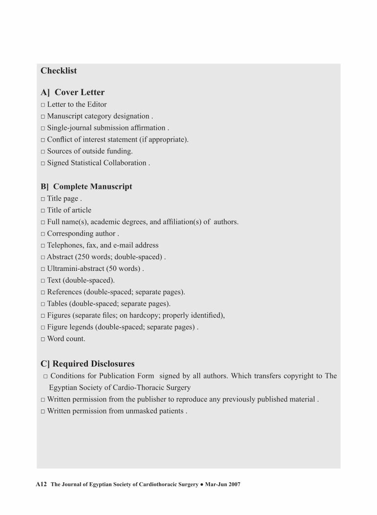
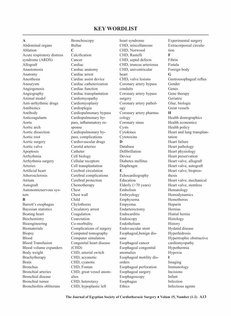

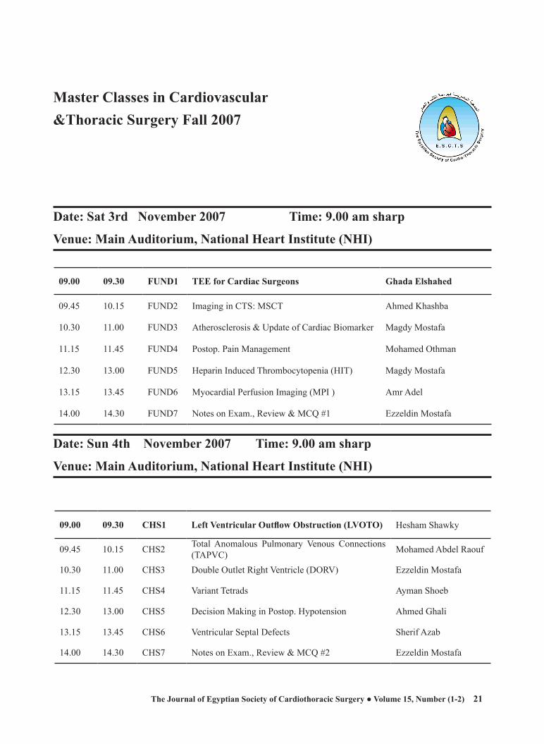
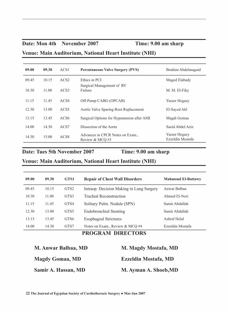
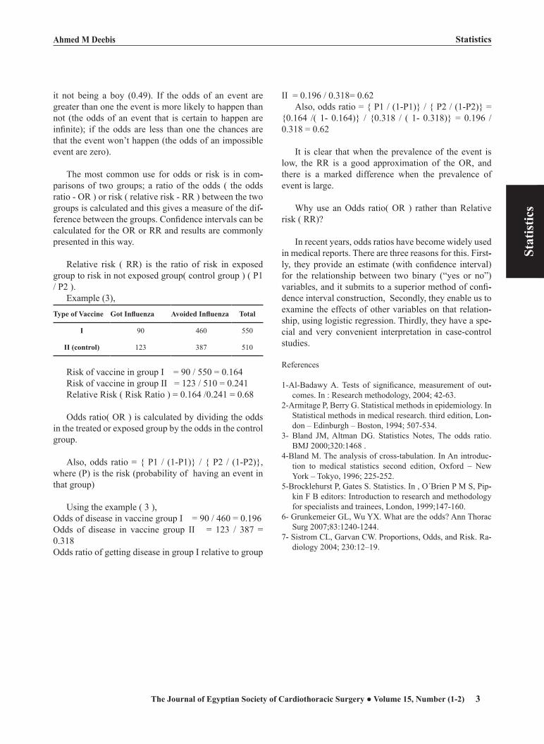
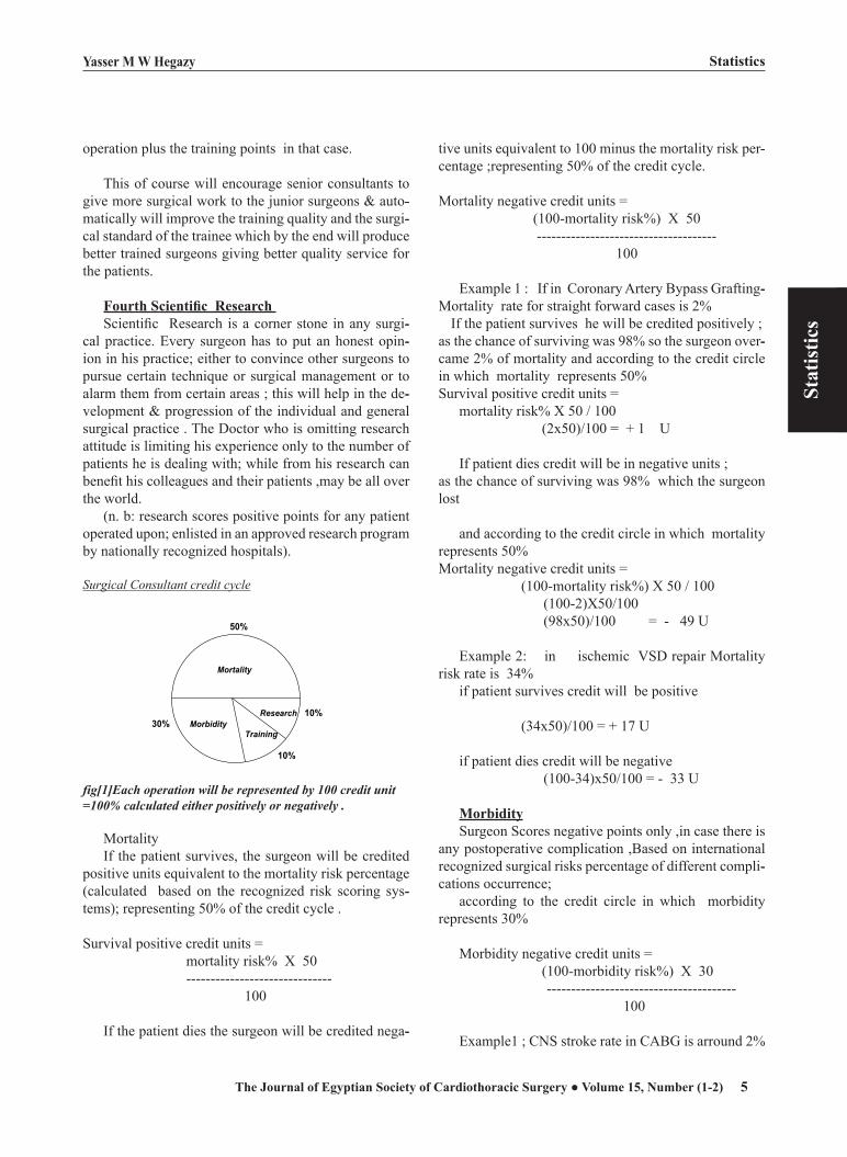
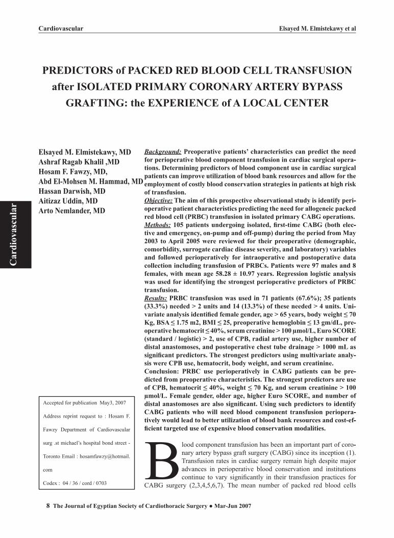
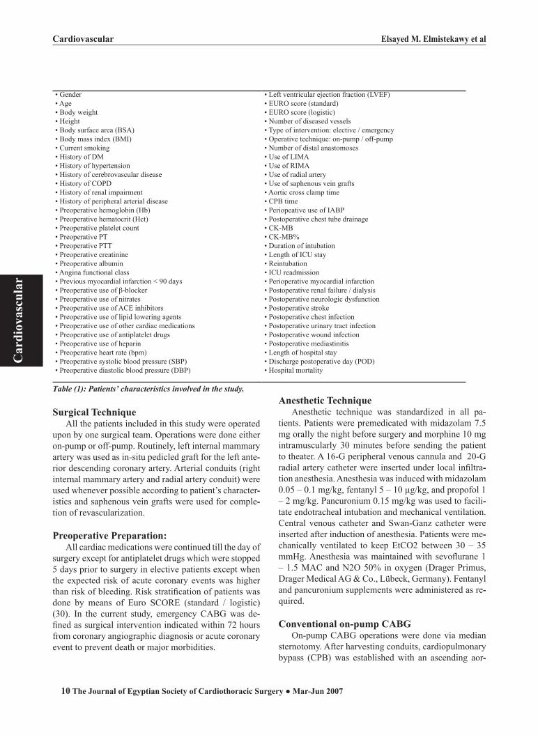
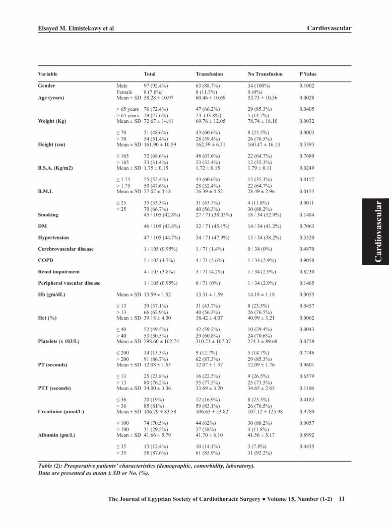

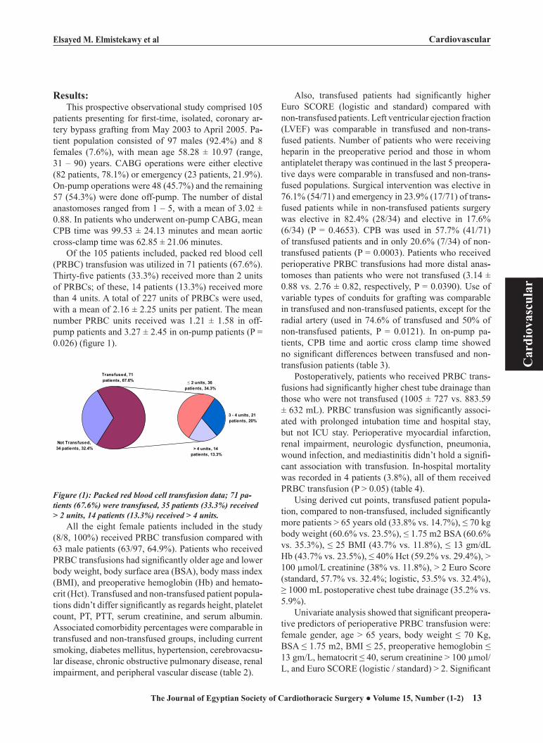

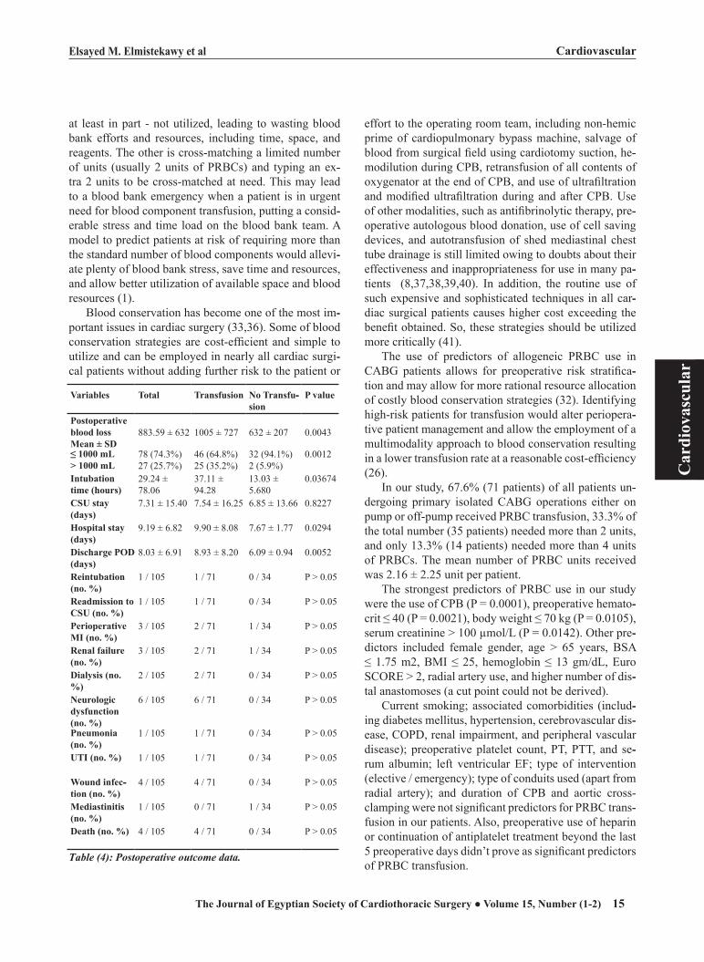
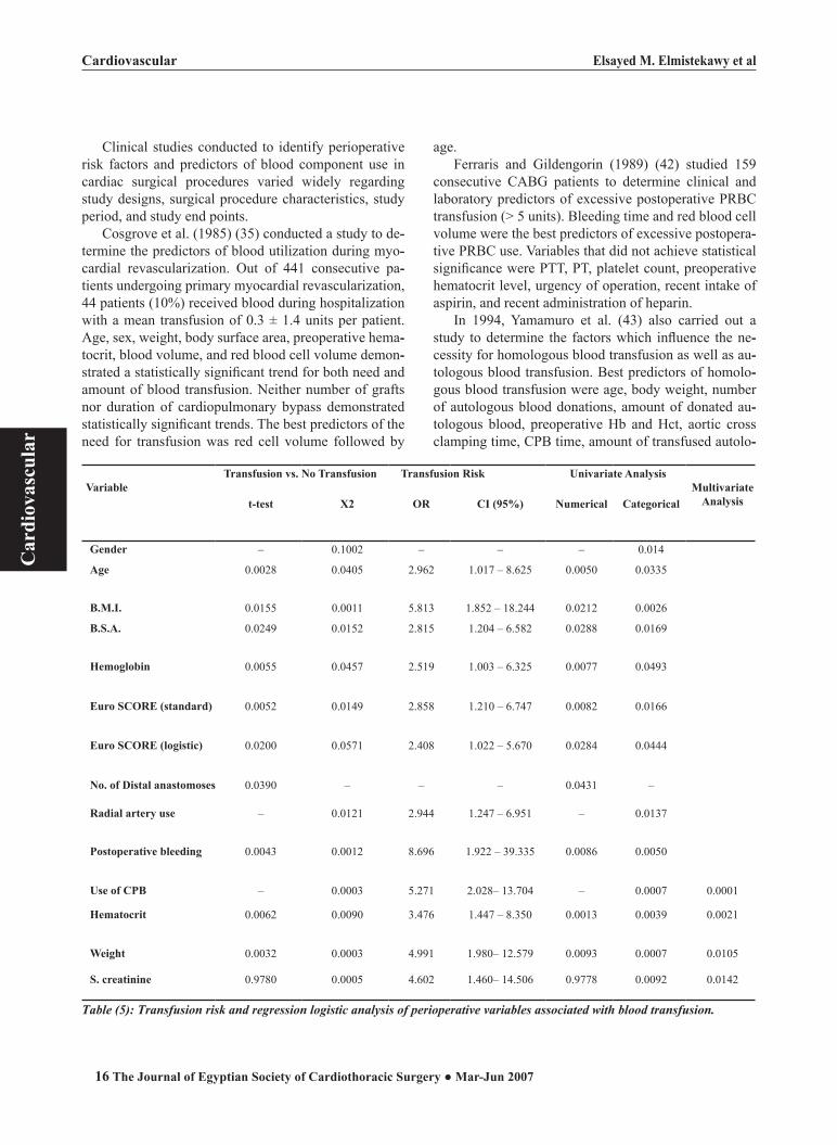


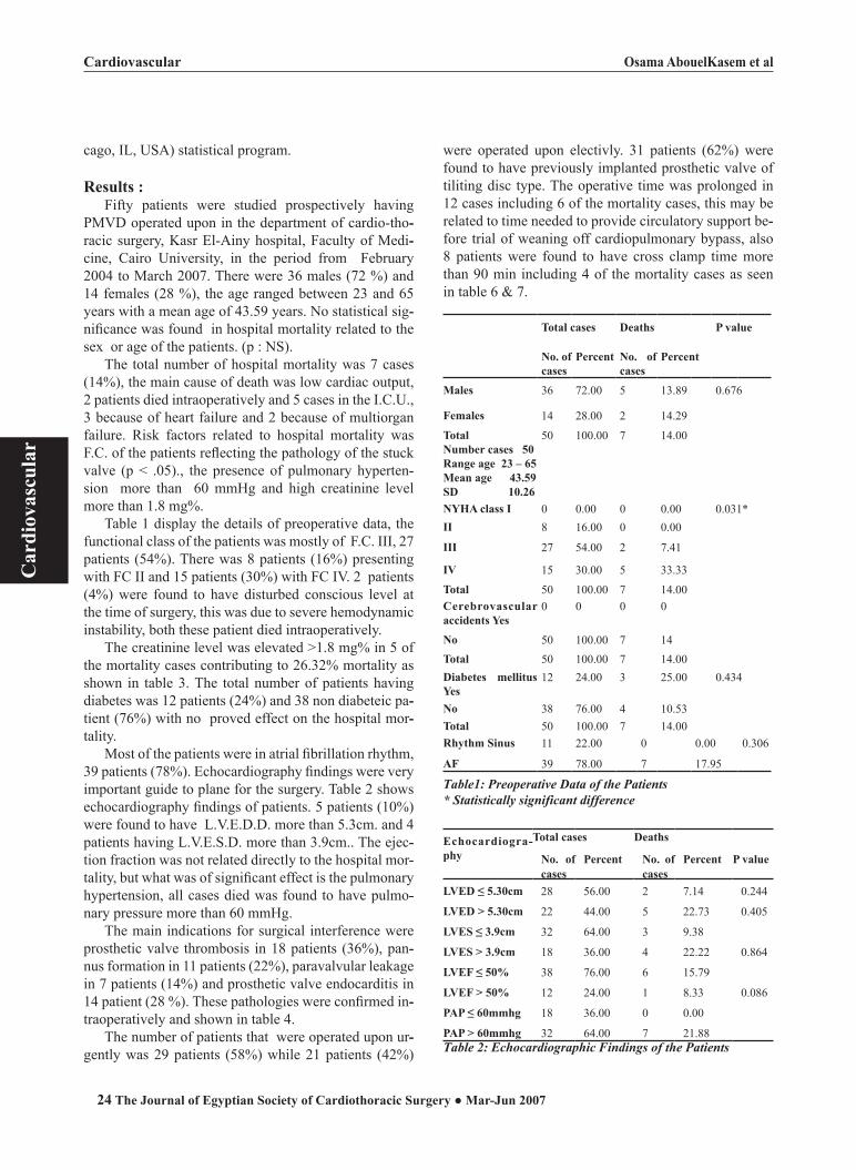
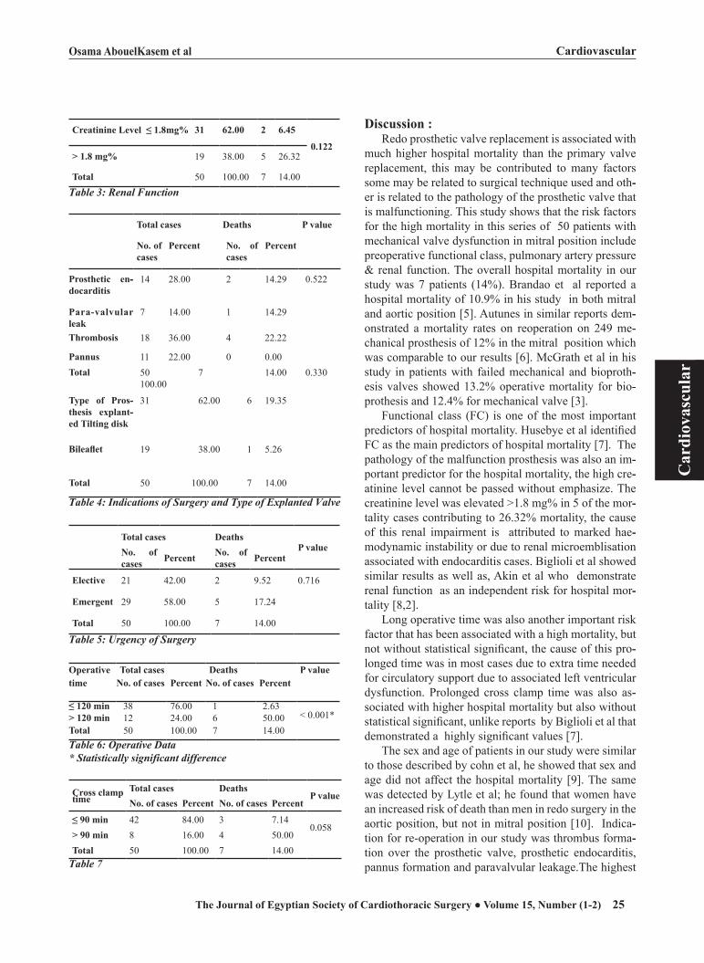

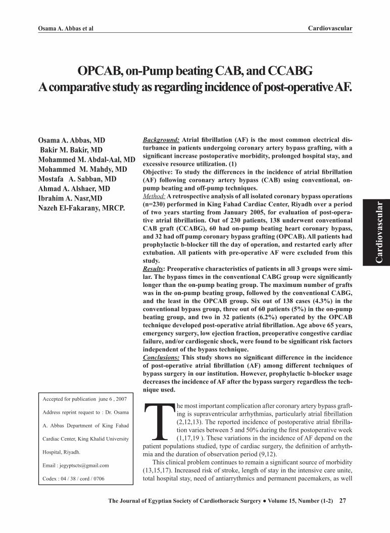
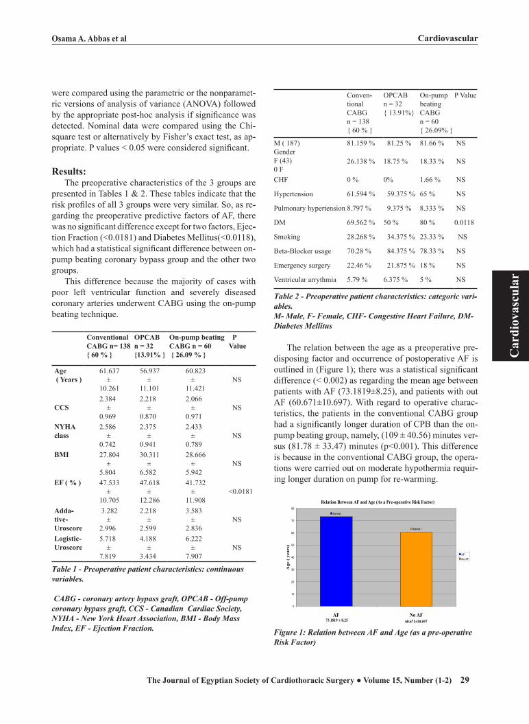
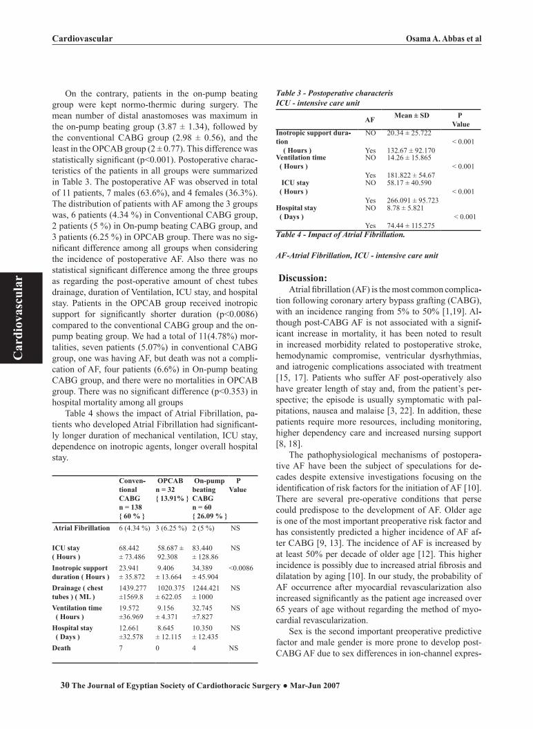
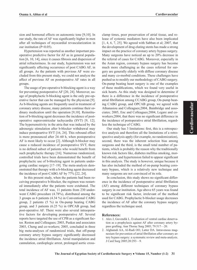
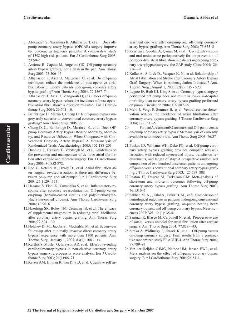

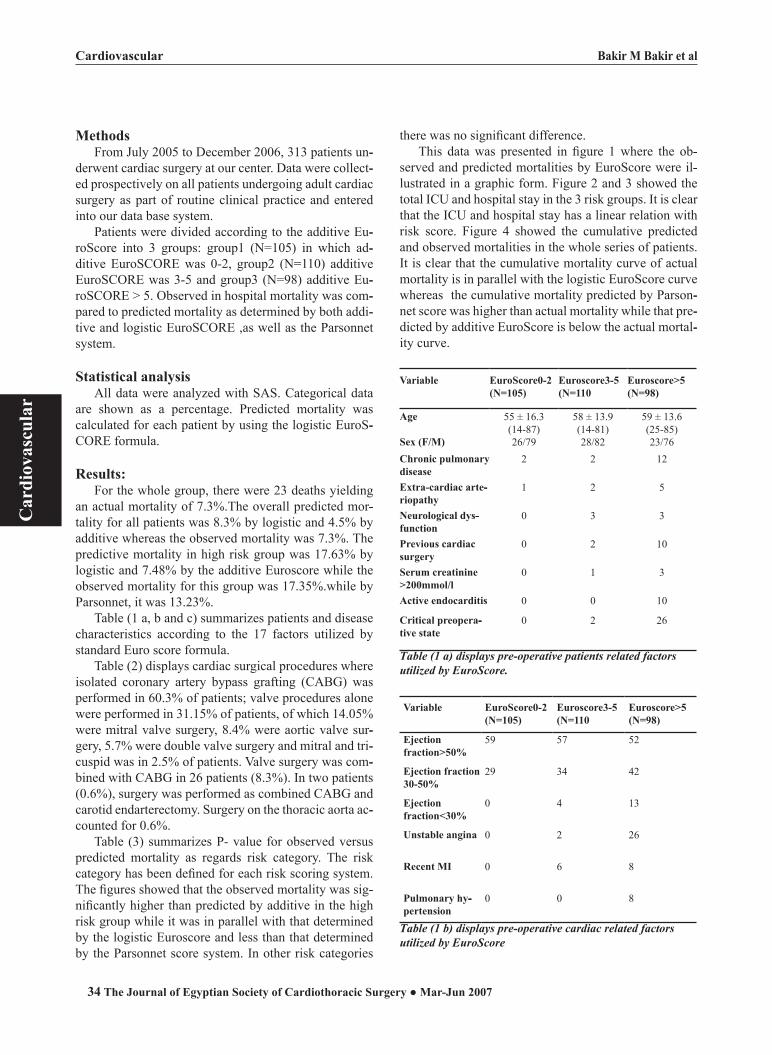
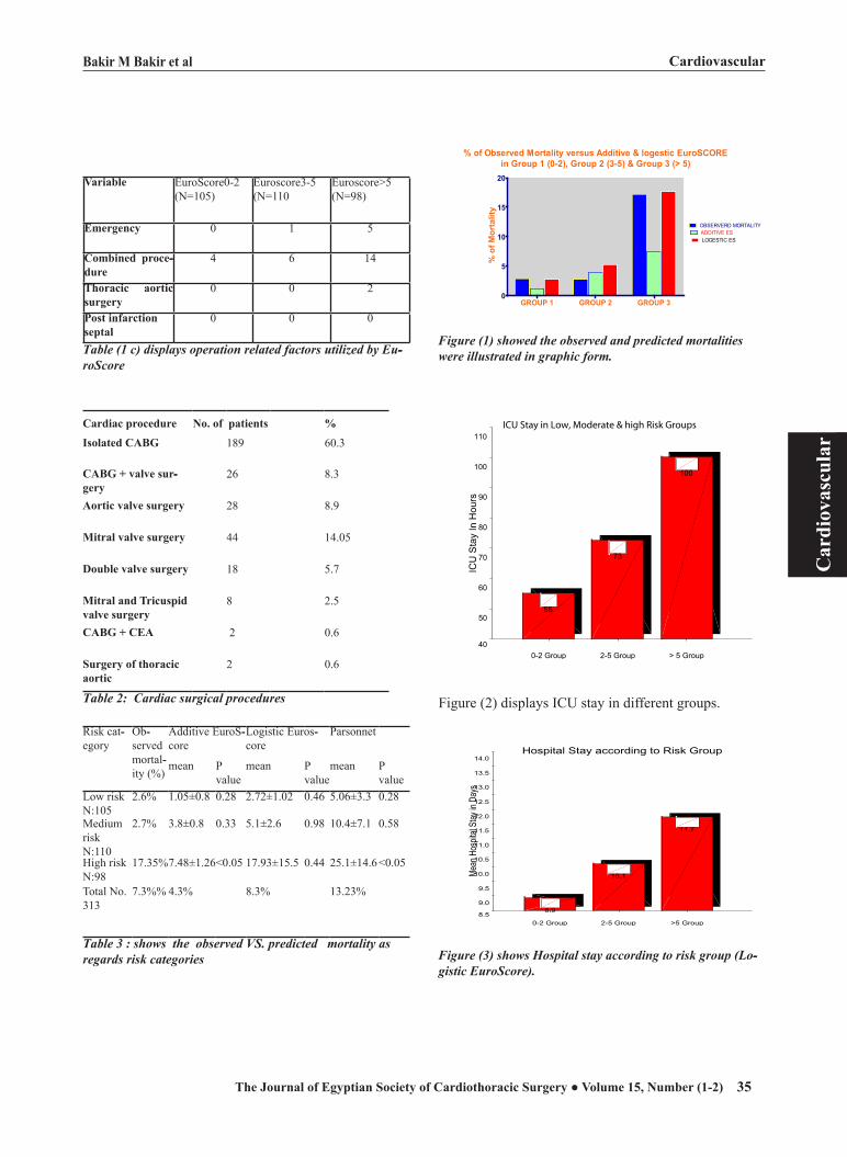
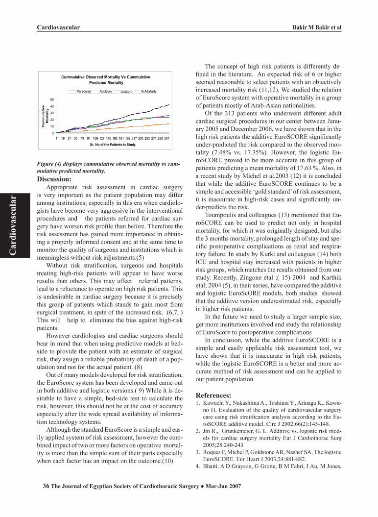
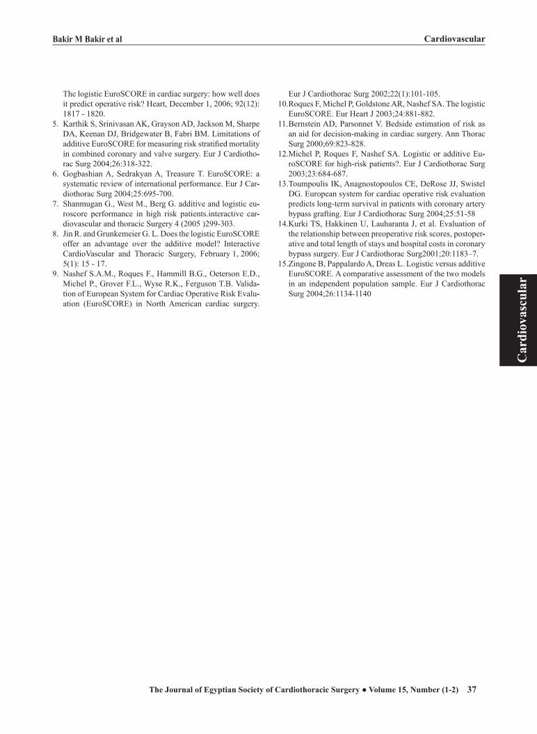
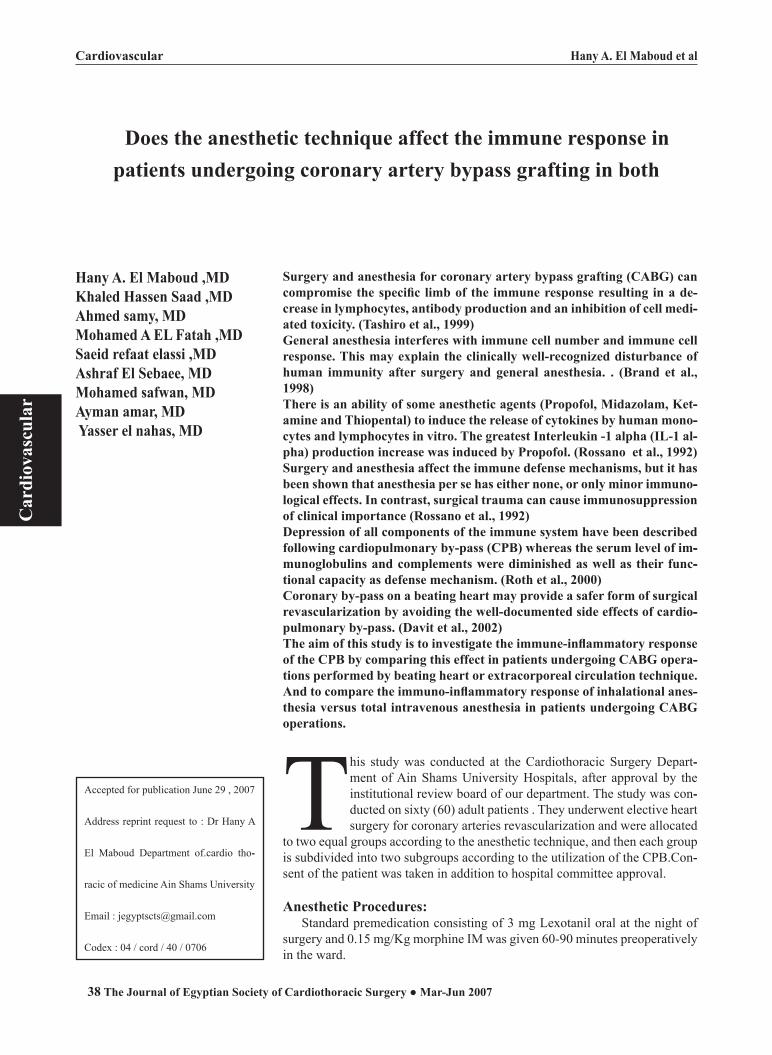
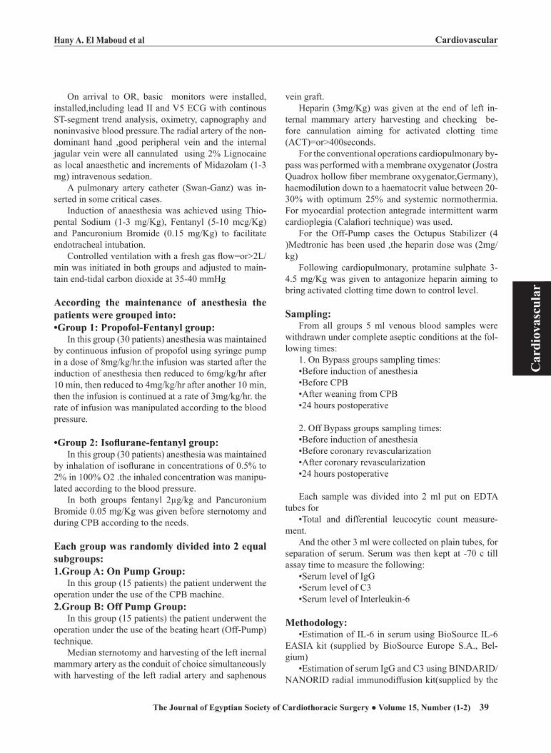

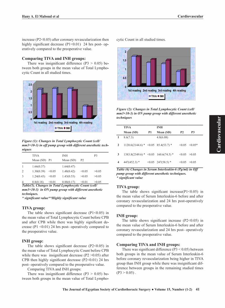
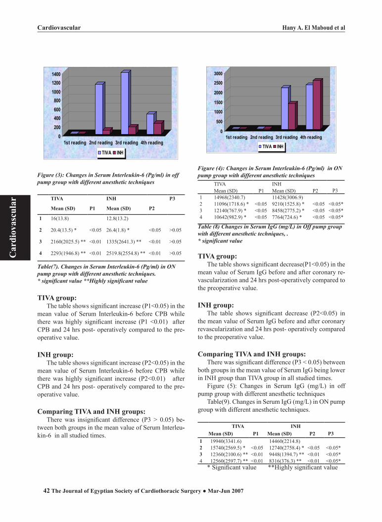
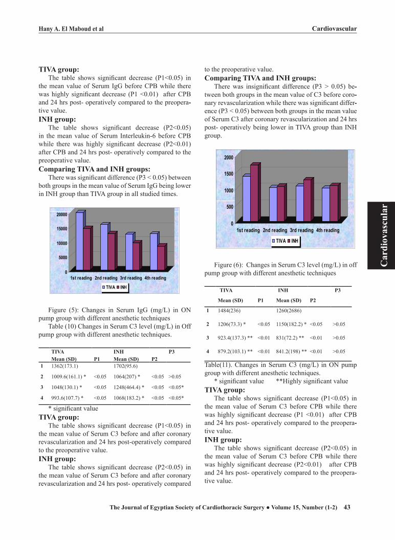
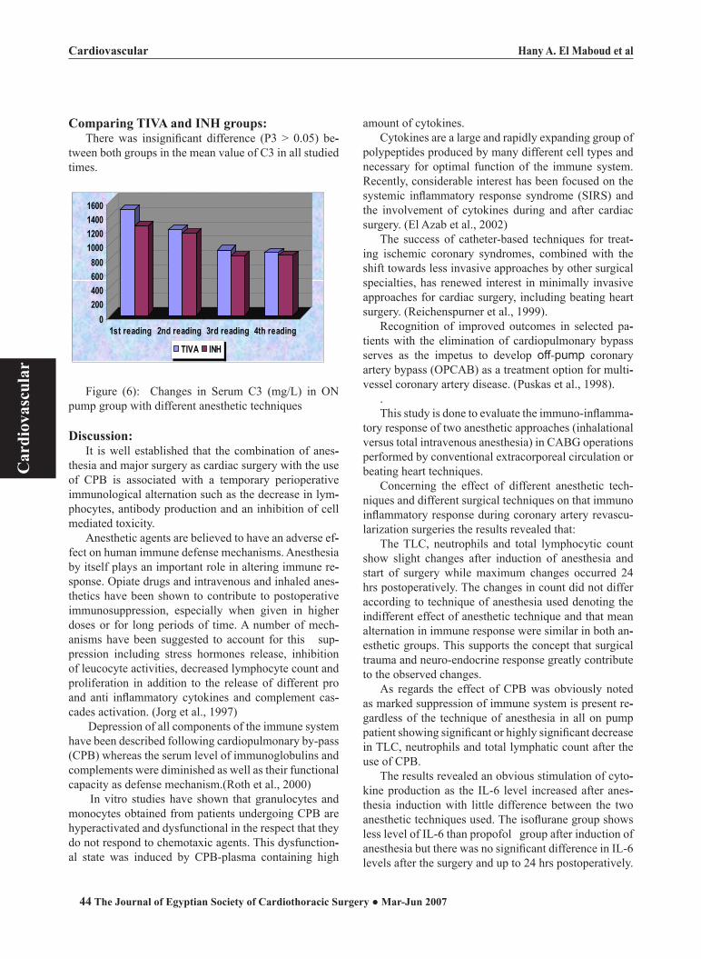


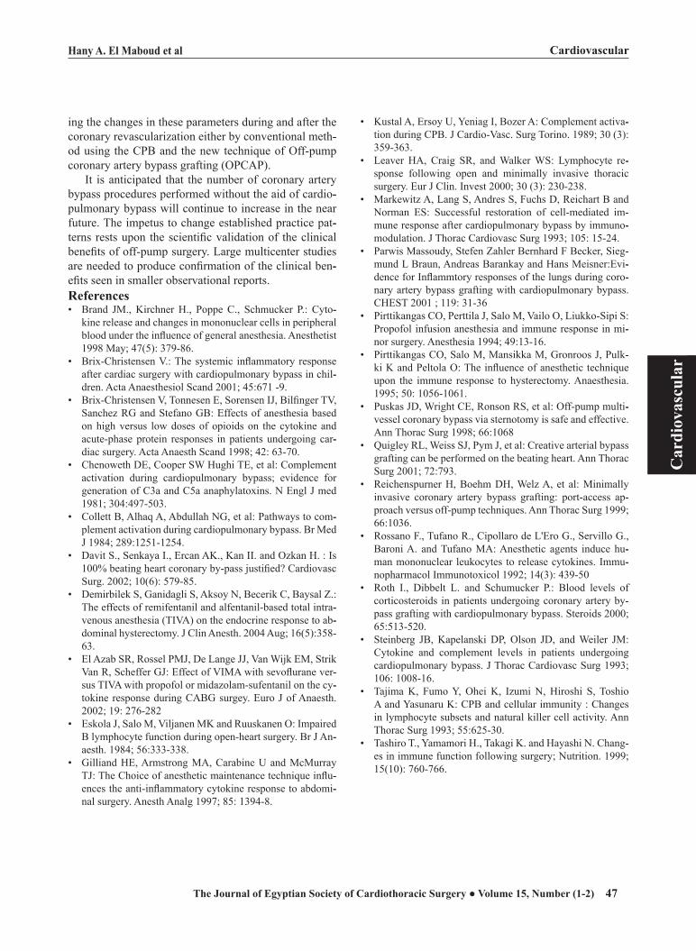

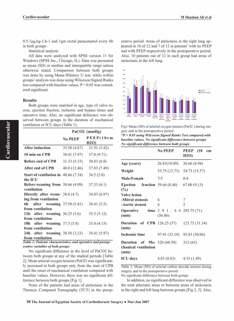
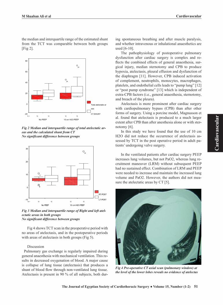
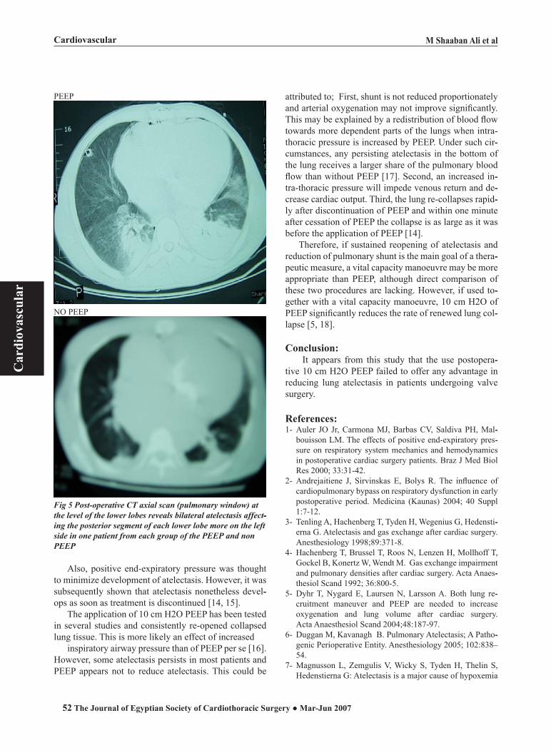
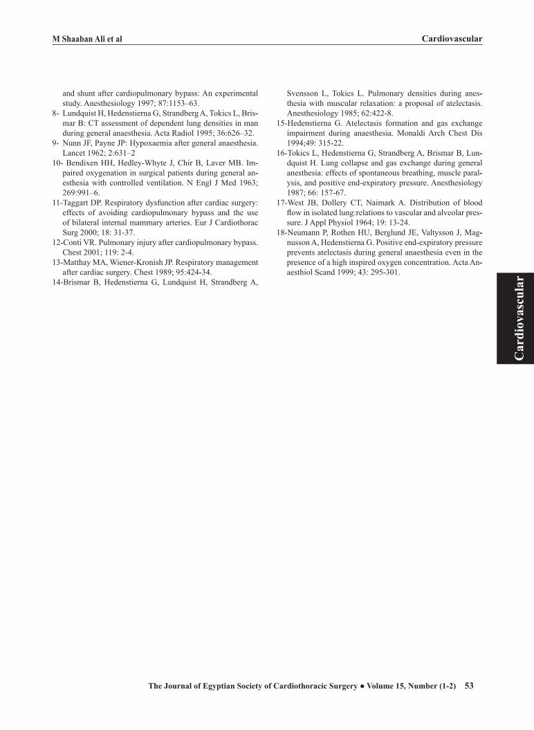
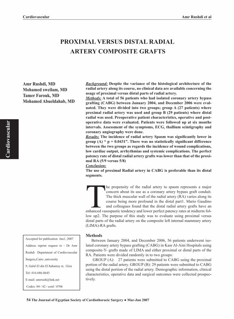
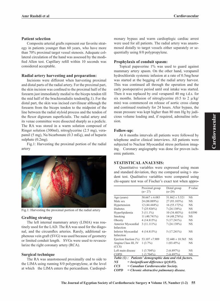
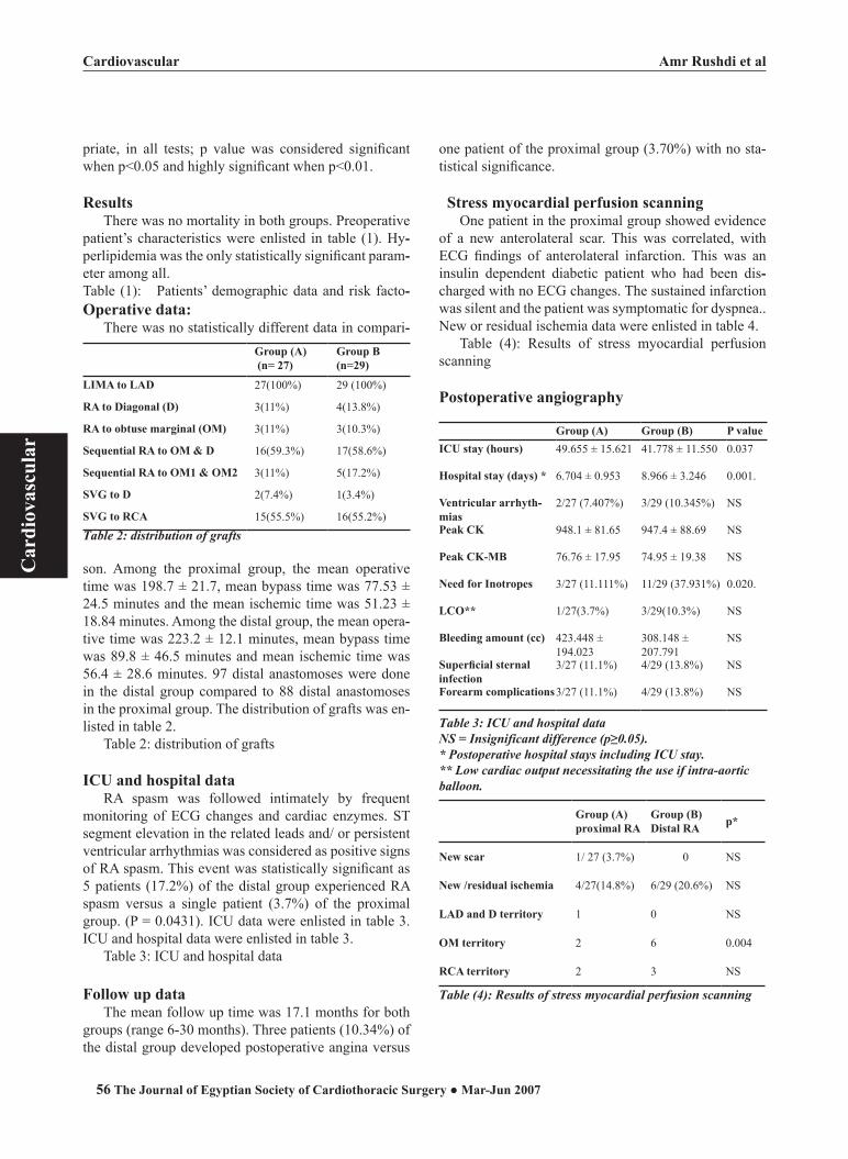
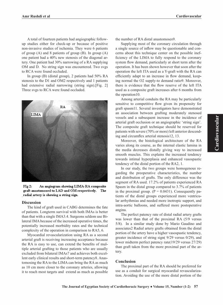

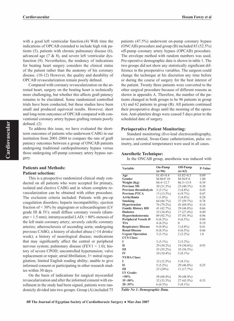

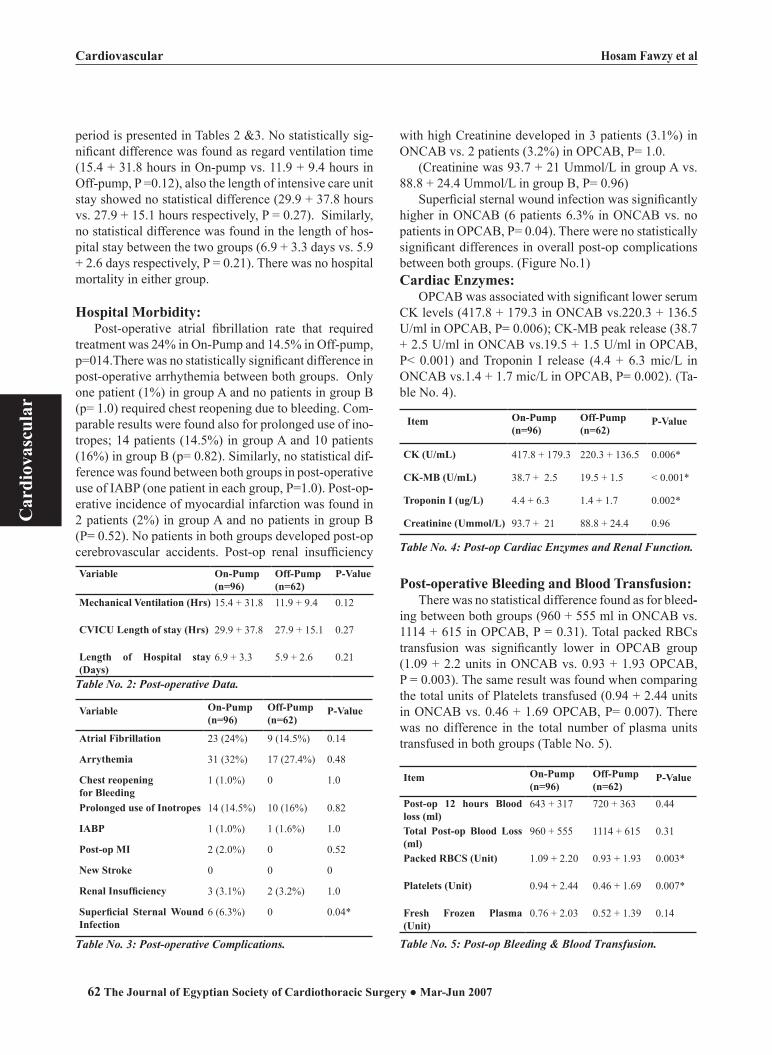
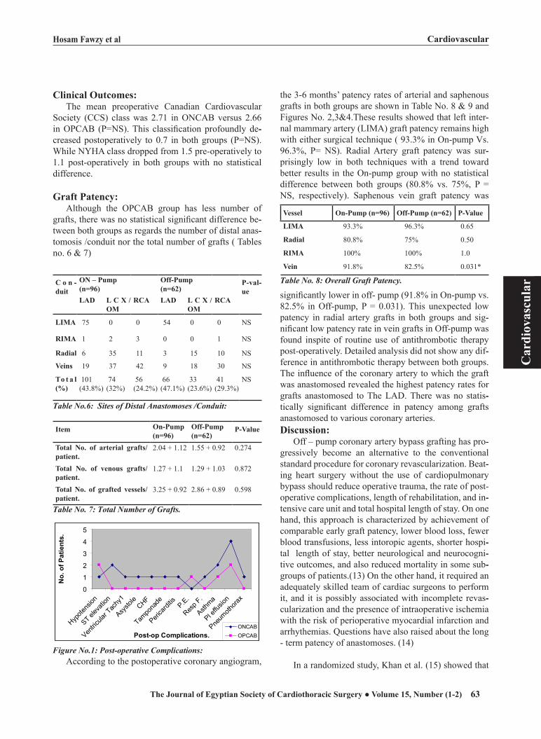
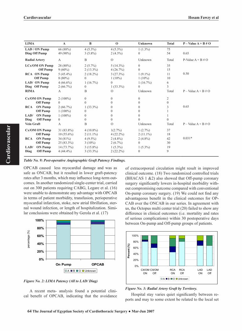


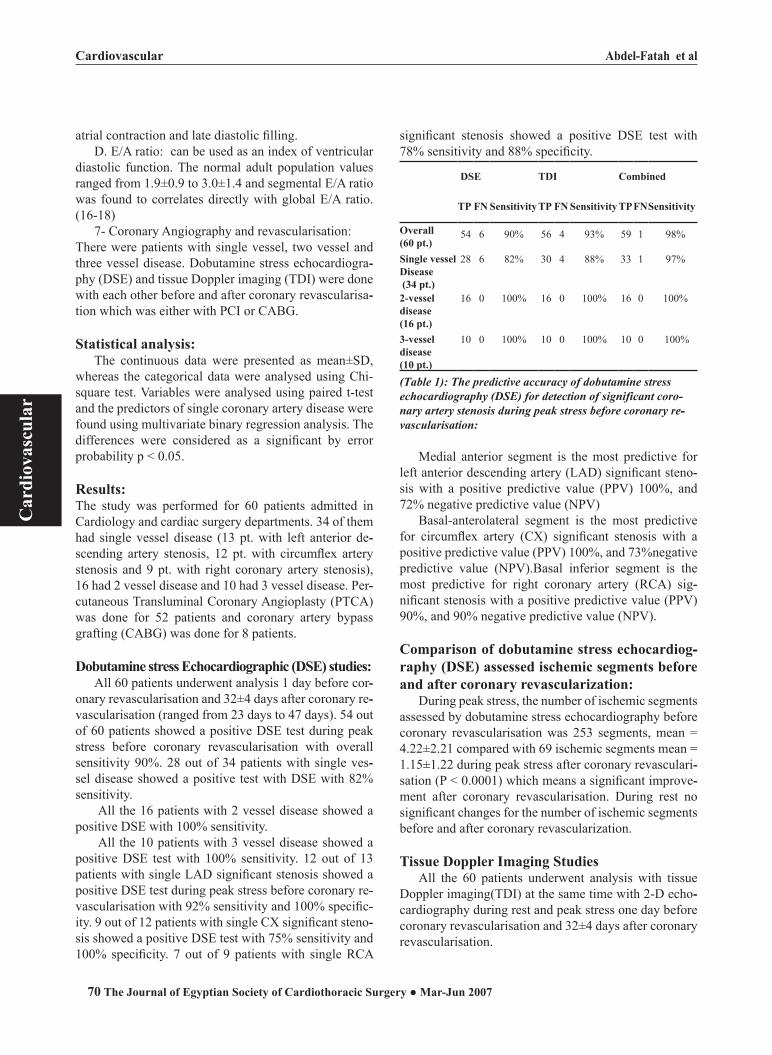

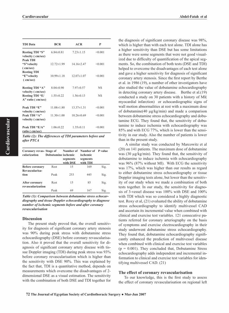


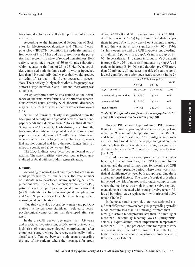


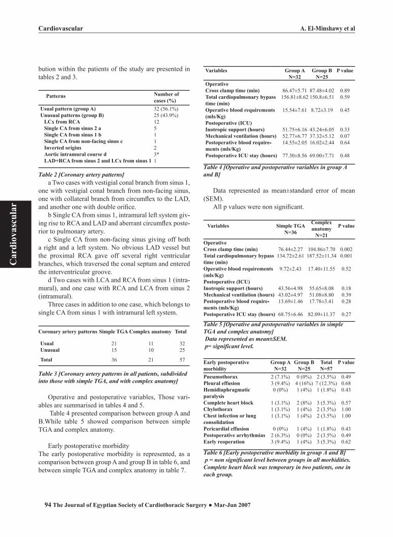
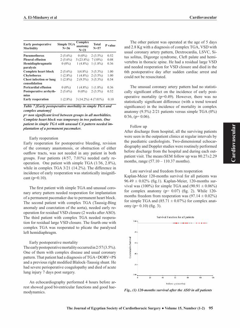
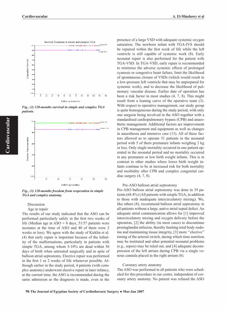
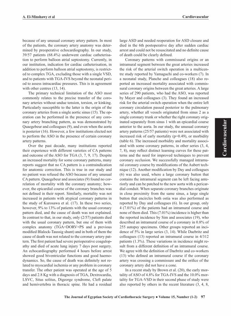


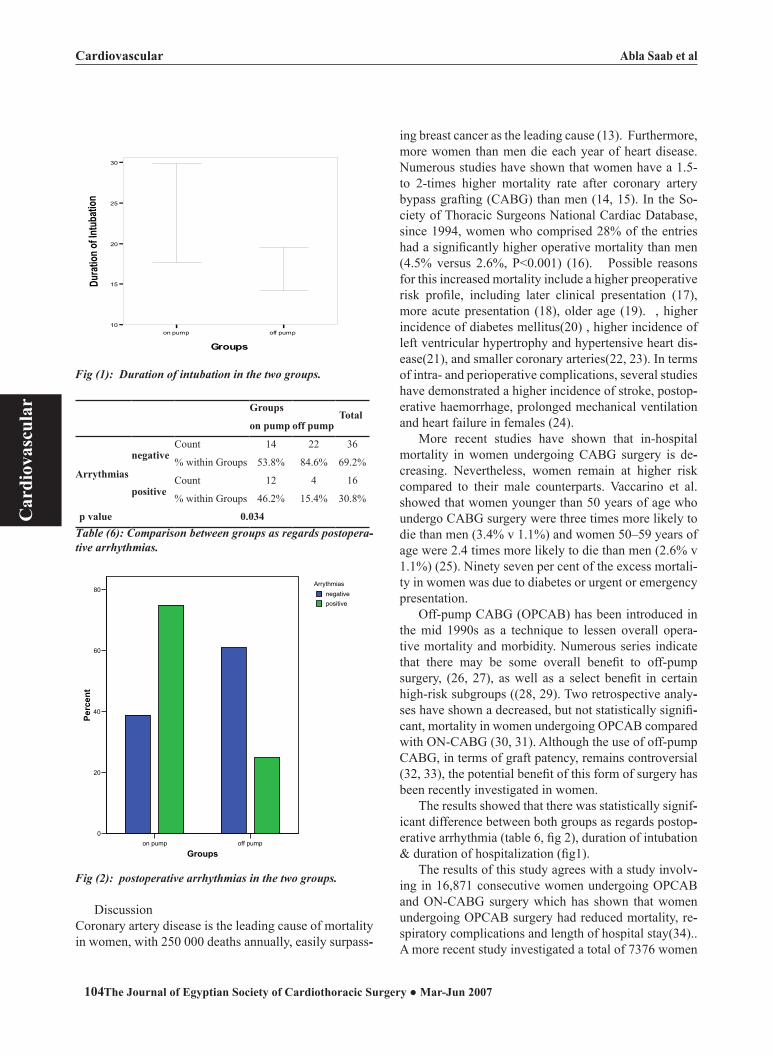

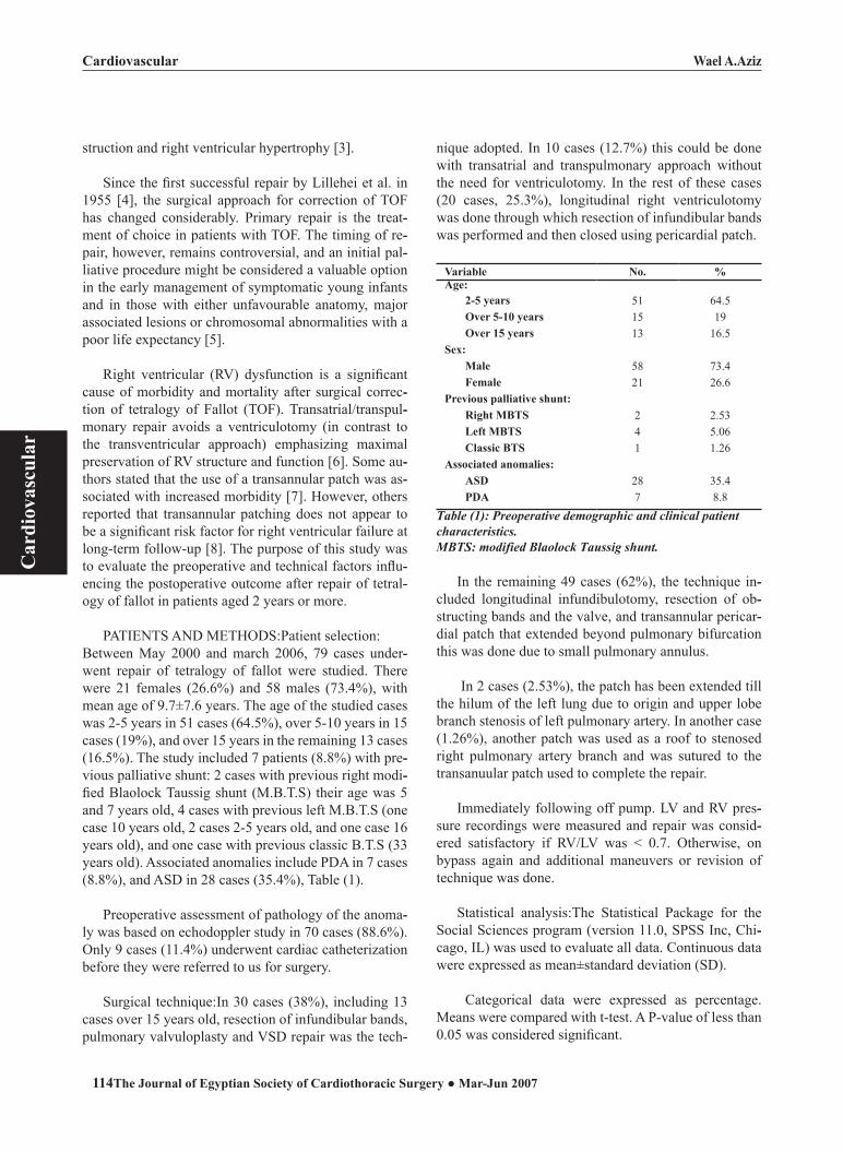
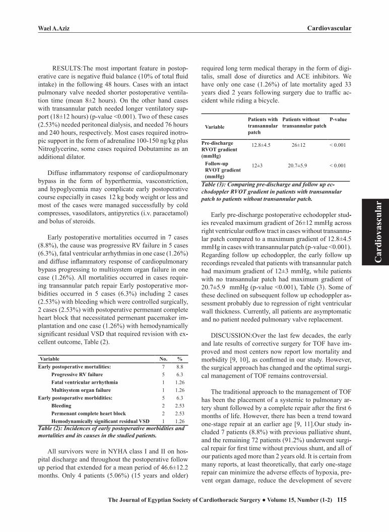

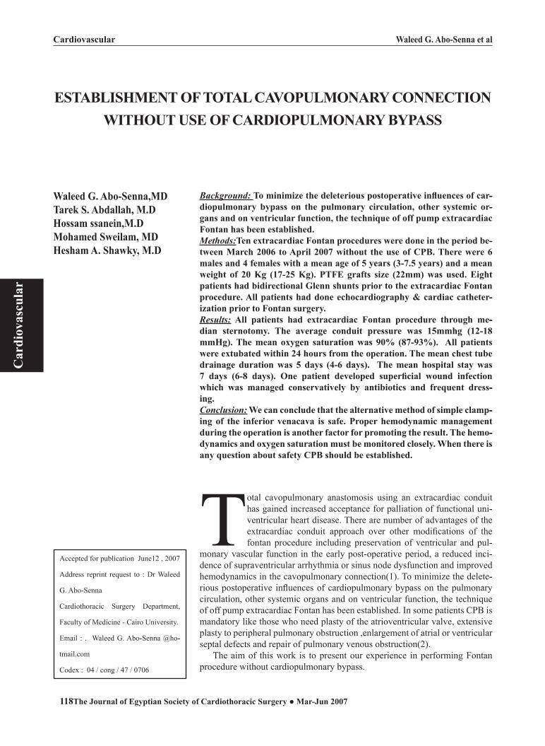
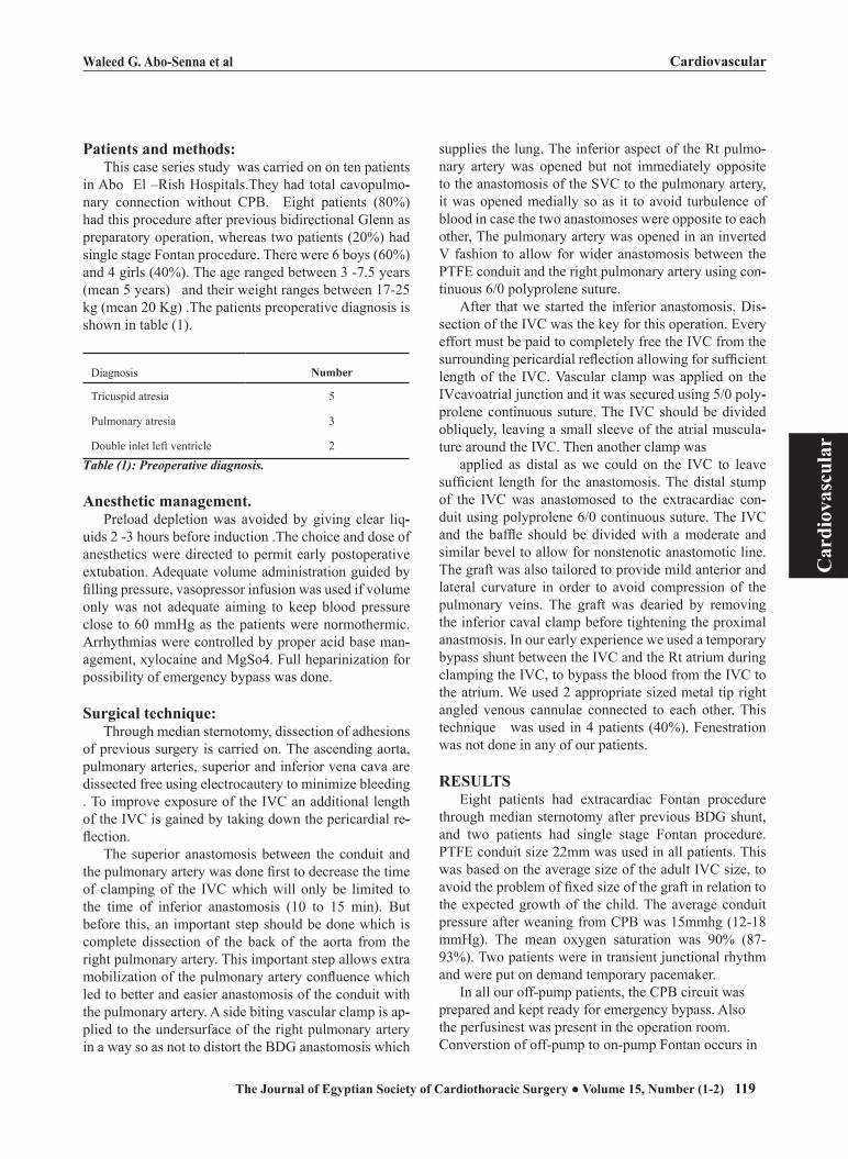


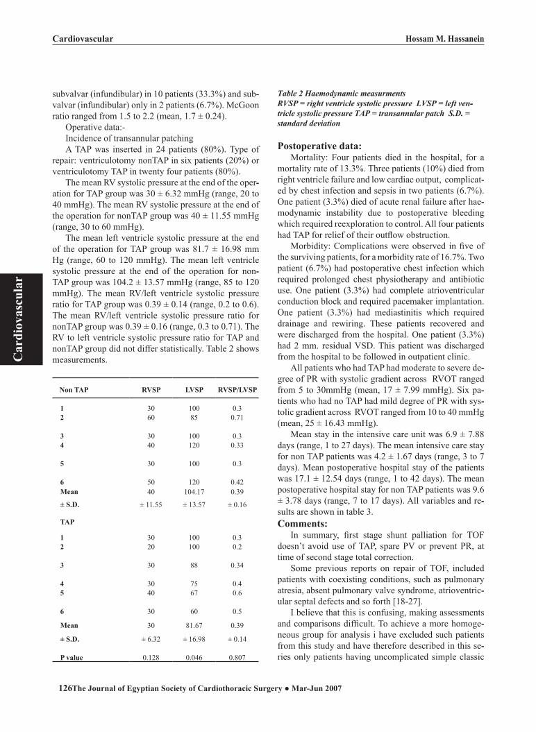
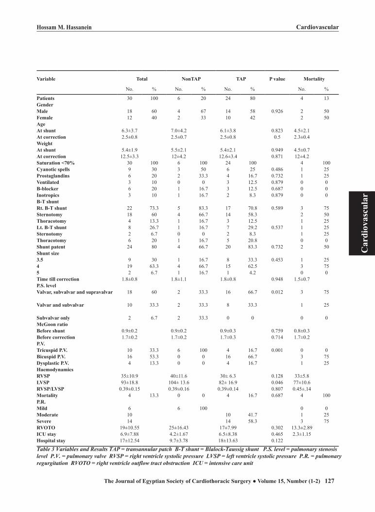


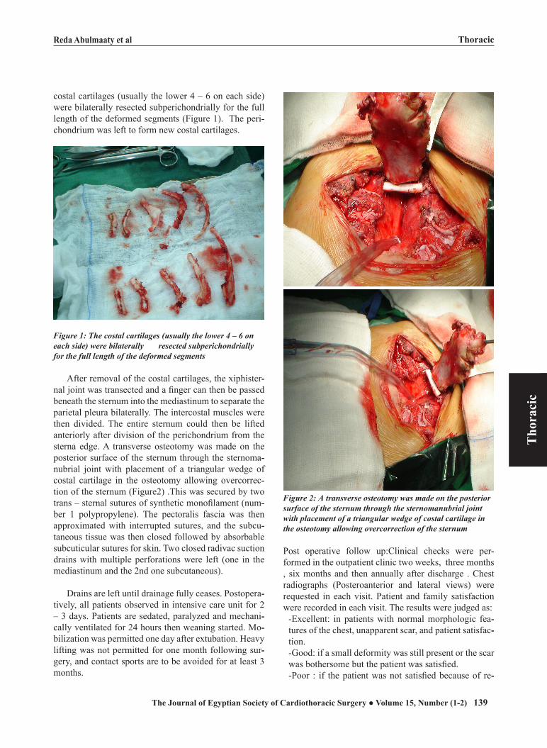
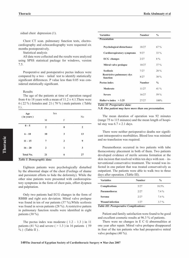
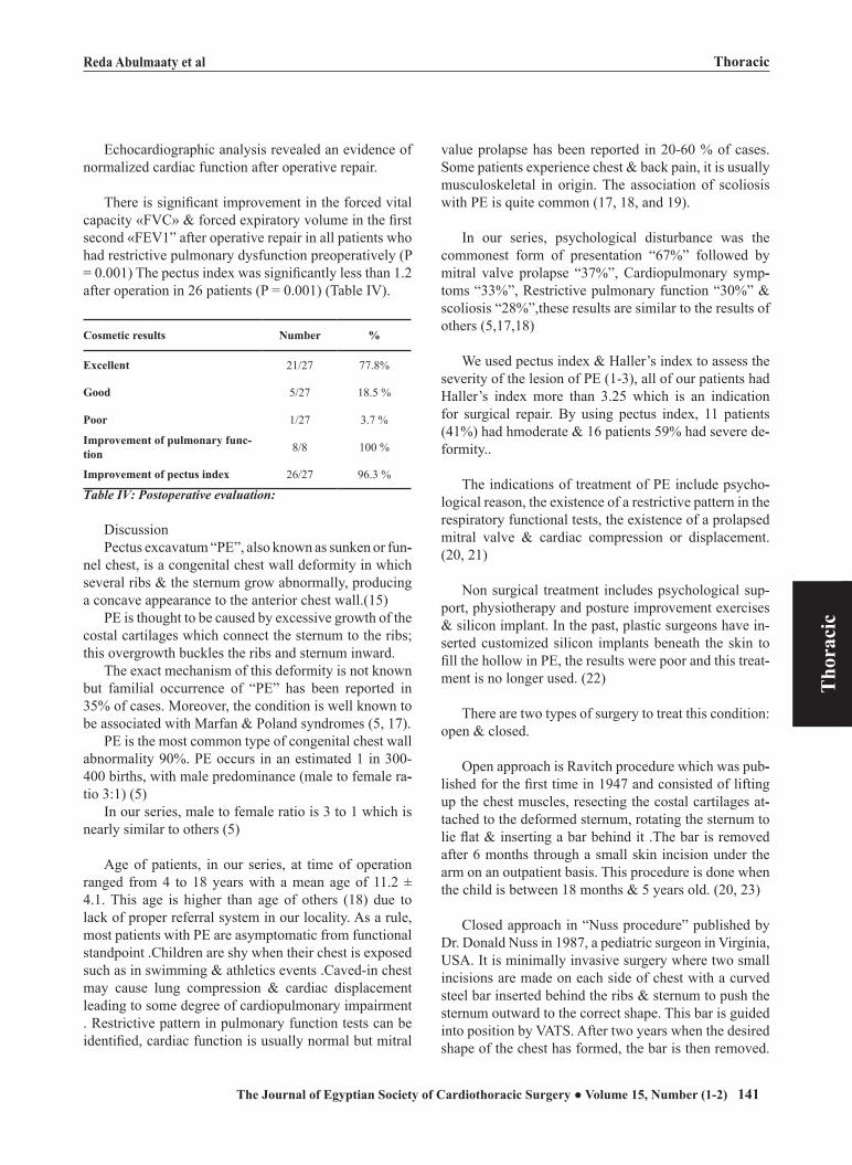

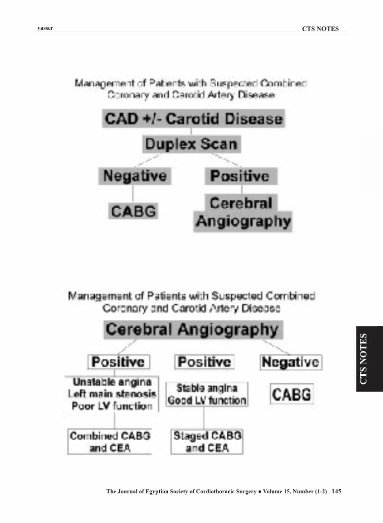
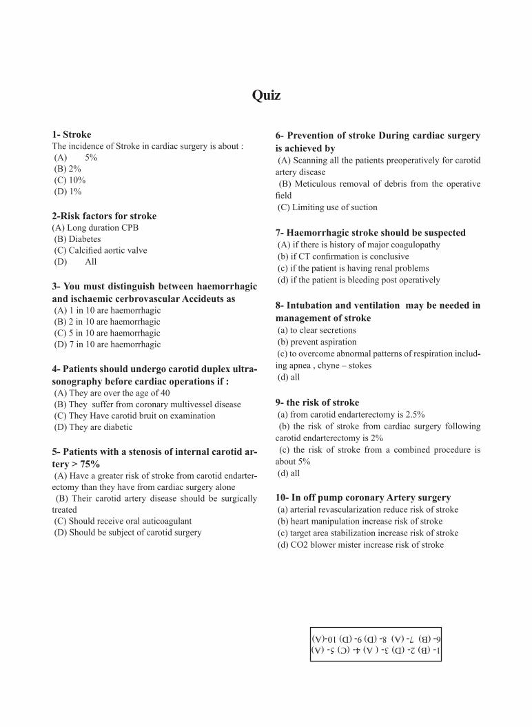


![[Empanelment of the hospital cancelled w.e.f. 16.01.2009]. · 12, NOIDA General and specialised Cardiological services including Cardio-thoracic surgery. 11. Metro Multi Specialty](https://static.documents.pub/doc/80x56/5e70544db7bed33a0c09c9ab/empanelment-of-the-hospital-cancelled-wef-16012009-12-noida-general-and.jpg)


