Journal of Trace Elements in Medicine and Biology 40 (2017) 30–36
Contents lists available at ScienceDirect
Journal of Trace Elements in Medicine and Biology
jo ur nal homep age: www.elsev ier .com/ locate / j temb
oxicology
luminium in brain tissue in familial Alzheimer’s disease
mbreen Mirzaa, Andrew Kingb,c, Claire Troakesc, Christopher Exleya,∗
The Birchall Centre, Lennard-Jones Laboratories, Keele University, Staffordshire, ST5 5BG, United KingdomDepartment Of Clinical Neuropathology, King’s College Hospital, London, SE5 9RS, United KingdomMRC London Neurodegenerative Diseases Brain Bank, Institute of Psychiatry, Psychology and Neuroscience, King’s College, London, SE5 8AF, Unitedingdom
r t i c l e i n f o
rticle history:eceived 21 November 2016ccepted 7 December 2016
The genetic predispositions which describe a diagnosis of familial Alzheimer’s disease can be consideredas cornerstones of the amyloid cascade hypothesis. Essentially they place the expression and metabolismof the amyloid precursor protein as the main tenet of disease aetiology. However, we do not know thecause of Alzheimer’s disease and environmental factors may yet be shown to contribute towards itsonset and progression. One such environmental factor is human exposure to aluminium and aluminiumhas been shown to be present in brain tissue in sporadic Alzheimer’s disease. We have made the firstever measurements of aluminium in brain tissue from 12 donors diagnosed with familial Alzheimer’sdisease. The concentrations of aluminium were extremely high, for example, there were values in excessof 10 �g/g tissue dry wt. in 5 of the 12 individuals. Overall, the concentrations were higher than all
luminium-selective fluorescenceicroscopy
previous measurements of brain aluminium except cases of known aluminium-induced encephalopathy.We have supported our quantitative analyses using a novel method of aluminium-selective fluorescencemicroscopy to visualise aluminium in all lobes of every brain investigated. The unique quantitative dataand the stunning images of aluminium in familial Alzheimer’s disease brain tissue raise the spectre ofaluminium’s role in this devastating disease.
Genetic mutations associated with both the expression [1] andetabolism [2] of amyloid precursor protein (APP) are, in general,
he basis for a diagnosis of familial Alzheimer’s disease (fAD). They,long with evidence from Down’s syndrome [3], provide strongupport for the amyloid cascade hypothesis [4,5] and a central roleor the neuropathology and biochemistry of amyloid-beta (A�) inlzheimer’s disease [6]. In many ways, familial AD has been useds a blueprint for understanding and treatment of sporadic or late-nset AD.
Aluminium is present in human brain tissue [7] and in a recenttudy involving 60 human brains the median aluminium contentf 712 tissues across all four main lobes was 1.02 �g/g dry wt. with5% of all values being <2.01 �g/g dry wt. tissue [8]. The associa-ion of aluminium and AD has a significant history [9,10] and yet
here remains no consensus as to a role for this known neurotoxinn the disease [11]. However, recent reports concerning sporadicD [12] and environmental [13] and occupational [14] exposure to
aluminium have allowed the conclusion to be drawn that, undercertain conditions, it is inevitable that aluminium will contributetowards AD [11,15]. The suggestion is made that wherever in thebrain the concentration of aluminium is pathologically-concerning(>2.00 �g/g dry wt.) that this aluminium will contribute towardsany ongoing AD and will result in the disease being earlier in onsetwith a more aggressive aetiology [15].
Familial AD is characterised by an earlier age of onset and yetthere are no data to describe the aluminium content of brain tissuein this ‘signature’ form of AD. Herein we have obtained brain tissuefrom 12 autopsy-confirmed cases of familial AD and we have car-ried out the first ever measurements of brain aluminium contentin familial AD. We have also supported our quantitative measure-ments with imaging of brain aluminium by aluminium-selectivefluorescence microscopy.
2. Materials and methods
There was ethical approval from The MRC London Neu-
rodegenerative Diseases Brain Bank at King’s College, London(08/MRE09/38 + 5).
Samples of cortex of approximately 1 g frozen weight from tem-poral, frontal, parietal and occipital lobes were obtained from 12
le under the CC BY-NC-ND license (http://creativecommons.org/licenses/by-nc-nd/
A. Mirza et al. / Journal of Trace Elements in Medicine and Biology 40 (2017) 30–36 31
Fig. 1. Representative images of aluminium in frontal cortex. Light (A&D) and fluorescence (B&E) microscopy images of lumogallion-stained sections of frontal cortex. Asterisklabel suggested intracellular deposits while arrows show diffuse deposits. Fluorescence microscopy of un-stained adjacent tissue sections (C&F) show autofluorescence. Scalebars are all 100 �m.
F scencel rescenS
cwtatreriwaa
ig. 2. Representative images of aluminium in parietal cortex. Light (A&D) and fluoreabel suggested intracellular deposits associated with both living and dead cells. Fluocale bars are 100 �m (A–C) and 50 �m (D–F).
ases of autopsy-confirmed familial Alzheimer’s disease. Donorsith familial Alzheimer’s disease are extremely rare and we had
he privilege of obtaining tissue from the only 12 cases availablet the brain bank with the year of diagnosis ranging from 1991hrough to 2009. There were 7 female and 5 male donors in the ageange 42–86. The bases for a diagnosis of familial Alzheimer’s dis-ase are given in Table 1. Where no definite genetic mutation wasecorded at the time of autopsy we have defined probable famil-al Alzheimer’s disease as either (i) having two first degree relatives
ith a dementing illness or (ii) where the patient or first degree rel-
tive had developed symptoms of dementia at less than 60 years ofge or (iii) where the patient had one first and one second degree
(B&E) microscopy images of lumogallion-stained sections of frontal cortex. Asteriskce microscopy of un-stained adjacent tissue sections (C&F) show autofluorescence.
relative with dementia and one with symptoms of dementia at lessthan 60 years of age.
The aluminium content of these tissues was measured byan established and fully validated method [8] which herein isdescribed briefly. Thawed tissues were cut using a stainless steelblade to give individual samples of ca 0.3 g (3 sample replicatesfor each lobe) wet weight and dried to a constant weight at 37 ◦C.Dried and weighed tissues were digested in a microwave (MARSXpress CEM Microwave Technology Ltd.) in a mixture of 1 mL15.8 M HNO3 (Fisher Analytical Grade) and 1 mL 30% w/v H2O2 (BDH
Aristar). Digests were clear with no fatty residues and, upon cool-ing, were made up to 5 mL volume using ultrapure water (cond<0.067 �S/cm). Total aluminium was measured in each sample
32 A. Mirza et al. / Journal of Trace Elements in Medicine and Biology 40 (2017) 30–36
Fig. 3. Representative images of aluminium in cortex. Light (A&D) and fluorescence (B&E) microscopy images of lumogallion-stained sections of frontal (A&B) and temporal(D&E) cortex. Asterisk label suggested intracellular deposits and arrows diffuse deposits. Fluorescence microscopy of un-stained adjacent frontal (C) and temporal (F) tissuesections show autofluorescence. Scale bars are 50 �m (A–C) and 100 �m (D–F).
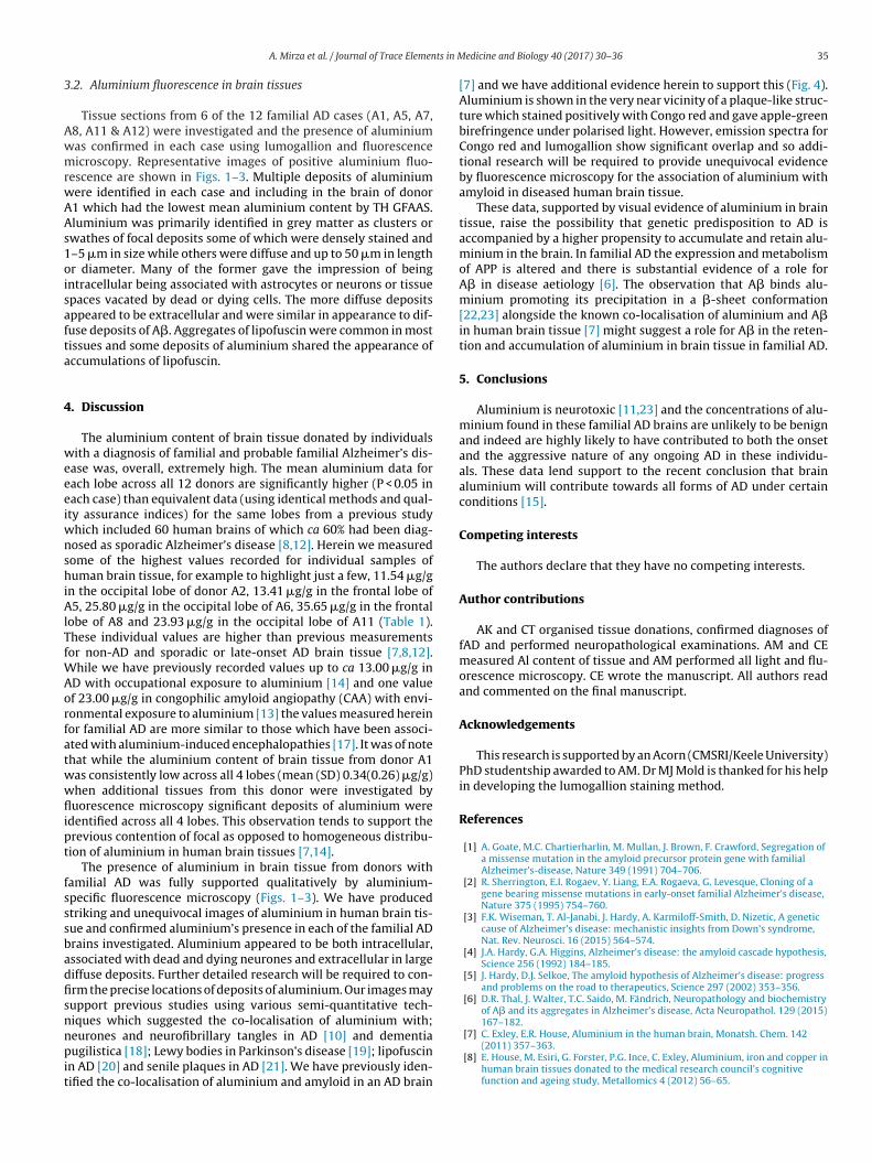
Fig. 4. Co-localisation of amyloid and aluminium in occipital cortex. (A) Light microscopy image of Congo red-stained tissue showing (arrow) senile plaque-like amyloidd rrow)
F e plaqt ium. S
bte
Aram
eposit. (B) Polarising microscopy image of Congo red-stained image showing (aluorescence microscopy image of Congo red-stained tissue showing (arrow) senilissue stained with lumogallion and showing (arrow) significant deposits of alumin
y transversely-heated graphite furnace atomic absorption spec-rometry (TH GFAAS) using matrix-matched standards and anstablished analytical programme [8].
Formalin-fixed brain tissue for 6 cases of familial AD (A1, A5,7, A8, A11 & A12) was supplied as pre-embedded paraffin blocks
epresenting cortical tissue from frontal, parietal, and temporalnd occipital lobes. A recently developed, optimised and validatedethod using lumogallion and fluorescence microscopy was used
apple-green birefringence characteristic of amyloid in � sheet conformation. (C)ue-like amyloid deposit. (D) Fluorescence microscopy image of adjacent section ofcale bars are all 50 �m.
to identify the presence of aluminium in tissues [16]. This methodwas shown to be specific for aluminium with no interferencefrom any other metals and no issues relating to autofluorescence.Slides of serial adjacent sections of tissue were imaged using anOlympus BX50 fluorescence microscope with a BX-FLA reflected
light fluorescence attachment, equipped with a mercury burnerand a vertical illuminator. For lumogallion (and autofluorescence)imaging a U-MNIB3 fluorescence filter cube was used (bandpass
Aluminium was found in all 144 tissues and its concentrationanged from 0.01 to 35.65 �g/g dry wt. (Table 1). The mean alu-
able 1he content of aluminium in each of 4 lobes (O – occipital; F – frontal; T – temporal; P – parioncerning or pathologically-significant are given in italics or bold typescript respectively.PP – amyloid precursor protein, BNE – Brain Net Europe.
Donor ID Pathology and genetics Gender
A1 Braak/BNE stage VI M
Grandfather, father, brother died
with dementia,brother had a
PSEN-1 mutation
A2 Braak Stage VI F
Father and nephew died with
dementia, nephew developed
dementia at young age
A3 Braak Stage V-VI M
Father died at young age with
AD
A4 Braak Stage V-VI M
PSEN-1(DELTA4)
A5 Braak Stage at least IV M
Mother and brother died in their
50 s with dementia
A6 Braak stage V-VI F
Daughter died in 30 s withdementia
edicine and Biology 40 (2017) 30–36 33
minium content for whole brains (n = 12) ranged from 0.34(0.26)for individual A1 to 6.55(9.59) �g/g dry wt. for individual A8.Approximately 40% of tissues (57/144) had an aluminium con-tent considered as pathologically-concerning (≥2.00 �g/g dry wt.)while approximately 58% of these tissues had an aluminium con-tent considered as pathologically-significant (≥3.00 �g/g dry wt.).The brains of 11 out of 12 individuals had at least one tissue with
a pathologically-significant content of aluminium. The brains of9 individuals had at least one tissue with an aluminium content≥5.00 �g/g dry wt. while 5 of these had at least one tissue with analuminium content ≥10.00 �g/g dry wt. (Table 1). The mean (SD)
etal) of 12 cases of familial Alzheimer’s disease. Values considered as pathologically- Details of pathology and known genetic mutations are also given. PSEN – presenilin,
Age Lobe Replicate [Al] �g/g dry wt.
47 O 1 0.472 0.673 0.03
F 1 0.842 0.523 0.13
T 1 0.162 0.023 0.29
P 1 0.532 0.283 0.13Mean(SD) 0.34(0.26)
76 O 1 10.122 8.323 11.54
F 1 1.752 4.483 0.48
T 1 0.692 0.553 0.16
P 1 0.272 1.253 1.60Mean(SD) 3.43(4.17)
53 O 1 2.072 2.833 7.97
F 1 3.232 1.553 3.93
T 1 0.702 1.413 2.72
P 1 3.182 2.173 5.32Mean(SD) 3.09(1.97)
42 O 1 0.102 4.793 0.52
F 1 3.112 0.853 1.48
T 1 0.482 2.373 2.51
P 1 8.402 1.113 0.39Mean(SD) 2.18(2.39)
60 O 1 0.592 0.733 0.14
F 1 1.242 6.513 13.41
T 1 0.772 0.343 0.25
P 1 2.612 0.413 0.54Mean(SD) 2.30(3.93)
86 O 1 25.802 0.663 2.25
F 1 3.912 9.893 1.61
34 A. Mirza et al. / Journal of Trace Elements in Medicine and Biology 40 (2017) 30–36
Table 1 (Continued)
Donor ID Pathology and genetics Gender Age Lobe Replicate [Al] �g/g dry wt.
T 1 0.242 2.543 1.05
P 1 0.892 1.873 1.23Mean(SD) 4.33(7.23)
A7 Braak/BNE stage VI,cotton wool F 69 O 1 1.22plaques (PSEN mutation) and 2 1.62limbic Lewy bodies 3 0.97dementia diagnosed at age 46 F 1 1.08
2 1.113 3.00
T 1 2.692 1.193 1.56
P 1 0.542 0.013 0.32Mean(SD) 1.28(0.87)
A8 Braak stage VI with Lewy bodies F 65 O 1 2.442 1.73
PSEN-1 (E280G) 3 2.12F 1 5.06
2 2.533 35.65
T 1 10.432 6.313 6.77
P 1 3.822 0.363 1.38Mean(SD) 6.55(9.59)
A9 Braak Stage V–VI, Lewy bodies F 72 O 1 2.532 2.93
APP717(VAL-ILE) 3 0.81F 1 8.56
2 1.283 1.46
T 1 2.482 0.653 2.61
P 1 1.362 5.443 0.96Mean(SD) 2.59(2.30)
A10 Braak Stage VI, Lewy bodies F 49 O 1 2.382 4.14
PSEN-1 (L153V) 3 2.40F 1 2.60
2 0.633 1.26
T 1 2.372 0.123 1.99
P 1 1.142 0.763 0.28Mean(SD) 1.67(1.18)
A11 Braak Stage V-VI F 69 O 1 23.932 0.85
APP717(VAL-ILE) 3 3.15F 1 1.45
2 0.843 1.02
T 1 1.412 0.413 6.21
P 1 0.552 2.273 0.37Mean(SD) 3.54 (6.63)
A12 Braak at least Stage IV M 61 O 1 4.572 1.93
APP717(VAL-GLY) 3 0.85F 1 1.81
2 2.543 0.84
T 1 6.002 0.273 2.47
a3t
luminium content across all 12 individuals for each lobe were.89(5.86), 3.66(6.18), 2.03(2.35) and 1.61(1.90) �g/g dry wt. forhe occipital, frontal, temporal and parietal lobes respectively. The
P 1 5.412 0.663 0.01Mean(SD) 2.28(2.03)
aluminium content of the parietal lobe was statistically differentto the occipital lobe (P = 0.029; df = 70) but there were no otherstatistically significant differences in lobe aluminium content.
ts in M
3
AwmrwAAs1oisafta
4
weeeiwnshiAlTfWAorfatwwflipt
fsssbadfisnnpit
A. Mirza et al. / Journal of Trace Elemen
.2. Aluminium fluorescence in brain tissues
Tissue sections from 6 of the 12 familial AD cases (A1, A5, A7,8, A11 & A12) were investigated and the presence of aluminiumas confirmed in each case using lumogallion and fluorescenceicroscopy. Representative images of positive aluminium fluo-
escence are shown in Figs. 1–3. Multiple deposits of aluminiumere identified in each case and including in the brain of donor1 which had the lowest mean aluminium content by TH GFAAS.luminium was primarily identified in grey matter as clusters orwathes of focal deposits some of which were densely stained and–5 �m in size while others were diffuse and up to 50 �m in lengthr diameter. Many of the former gave the impression of beingntracellular being associated with astrocytes or neurons or tissuepaces vacated by dead or dying cells. The more diffuse depositsppeared to be extracellular and were similar in appearance to dif-use deposits of A�. Aggregates of lipofuscin were common in mostissues and some deposits of aluminium shared the appearance ofccumulations of lipofuscin.
. Discussion
The aluminium content of brain tissue donated by individualsith a diagnosis of familial and probable familial Alzheimer’s dis-
ase was, overall, extremely high. The mean aluminium data forach lobe across all 12 donors are significantly higher (P < 0.05 inach case) than equivalent data (using identical methods and qual-ty assurance indices) for the same lobes from a previous study
hich included 60 human brains of which ca 60% had been diag-osed as sporadic Alzheimer’s disease [8,12]. Herein we measuredome of the highest values recorded for individual samples ofuman brain tissue, for example to highlight just a few, 11.54 �g/g
n the occipital lobe of donor A2, 13.41 �g/g in the frontal lobe of5, 25.80 �g/g in the occipital lobe of A6, 35.65 �g/g in the frontal
obe of A8 and 23.93 �g/g in the occipital lobe of A11 (Table 1).hese individual values are higher than previous measurementsor non-AD and sporadic or late-onset AD brain tissue [7,8,12].
hile we have previously recorded values up to ca 13.00 �g/g inD with occupational exposure to aluminium [14] and one valuef 23.00 �g/g in congophilic amyloid angiopathy (CAA) with envi-onmental exposure to aluminium [13] the values measured hereinor familial AD are more similar to those which have been associ-ted with aluminium-induced encephalopathies [17]. It was of notehat while the aluminium content of brain tissue from donor A1as consistently low across all 4 lobes (mean (SD) 0.34(0.26) �g/g)hen additional tissues from this donor were investigated byuorescence microscopy significant deposits of aluminium were
dentified across all 4 lobes. This observation tends to support therevious contention of focal as opposed to homogeneous distribu-ion of aluminium in human brain tissues [7,14].
The presence of aluminium in brain tissue from donors withamilial AD was fully supported qualitatively by aluminium-pecific fluorescence microscopy (Figs. 1–3). We have producedtriking and unequivocal images of aluminium in human brain tis-ue and confirmed aluminium’s presence in each of the familial ADrains investigated. Aluminium appeared to be both intracellular,ssociated with dead and dying neurones and extracellular in largeiffuse deposits. Further detailed research will be required to con-rm the precise locations of deposits of aluminium. Our images mayupport previous studies using various semi-quantitative tech-iques which suggested the co-localisation of aluminium with;
eurones and neurofibrillary tangles in AD [10] and dementiaugilistica [18]; Lewy bodies in Parkinson’s disease [19]; lipofuscin
n AD [20] and senile plaques in AD [21]. We have previously iden-ified the co-localisation of aluminium and amyloid in an AD brain
edicine and Biology 40 (2017) 30–36 35
[7] and we have additional evidence herein to support this (Fig. 4).Aluminium is shown in the very near vicinity of a plaque-like struc-ture which stained positively with Congo red and gave apple-greenbirefringence under polarised light. However, emission spectra forCongo red and lumogallion show significant overlap and so addi-tional research will be required to provide unequivocal evidenceby fluorescence microscopy for the association of aluminium withamyloid in diseased human brain tissue.
These data, supported by visual evidence of aluminium in braintissue, raise the possibility that genetic predisposition to AD isaccompanied by a higher propensity to accumulate and retain alu-minium in the brain. In familial AD the expression and metabolismof APP is altered and there is substantial evidence of a role forA� in disease aetiology [6]. The observation that A� binds alu-minium promoting its precipitation in a �-sheet conformation[22,23] alongside the known co-localisation of aluminium and A�in human brain tissue [7] might suggest a role for A� in the reten-tion and accumulation of aluminium in brain tissue in familial AD.
5. Conclusions
Aluminium is neurotoxic [11,23] and the concentrations of alu-minium found in these familial AD brains are unlikely to be benignand indeed are highly likely to have contributed to both the onsetand the aggressive nature of any ongoing AD in these individu-als. These data lend support to the recent conclusion that brainaluminium will contribute towards all forms of AD under certainconditions [15].
Competing interests
The authors declare that they have no competing interests.
Author contributions
AK and CT organised tissue donations, confirmed diagnoses offAD and performed neuropathological examinations. AM and CEmeasured Al content of tissue and AM performed all light and flu-orescence microscopy. CE wrote the manuscript. All authors readand commented on the final manuscript.
Acknowledgements
This research is supported by an Acorn (CMSRI/Keele University)PhD studentship awarded to AM. Dr MJ Mold is thanked for his helpin developing the lumogallion staining method.
References
[1] A. Goate, M.C. Chartierharlin, M. Mullan, J. Brown, F. Crawford, Segregation ofa missense mutation in the amyloid precursor protein gene with familialAlzheimer’s-disease, Nature 349 (1991) 704–706.
[2] R. Sherrington, E.I. Rogaev, Y. Liang, E.A. Rogaeva, G. Levesque, Cloning of agene bearing missense mutations in early-onset familial Alzheimer’s disease,Nature 375 (1995) 754–760.
[3] F.K. Wiseman, T. Al-Janabi, J. Hardy, A. Karmiloff-Smith, D. Nizetic, A geneticcause of Alzheimer’s disease: mechanistic insights from Down’s syndrome,Nat. Rev. Neurosci. 16 (2015) 564–574.
[4] J.A. Hardy, G.A. Higgins, Alzheimer’s disease: the amyloid cascade hypothesis,Science 256 (1992) 184–185.
[5] J. Hardy, D.J. Selkoe, The amyloid hypothesis of Alzheimer’s disease: progressand problems on the road to therapeutics, Science 297 (2002) 353–356.
[6] D.R. Thal, J. Walter, T.C. Saido, M. Fändrich, Neuropathology and biochemistryof A� and its aggregates in Alzheimer’s disease, Acta Neuropathol. 129 (2015)167–182.
[7] C. Exley, E.R. House, Aluminium in the human brain, Monatsh. Chem. 142(2011) 357–363.
[8] E. House, M. Esiri, G. Forster, P.G. Ince, C. Exley, Aluminium, iron and copper inhuman brain tissues donated to the medical research council’s cognitivefunction and ageing study, Metallomics 4 (2012) 56–65.
11] C. Exley, What is the risk of aluminium as a neurotoxin? Expert Rev.Neurother. 14 (2014) 589–591.
12] C. Exley, E. House, A. Polwart, M.M. Esiri, Brain burdens of aluminium, ironand copper and their relationships with amyloid-� pathology in 60 humanbrains, J. Alzheimers Dis. 31 (2012) 725–730.
13] C. Exley, M. Esiri, Severe cerebral congophilic angiopathy coincident withincreased brain aluminium in a resident of Camelford, Cornwall, UK, J. Neurol.Neurosurg. Psychiatry 77 (2006) 877–879.
14] C. Exley, T. Vickers, Elevated brain aluminium and early onset Alzheimer’sdisease in an individual occupationally exposed to aluminium: a case report,
J. Med. Case Rep. 8 (2014) 41.
15] C. Exley, Why industry propaganda and political interference cannot disguisethe inevitable role played by human exposure to aluminium inneurodegenerative diseases including Alzheimer’s disease, Front. Neurol. 5(2014) 212.
[
[
[
edicine and Biology 40 (2017) 30–36
16] A. Mirza, A. King, C. Troakes, C. Exley, The identification of aluminium inhuman brain tissue using lumogallion and fluorescence microscopy, J.Alzheimers Dis. 54 (2016) 1333–1338.
17] E. Reusche, U. Seydel, Dialysis-associated encephalopathy-light andelectron-microscopic morphology and topography with evidence ofaluminium by laser microprobe mass analysis, Acta Neuropathol. 86 (1993)249–256.
18] C. Bouras, P. Giannakopoulos, P.F. Good, A. Hsu, P.R. Hof, D.P. Perl, A lasermicroprobe mass analysis of brain aluminium and iron in dementiapugilistica: comparison with Alzheimer’s disease, Eur. Neurol. 38 (1997)53–58.
19] E.C. Hirsch, J.P. Brandel, P. Galle, F. Javoyagid, Y. Agid, Iron and aluminiumincrease in the substantia-nigra of patients with Parkinson’s disease – anx-ray-microanalysis, J. Neurochem. 56 (1991) 446–451.
20] S. Tokutake, S. Oyanagi, Accumulation of aluminium and silicon in lipofuscingranules, Gerontology 41 (1995) 131–142.
21] S. Yumoto, S. Kakimi, A. Ohsaki, A. Ishikawa, Demonstration of aluminium inamyloid fibres in the cores of senile plaques in the brains of patients with
Alzheimer’s disease, J. Inorg. Biochem. 103 (2009) 1579–1584.
22] C. Exley, N.C. Price, S.M. Kelly, J.D. Birchall, An interaction of �-amyloid withaluminium in vitro, FEBS Lett. 324 (1993) 293–295.
23] C. Exley, The coordination chemistry of aluminium in neurodegenerativedisease, Coord. Chem. Rev. 256 (2012) 2142–2146.