THE JOURNAL @ 1992 by The American Soclety for Biochemistry and Molecular Biology, OF BIOLOGICAL CHEMISTRY Inc. Vol. 267, No. 26, Ienue of September 16, PP. 1S902-18907,1992 Printed in U. S. A. Activation of Complement Components C3 and C5 by a Cysteine Proteinase (Gingipain- 1) from Porphyromonus (Bacteroides) gingivalis+ (Received for publication, March 23, 1992) James A. Wingrove$, Richard G. DiScipioS, Zuxiong Chen#, Jan Potempan, James Travis#, and Tony E. Hugli$ 11 From the $Department of Immunology, The Institute of Scripps Clinic, La Jolla, Californin 92037,llJagielloninn University, Institute of Molecular Biobm. Cracow. Poland. and the $Department of Biochemistry, University of Georgia, -I. Athens, Giorgia 30602 Complement components C3 and C5 are susceptible to limited proteolysis by an arginine-specific cysteine proteinase isolated from Porphyromonas gingivulis. This bacterium is an anaerobe commonly associated with severe periodontal disease. Infection by P. gin- givalie is accompanied by an acute inflammatory re- sponse, complete with extensive neutrophil involve- ment. This prompted us to investigate a possible direct role for complement in peridontitis evoked by P. gin- givulis. Exposure of C3 and C5 to the cysteine protein- ase at molar ratios between 1:25 and 1:lOO (enzyme to substrate ratios) resulted in a time-dependent, limited degradation of each component. C3 was converted in a stepwise manner to C3a-like and C3b-like fragments with evidence of extensive further degradation of the C3a-like portion of the molecule. We were unable to demonstrate C3a activity in the C3 digestion mixtures. C3 degradation appears to involve primarily the a- chain. Proteolysis of C5 also progresses in a stepwise manner producing an initial internal cleavage of the a-chain to generate 30- and 86-kDa fragments. Fur- ther digestion of the 86-kDa amino-terminal fragment of the a-chain leads to the release of C5a or a C5a-like fragment that is biologically active for neutrophil ac- tivation. The fact that a potent chemotactic factor, i.e. CSa, can be generated from C5 by a proteinase derived from P. gingivalie suggests a recruiting mechanism for attracting neutrophils to the gingival lesion site in periodontal disease. An arginine-specific cysteine proteinase from Porphyro- mom gingivalis (gingipain-1)’ has recently been isolated and fully characterized by Chen et al. (1). This enzyme is a 50- * This work wassupported in part by National Institutes of Health Grants AI17354, HL25658, and HL16411 (to T. E. H.), A122415 (to R. G. D.), and HL26148 and HL37090 (to J. T.). This is publication ‘7094-1” from the Department of Immunology, The Scripps Re- search Institute. The costs of publication of this article were defrayed in part by the payment of page charges. This article must therefore be hereby marked “aduertisement” in accordance with 18 U.S.C. Section 1734 solelyto indicate this fact. )I To whom correspondence and reprint requests should be ad- dressed The Scripps Research Institute, Dept. of Immunology IMM 18, 10666 N. Torrey Pines Rd., La Jolla, CA92037. Tel.: 619-554- 8158; Fax: 619-554-6705. The abbreviations used are: gingipain-1, cysteinyl proteinase from P. gingivalis; SDS, sodium dodecyl sulfate; PAGE, polyacrylamide gel electrophoresis; MOPS, N-morpholinepropanesulfonic acid; EBSS, Earle’s balanced salt solution; PMN, polymorphonuclear leukocytes; TLCK, tosyl-L-lysine chloromethyl ketone. kDa proteinase dependent on a thiol-reducing agent and metal ions (Ca2+) for optimal activity. Since the anaerobic bacterium P. gingivalis is a pathogen commonly associated with perio- dontitis in adults (2-8), we were interested in evaluating the ability of this isolated cysteinyl enzyme to convert comple- ment components that may participate in the inflammatory response. Progressive periodontitis is characterized by acute tissue degradation promoted by collagen digestion and a vig- orous inflammatory response characterized by excessive neu- trophil infiltration (9). Gingival crevicular fluid accumulates in periodontitis as gingival tissue erosion progresses a t the foci of the infection, and numerous plasma proteins are exposed to proteinases expressed by the bacteria at theinjury site. It was speculated that neutrophils may have been recruited to the gingiva, in part, by the humoral chemotactic factor C5a. The complement components C3 and C5 are activated by complex plasma proteases with “trypsin-like” specificities called convertases (10). The plasma convertases cleave the a-chains of C3 and C5 at a specific sitegenerating biologically active factors known as anaphylatoxins (i.e. C3a and C5a). The anaphyla- toxins are potentproinflammatory factors exhibiting chemo- tacticand/or spasmogenic activities as well as promoting increased vascular permeability (11). The larger products from C3 and C5 cleavage (i.e. C3b and C5b) participate in functions including complement cascade activation, opsini- zation, and lytic complex formation. We examined the deg- radative action of gingipain-1 on components C3 and C5 and characterized limited proteolysis of these plasma proteins. Cleavage of component C5 by this enzyme generated activities characteristic of factor C5a, based on leukocyte polarization and/or chemotactic responses obtained from boththe C5 digestion mixture and from a low molecular weight fraction recovered after gel filtration of the mixture. MATERIALS AND METHODS Preparation of C3a and C5a”Human C5a and C3a were prepared according to the methods described by Hugli and co-workers (12-14). Preparation of C3 and C5“Human C3 was isolated according to the procedures described by Tack and Prahl(15), and C5 was isolated by the methods of DiScipio (16), as modified by Parkes et al. (17). Outdated human plasma was treated with barium citrate, and C3 and C5 were precipitated with 4-12% polyethylene glycol. The prep- aration was then fractionated on a column of DEAE-Sephadex fol- lowed by gel filtration through Sephacryl S-300. The C3, C ~ U , and C5 were separated by sulfated Sepharose. Contaminating C3u was removed from C5 bypassage through a column of rabbit anti-C3 IgG- Sepharose. The C3 and C5 were further purified to apparent homo- geneity, as visualized by sodium dodecyl polyacrylamide gel electro- phoresis (SDS-PAGE), by fast protein liquid chromatography using 18902
Transcript
THE JOURNAL @ 1992 by The American Soclety for Biochemistry and Molecular Biology,
OF BIOLOGICAL CHEMISTRY Inc.
Vol. 267, No. 26, Ienue of September 16, PP. 1S902-18907,1992 Printed in U. S. A.
Activation of Complement Components C3 and C5 by a Cysteine Proteinase (Gingipain- 1) from Porphyromonus (Bacteroides) gingivalis+
(Received for publication, March 23, 1992)
James A. Wingrove$, Richard G. DiScipioS, Zuxiong Chen#, Jan Potempan, James Travis#, and Tony E. Hugli$ 11 From the $Department of Immunology, The Institute of Scripps Clinic, La Jolla, Californin 92037, llJagielloninn University, Institute of Molecular Biobm. Cracow. Poland. and the $Department of Biochemistry, University of Georgia,
-I.
Athens, Giorgia 30602
Complement components C 3 and C5 are susceptible to limited proteolysis by an arginine-specific cysteine proteinase isolated from Porphyromonas gingivulis. This bacterium is an anaerobe commonly associated with severe periodontal disease. Infection by P . gin- givalie is accompanied by an acute inflammatory re- sponse, complete with extensive neutrophil involve- ment. This prompted us to investigate a possible direct role for complement in peridontitis evoked by P . gin- givulis. Exposure of C3 and C5 to the cysteine protein- ase at molar ratios between 1:25 and 1: lOO (enzyme to substrate ratios) resulted in a time-dependent, limited degradation of each component. C 3 was converted in a stepwise manner to C3a-like and C3b-like fragments with evidence of extensive further degradation of the C3a-like portion of the molecule. We were unable to demonstrate C3a activity in the C3 digestion mixtures. C3 degradation appears to involve primarily the a- chain. Proteolysis of C5 also progresses in a stepwise manner producing an initial internal cleavage of the a-chain to generate 30- and 86-kDa fragments. Fur- ther digestion of the 86-kDa amino-terminal fragment of the a-chain leads to the release of C5a or a C5a-like fragment that is biologically active for neutrophil ac- tivation. The fact that a potent chemotactic factor, i.e. CSa, can be generated from C5 by a proteinase derived from P. gingivalie suggests a recruiting mechanism for attracting neutrophils to the gingival lesion site in periodontal disease.
An arginine-specific cysteine proteinase from Porphyro- m o m gingivalis (gingipain-1)’ has recently been isolated and fully characterized by Chen et al. (1). This enzyme is a 50-
* This work was supported in part by National Institutes of Health Grants AI17354, HL25658, and HL16411 (to T. E. H.), A122415 (to R. G . D.), and HL26148 and HL37090 (to J. T.). This is publication ‘7094-1” from the Department of Immunology, The Scripps Re- search Institute. The costs of publication of this article were defrayed in part by the payment of page charges. This article must therefore be hereby marked “aduertisement” in accordance with 18 U.S.C. Section 1734 solely to indicate this fact.
)I To whom correspondence and reprint requests should be ad- dressed The Scripps Research Institute, Dept. of Immunology IMM 18, 10666 N. Torrey Pines Rd., La Jolla, CA 92037. Tel.: 619-554- 8158; Fax: 619-554-6705.
The abbreviations used are: gingipain-1, cysteinyl proteinase from P. gingivalis; SDS, sodium dodecyl sulfate; PAGE, polyacrylamide gel electrophoresis; MOPS, N-morpholinepropanesulfonic acid; EBSS, Earle’s balanced salt solution; PMN, polymorphonuclear leukocytes; TLCK, tosyl-L-lysine chloromethyl ketone.
kDa proteinase dependent on a thiol-reducing agent and metal ions (Ca2+) for optimal activity. Since the anaerobic bacterium P. gingivalis is a pathogen commonly associated with perio- dontitis in adults (2-8), we were interested in evaluating the ability of this isolated cysteinyl enzyme to convert comple- ment components that may participate in the inflammatory response. Progressive periodontitis is characterized by acute tissue degradation promoted by collagen digestion and a vig- orous inflammatory response characterized by excessive neu- trophil infiltration (9).
Gingival crevicular fluid accumulates in periodontitis as gingival tissue erosion progresses a t the foci of the infection, and numerous plasma proteins are exposed to proteinases expressed by the bacteria at the injury site. It was speculated that neutrophils may have been recruited to the gingiva, in part, by the humoral chemotactic factor C5a. The complement components C3 and C5 are activated by complex plasma proteases with “trypsin-like” specificities called convertases (10). The plasma convertases cleave the a-chains of C3 and C5 at a specific site generating biologically active factors known as anaphylatoxins (i.e. C3a and C5a). The anaphyla- toxins are potent proinflammatory factors exhibiting chemo- tactic and/or spasmogenic activities as well as promoting increased vascular permeability (11). The larger products from C3 and C5 cleavage (i.e. C3b and C5b) participate in functions including complement cascade activation, opsini- zation, and lytic complex formation. We examined the deg- radative action of gingipain-1 on components C3 and C5 and characterized limited proteolysis of these plasma proteins. Cleavage of component C5 by this enzyme generated activities characteristic of factor C5a, based on leukocyte polarization and/or chemotactic responses obtained from both the C5 digestion mixture and from a low molecular weight fraction recovered after gel filtration of the mixture.
MATERIALS AND METHODS
Preparation of C3a and C5a”Human C5a and C3a were prepared according to the methods described by Hugli and co-workers (12-14).
Preparation of C3 and C5“Human C3 was isolated according to the procedures described by Tack and Prahl(15), and C5 was isolated by the methods of DiScipio (16), as modified by Parkes et al. (17).
Outdated human plasma was treated with barium citrate, and C3 and C5 were precipitated with 4-12% polyethylene glycol. The prep- aration was then fractionated on a column of DEAE-Sephadex fol- lowed by gel filtration through Sephacryl S-300. The C3, C ~ U , and C5 were separated by sulfated Sepharose. Contaminating C3u was removed from C5 by passage through a column of rabbit anti-C3 IgG- Sepharose. The C3 and C5 were further purified to apparent homo- geneity, as visualized by sodium dodecyl polyacrylamide gel electro- phoresis (SDS-PAGE), by fast protein liquid chromatography using
18902
C3 and C5 Cleavage by a P. gingivalis Proteinase a Mono Q (5 X 55 mm) anion-exchange column (Pharmacia LKB Biotechnology Inc.).
Proteinase Isolation from P. gingiualis-The enzyme gingipain-1 was purified as described by Chen et al. (1) including the passage over an arginine-Sepharose affinity column to remove extraneous protein- ases.
Initial Digests of C3 and C5"Human C3 or C5 were incubated with the cysteine proteinase (on a molar basis of substrate to gingi- pain-1 of either 251 (using preparation A)* or 1001 (using prepara- tion B) as indicated). Incubations were carried out in 10 mM Tris/ HCl, 1 mM cysteine, 5 mM CaC12, and with or without 50 mM glycyl- glycine at pH 7.0 and 37 "C. This incubation mixture exclusive of the glycyl-glycine will be referred to as the digestion buffer. A t designated time points aliquots were removed, and proteolysis was inhibited by adding TLCK at a 2 mM final concentration.
Sequence Analysis of C5 Fragments-Amino acid sequence analyses of the cy-chain fragments of C5 cleaved by gingipain-1 were obtained after SDS-PAGE separation. The fragments were blotted onto Im- mobilon transfer membranes (Millipore Corp., Bedford, MA). Eight to 10 cycles of automated Edman degradation (18) were performed using an Applied Biosystems 470A Protein Sequencer. The amino termini of C5 fragments were assigned based on the known primary structure of C5 (19).
Electrophoresis-SDS-PAGE was performed according to the method of Laemmli (20). Prior to electrophoresis the samples were boiled in a buffer containing 20% glycerol, 4% SDS, and 0.1% brom- phenol blue. The samples were run under reducing conditions by adding 2% 8-mercaptoethanol unless otherwise noted. Samples were heated for 5 min at 100 "C prior to loading onto gels. A 5-15% gradient gel was used for the initial digests of C3 and C5, and the gels were subsequently stained with Coomassie Brilliant Blue R. The C5 digest used to visualize breakdown products before and after reduction of the disulfide bonds was electrophoresed in an 8% gel. Attempts to visualize C5a in the C5 digest were carried out using 13% gels that were developed with silver stain according to the method described by Merril et al. (21).
Electrophoresis on cellulose acetate strips was performed in 0.075 M barbital buffer a t pH 8.6 and 4 "C for 30 min at 200 V. The Beckman Microzone apparatus (model R101) was used for the elec- trophoresis of the protein, and the strips were stained using Amido Black.
Neutrophil Isolation-Neutrophils (polymorphonuclear leukocytes, PMNs) were isolated from peripheral blood of healthy human donors according to the method described by Fehr and Dahinden (22). Blood was drawn into syringes containing a final concentration of 10 mM EDTA. Blood was mixed in a 50-ml conical tube containing an equal volume of sterile, nonpyrogenic 6% dextran and 0.9% saline (Baxter). Cells were allowed to sediment for 60 min at room temperature. The leukocyte-rich upper layer was collected, and 30 ml was carefully layered over 15 ml of Ficoll-Paque (Pharmacia) and centrifuged for 25 min at 300 X g in a Sorvall RT6000B centrifuge. The pellet containing PMNs was depleted of red blood cells by hypotonic lysis. The PMNs were then washed twice in Earle's balanced salt solution (EBSS, GIBCO) containing 10 mM MOPS/HCI at pH 7.3. The PMNs to be used in the polarization assay were in EBSS and MOPS/HCl, and cells used for the chemotaxis assay were resuspended in EBSS containing 1% bovine serum albumin (Sigma).
Polarization Assay-The effect of products in the C5 digest on the morphology of PMNs was measured according to the assay described by Haston and Shields (23). The C5 (90 pg) was incubated with the cysteine proteinase (251; molar ratio of C5 to enzyme) in 200 pl of the digestion buffer for 180 min at 37 "C. Controls devoid of either the enzyme or C5 were incubated under identical conditions. PMNs (4 X 10⁶/ml) were incubated with aliquots of the C5 digest for 30 min at 37 "C, then fixed with 2.5% ice-cold glutaraldehyde (2.5% glutar- aldehyde in 0.9% saline, Fisher) for 2 h a t 4 "C. Cells were examined microscopically, and cells that deviated from the typical spherical shape were scored as polarized. The results are expressed as a percent of cells polarized (200 cells were counted per sample).
Chemotaxis Assay-Chemotaxis of PMNs was measured as de-
* Two preparations of gingipain-l were used in this study. Prepa- ration A had a specific activity of 728 units, and preparation B had a specific activity of 1,123 units. A unit of gingipain-1 enzymatic activity is based on the spectroscopic assay using benzoyl-Arg-p- nitroanilide as substrate and recording A absorbance units a t 405 nm/min/absorbance unit a t 280 nm according to the method of Chen et al. (1).
18903 scribed by Dahinden et al. (24) in modified Boyden chambers (Adaps, Inc., Dedham, MA, models P1 and 1/2SC). The C5 was incubated with the proteinase at a 1001 molar ratio in the digestion buffer for 90 min at 37 "C. Controls were run as above.
Cells that migrated through the entire thickness of an 8-pm micro- pore filter (Sartorius, Gottingen, Federal Germany of Germany) were counted after 90 min in an incubator a t 5% Con and 37 "C. The upper chamber contained the purified PMNs (3 X 1Ofi/ml), and the lower chamber contained the chemoattractant in EBSS/albumin buffer. Zymosan-activated serum was used at 1:5 dilution as the reference chemoattractant. Results are expressed as a percentage of the 1:5 diluted zymosan-activated serum control. The buffer control typically gave 2% of the zymosan-activated serum cell response. The number of cells migrating to the lower chamber was determined by a Sysmex F-300 hematology analyzer (TOA Medical Electronics, Kobe, Japan).
Isolation of C5a-like Peptide-The C5 (400 pg) was incubated with gingipain-1 (at a 1001 molar ratio) in the digestion buffer under the conditions described above. Approximately 2 X IO5 cpm of '2sI-labeled C5 was included in the digestion mixture. C5a labeled with "'1 (5 X lo5 cpm) was added to the digest immediately before it was applied to a P-60 column (1.5 X 55 cm, Bio-Rad) equilibrated with 20 mM imidazole HCI, containing 0.3 M NaC1, at pH 7.0. The gel filtration was performed at 4 "C using a flow rate of 10 ml/h. Fractions containing radiolabeled C5a were pooled and dialyzed extensively against distilled water a t 4 "C. This sample was lyophilized to dryness and resuspended in a 4-pl volume of water for analysis.
RESULTS
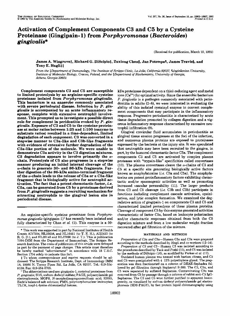
Digestion of C3 by P. gingiualis Proteinuse-Purified human C3 was digested with the cysteine proteinase at a molar ratio of 25:l in digestion buffer. Since it has been shown that the enzyme cleaves substrates such as benzoyl-Arg-p-nitroanilide more rapidly in the presence of glycyl-glycine (Gly-Gly), we carried out the time course digests both in the presence and absence of the dipeptide. The cysteine proteinase selectively cleaved the a-chain generating what initially appears to be the a'-chain of C3b (10). This result indicates that a fragment approximately the size of C3a (ie. 9 kDa) had been released (see Fig. 1). The rate of digestion with Gly-Gly present ap- pears only slightly more rapid than without the dipeptide. The C3 a-chain is nearly all converted to the a'-chain after 1 h of digestion. Further breakdown fragments of the C3 a'- chain are evident in the gel, and a decreasing intensity of the a'-band suggests that degradation continues. Visual evidence suggests that the C3 P-chain is resistant to this proteinase whether Gly-Gly is present or not.
Attempts to demonstrate C3a activity in the C3 digestion mixture were unsuccessful. A digest containing 300 pg of C3 was applied to a guinea pig ileal strip with no response.
time (min.)
-!3 75
+ Gly-Gly - Gly-Gly
FIG. 1. Digestion of human C3 by gingipain-1 from P. gin- giualis. Human C3 (13.3 pg) was exposed to the proteinase (142 ng of enzyme, specific activity 728 units) a t a 1:25 mol to mol ratio. The digestion mixture (0.5 ml) contained 10 mM Tris/HCl, 1 mM cysteine, 5 mM CaC12, and 50 mM glycyl-glycine at pH 7.0. The mixture was incubated at 37 "C, 45-4 aliquots were removed at designated times, and the enzyme was inhibited by adding TLCK at a 2 mM final concentration. Electrophoresis was performed under reducing condi- tions in a gradient (5-15%) SDS-polyacrylamide gel. The gel was subsequently stained with Coomassie Blue to visualize the cy- and 8- chain products of C3.
18904 C3 and C5 Cleavage by a P. gingivalis Proteinase
Sensitivity of the ileum would have detected approximately 1% of the C3a potentially generated from the digest. When the C3 digest was applied to a 13% gel (SDS-PAGE) and developed with silver stain, no material was detected in the C3a region (data not shown). We concluded that the C3a-like fragment released from the a-chain undergoes extensive deg- radation.
Digestion of C5 by the P. gingiualis Proteinase-Human C5 was digested by gingipain-1 at a 251 molar ratio, and the degradation pattern obtained is shown in Fig. 2. Initial cleav- age was specific for the C5 a-chain, as in the case of C3, except that the cleavage occurred internally generating frag- ments of 86 kDa (a-1) and 30 kDa (a-21, respectively. The dominant bands observed in SDS-PAGE gels after 1 h of digestion were the @-chain and the a-1 and a-2 fragments of C5. The amino-terminal sequences of the a-1 and a-2 frag- ments were determined as G-Y-G-D-S-N-Y-K- and T-L-Q- K-K-I-E-E-I-A- , respectively. The a-1 (86 kDa) and the a-2 (30 kDa) fragments are the first polypeptides to be formed from cleavage of C5 by gingipain-1, and they equal the molec- ular weight of the intact a-chain; therefore it is concluded that the initial site of cleavage in the C5 a-chain occurs within an intrachain or interchain disulfide loop located near the carboxyl-terminal end of this polypeptide chain. According to the banding pattern of C5 digested under nonreducing con- ditions, the fragmented a-chain and intact @-chain remain covalently attached after limited digestion (see Fig. 3). When a 13% SDS-PAGE gel of C5 after 2 and 3 h of digestion was stained with silver reagent, a band in the size range of C5a was observed (data not shown).
A scheme for the cleavage of C5 by gingipain-1 is shown in Fig. 4. The enzyme attacks primarily the a-chain of C5. The first peptide bond cleaved is between arginine 715 and glycine 716 of the a-chain. Subsequently, other sites are attacked including the bond between positions 74 and 75, which gen- erates C5a.
Both human C3a and C5a were submitted to proteolysis by gingipain-1 (specific activity 1,123 units) at 1001 molar ratios, and degradation was evaluated after electrophoresis on cel- lulose acetate strips. The C3a was extensively degraded after
116"- - -. - .. -a 116
84- c
58- 48-
37- 27-
-a1 86 -p 80
-a2 30
FIG. 2. Digestion of human C5 by gingipain-1 from P. gin- givali.9. Human C5 (16.2 pg) was exposed to the proteinase (173 ng of enzyme, specific activity 728 units) a t a 25:l mol to mol ratio. The digestion mixture (0.5 ml) contained 10 mM Tris/HCl, 1 mM cysteine, and 5 mM CaC12 at pH 7.0. The mixture was incubated at 37 "C, 45- pl aliquots were removed at designated times, and the enzyme was inhibited by adding TLCK at a 2 mM final concentration. Electro- phoresis was performed under reducing conditions in a gradient (5- 15%) SDS-polyacrylamide gel. The gel was developed with Coom- masie stain to visualize the a- and @-chain products of C5.
time (min.) 0 60 120 180 0 60 120 180 (kD)
M.W. - - 180
-a 116
-a1 86 -P
+SH -SH
FIG. 3. Human C5 (9.4 pg) was incubated with 100 ng of gingipain-1 (specific activity 728 units and a 50:l molar ratio) at 37 "C in (0.5 ml) digestion buffer at pH 7.0. Fifty-pl aliquots were removed at designated times, and the enzyme was inhibited by adding 2 mM TLCK. Samples were heated at 100 "C for 5 min in sample buffer with and without 1 mM cysteine and run on 8% SDS-PAGE gel. The stained gel indicated cleavage of the a-chain of C5a, as in the reduced gel (see Fig. 2). The unreduced material remained covalently intact after cleavage.
csa CSb n t 1
I S- 6 s-s
FIG. 4. A model is proposed for the cleavage of C5 by gin- gipain-1. The two chains of C5 are represented by bars, and the kxagom containing CHO indicate the attachment sites of the aspar- aginyl-linked oligosaccharide units. The arrows designated 1' and 2" denote the primary and secondary gingipain-1 cleavage sites in C5. The secondary site between residues 74 and 75 is not the only site of cleavage as degradation progresses but is identified as a known site of cleavage based on the release of the functional factor C5a.
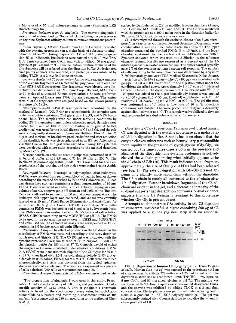
a 30-min incubation, both in the presence and absence of Gly- Gly (Fig. 5A). In the presence of glycyl-glycine the C3a appeared partially degraded after 10 min and was nearly destroyed after 60 min. Cleavage of C3a by the protease explains why activity could not be demonstrated in the C3 digestion mixtures; presumably the C3a fragment was released and then destroyed as the digestion continues.
C5a is more resistant to the gingipain-1 than C3a, based on the rate of degradation. After 30 min of digestion the C5a band does appear to get somewhat fainter (Fig. 5B). However, the majority of C5a remains intact even after 60 min of digestion, indicating that when C5 is subjected to prolonged digestion by gingipain-1, functional C5a may accumulate in the digestion mixture without further appreciable degrada- tion.
Evidence of C5a Generation-A digest of C5 was prepared by treating the plasma protein with gingipain-1 at a molar ratio of 25:l for 3 h. Aliquots of the digestion mixture were diluted and incubated with human neutrophils. Morphologic changes in the cells, known as polarization, were scored by counting the deformed cells as a ratio of the normally rounded cells. The neutrophil response to the digestion mixture indi- cated that a factor with activity like that of C5a was present
C3 and C5 Cleavage by a P. gingivalis Proteinase 18905
FIG. 5. Human C3a and C5a were incubated with gingipain- 1 (specific activity 1,123 units and at a 1OO: l molar ratio) in 0.2 ml of the pH 7.0 digestion buffer at 37 "C, with and without 60 mM glycyl-glycine. Ali- quots were removed, and TLCK was added (2 mM final concentration). Sam- ples were lyophilized to dryness and re- dissolved in 10 pl of water. Cellulose acetate strip electrophoresis was per- formed for 30 min at 200 V. Strips were subsequently stained in Amido Black to visualize digestion of the anaphylatox- ins. Panel A, C3a digest indicates exten- sive degradation versus time. Panel B, C5a digest indicates that little degrada- tion has occurred.
A.C3a without Gly-Gly
time (min.) 60 45 30 20 10 0
+ 0
60 45 30 20 10 0
I I C 5 dioest I h ~ 9 enzynie control ; 80 EZd C 5 control
0
'C 0 0 a
+ I
60 - m 40 - - 0 0 r ; 20
0 1:lO 1:lOO 1 :lo00 Buffer
Dilution
FIG. 6. Human C5 (90 pg) was digested with gingipain-1 (specific activity 728 units at a 25:l molar ratio) in 0.2 ml of digestion buffer at pH 7.0 and 37 "C. The mixture was incubated for 3 h, and proteolysis was inhibited by TLCK (to 2 mM). Fifty pl of EBSS containing 10 mM MOPS at pH 7.3 was added to the mixture. Neutrophil polarization was assayed on dilutions of the mixture as indicated. Controls were the same mixture except devoid of either C5 (enzyme control) or proteinase (C5 control) a t corresponding dilu- tions.
(Fig. 6). Calculations of the activity expressed by the C5 digest (EDSO for a 1:lOO dilution) compared with purified C5a (25) indicated that approximately 25% of the potential C5a activity had been generated.
A separate C5 digest was prepared for the chemotaxis assay using a different enzyme preparation. This enzyme prepara- tion had a somewhat higher specific activity (1,123 units) than previous preparations and was used at a 1001 molar ratio. Aliquots of the digestion mixture were diluted, and neutrophil migration was evaluated using a modified Boyden chamber assay (Fig. 7). Estimates of the recovery of activity relative to purified C5a indicated a yield of 20%.
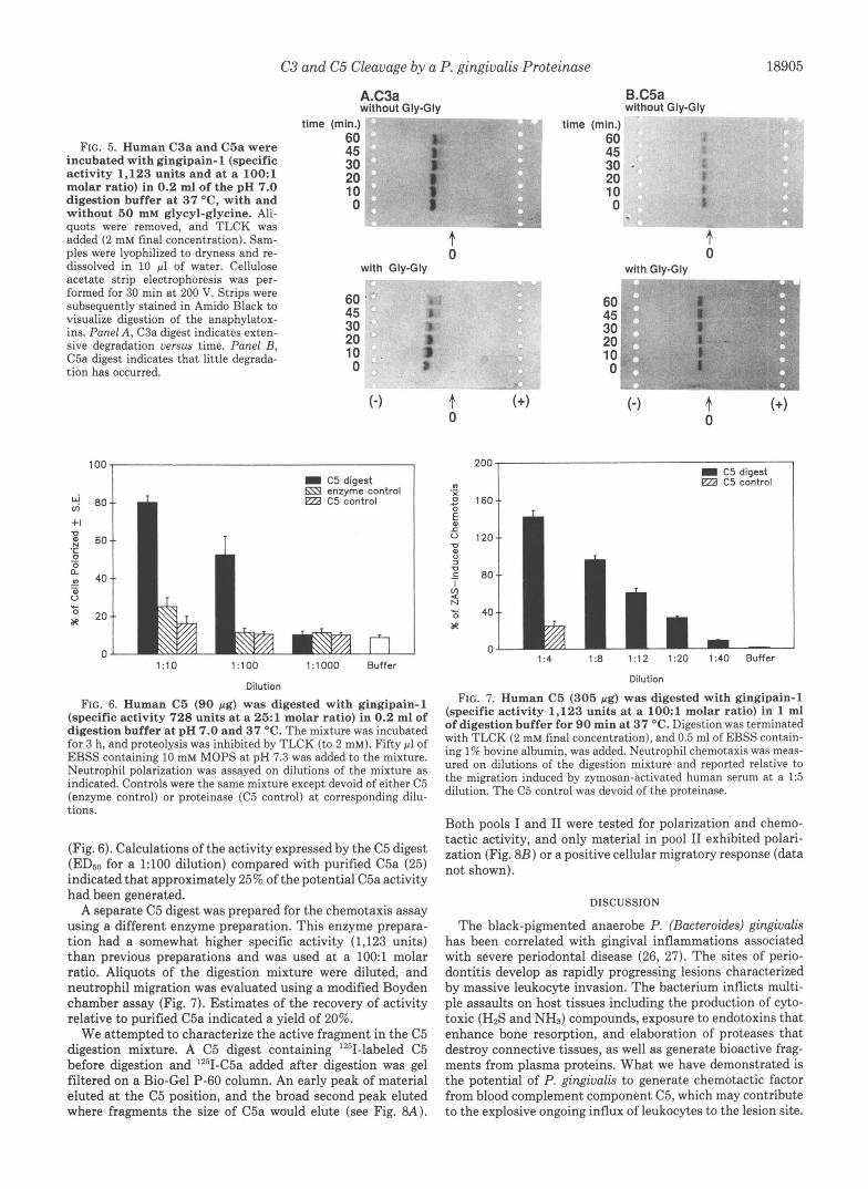
We attempted to characterize the active fragment in the C5 digestion mixture. A C5 digest containing lZ5I-labeled C5 before digestion and lZ5I-C5a added after digestion was gel filtered on a Bio-Gel P-60 column. An early peak of material eluted at the C5 position, and the broad second peak eluted where fragments the size of C5a would elute (see Fig. 8A) .
0
B.CSa without Gly-Gly
time (min.) 60 45 30 20 10 0
+ 0
With Gly-Gly p;, . - . ,
60 45 30 20 10 0
+ 0
200 - I C 5 digest
.- m ezd C 5 control
X 1 0 160"
1 :4 1 % 1 :12 1:ZO 1 :40 Buffer
Dilution
FIG. 7. Human C5 (305 pg) was digested with gingipain-1 (specific activity 1,123 units at a 1 O O : l molar ratio) in 1 ml of digestion buffer for 90 min at 37 "C. Digestion was terminated with TLCK (2 mM final concentration), and 0.5 ml of EBSS contain- ing 1% bovine albumin, was added. Neutrophil chemotaxis was meas- ured on dilutions of the digestion mixture and reported relative to the migration induced by zymosan-activated human serum a t a 1:5 dilution. The C5 control was devoid of the proteinase.
Both pools I and I1 were tested for polarization and chemo- tactic activity, and only material in pool I1 exhibited polari- zation (Fig. 8B) or a positive cellular migratory response (data not shown).
DISCUSSION
The black-pigmented anaerobe P. (Bacteroides) gingiualis has been correlated with gingival inflammations associated with severe periodontal disease (26, 27). The sites of perio- dontitis develop as rapidly progressing lesions characterized by massive leukocyte invasion. The bacterium inflicts multi- ple assaults on host tissues including the production of cyto- toxic (H2S and NH3) compounds, exposure to endotoxins that enhance bone resorption, and elaboration of proteases that destroy connective tissues, as well as generate bioactive frag- ments from plasma proteins. What we have demonstrated is the potential of P. gingiualis to generate chemotactic factor from blood complement component C5, which may contribute to the explosive ongoing influx of leukocytes to the lesion site.
18906 C3 and C5 Cleavage by a P. gingivalis Proteinase
s A
Elution Volume (mi)
B 100,
Dilution
FIG. 8. A and B, human C5 (400 pg containing 20,000 cpm of Iz5I- labeled C5) was digested with gingipain-1 (specific activity 728 units a t a 25:l molar ratio) in 1 ml of digestion buffer for 90 min at 37 'C. The proteinase inhibitor TLCK was added along with 5 X lo6 cpm of "'I-labeled C5a, and the sample was applied to the gel filtration column. The mixture was eluted from a 1.5 X 55-cm column of Bio- Gel P-60 with 20 mM imidazole HCI containing 0.3 M NaCl at pH 7.0. The chromatography was performed at 4 "C and a flow rate of 10 ml/h. Panel A, the elution profile of the digestion mixture was monitored by radioactivity. Pools were made of the early C5 peak I and the later eluting peak 11; peak I1 represents the elution volume of C5a. Panel B, polarization activity for dilutions of the material collected in pool 11. Pool I was inactive and gave results identical to the buffer control (data not shown).
We propose that once the initial injury has been made to the gingival connective tissue and plasma gains access to the infected region, the following series of events occur. The contact of gingipain-1 with plasma promotes complement activation, and active chemotactic factor (i.e. C5a) is gener- ated. The C5a recruits leukocytes to the lesion site directly and then indirectly amplifies recruitment by stimulating the release of other potent chemotactic factors such as leukotriene B and platelet-activating factor from neutrophils and inter- leukin-8 from monocytes (28). These events of cellular se- questration result in severe tissue damage to the gingiva from proteolytic attack by the massive influx of leukocytic enzymes.
Our results indicate that a cysteine proteinase from P. gingiualis has the capacity to degrade both complement com- ponents C3 and C5. Previous studies have identified a trypsin- like protease(s) from P. gingiualis which cleaves the substrate benzoyl-L-arginine-p-nitroanilide and appears to be a cystei- nyl proteinase but ranges in estimated size from 35 to more than 100 kDa (29-31). These may or may not be the same proteinase as recently isolated and characterized by Chen et
al. (1). It is well known that exposure of immunoglobulins, complement factors, and other serum components to bacteria of the genera Bacteroides results in degradation (32). A spe- cific study of the capacity of P. gingiualis to convert purified C3 indicated that different strains had a variable capacity for cleaving the C3 molecule (33). These studies failed to identify the proteinase responsible for the proteolysis of the serum proteins.
The nature of the degradative process has now been ex- plored using purified enzyme. The cleavage patterns for C3 and C5 are consistent with limited proteolysis leading to generation of the C3a- and C5a-like fragments, respectively, in the course of the degradative process. Unlike the results of Chen et al. (1) who observed a marked enhancement in the amidolytic activity of gingipain-1 when Gly-Gly is added, we recorded only a minor effect from the addition of dipeptide on the cleavage of C3 and C5. It is suggested that the role of the Gly-Gly is to eliminate nonproductive binding of the peptide substrates. This may be less important when a larger protein substrate is used. Gingipain-1 digestion of C3 leads to specific cleavage of the a-chain at or near the C3a-C3b scis- sion site, this is the same site that is susceptible to the serum complement convertases (10,l l) .
Although the apparent initial site of C3 cleavage by the P. gingiualis protease is at or near the C3a-C3b bond junction, the C3a portion fails to accumulate in the digestion mixture. We have shown that the C3a molecule is readily susceptible to degradation by the proteinase and does not remain func- tional in the digestion mixture. The C3b portion of the mol- ecule appears to have a relatively long lifetime in the presence of the proteinase with considerable quantities of C3b, repre- sented by intact a'- and @-chains, surviving for up to 3 h of exposure to 1% (w/w) enzyme.
The C5 molecule undergoes a limited degradation differing from that evoked by serum convertase. The initial site of cleavage is not the C5a-C5b junction but rather an internal bond between positions 715 and 716 in the a-chain. This site of cleavage is identical to the site at which plasmin cleaves C5b (34). The fragments of the C5 a-chain are further de- graded, and C5a is generated from the 86 kDa fragment. Evidence that after the initial cleavage the C5 a-chain frag- ments remain covalently attached supports the hypothesis that cleavage occurs near the carboxyl-terminal end of the a- chain and within either interchain or intrachain disulfide linkages. C5a is less susceptible to attack by gingipain-1 than is C3a, and C5a can accumulate during the digestion of C5 by the proteinase. We have measured C5a activity in the digests equivalent to 20-25% of the potential levels predicted from the C5. Evidence that the activity detected in these digests was associated with the C5a molecule was provided by meas- uring activity after separation on a gel filtration column. The low molecular weight fraction contained all of the biological activity.
Pool I1 is assumed to contain intact C5a based on previous evidence that human C5a is approximately 10-30 fold more active in a chemotactic assay than is C5a missing the car- boxyl-terminal arginyl residue (i.e. C5ad,. A ~ ~ ) . Furthermore, the derivative C5a 1-69, missing the five carboxyl-terminal residues M-Q-L-G-R, is less than 0.1% as active as C5a in the same assay (35). Synthetic peptide studies have confirmed that the biologic effector site in C5a is located in the carboxyl- terminal region including residues M-Q-L-G-R. These studies also showed that extension of the sequence beyond the ter- minal arginine by adding even a glycyl residue reduced activity by 12-20-fold (25). Consequently, cleavage of the C5 a-chain by gingipain-1 at a basic residue amino-terminal or carboxyl-
C3 and C5 Cleavage by a P. gingivalis Proteinase 18907
terminal to the C5a scission site, that is located between residues 74-75, would predictably result in a C5 fragment exhibiting markedly less biological activity than was observed here.
Taken together, these data give compelling evidence that a cysteinyl proteinase from the P. gingivalis bacterium (gingi- pain-1) is capable of efficiently cleaving C3 and C5. It appears that this particular proteinase may be, in part, responsible for the proteolytic ability of the bacteria to degrade numerous serum proteins. This is the first indication that proteinases from P. gingivalis are capable of generating bioactive factors from serum components. Action of gingipain-1 on C5 releases a potent chemotactic factor, which, if generated at the site of infection, could result in a significant leukocytic infiltration and help to explain the rapid onset of the inflammatory response associated with progressive periodontitis.
1.
2. 3.
4.
5.
6. 7.
8.
REFERENCES
10. 9.
11. 12.
13. 14.
15. 16. 17.
18. 19.
20. 21.
22. 23.
24. 25.
26. ~.
0.7
Chen, Z., Potempa, J., Polanowski, A., Wikstrom, M., and Travis, J. (1992)
Mayrand, D., and Holt, S. C. (1988) Microbiol. Reu. 62,134-152 J. Biol. Chem. 2 6 7 , 18896-18901 28.
Moore, W. E. C., Holdeman, L. V., Smibert, R. M., Hash, D. E., Burmeister, 29.
Zambon, J. J., Reynolds, H. S., a n f Slots, J. (1981) Infect. Immun. 3 2 , 30. J. A., and Ranney, R. R. (1982) In ect Immun 38,1137-1148
19R-2nR
1,.
Marsh, P. D., McKee, A. S., McDermid, A. S., and Dowsett, A. B. (1989) 31.
Grenier, D., and Mayrand, D. (1987) J. Clin. Microbiol. 26 , 738-740 Birkedal-Hansen, H., Taylor, R. E., Zambon, J. J., Barwa, P. K., and
32.
Sundavist. G.. Carlsson. J.. and Hanstrom. L. (1987) J. Periodontal Res. 34. Neiders, M. E. (1988) J. Periodontal Res. 23,258-264 33.
-.- "_ FEMS Microbiol. Lett. 59, 181-186
, , 22;300-306
, . . 35.
White, D., and Mayrand, D. (1981) J. Periodontal Res. 16,259-265 Muller-Eberhard, H. J. (1988) Annu. Reu. Biochem. 57,321-347 Hugli T. E. (1986) Corn lement 3,111-127 Hu li: T. E., Vallota, If H., and Muller-Eberhard, H. J. (1975) J. Biol.
Fernandez, H. N., and Hugli, T. E. (1976) J. Immunol. 117 , 1688-1694 Hugli, T. E., Gerard, C., Kawahara, M., Scheetz, M. E., Barton, R., Briggs,
S., Koppel, G., and Russell, S. (1981) MOL Cell. Biochem. 41,59-66 Tack, B. F., and Prahl, J. W. (1976) Biochemistry 16,4513-4521 DiScipio, R. G. (1981) Biochem. J. 199,485-496 Parkes, C., DiScipio, R. G., Kerr, M. A., and Prohaska, R. (1981) Biochem.
Edman, P., and Begg, G. (1967) Eur. J. Biochem. 1,80-91 Haviland, D. L., Haviland, J. C., Fleischer, D. T., Hunt, A., and Wetsel, R. Laemmli, U. K. (1970) Nature 227,680-685 Merril, C. R., Switzer, R. C., and Van Keuren, M. L. (1979) Proc. Natl.
Fehr, J., and Dahinden, C. (1979) J. Clin. Inuest. 6 4 , 8-16 Haston, W. S., and Shields, J. M. (1985) J. Immunol. Methods 8 1 , 229-
Dahinden, C., Galanos, C., and Fehr, J. (1983) J. Immunol. 130,857-862 Ember. J. A.. Sanderson. D. G.. Tavlor. S.. Kawahara. M.. and Hueli. T. E.
&em. 260,1472-1478
J. 193,963-970
A. (1991) J. Immunol. 146,362-368
Acad. Sci. U. S. A. 76,4335-4340
237
(1992) J. ~mmunol., 148,3165-5173 '
tol. 13,570-577
(1989) Oral Microbiol. Immun. 4. 178-179
, , I .
Slots, J., Bragd, L., Wikstrom, M., and Dahlen, G. (1986) J. Clin. Periodon-
Smalley, J. W., Birss, A. J., Kay, H. M., McKee, A. S., and Marsh, P. D.
Morgan; E. L., Sanderson, S. D., Scholz, W., Noonan, D. J., Hobbs, M. V., Weigle, W. O., and Hugli, T. E. (1992) FASEB J. 6, A1121 (Abst. 1075)
Yoshimura. F.. Nishlkata. M.. Suzuki. T.. Hoover C. I.. and Newbrun, E. (1984) Arch.' Oral Biol. 29,559-564.
Tsutsui. H.. Kinouchi. T.. Wakano. Y.. and Ohnishi. Y. (1987) Infect.
Otsuka, M., Endo, J., Hinode, D., Na a h , A., Maehara, R., Sato, M., and
Sundqvist, G., Carlsson, J., Herrmann, B., and Tarnvik, A. (1985) J. Med. Nakamura, R. (1987) J. PeriodontafRes. 22,491
Schenkein, H. A. (1988) J. Periodontal Res. 2 3 , 187-192 DiScipio, R. G. (1992) J. Biol. Chem. 267 , 17087-17094 Chenoweth, D. E., and Hugli, T. E. (1980) Mol. Immunol. 1 7 , 151-161