Family-specific Kinesin Structures Reveal Neck-linker LengthBased on Initiation of the Coiled-coil*
Received for publication, May 26, 2016, and in revised form, July 7, 2016 Published, JBC Papers in Press, July 26, 2016, DOI 10.1074/jbc.M116.737577
Rebecca K. Phillips‡, Logan G. Peter‡, Susan P. Gilbert§, and X Ivan Rayment‡1
From the ‡Department of Biochemistry, University of Wisconsin, Madison, Wisconsin 53706 and the §Department of BiologicalSciences, Rensselaer Polytechnic Institute, Troy, New York 12180
Kinesin-1, -2, -5, and -7 generate processive hand-over-hand8-nm steps to transport intracellular cargoes toward the micro-tubule plus end. This processive motility requires gating mech-anisms to coordinate the mechanochemical cycles of the twomotor heads to sustain the processive run. A key structural ele-ment believed to regulate the degree of processivity is the neck-linker, a short peptide of 12–18 residues, which connects themotor domain to its coiled-coil stalk. Although a shorter neck-linker has been correlated with longer run lengths, the struc-tural data to support this hypothesis have been lacking. To testthis hypothesis, seven kinesin structures were determined byx-ray crystallography. Each included the neck-linker motif, fol-lowed by helix �7 that constitutes the start of the coiled-coilstalk. In the majority of the structures, the neck-linker lengthdiffered from predictions because helix �7, which initiates thecoiled-coil, started earlier in the sequence than predicted. A fur-ther examination of structures in the Protein Data Bank revealsthat there is a great disparity between the predicted andobserved starting residues. This suggests that an accurate pre-diction of the start of a coiled-coil is currently difficult toachieve. These results are significant because they now excludesimple comparisons between members of the kinesin superfam-ily and add a further layer of complexity when interpreting theresults of mutagenesis or protein fusion. They also re-emphasizethe need to consider factors beyond the kinesin neck-linkermotif when attempting to understand how inter-head commu-nication is tuned to achieve the degree of processivity requiredfor cellular function.
The coiled-coil was the first quaternary structural arrange-ment described and has been predicted to occur in �3% of allproteins (1, 2). This apparently simple motif shows consider-able structural variation, but because of the characteristic dis-tribution of hydrophobic and polar residues, it can be detectedreadily through sequence analysis (3–5). Coiled-coils play
diverse functional roles, but in many cases, they serve as oligo-merization domains. Under these circumstances, the exactlength or starting point of the coiled-coil is structurally andfunctionally important. Unfortunately, this parameter is notwell defined or uniformly predicted by any computationalalgorithm.
The precise starting point of the coiled-coil is particularlyimportant in the kinesin and myosin family of molecularmotors, where the dimerization module coordinates the activ-ities of individual motor domains on separate polypeptidechains. Typically, in these motor families, there is a flexiblesection of the polypeptide that connects the motor to thedimerization module. The length of this connecting unit plays acritical role in processive molecular motors and is especiallyimportant for many members of the kinesin superfamily inwhich this domain is known as the neck-linker.
Kinesin motor proteins are classified into 15 different kinesinfamilies, which share a structurally conserved kinesin motordomain (6 –10). These families perform a diverse set of cellularfunctions, all of which involve moving along a microtubuletrack for cargo transport or modulating microtubule dynamics(11–17). Part of the classification is dictated by the location oftheir motor domains. N-terminally located motors comprisethe majority of kinesin families. The exceptions are the kinesin-14A and -14B families that contain C-terminal motors and thekinesin-13 family in which the motor domain is located in themiddle of the polypeptide (6).
This study is focused on N-terminal kinesins, which are com-posed of an N-terminal motor domain connected to a long�-helical region that dimerizes into a coiled-coil stalk that endswith a C-terminal cargo domain that may interact with otherpartner proteins or substrates. The N-terminal motor domainis responsible for ATP turnover coupled to force production.This group of motors is typically dimeric and shows processivemovement along microtubules tracks. The ability of the N-ter-minal kinesin family to remain on the microtubule lattice iscritical to their function.
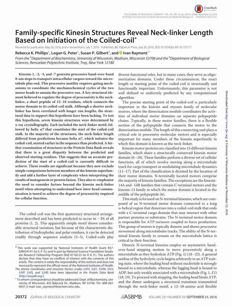
Dimeric N-terminal kinesins employ an asymmetric hand-over-hand stepping motion to move processively along amicrotubule as they hydrolyze ATP (Fig. 1) (18 –23). A generaloutline of the hydrolytic cycle begins arbitrarily in an ATP wait-ing state, where the leading head without nucleotide is stronglybound to a microtubule, whereas the lagging head is bound toADP, but only weakly associated with a microtubule (Fig. 1, E1)(18, 19). To proceed with stepping, the leading head binds ATP,and the dimer undergoes a structural transition transmittedthrough the neck-linker motif, a 12–18-amino acid flexible
* This work was supported by National Institutes of Health Grant R37-GM054141 (to S. P. G.) and in part by National Science Foundation Gradu-ate Research Fellowship Program DGE-0718123 (to R. K. P.). The authorsdeclare that they have no conflicts of interest with the contents of thisarticle. The content is solely the responsibility of the authors and does notnecessarily represent the official views of the National Institutes of Health.
The atomic coordinates and structure factors (codes 5JVU, 5JX1, 5JVM, 5JV3,5JVP, 5JVS, and 5JVR) have been deposited in the Protein Data Bank(http://wwpdb.org/).
1 To whom correspondence should be addressed: Dept. of Biochemistry, Uni-versity of Wisconsin, 433 Babcock Dr., Madison, WI 53706. Tel.: 608-262-0437; E-mail: [email protected].
crossmarkTHE JOURNAL OF BIOLOGICAL CHEMISTRY VOL. 291, NO. 39, pp. 20372–20386, September 23, 2016
peptide that connects the motor domain to the coiled-coil stalk(Fig. 1, E2) (24, 25). This structural transition, designated neck-linker docking, shifts the lagging unbound head forward 16 nm
to the next microtubule-binding site toward the microtubuleplus end (Fig. 1, E3) (24, 26). Subsequently, ADP is releasedfrom this new leading head resulting in both heads bound to themicrotubule (Fig. 1, E4 and E5) (27).
When both heads are bound to the microtubule, the neck-linker domains are oriented in opposite directions (28, 29). Theneck-linker of the leading nucleotide-free head is orientedbackward, inhibiting ATP binding and hydrolysis by the fronthead until the rear head detaches from the microtubule (Fig. 1,E6) (30). The neck-linker of the lagging ATP-bound headremains docked onto the catalytic core and directed forward.ATP hydrolysis on the microtubule-bound lagging head resultsin another structural rearrangement that leads to phosphaterelease. Thereby, the lagging head transitions into a weaklybound ADP state and detaches from the microtubule, thusstarting the cycle anew (Fig. 1, E7).
During the stepping cycle, each motor domain must remainout-of-phase with the other. If both motor domains enter anunbound state at the same time, the processive run ends. Therun length indicates the distance a motor steps along the micro-tubule before dissociation and gives a measure of processivity.Each kinesin family member has different average run lengthsranging from �0.2 �m, as in Eg5, to 2.1 �m, as in conventionalkinesin-1, although this is construct-dependent for kinesin-1and can range from 1.3 to 2.1 �m (25, 31, 32). Despite thestructural conservation between different kinesin motors, thereare clear kinetic differences between the families.
One domain hypothesized to contribute to processivity is thekinesin neck-linker, a small flexible peptide consisting of 12–18amino acids (33–35). The neck-linker connects each motordomain to the coiled-coil stalk where the junction betweenthese two entities has been assumed to occur at the same posi-tion in the sequence as that observed in kinesin-1 (36). Theneck-linker itself undergoes a series of structural transitions asoutlined in the kinesin mechanochemical cycle. It is hypothe-sized that longer neck-linkers increase the diffusional searcharea and therefore could slow down stepping, allowing time forthe forward head to release from the microtubule (37).Recently, alterations in neck-linker length were shown to affectthe kinetic cycle (38). Increasing neck-linker length resulted inincreased rear head binding, and decreasing neck-linker lengthresulted in slower release of ADP from the unbound head. Botheffects result in slowing the productive kinetic cycle (38).Because the neck-linker is involved in connecting the motordomain and coiled-coil stalk, changes in the length of the neck-linker are expected to alter communication between the twomotor heads.
Given that the overall size of the kinesin motor domain issimilar across all families and that they bind to microtubules ina similar manner, it is surprising that the predicted length of theneck-linker between families shows considerable variationeven though within each family the neck-linker/�7 sequencesare almost completely conserved (36). This is especially puz-zling because a multitude of studies show increasing the neck-linker for a given kinesin by even one residue results indecreases in run length and processivity (25, 31, 34, 39, 40). Thisconundrum is particularly evident in members of the kinesin-2family, KIF3AB and KIF3AC, which are unique in forming a
FIGURE 1. Schematic representation of the kinesin chemomechanicalcycle that illustrates key transitions that influence processivity. A pro-cessive run is started by binding of either head followed by rapid ADP releaseto form the E1 intermediate where the leading head that forms the initialcontact is microtubule-bound but nucleotide-free (Ø), whereas the trailinghead is detached with ADP tightly bound. ATP binding to the leading headinduces a series of structural transitions, including neck-linker docking thatallows the trailing ADP-head to move 16 nm ahead to its new microtubule-binding site (E2–E4). ADP release from the new leading head (E4 and E5)results in the E5 two-head bound state, thereby generating intermolecularstrain, which inhibits ATP binding at the now leading head. ATP hydrolysis atthe trailing head followed by phosphate (Pi) release generates a microtubuleweakly bound ADP state (E6 and E7). Detachment of the trailing head relievesthe intermolecular strain (E7), and initiates the next motor cycle.
Coiled-coil Initiation in Kinesin Motors
SEPTEMBER 23, 2016 • VOLUME 291 • NUMBER 39 JOURNAL OF BIOLOGICAL CHEMISTRY 20373
heterodimeric motor. KIF3AB and KIF3AC further interactwith an adaptor protein to bind a variety of cargoes for intrafla-gellar and neuronal transport (11, 15, 41– 45). KIF3AC in par-ticular has been implicated in neuronal repair (11). BothKIF3AB and KIF3AC are highly processive motors (46). Yettheir neck-linker is predicted to be three residues longer thanconventional kinesin, which would suggest that these motorsshould not be so processive (36). KIF3AB and KIF3AC haveidentical neck-linker lengths and nearly identical neck-linkersequences, differing only at Thr-380 in KIF3C, which is an ala-nine in KIF3A and KIF3B. Both motors are processive; KIF3ABand KIF3AC have run lengths of 1.6 and 1.2 �m, respectively(37, 46). However, the kinetic parameters vary significantlybetween the two motors (46 – 48). Furthermore, homodimericspecies KIF3AA, KIF3BB, and KIF3CC also exhibit vastly dif-ferent processivity parameters. KIF3AA and KIF3BB are highlyprocessive, but KIF3CC travels at only 7.5 nm/s with an averagerun length of just 0.6 �m, which suggests that the neck-linker isnot the only determinant of processivity (46). These observa-tions prompted an investigation of the true length of the neck-linker in the best studied classes of N-terminal kinesin families.
The original estimates of the neck-linker length made theassumption that the coiled-coil would begin on a hydrophobicresidue that lie in either the a or d position of the coiled-coilheptad repeat (36). However, the predictions of the first residueto adopt a helical conformation in any coiled-coil are ambigu-ous, even though the body of a coiled-coil is well indicated bycurrent software (3, 4). Prediction software recognizes the hep-tad repeat in a coiled-coil domain. However, because the begin-ning and end of sequences may not follow that pattern, there isinsufficient information to make accurate predictions of thestart or of the end of a native coiled-coil. Given the considerablevariation in the amino acid sequence of neck-linkers and asso-ciated coiled-coils, this raises the question whether the truelength of the neck-linker has been accurately identified acrossthe kinesin superfamily with the original assumption describedabove.
There is only one structure for a dimeric N-terminal kinesin,rat kinesin-1 (3KIN) because dimeric kinesins are difficult tocrystallize (14). The 3KIN structure provided the first picture ofthe true �7 start and neck-linker length in the context of adimeric motor. Most kinesin motor structures are monomeric,allowing the neck-linker to adopt varying conformations thatmay not reflect that experienced by the dimeric motor in vivo.For example, in the structure of the kinesin-2 KIF3B (3B6U) the
neck-linker includes a cis-proline; therefore, this structure isunlikely to reflect the native neck-linker conformation. Thisstudy is directed toward providing an experimental foundationfor determining the start site for the coiled-coil and by infer-ence the length of the neck-linker. We determined the struc-tures of the neck-linker and �7 helix from the following fourdifferent kinesin families: kinesin-1, -2, -5, and -7. Kinesin-1 isthe canonical N-terminal processive kinesin motor. Kinesin-2family members, KIF3A and KIF3C, are involved in long rangetransport and neuronal repair (11, 15). Kinesin-5 family mem-ber, Eg5, is unique within this subset as it is a bipolar tetramerickinesin whose role is to cross-link microtubules during celldivision (12, 13, 49). Kinesin-7 family member CENP-E isresponsible for transporting misaligned chromosomes duringcongression in mitosis (16). In addition, we compared struc-tures in the PDB2 that include a native start to their coiled-coilwith the predictions generated from either MARCOIL orCOILS-28 (3, 4). Overall, we find that structures of proteins arenecessary to determine the true start site of the coiled-coilrather than relying on prediction software alone.
Results
Crystal Structures of Kinesin-1, -2, -5, and -7 Neck-linker �7Helix Proteins—All neck-linker structures were homodimericand solved to a resolution of 2.3 Å or higher allowing for accu-rate determination of secondary structure transitions. Theextent of the ordered structure in each construct is given inTable 1. Several structures had multiple monomers in theasymmetric unit. Individual monomers were similar as shownin Table 1. For each neck-linker structure, the start of thecoiled-coil helix was determined using the Dictionary of Sec-ondary Structure of Proteins algorithm (50). In each structure,varying lengths of the neck-linker were ordered; thus wefocused on the initiation point of the �7 helix to determineneck-linker length.
All of the structures were determined as fusions with theC-terminal dimerization domain of EB1. Previous studies haveshown that inclusion of globular folding domains considerablyincreases the ability to express and crystallize sections of coiled-coil proteins and that they do not perturb the structure morethan one heptad from the point of fusion (51, 52).
2 The abbreviations used are: PDB, Protein Data Bank; MEPEG, methoxypolyethylene glycol; PEE, pentaerythritol ethoxylate; PEG, polyethyleneglycol; TCEP, tris(2-carboxyethyl)phosphine; BTP, 1,3-bis[tris(hydro-xymethyl)methylamino]propane.
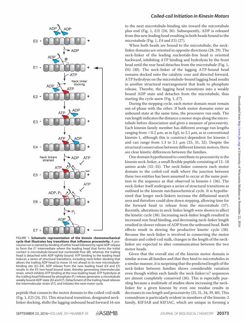
TABLE 1Average pairwise root means square differences between independent chains within each structural determinationFirst ordered residue is listed for the longest monomer and may not be ordered in other monomers in the asymmetric unit. r.m.s.d. is root mean square deviation. NA is notapplicable.
ConstructMonomers in
asymmetric unit Average r.m.s.d. First ordered residue
ÅCENP-E 6 0.862 Asn-336a
Eg5 4 0.450 Asn-336a
KIF3A-kinesin-1 8 0.739 Asn-352 (KIF3A)a
KIF3C 2 0.578 Asn-374a
KIF3A 1 NA Asp-354Kinesin-1 2 0.529 Asn-340Kinesin-1 � DAL 1 NA Asp of DAL (Ala-345-2)
a In these structures two residues that remain from the recombinant tobacco etch virus cleavage site were also observed.
Coiled-coil Initiation in Kinesin Motors
20374 JOURNAL OF BIOLOGICAL CHEMISTRY VOLUME 291 • NUMBER 39 • SEPTEMBER 23, 2016
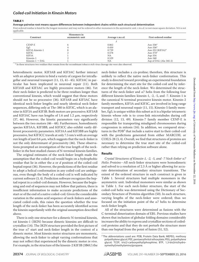
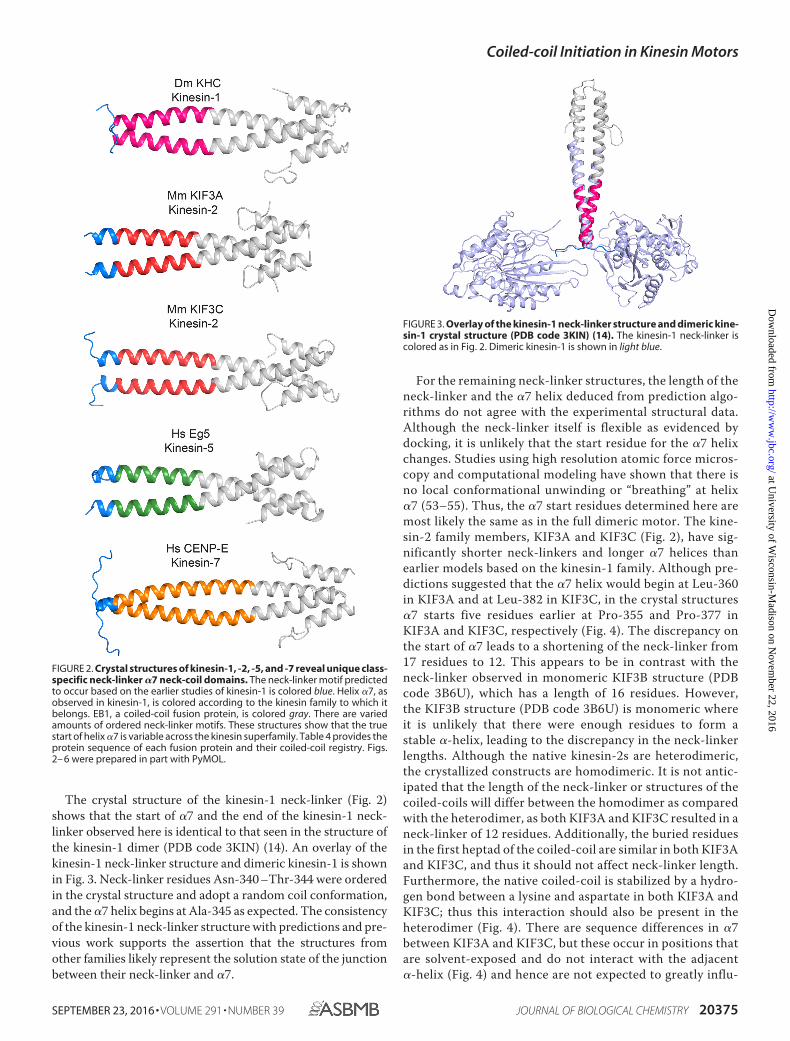
The crystal structure of the kinesin-1 neck-linker (Fig. 2)shows that the start of �7 and the end of the kinesin-1 neck-linker observed here is identical to that seen in the structure ofthe kinesin-1 dimer (PDB code 3KIN) (14). An overlay of thekinesin-1 neck-linker structure and dimeric kinesin-1 is shownin Fig. 3. Neck-linker residues Asn-340 –Thr-344 were orderedin the crystal structure and adopt a random coil conformation,and the �7 helix begins at Ala-345 as expected. The consistencyof the kinesin-1 neck-linker structure with predictions and pre-vious work supports the assertion that the structures fromother families likely represent the solution state of the junctionbetween their neck-linker and �7.
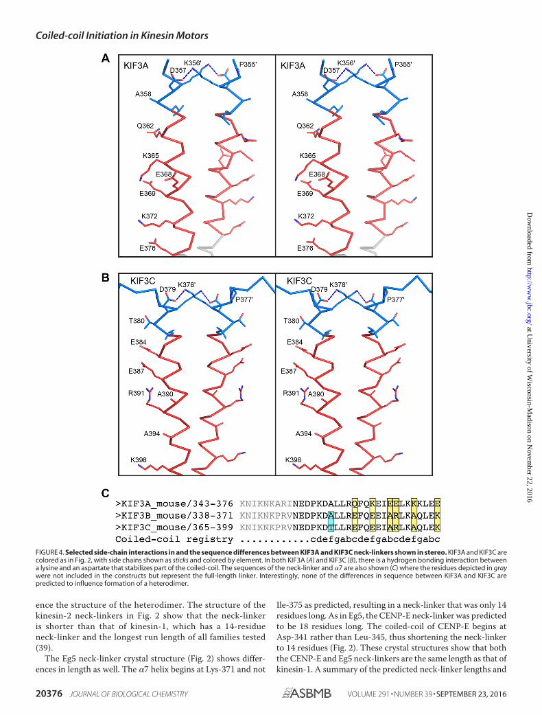
For the remaining neck-linker structures, the length of theneck-linker and the �7 helix deduced from prediction algo-rithms do not agree with the experimental structural data.Although the neck-linker itself is flexible as evidenced bydocking, it is unlikely that the start residue for the �7 helixchanges. Studies using high resolution atomic force micros-copy and computational modeling have shown that there isno local conformational unwinding or “breathing” at helix�7 (53–55). Thus, the �7 start residues determined here aremost likely the same as in the full dimeric motor. The kine-sin-2 family members, KIF3A and KIF3C (Fig. 2), have sig-nificantly shorter neck-linkers and longer �7 helices thanearlier models based on the kinesin-1 family. Although pre-dictions suggested that the �7 helix would begin at Leu-360in KIF3A and at Leu-382 in KIF3C, in the crystal structures�7 starts five residues earlier at Pro-355 and Pro-377 inKIF3A and KIF3C, respectively (Fig. 4). The discrepancy onthe start of �7 leads to a shortening of the neck-linker from17 residues to 12. This appears to be in contrast with theneck-linker observed in monomeric KIF3B structure (PDBcode 3B6U), which has a length of 16 residues. However,the KIF3B structure (PDB code 3B6U) is monomeric whereit is unlikely that there were enough residues to form astable �-helix, leading to the discrepancy in the neck-linkerlengths. Although the native kinesin-2s are heterodimeric,the crystallized constructs are homodimeric. It is not antic-ipated that the length of the neck-linker or structures of thecoiled-coils will differ between the homodimer as comparedwith the heterodimer, as both KIF3A and KIF3C resulted in aneck-linker of 12 residues. Additionally, the buried residuesin the first heptad of the coiled-coil are similar in both KIF3Aand KIF3C, and thus it should not affect neck-linker length.Furthermore, the native coiled-coil is stabilized by a hydro-gen bond between a lysine and aspartate in both KIF3A andKIF3C; thus this interaction should also be present in theheterodimer (Fig. 4). There are sequence differences in �7between KIF3A and KIF3C, but these occur in positions thatare solvent-exposed and do not interact with the adjacent�-helix (Fig. 4) and hence are not expected to greatly influ-
FIGURE 2. Crystal structures of kinesin-1, -2, -5, and -7 reveal unique class-specific neck-linker �7 neck-coil domains. The neck-linker motif predictedto occur based on the earlier studies of kinesin-1 is colored blue. Helix �7, asobserved in kinesin-1, is colored according to the kinesin family to which itbelongs. EB1, a coiled-coil fusion protein, is colored gray. There are variedamounts of ordered neck-linker motifs. These structures show that the truestart of helix �7 is variable across the kinesin superfamily. Table 4 provides theprotein sequence of each fusion protein and their coiled-coil registry. Figs.2– 6 were prepared in part with PyMOL.
FIGURE 3. Overlay of the kinesin-1 neck-linker structure and dimeric kine-sin-1 crystal structure (PDB code 3KIN) (14). The kinesin-1 neck-linker iscolored as in Fig. 2. Dimeric kinesin-1 is shown in light blue.
Coiled-coil Initiation in Kinesin Motors
SEPTEMBER 23, 2016 • VOLUME 291 • NUMBER 39 JOURNAL OF BIOLOGICAL CHEMISTRY 20375
ence the structure of the heterodimer. The structure of thekinesin-2 neck-linkers in Fig. 2 show that the neck-linkeris shorter than that of kinesin-1, which has a 14-residueneck-linker and the longest run length of all families tested(39).
The Eg5 neck-linker crystal structure (Fig. 2) shows differ-ences in length as well. The �7 helix begins at Lys-371 and not
Ile-375 as predicted, resulting in a neck-linker that was only 14residues long. As in Eg5, the CENP-E neck-linker was predictedto be 18 residues long. The coiled-coil of CENP-E begins atAsp-341 rather than Leu-345, thus shortening the neck-linkerto 14 residues (Fig. 2). These crystal structures show that boththe CENP-E and Eg5 neck-linkers are the same length as that ofkinesin-1. A summary of the predicted neck-linker lengths and
FIGURE 4. Selected side-chain interactions in and the sequence differences between KIF3A and KIF3C neck-linkers shown in stereo. KIF3A and KIF3C arecolored as in Fig. 2, with side chains shown as sticks and colored by element. In both KIF3A (A) and KIF3C (B), there is a hydrogen bonding interaction betweena lysine and an aspartate that stabilizes part of the coiled-coil. The sequences of the neck-linker and �7 are also shown (C) where the residues depicted in graywere not included in the constructs but represent the full-length linker. Interestingly, none of the differences in sequence between KIF3A and KIF3C arepredicted to influence formation of a heterodimer.
Coiled-coil Initiation in Kinesin Motors
20376 JOURNAL OF BIOLOGICAL CHEMISTRY VOLUME 291 • NUMBER 39 • SEPTEMBER 23, 2016
actual neck-linker lengths along with the average run length islisted in Table 2.
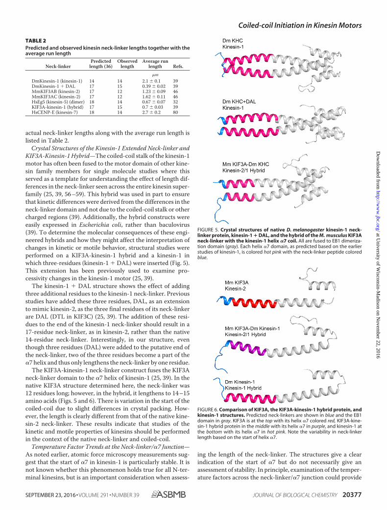
Crystal Structures of the Kinesin-1 Extended Neck-linker andKIF3A-Kinesin-1 Hybrid—The coiled-coil stalk of the kinesin-1motor has often been fused to the motor domain of other kine-sin family members for single molecule studies where thisserved as a template for understanding the effect of length dif-ferences in the neck-linker seen across the entire kinesin super-family (25, 39, 56 –59). This hybrid was used in part to ensurethat kinetic differences were derived from the differences in theneck-linker domain and not due to the coiled-coil stalk or othercharged regions (39). Additionally, the hybrid constructs wereeasily expressed in Escherichia coli, rather than baculovirus(39). To determine the molecular consequences of these engi-neered hybrids and how they might affect the interpretation ofchanges in kinetic or motile behavior, structural studies wereperformed on a KIF3A-kinesin-1 hybrid and a kinesin-1 inwhich three-residues (kinesin-1 � DAL) were inserted (Fig. 5).This extension has been previously used to examine pro-cessivity changes in the kinesin-1 motor (25, 39).
The kinesin-1 � DAL structure shows the effect of addingthree additional residues to the kinesin-1 neck-linker. Previousstudies have added these three residues, DAL, as an extensionto mimic kinesin-2, as the three final residues of its neck-linkerare DAL (DTL in KIF3C) (25, 39). The addition of these resi-dues to the end of the kinesin-1 neck-linker should result in a17-residue neck-linker, as in kinesin-2, rather than the native14-residue neck-linker. Interestingly, in our structure, eventhough three residues (DAL) were added to the putative end ofthe neck-linker, two of the three residues become a part of the�7 helix and thus only lengthens the neck-linker by one residue.
The KIF3A-kinesin-1 neck-linker construct fuses the KIF3Aneck-linker domain to the �7 helix of kinesin-1 (25, 39). In thenative KIF3A structure determined here, the neck-linker was12 residues long; however, in the hybrid, it lengthens to 14 –15amino acids (Figs. 5 and 6). There is variation in the start of thecoiled-coil due to slight differences in crystal packing. How-ever, the length is clearly different from that of the native kine-sin-2 neck-linker. These results indicate that studies of thekinetic and motile properties of kinesins should be performedin the context of the native neck-linker and coiled-coil.
Temperature Factor Trends at the Neck-linker/�7 Junction—As noted earlier, atomic force microscopy measurements sug-gest that the start of �7 in kinesin-1 is particularly stable. It isnot known whether this phenomenon holds true for all N-ter-minal kinesins, but is an important consideration when assess-
ing the length of the neck-linker. The structures give a clearindication of the start of �7 but do not necessarily give anassessment of stability. In principle, examination of the temper-ature factors across the neck-linker/�7 junction could provide
TABLE 2Predicted and observed kinesin neck-linker lengths together with theaverage run length
FIGURE 5. Crystal structures of native D. melanogaster kinesin-1 neck-linker protein, kinesin-1 � DAL, and the hybrid of the M. musculus KIF3Aneck-linker with the kinesin-1 helix �7 coil. All are fused to EB1 dimeriza-tion domain (gray). Each helix �7 domain, as predicted based on the earlierstudies of kinesin-1, is colored hot pink with the neck-linker peptide coloredblue.
FIGURE 6. Comparison of KIF3A, the KIF3A-kinesin-1 hybrid protein, andkinesin-1 structures. Predicted neck-linkers are shown in blue and the EB1domain in gray. KIF3A is at the top with its helix �7 colored red, KIF3A-kine-sin-1 hybrid protein in the middle with its helix �7 in purple, and kinesin-1 atthe bottom with its helix �7 in hot pink. Note the variability in neck-linkerlength based on the start of helix �7.
Coiled-coil Initiation in Kinesin Motors
SEPTEMBER 23, 2016 • VOLUME 291 • NUMBER 39 JOURNAL OF BIOLOGICAL CHEMISTRY 20377
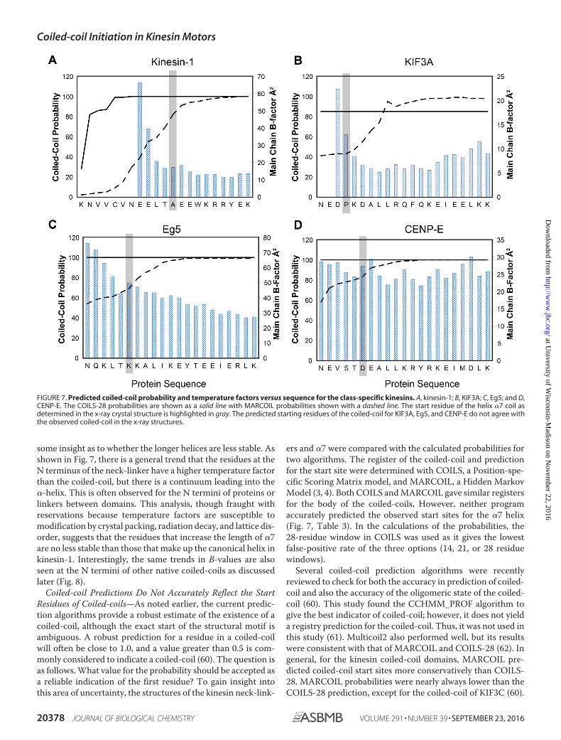
some insight as to whether the longer helices are less stable. Asshown in Fig. 7, there is a general trend that the residues at theN terminus of the neck-linker have a higher temperature factorthan the coiled-coil, but there is a continuum leading into the�-helix. This is often observed for the N termini of proteins orlinkers between domains. This analysis, though fraught withreservations because temperature factors are susceptible tomodification by crystal packing, radiation decay, and lattice dis-order, suggests that the residues that increase the length of �7are no less stable than those that make up the canonical helix inkinesin-1. Interestingly, the same trends in B-values are alsoseen at the N termini of other native coiled-coils as discussedlater (Fig. 8).
Coiled-coil Predictions Do Not Accurately Reflect the StartResidues of Coiled-coils—As noted earlier, the current predic-tion algorithms provide a robust estimate of the existence of acoiled-coil, although the exact start of the structural motif isambiguous. A robust prediction for a residue in a coiled-coilwill often be close to 1.0, and a value greater than 0.5 is com-monly considered to indicate a coiled-coil (60). The question isas follows. What value for the probability should be accepted asa reliable indication of the first residue? To gain insight intothis area of uncertainty, the structures of the kinesin neck-link-
ers and �7 were compared with the calculated probabilities fortwo algorithms. The register of the coiled-coil and predictionfor the start site were determined with COILS, a Position-spe-cific Scoring Matrix model, and MARCOIL, a Hidden MarkovModel (3, 4). Both COILS and MARCOIL gave similar registersfor the body of the coiled-coils, However, neither programaccurately predicted the observed start sites for the �7 helix(Fig. 7, Table 3). In the calculations of the probabilities, the28-residue window in COILS was used as it gives the lowestfalse-positive rate of the three options (14, 21, or 28 residuewindows).
Several coiled-coil prediction algorithms were recentlyreviewed to check for both the accuracy in prediction of coiled-coil and also the accuracy of the oligomeric state of the coiled-coil (60). This study found the CCHMM_PROF algorithm togive the best indicator of coiled-coil; however, it does not yielda registry prediction for the coiled-coil. Thus, it was not used inthis study (61). Multicoil2 also performed well, but its resultswere consistent with that of MARCOIL and COILS-28 (62). Ingeneral, for the kinesin coiled-coil domains, MARCOIL pre-dicted coiled-coil start sites more conservatively than COILS-28. MARCOIL probabilities were nearly always lower than theCOILS-28 prediction, except for the coiled-coil of KIF3C (60).
FIGURE 7. Predicted coiled-coil probability and temperature factors versus sequence for the class-specific kinesins. A, kinesin-1; B, KIF3A; C, Eg5; and D,CENP-E. The COILS-28 probabilities are shown as a solid line with MARCOIL probabilities shown with a dashed line. The start residue of the helix �7 coil asdetermined in the x-ray crystal structure is highlighted in gray. The predicted starting residues of the coiled-coil for KIF3A, Eg5, and CENP-E do not agree withthe observed coiled-coil in the x-ray structures.
Coiled-coil Initiation in Kinesin Motors
20378 JOURNAL OF BIOLOGICAL CHEMISTRY VOLUME 291 • NUMBER 39 • SEPTEMBER 23, 2016
Both programs poorly predicted the KIF3C coiled-coil.MARCOIL yielded a probability of 0.09 for the propensity ofPro-377 to form a coiled-coil, and COILS predicted a probabil-ity of 0.06 for the 28-residue window.
For kinesin-1, where the structure was previously known, thealgorithms differ in the coiled-coil probabilities. Ala-345 is the�7 helix start. MARCOIL gives a conservative probability of0.82, although COILS reaches a probability of 1, five residuesearlier in the sequence where there is no coiled-coil. COILStended to over-predict the neck-linker coiled-coil, reachinghigh probabilities earlier in the sequence. MARCOIL is a better
estimator, but its predictions for the coiled-coil start sitesranged from 0.09 to 0.83. Neither program yielded reliable pre-dictions for the start site of coiled-coils. The variation betweenpredictive approaches creates a dilemma for deciding thecoiled-coil start site based on bioinformatics approaches.Indeed, structural results reveal a fundamental weakness inthe prediction algorithms because they are unable to cate-gorically indicate the first residue that will adopt the �-hel-ical conformation.
To see whether this problem of predicting the coiled-coilstart sites in kinesins is a general phenomenon, structures of
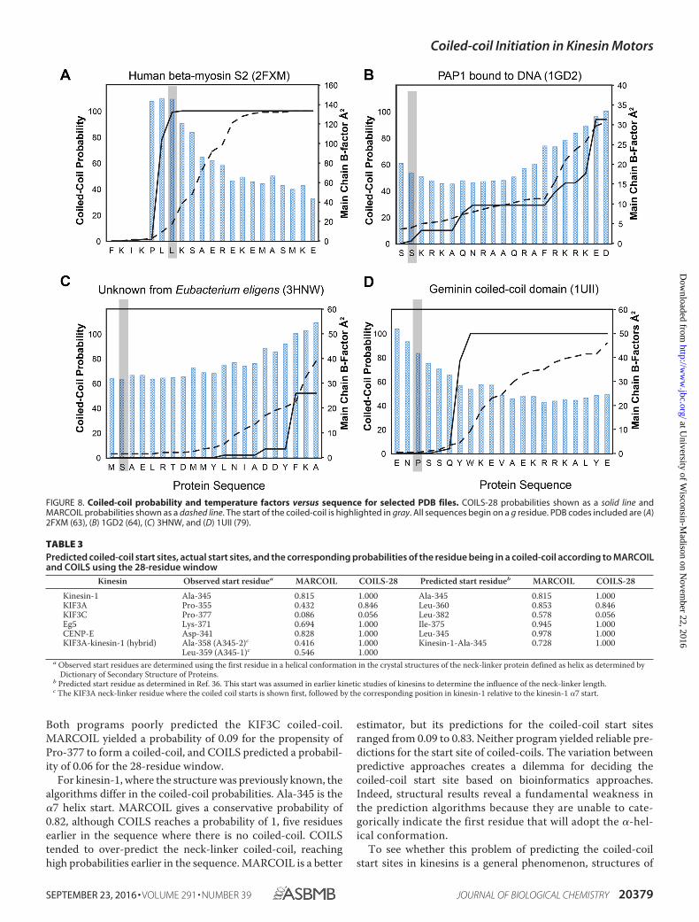
FIGURE 8. Coiled-coil probability and temperature factors versus sequence for selected PDB files. COILS-28 probabilities shown as a solid line andMARCOIL probabilities shown as a dashed line. The start of the coiled-coil is highlighted in gray. All sequences begin on a g residue. PDB codes included are (A)2FXM (63), (B) 1GD2 (64), (C) 3HNW, and (D) 1UII (79).
TABLE 3Predicted coiled-coil start sites, actual start sites, and the corresponding probabilities of the residue being in a coiled-coil according to MARCOILand COILS using the 28-residue window
Leu-359 (A345-1)c 0.546 1.000a Observed start residues are determined using the first residue in a helical conformation in the crystal structures of the neck-linker protein defined as helix as determined by
Dictionary of Secondary Structure of Proteins.b Predicted start residue as determined in Ref. 36. This start was assumed in earlier kinetic studies of kinesins to determine the influence of the neck-linker length.c The KIF3A neck-linker residue where the coiled coil starts is shown first, followed by the corresponding position in kinesin-1 relative to the kinesin-1 �7 start.
Coiled-coil Initiation in Kinesin Motors
SEPTEMBER 23, 2016 • VOLUME 291 • NUMBER 39 JOURNAL OF BIOLOGICAL CHEMISTRY 20379
coiled-coil proteins in the PDB that contain a native transitionfrom random-coil to parallel coiled-coil were also examined (Fig. 8and Table 4). Although there are a large number of structures ofdimeric coiled-coils in the Protein Data Bank, there are only abouta dozen that contain the native start sequence. Most structures inthe PDB represent fragments of a larger protein or are fused to thecanonical coiled-coil found in GCN4. Interestingly, the perfor-mance of the algorithms on this restricted set was similar to thatobserved for the kinesin neck-linker proteins.
In almost every case, the algorithms miss the start site of thecoiled-coil. As with the neck-linker proteins, COILS-28 over-predicts the propensity for coiled-coil, and MARCOIL is muchmore conservative. Neither algorithm accurately predicted thecoiled-coil start, often only reaching a reasonable probability ofcoiled-coil formation until 10 or more residues after the struc-turally observed start site.
A notable exception is 2FXM, the N-terminal region of the S2fragment of cardiac �-myosin II (63). COILS-28 reaches a prob-ability of 99% for the start of the coiled-coil to begin at thesite corresponding with the coiled-coil start in the structure.MARCOIL is close, but still under-predicts the probability of acoiled-coil in that region.
In 1GD2, a structure of the bZIP transcription factor, bothCOILS and MARCOIL under-predict the possibility of acoiled-coil and do not reach an �90% probability until17 residues later in the sequence, corresponding to �2.5heptads of coiled-coil missed (64). Results are similar for3HNW, a coiled-coil protein from Eubacterium eligens withunknown function. Neither COILS nor MARCOIL reaches areasonable coiled-coil prediction until more than three hep-tads into the coiled-coil domain. Overall, it is clear that algo-rithms can predict the presence and the register of a coiled-coil but do not provide an accurate guide to the start of thehelical structure.
Discussion
In this study, the crystal structures of seven kinesin neck-linker �7 helices were determined from four different kinesinfamilies representing N-terminal kinesin motors. In thesestructures, there are differences in the length of the neck-linkerand the start of the coiled-coil stalk as compared with previouspredictions. These differences are most likely the result of inac-curate assumptions in the prediction of coiled-coil start sites.The coiled-coil algorithms, COILS-28 and MARCOIL, werefurther investigated with respect to start site accuracy forother non-kinesin proteins and were shown to be unable topredict the start sites for the majority of coiled-coil-contain-ing structures.
In the kinesin neck-linker structures, the coiled-coil start sitewas 4 –5 residues earlier in the sequence than was predicted.The original assignment of neck-linker length by Hariharanand Hancock in 2009 (36) was based upon the assumption thatthe �7 helix would begin on an a or d residue in the coiled-coilregistry, and this was true for the kinesin-1 motors. However,the choice of this residue registry was arbitrary and as shown bythe results in this paper does not universally apply. KIF3A andKIF3C both begin on a c position, CENP-E and Eg5 begin on dpositions, and the KIF3A-kinesin-1 hybrid begins on an f posi-tion. The addition of the DAL to the kinesin-1 neck-linkerchanges the start site from an a position to an f position. Of thenon-kinesin proteins also examined, the most common startresidue was the d position consistent with the assumption of a dposition beginning the coiled-coil. The varying start sites showthat most positions in the heptad are utilized for the start site ofa coiled-coil domain.
Numerous studies have shown differences in the neck-linkerlength can drastically alter the single molecular parameters (25,
TABLE 4COILS-28 and MARCOIL prediction for structures of coiled-coil con-taining proteins that contain a native start sequenceAll sequences are in the same register as listed in the top chart. The start residues foreach coiled-coil as determined by Dictionary of Secondary Structure of Proteins andthe PDB structure are shaded and underlined. Probabilities (�100) as predicted byCOILS-28 and MARCOIL are shown successively below each amino acid sequence.References for each PDB if available are included below the PDB code.
Coiled-coil Initiation in Kinesin Motors
20380 JOURNAL OF BIOLOGICAL CHEMISTRY VOLUME 291 • NUMBER 39 • SEPTEMBER 23, 2016
31, 37, 39, 65). However, our study suggests that rather than adirect association of shorter neck-linkers leading to greater pro-cessivity, it may be that neck-linker length is tuned to its motorand relative changes in length can increase or decrease theprocessivity.
Many studies have inserted residues in or deleted residuesfrom the presumptive end point of the neck-linker where it wasassumed that the additional residues would add to the neck-linker and have no effect on the coiled-coil (25, 39). As shownin this study, inserted residues can be incorporated directlyinto the �7 helix, rather than increasing the length of theneck-linker. Even when there are added residues at theappropriate end of the neck-linker, as in kinesin-1, the resi-dues may be incorporated into the coiled-coil domain, ratherthan extending the neck-linker. It is possible that the addedresidues may disrupt coiled-coil formation because thecoiled-coil motif depends on specific residues at each posi-tion to fulfill the canonical knobs-into-holes packing. Thus,the disruption of coiled-coil formation, rather than directaltering of neck-linker length, could be leading to artificiallengthening of the neck-linker. The changes in coiled-coilformation may also underlie the changes in processivity seenin other studies (25, 57, 58).
Furthermore, we have shown that fusing the �7 helix fromkinesin-1 to kinesin-2 results in changes in the true neck-linkerlength. This accounts for the observed difference in kineticand motile activities between synthetic and native fusions. Ina study where the KIF3A motor domain and neck-linkerwere fused to the kinesin-1 �7 helix, the average run lengthwas 0.7 �m, although a separate study showed the run lengthwas nearly 50% longer at 1 �m when using the native coiled-coil (39, 46, 65). In contrast, the speed of the KIF3A-kine-sin-1 construct, 480 nm/s, was nearly twice as fast as thenative, 240 nm/s, showing that there are clear alterations inkinetic properties when proteins are fused to differentcoiled-coil domains (39, 46).
Our study also shows that although the coiled-coil predictionalgorithms, COILS and MARCOIL, are able to predict both theoccurrence of a coiled-coil accurately and the registry, bothalgorithms struggle to identify the start site of a coiled-coil. Theprobabilities given by both MARCOIL and COILS cannot berelied upon to yield the correct initiation point because there isno a priori way to decide which probability should be chosen toindicate the absolute start of the helical conformation. Thus,when evaluating sequences through bioinformatics, it shouldbe understood that there is considerable ambiguity in where thecoiled-coil starts. This might not be important for many pro-teins that include coiled-coils, but it is critical when the junc-tion between globular domains and coiled-coils plays a role infunction, as seen in motor proteins.
Kinesin neck-linker domains are clearly important for pro-cessivity, but it is not as simple as a short neck-linker leading toincreased processivity. Further studies must consider thelength of the neck-linkers carefully and ensure that when muta-tions or substitutions are made for kinetic studies the con-structs are altered in the way that was intended.
Experimental Procedures
Construct Preparation—Mus musculus KIF3A cDNA was agenerous gift from William O. Hancock (Pennsylvania StateUniversity, University Park, PA). M. musculus KIF3C cDNAwas synthesized by Open Biosystems (GE Healthcare, Lafa-yette, CO). Homo sapiens Eg5 was obtained from an expressionplasmid containing Eg5(1–513) (66). H. sapiens CENP-E wasobtained from an expression plasmid containing CENP-E(1–407) (67). Drosophila melanogaster kinesin-1 neck-linker wassynthesized by Integrated DNA Technologies (Coralville, IA).Kinesin-1 neck-linker mutagenesis was accomplished througha QuikChange-like protocol to introduce gatgcgctg for the�DAL mutation and to mutate cysteine 338 to alanine.
Neck-linker proteins were cloned using a modified pET-31vector (Novagen) containing an N-terminal 8-histidine taglinked to the protein via a tobacco etch virus protease site and aC-terminal EB1 protein used as a coiled-coil fusion. EB1 is acoiled-coil protein with a C-terminal globular domain used toimprove crystallization and expression of coiled-coil-contain-ing proteins (46, 51, 68, 69). Great care was taken to maintainthe coiled-coil registration across the fusion boundary and toavoid conflicts between structurally adjacent residues (68). Acomplete description of all constructs is included in Table 5.Cloning was accomplished using a protocol similar to theQuikChange method (Agilent) as described previously (68).Briefly, the QuikChange method allows genes to be insertedinto vectors via linear amplification using PfuUltra II Fusion HFpolymerase (Agilent), avoiding the introduction of cloning arti-facts and resulting in faster preparation of constructs (70, 71).The sequences of the constructs and coiled-coil registry aredetailed in Table 5.
Neck-linker proteins were expressed in an E. coli BL21-CodonPlus (DE3)-RIL strain (Agilent). For natively expressedneck-linker proteins, 6 liters of Lysogeny Broth culture wereinoculated with an overnight culture formed from a single col-ony and allowed to grow to an A600 between 0.6 and 1.0 at 37 °C.Upon reaching the appropriate A600, the cells were chilled to16 °C, induced with 0.5 mM isopropyl �-D-1-thiogalactopyra-noside, and grown at 16 °C for 18 h before harvesting via cen-trifugation. For production of selenomethionine-derived pro-teins, 6 liters of M9 media were inoculated with 50 ml per 500ml of overnight culture. Cells were grown to an A600 between0.6 and 1.0 at 37 °C. Upon reaching the appropriate A600, thecells were chilled to 16 °C, and 5 ml of an amino acid mixture(100 mg of lysine, 100 mg of threonine, 100 mg of phenylala-nine, 50 mg of leucine, 50 mg of isoleucine, 50 mg of valine, 50mg of selenomethionine per 30 ml of mixture) was added. Cellswere grown with shaking at 16 °C for 30 min before inductionwith 1 mM isopropyl �-D-1-thiogalactopyranoside. Cells weregrown for 24 h before harvesting via centrifugation.
Protein Purification—All purification steps occurred at 4 °C.Ten g of cell paste were lysed via sonication in 100 ml of lysisbuffer (20 mM Tris-HCl, pH 8.0, 300 mM NaCl, 0.1 mM EGTA,0.2 mM tris(2-carboxyethyl)phosphine (TCEP), 30 mM
imidazole) with 1 mM phenylmethylsulfonyl fluoride, 50 nM
yl)benzenesulfonyl fluoride (Gold BioTechnology). The lysatewas clarified via centrifugation at 125,000 � g for 30 min at 4 °C.The supernatant was loaded onto a 7-ml nickel-nitrilotriaceticacid (Qiagen) column at 1 ml/min and washed with 20 columnvolumes of lysis buffer. Protein was eluted in 4 column volumesof elution buffer (lysis buffer with 200 mM imidazole). The octa-histidine tag was cleaved using a 1:100 molar ratio of a recom-binant tobacco etch virus protease and was dialyzed againstlysis buffer without imidazole (72). After overnight digestion at4 °C, the octa-histidine tag was removed via a second 7-ml nickel-nitrilotriacetic acid column. Protein was loaded at 1 ml/minand washed with 3 column volumes of buffer (20 mM Tris-HCl,pH 8.0, 300 mM NaCl, 0.1 mM EGTA, 0.2 mM TCEP, 30 mM
imidazole), followed by 3 column volumes of lysis buffer. Theprotein was concentrated using an Amicon Ultra-15 Centrifu-gal Filter Unit with Ultracel-10 membrane (Millipore). Theconcentrated protein was dialyzed against 10 mM Tris-HCl, pH8.0, 100 mM NaCl, 0.1 mM EGTA, 0.2 mM TCEP and flash-frozen in 30-�l droplets in liquid nitrogen and stored at �80 °Cprior to crystallization.
Crystallization—Kinesin-1 neck-linker crystals were grownat 20 °C via vapor diffusion from a 1:1 mixture of 15 mg/mlprotein solution and 20% (w/v) methoxy polyethylene glycol(MEPEG) 5000, 100 mM Li2SO4, 100 mM MES/acetate, pH 5.5,10 mM �-caprolactone. Kinesin-1 crystals appeared spontane-ously after 1–2 days. Crystals were layered plates reaching max-imal dimensions of 200 � 200 � 25 �m. Crystals were flash-frozen in liquid nitrogen in synthetic mother liquor (20% (w/v)MEPEG 5000, 100 mM Li2SO4, 100 mM MES/acetate, pH 5.5, 10mM �-caprolactone) supplemented with 10% ethylene glycol.
Kinesin-1 � DAL crystals were grown at 20 °C via vapor dif-fusion from a 1:1 mixture of 15 mg/ml protein solution and 100mM sodium acetate, pH 5.0, 24% (w/v) polyethylene glycol(PEG) 400, 80 mM MgCl2, 3 mM ZnSO4. Drops were streak-seeded after 24 h, and cube-like crystals grew to a maximum
size of 100 � 100 �100 �m after 1 week. Crystals were flash-frozen in liquid nitrogen with synthetic mother liquor (100 mM
sodium acetate, pH 5.0, 24% (w/v) polyethylene glycol (PEG)400, 80 mM MgCl2, 3 mM ZnSO4) supplemented with 8% (w/v)ethylene glycol.
KIF3A crystals were grown via vapor diffusion from a 1:1mixture of 15 mg/ml protein solution at 20 °C in 24% (w/v)2-methyl-2,4-pentanediol, 10% (w/v) PEG 4000, 100 mM CaCl2,100 mM MOPS, pH 7.0, 1 mM CdCl2. Drops were streak-seededafter 24 h, and rod-shaped crystals reached a maximum size of200 � 20 � 20 �m within 2 days and were flash-frozen for datacollection directly from the drop.
KIF3A-kinesin-1 hybrid (KIF3A-KHC) crystals were grownat 20 °C via vapor diffusion from a 1:1 mixture of 15 mg/mlprotein solution in 32% (w/v) polyethylene glycol dimethylether 500, 30 mM MgCl2, 10 mM diethylenetriamine, 100 mM
sodium 3-[4-(2-hydroxyethyl)-1-piperazinyl]propanesulfonicacid, pH 8.5. Drops were streak-seeded after 24 h, and rod-shaped crystals reached a maximum size of 350 � 20 � 20 �mwithin 2 days and were flash-frozen in liquid nitrogen for datacollection directly from the drop.
KIF3C crystals were grown via vapor diffusion from a 1:1mixture of 15 mg/ml protein solution at 20 °C in 30% (w/v)pentaerythritol ethoxylate (PEE) 797, 1.5% (w/v) ethylene glycolmonoethyl ether, 400 mM MgCl2, 100 mM BTP, pH 9.0. Dropswere streak-seeded after 24 h, and rod-shaped crystals reacheda maximum size of 200 � 30 � 30 �m. For data collection,crystals were flash-frozen in liquid nitrogen directly from thedrop.
Eg5 crystals were grown at 4 °C via vapor diffusion from a 1:1mixture of 15 mg/ml protein solution in 24% (w/v) PEE 797, 100mM CaCl2, 100 mM MOPS, pH 7.0, 0.1% (w/v) octyl glucoside.Crystals were streak-seeded after 24 h and cube-shaped crystalsreached maximum dimensions of 100 � 100 � 100 �m after 3days. Crystals were flash-frozen in liquid nitrogen in synthetic
TABLE 5Neck-linker construct sequences and coiled-coil registryThe neck-linker and �7 helices are shown in black text; His8 tags are shown in purple; the recombinant tobacco etch virus protease recognition site and linker are shownin red, and EB1 is depicted in blue.
a The native kinesin-1 neck-linker includes a cysteine at residue 338. Initial structural studies of this construct yielded a structure that included a spurious cross-link betweenadjacent chains. This problem was solved by mutating this residue (underlined) to an alanine to avoid this crystallization artifact.
Coiled-coil Initiation in Kinesin Motors
20382 JOURNAL OF BIOLOGICAL CHEMISTRY VOLUME 291 • NUMBER 39 • SEPTEMBER 23, 2016
The CENP-E selenomethionine crystals were grown viavapor diffusion from a 1:1 mixture of 10 mg/ml protein solutionat 20 °C in 18% (w/v) MEPEG 2000, 175 mM Li2SO4, 100 mM
MES, pH 6.0, 1 mM CdCl2. Drops were streak-seeded after 24 h,and hexagonal crystals reached maximum dimensions of 100 �100 � 100 �m after 5 days. CENP-E selenomethionine crystalswere flash-frozen in liquid nitrogen for data collection in syn-thetic mother liquor (20 °C in 18% (w/v) MEPEG 2000, 175 mM
Li2SO4, 100 mM MES, pH 6.0, 1 mM CdCl2) supplemented with15% (w/v) ethylene glycol.
Structure Determination—The x-ray diffraction data for allneck-linker structures were collected at the SBC 19-ID beamline at the Advanced Photon Source (Argonne, IL). The datasetswere integrated and scaled with the program HKL2000 (73).The kinesin-1, KIF3A-kinesin1 hybrid, KIF3A, KIF3C, and Eg5neck-linker structures were solved via molecular replacementusing Phaser with PDB structure 1YIB (74, 75). The CENP-Eneck-linker structure was solved independently using sel-enomethionine-containing crystals, where single anomalousdiffraction data were processed using Phaser (75). After theinitial solutions were obtained, structures were refined by iter-ative cycles of manual model building in Coot and refinementwith phenix.refine (76, 77). Data collection and refinement sta-tistics for all structural determinations are given in Table 6.Secondary structure assignment was calculated with the Dic-tionary of Secondary Structure of Proteins Algorithm (50).Structural overlays were done using Superpose (78).
Author Contributions—R. K. P., L. G. P., S. P. G., and I. R. designed theresearch. R. K. P. and L. G. P. performed the research, and R. K. P.,S. P. G., and I. R. analyzed the data and wrote the manuscript.
Acknowledgments—Use of the Structural Biology ID19 and BM19beamlines, Argonne National Laboratory Advanced Photon Source,was supported by the United States Department of Energy, Office ofEnergy Research, under Contract No. W-31-109-ENG-38. We alsothank Dr. Alessandro Senes (University of Wisconsin-Madison) forenthusiastic and helpful discussions.
References1. Crick, F. H. (1953) The packing of �-helices: simple coiled-coils. Acta
Crystallogr. 6, 689 – 6972. Rackham, O. J., Madera, M., Armstrong, C. T., Vincent, T. L., Woolfson,
D. N., and Gough, J. (2010) The evolution and structure prediction ofcoiled coils across all genomes. J. Mol. Biol. 403, 480 – 493
3. Delorenzi, M., and Speed, T. (2002) An HMM model for coiled-coil do-mains and a comparison with PSSM-based predictions. Bioinformatics 18,617– 625
4. Lupas, A., Van Dyke, M., and Stock, J. (1991) Predicting coiled coils fromprotein sequences. Science 252, 1162–1164
5. Parry, D. A., Fraser, R. D., and Squire, J. M. (2008) Fifty years of coiled-coilsand �-helical bundles: a close relationship between sequence and struc-ture. J. Struct. Biol. 163, 258 –269
6. Hirokawa, N., Noda, Y., Tanaka, Y., and Niwa, S. (2009) Kinesin superfam-ily motor proteins and intracellular transport. Nat. Rev. Mol. Cell Biol. 10,682– 696
7. Lawrence, C. J., Dawe, R. K., Christie, K. R., Cleveland, D. W., Dawson,
S. C., Endow, S. A., Goldstein, L. S., Goodson, H. V., Hirokawa, N., How-ard, J., Malmberg, R. L., McIntosh, J. R., Miki, H., Mitchison, T. J., Okada,Y., et al. (2004) A standardized kinesin nomenclature. J. Cell Biol. 167,19 –22
8. Endow, S. A., Kull, F. J., and Liu, H. (2010) Kinesins at a glance. J. Cell Sci.123, 3420 –3424
9. Kull, F. J., Sablin, E. P., Lau, R., Fletterick, R. J., and Vale, R. D. (1996)Crystal structure of the kinesin motor domain reveals a structural similar-ity to myosin. Nature 380, 550 –555
10. Marx, A., Muller, J., and Mandelkow, E. (2005) The structure of microtu-bule motor proteins. Adv. Protein Chem. 71, 299 –344
11. Gumy, L. F., Chew, D. J., Tortosa, E., Katrukha, E. A., Kapitein, L. C.,Tolkovsky, A. M., Hoogenraad, C. C., and Fawcett, J. W. (2013) Thekinesin-2 family member KIF3C regulates microtubule dynamics andis required for axon growth and regeneration. J. Neurosci. 33,11329 –11345
12. Kashina, A. S., Rogers, G. C., and Scholey, J. M. (1997) The bimC family ofkinesins: essential bipolar mitotic motors driving centrosome separation.Biochim. Biophys. Acta 1357, 257–271
13. Kashina, A. S., Baskin, R. J., Cole, D. G., Wedaman, K. P., Saxton, W. M.,and Scholey, J. M. (1996) A bipolar kinesin. Nature 379, 270 –272
14. Kozielski, F., Sack, S., Marx, A., Thormahlen, M., Schonbrunn, E., Biou, V.,Thompson, A., Mandelkow, E. M., and Mandelkow, E. (1997) The crystalstructure of dimeric kinesin and implications for microtubule-dependentmotility. Cell 91, 985–994
15. Muresan, V., Abramson, T., Lyass, A., Winter, D., Porro, E., Hong, F.,Chamberlin, N. L., and Schnapp, B. J. (1998) KIF3C and KIF3A form anovel neuronal heteromeric kinesin that associates with membrane vesi-cles. Mol. Biol. Cell 9, 637– 652
16. Schaar, B. T., Chan, G. K., Maddox, P., Salmon, E. D., and Yen, T. J. (1997)CENP-E function at kinetochores is essential for chromosome alignment.J. Cell Biol. 139, 1373–1382
17. Vale, R. D., Reese, T. S., and Sheetz, M. P. (1985) Identification of a novelforce-generating protein, kinesin, involved in microtubule-based motility.Cell 42, 39 –50
18. Hackney, D. D. (1994) Evidence for alternating head catalysis by kinesinduring microtubule-stimulated ATP hydrolysis. Proc. Natl. Acad. Sci.U. S. A. 91, 6865– 6869
19. Gilbert, S. P., Webb, M. R., Brune, M., and Johnson, K. A. (1995) Pathwayof processive ATP hydrolysis by kinesin. Nature 373, 671– 676
20. Schnitzer, M. J., and Block, S. M. (1997) Kinesin hydrolyses one ATP per8-nm step. Nature 388, 386 –390
21. Asbury, C. L., Fehr, A. N., and Block, S. M. (2003) Kinesin moves by anasymmetric hand-over-hand mechanism. Science 302, 2130 –2134
22. Kaseda, K., Higuchi, H., and Hirose, K. (2003) Alternate fast and slowstepping of a heterodimeric kinesin molecule. Nat. Cell Biol. 5, 1079 –1082
23. Yildiz, A., Tomishige, M., Vale, R. D., and Selvin, P. R. (2004) Kinesin walkshand-over-hand. Science 303, 676 – 678
24. Rice, S., Lin, A. W., Safer, D., Hart, C. L., Naber, N., Carragher, B. O., Cain,S. M., Pechatnikova, E., Wilson-Kubalek, E. M., Whittaker, M., Pate, E.,Cooke, R., Taylor, E. W., Milligan, R. A., and Vale, R. D. (1999) A structuralchange in the kinesin motor protein that drives motility. Nature 402,778 –784
25. Shastry, S., and Hancock, W. O. (2011) Interhead tension determines pro-cessivity across diverse N-terminal kinesins. Proc. Natl. Acad. Sci. U. S. A.108, 16253–16258
26. Muretta, J. M., Jun, Y., Gross, S. P., Major, J., Thomas, D. D., and Rosenfeld,S. S. (2015) The structural kinetics of switch-1 and the neck linker explainthe functions of kinesin-1 and Eg5. Proc. Natl. Acad. Sci. U. S. A. 112,E6606 –E6613
27. Mickolajczyk, K. J., Deffenbaugh, N. C., Arroyo, J. O., Andrecka, J., Kukura,P., and Hancock, W. O. (2015) Kinetics of nucleotide-dependent struc-tural transitions in the kinesin-1 hydrolysis cycle. Proc. Natl. Acad. Sci.U. S. A. 112, E7186 –E7193
28. Hoenger, A., Sack, S., Thormahlen, M., Marx, A., Muller, J., Gross, H., andMandelkow, E. (1998) Image reconstructions of microtubules decoratedwith monomeric and dimeric kinesins: comparison with x-ray structureand implications for motility. J. Cell Biol. 141, 419 – 430
Coiled-coil Initiation in Kinesin Motors
20384 JOURNAL OF BIOLOGICAL CHEMISTRY VOLUME 291 • NUMBER 39 • SEPTEMBER 23, 2016
29. Skiniotis, G., Surrey, T., Altmann, S., Gross, H., Song, Y. H., Mandelkow,E., and Hoenger, A. (2003) Nucleotide-induced conformations in the neckregion of dimeric kinesin. EMBO J. 22, 1518 –1528
30. Dogan, M. Y., Can, S., Cleary, F. B., Purde, V., and Yildiz, A. (2015) Kine-sin’s front head is gated by the backward orientation of its neck linker. CellRep. 10, 1967–1973
31. Yildiz, A., Tomishige, M., Gennerich, A., and Vale, R. D. (2008) Intramo-lecular strain coordinates kinesin stepping behavior along microtubules.Cell 134, 1030 –1041
32. Valentine, M. T., Fordyce, P. M., Krzysiak, T. C., Gilbert, S. P., and Block,S. M. (2006) Individual dimers of the mitotic kinesin motor Eg5 step pro-cessively and support substantial loads in vitro. Nat. Cell Biol. 8, 470 – 476
33. Romberg, L., Pierce, D. W., and Vale, R. D. (1998) Role of the kinesin neckregion in processive microtubule-based motility. J. Cell Biol. 140,1407–1416
34. Thorn, K. S., Ubersax, J. A., and Vale, R. D. (2000) Engineering the pro-cessive run length of the kinesin motor. J. Cell Biol. 151, 1093–1100
35. Tomishige, M., and Vale, R. D. (2000) Controlling kinesin by reversibledisulfide cross-linking: identifying the motility-producing conformationalchange. J. Cell Biol. 151, 1081–1092
36. Hariharan, V., and Hancock, W. O. (2009) Insights into the mechanicalproperties of the kinesin neck linker domain from sequence analysis andmolecular dynamics simulations. Cell Mol. Bioeng. 2, 177–189
37. Andreasson, J. O., Milic, B., Chen, G.-Y., Guydosh, N. R., Hancock, W. O.,and Block, S. M. (2015) Examining kinesin processivity within a generalgating framework. Elife 4, e07403
38. Isojima, H., Iino, R., Niitani, Y., Noji, H., and Tomishige, M. (2016) Directobservation of intermediate states during the stepping motion of kine-sin-1. Nat. Chem. Biol. 12, 290 –297
39. Shastry, S., and Hancock, W. O. (2010) Neck linker length determines thedegree of processivity in kinesin-1 and kinesin-2 motors. Curr. Biol. 20,939 –943
40. Duselder, A., Thiede, C., Schmidt, C. F., and Lakamper, S. (2012) Neck-linker length dependence of processive kinesin-5 motility. J. Mol. Biol.423, 159 –168
41. Aizawa, H., Sekine, Y., Takemura, R., Zhang, Z., Nangaku, M., and Hiro-kawa, N. (1992) Kinesin family in murine central nervous system. J. CellBiol. 119, 1287–1296
42. Kondo, S., Sato-Yoshitake, R., Noda, Y., Aizawa, H., Nakata, T., Matsuura,Y., and Hirokawa, N. (1994) KIF3A is a new microtubule-based antero-grade motor in the nerve axon. J. Cell Biol. 125, 1095–1107
43. Yamazaki, H., Nakata, T., Okada, Y., and Hirokawa, N. (1995) KIF3A/B: aheterodimeric kinesin superfamily protein that works as a microtubuleplus end-directed motor for membrane organelle transport. J. Cell Biol.130, 1387–1399
44. Cole, D. G., Diener, D. R., Himelblau, A. L., Beech, P. L., Fuster, J. C., andRosenbaum, J. L. (1998) Chlamydomonas kinesin-II– dependent intrafla-gellar transport (IFT): IFT particles contain proteins required for ciliaryassembly in Caenorhabditis elegans sensory neurons. J. Cell Biol. 141,993–1008
45. Carpenter, B. S., Barry, R. L., Verhey, K. J., and Allen, B. L. (2015) Theheterotrimeric kinesin-2 complex interacts with and regulates GLI pro-tein function. J. Cell Sci. 128, 1034 –1050
46. Guzik-Lendrum, S., Rank, K. C., Bensel, B. M., Taylor, K. C., Rayment, I.,and Gilbert, S. P. (2015) Kinesin-2 KIF3AC and KIF3AB can drive long-range transport along microtubules. Biophys. J. 109, 1472–1482
47. Albracht, C. D., Rank, K. C., Obrzut, S., Rayment, I., and Gilbert, S. P.(2014) Kinesin-2 KIF3AB exhibits novel ATPase characteristics. J. Biol.Chem. 289, 27836 –27848
48. Zhang, P., Rayment, I., and Gilbert, S. P. (2016) Fast or slow, either headcan start the processive run of kinesin-2 KIF3AC. J. Biol. Chem. 291,4407– 4416
49. Scholey, J. E., Nithianantham, S., Scholey, J. M., and Al-Bassam, J. (2014)Structural basis for the assembly of the mitotic motor kinesin-5 into bi-polar tetramers. eLife 3, e02217
50. Kabsch, W., and Sander, C. (1983) Dictionary of protein secondary struc-ture: pattern recognition of hydrogen-bonded and geometrical features.Biopolymers 22, 2577–2637
51. Frye, J., Klenchin, V. A., and Rayment, I. (2010) Structure of the tropomy-osin overlap complex from chicken smooth muscle: insight into the diver-sity of N-terminal recognition. Biochemistry 49, 4908 – 4920
52. Korkmaz, E. N., Taylor, K. C., Andreas, M. P., Ajay, G., Heinze, N. T., Cui,Q., and Rayment, I. (2016) A composite approach towards a completemodel of the myosin rod. Proteins 84, 172–189
53. Hyeon, C., and Onuchic, J. N. (2007) Mechanical control of the directionalstepping dynamics of the kinesin motor. Proc. Natl. Acad. Sci. U. S. A. 104,17382–17387
54. Hyeon, C., and Onuchic, J. N. (2007) Internal strain regulates the nucleo-tide binding site of the kinesin leading head. Proc. Natl. Acad. Sci. U.S.A.104, 2175–2180
55. Bornschlogl, T., Woehlke, G., and Rief, M. (2009) Single molecule me-chanics of the kinesin neck. Proc. Natl. Acad. Sci. U. S. A. 106, 6992– 6997
56. Arpag, G., Shastry, S., Hancock, W. O., and Tuzel, E. (2014) Transport bypopulations of fast and slow kinesins uncovers novel family-dependent motorcharacteristics important for in vivo function. Biophys. J. 107, 1896–1904
57. Chen, G.-Y., Arginteanu, D. F., and Hancock, W. O. (2015) Processivity ofthe kinesin-2 KIF3A results from rear head gating and not front headgating. J. Biol. Chem. 290, 10274 –10294
58. Chen, Y., and Hancock, W. O. (2015) Kinesin-5 is a microtubule polymer-ase. Nat. Commun. 6, 8160
59. Hoeprich, G. J., Thompson, A. R., McVicker, D. P., Hancock, W. O., andBerger, C. L. (2014) Kinesin’s neck-linker determines its ability to navigateobstacles on the microtubule surface. Biophys. J. 106, 1691–1700
60. Li, C., Ching Han Chang, C., Nagel, J., Porebski, B. T., Hayashida, M.,Akutsu, T., Song, J., and Buckle, A. M. (2016) Critical evaluation of in silicomethods for prediction of coiled-coil domains in proteins. Brief. Bioin-form. 17, 270 –282
61. Bartoli, L., Fariselli, P., Krogh, A., and Casadio, R. (2009) CCHMM_PROF:a HMM-based coiled-coil predictor with evolutionary information. Bioin-formatics 25, 2757–2763
62. Trigg, J., Gutwin, K., Keating, A. E., and Berger, B. (2011) Multicoil2:Predicting coiled coils and their oligomerization states from sequence inthe twilight zone. PLoS ONE 6, e23519
63. Blankenfeldt, W., Thoma, N. H., Wray, J. S., Gautel, M., and Schlichting, I.(2006) Crystal structures of human cardiac �-myosin II S2- provideinsight into the functional role of the S2 subfragment. Proc. Natl. Acad. Sci.U. S. A. 103, 17713–17717
64. Fujii, Y., Shimizu, T., Toda, T., Yanagida, M., and Hakoshima, T. (2000)Structural basis for the diversity of DNA recognition by bZIP transcrip-tion factors. Nat. Struct. Biol. 7, 889 – 893
65. Andreasson, J. O., Shastry, S., Hancock, W. O., and Block, S. M. (2015) Themechanochemical cycle of mammalian kinesin-2 KIF3A/B under load.Curr. Biol. 25, 1166 –1175
66. Krzysiak, T. C., Wendt, T., Sproul, L. R., Tittmann, P., Gross, H., Gilbert,S. P., and Hoenger, A. (2006) A structural model for monastrol inhibitionof dimeric kinesin Eg5. EMBO J. 25, 2263–2273
67. Sardar, H. S., Luczak, V. G., Lopez, M. M., Lister, B. C., and Gilbert, S. P.(2010) Mitotic kinesin CENP-E promotes microtubule plus-end elonga-tion. Curr. Biol. 20, 1648 –1653
68. Klenchin, V. A., Frye, J. J., Jones, M. H., Winey, M., and Rayment, I. (2011)Structure function analysis of the C-terminal domain of CNM67, a corecomponent of the Saccharomyces cerevisiae spindle pole body. J. Biol.Chem. 286, 18240 –18250
69. Taylor, K. C., Buvoli, M., Korkmaz, E. N., Buvoli, A., Zheng, Y., Heinze,N. T., Cui, Q., Leinwand, L. A., and Rayment, I. (2015) Skip residuesmodulate the structural properties of the myosin rod and guide thickfilament assembly. Proc. Natl. Acad. Sci. U.S.A. 112, E3806 –E3815
70. Chen, G. J., Qiu, N., Karrer, C., Caspers, P., and Page, M. G. (2000) Restric-tion site-free insertion of PCR products directionally into vectors. Bio-Techniques 28, 498 –500
71. van den Ent, F., and Lowe, J. (2006) RF cloning: A restriction-free methodfor inserting target genes into plasmids. J. Biochem. Biophys. Methods 67,67–74
72. Blommel, P. G., and Fox, B. G. (2007) A combined approach to improvinglarge-scale production of tobacco etch virus protease. Protein Expr. Purif.55, 53– 68
Coiled-coil Initiation in Kinesin Motors
SEPTEMBER 23, 2016 • VOLUME 291 • NUMBER 39 JOURNAL OF BIOLOGICAL CHEMISTRY 20385
73. Otwinowski, Z., and Minor, W. (1997) Processing of x-ray diffraction datacollected in oscillation mode. Methods Enzymol. 276, 307–326
74. Slep, K. C., Rogers, S. L., Elliott, S. L., Ohkura, H., Kolodziej, P. A., and Vale,R. D. (2005) Structural determinants for EB1-mediated recruitment ofAPC and spectraplakins to the microtubule plus end. J. Cell Biol. 168,587–598
75. McCoy, A. J., Grosse-Kunstleve, R. W., Adams, P. D., Winn, M. D., Sto-roni, L. C., and Read, R. J. (2007) Phaser crystallographic software. J. Appl.Crystallogr. 40, 658 – 674
76. Emsley, P., Lohkamp, B., Scott, W. G., and Cowtan, K. (2010) Features anddevelopment of Coot. Acta Crystallogr. D Biol. Crystallogr. 66, 486 –501
77. Adams, P. D., Afonine, P. V., Bunkoczi, G., Chen, V. B., Davis, I. W., Echols,N., Headd, J. J., Hung, L.-W., Kapral, G. J., Grosse-Kunstleve, R. W., Mc-Coy, A. J., Moriarty, N. W., Oeffner, R., Read, R. J., Richardson, D. C., et al.(2010) PHENIX: a comprehensive Python-based system for macromolec-ular structure solution. Acta Crystallogr. D Biol. Crystallogr. 66, 213–221
78. Maiti, R., Van Domselaar, G. H., Zhang, H., and Wishart, D. S. (2004)SuperPose: a simple server for sophisticated structural superposition. Nu-cleic Acids Res. 32, W590 –W594
79. Saxena, S., Yuan, P., Dhar, S. K., Senga, T., Takeda, D., Robinson, H.,Kornbluth, S., Swaminathan, K., and Dutta, A. (2004) A dimerized coiled-coil domain and an adjoining part of geminin interact with two sites onCdt1 for replication inhibition. Mol. Cell 15, 245–258
80. Gudimchuk, N., Vitre, B., Kim, Y., Kiyatkin, A., Cleveland, D. W.,Ataullakhanov, F. I., and Grishchuk, E. L. (2013) Kinetochore kinesin
CENP-E is a processive bi-directional tracker of dynamic microtubuletips. Nat. Cell Biol. 15, 1079 –1088
81. Bigalke, J. M., Dames, S. A., Blankenfeldt, W., Grzesiek, S., and Geyer, M.(2011) Structure and dynamics of a stabilized coiled-coil domain in theP-TEFb regulator Hexim1. J. Mol. Biol. 414, 639 – 653
82. Cottee, M. A., Muschalik, N., Johnson, S., Leveson, J., Raff, J. W., and Lea,S. M. (2015) The homo-oligomerisation of both Sas-6 and Ana2 is re-quired for efficient centriole assembly in flies. eLife 4, e07236
83. van Breugel, M., Wilcken, R., McLaughlin, S. H., Rutherford, T. J., andJohnson, C. M. (2014) Structure of the SAS-6 cartwheel hub from Leish-mania major. eLife 3, e01812
84. Kitagawa, D., Vakonakis, I., Olieric, N., Hilbert, M., Keller, D., Olieric, V.,Bortfeld, M., Erat, M. C., Fluckiger, I., Gonczy, P., and Steinmetz, M. O.(2011) Structural basis of the 9-fold symmetry of centrioles. Cell 144,364 –375
85. Tarbouriech, N., Curran, J., Ruigrok, R. W., and Burmeister, W. P. (2000)Tetrameric coiled coil domain of Sendai virus phosphoprotein. Nat.Struct. Biol. 7, 777–781
86. Day, C. L., and Alber, T. (2000) Crystal structure of the amino-terminalcoiled-coil domain of the APC tumor suppressor. J. Mol. Biol. 301,147–156
87. Chang, J. F., Hall, B. E., Tanny, J. C., Moazed, D., Filman, D., and Ellen-berger, T. (2003) Structure of the coiled-coil dimerization motif of Sir4and its interaction with Sir3. Structure 11, 637– 649
Coiled-coil Initiation in Kinesin Motors
20386 JOURNAL OF BIOLOGICAL CHEMISTRY VOLUME 291 • NUMBER 39 • SEPTEMBER 23, 2016