101
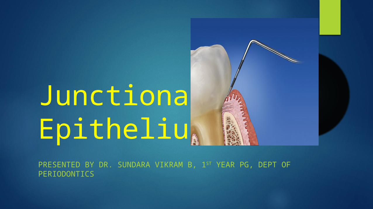
Junctional Epithelium PRESENTED BY DR. SUNDARA VIKRAM B, 1 ST YEAR PG, DEPT OF PERIODONTICS
| Date post: | 15-Feb-2017 |
| Category: |
Health & Medicine |
| Upload: | vikram-buddhanesan |
| View: | 3,352 times |
| Download: | 1 times |
Junctional EpitheliumPRESENTED BY DR. SUNDARA VIKRAM B, 1ST YEAR PG, DEPT OF PERIODONTICS
Introduction:
There are three types of mucous membranes (masticatory, lining, and specialized) line the oral cavity and form the structural boundary between the body and the external environment.
Epithelia exhibit considerable differences in their histology, thickness and differentiation suitable for the functional demands of their location

The gingival epithelium around a tooth is divided into three functional compartments– outer, sulcular, and junctional epithelium
The outer epithelium extends from the mucogingival junction to the gingival margin where crevicular/sulcular epithelium lines the sulcus
At the base of the sulcus connection between gingiva and tooth is mediated with JUNCTIONAL EPITHELIUM

Introduction:
Mucosal epithelia are composed of continuously dividing and shedding populations of keratinocytes.
The junctional epithelium is attached to the tooth surface by a distinct mechanism known as the epithelial attachment apparatus.
Introduction: It is commonly accepted that the junctional epithelium
exhibits several unique structural and functional features that contribute to preventing pathogenic bacterial flora from colonizing the subgingival tooth surface

HISTORICAL ASPECTS
Historical aspects of JE:
Gottlieb (1921) was the first to describe the junctional epithelium
Schroeder and Listgarten (1977) clarified the anatomy and histology of the dentogingival junction in their monograph: ‘Fine structure of developing epithelial attachment of human teeth’.

GOTTLIEB -1921
Epithelial attachment is
organically united to the tooth surface

WAERHAUG -1952
Based on his animal experiments(in dogs) he postulated that the cells of the epithelial attachment adhere weakly to the tooth surface and it forms
the lining of the physiologic pocket
Orban’s concept (1953)
He stated that the separation of the epithelial attachment cells from the tooth surface involved preparatory degenerative changes in the epithelium.
Waerhaug’s concept (1960)
He presented the concept of epithelial cuff. This concept was based on insertion of thin blades between the surface of tooth and the gingiva
Blades could be easily passed apically to the connective tissue attachment at CEJ without resistance.
It was concluded that gingival tissue and tooth are closely adapted but not organically united.
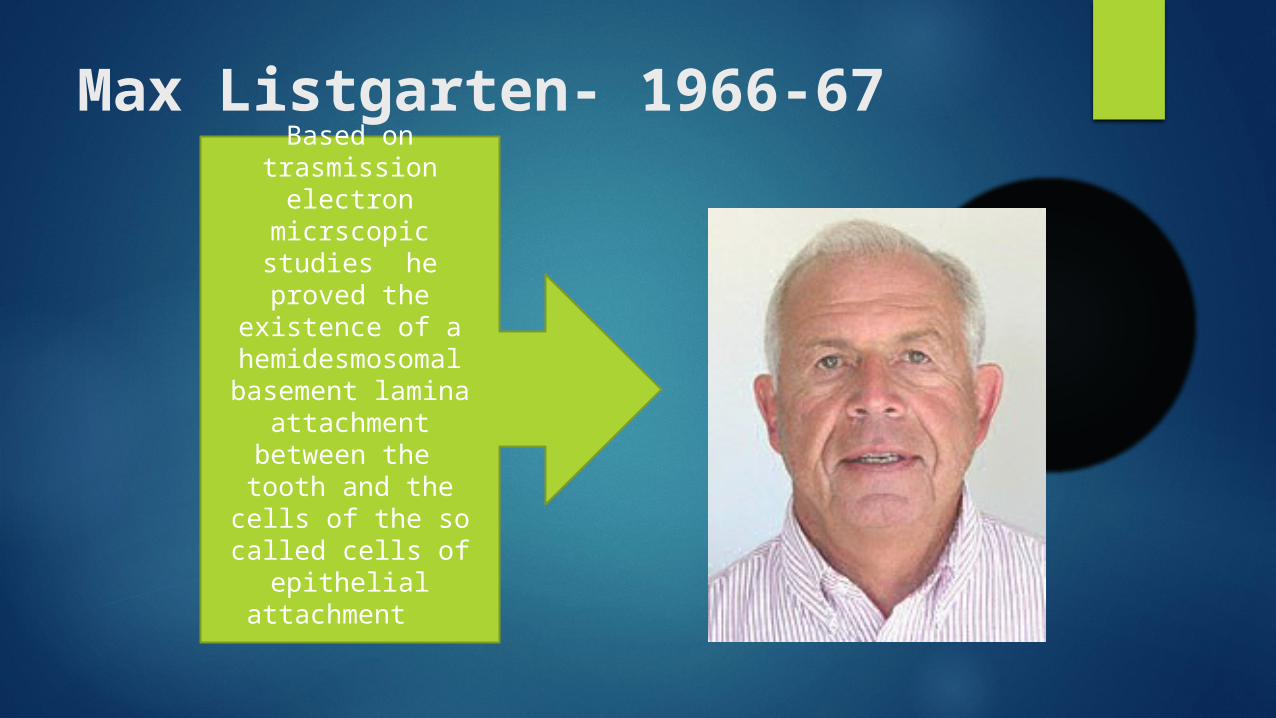
Max Listgarten- 1966-67Based on
trasmission electron micrscopic studies
he proved the existence of a
hemidesmosomal basement lamina
attachment between the tooth and the
cells of the so called cells of epithelial
attachment
Schroeder and Listgarten concept (1971)
The previous controversy was resolved after evolution of transmission electron microscopy.
Primary epithelial attachment refers to the epithelial attachment lamina released by the REE. It lies in direct contact with enamel and epithelial cells attached to it by hemi-desmosomes.
When REE cells transform into JE cells the primary epithelial attachment becomes secondary epithelial attachment . It is made of epithelial attachment between basal lamina and hemi-desmosomes.
Terminologies:
Epithelial attachment – Gottlieb 1921
Epithelial cuff -Waerhaug 1952
Attached epithelial cuff- Orban 1956
Attachment epithelium- Grant, Stern 1968
Junctional epithelium- Anderson and Stern1967

What is Junctional Epithelium?
It is the third (and most intriguing) component of the epithelial integument of the periodontium, in addition to the oral gingival epithelium and the oral sulcular epithelium.
It consists of collar like band of stratified squamous nonkeratinizing epithelium.
What is Junctional Epithelium?
It is 3 to 4 layers thick in early life, but the number of layers increases with age to 10 or even 20 years.
JE tapers from its coronal end, which may be 10 to 29 cells wide to 1 or 2 cells at its apical termination, located at the cemento enamel junction in healthy tissue.

What is Junctional Epithelium?
Those cells are grouped into two strata: THE BASAL LAYER facing the connective tissue and THE SUPRABASAL LAYER extending to the tooth
surface. The length of the JE ranges from 0.25 to 1.35mm.
What is Junctional Epithelium?
It provides the attachment mechanism of the epithelium to the surface of tooth hard substance.
It also provides a protective function relative to the subjacent periodontal ligament.

Definitions for JE:Junctinal epithelium is the non keratinised
stratified squamous epithelium which attaches and form a collar around the cervical portion of
the tooth that follows CEJCarranza’s clinical periodontology
Epithelial attachment is the structural complex by which junctional epithelium is attached to the
tooth surface
What are it’s functions?
First, JE is firmly attached to the tooth and thus forms an epithelial barrier against the plaque bacteria.
Second, it allows the access of GCF, inflammatory cells and components of the immunological host defense to the gingival margin.
Third, JE cells exhibit rapid turnover, which contributes to the host parasite equilibrium and rapid repair of damaged tissue
Genco RJ et al AAP 1996
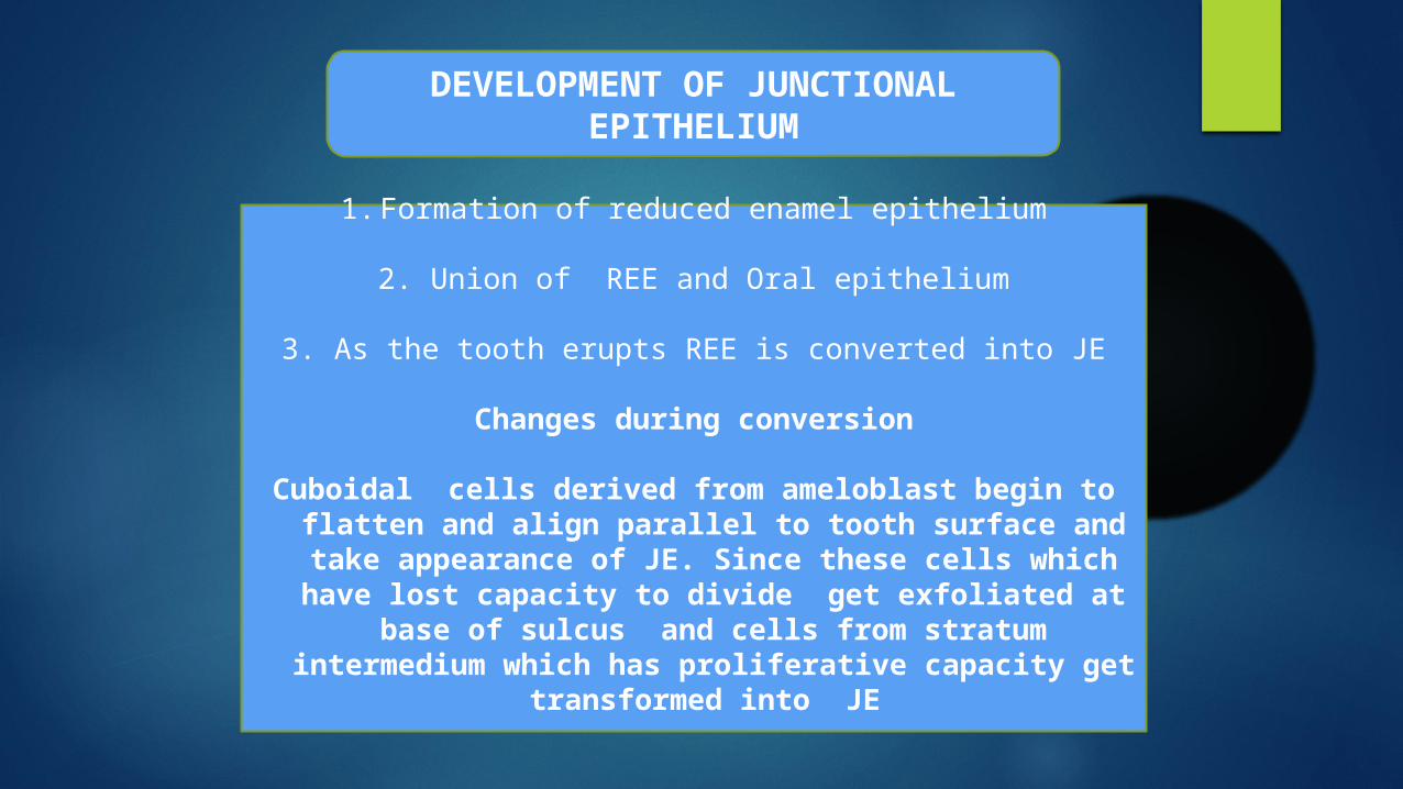
DEVELOPMENT OF JUNCTIONAL EPITHELIUM
1. Formation of reduced enamel epithelium
2. Union of REE and Oral epithelium
3. As the tooth erupts REE is converted into JE
Changes during conversion
Cuboidal cells derived from ameloblast begin to flatten and align parallel to tooth surface and take appearance of JE. Since these cells which have lost capacity to divide get exfoliated at base of sulcus
and cells from stratum intermedium which has proliferative capacity get transformed into JE

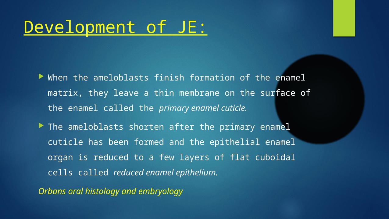
Development of JE:
When the ameloblasts finish formation of the enamel matrix, they leave a thin membrane on the surface of the enamel called the primary enamel cuticle.
The ameloblasts shorten after the primary enamel cuticle has been formed and the epithelial enamel organ is reduced to a few layers of flat cuboidal cells called reduced enamel
epithelium.
Orbans oral histology and embryology
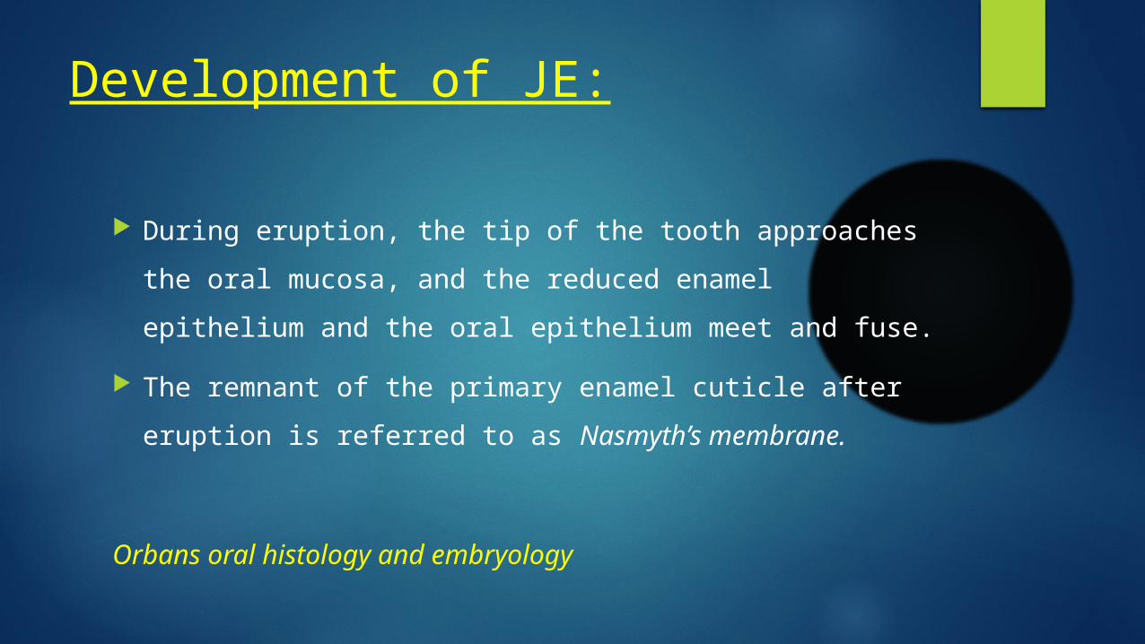
Development of JE:
During eruption, the tip of the tooth approaches the oral mucosa, and the reduced enamel epithelium and the oral epithelium meet and fuse.
The remnant of the primary enamel cuticle after eruption is referred to as Nasmyth’s membrane.
Orbans oral histology and embryology
Development of JE:
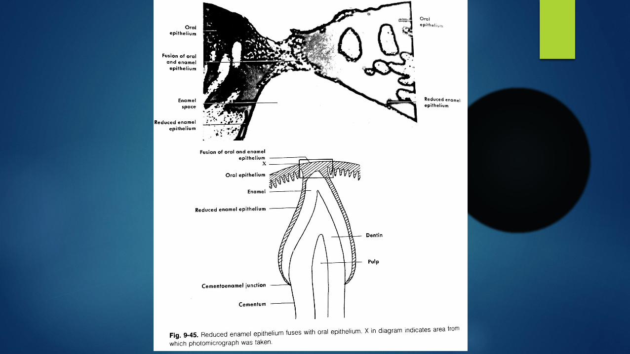
The epithelium that covers the tip of the crown degenerates in its center and the crown emerges through this perforation into the oral cavity.
The REE remains organically attached to the part of the enamel that has not yet erupted. Once the tip of the crown has emerged, the REE is termed as Primary
attachment epithelium (Junctional epithelium).
Development of JE:
At the margin of the gingiva, the junctional epithelium is continuous with the oral epithelium.
As the tooth erupts, the REE grows gradually shorter. Gingival sulcus may develop between the gingiva and
the surface of the tooth and extend around its circumference.

Development of JE:
It is bounded by the JE at its base and by the gingival margin laterally. The gingiva encompassing the sulcus is the free, or marinal gingiva.
Anatomical features:
JE forms a collar peripheral to cervical region of tooth of about 0.75 to 1.35 mm
Interproximally JE of adjacent teeth fuse to form the lining of the col area
Epithelial connective tissue interface is smooth (no rete pegs)
JE is thickest at bottom of sulcus and tapers of in apical direction
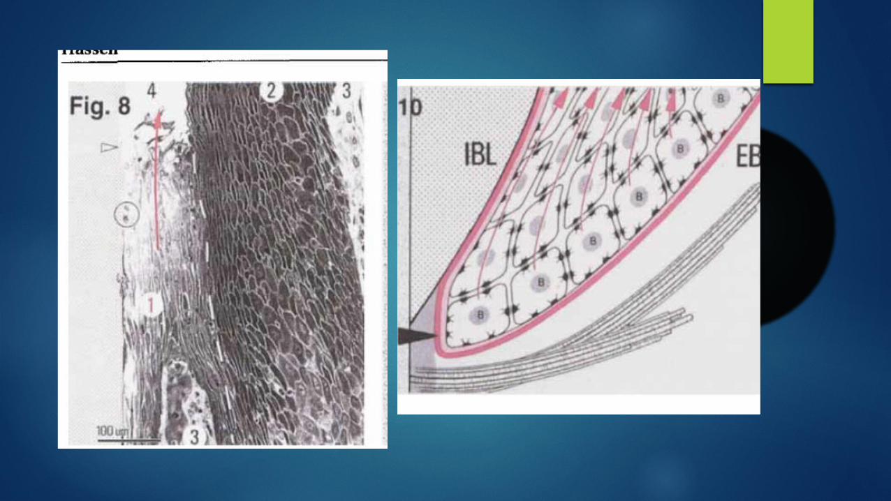
Microscopic features It has 15-30 cell layers coronally and 1-3 layers at apical termination
It has two strata- stratum basale and stratum suprabasale
The basal and adjacent 1-2 suprabasal cells are cuboidal to slightly spindle shaped and all the remaining cells are flat and oriented parallel to the tooth surface
The innermost suprabasal cells(facing the tooth surface) also called DAT cells(Salonen et al 1994) form and maintain the epithelial attachment apparatus
It has two basal lamina – External basal lamina and internal basal lamina

Transmission electron microscopic features:
Lysosomal bodies are in large numbers Golgi fields are large Poly ribosomes are numerous Keratinosomes (Odland bodies) are absent Cytokeratin bundles are scarce Desmosomes are less Inter cellular spaces are more wider and occupied by
inflammatory cells
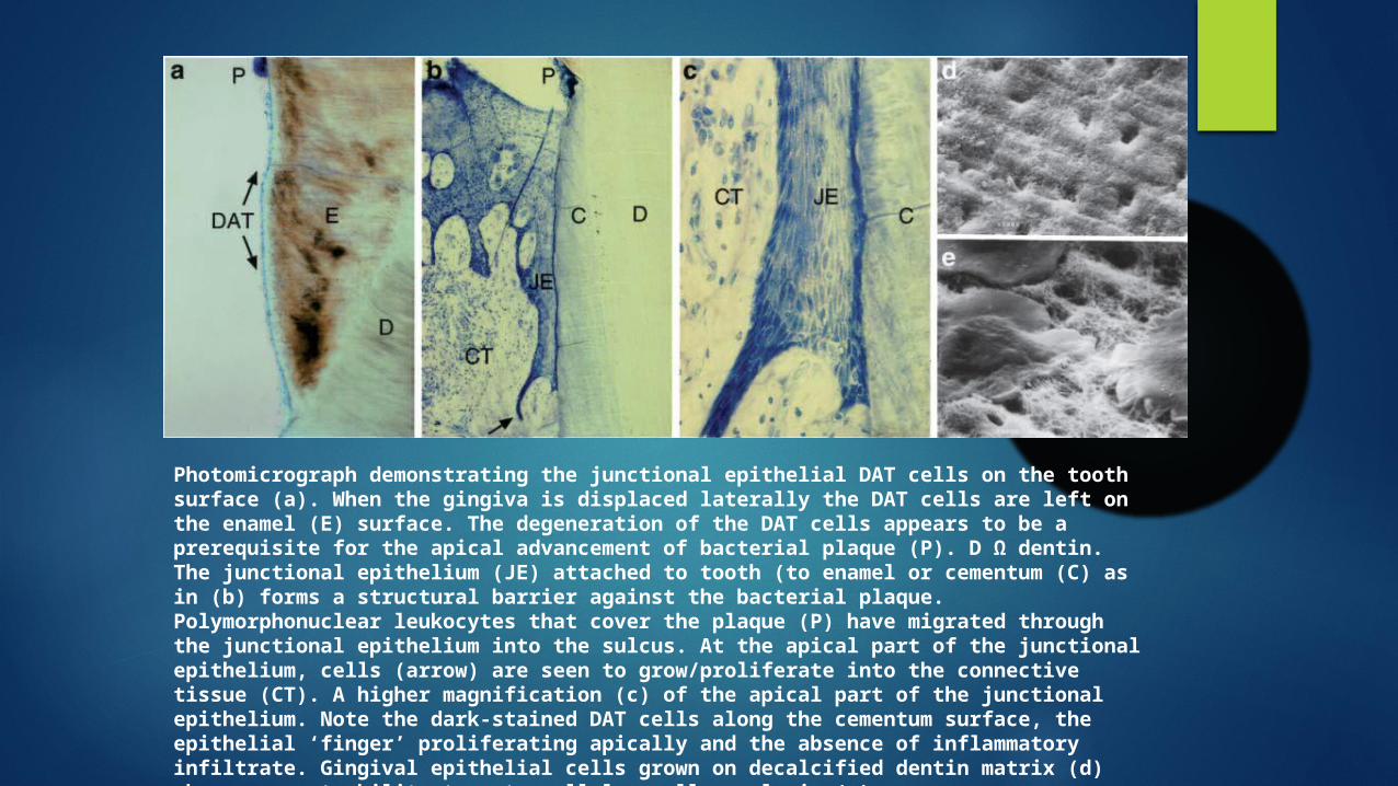
Photomicrograph demonstrating the junctional epithelial DAT cells on the tooth surface (a). When the gingiva is displaced laterally the DAT cells are left on the enamel (E) surface. The degeneration of the DAT cells appears to be a prerequisite for the apical advancement of bacterial plaque (P). D Ω dentin. The junctional epithelium (JE) attached to tooth (to enamel or cementum (C) as in (b) forms a structural barrier against the bacterial plaque. Polymorphonuclear leukocytes that cover the plaque (P) have migrated through the junctional epithelium into the sulcus. At the apical part of the junctional epithelium, cells (arrow) are seen to grow/proliferate into the connective tissue (CT). A higher magnification (c) of the apical part of the junctional epithelium. Note the dark-stained DAT cells along the cementum surface, the epithelial ‘finger’ proliferating apically and the absence of inflammatory infiltrate. Gingival epithelial cells grown on decalcified dentin matrix (d) show apparent ability to extracellular collagenolysis (e)
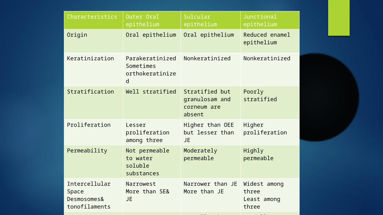
Characteristics Outer Oral epithelium
Sulcular epithelium
Junctional epithelium
Origin Oral epithelium Oral epithelium Reduced enamel epithelium
Keratinization ParakeratinizedSometimes orthokeratinized
Nonkeratinized Nonkeratinized
Stratification Well stratified Stratified but granulosam and corneum are absent
Poorly stratified
Proliferation Lesser proliferation among three
Higher than OEE but lesser than JE
Higher proliferation
Permeability Not permeable to water soluble substances
Moderately permeable
Highly permeable
Intercellular SpaceDesmosomes& tonofilaments
Narrowest More than SE& JE
Narrower than JEMore than JE
Widest among threeLeast among three
Retepegs Present Normally absent, appears in inflammation
Normally absent, appears in inflammation

PRIMARY EPITHELIAL ATTACHMENTAttachment of reduced enamel epithelium to
enamel of unerupted crown
SECONDARY EPITHELIAL ATTACHMENTAfter the conversion of REE to JE the attachment is referred as secondary epithelial attachment
EPITHELIAL ATTACHMENT APPARATUSMediated by hemidesmosomes of DAT (Directly
Attached to Tooth) cells and internal basal lamina
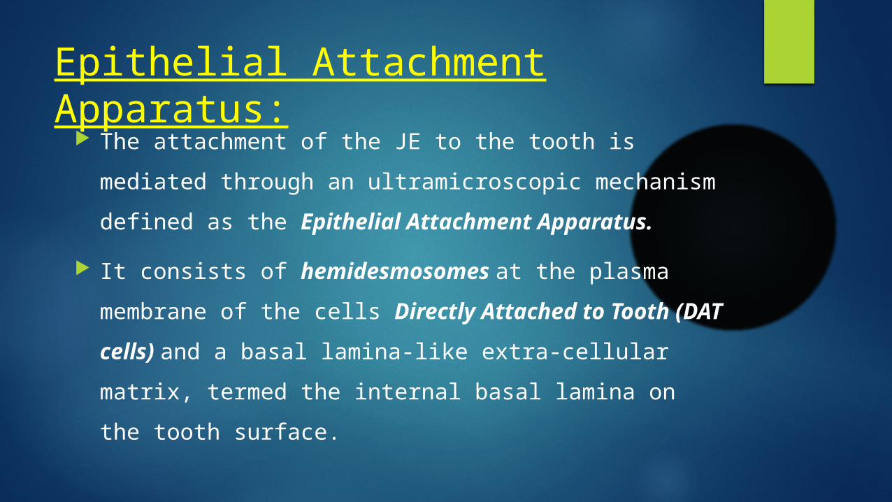
Epithelial Attachment Apparatus: The attachment of the JE to the tooth is mediated
through an ultramicroscopic mechanism defined as the Epithelial Attachment Apparatus.
It consists of hemidesmosomes at the plasma membrane of the cells Directly Attached to Tooth (DAT cells) and a basal lamina-like extra-cellular matrix, termed the internal basal lamina on the tooth surface.
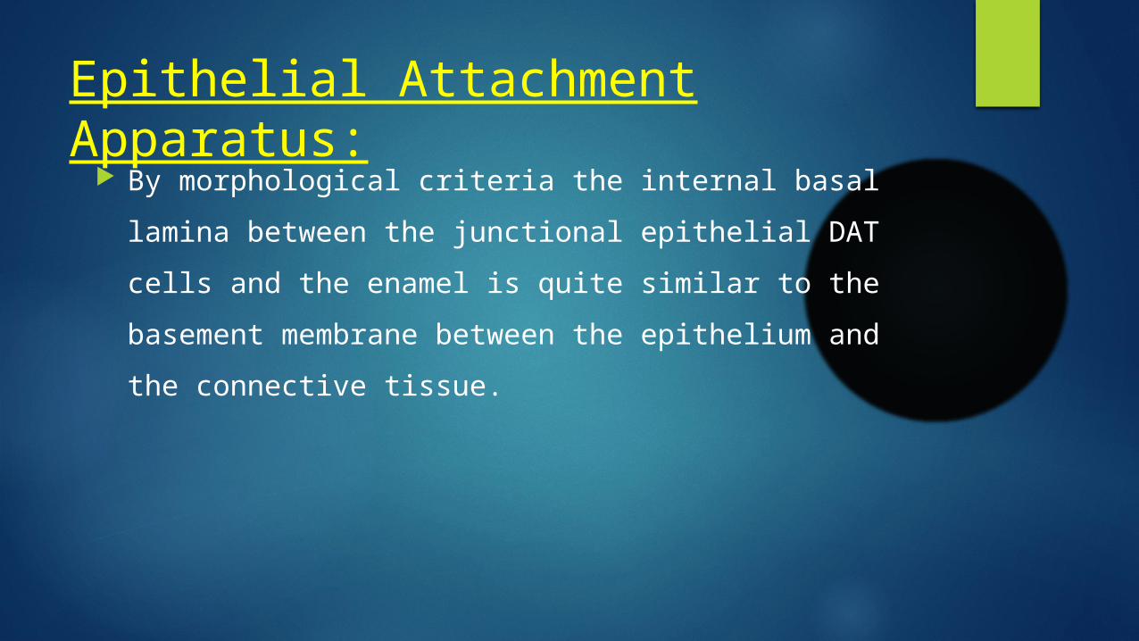
Epithelial Attachment Apparatus: By morphological criteria the internal basal lamina
between the junctional epithelial DAT cells and the enamel is quite similar to the basement membrane between the epithelium and the connective tissue.
Epithelial Attachment Apparatus:
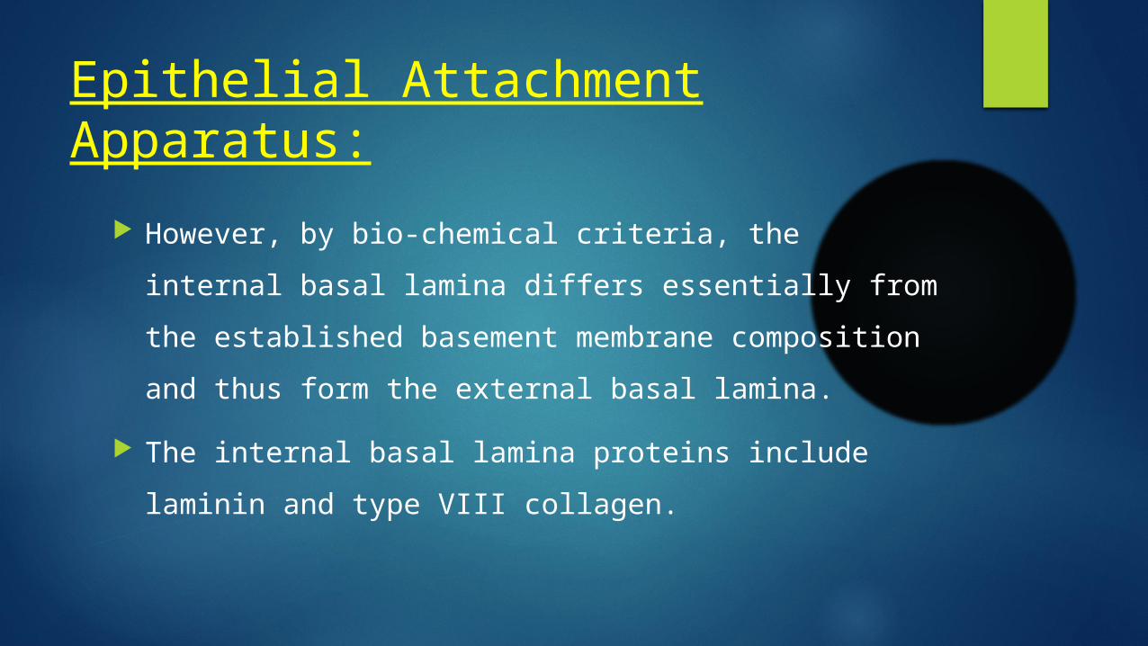
However, by bio-chemical criteria, the internal basal lamina differs essentially from the established basement membrane composition and thus form the external basal lamina.
The internal basal lamina proteins include laminin and type VIII collagen.
Epithelial Attachment Apparatus:
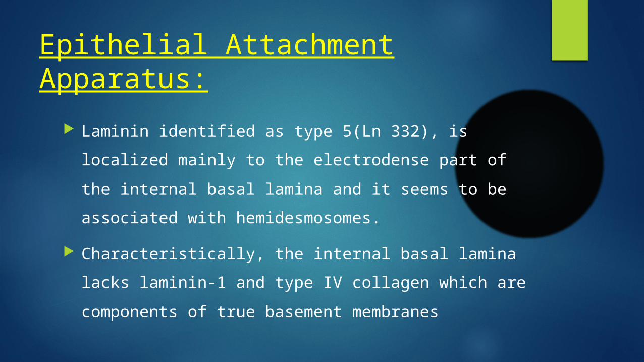
Laminin identified as type 5(Ln 332), is localized mainly to the electrodense part of the internal basal lamina and it seems to be associated with hemidesmosomes.
Characteristically, the internal basal lamina lacks laminin-1 and type IV collagen which are components of true basement membranes
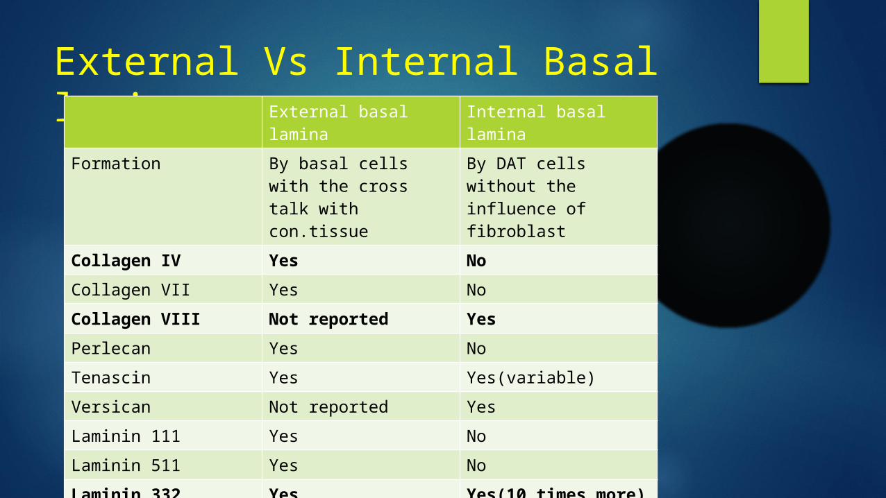
External Vs Internal Basal laminaExternal basal lamina Internal basal lamina
Formation By basal cells with the cross talk with con.tissue
By DAT cells without the influence of fibroblast
Collagen IV Yes NoCollagen VII Yes NoCollagen VIII Not reported YesPerlecan Yes NoTenascin Yes Yes(variable)Versican Not reported YesLaminin 111 Yes NoLaminin 511 Yes NoLaminin 332 (Laminin 5)
Yes Yes(10 times more)
Hemidesmosomes:
Hemidesmosomes have a decisive role in the firm attachment of the cells to the internal basal lamina on the tooth surface.
It may also act as specific sites of signal transduction and thus participate in regulation of gene expression, cell proliferation and cell differentiation.
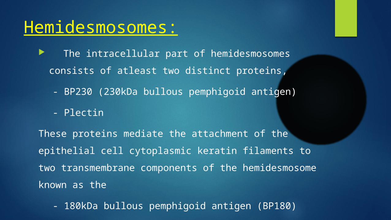
Hemidesmosomes: The intracellular part of hemidesmosomes consists
of atleast two distinct proteins,
- BP230 (230kDa bullous pemphigoid antigen)
- Plectin
These proteins mediate the attachment of the epithelial cell cytoplasmic keratin filaments to two transmembrane components of the hemidesmosome known as the
- 180kDa bullous pemphigoid antigen (BP180)

Hemidesmosomes:
In general, the interaction between the different components of the extracellular matrix and the cell surface molecules linked to the intercellular cytoskeleton is fundamental for cell adhesion, cell motility, synthetic capacity, tissue stability, regeneration and responses to external signals.
Hemidesmosomes:
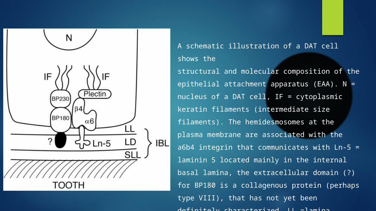
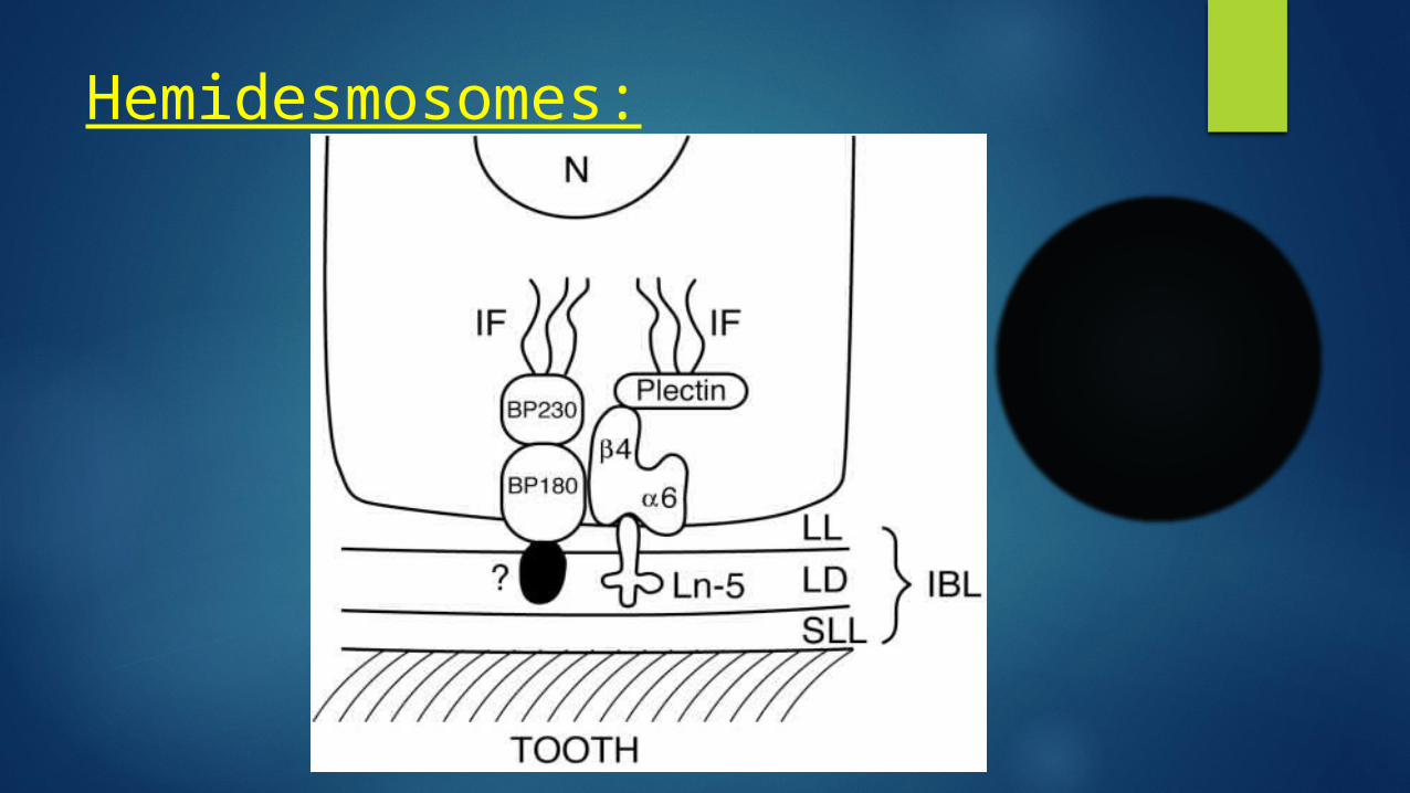
A schematic illustration of a DAT cell shows thestructural and molecular composition of the epithelial attachment apparatus (EAA). N = nucleus of a DAT cell, IF = cytoplasmic keratin filaments (intermediate size filaments). The hemidesmosomes at the plasma membrane are associated with the a6b4 integrin that communicates with Ln-5 = laminin 5 located mainly in the internal basal lamina, the extracellular domain (?) for BP180 is a collagenous protein (perhaps type VIII), that has not yet been definitely characterized. LL =lamina lucida, LD = lamina densa, SLL =sublamina lucida, IBL = internal basal lamina.

Turnover of the JE cells with DAT cells:
The turnover of the junctional epithelium is exceptionally rapid. At the coronal part of the JE, the DAT cells typically express a
high density of transferrin receptors. DAT cells have more important role in tissue dynamics and
reparative capacity of the junctional epithelium than has previously been thought.

Turnover of the JE cells with DAT cells:
Any structural or molecular changes in the internal basal lamina can potentially influence the vital functions of the DAT cells and contribute to the effectiveness or failure of the junctional epithelial defense or vice versa.
Changes in the cell metabolism may affect the Internal Basal Lamina (IBL).
Morphological studies of the internal basal lamina of teeth extracted because of advanced periodontitis have shown that remnants of the internal basal lamina can be detected even adjacent to severely degenerated DAT cells (Overman DO et all)

The mechanism of DAT cell turnover is not fullyunderstood. Considering the fact that the DAT cells are able to divide and migrate, three possible mechanisms can be proposed. These are
(1) the daughter cells producedby dividing DAT cells replace degenerating cells onthe tooth surface,
(2) the daughter cells enter the exfoliationpathway and gradually migrate coronally betweenthe basal cells and the DAT cells to eventually break offinto the sulcus, or
(3) epithelial cells move/migrate in thecoronal direction along the tooth surface and are replaced by basal cells migrating round the apical termination of the junctional epithelium.

JE in the Anti-Microbial Defense: Junctional epithelium consists of active populations of
cells and antimicrobial functions, which together form the first line of defense against microbial invasion into tissue.
Even though junctional epithelial cell layers provide a barrier against bacteria many bacterial substances, such as lipopolysaccharide, pass easily through the epithelium but have only limited access through the external basal lamina into the connective tissue (Shwartz et al 1972).

Rapid turnover, as such, is an important factor in the microbial defense of junctional epithelium.
The area covered by the dividing cells in the junctional epithelium is at least 50 times larger than the area through which the epithelial cells desquamate into the gingival sulcus, there is a strong funnelling effect that contributes to the flow of epithelial cells (Schroder et al 1967).
Rapid shedding and effective removal of bacteria adhering to epithelial cells is therefore an important part of the antimicrobial defense mechanisms at the dentogingival junction.

Role of Enzymes in the Anti-Microbial Defense of JE:
There is increasing evidence indicating that several specific antimicrobial defense systems exist in the oral mucosa.
Junctional epithelium, have been found to contain enzyme-rich lysosomes.
Their fusion with plasma membrane is triggered by elevation of the intracellular calcium concentration (Rodriguez et al 1997)
In rats, the lysosomes have been demonstrated to contain cysteine proteinases (cathepsin B and H) active at acidic pH (Yamaza T et al 1997).
Recently, it has been found that the junctional epithelial cells lateral to DAT cells produce matrilysin (matrix metalloproteinase-7) (Uitto VJ et al 2002).
Matrilysin contributes to the mucosal defense by the release of bioactive molecules from the cell surfaces which play a role in the inflammatory reaction.
Role of Enzymes in the Anti-Microbial Defense of JE:

Several antimicrobial mechanisms exist in thejunctional epithelium. In the coronal part of the junctional epithelium quick cell exfoliation(1)because of rapid cell division
(2)and funnelling of junctional epithelial cells towards the sulcus hinder bacterial colonization. Laterally, the (external) basement membrane forms an effective barrier against invading microbes
(3). Active antimicrobial substances are produced in junctional epithelial cells. These include defensins andlysosomal enzymes
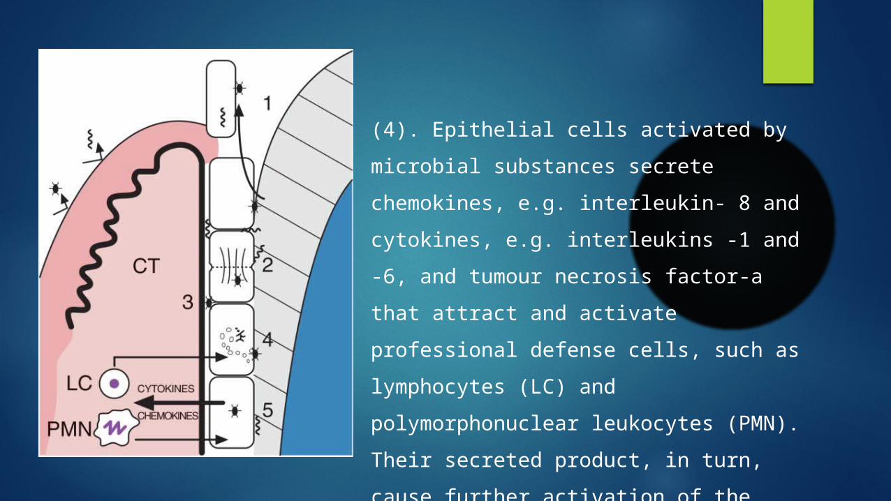
(4). Epithelial cells activated by microbial substances secrete chemokines, e.g. interleukin- 8 and cytokines, e.g. interleukins -1 and -6, and tumour necrosis factor-a that attract and activate professional defense cells, such as lymphocytes (LC) and polymorphonuclear leukocytes (PMN). Their secreted product, in turn,cause further activation of the junctional epithelial cells.

Role of Junctional Epithelium in Disease

Problems in JE:

THE DETACHMENT OF THE DAT CELLSFROM THE TOOTH SURFACE:
Role of the gingival crevice fluid
Role of the polymorphonuclear leukocytes
Role of host proteinases and inflammatory mediators
Role of bacterial products
Role of risk factors for periodontal disease
Role of GCF:
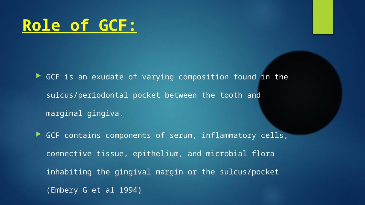
GCF is an exudate of varying composition found in the
sulcus/periodontal pocket between the tooth and marginal
gingiva.
GCF contains components of serum, inflammatory cells,
connective tissue, epithelium, and microbial flora inhabiting the
gingival margin or the sulcus/pocket (Embery G et al 1994)
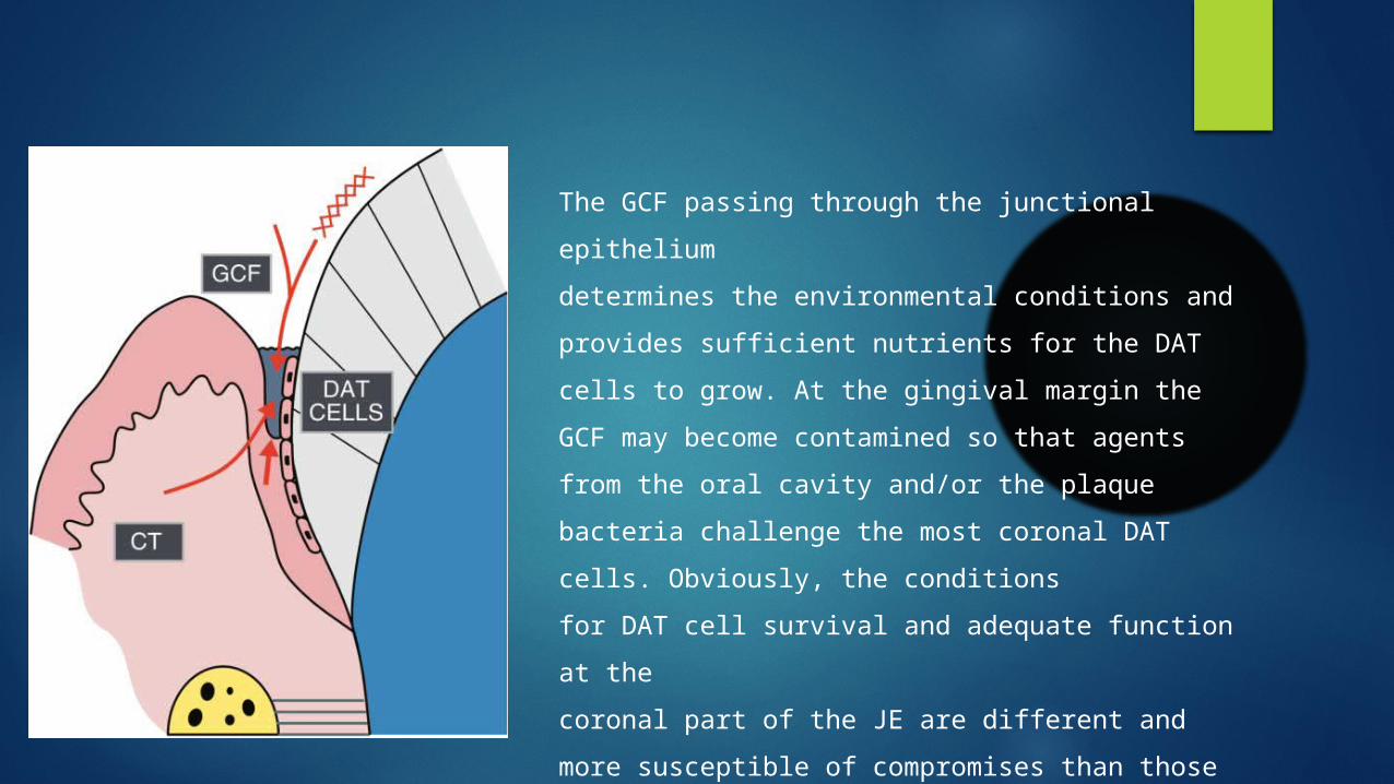
The GCF passing through the junctional epitheliumdetermines the environmental conditions and provides sufficient nutrients for the DAT cells to grow. At the gingival margin the GCF may become contamined so that agents from the oral cavity and/or the plaque bacteria challenge the most coronal DAT cells. Obviously, the conditionsfor DAT cell survival and adequate function at thecoronal part of the JE are different and more susceptible of compromises than those for the basal cells living in the vicinity of the connective tissue (CT) and the blood circulation.

Role of GCF:
In the healthy sulcus the amount of GCF is very small. However,
its constituents participate in the normal maintenance of function
of the junctional epithelium throughout its lateral and vertical
dimensions, including the most coronal DAT cells.
During inflammation the GCF flow increases and its composition
starts to resemble that of an inflammatory exudate (Cimasoni et
al 1983).

Role of GCF:
Although all the junctional epithelial cells are constantly exposed
to the GCF and its various constituents, the nutritional and other
vital conditions in the different parts of the junctional epithelium
depend on a large number of local factors.

Role of GCF Main route of GCF diffusion is
through the EBL and inter cellular spaces
GCF passing through JE gives nutrients to DAT cells
Increased GCF flow during inflammation will have a flushing action against bacteria and its products
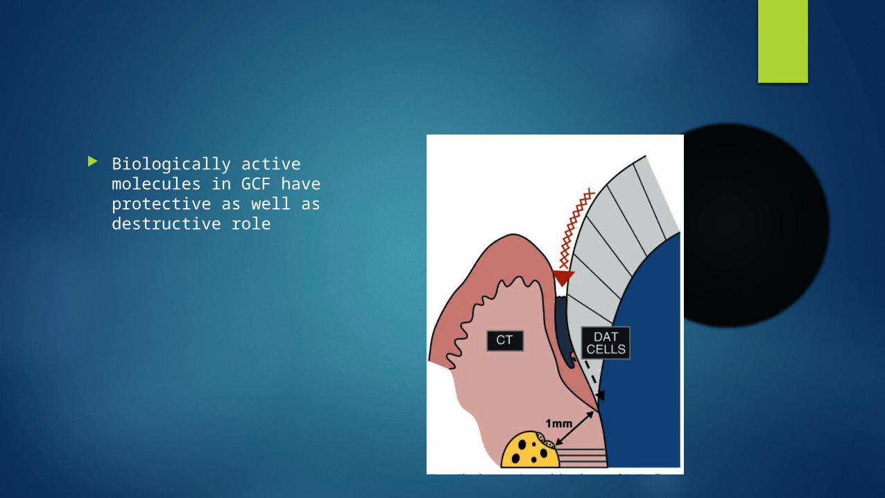
Biologically active molecules in GCF have protective as well as destructive role
Role of the polymorphonuclearleukocytes:
Polymorphonuclear leukocytes form the most important line of defense against bacterial plaque at the gingival margin (Page RC et al).
Polymorphonuclear leukocytes are a major contributor in the host–parasite equilibrium but have a limited capacity to reclaim any tooth surface once lost to the plaque bacteria.
Role of the polymorphonuclearleukocytes:
The polymorphonuclear leukocytes are most effective in aerobic conditions close to the gingival margin (Dennison et al 1997), suggesting a different role for them in anaerobic periodontal lesions.
Lactoferrin is an important antimicrobial protein present in the secondary granules of polymorphonuclear leukocytes.

Role of the polymorphonuclearleukocytes:
High concentrations of lactoferrin do, however, hamper epithelial cell growth by interfering with their adhesion and spreading. The molecule may, thus, have a role in delaying the repair of the junctional epithelium/DAT cell population during severe inflammation.
Role of host proteinases andinflammatory mediators:
Degradation of extracellular matrix during periodontal inflammation is a multistep process that involves several proteolytic enzymes.
Different cell types of periodontal tissue produce matrix metalloproteinases (collagenases, stromelysins, gelatinases, membrane-type metalloproteinases), plasminogen activator, cathepsins and elastase (Birkedal Hansen et al, Suomaleinin et al).

Role of host proteinases andinflammatory mediators:
Neutrophil elastase and cathepsin G are capable of degrading basement membrane type IV collagen and laminin, and also type VIII collagen, found in the internal basal lamina (Heck & Blackburn et al, Kittlecherger et al).
However, electron microscopic studies on DAT cells attached to teeth extracted because of advanced periodontitis do not support the idea that
Role of host proteinases andinflammatory mediators:
enzymatic degradation of the epithelial attachment apparatus precedes the degeneration of DAT cells (Overman et al).
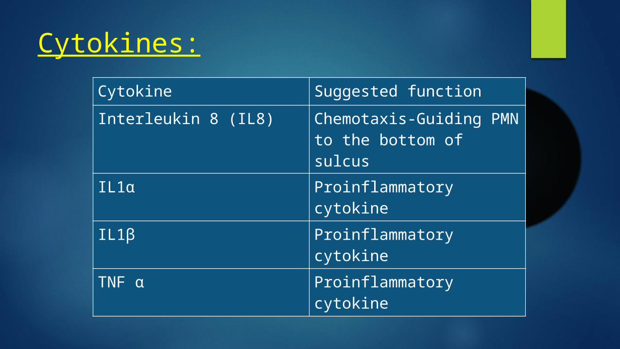
Cytokines:Cytokine Suggested functionInterleukin 8 (IL8) Chemotaxis-Guiding PMN
to the bottom of sulcusIL1α Proinflammatory cytokine
IL1β Proinflammatory cytokine
TNF α Proinflammatory cytokine

Problems in Junctional Epithelium:
If the junctional epithelium is repeatedly or continuously exposed to bacterial challenges, which may lead to the failure of JE, subgingival plaque formation, Conversion of the gingival sulcus into a periodontal pocket, and Increase in the inflammatory focus in the connective tissue.
Role of JE in passive eruption:
Role of JE in passive eruption:
Passive eruption is the exposure of the teeth by apical migration of the gingiva.
This concept distinguishes between the anatomic crown (the portion of the tooth covered by enamel) and the anatomic root (the portion of the tooth covered by cementum) and between the
Role of JE in passive eruption:
clinical crown (the part of the tooth that has been denuded of its gingiva and projects into the oral cavity) and clinical root (the portion of the tooth covered by periodontal tissues).
Role of JE in passive eruption:
When the teeth reach their functional antagonists, the gingival sulcus and junctional epithelium are still on the enamel and the clinical crown is approximately two thirds of the anatomic crown.
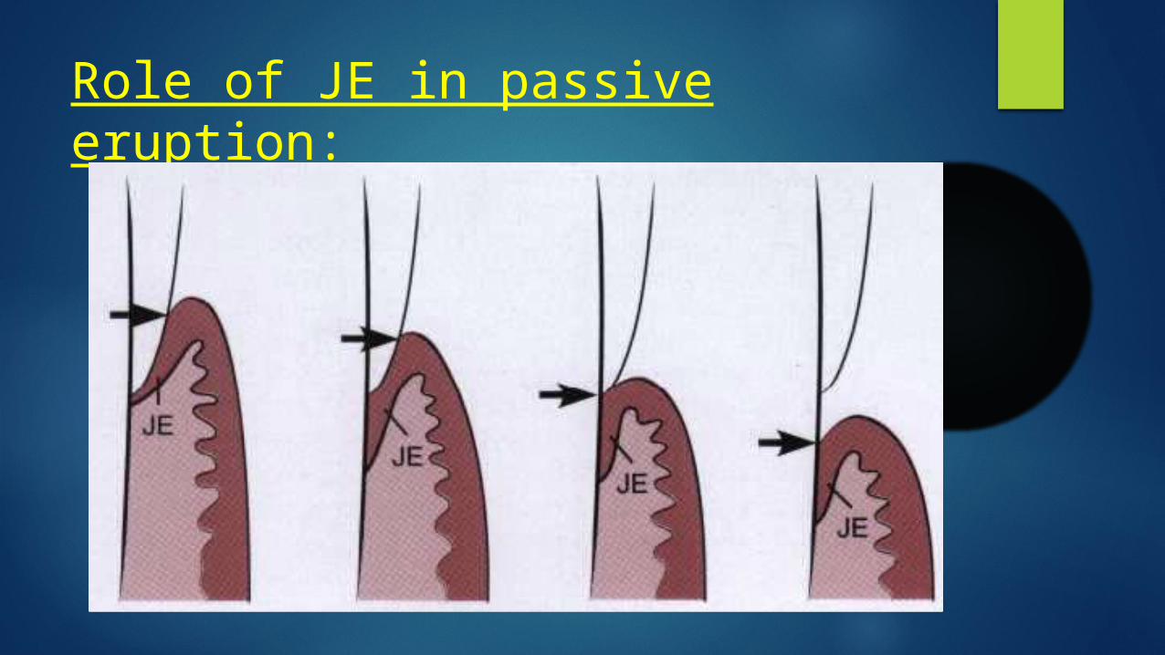
Passive eruption is divided into four stages. Although this was originally thought to be a normal physiologic process, it is now considered a pathologic process.

Role of JE in passive eruption: Stage 1: The teeth
reach the line of occlusion. The junctional epithelium and base of the gingival sulcus are on the enamel.

Role of JE in passive eruption: Stage 2: The junctional
epithelium proliferates so that part is on the cementum and part is on the enamel. The base of the sulcus is still on the enamel.
Role of JE in passive eruption:
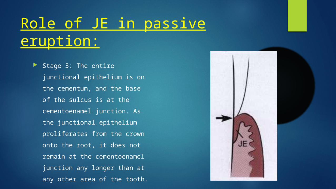
Stage 3: The entire junctional epithelium is on the cementum, and the base of the sulcus is at the cementoenamel junction. As the junctional epithelium proliferates from the crown onto the root, it does not remain at the cementoenamel junction any longer than at any other area of the tooth.
Role of JE in passive eruption: Stage 4: The junctional
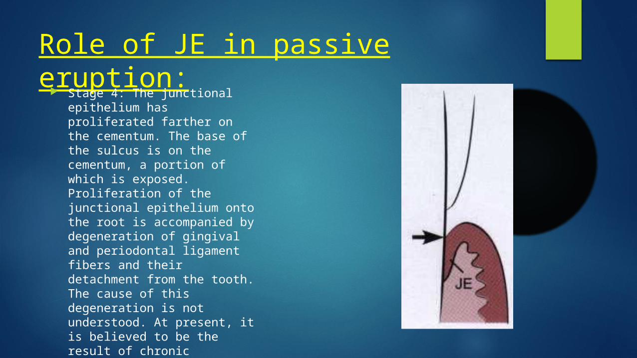
epithelium has proliferated farther on the cementum. The base of the sulcus is on the cementum, a portion of which is exposed. Proliferation of the junctional epithelium onto the root is accompanied by degeneration of gingival and periodontal ligament fibers and their detachment from the tooth. The cause of this degeneration is not understood. At present, it is believed to be the result of chronic inflammation and therefore a pathologic process.

JE in pocket formation: Conversion of JE to pocket epithelium is regarded as the
hallmark in the development of periodontitis
Infla
mm
atio
nSupracrestal collagen destruction
Loss
of c
onta
ct in
hibi
tion,
Ov
er e
xpre
ssio
n of
EGFApical
migration of JE
Infla
mm
ator
y ce
ll in
filtra
tionCoronal
detachment of JE
Apical migration of JE:
Due to - Loss of contact inhibition -due to supra crestal collagen
destruction
-Increased expression of EGF and its receptors due to cytokine stimulation
Epithelial cell at apical end of migrating JE have no internal and external basal lamina
Coronal detachment of JE:
Due to - More pooling of inflammatory cells at the coronal end -Degeneration of cell adhesion molecules by inflammatory
mediators -Destruction of cell adhesion complexes directly by bacterial
enzymes such as gingipains

JE in pocket:
Shorter than normal Cells are mostly in normal condition May exhibit slight degeneration
Role of JE in Gingivitis:
During the initial lesion of gingivitis, Leukocytes, mainly PMNs leukocytes leave the capillaries by migrating trough the walls ( Lindhe J perio res)
They can be seen in increased quantities in the connective tissue, and the junctional epithelium and the gingival sulcus.
Role of JE in Gingivitis:
The junctional epithelium becomes densely infiltrated with neutrophils and it may begin to show development of rete pegs or ridges in the early lesion of gingivitis.
During the established lesion of gingivitis, the junctional epithelium reveals widened intercellular spaces filled with granular cellular debris, including lysosomes derived from disrupted neutrophils, lymphocytes and monocytes.
Role of JE in Gingivitis:
JE forms rete pegs or ridges that protrude into the connective tissue and the basal lamina is destroyed in some areas.

JE in Necrotizing Ulcerative Gingivitis:
Surface epithelium is destroyed And it is replacd by a meshwork of fibrin, necrotic
epithelial cells, PMNs and neutrophils and various types of microorganisms
The epithelium becomes edematous and there is infilteration of PMNs in the intercellular spaces.
JE in Necrotizing Ulcerative Gingivitis:
Surface epithelium is destroyed And it is replacd by a meshwork of fibrin, necrotic
epithelial cells, PMNs and neutrophils and various types of microorganisms
The epithelium becomes edematous and there is infilteration of PMNs in the intercellular spaces.

What happens to JE in Trauma From Occlusion:
TFO causes widening of the marginal PDL space, a narrowing of the interproximal alveolar bone.
In case of TFO, the junctional epithelium will be intact and there will be no degeneration of the epithelial tissues unless there is any plaque accumulation.

Syndromes affecting JE:
Kindler syndrome: A rare skin blistering disorder along with early
onset aggressive periodontitis. Due to loss of kindlin-1 protein which is involved in
integrin activation. JE fails to attach to the tooth surface

Regeneration of JE
Injury to JE may occur due to intentional or accidental trauma.
Accidental trauma can occur during probing, flossing or tooth margin preparations for restorations.
Intentional trauma occurs during periodontal surgeries where the JE is completely lost.
Many studies have been done to investigate the renewal of JE. These include studies done on renewal of JE on tooth and implant surface after mechanical detachment by probing.
Studies have been done on mechanical trauma during flossing and on regeneration of JE after gingivectomy procedure which completely removes JE.

Taylor and Campbell 1972: A new and complete attachment indistinguishable from that in control was established 5 days after complete separation of the JE from the tooth surface.
Frank et al 1972: A study demonstrated that newly differentiated attachment apparatus with normal hemidesmosomal attachment is possible following surgery. This new attachment apparatus was seen on cementum as well as dentin.
Listgarten 1972:Hemidesmosomes appeared to form prior to the basal lamina. The basal lamina is initially formed in close proximity to the hemidesmosomes at both the tooth and connective tissue interface. At 4 to 7 weeks, the basal lamina appeared complete. Studies have shown that regeneration of JE after procedure usually occurs within 20 days.

JE AROUND IMPLANTS
The junctional epithelium around implants always originates from epithelial cells of the oral mucosa, as opposed to the junctional epithelium around teeth which originates from the reduced enamel epithelium.
Despite different origins of the 2 epithelia, a functional adaptation occurs when oral epithelia form an epithelial attachment around implants.

NATURAL TOOTH
Epithelium tapers towards the depth
Large number of cell organelles
Fibers are arranged perpendicular
IMPLANT
•Epithelium is thicker
•Few organelles
•Fibers are arranged parallely
•Numerous kerato-hyalin granules
CONCLUSION
Junctional Epithelium is important because of its anatomical location.
It is the site of host-bacterial interaction in initiation of periodontal disease.
There is a constant presence of bacteria and their products in the gingival sulcus which makes this an important structural component of periodontal defense mechanism.
The conversion of the junctional epithelium to pocket epithelium is regarded as hallmark in the development of periodontitis.
References Moon-Il Cho & Philias R. Garant. Development and general
structure of the periodontium. Periodontology 2000, Vol. 24, 2000, 9–27.
Mark Bartold, Laurence J. Walsh & A. Sampath Narayanan. Molecular and cell biology of the gingiva.P. Periodontology 2000, Vol. 24, 2000, 28–55.
Huberte . Schroede & R M Listgarten. The gingival tissues: The architecture of periodontal Protection. Periodontology 2000, Vol. 13, 1997, 91-120.
Takashi Sawada1 and Sadayuki Inoue. Ultrastructure of Dentogingival Border of Normal and Replanted Tooth and Dental Implant, chapter 11 www.intechopen.com/books/implantdentistry
Carranza’s clinical periodontology 11th edition Orban’s oral anatomy and histology

Thank you…….

![Junctional Epithelium: A dynamic seal around the tooth · The Junctional Epithelium forms with the eruption of tooth crown into the oral cavity [6]. It arises from the Reduced Enamel](https://static.documents.pub/doc/80x56/5fb280b2f9e57e0dca5d4e7d/junctional-epithelium-a-dynamic-seal-around-the-tooth-the-junctional-epithelium.jpg)


