58
June 2016, Volume: 1, Issue:1 KARNATAKA PROSTHODONTIC JOURNAL www.kpjonline.com www.kpjonline.com JUNE 2016 VOLUME 1 ISSUE 1

June 2016 Volume 1 Issue1 KARNATAKA PROSTHODONTIC JOURNAL wwwkpjonlinecom
wwwkpjonlinecom
JUNE 2016
VOLUME 1
ISSUE 1
June 2016 Volume 1 Issue1 KARNATAKA PROSTHODONTIC JOURNAL wwwkpjonlinecom
CONTENTS
1
Rugae Duplication ndash Different Techniques Of
Customizing Palatal Rugae in Maxillary Complete
Denture to Enhance Phonetics Dr Anupama Neelakantan Dr Sunil Dhaded
1
2 Rehabilitation Of A Patient With An Interim Haryngeal
ObturatorA Case Report Dr Thilak Shetty B Dr Shobha Rodrigues Dr Sharon Saldanha
8
3
Prosthetic Rehabilitation of a Patient with Atrophic
Ridges A Clinical Report
Vaibhav Gupta Dr Shobha Rodrigues Dr Vidya K
Shenoy Dr Thilak Shetty Dr Sharon Saldanha Dr
Mahesh M
Dr Puneeth Hegde
13
4
Prosthodontic Management Of Marginal
Hemimandibulectomy With Surgically Induced Lip
Drop
Dr Krishna Prasad D Dr Anupama Prasad D
Dr Anshul Bardia
20
5 Short Dental Implants ndash A Review Of Clinical Performance
Biomechanical Aspects And Risk Factors For Survival
Dr Talreja Karishma S Dr Rodrigues Shobha J Dr Pai Umesh Y
28
6
Knowledge Attitude and Oral Hygiene Practice Among
Patients Wearing Fixed Partial Dentures In South
Coastal Karnataka Region
Dr Manoj Shetty Dr Krishna Prasad D Dr Chethan Hegde
Dr Nikhila Thulasida
37
7 Aesthetic And Functional Rehabilitation Of A Severely
Mutilated Dentition
Dr Nirupama R Prof (Dr) Manoj Shetty
42
8
SynCone- A New Dimension In Implant Overdenture A
Case Report Prof(Dr) Manoj Shetty Dr Azlinder Prof(Dr)Rakshith Hegde
Prof (Dr) Chethan Hegde Dr Nivya John
48
June 2016 Volume 1 Issue1 KARNATAKA PROSTHODONTIC JOURNAL wwwkpjonlinecom
1
Rugae Duplication ndash Different Techniques Of
Customizing Palatal Rugae in Maxillary Complete
Denture to Enhance Phonetics
Dr Anupama Neelakantan1 Dr Sunil Dhaded
2
Department of Prosthodontics AMErsquos Dental College Near Government Polytechnic
College Bijengere Road Raichur Karnataka
ABSTRACT
Speech is imperative for human communication Therefore phonetics must be considered
along with mechanics and esthetics as the integral factors in contributing to the success of a dental
prothesis Palatal rugae contours have a very important role in phonetics by production of linguo-
palatal sounds that involves the contact between tongue and palate By customizing palatal contours
of a maxillary denture to the tongue the patient may easily adapt to the definitive denture contour
which in turn shortens or eliminates the adjustment period for the achievement of proper speech This
review article deals with different methods of palatine rugae duplication in complete denture
prosthesis to improve phonetics besides briefly describing its role in various other fields such as sex
determination orthodontics and forensic odontology
KEY WORDS Palatine rugae rugae duplication phonetics linguopalatal sounds forensic
odontology
INTRODUCTION
Speech is an integral part of human communication which makes the human species
superior to other life forms Although every prosthodontist aims at providing excellent complete
denture prosthesis in terms of esthetics functional efficiency and comfort a thorough evaluation of
phonetics is too often neglected with greater emphasis placed on other three components1
1st Year Post Graduate student 1 Professor and Head
2
June 2016 Volume 1 Issue1 KARNATAKA PROSTHODONTIC JOURNAL wwwkpjonlinecom
2
Among the various anatomical landmarks of the oral cavity Palatine rugae are perhaps one
of the least understood or unexplored regions of the oral mucous membrane Due to this they have
been arbitrarily associated with functions like speech adaptation proprioception and taste2
Palatal rugae also called plicae palatinae transversae and rugae palatina refer to the ridges on
the anterior part of the palatal mucosa each side of the median palatal raphe and behind the
incisive papilla 34
Palatine rugae are elevations of the mucous membrane and are very prominent in
most of the animals where they help in gripping the food before tearing it with brute force
Optimal phonetics can best be achieved by obtaining a proper occlusal vertical dimension (OVD) and
occlusal plane correctly positioning the anterior and posterior teeth to suit best the functional and
esthetic requirements as well as adequately contouring the palatal surface Because the lack of texture
on the palatal portion of a complete denture can impede proper articulation one solution is to add
palatal rugae1 5
CLASIFICATION OF RUGAE
Identification of palatal rugae pattern is based on classification by Thomas et al This
classification includes number length shape and identification pattern of rugae By determing the
length of all rugae three categories are identified6 7
1 Primary rugae (5-10 mm)
2 Secondary rugae (3-5 mm)
3 Fragmentary rugae (less than 3 mm)
The shape of individual rugae are classified into four major types
1 Straight ndash Runs directly from origin to termination
2 Curvy ndash Simple crescent shape that was curved gently
3 Circular ndash Definite continuous ring formation diameter from origin to termination is considered
4 Wavy ndash Serpentile form
The unification pattern is further subdivided into diverging and converging types89
Diverging pattern occurs when two rugae begin from the same origin but diverge transversely1011
Converging pattern occurs when two rugae arise from different regions and converge
transversely89
June 2016 Volume 1 Issue1 KARNATAKA PROSTHODONTIC JOURNAL wwwkpjonlinecom
3
METHODS OF RUGAE DUPLICATION
Characterization of the complete denture is necessary to give the dentures a life like
appearance to make it appear more natural12
Palatal rugae can be characterized and incorporated in
the maxillary complete denture by different techniques
RUGAE DUPLICATION USING PUTTY IMPRESSION TECHNIQUE
The primary impression is made in impression compound using stock tray and cast is poured
Putty is adapted over rugae area of maxillary cast to record prominent rugae on the palateModelling
wax is melted and poured over the putty impression slowly and carefully to record the imprints of
rugae over the impression Before flasking of denture wax imprint of rugae was placed on maxillary
trial denture base adapted carefully on the palatal portion of the maxillary trial denture base13
RUGAE DUPLICATION USING DENTAL FLOSS
An ideal protocol for complete denture fabrication was followed till the stage of obtaining the
secondary castThenmark the rugae patterns in definitive maxillary cast using permanent marker
Apply auto-polymerizing resin (clear) in sprinkle on method on the rugae portion in the cast The
markings will be seen through the transparent resin in the cast The thickness of resin added should
not exceed 1 mm Apply auto-polymerizing resin (pink) in sprinkle on method on the rest of cast and
fabricate the record base in the usual manner Proceed with the tentative jaw relation and teeth
arrangement Trial denture verification is done Demount the maxillary cast from articulator Cut
dental floss as per the required lengths and lute them over the rugae marking seen through the record
base using inlay casting wax Proceed with fabrication of denture in conventional manner The rugae
pattern is duplicated in the denture14
June 2016 Volume 1 Issue1 KARNATAKA PROSTHODONTIC JOURNAL wwwkpjonlinecom
4
RUGAE DUPLICATION USING TIN FOIL
NEW PROSTHESIS
Cut tinfoil (0001 tinfoil) to the desired shape and adapt it to the rugae area on the master cast
with prominent rugae Tinfoil pattern is removed from the cast and is sealed to the palatal area of the
completed wax-up with hot baseplate wax Then it is flasked processed finishedand polished as
usual15 16
EXISTING PROSTHESIS
Adapt tinfoil on the cast with prominent rugae flow hot baseplate wax over the surface to
reinforce the tinfoil Remove wax reinforced tinfoil from the cast and trim to desired
shapeAutopolymerizing acrylic resin is applied on the underside of the tinfoil pattern to fabricate
rugae When cured remove the tinfoil and secure acrylic rugae to the palatal area of the existing
prosthesis with autopolymerizing acrylic resin Refine finish and polish15s
June 2016 Volume 1 Issue1 KARNATAKA PROSTHODONTIC JOURNAL wwwkpjonlinecom
5
DISCUSSION
The procedure of electroplating to form metal palate that duplicates patientsrsquo palate is limited
in that it does not apply to dentures made of acrylic resin 17
Another procedure uses an impression of
maxillary cast to make custom acrylic resin pattern to capture patientrsquos anatomy But this involves
making additional impression or duplication of cast Missing lingual contours of denture teeth should
be added during waxing up of trial dentures in this method 18
Use of palatogram and acrylic resin to
modify palatal portion of denture has been done19-21
The production of palatolingual group of sounds involves firm contact of the tip of the tongue
against the rugae When these rugae and the hard palate are covered by the denture proprioceptive
feedback may be changed Therefore phonetics may be affected by the presence of denture Copying
of the rugae on the palatal surface of the denture reduces this problem2223
Accurate approximation of palatal contours of a maxillary complete denture to patientrsquos
tongue can improve intelligibility if other factors such as tooth position occlusal plane and vertical
dimension are satisfactory24
A method for functionally modifying the contour of the palatal vault of
maxillary complete denture can be achieved at the trail stage of denture construction and incorporated
in the finished denture 25
Artificial duplication can be done using corrugated metal plates plastic palate forms free
hand wax carving of anatomical palate forms etc These artificial rugae may cause interference with
speech if they are made too prominent22
The use of ribbed features when made from a significantly stiffer material and designed to
mimic palatal rugae offer an acceptable method of providing significant improvement in speech as
well as rigidity to the maxillary denture22
Besides phonetics the authors believe that they may play important role in biological
adaptation of the tongue to the denture and important contributor in taste perception Palatine rugae
when duplicated on the denture improved patientrsquos ability to identify flavors especially sour foods
Both responsetimes as well as qualities of perception of sour taste improved with denture that was
characterized with Palatine rugae Understanding the perception of sour taste has received less
attention than sweetness and bitterness particularly for mammals 16
Multiple mechanisms have been
proposed to explain how hydrogen ions interact with taste receptor cells to cause a response2223
Although it has been widely accepted that the hydrogen ion is the chemical entity responsible for the
June 2016 Volume 1 Issue1 KARNATAKA PROSTHODONTIC JOURNAL wwwkpjonlinecom
6
sour taste many physiological studies have indicated the involvement of protonated organic acids as a
stimulus for sour taste as well Irrespective of the mechanism for the sour taste of tongue the patient
was able to perceive the sour taste soon as well as better The denture
with palatine rugae provides an irregular surface against which the tongue is locked appropriately than
with the flat surface Once the tongue is locked in place a negative pressure is developed by it so that
the flavor from the foodstuff is sucked This is especially true for the sour taste Another
reason for better perception would be that when the tongue touches irregular surface of the palatine
rugae the elevations and depressions on the denture open up the microvilli by stretching them away
from each other This allows the hydrogen ion from food to come in contact with the taste receptor
cells that are oriented perpendicular to the surface in a parallel arrangement2
CONCLUSION
Phonetics is one of the important factors in complete denture construction However this
factor is neglected because of the adaptability of patients It is true that most patients can learn to
produce satisfactory speech in spite of an unsatisfactory denture The need to consider phonetics is not
recognized in most instances until a patient complains of inability to produce certain sounds with the
dentures Completely edentulous individuals using dental prosthesis tend to mispronounce certain
sounds pronunciation of which depends upon the rugae pattern and also the palatal contour Thus
prosthodontists need to create the customized rugae and palatal contours in complete dentures with
care for achieving speech which is much more normal and also eliminate the waiting and training
period after denture insertion To aid the dentist in minimizing these speech problems the importance
of phonetics in dental prosthesis has been discussed
REFERENCES
1 Vaswani Priya Pronob Sanyal and Ankur Prajapati Comparison of speech articulation and
intelligibility in palatally contoured dentures using a novel rugae duplication technique A clinical
study International Journal of Dental Research 32 (2015) 15-20
2 Mattoo Khurshid and Dr Pooja Arora Shujaurahman duplicating palatine rugae in
complete denture prosthesis to enhance the relationship between food and taste receptors
3 Lysell L Plicae palatinae transversae and papilla incisiva in man A morphologic and genetic
studyActaOdontol Scand 1955 13 (Suppl 18) 5ndash137
4 Thomas CJ Kotze TJ The palatal rugaepattern A new classification J Dent Assoc South
Afr1983 38153ndash7
June 2016 Volume 1 Issue1 KARNATAKA PROSTHODONTIC JOURNAL wwwkpjonlinecom
7
5 Martone AL (1963) Clinical applications of concepts of Functional Anatomy and Speech
Science to complete denture prosthodonticsJ Prosthet Dentistry 13 4-33
6 Shetty Divya et al Assessment of palatal rugae pattern and their reproducibility for
application in forensic analysis Journal of forensic dental sciences 52 (2013) 106
7 Thomas CJ Kotze TJ The palatal rugae pattern A new classification J Den Assoc S Afr
198338153‑ 7
8 Bharath ST Sex determination by discriminant function analysis of palatal rugae from a
population of coastal Andhra J Forensic Dent Sci 2011358‑ 62
9 Robison WR Summitt SF Oesterle JB Brannon LJ Morlang RBIndividuality of human
palatal rugae J Forensic Sci 1988 33718‑ 26
10 Limsom KS Thomas CJ Kotze TJ Computerized recording of the palatal rugae pattern and
evaluation of its application in forensic identification J Forensic Odonto‑ Stomatol 20042231
11 Allen H The palatal rugae in man Dental Cosmos 18893166‑ 80
12 Fernandez Teny et al A technique for palatal rugae transfer during characterization of
complete dentures SIS Index ID 833 (2015) 89
13 Deo Pratibha Katiyar Dr Krishna and Ritu Mohindra Duplication of Important Landmark-
Palatine Rugae World 31 (2012) 95-96
14 Vijayaraghavan Vasantha and P Chandni A Simple Method for Palatal Rugae Carving in
Complete Dentures Journal of Indian Prosthodontic Society 132 (2013) 137
15 Gitto Christina A Salvatore J Esposito and Julius M Draper A simple method of adding
palatal rugae to a complete denture The Journal of prosthetic dentistry 812 (1999) 237-239
16 Singh Niyati et al Unconventional-True to Life Interpretation of Esthetics in Single
Complete Denture A Case Report Indian Journal of Contemporary Dentistry 21 (2014) 106
17 Rogers OW (1970) Electroformed metal plates for complete dentures J Prosthet Dent
23207ndash217
18 White KC Connelly ME (1986) Duplicating natural palatal contours in acrylic resin complete
dentures Prosthet Dent 61508ndash510
19 Palmer JM (1979) Structural changes for speech improvement in complete upper denture
fabrication J Prosthet Dent 41507ndash510
20 Kong HJ Hansen CA (2008) Customizing palatal contours of a denture to improve speech
intelligibility J Prosthet Dent 99243ndash248
21 Sanjay VB Priti S Sadekh A (2012) Reproducing functional palatal contours in complete
dentures to improve speechmdasha case report J Indian Dent Assoc 6(2)111ndash114
22 Krishna Vamsi et al Dentures with phonetically contoured palate a simple technique of
adding customized rugae and palatal contours to the maxillary denture The journal of
contemporary dental practice 132 (2012) 216-218
23 Tanaka H Speech patterns of edentulous and morphology of the palate in relation to
phonetics J Prosthet Dent 19732916-28
24 Kong HJ Hansen CA Customizing palatal contours of a denture to improve speech
intelligibility J Prosthet Dent Mar 2008 99(3)243-48
25 Goyal BK Greenstein P Functional contouring of the palatalvault for improving speech with
complete dentures J Prosthet Dent 198248641-46
26 Meenu Merry C Paul A simple technique of fabricating customized palatal rugae contours in
complete dentures for enhancing phonetics KDJ April 201033(2)110
27 White JA Bond IP Jagger DC Improving mechanical properties of maxillary complete
dentures through a bioinspired engineering design Int J Prosthodont Nov-Dec 201124(6)589-98
June 2016 Volume 1 Issue1 KARNATAKA PROSTHODONTIC JOURNAL wwwkpjonlinecom
8
Rehabilitation Of A Patient With An Interim Haryngeal
Obturator A Case Report
Dr Thilak Shetty B1 Dr Shobha Rodrigues
2 Dr Sharon Saldanha
3
Department Of Prosthodontics Manipal College of Dental Sciences Manipal University
Mangalore- 575001India
ABSTRACT
An interim prosthesis is used to rehabilitate a patient with partial or total soft palate defect
generally as soon as possible after surgery This article describes a stage by stage technique of
fabrication an interim pharyngeal obturator with a speech bulb for a patient with a partial soft palate
defect
INTRODUCTION
Cancers of the mouth tongue oropharynx nasopharynx and larynx comprise approximately
5 of all cancers1-3
Most treatment methods to eliminate the cancers would involve surgical resection
and concomitant radiation resulting in incapacitating defects compromising the integrity and function
of the oral cavity requiring immediate short or long term management and rehabilitation procedures
While restoration of the defect is fairly straightforward in case of the hard palate it becomes more
complicated and challenging when involving the soft palate Among soft palate defects the complete
soft palate defect is easier to trace and obturate than compared with a soft palate that has been
partially resected and is dysfunctional4 A partial soft palate defect may result from the surgical
resection of the posterior border from the medial or lateral posterior portion of the soft palate5
Median posterior border defects occur after the resection from the uvula and posterior soft palate In
contrast lateral defects occur when the anterior tonsillar pillar and retromolar trigone are resected
Professor 1
Professor ampHead 2
Associate Professor 3
June 2016 Volume 1 Issue1 KARNATAKA PROSTHODONTIC JOURNAL wwwkpjonlinecom
9
Rehabilitation of such patients may be accomplished surgically or prosthetically Surgical
reconstruction includes microvascular flap techniques using vascularized or nonvascularized soft
tissue flaps4 However in many instances these flaps may unsuccessfully obturate the nasopharyngeal
port and patients may then be referred to the maxillofacial prosthodontist for evaluation and
treatment The presence of the flap may complicate the successful prosthetic obturation of these
surgically reconstructed defects
This article describes a stage by stage rehabilitation of an acquired lateral soft palate defect
with an interim pharyngeal obturator and a speech bulb This prosthesis made the rehabilitation
comfortable and served as a transitional and training denture prior to insertion of the more definitive
prosthesis The prosthesis helped to alleviate speech problems and assisted in the masticatory
function The speech bulb was easy to insert and remove for the patient It was also easy to fabricate
and adjust to the denture base
CLINICAL REPORT
A 29-year-old man diagnosed with adenoid cystic carcinoma of the minor palatal salivary
glands had undergone a partial maxillectomy and excision of the soft palate (Fig1) and was referred
to the Department of Prosthodontics Manipal College of Dental Sciences Mangalore India
Immediate surgical reconstruction was not recommended due to the need for further treatment with
radiation therapy The patient received postoperative external beam radiation therapy by anterior
direct beam on a telecobalt machine with a total dose of 60 Gy in 30 fractions over a period of 6
weeks The patient tolerated the radiation well and was subsequently referred for possible prosthetic
restoration of the oral defect after radiation therapy On examination of the defect laterally resected
and dysfunctional soft palate along with partial maxillectomy on the right side was noted Various
modalities of prosthetic reconstruction were discussed with the patient and the patient indicated a
June 2016 Volume 1 Issue1 KARNATAKA PROSTHODONTIC JOURNAL wwwkpjonlinecom
10
desire for an economical solution Hence heat-polymerizing interim acrylic resin prosthesis was
planned and the expectations of this prosthesis were explained to the patient
To improve patient adaptation the speech-aid device was constructed stage by stage In the
first stage the impression of the defect was obtained with irreversible hydrocolloid (Imprint Dental
Products of India Ltd) The impression was removed and poured in Type III dental stone (Dentstone
Pankaj Industries Mumbai Maharashtra India) Undercuts were blocked in Type II dental stone (Fig
2) Maxillomandibular jaw relations were obtained and prosthesis was waxed to form A heat
polymerized clasp retained acrylic-resin maxillary prosthesis was delivered to the patient (Fig 3)
After 3 weeks an acrylic-resin extension was added to the posterior border of this prosthesis This was
extended posteriorly to the intact residual soft palate and parallel to the soft tissue in the nasopharynx
approximately 3 to 4 mm short of the adjacent tissues at the maximum level of contraction Some
pressure was exerted to slightly elevate the remaining soft palate to compensate for the thickness of
the material and not to encroach on the tongue space The contours of the defect and velopharyngeal
musculature were functionally recorded with modeling compound to form the speech bulb The bulb
was placed within the nasopharynx at the plane of velopharyngeal (VP) closure The patient was
instructed in repeated swallowing so that the bulb was grossly molded but still underextended The
bulb was designed to be slightly superior to the level of the VP closure and to approximate the
pharyngeal walls so as to allow competent VP valving during speech but leave free nasal breathing
and production of nasalized speech sounds The denture base with the extension was chilled in cold
water and preparations were made for conversion of the bulb into acrylic resin Type II stone was
placed around the obturator impression to include the intaglio side of the denture base (Fig 4) The
impression material was replaced with heat-polymerizing acrylic resin (Fig 5) The patient was
instructed to wear the prosthesis at all times during the day including at meals and to remove it at
night The adaptation of the patient to the prosthesis was prompt and good although he reported
impeded breathing during strenuous activities
June 2016 Volume 1 Issue1 KARNATAKA PROSTHODONTIC JOURNAL wwwkpjonlinecom
11
The patient was scheduled for the first post-insertion adjustment 3 days after the insertion At
the first post-insertion appointment the surgical wound was observed to ensure health of the tissues to
relieve the prosthesis for pressure areas on the tissues to compensate for processing changes and to
emphasize hygiene and home care The patient was placed on a 3-month recall for evaluation and
observation of any recurrence
SUMMARY
This clinical report describes a multistep procedure for prosthetic rehabilitation of a soft
palate defect with an interim pharyngeal obturator and speech bulb The advantages of this prosthesis
are that the technique is noninvasive cost-effective tissue tolerant comfortable to use and easy to
fabricate and clean The prosthesis coupled with the patients compensatory phenomenon improved
the quality of life and provided appropriate and effective nasopharyngeal obturation
FIGURES
Fig 1 Preoperative view of the defect
Fig 2 Blocked out cast
June 2016 Volume 1 Issue1 KARNATAKA PROSTHODONTIC JOURNAL wwwkpjonlinecom
12
REFERENCES
1 Parker S Tong T Bolden S Wingo PA Cancer statistics 1997 CA Cancer J Clin 1997475-
27
2 Cancer Incidence by site Age-Standardized Rate per 100000 Ottawa Statistics Canada and
the Canadian Council of Cancer Registries Health protection Branch- Laboratory Centre for
Disease Control 2000
3 WHO Mortality Database Age- Standardized Rate per 100000 Geneva WHO Databank
1999
4 Chambers MS Lemon JC Martin JW Obturation of the partial soft palate defect J Prosthet
Dent 20049175-9
5 Curtis TA Beumer J Speech velopharyngeal function and restoration of soft palate defects
In Maxillofacial rehabilitation prosthodontic and surgical considerations eds Beumer J Curtis TA
and Marunick MT Isbiyaku EuroAmerica St Louis 1996 304-319
Fig 3 Clasp retained acrylic removable
partial denture
Fig 4 Cast with stone placed around the
obturator impression
Fig 5 Prosthesis in situ
Fig 6 Postoperative view in occusion
June 2016 Volume 1 Issue1 KARNATAKA PROSTHODONTIC JOURNAL wwwkpjonlinecom
13
Prosthetic Rehabilitation of a Patient with Atrophic
Ridges A Clinical Report
Vaibhav Gupta Dr Shobha Rodrigues
1 Dr Vidya K Shenoy
2
Dr Thilak Shetty3 Dr Sharon Saldanha
4 Dr Mahesh M
4
Dr Puneeth Hegde5
Professoramp HOD Department of Prosthodontics MCODSMangalore1
Professor amp HOD Department of Prosthodontics A J Institute Of Dental Sciences
Mangalore2
Professor Department of Prosthodontics MCODSMangalore3
Associate Professor Department of Prosthodontics MCODSMangalore4
Assistant Professor Department of Prosthodontics MCODSMangalore5
Intern Department of Prosthodontics MCODS Mangalore
ABSTRACT
Extreme resorption of the maxillary and mandibular denture-bearing area may lead to
problems with prosthetic rehabilitation As resorption progresses there is a resultant narrow more
constricted upper residual ridge opposed by a wider lower residual ridge decreased supporting
tissues that results in a large restorative space between the opposing residual ridges
This clinical report describes a method for prostheticrehabilitation of a completely edentulous
patient with hollow maxillary denture and a conventional mandibular denture contours of which are
in harmony with the neutral zone
INTRODUCTION
India has a large geriatric population of 77 million comprising 77 of its total population In
a community based study1 planned to assess the level of edentulousness denture need and denture
wear it was found that although level of edentulousness was high there was a low level of denture
wearingAmong the many factors elucidated for poor denture wear one contributory factor may be
the extremeresorption of the edentulous ridges resulting in dentures functioning as oral
acrobaticsNevertheless treatment options for prosthodontic rehabilitation of completely edentulous
patients include conventional complete dentures and implant supported fixed orremovable prosthesis
June 2016 Volume 1 Issue1 KARNATAKA PROSTHODONTIC JOURNAL wwwkpjonlinecom
14
Complete dentures are mechanical devices that must function in harmony with the
surrounding orofacial musculature In addition they must fulfillthe basic objectives of Prosthodontics
including retention stability support aesthetics and preservation of remaining tissues However
extreme resorption of the maxillaryand mandibular denture-bearing area may lead to problems with
prosthetic rehabilitation As resorption progresses there is a resultant narrow more constricted upper
residual ridge opposed by a wider lower residual ridge decreased supporting tissues that results in a
large restorative space between the opposing residualridges This may result in heavy complete
dentures that may compound to the poor denture-bearing ability of the tissues and lead to decreased
retention and resistance
The extensive volume of the denture base material in prostheses provided to patients with
severe residual ridge resorption necessitates making the denture base hollow to reduce the prosthesis
weight There are numerous references in the literature that propagate the merits of dentures
constructed in harmony with the neuromuscular function as well as describe various materials and
methods for fabrication of hollow prostheses2-18
Although controversial19
it has been suggested that
gravity and the addition of weight to the mandibular complete denture may aid in prosthesis
retention2021
In additionthe coordination of complete dentures with the neuromuscular function and
arrangement of teeth in the neutral zone is highly effective in an atrophic mandible 2andis the
foundation of a successful stable denture
Previously described techniques for weight reduction include using a solid 3-dimensional
spacer including dental stone3-13
cellophane wrapped asbestos14
silicone putty1516
or modelling
clay1718
during laboratory processing to exclude denture base material from the planned hollow cavity
of the prosthesis
Mahdy3
also presented a double flask technique that allows forthe complete fabrication of the
obturator from the waxtry-in stage to completion of the prosthesisThe primary disadvantage of such
techniques is that the long junction between the two previously polymerized portions of the denture
that is luted with autopolymerising resinis apotential site for leakage and discolouration The need for
a lightweight hollow maxillary denture and stable mandibular denture fabricated from a strong
durable material is quite evident
This clinical report describes a method for prostheticrehabilitation of a completely edentulous
patient with hollow maxillary denture and a conventional mandibular denture contours of which are
in harmony with the neutral zone
June 2016 Volume 1 Issue1 KARNATAKA PROSTHODONTIC JOURNAL wwwkpjonlinecom
15
CLINICAL REPORT
A 69-year-old man completely edentulous male patient (Fig 1 amp 2) with a history of denture
wear for the past 10years was referred to the Departmentof Prosthodontics of this Institution for
prosthetic rehabilitation of severely resorbed ridges Past medical history was noncontributory Dental
history revealed unstable and loose maxillary and mandibular dentures Intraoral examination
revealed severely resorbed ridges with increased interridge space (Fig3)Variousmodalities of
prosthetic reconstruction were discussedwith the patient and the patient indicated a desire for
aneconomical solution Hence a heat-polymerized hollow maxillary denture and a weighted
mandibular denture with prosthetic teeth arranged in the neutral zone was planned and the
expectations of this prosthesis were explained to the patient
The traditional sequence of denture construction was followed till the definitive impressions
were made and the master casts were constructed and indexed in the land areaMaxillary occlusal rim
was constructed with modelling wax (Hindustan Modelling wax Hindustan dental products
Hyderabad India) The lower wax rim was constructed on a stabilized record base with low fusing
compound(Pinnacle Dental Products of India Ltd Mumbai India) softened at 135degF and shaped
similar to a wax occlusal rim The tray and modelling compound was placed in the mouth and the
patient was instructed to swallow and purse the lips The modelling compound was hardened in the
mouth sufficiently to prevent distortionJaw relations were recorded and the casts were mounted on a
mean value articulator(Fig 4)The modelling compound was lubricated and encased in a templateof
vinyl polysiloxane putty (Reprosil Dentsply Caulk Milford Del) which serves as an index for future
teeth arrangement(Fig5)The low fusing compound was replaced with modelling wax within the
confines of the prepared index Prosthetic teeth arrangement (Premadent Super Dental Products
Delhi India) was done and the dentureswere fabricated in the conventional manner till the verification
appointmentThe mandibular denture was then processed in the conventional manner as per the
manufacturerrsquos instructions Two identical flasks were used to fabricate a hollow maxillary denture on
lines of a described article 13
For this the trial denture was processed in the standard manner
through the wax elimination stage Two layers of baseplate wax was thenadapted (Supernal SD
Dental Corporation Lucknow India) to the definitive cast in the drag conforming to the border
extensions (Fig6 A) A second identical flask was used to invest the baseplate wax and again the wax
elimination process was completed The cope and second drag was packed with heat-polymerized
acrylic resin (DPI-heat cure Dental Products of India Ltd)Similarly a minimal thickness of acrylic
resin was processed around the teeth using a different cope(Fig6 B) The original cope was seated on
the original drag and complete closure of the flask was ascertained (Fig 6 C) A thin feather edge
margin was created along the visible junction to minimize the thickness of the autopolymerising
June 2016 Volume 1 Issue1 KARNATAKA PROSTHODONTIC JOURNAL wwwkpjonlinecom
16
resinThe visiblejunction between the two previously polymerized portions was luted with
autopolymerising acrylic resin (DPI-RR Dental Products of India Ltd)The whole prosthesis was
recovered and the palatal surface was luted in a similar manner The entire junction was waxed and
reprocessed so that the seam that seals the two sections is completely covered with heat-processed
acrylic resin minimizing the stain and leakage around the area of the seam and increasing the
durability and longevity of the prosthesisThe dentures were deflasked equilibrated and a hollow
maxillary denture and weighted mandibular denture was delivered to the patient (Fig 7amp Fig8)The
patientwas instructed on home care and prosthesis maintenance
SUMMARY
This clinical reportdescribes a method for prosthetic rehabilitation of a completely edentulous
patientwith resorbed ridges and excessiveinterarchspacewith a hollow maxillary denture and a
weighted mandibulardenture contours of which are in harmony with the neutral zoneControlling the
thickness of the hollowportion without the use of any three dimensional spacer and eliminating
leakage and discoloration are severaladvantages of this technique An additional laboratory step is
however required for the final culmination of the prosthesis
FIGURES
Fig 1 Preoperative view of
patient
Fig2 Profile of the patient
June 2016 Volume 1 Issue1 KARNATAKA PROSTHODONTIC JOURNAL wwwkpjonlinecom
17
Fig 3 Intraoral view
Fig 4 Maxillomandibular jaw
relations recorded
Fig 5 Putty index for the
neutral zone
Fig6 A Adaptation of baseplate
wax to the definitive cast
Fig 6 B Acrylic resin processed
around denture teeth
Fig 6 C Denture processing using
three identical sections of two
denture flasks
June 2016 Volume 1 Issue1 KARNATAKA PROSTHODONTIC JOURNAL wwwkpjonlinecom
18
REFERENCES
1 Shah N Parkash H Sundaram KR Edentulousness denture wear and denture needs of
Indian elderly a community based study Journal of Oral Rehabilitation200431467-476
2 Gahan MJ Walmsley AD The neutral zone impression revisited Br Dent J 2005198269-
272
3 El Mahdy AS Processing a hollow obturator J Prosthet Dent 196922682-686
4 Brown KE Fabrication of a hollow-bulb obturator J Prosthet Dent 19692197-103
5 Ackerman AJ Prosthetic management of oral and facial defects following cancer surgery J
Prosthet Dent 19555413-432
6 Nidiffer TJ Shipman TH Hollow bulb obturator for acquired palatal openings J Prosthet
Dent 19577126-134
7 Rahn AO Boucher LJ Maxillofacial prosthetics principles and concepts St Louis Elsevier
1970 p 95
8 Chalian VA Drane JB Standish SM Intraoral prosthetics In Maxillofacial prosthetics
multidisciplinary practice edsChalian VA Drane JB Standish SM Baltimore Williams amp
Wilkins 1971 133-157
9 Buckner H Construction of a denture with hollow obturator lid and soft acrylic lining J
Prosthet Dent 19743195-99
10 Browning JD Kinderknecht J Fabrication of a hollow obturator with fluid resin J Prosthet
Dent 198452891-895
11 Fattore LD Fine L Edmonds DC The hollow denture an alternative treatment for atrophic
maxillae J Prosthet Dent 198859514-516
12 Gardner LK Parr GR Rahn AO Simplified technique for the fabrication of a hollow
obturator prosthesis using vinyl polysiloxane J Prosthet Dent 19916660-62
13 McAndrew KS Rothenberger S Minsley GE An innovative investment method for the
fabrication of a closed hollow obturator prosthesis J Prosthet Dent 199880129-132
Fig 7 Post-operative view of the
patient
Fig8 Hollow maxillary denture and
weighted mandibular denture
June 2016 Volume 1 Issue1 KARNATAKA PROSTHODONTIC JOURNAL wwwkpjonlinecom
19
14 Worley JL Kniejski ME A method for controlling the thickness of hollow obturator
prostheses J Prosthet Dent 198350227-229
15 Holt RA Jr A hollow complete lower denture J Prosthet Dent 198145452-454
16 Jhanji A Stevens ST Fabrication of one-piece hollow obturators J Prosthet Dent
199166136-138
17 Elliott DJ The hollow bulb obturator its fabrication using one denture flask Quintessence
Dent Technol 1983713-14
18 DaBreo EL A light-cured interim obturator prosthesis A clinical report J Proshet Dent
199063371-373
19 Ohkubo C Hosoi T Effect of weight change of mandibular complete dentures on chewing
and stability a pilot study J Prosthet Dent 199982636-642
20 Jacobson TE Krol AJ A contemporary review of the factors involved in complete denture
retention stability and support Part I retention J Prosthet Dent 1983495-15
21 Wormley JH Brunton DA Weighted mandibular dentures J Prosthet Dent 197432101-102
June 2016 Volume 1 Issue1 KARNATAKA PROSTHODONTIC JOURNAL wwwkpjonlinecom
20
Prosthodontic Management of Marginal
Hemimandibulectomy With Surgically Induced Lip Drop
Dr Krishna Prasad D1 Dr Anupama Prasad D
2 Dr Anshul Bardia
3
Department of Prosthodontics and Crown amp Bridge ABShetty Memorial Institute of Dental
Sciences Nitte UniversityDeralakatteMangaluruKarnataka India -575018
ABSTRACT
Loss of continuity of the mandible destroys the balance and symmetry of mandibular
function leading to altered mandibular movements and deviation of the residual fragment towards the
resected side The rehabilitation of these cases must be carefully planned and the treatment requires a
denture construction in such a way to get maximum retention support stability esthetics and
function This case report describes the treatment of a patient with partially resected edentulous
mandible combining functional and esthetic requirements A removable prosthetic appliance was
fabricated to maintain the lip in its normal position thereby helping the patient to perform normal
functions All basic principles of rehabilitation are applied and interpretation was based on altered
anatomic and functional situation An esthetic configuration with ideal function was achieved and the
occlusion showed a satisfactory stability
KEYWORDS Hemimandibulectomy maxillofacial rehabilitation esthetics
INTRODUCTION
One of the most challenging and demanding maxillofacial endeavours is the construction of
functional complete dentures for the edentulous patient who has undergone a mandibular resection
Loss of continuity of the mandible destroys the balance of the mandibular movement and function
leading to altered mandibular movement and deviation of the residual fragment towards the surgical
side The greater the loss of tissues greater will be the deviation of the mandible to the resected side
thus compromising the prognosis of the prosthetic rehabilitation to a greater extent
Professor1 Reader
2 Former Post graduate student
3
Corresponding author DrAnupama Prasad D Reader Department of Prosthodontics
and Crown amp Bridge A B Shetty Memorial Institute of Dental Sciences Nitte University
Deralakatte Mangaluru -575018 Email anupamaprasaddymailcom
Apart from deviation other dysfunctions in such patients are observed in swallowing speech control
of saliva mandibular movements mastication respiration and psychic functioning1
June 2016 Volume 1 Issue1 KARNATAKA PROSTHODONTIC JOURNAL wwwkpjonlinecom
21
Based on the nature of resection Cantor and Curtis (1971) devised a prosthetic classification
that is as follows2
Class I Radical alveolectomy with preservation of mandibular continuity
Class II Lateral resection of the mandible distal to the cuspid
Class III Lateral resection of the mandible and maxilla
Class IV Lateral bone graft surgical reconstruction
Class V Anterior bone graft surgical reconstruction
Class VI Resection of the anterior portion of the mandible without reconstructive surgery to unite the
lateral fragments
In cases with class II III IV V guide flange prosthesis would be a treatment modality One
of the basic objectives in rehabilitation is to retrain the muscles for mandibular denture control and
repeated occlusal approximation
This article highlights prosthetic rehabilitation of a class I hemimandibulectomy patient for
whom a mandibular prosthesis is fabricated with loop on corner of mouth for lip support
CASE REPORT
A 74 year-old completely edentulous female patient was referred to the Department of
Prosthodontics after extraction of remaining natural teeth which were periodontally compromised for
maxillofacial rehabilitation with a chief complaint of difficulty in eating and speaking Her medical
history revealed that she was diagnosed for squamous cell carcinoma on the right side of the
mandible for which she had undergone marginal resection of mandible on right side 3 years back
The patientrsquos history indicated that she had a tobacco-chewing habit since 40 years An extra oral
examination revealed asymmetrical face and a convex profile There was a deviation of the mouth to
the right side that is toward resected side
On intraoral examination it was found that the maxillary and mandibular arches were
completely edentulous On palpation the mandibular ridge was present till first premolar region On
evaluation of pre extraction diagnostic ortho-pantamogram segmental absence of the mandible distal
to the first premolar to the last molar on the same side was noted This particular case represents to
class I Cantor and Curtis classification
June 2016 Volume 1 Issue1 KARNATAKA PROSTHODONTIC JOURNAL wwwkpjonlinecom
22
CLINICAL PROCEDURE
Preliminary impressions were made with irreversible hydrocolloid material (Zelgan Plus
Dentsply Gurgaon India) using stock trays Casts were prepared (Fig 1) and self-cure clear acrylic
resin (RR Dentsply India) custom trays were constructed The tray was border-molded with
modeling plastic (DPI Tracing stick Dental products of India Mumbai India) taking care to avoid
overextension Final impressions were made with light-body vinyl polysiloxane (AquasilDentsply
Caulk Milford DE) While in case of mandibular final impression tray handle was extended with
autopolymerizing resin and a cylindrical mandibular rest is fabricated in the posterior region with an
increased vertical height Then softened impression compound was placed on the top of the
mandibular rests and inserted in the patientrsquos mouth Patient was advised to close her mouth so that
the mandibular rest fit against the maxillary alveolar ridge
This was done to stabilize the tray in position by preventing anterioposterior and mediolateral
displacement of the tray during final impression and which was made using the light body material
and the patient was asked to close the mouth such that cylindrical rest will fit over maxillary ridge
For recording the functional state patient was instructed to run his tongue along her lips suck in her
cheeks pull in her lips and swallow by keeping her mouth closed till the impression material
hardened
Master casts were poured with Type III dental stone (DPI Mumbai India) Stabilized record
bases were made with self-cure acrylic (DPI Mumbai India) using the sprinkle-on technique Wax
rims were adjusted until a tentative occlusal vertical dimension was established Face bow transfer
was made to orient the maxillary cast to the semi-adjustable articulator (Artexsemiadjustable
articulator rotofix face bow) Maxillomandibular relations were recorded with wax interocclusal
records The patient tactile sense and sense of comfort was used to assess the vertical dimension of
occlusion The patient was asked to move her mandible as far possible to the untreated side and then
gently close her jaw into position to record a functional maxillomandibular relationship
The teeth were arranged in the usual manner semi-anatomic posterior teeth (Acryrock Pyrax
polymers Roorkee India) were used Maxillary and mandibular teeth were arranged to achieve
balanced occlusion Occlusal table on resected side was up to the second premolar just to establish
the cross arch stability and balance in the right lateral excursive movements A wax set-up was tried
in the mouth and was checked for esthetics phonetics occlusal vertical dimension and balanced
occlusion The basic objective is to achieve an occlusal scheme which will have a multiplicity of
occlusal contacts in centric position Long centric concept and to a slightly decreased vertical
June 2016 Volume 1 Issue1 KARNATAKA PROSTHODONTIC JOURNAL wwwkpjonlinecom
23
dimension of occlusion in an attempt to decrease occlusal force is given The level of the occlusal
plane especially in edentulous patients should be acceptable to the remaining portion of the tongue to
permit easier distribution and control of food on the occlusal table and control of complete denture
prosthesis3 A posterior palatal seal was recorded and the dentures were waxed processed (DPI RR
heat cure DPI India) and remounted and the occlusion was refined Freedom of movement and lack
of cuspalintercuspation was checked before denture insertion The dentures were evaluated intraorally
and the mandible was manipulated to the static centric position area4 Any interference in normal
movements was corrected During insertion to improve the tissue contact situation resilient liner
(UfigelVOCO gmbh ) was used to reline the mandibular denture by keeping the mandible into the
maximum intercuspation position The sealer was applied once over the polymerized surface of the
resilient liner which prevents water sorption by the liner and helps in maintaining the softness for a
longer period of time The dentures were removed repolished and then reinserted
The prosthesis design is composed of snapfit buttons (Fig 2) which includes male and female
component in which male component is attached to lower mandibular denture in premolar region and
female component in removable segment (Fig 3) to which stainless steel wire is attached curving
out at the anterior end to form a loop supporting the lip extraorally The removable component of
prosthesis was fabricated with self-cure acrylic resin and it was designed such that patient can easily
remove the removal component during mastication
Addition of a 21-gauge stainless steel wire in the form of a J-shaped buccal loop to engage the
corner of the mouth of the unaffected side in order to pull the corner of the mouth and achieve an
esthetically pleasing appearance The wire loop was embedded in the acrylic of the buccal flange of
the removal segment of the denture5 (Fig 4) It was adjusted to ensure that its position provided
circumoral symmetry and esthetics without compromising comfort and simulated functional jaw
movements The extraoral wire components were relined with permanent tissue conditioner to reduce
the shine so as to blend with the skin Follow-up appointments were carried out routinely to ensure
patient comfort and satisfaction No discomfort or any problems in mouth opening or mastication
were noted resulting from the J-shaped loop and the patient was quite happy with the prosthesis
The patient was instructed to chew only on the non-resected side to avoid denture instability
It may be necessary to accept an occlusion that is not bilaterally balanced in eccentric occluding
positions for an edentulous resected maxilla or mandible The patient was given routine post insertion
instructions and was motivated to make efforts to learn to adapt to the new dentures Simple exercises
were suggested to the patient such as repeated opening and closing of mandible This helped the
patient learn to manipulate the lower prosthesis into the proper position Initially retention of the
June 2016 Volume 1 Issue1 KARNATAKA PROSTHODONTIC JOURNAL wwwkpjonlinecom
24
dentures especially of the lower one was a problem but this improved with constant use Within a
week the patient expressed satisfaction in mastication phonetics and esthetics and drastic
improvement is seen from initial stages of prosthesis planning (Fig 5) and after the fit and insertion of
the final prosthesis (Fig 6)
DISCUSSION
The prosthetic rehabilitation of a hemimandibulectomy subject is a difficult task for a
prosthodontist as the normal physiological functions like swallowing speech mandibular movements
mastication control of saliva and respiration are adversely affected by radical mandibular surgery
These dysfunctions radically alter the prosthetic prognosis Surgical reconstruction by implants and
grafts of various types is the ideal treatment when feasible
In the present case the OPG (Fig7) and intra oral pictures (Fig8) revealed the absence of
mandibular segment As the surgical reconstruction is not always feasible in every patient
prosthodontic approach has to be considered to restore the esthetic and function in such subject
Because of the loss of the normal anatomy and physiology of the oral cavity many principles of
complete denture prosthesis must be compromised Since the mandibulectomypatients have reduced
masticatory strength and little soft and hard tissue support it is important to record and utilize as
broad denture base as possible within the physiological limits
Closed-mouth impression techniques have been suggested but these were designed for
making accurate static impressions6 The column trays described in this article are similar in form but
they are used to record the muscular dynamics of the postsurgical lower denture space The reasons
for increasing the height of the lateral columns of the custom trays are as follows-
1 To reduce the amount of force exerted by the remaining muscles of mastication
2 To make swallowing more difficult
This type of ―stress swallowing will cause extreme muscular activity of the residual tongue
and floor of the mouth An impression of this functional activity should help prevent future denture
displacement In the final denture form the tissue conditioner placed on the dentures when they are
first inserted provides comfort during the adjustment period corrects any tissue surface discrepancies
resulting from the impression material and refines the final denture form during function
Lott and Levin stated that retention will increase in proportion to an increase in the area
covered by the denture Boucher states that the amount of biting force tolerated by a denture is
proportional to the size of the tissue-bearing area Since hemimandibulectomy patients have markedly
June 2016 Volume 1 Issue1 KARNATAKA PROSTHODONTIC JOURNAL wwwkpjonlinecom
25
reduced masticatory strength and little soft and hard-tissue support it is important to record and
utilize as broad denture base as is possible The use of a tissue-conditioning agent facilitates the
extension of a functional denture form to the maximum size tolerated by the oral tissues This form
should enhance the patientrsquos ability to manipulate the prosthesis and to realize maximal masticatory
potential2
Facial symmetry could be improved with the use of removable prostheses Esthetics has to be
compromised however because labial commissural sag is necessary if a functional seal is to be
maintained between the lips Without this seal drinking and speaking appear to be much more
difficult7 In this particular case an effort was made to restore the patientrsquos appearance and comfort by
repositioning and supporting the lip in a natural position with the described prosthesis Since the J-
hook was lined with permanent tissue conditioner which does not cause any irritation and it has to be
changed every 6 month patient has to wear for full time except during mastication of hard food
CONCLUSION
The described technique offers an inexpensive simple and expedient approach to manage the
hemimandibulectomy patient The availability of well-formed edentulous ridges and an excellent
peripheral seal permitted excellent retention and stability of the dentures and the presence of the loop
to support oral commisure The philosophical approach to the treatment and rehabilitation of
edentulous patients with resected mandibles is not in concentrating on what has been sacrificed in the
eradication of the disease but rather in taking full advantage of the remaining structures
FIGURES
Figure 1 Maxillary and
mandibular diagnostic cast
Figure 2 Snapfit button include male
and female component
June 2016 Volume 1 Issue1 KARNATAKA PROSTHODONTIC JOURNAL wwwkpjonlinecom
26
Figure 4 Prosthetic design with extroral
loop
Figure 3 Male component in the
mandibular denture and female
component in the removable segment
Figure 5 Preoperative photograph
Figure 6 Postoperative
photograph
Figure 7 Pre extraction
diagnostic OPG
Figure 8 Intra oral view
June 2016 Volume 1 Issue1 KARNATAKA PROSTHODONTIC JOURNAL wwwkpjonlinecom
27
REFERENCES
1 Beumer J Curtis T Firtell D Maxillofacial rehabilitation St Louis Mosby 1979 p 90-169
2 Cantor R Curtis TA Prosthetic management of edentulous mandibulectomy patients Part 1
Anatomic physiologic and psychologic consideration J Prosthet Dent 1971 25446-57
3 Ronald P Desjardins Occlusal considerations for the partial mandibulectomy patient J Prosthet
Dent 1979 41308-315
4 Cantor R Curtis TA Prosthetic management of edentulous mandibulectomy patients Part II
Clinical procedures J Prosthet Dent 1971 25546-55
5 Bagchi G Nath DK Restoration of facial symmetry in a patient with bell palsy using a modified
maxillary complete denture a case report Int J Prosthodont 2012 25(3)290-3
6 MacMillin J J Closed Mouth Technique for Impressions of the Lower Jaw J Amer Dent Ass
34 715 1947
7 Larsen SJ Carter JF Abrahamian HA Prosthetic support for unilateral facial paralysis J Prosthet
Dent 1976 35(2)192-201
June 2016 Volume 1 Issue1 KARNATAKA PROSTHODONTIC JOURNAL wwwkpjonlinecom
28
Short Dental Implants ndash A Review Of Clinical
Performance Biomechanical Aspects And Risk Factors
For Survival
Dr Talreja Karishma S Dr Rodrigues Shobha J2 Dr Pai Umesh Y
3
Department of Prosthodontics Manipal College of Dental Sciences Mangalore Manipal
University
INTRODUCTION
Implant supported prosthesis are gradually becoming the norm for restoration of missing
teeth1
The posterior edentulous arches are a biologically and mechanically challenging area for
rehabilitation with implant supported prostheses These regions have unfavourable bone quality and
lesser bone volume as compared to anterior edentulous sites compelling the operator to place shorter
implants The poor bone quality limits the number of implants placed thus increasing bending forces
on individual implants Furthermore occluding force increases the closer the teeth are placed to the
temporomandibular joint2
The obsolete protocol of placing the longest possible implant within anatomical limitations
has lead authors to employ procedures like distraction osteogenesis bone grafting guided bone
regeneration sinus floor elevation and mandibular nerve repositioning to gain adequate residual ridge
height at these sites These techniques have a variable degree of success and require considerable
dexterity and skill from the operator Short dental implants open up an exciting portal out of
complicated surgical procedures involved in implant site preparation in posterior atrophic arches
Short Dental implants (SDI) are a more cost-effective alternative that reduces treatment time
and rules out complications related to surgical and grafting procedures Authors in their studies have
quoted different lengths however considering 10mm as the standard length an implant less than
10mm in length is considered a Short Dental Implant and is usually applied in alveolar ridges with
decreased bone height3
The biomechanical rationale in support of SDIs is that loading bearing forces are concentrated
on the crestal portion of the implant and an increase of implant length from 7 to 10mm does not
significantly improve its anchorage 4 Instead with an increase of every 1mm in the implant diameter
the functional surface area increases by 30-200 thus improving the load dissipation ability of the
implant5 Recent Finite Element Analyses has demonstrated that implant length had no effect on stress
concentration on crestal bone around an implant hence a SDI may be a sound choice6
BDS 3rd
year Post Graduate Student Head of Department2
Associate Professor3
Corresponding Author Dr Karishma S Talreja Department of Prosthodontics
ManipalCollege of Dental Sciences Light House Hill Rd Mangalore-575001
Contact no- +919930794574 Email address- karishmatalreja90gmailcom
June 2016 Volume 1 Issue1 KARNATAKA PROSTHODONTIC JOURNAL wwwkpjonlinecom
29
Friberg and Jemt 27
were among the early authors to note high failure rates in both arches
with short fixtures (7mm) Early failure rate was pronounced in resorbed arches with poor bone
quality However the implants used in this study were of narrow diameter and had a smooth
machined surface SDIs are designed to provide an increased Bone-to-implant contact by virtue of an
increased diameter Newer SDIs overcome such limitations by incorporation of surface modifications
like acid-etching that increase the surface area for osseointegration
This article is a review of the many aspects of risk factors for success and performance of
SDIs under various clinical scenarios
RISK FACTORS
The risk factors for failure of SDIs may be broadly divided into endogenous (systemic or
local) and exogenous (operator or biomaterial-related) factors8
Endogenous factors-
SMOKING
Mezzomo et al9 in a meta-analysis on success rate of single crowns found a higher failure
percentage in studies wherein smokers were included as compared to studies that excluded them
Strietzel amp Reichert found a significant association between heavy smoking (gt10 cigarettesday) and
frequency of implant loss10
SYSTEMIC DISEASES
Most studies exclude pregnant women immunocompromised patients and those under
medication from their sample size This impairs the assessment of implant survival in such patients
For single crowns supported by SDIs no statistically significant difference was found in the failure
percentage in systemically compromised patients9
BRUXISM
Twail et al11
found more incidences of prosthetic failures like veneer fractures and screw
loosening in bruxer groups however no statistically significant difference was found on inter group
comparison between buxer non-bruxer and occasional bruxer groups
PERIODONTAL DISEASE
The biological failure proportion of studies that included periodontal patients did not show a
statistically significant upward trend as compared to studies that did not include periodontal patients
Marginal bone loss in periodontal groups however was found to be significantly higher
Perimplantitis and persistent periodontal disease are major risk factors for the loss of integration of
SDIs9
BONE QUALITY
Studies have failed to find an association between high failure rates and low quality
bone101218
On the other hand higher failure rates were associated with machines surface implants as
compared to rough surface implants in poor quality bone11
The density of the bone directly correlates
to the strength of the bone with less density demonstrating strength reduction of 50 -80 compared
June 2016 Volume 1 Issue1 KARNATAKA PROSTHODONTIC JOURNAL wwwkpjonlinecom
30
to high density bone types13
Weng et al14
noted a 25 failure rate of SDI (machined surface)
supported prostheses in the posterior maxilla failures occurring within 18 months of loading Hence
rougher surfaces for implants are preferred in poor bone quality Finite element analysis has found
that maximum Von-Mises stress variability was minimal when the diameter of SDIs was within 55
and 71mm Peak stress on the implant-bone interface is seen to increase with reduction in bone
density3 Osteopenic bone has thin cortices and reduced spongiosa hence needs larger diameters for
optimal load bearing capacity Implant diameter in excess of 4mm and length more than 9mm are
optimal properties for screwed implants in type IV bone15
OPERATOR RELATED RISK FACTORS
Operator related risk factors include the surgical technique prosthetic design and loading
protocol undertaken in the placement of the implant
SURGICAL TECHNIQUE
Misch et al16
proposed employing a one stage approach in D2 bone by adding a permucosal
extension at the time of surgery and a two-stage approach in D4 bone While a two-stage implant
placement approach has been suggested by some authors17
no significant difference has been found in
failure rates between single-stage and two-stage implants Also in fully edentulous patients two-stage
implants are preferred4918
Esposito et al 19
concluded that a submerged approach may be preferable in
implants that do not achieve optimal primary stability and in completely edentulous cases
CROWNIMPLANT RATIO
The crown height is a vertical cantilever and when increased from 10 to 20mm the force on
the implant is increased by 100 An angled prosthetic load is also a force magnifier on the implant
Hence detrimental effects of non-axial forces on crestal bone increase with increase in crown
height16
A high crown- to- implant ratio was assumed to have a negative biological effect on crestal
bone loss20
Peri-implant bone resorption is similar in all implant-to-crown ratio groups even when
increased by 2 to 3 times provided non axial forces were controlled11
Rossi Tawil Mertens and
Deporter et al claimed that increased CI ratio placed no deterimental effects on the health of the
implant11202122
Nedir and Birdi et al2324
evaluated crown-implant ratios ranging from 105 to 180
and 09 to 32 respectively to find no detrimental effects on surrounding bone Current research has
rejected crown-implant ratio as a major biomechanical risk factor as long as occlusal contacts are
placed as close as possible to the long axis of the implant and favourable force orientation and load
distribution is maintained11
Crown height space on the other hand is a more reliable indicator of detrimental effects on
marginal bone when crown height spaces exceed 15-mm length25
For each additional millimeter of
crown height stress concentration at the implant neck may increase by 2026
Hof et al 27
observed
greater bone loss in the anterior maxilla with increased crown-to-implant ratio than the posterior
areas This may be possibly explained by off-axis loading at the implant-bone interface
June 2016 Volume 1 Issue1 KARNATAKA PROSTHODONTIC JOURNAL wwwkpjonlinecom
31
PLATFORM SWITCHING
Platform switching shifts the stress concentration zone from the crest bone-implant interface
to the axis of the implant thus reducing stress levels at the cervical bone area28
Telleman et al29
from
the results of a randomized control trial found that 1 year post loading inter proximal bone levels were
better maintained at implants restored according to the platform switching concept
IMPLANT NUMBER AND SPLINTING
Factors contributing to marginal bone loss around dental implants include surgical trauma
faulty implant positioning occlusal overloading or non-axial loading30-32
Stress level in bone around
splinted implants is found to be lower than bone around unsplinted implants by a factor of 933
A
positive influence of splinting and number of splinted implants has been observed on success rate of
SDIs in atrophic posterior arches up to a 10 year follow up period1620273435
Placement of additional
implants increases the effective surface area for stress distribution Hence one implant for each
missing premolar and two for each missing molar were suggested16
To further capitalize on functional
area these must be splinted
WIDTH OF OCCLUSAL TABLE AND TYPE OF OCCLUSION-
Within 54 and 83mm the width of the occlusal table did not significantly affect peri implant
bone loss11
Axial forces distribute stress more evenly throughout the implant as compared to bending
moments Occlusion should be mutually protected and prostheses should be free of non axial
loading1116
CANTILEVER FORCES
The length of the posterior cantilever in the mandible is directly related to complications
andor failure of the prosthesis3637
Romeo et al 38
found no detrimental effects of cantilevers
provided cantilever length was appropriate and occlusal function was under control Mesial and distal
cantilever lengths limited to 275 +-165 and 224+-160mm respectively have found to cause
marginal bone loss within acceptable limits11
LOADING PROTOCOL
Most authors follow and recommend a delayed loading protocol for SDIs Rossi et al21
conducted a study using SLActive straumann 6mm implants that were early loaded (6 weeks after
insertion) These implants yielded high survival rates and moderate loss of bone after two years of
loading However long-term follow-up larger sample size and randomized trials are required to
provide concrete evidence for incorporation of early loading protocols into clinical use
BIOMATERIAL RELATED FACTORS
Implant length implant diameter surface topography and implant thread pitch are important
parameters that influence the selection of the most fitting implant in a given clinical situation
June 2016 Volume 1 Issue1 KARNATAKA PROSTHODONTIC JOURNAL wwwkpjonlinecom
32
IMPLANT LENGTH-
Implant length is defined as the length between the implant neck and the implant apex
Increase in implant length has found to have minimal beneficial effect on load distribution around the
crestal portion of the implant6 Mezzemo et al
9 in a meta-analysis stated that short implants supporting
single crowns obtained similar if not superior survival rates as compared to standard length implants
Few studies exist on implants of 5 and 6mm length thus limiting the data obtained from systematic
reviews A two year trial of implants of four millimeter length with SLActive surfaces has yielded
survival rates of 957 after 1 year and 923 at the end of the trial 34
Ling Sun et al18
have reported
highest survival rate for implant lengths of 75 and 9mm But no statistically significant difference
exists based on length
IMPLANT DIAMETER
For every 1mm increase in diameter functional SA is increased by 30 ndash 200 along with
BIC5 Sato et al
39 on the basis of an in vitro study stated that wide implants are capable of bearing
larger loads and perform better than implants of smaller diameter under tensile forces Wider
diameters of implants are hence referred for reduced bone density This however is limited by the
bucco-lingual width of the residual ridge
IMPLANT SURFACE
Griffin and Cheung40
suggested ―the implant maximized surface area as the main contributing
factor to the high success rate Rougher surfaces offer extensive area for osseointegration and have
better bone-implant-contact as compared to machined or acid ndash etched surfaces16-18
Various surface
modified implants like SLActive surfaces213441
TiOblast implants Astra Tech20
and bioabsorbable
HA blasted implants 16
have shown better results as compared to the poor results seen with machined
surface implants7
IMPLANT THREAD PITCH-
Thread pitch is defined as the distance between adjacent threads or the number of threads per
unit length in the same axial plane and on the same side of the axis5 Hence the greater the implant
pitch the greater the surface area available for osseointegration and load dissipation Another implant
thread geometry parameter worth consideration in this context is thread depth16
Misch16
has suggested a protocol for the reduction of stress at the bone-implant interface for
SDIs they include-
1) no cantilevers on the prostheses
2) no angled forces to the posterior restorations
3) splinting multiple implants together
4) implant surface modification
5) increase implant thread pitch
June 2016 Volume 1 Issue1 KARNATAKA PROSTHODONTIC JOURNAL wwwkpjonlinecom
33
INDICATIONS
Annibali et al4 in a systematic review reported successful results for short dental implants
with a pooled survival rate of 991 and a low incidence of biological and biomechanical
complications after a mean follow-up period of 32+- 17 years
Studies have evaluated the efficiency of 6mm vs 10mm implants supporting fixed partial
dentures in augmented bone41
6mm vs 11mm implants combined with sinus floor elevation
supporting single crowns42
and 6 5mm implants rehabilitating bilateral atrophic posterior arches vs
longer implants in augmented bone 4344
to find similar if not better performance of SDIs with fewer
post-operative complications in comparsion to conventional implants in augmented bone Based on
the results of randomized control trials and clinical studies the following indications of SDIs in
atrophic arches can be put forth41-44
1) implant supported single crowns
2) implant supported fixed partial dentures
3) implant supported overdentures
The need for long term follow-up studies is quintessential to evaluate the effect of bone loss
on the survival of the SDIs While the loss of 2mm of crestal bone has minimal impact on the stability
of a 10 mm or longer implant a similar bone loss pattern on a 7mm implant for example leaves
behind a considerably lesser bone volume for load dissipation
The assessment of failure rates of SDIs should consider the poor quality of bone that is
commonly observed in atrophic arches indicated for SDIs in comparison to bone found in regions
indicated for conventional implants and rather be compared to the outcome of implants placed in
grafted sites5
ADVANTAGES OF SDIs-16
Lack of bone grafting reduces cost and duration of treatment
Surgical risk of sinus perforation mandibular paresthesia is eliminated along with decreased
chances of overheating the osteotomy site or damage to dilacerated adjacent tooth root
No need for additional inventory and decreased surgical complexity
Implant placement in smaller interarch space
LIMITATIONS
The reversed crown to implant ratio may not be an esthetic concern in the posterior
quadrants however it may not be acceptable in the anterior maxilla Here the morbidity related to an
autologous bone graft for reconstruction must be considered20
Other than this there is a draught of
data on results of long term clinical trials of SDIs in poor quality bone Also management of atrophic
ridges that have horizontal ridge insufficiency with SDIs is a question that still remains unanswered
June 2016 Volume 1 Issue1 KARNATAKA PROSTHODONTIC JOURNAL wwwkpjonlinecom
34
REFERENCES
1 Jemt T Lekholm U Adell R Osseointegrated implants in the treatment of partially
edentulous patients A preliminary study on 876 consecutively placed fixtures Int J Oral
Maxillofac Implants 19894211-217
2 Jemt T Lekholm U Oral implant treatment in posterior partially edentulous jaws A 5-year
follow-up report Int J Oral Maxillofac Implants 19938635-640
3 Kang N Wu YY Gong P Yue L O GM A study of force distribution of loading stresses on
implant-bone interface on short implant length using 3-dimensional finite element analysis Oral
Surg Oral Med Oral pathol Oral Radiol 2014 118519-523
4 Annibali S Cristalli MP DellrsquoAquila D Bignozzi I La Monaca G Pilloni A Short dental
implants A Systematic Review J Dent Res 91(1)25-322012
5 Misch C Bidez MW Contemporary Implant Dentistry St Louis Mo CV Mosby 1999
6 Fugazzotto PA Shorter implants in clinical practice Rationale and treatment results Int J
Oral Maxillofac Implants 200823487-496
7 Friberg B Jemt T Lekholm U Early failures in 4641 consecutively placed Branemark dental
implants A Study from stage 1 surgery to the connection of completed prostheses Int J Oral
Maxillofac Implants 19916142-146
8 Esposito M Hirsh J-M Lekholm U Thomsen P Biological factors contributing to failures of
osseointergrated oral implants Euro Oral Sci 1998106721-764
9 Mezzomo et al Meta-analysis of single crowns supported by short (lt10mm) implants in the
posterior region J Clin Periodontol 2014 41 191-213
10 Strietzel F P amp Reichert P A (2007) Oral rehabilitation using Camlog screw-cylinder
implants with a particle-blasted and acid-etched microstructured surface Results from a
prospective study with special consideration of short implants Clinical Oral Implants Research 18
591ndash600
11 Tawil et al Influence of prosthetic parameters on the survival and complication rates of Short
Dental Implants Int J Oral Maxillofac Implants 200621275-282
12 Nicolau P Korostoff J Ganeles J Jackowski J Krafft T Neves M Divi J Rasse M
Guerra F amp Fischer K (2013) Immediate and early loading of chemically modified implants in
the posterior jaws 3-year results from a prospective randomized multicenter study Clinical
Implant Dentistry and Related Research 15 600ndash612
13 Misch CE Qu Z Bidez MW Mechanical properties of trabecular bone in the human
mandible Implications for dental implant treatment planning and surgical placement J Oral
Maxillofac Surg 199957700-706
14 Weng D Jacobson Z Tarnow D et al a prospective multicenter clinical trial of 3i machined-
surface implants Results after 6 years of follow-up Int J Oral Maxillofac Implants 200318417-
423
15 Li T Kong L Wang Y Hu K Song L et al Selection of optimal dental implant diameter and
length in type IV bone a three dimensional finite element analysis Int J Oral Maxillofac Surg
2009381077-1083
16 Misch C Steigenga J Barboza E Misch-Dietsh F Cianciola L Kazor C Short dental
implants in posterior partial edentulism a multicenter retrospective 6-year case series study J
Periodontol 2006771340-1347
17 Gentile MA Chuang SK Dodson TB Survival estimates and risk factors for failure with 6 times
57-mm implants Int J Oral Maxillofac Implants 200520930ndash937
June 2016 Volume 1 Issue1 KARNATAKA PROSTHODONTIC JOURNAL wwwkpjonlinecom
35
18 Sun H Huang C Wu Y Shi B Failure rate of short (lt= 10mm) dental implants and factors
influencing their failure A Systematic Review Int J Oral Maxillofac Implants 200126816-825
19 Esposito M Grusovin MG Chew YS Coulthard P Worthington HV One-stage versus two-
stage implant placement A Cochrane systematic review of randomised controlled clinical trials
Eur J Oral Implantol 2009291ndash99
20 Mertens C Meyer-Baumer A Kappel H Hoffmann J Steveling H Use of 8-mm and 9-mm
Implants in atrophic alveolar rides 10-year results Int J Oral Maxillofac Implants 2012271501-
1508
21 Rossi F Ricci E Marchetti C Lang N P amp Botticelli D (2010) Early loading of single
crowns supported by 6-mm-long implants with a moderately rough surface a prospective 2-year
follow-up cohort study Clinical Oral Implants Research 21 937ndash943
22 Deporter D A Kermalli J Todescan R amp Atenafu E (2012) Performance of sintered
poroussurfaced press-fit implants after 10 years of function in the partially edentulous posterior
mandible International Journal of Periodontics and Restorative Dentistry 32 563ndash570
23 Nedir RBischof MBriaux JMBeyer SBernard JPA 7-year life table analysis from a
prospective study on ITI implants with special emphasis on the use of short implants Clin Oral
Implants Res 200415150ndash157
24 Birdi H Schulte J Kovacs A Weed M Chuang SK Crown-to-Implant ratios of short length
implants J Oral Implant VolXXXVINo Six2010
25 Kim YOh TJMisch CEWang HLOcclusal considerations in implant therapy clinical
guidelines with biomechanical rationale Clin Oral Implants Res 2005 1626ndash35
26 Blanes RJ Bernard JP Blanes ZM Belser UC A 10-year prospective study of ITI dental
implants placed in the posteriorregionIIinfluenceof thecrown-to-implantratioand different
prosthetic treatment modalities on crestal bone loss Clin Oral Implants Res 2007 18707ndash714
27 Hof M Pommer B Zukic N Vasak C Lorenzoni M Zechner W Influence of Prosthetic
Parameters on Peri ndashImplant Bone Resorption in the first year of loading A Multifactorial
Analysis Clinical Implant Dentistry and Related Reasearch Vol 17 Supplement 1 (2015)
28 Maeda Y Miura J Taki I amp Sogo M (2007) Biomechanical analysis on platform
switching is there any biomechanical rationale Clinical Oral Implants Research 18 581ndash584
29 Telleman et al Impact of platform switching on inter-proximal bone levels around short
implants in the posterior region I year results from a randomized clinical trial J Clin Periodontol
2012 39688-697
30 Tomasi C Sanz M Cecchinato D et al Bone dimensional variations at implants placed in
fresh extraction sockets a multilevel multivariate analysis Clin Oral Implants Res 2010 2130ndash
36 6 Nissan J Ghelfan O Gross O Priel I Gross M Chaushu G
31 The effect of crownimplant ratio and crown height space on stress distribution in unsplinted
implant supporting restorations J Oral Maxillofac Surg 2011 691934ndash1939
32 Pozzi A Agliardi E Tallarico M Barlattani A Clinical and radiological outcomes of two
implants with different prosthetic interfaces and neck configurations randomized controlled split-
mouth clinical trial Clin Implant Dent Relat Res 2012 [Epub ahead of print]
33 Bergkvist G Simonsson K Rydberg K Johansson F Derand T A finite element analysis of
stress distribution in bone tissue surrounding uncoupled or splinted dental implants Clin Implant
Dent Relat Res 20081040ndash46
34 Slotte C Gronningsaeter A Halmoy A Ohrnell L Stroh G Isaksson S Johansson L Four-
millimeter implants supporting fixed partial dental prostheses in the severely resorbed posterior
June 2016 Volume 1 Issue1 KARNATAKA PROSTHODONTIC JOURNAL wwwkpjonlinecom
36
mandible two-year results Clinical Implant Dentistry and Related Research Vol 14 Supplement
12012
35 Nissan J Gross O Ghelfan O Priel I Gross M Chaushu G The effect of splinting implant-
supported restorations on stress distribution of different crown-implant ratios and crown height
spaces J Oral Maxillofac Surg 2011 692990ndash 2994
36 White S Caputo AA Anderkvist T Effect of cantilever length on stress transfer by implant
supported prostheses J Prosthet Dent 199471493-499
37 Rangert BEng MSullivan RJemt TLoad factor control for implants in the posterior partially
edentulous segmentInt J Oral Maxillofac Implants 199712360ndash370
38 Romeo ELops DMargutti EGhisolfi MChiapasco MVogel G Implant-supported fixed
cantilever prostheses in partially edentulous archesA seven year prospective studyClin Oral
Implants Res 200314303ndash311
39 Sato Y Shindoi N Hosokawa R Tsuga K Akagawa Y A biomechanical effect of wide
implant placement and offset placement of three implants in the posterior partially edentulous
region J Oral Rehabil 20002715ndash21
40 Griffin TJ Cheung WS (2004) The use of short wide implants in posterior areas with
reduced bone height a retrospective investigation J Prosthet Dent 92139-144
41 Romeo E Storelli S Casano G Scanferla M Botticelli D six-mm versus 10-mm long
implants in the rehabilitation of posterior edentulous jaws a 5-year follow-up of a randomised
control trial
42 Gulje F Raghoebar G Vissink A Meijer H single crowns in the resorbed posterior maxilla
supported by either 6-mm implants or by 11-mm implants combined with sinus floor elevation
surgery a 1-year randomised control trial Eur J Oral Implantol 20147(3)247-255
43 Esposito M Cannizzaro G Soardi E Pistilli R Piattelli M Corvino V Felice P Posterior
atrophic jaws rehabilitated with prostheses supported by 6 mm-long 4 mm-wide implants or by
longer implants in augmented bone Preliminary results from a pilot randomised controlled trial
Eur J Oral Implantol 20125(1)19ndash33
44 Pistilli R Felice P Cannizzaro G Piattelli M Corvino V et al Posterior atrophic jaws
rehabilitated with prostheses supported by 6 mm long 4 mm wide implants or by longer implants
in augmented bone One-year post-loading results from a pilot randomised controlled trial Eur J
Oral Implantol 20136(4)359ndash372
45 Chan M Narhi C Baat T Kalk W Treatment of atrophic edentulous Maxilla with implant
supported overdentures a review of literature Int J Prosthodont 1998117-15
June 2016 Volume 1 Issue1 KARNATAKA PROSTHODONTIC JOURNAL wwwkpjonlinecom
37
Knowledge Attitude and Oral Hygiene Practice Among
Patients Wearing Fixed Partial Dentures In South Coastal
Karnataka Region
Dr Manoj Shetty1 Dr Krishna Prasad D
2Dr Chethan Hegde
3 Dr Nikhila Thulasida
MDSProfessor and HOD Dept of Oral Implantology1
MDS Professor Dept of Prosthodontic2
MDSProfessor and HOD Dept of Prosthodontics3
PG studentDept of Prosthodontics4
A B Shetty Memorial Institute of dental sciencesDeralakatte Mangalore
ABSTRACT
The purpose of this study was to assess the knowledge attitude and the oral hygiene measures
undertaken by the patient wearing fixed partial denture visiting the Department of Prosthodontics in
AB Shetty Memorial Institute of Dental Sciences Deralakatte which is located in the south coastal
Karnataka region To improve the oral hygiene practices and to impart further knowledge among the
patients it is necessary to know the current status The sample size of the present study was 200 and a
written consent form was obtained from the patients before conducting the survey using a
questionnaire
The study showed that 35 of the sample chose fixed prosthesis by their choice 55 as it
was suggested by the dentist and the rest 10 due to othersrsquo suggestions 40 preferred fixed
prosthesis due to esthetic and functional reasons whereas 36 solely because of functional benefits
and 24 for esthetic reasons615 of the population knew the material with which their prosthesis is
fabricated which indicates that majority of the people are aware of prosthesis they have Among
this796 had metal-ceramic crowns 154 had all metal and 48 have all ceramic crowns which
shows that the patients are more inclined towards esthetic restorations From the questions regarding
the fit of prosthesis colour matching and shape of prosthetic teeth 85 patient found the quality of
prosthesis excellent525 good355 average and37 poor Regarding the oral hygiene practices
72 of the sample brushes only once a day 275 twice a day and 05 more than twice Also 80
were unaware of the type of bristles in their brushes
KEYWORDS Fixed partial denture knowledge attitude oral hygiene practices
June 2016 Volume 1 Issue1 KARNATAKA PROSTHODONTIC JOURNAL wwwkpjonlinecom
38
INTRODUCTION
Teeth play an important role in the maintenance of a positive self image1Loss of teeth had
hampered the social life of people But with the advancement of technology in the field of prosthetic
rehabilitation increased awareness among people and improvements in the health care system people
are able to achieve better treatment for lost teeth through fixed dental prosthesis In order to impart
further knowledge among the patients and to improve the oral hygiene practices it is necessary to
access the knowledge attitude and hygiene practices among the patients wearing fixed partial
dentures
The purpose of this study was to assess the knowledge attitude and the oral hygiene measures
undertaken by the patient wearing fixed partial denture visiting the Department of Prosthodontics in
AB Shetty Memorial Institute of Dental Sciences Deralakatte which is located in the south coastal
Karnataka region The study was conducted to evaluate the knowledge or source of knowledge about
the fixed prosthesis to assess the level of satisfaction and efficiency of fixed prosthesis post insertion
and to assess the hygiene practices and maintenance of fixed prosthesis among the patients
MATERIALS AND METHODS
SAMPLE SELECTION 200 patients visiting the Department of Prosthodontics in AB Shetty
Memorial Institute of Dental Sciences Deralakatte were selected A written informed consent was
obtained from each patient prior to starting the survey
After obtaining the consent of the patients who are having a fixed dental prosthesis to
participate in the study patients were asked to fill the questionnaire form which was later evaluated to
assess the knowledge attitude and oral hygiene practices among the patients
RESULTS
Among the 200 fixed partial denture wearers 110 chose fixed prosthesis as it was suggested
by the dentist and 70 by their own choice and 20 due to other reasons(Fig 1) This indicates a lack of
knowledge or decision making ability of the patient
48200 patients preferred fixed prosthesis due to esthetic reasons 72100 due to the functional
benefits and 80100 due to both(Fig 2) Among 615 who knew the material with which their
June 2016 Volume 1 Issue1 KARNATAKA PROSTHODONTIC JOURNAL wwwkpjonlinecom
39
prosthesis is made of 796 have metal ceramic crowns 154 have all metal and 48 have all
ceramic crowns(Fig 3)
Evaluation of the attitude of the patients towards the fixed dental prosthesis was found to be
good 85 patient found the quality of prosthesis excellent 525 good 355 average and37
poor(Fig 4)
Assessment of oral hygiene practices among the fixed partial denture wearers showed that
143200 patients brush once a day 55200 twice a day and 2200 more than twice 16200 patients
uses other aids to clean the prosthetic area whereas the remaining patients do not use any other
aids(Fig 5) Among the 8 who uses additional aids 1416 uses floss and 216 uses interproximal
brushes(Fig 6)
DISCUSSION
Various studies have been carried out in the country and various parts of the world to
determine the prevalence prosthetic status and attitude of patients towards fixed prosthetic treatment
There has been an increased trend towards fixed prosthesis compared to removable prosthesis in
recent years
In this study the knowledge attitude and oral hygiene practices among the fixed prosthesis
wearers in south coastal region were evaluated using a questionnaire The sample size of the present
study is 200 and a written consent form was obtained from the patients before conducting the survey
According to the survey 35 of the sample chose fixed prosthesis by their choice 55 as it was
suggested by the dentist and the rest 10 due to others suggestions This may be due to the lack of
education and decreased accessibility to the internet sources in the rural areas Knowledge among the
rural population should be improved so that patients can have better decision making skills regarding
their prosthesis 40 preferred fixed dental prosthesis due to esthetic and functional reasons whereas
36 solely because of functional benefits and 24 for esthetic reasons615 of the population knew
the material with which their prosthesis is fabricated which indicates that majority of the people are
aware of prosthesis they have Among this796 had metal-ceramic crowns 154 had all metal and
48 have all ceramic crowns which shows that the patients are more inclined towards esthetic
restorations The cost factor was the major reason that prompted few patients to opt for all metal
prosthesis The study conducted by Napankangas et al in Finland showed overall 124 of men and
121 of women had single crowns while 48 of men and 80 of women had FPDs A logistic
regression analysis showed that the presence of crowns and FPDs was significantly associated with a
June 2016 Volume 1 Issue1 KARNATAKA PROSTHODONTIC JOURNAL wwwkpjonlinecom
40
southern place of residence high and middle levels of education and high frequency of dental visits2
The finding from the study conducted by Shigli et al in Belgaum also indicate that awareness need to
be increased regarding esthetics and phonetics3 A higher frequency of removable restorations was
present in older age groups in subjects living in rural areas in those from a lower socio-economic
status and in subjects with less education and lower incomes according to the study by Zitzmann et
al4
From the questions regarding the fit of prosthesis colour matching and shape of prosthetic
teeth 85 patient found the quality of prosthesis excellent525 good355 average and37
poor This data shows a positive attitude towards the fixed prosthetic treatment Imparting knowledge
about the fixed prosthesis will allow the population to make better judgement and decision regarding
their prosthetic rehabilitation
Regarding the oral hygiene practices 72 of the sample brushes only once a day 275
twice a day and 05 more than twice also 80 were unaware of the type of bristles in their brushes
It is mainly due to the ignorance and lack of literacy among the population Only 8 of patients use
other aids to clean the prosthetic area in the oral cavity This indicates a need to increase the
awareness among the population regarding oral hygiene practices so as to increase the longevity of the
prosthesis and improving oral health Even the study by Patil VV et al emphasized the need for
improved dental health awareness and availability of dental facilities to industrial workers in
Belgaum5Cross sectional study by Gutshow et al also found a highly significant association between
level of school education and the need of prosthetic treatment6
CONCLUSION
There has been a dramatic increase in the number of crowns and fixed dental prosthesis
provided according to the studies conducted in Scotland and Finland78
The same trend is seen all over
the world regarding the prosthetic rehabilitation
According to the survey conducted among the patients visiting the Department of
Prosthodontics in AB Shetty memorial institute of Dental Sciences it was found that knowledge
among the patients wearing fixed partial dentures about the prosthesis were good Majority of the
patients were well satisfied with the prosthesis and was having a positive attitude towards the fixed
prosthetic replacement The awareness about oral hygiene practices were comparatively less among
the conducted sample and require further reinforcement regarding the same Though variety of
cultural influences attitudes beliefs educational background and financial status determine the
June 2016 Volume 1 Issue1 KARNATAKA PROSTHODONTIC JOURNAL wwwkpjonlinecom
41
treatment received by the patient increase of awareness among the patients will definitely help in
enhancing the oral health and social attitude of the patients
REFERENCES
1 Roessler D MComlpete denture success for patients and dentistsInt Dent J 200353340-5
2 Napankangas R Haikola B Oikarinen K Soderholm ALPrevalence of single crowns and
fixed partial dentures in elderly citizens in the southern and northern parts of Finland J Oral
Rehabil 2011 38 328-32
3 Shigli K Hebbal M Angadi GSAttitudes Towards Replacement of Teeth Among Patients at
the Institute of Dental Sciences Belgaum India J Dent Educ 2007 71(11) 1467-75
4 Zitzmann NU Hagmann E Weiger RWhat is the prevalence of various types of prosthetic
dental restorations in Europe Clin Oral Impl Res 2007 18(3) 20-33
5 Patil VV Shigli K Hebbal M Agrawal NTooth loss prosthetic status and treatment needs
among industrial workers in Belgaum Karnataka India J Oral Sci 2012 54 285-92
6 Gutschow F Jakstat HCross-sectional study on the prevalence of prosthodontic
reconstructions in young men Deutsche Zahnarztliche Zeitschrift 199156 602ndash605
7 Fyffe HEProvision of crowns in Scotland ndash a ten year longitudinal study Community Dent
Health 19929159ndash16
8 Ranta K Tuominen R Paunio IRehabilitation with fixed prostheses among Finnish adults
Community Dent Health 19864137ndash142
June 2016 Volume 1 Issue1 KARNATAKA PROSTHODONTIC JOURNAL wwwkpjonlinecom
42
Aesthetic And Functional Rehabilitation Of A Severely
Mutilated Dentition
Dr Nirupama R1 Prof (Dr) Manoj Shetty
2
Department of Prosthodontics A B Shetty Memorial Institute of dental sciences
Deralakatte Mangalore
ABSTRACT
Increased life expectancies have led to an increased demand for prosthetic
rehabilitation of the elderly citizens who also are active socially The subjects demand rehabilitation
not only for function but also for aestheticsThe present case report describes treatment modality to
restore a severely compromised dentition with the complete occlusal rehabilitation in a fifty five year
old male patient with missing posterior teeth and fractured anterior fixed partial denture prosthesis
The patients aesthetic and functional expectations were fulfilled by adapting Pankey Mann Schuyler
Philosophy
KEY WORDS Full mouth rehabilitation aesthetics severely compromised dentitionBroadricks
Occlusal Plane Analyzer Occlusal Plane
INTRODUCTION
Tooth surface loss may occur either as a physiological process due to ageing or due to
pathological processes such as caries or non carious lesionsOcclusal disease is the loss of the
anatomical parts of occluding tooth surfaces which ultimately result in functional
impairmentPhysiological wear results in progressive but very slowexcessive wear refers to any level
of occlusal wear that can be expected to require corrective intervention in order to preserve the
dentition The physiological wear of teeth is probably an age-related phenomenon As the teeth
continue to function and be challenged by erosive attritive and abrasive factors there will be change
to the surfaces of teeth
MDS Sr lecturer1
MDS2
June 2016 Volume 1 Issue1 KARNATAKA PROSTHODONTIC JOURNAL wwwkpjonlinecom
43
Patients with severely worn dentition and loss of multiple posterior teeth may result in
reduced vertical dimension and occlusal instabilityRaising the vertical dimension is then required to
correct the occlusal relationship to restore function as well as aesthetics
Reconstruction of a worn dentition is essential In any restorative treatment plan the first
decision to be made is whether or not the restorations are designed to harmonize with the existing
occlusion or make a change towards an ideal occlusionThe treatment can be either active or passive
The passive approach involves monitoring the degree of wear and plansvarious preventive strategies
Monitoring involves taking a series of repetitive examinations and certain measurements over a period
of time in order to asses if a condition is progressive Standardized intra-oral photographs study
models and measuring lesion dimensions are all potential approaches
Performing a successful occlusal rehabilitation is an arduous task that entails meticulous
treatment planning resulting in preservation of remaining natural teeth and healthy maintenance of
supporting structures
The present case report explains a simplified multidisciplinary approach to a functional and
aesthetics restoration of a severely compromised dentition by adapting Pankey Mann Schuyler (PMS)
philosophy with minor modifications
CASE REPORT
A 55 year old male patient who was moderately built reported to the department of
prosthodontics His chief complaint was difficulty in chewing food because of loss of posterior teeth
and poor aesthetics due to loss of old fixed partial denture in relation to maxillary anterior region
Patientrsquos personal and medical history was non contributory
On intra oral examination the maxillary arch showed fractured fixed partial denture in
relation to 11 12 13 21 22 and 23 Remaining teeth present were 11 13 1422232425 and 27 11
13 22 and 23 were subjected for root canal treatment (RCT) later followed by new fixed partial
denture Old crowns present on 14 and 25 were removed and also subjected for RCT along with 24
followed by crowns which were used as abutments for cast partial denture (CPD)
In mandibular arch 35 and 36 were missing and temporary filling in relation to 36 was seen
later subjected to RCT All anterior teeth (31 32 33 41 42 and 43) were attrided and referred for
intentional RCT in order to receive crowns
June 2016 Volume 1 Issue1 KARNATAKA PROSTHODONTIC JOURNAL wwwkpjonlinecom
44
Over all on intra oral examination there was decrease in vertical dimension due to loss of
many posterior teeth and attrition of lower anterior teeth(Figure I)A diagnostic OPG(Figure II) was
made andevaluated
Diagnostic casts were mounted on a semi-adjustable articulator and wax mock up was done
for the purpose of treatment planning It was decided thereafter to adapt and modify PMS philosophy
to rehabilitate the dentition and increase the vertical dimension by 2mm (Figure III)Broadrickrsquos
occlusal plane analysis was then done to establish the occlusal plane (Figure IV)
In the next appointment the lower anteriors were prepared and temporisation was
doneSimultaneously the root canal treated upper anterior teeth were subjected to post and core
treatment in order to minimise the total number of visits In the subsequent visit permanent
cementation of the lower anterior restorations and tooth preparation and temporisation of the upper
anterior teeth were accomplished(Figure V) An interim removable partial denture was also provided
to the patient in order to increase the vertical dimension
After the evaluation of the patientrsquos adaptation and tolerance to the increased VD
cementation of the upper anterior restorations was done and the incisal guidance was established The
lower posterior teeth were then prepared and restored according to Broadrickrsquosocclusal plane
analysis(FigureV)
In the next visit preparation of the upper premolar teeth which were to be used as abutments
for the upper CPD was doneThe crowns with the rest seats were then temporarily cemented onto the
abutments in order to make a pick up impression The master cast thus obtained was surveyed and
metal milling was done(Figure VII) A conventional upper cast partial denture was then fabricated and
inserted(FigureVIII)
CONCLUSION
Managing full mouth rehabilitation cases are most challenging in dental practice Several
decisions have to be made concerning the occlusion The clinician must take in to consideration not
only the aesthetic and functional aspects but also ensure that the physiological restoration is in
harmony with the stomatognathic system We must also remember that not all patients can be
successfully treated with a single preconceived treatment philosophy Thus this report presents an
adaptation of PMS in order to successfully rehabilitate the above severely mutilated dentition case
(Figure IX)
June 2016 Volume 1 Issue1 KARNATAKA PROSTHODONTIC JOURNAL wwwkpjonlinecom
45
Figure I Pre-operative view of maxillary and mandibular arch intra orally
Figure II Pre-operative OPG
veiw
Figure III Diagnostic mounting and mock up done on the
same according to broadricks occlusal plane analysis
June 2016 Volume 1 Issue1 KARNATAKA PROSTHODONTIC JOURNAL wwwkpjonlinecom
46
Figure IV Mandibular arch mock up done according to Broadricks occlusal plane analysis
Figure VII Metal milling on
abutment teeth for maxillary arch
CPD posteriorly cation
Figure V Permanent cementation of the lower anterior restorations and tooth
preparation and temporisation of the upper anterior teeth
Figure VI Analysing Brodricks
Occlusal plane before
cementation of lower posterior
restoration
June 2016 Volume 1 Issue1 KARNATAKA PROSTHODONTIC JOURNAL wwwkpjonlinecom
47
REFERENCES
1 Peter EDawson Evaluation diagnosis and treatment of occlusal problems 2nd Ed Cv
Mosby Company St Louis BaltimoreToronto 1989
2 Bloom DR Padayachy JN Increasing occlusal vertical dimension - Why when amp
howBritish Dental Journal 2006200251-6] Eckert SE Carr AB Single maxillary complete
denture Dent Clin North Am 200448
3 Wassel RW Steele JG Welsh G Considerations when planning occlusal rehabilitation A
review of the literature International Dental Journal 1998 48
4 Paul H Pokorny Jonathan P Wiens and Harold Litvak Occlusion for fixed
prosthodonticsA historical perspective of the gnathological influence
J Prosthet Dent 200899
5 Rivera-Moreles WC Mohl ND Restoration of vertical dimension of occlusion in the
severely worn dentition Dent Clin North Am 199236(3)
Figure VIII Post-operative view of maxillary and mandibular arch intraoraly
FigureIX Pre-operative and post-operative view after final
restoration
June 2016 Volume 1 Issue1 KARNATAKA PROSTHODONTIC JOURNAL wwwkpjonlinecom
48
SynCone- A New Dimension In Implant Overdenture A
Case Report
Prof(Dr) Manoj Shetty1 Dr Azlinder
2 Prof(Dr)Rakshith Hegde
3 Prof (Dr) Chethan
Hegde4 Dr Nivya John
ABSTRACT
AnkylosSynCone concept has become an emerging trend among implantologistsinorder to
provide rehabilitation of edentulous ridges With the luxury of friction fit telescoping crowns and the
immediate loading concept SynCone concept thrives to be the next big thing in the implant
worldhere is a case report on mandibular edentulous rehabilitation with SynConeconceptThe
telescopic crown technique has benefits such as excellent three dimensional immobilization of the
restoration flexibility of design and optimum access for oral hygiene with cost effective
proceduresWith these features we can forsee a new dimension in rehabilitation of missing natural
teeth
KEYWORDS SynCone degunomesimmediateloadingtelescopic copings
INTRODUCTION
Implants have now become the forefront of modern day dentistry when it comes to
rehabilitation of the missing tooth or teethVarious implant concepts provided by various implant
systems gives us a wide range of options for treatment planning
One such newly trending concept is the SynCone conceptconcept is one of the recent trends
in implant dentistry for the rehabilitation of edentulous ridges This concept attributes to
theplacement of implants and immediately loading the prosthesis
The benefit of SynCone concept is its versatilityThey provide fast and cost efficient
restorations of edentulous ridgesMinimally invasive treatment makes it possible to load the
prosthesis under Local anaesthesia on the same dayFor delayed restorations it serves as a
prefabricated retaining element for the maxillary and mandibular ridgeNew abutment angulations
provide improved parallelization1
Here is a case report on rehabilitation of the edentulous mandible with implant placements
interforaminally using the SynCone concept
Professor and Head Of Oral ImplantologyMDS1 BDS
2 ProfessorMDS
3 Head Of The
Department MDS4
Final Year Post Graduate
CASE REPORT
June 2016 Volume 1 Issue1 KARNATAKA PROSTHODONTIC JOURNAL wwwkpjonlinecom
49
A 45 year old female patient(fig 1) reported at the out patient department of Prosthodontics
and Implantology with periodontally damaged lower teeth and maxillary ridge was rehabilitated with
fixed restorations(Fig 2)
Patients chief complaint was poor esthetics and difficulty in chewing due to missinglower
back teethPatient was in a good health condition Blood picture showed normal values for the
surgicaltreatmentIntra oral clinical examination revealed Grade II mobility with respect to all the
teeth of the mandibular arch Bone density bone heightand bone width were analysed using the
preoperative radiograph(fig 3) Implant selection was done accordingly
MATERIAL AND METHODS
TREATMENT PLAN
Extraction of all teeth and immediate implant placement
Placement of 4 implants in the mandibular region with immediate loading-AnkylosSynCone
Concept
The treatment plan was explained to the patient and the consent was taken
Pre surgical antibiotic prophylaxis was commenced one day before surgery and 1hour before the
surgeryThe patient was prescribed (Cap Amox 500Mg Thrice daily )
THE SURGICAL PHASE
The patient was prepared and sterilized surgical instruments were arranged(fig 4)Atruatmatic
extraction was carried out with respect to mandibular anterior teeth under local anaesthesia(fig 5)
Implant osteotomies were carried out with recommended sequence of drills with copious
irrigation Lindermandrill was used to prepare the osteotomy site(Fig 6a) Trispadedrill(35mm) was
used to extend the osteotomy site(fig 6b) Paralleling pins were placed in each osteotomy indicating
their parallelism(fig 7)Bone reamer was then used and implants (ankylos A95A11A11A95 wrt
34334344 ) were placed avoiding the mental foramen(fig 8)
Prefabricated 4o
SynCone abutments were then placed(fig 9)The correct positions of the
implant and the SynCone abutments were then checked with four paralleling pinsVicryl 30 sutures
were then placed
THE PROSTHETIC PHASE
The abutments were then isolated with rubberdam(fig 10) petroleum jelly was applied to the
mandibular ridge inorder to protect the mucosa
SynCone gold degunomeswere then inserted (fig 11)
June 2016 Volume 1 Issue1 KARNATAKA PROSTHODONTIC JOURNAL wwwkpjonlinecom
50
A window was prepared in the mandibular denture that involved the areas with the
degunomes which was later picked up by self cure acrylic resin(fig 12)The denture was then
trimmed of the excess material polished and loaded immediately in the patient(fig 13)
The post op radiograph confirmed ideal placement of the implants(fig 14)
POST SURGICAL TREATMENT
The patient was adviced to follow the prescription below
Cap Amox (500mg)---------------------- 15
Three times daily for 5 days
Tab Imol Plus ------------------------------10
Two times daily for 5 days
Chlorhexidine mouthwash was adviced to be gargled twice daily for 15 days
Patient was given instructions on maintenance of oral hygiene and recalled after one week
one month and three months(fig 15)
DISCUSSION
SynCone Concept has become an emerging trend in implant dentistry Mostly because of its
unique abutment retainer systemBecause the implants have morse taper connection the
SynConeabutment retains a full degree of rotational movement 1In this system pre machined
titanium abutment is used which is available in a 4 5 or a 6 taper The abutments are also fabricated
in a 15 and 225 and 30 angulations when correction of angulations is needed especially in the
maxillary archThe SynCone abutments are available with sulcus heights of 15mm 3 mm 45mm to
accommodate variability in sulcus heights and also to subcrestally place implants1
The long term retentive characteristics of the abutments was assessed by Zhang et al Authors
concluded that inspite of the removal and cleaning pf the denture a constant retentive force was
expected for 5yrs 3Huan and jhu reported no adverse effects on 12 -24 month follow up of
immediately replaced mandibular overdenture 5Marco et al in his studies showed a 989 success
rate of implant supported overdenture using SynCone concept
The precision fit provided by the gold copings and the SynCone abutments in the denture
prevents excessive horizontal forces on the implant which may alter the course of an otherwise
uneventful osseointegration This treatment concept can be applied in significantly non parallel
divergent implant placement due to 4 and 6 degree SynCone abutment taper and the use of angled
abutments
June 2016 Volume 1 Issue1 KARNATAKA PROSTHODONTIC JOURNAL wwwkpjonlinecom
51
The AnkylosSyncone provides an immediately functional overdenture chair side while the
patient is still anesthetized This is one of the most important characteristics of using Syncone
abutments that the patient can leave the clinic with a fully functional and esthetically pleasing
prosthesis
The AnkylosSyncone Concept is gaining popularity because of the possibility of immediately
loading and a new system of telescopic crown techniqueThe friction fit of the degunomes and the
abutments gives it a high end retention The telescopic design of the coping and its ability to be
attached to the SynCone abutment without the use of any cement or a screw gives it a cutting edge
among other treatments These two features enable the prosthodontist to fabricate a restoration that is
extremely stable and performs as well as a fixed restoration yet at the same time can be removed by
the patient for daily maintenance
The concept of immediate loading provides this concept its efficiency6The telescopic crown
technique has benefits such as excellent three dimensional immobilization of the restoration
flexibility of design and optimum access for oral hygiene with cost effective proceduresWith these
features we can forsee a new dimension in rehabilitation of missing natural teeth
CONCLUSION
The rapid technological advances in the field of dentistry have resulted in the wide use of
implants to support and retain fixed and removable prosthesis One of the options in implant ndash
supported removable prosthesis within the Ankylos Dental Implant system is the Syncone overdenture
conceptTheexcellent immediate functional and esthetic result of SynCone concept marks a turning
point in the future of implant dentisty
FIGURES
Fig 1 Showing the patient
June 2016 Volume 1 Issue1 KARNATAKA PROSTHODONTIC JOURNAL wwwkpjonlinecom
52
Fig 2a Showing the mandibular
teeth Fig 2b Showing the maxillary restorations
Fig 3 Showing the pre-op Radiograph
June 2016 Volume 1 Issue1 KARNATAKA PROSTHODONTIC JOURNAL wwwkpjonlinecom
53
Fig 4 Showing the sterilized armamentarium
Fig 5a Showing atruamatic extraction of
mandibular anterior teeth
Fig 5b Showing the extracted teeth
Fig 6a Fig 6b
Fig 6 Osteotomy site showing pilot drill(a) and trispade drill(b)
June 2016 Volume 1 Issue1 KARNATAKA PROSTHODONTIC JOURNAL wwwkpjonlinecom
54
Fig 7 Placement of paralleling pins
Fig 8 Showing ankylos implant
being placed
Fig 9 Showing the SynCone abutments in
placed
Fig 10 Showing the placement of sutures
Fig 11 Showing rubber dam placement Fig 12 Showing the inserted gold
degunomes
June 2016 Volume 1 Issue1 KARNATAKA PROSTHODONTIC JOURNAL wwwkpjonlinecom
55
Fig 13 Showing the pick up
impression with cold cure acrylic
resin
Fig 14 Showing the finished and
polished overdenture
Fig 14 Post op radiograph showing the fit of the abutments and
degunomes
Fig 14 Patient after treatment
June 2016 Volume 1 Issue1 KARNATAKA PROSTHODONTIC JOURNAL wwwkpjonlinecom
56
REFERENCES
1) Syncone concept DentsplyManual of AnkylosSynCone Concept
2) Marco DDiego NImmediate rehabilitation of the edentulaous mandible using the
ankylosteklescopic copings and intra oral welding a pilot study conceptInt J Periodont And
Restor Dent201232189-94
3) Zhang RG Hannak WB Roggensack M Freesmeyer WB Retentive characteristics of
AnkylosSynCone conical crown system over long-term use in vitro Eur J ProsthodontRestor Dent
20081661ndash66 2
4) Huang JS Zhu XB Immediate implantsupport and overdenture retained by conical crowns
Three case reports [in Chinese] Hua Xi Kou Qiang Yi XueZaZhi 200927461ndash464
5) Branemark PI Osseointegration and its experimental background J Prosthet Dent
198350399ndash410
6) Jaffin RA Kumar A BermanCL Immediate loading ofimplants in partially and fully
edentulous jaws a series of 27case reports J Periodontol 200071833ndash838
7) Steenberghe D NaertI Andersson M Brajnovic I VanCleynenbreugel J Suetens P A
]custom template and definitiveprosthesis allowing immediateimplant loading in the maxillaa
clinical report Int J Oral MaxillofacImplants 200217663ndash670
8) Degidi M Piattelli A Immediatefunctional and non-functionalloading of dental implants a 2-
to 60 month follow-up studyof 646 titanium implants J Periodontol200374225ndash241
9) Degidi M Nardi D Piattelli A Immediate rehabilitation of the edentulous mandible with a
definitive prosthesis supported by an intraorally welded titanium bar Int J Oral Maxillofac
Implants 200924 342ndash347
10) Klee de Vasconcellos D Bottino MA Saad PA Faloppa FF A new device in immediately
loaded implant treatment in the edentulous mandible Int J Oral Maxillofac Implants 2006
21615ndash622

June 2016 Volume 1 Issue1 KARNATAKA PROSTHODONTIC JOURNAL wwwkpjonlinecom
2
Among the various anatomical landmarks of the oral cavity Palatine rugae are perhaps one
of the least understood or unexplored regions of the oral mucous membrane Due to this they have
been arbitrarily associated with functions like speech adaptation proprioception and taste2
Palatal rugae also called plicae palatinae transversae and rugae palatina refer to the ridges on
the anterior part of the palatal mucosa each side of the median palatal raphe and behind the
incisive papilla 34
Palatine rugae are elevations of the mucous membrane and are very prominent in
most of the animals where they help in gripping the food before tearing it with brute force
Optimal phonetics can best be achieved by obtaining a proper occlusal vertical dimension (OVD) and
occlusal plane correctly positioning the anterior and posterior teeth to suit best the functional and
esthetic requirements as well as adequately contouring the palatal surface Because the lack of texture
on the palatal portion of a complete denture can impede proper articulation one solution is to add
palatal rugae1 5
CLASIFICATION OF RUGAE
Identification of palatal rugae pattern is based on classification by Thomas et al This
classification includes number length shape and identification pattern of rugae By determing the
length of all rugae three categories are identified6 7
1 Primary rugae (5-10 mm)
2 Secondary rugae (3-5 mm)
3 Fragmentary rugae (less than 3 mm)
The shape of individual rugae are classified into four major types
1 Straight ndash Runs directly from origin to termination
2 Curvy ndash Simple crescent shape that was curved gently
3 Circular ndash Definite continuous ring formation diameter from origin to termination is considered
4 Wavy ndash Serpentile form
The unification pattern is further subdivided into diverging and converging types89
Diverging pattern occurs when two rugae begin from the same origin but diverge transversely1011
Converging pattern occurs when two rugae arise from different regions and converge
transversely89
June 2016 Volume 1 Issue1 KARNATAKA PROSTHODONTIC JOURNAL wwwkpjonlinecom
3
METHODS OF RUGAE DUPLICATION
Characterization of the complete denture is necessary to give the dentures a life like
appearance to make it appear more natural12
Palatal rugae can be characterized and incorporated in
the maxillary complete denture by different techniques
RUGAE DUPLICATION USING PUTTY IMPRESSION TECHNIQUE
The primary impression is made in impression compound using stock tray and cast is poured
Putty is adapted over rugae area of maxillary cast to record prominent rugae on the palateModelling
wax is melted and poured over the putty impression slowly and carefully to record the imprints of
rugae over the impression Before flasking of denture wax imprint of rugae was placed on maxillary
trial denture base adapted carefully on the palatal portion of the maxillary trial denture base13
RUGAE DUPLICATION USING DENTAL FLOSS
An ideal protocol for complete denture fabrication was followed till the stage of obtaining the
secondary castThenmark the rugae patterns in definitive maxillary cast using permanent marker
Apply auto-polymerizing resin (clear) in sprinkle on method on the rugae portion in the cast The
markings will be seen through the transparent resin in the cast The thickness of resin added should
not exceed 1 mm Apply auto-polymerizing resin (pink) in sprinkle on method on the rest of cast and
fabricate the record base in the usual manner Proceed with the tentative jaw relation and teeth
arrangement Trial denture verification is done Demount the maxillary cast from articulator Cut
dental floss as per the required lengths and lute them over the rugae marking seen through the record
base using inlay casting wax Proceed with fabrication of denture in conventional manner The rugae
pattern is duplicated in the denture14
June 2016 Volume 1 Issue1 KARNATAKA PROSTHODONTIC JOURNAL wwwkpjonlinecom
4
RUGAE DUPLICATION USING TIN FOIL
NEW PROSTHESIS
Cut tinfoil (0001 tinfoil) to the desired shape and adapt it to the rugae area on the master cast
with prominent rugae Tinfoil pattern is removed from the cast and is sealed to the palatal area of the
completed wax-up with hot baseplate wax Then it is flasked processed finishedand polished as
usual15 16
EXISTING PROSTHESIS
Adapt tinfoil on the cast with prominent rugae flow hot baseplate wax over the surface to
reinforce the tinfoil Remove wax reinforced tinfoil from the cast and trim to desired
shapeAutopolymerizing acrylic resin is applied on the underside of the tinfoil pattern to fabricate
rugae When cured remove the tinfoil and secure acrylic rugae to the palatal area of the existing
prosthesis with autopolymerizing acrylic resin Refine finish and polish15s
June 2016 Volume 1 Issue1 KARNATAKA PROSTHODONTIC JOURNAL wwwkpjonlinecom
5
DISCUSSION
The procedure of electroplating to form metal palate that duplicates patientsrsquo palate is limited
in that it does not apply to dentures made of acrylic resin 17
Another procedure uses an impression of
maxillary cast to make custom acrylic resin pattern to capture patientrsquos anatomy But this involves
making additional impression or duplication of cast Missing lingual contours of denture teeth should
be added during waxing up of trial dentures in this method 18
Use of palatogram and acrylic resin to
modify palatal portion of denture has been done19-21
The production of palatolingual group of sounds involves firm contact of the tip of the tongue
against the rugae When these rugae and the hard palate are covered by the denture proprioceptive
feedback may be changed Therefore phonetics may be affected by the presence of denture Copying
of the rugae on the palatal surface of the denture reduces this problem2223
Accurate approximation of palatal contours of a maxillary complete denture to patientrsquos
tongue can improve intelligibility if other factors such as tooth position occlusal plane and vertical
dimension are satisfactory24
A method for functionally modifying the contour of the palatal vault of
maxillary complete denture can be achieved at the trail stage of denture construction and incorporated
in the finished denture 25
Artificial duplication can be done using corrugated metal plates plastic palate forms free
hand wax carving of anatomical palate forms etc These artificial rugae may cause interference with
speech if they are made too prominent22
The use of ribbed features when made from a significantly stiffer material and designed to
mimic palatal rugae offer an acceptable method of providing significant improvement in speech as
well as rigidity to the maxillary denture22
Besides phonetics the authors believe that they may play important role in biological
adaptation of the tongue to the denture and important contributor in taste perception Palatine rugae
when duplicated on the denture improved patientrsquos ability to identify flavors especially sour foods
Both responsetimes as well as qualities of perception of sour taste improved with denture that was
characterized with Palatine rugae Understanding the perception of sour taste has received less
attention than sweetness and bitterness particularly for mammals 16
Multiple mechanisms have been
proposed to explain how hydrogen ions interact with taste receptor cells to cause a response2223
Although it has been widely accepted that the hydrogen ion is the chemical entity responsible for the
June 2016 Volume 1 Issue1 KARNATAKA PROSTHODONTIC JOURNAL wwwkpjonlinecom
6
sour taste many physiological studies have indicated the involvement of protonated organic acids as a
stimulus for sour taste as well Irrespective of the mechanism for the sour taste of tongue the patient
was able to perceive the sour taste soon as well as better The denture
with palatine rugae provides an irregular surface against which the tongue is locked appropriately than
with the flat surface Once the tongue is locked in place a negative pressure is developed by it so that
the flavor from the foodstuff is sucked This is especially true for the sour taste Another
reason for better perception would be that when the tongue touches irregular surface of the palatine
rugae the elevations and depressions on the denture open up the microvilli by stretching them away
from each other This allows the hydrogen ion from food to come in contact with the taste receptor
cells that are oriented perpendicular to the surface in a parallel arrangement2
CONCLUSION
Phonetics is one of the important factors in complete denture construction However this
factor is neglected because of the adaptability of patients It is true that most patients can learn to
produce satisfactory speech in spite of an unsatisfactory denture The need to consider phonetics is not
recognized in most instances until a patient complains of inability to produce certain sounds with the
dentures Completely edentulous individuals using dental prosthesis tend to mispronounce certain
sounds pronunciation of which depends upon the rugae pattern and also the palatal contour Thus
prosthodontists need to create the customized rugae and palatal contours in complete dentures with
care for achieving speech which is much more normal and also eliminate the waiting and training
period after denture insertion To aid the dentist in minimizing these speech problems the importance
of phonetics in dental prosthesis has been discussed
REFERENCES
1 Vaswani Priya Pronob Sanyal and Ankur Prajapati Comparison of speech articulation and
intelligibility in palatally contoured dentures using a novel rugae duplication technique A clinical
study International Journal of Dental Research 32 (2015) 15-20
2 Mattoo Khurshid and Dr Pooja Arora Shujaurahman duplicating palatine rugae in
complete denture prosthesis to enhance the relationship between food and taste receptors
3 Lysell L Plicae palatinae transversae and papilla incisiva in man A morphologic and genetic
studyActaOdontol Scand 1955 13 (Suppl 18) 5ndash137
4 Thomas CJ Kotze TJ The palatal rugaepattern A new classification J Dent Assoc South
Afr1983 38153ndash7
June 2016 Volume 1 Issue1 KARNATAKA PROSTHODONTIC JOURNAL wwwkpjonlinecom
7
5 Martone AL (1963) Clinical applications of concepts of Functional Anatomy and Speech
Science to complete denture prosthodonticsJ Prosthet Dentistry 13 4-33
6 Shetty Divya et al Assessment of palatal rugae pattern and their reproducibility for
application in forensic analysis Journal of forensic dental sciences 52 (2013) 106
7 Thomas CJ Kotze TJ The palatal rugae pattern A new classification J Den Assoc S Afr
198338153‑ 7
8 Bharath ST Sex determination by discriminant function analysis of palatal rugae from a
population of coastal Andhra J Forensic Dent Sci 2011358‑ 62
9 Robison WR Summitt SF Oesterle JB Brannon LJ Morlang RBIndividuality of human
palatal rugae J Forensic Sci 1988 33718‑ 26
10 Limsom KS Thomas CJ Kotze TJ Computerized recording of the palatal rugae pattern and
evaluation of its application in forensic identification J Forensic Odonto‑ Stomatol 20042231
11 Allen H The palatal rugae in man Dental Cosmos 18893166‑ 80
12 Fernandez Teny et al A technique for palatal rugae transfer during characterization of
complete dentures SIS Index ID 833 (2015) 89
13 Deo Pratibha Katiyar Dr Krishna and Ritu Mohindra Duplication of Important Landmark-
Palatine Rugae World 31 (2012) 95-96
14 Vijayaraghavan Vasantha and P Chandni A Simple Method for Palatal Rugae Carving in
Complete Dentures Journal of Indian Prosthodontic Society 132 (2013) 137
15 Gitto Christina A Salvatore J Esposito and Julius M Draper A simple method of adding
palatal rugae to a complete denture The Journal of prosthetic dentistry 812 (1999) 237-239
16 Singh Niyati et al Unconventional-True to Life Interpretation of Esthetics in Single
Complete Denture A Case Report Indian Journal of Contemporary Dentistry 21 (2014) 106
17 Rogers OW (1970) Electroformed metal plates for complete dentures J Prosthet Dent
23207ndash217
18 White KC Connelly ME (1986) Duplicating natural palatal contours in acrylic resin complete
dentures Prosthet Dent 61508ndash510
19 Palmer JM (1979) Structural changes for speech improvement in complete upper denture
fabrication J Prosthet Dent 41507ndash510
20 Kong HJ Hansen CA (2008) Customizing palatal contours of a denture to improve speech
intelligibility J Prosthet Dent 99243ndash248
21 Sanjay VB Priti S Sadekh A (2012) Reproducing functional palatal contours in complete
dentures to improve speechmdasha case report J Indian Dent Assoc 6(2)111ndash114
22 Krishna Vamsi et al Dentures with phonetically contoured palate a simple technique of
adding customized rugae and palatal contours to the maxillary denture The journal of
contemporary dental practice 132 (2012) 216-218
23 Tanaka H Speech patterns of edentulous and morphology of the palate in relation to
phonetics J Prosthet Dent 19732916-28
24 Kong HJ Hansen CA Customizing palatal contours of a denture to improve speech
intelligibility J Prosthet Dent Mar 2008 99(3)243-48
25 Goyal BK Greenstein P Functional contouring of the palatalvault for improving speech with
complete dentures J Prosthet Dent 198248641-46
26 Meenu Merry C Paul A simple technique of fabricating customized palatal rugae contours in
complete dentures for enhancing phonetics KDJ April 201033(2)110
27 White JA Bond IP Jagger DC Improving mechanical properties of maxillary complete
dentures through a bioinspired engineering design Int J Prosthodont Nov-Dec 201124(6)589-98
June 2016 Volume 1 Issue1 KARNATAKA PROSTHODONTIC JOURNAL wwwkpjonlinecom
8
Rehabilitation Of A Patient With An Interim Haryngeal
Obturator A Case Report
Dr Thilak Shetty B1 Dr Shobha Rodrigues
2 Dr Sharon Saldanha
3
Department Of Prosthodontics Manipal College of Dental Sciences Manipal University
Mangalore- 575001India
ABSTRACT
An interim prosthesis is used to rehabilitate a patient with partial or total soft palate defect
generally as soon as possible after surgery This article describes a stage by stage technique of
fabrication an interim pharyngeal obturator with a speech bulb for a patient with a partial soft palate
defect
INTRODUCTION
Cancers of the mouth tongue oropharynx nasopharynx and larynx comprise approximately
5 of all cancers1-3
Most treatment methods to eliminate the cancers would involve surgical resection
and concomitant radiation resulting in incapacitating defects compromising the integrity and function
of the oral cavity requiring immediate short or long term management and rehabilitation procedures
While restoration of the defect is fairly straightforward in case of the hard palate it becomes more
complicated and challenging when involving the soft palate Among soft palate defects the complete
soft palate defect is easier to trace and obturate than compared with a soft palate that has been
partially resected and is dysfunctional4 A partial soft palate defect may result from the surgical
resection of the posterior border from the medial or lateral posterior portion of the soft palate5
Median posterior border defects occur after the resection from the uvula and posterior soft palate In
contrast lateral defects occur when the anterior tonsillar pillar and retromolar trigone are resected
Professor 1
Professor ampHead 2
Associate Professor 3
June 2016 Volume 1 Issue1 KARNATAKA PROSTHODONTIC JOURNAL wwwkpjonlinecom
9
Rehabilitation of such patients may be accomplished surgically or prosthetically Surgical
reconstruction includes microvascular flap techniques using vascularized or nonvascularized soft
tissue flaps4 However in many instances these flaps may unsuccessfully obturate the nasopharyngeal
port and patients may then be referred to the maxillofacial prosthodontist for evaluation and
treatment The presence of the flap may complicate the successful prosthetic obturation of these
surgically reconstructed defects
This article describes a stage by stage rehabilitation of an acquired lateral soft palate defect
with an interim pharyngeal obturator and a speech bulb This prosthesis made the rehabilitation
comfortable and served as a transitional and training denture prior to insertion of the more definitive
prosthesis The prosthesis helped to alleviate speech problems and assisted in the masticatory
function The speech bulb was easy to insert and remove for the patient It was also easy to fabricate
and adjust to the denture base
CLINICAL REPORT
A 29-year-old man diagnosed with adenoid cystic carcinoma of the minor palatal salivary
glands had undergone a partial maxillectomy and excision of the soft palate (Fig1) and was referred
to the Department of Prosthodontics Manipal College of Dental Sciences Mangalore India
Immediate surgical reconstruction was not recommended due to the need for further treatment with
radiation therapy The patient received postoperative external beam radiation therapy by anterior
direct beam on a telecobalt machine with a total dose of 60 Gy in 30 fractions over a period of 6
weeks The patient tolerated the radiation well and was subsequently referred for possible prosthetic
restoration of the oral defect after radiation therapy On examination of the defect laterally resected
and dysfunctional soft palate along with partial maxillectomy on the right side was noted Various
modalities of prosthetic reconstruction were discussed with the patient and the patient indicated a
June 2016 Volume 1 Issue1 KARNATAKA PROSTHODONTIC JOURNAL wwwkpjonlinecom
10
desire for an economical solution Hence heat-polymerizing interim acrylic resin prosthesis was
planned and the expectations of this prosthesis were explained to the patient
To improve patient adaptation the speech-aid device was constructed stage by stage In the
first stage the impression of the defect was obtained with irreversible hydrocolloid (Imprint Dental
Products of India Ltd) The impression was removed and poured in Type III dental stone (Dentstone
Pankaj Industries Mumbai Maharashtra India) Undercuts were blocked in Type II dental stone (Fig
2) Maxillomandibular jaw relations were obtained and prosthesis was waxed to form A heat
polymerized clasp retained acrylic-resin maxillary prosthesis was delivered to the patient (Fig 3)
After 3 weeks an acrylic-resin extension was added to the posterior border of this prosthesis This was
extended posteriorly to the intact residual soft palate and parallel to the soft tissue in the nasopharynx
approximately 3 to 4 mm short of the adjacent tissues at the maximum level of contraction Some
pressure was exerted to slightly elevate the remaining soft palate to compensate for the thickness of
the material and not to encroach on the tongue space The contours of the defect and velopharyngeal
musculature were functionally recorded with modeling compound to form the speech bulb The bulb
was placed within the nasopharynx at the plane of velopharyngeal (VP) closure The patient was
instructed in repeated swallowing so that the bulb was grossly molded but still underextended The
bulb was designed to be slightly superior to the level of the VP closure and to approximate the
pharyngeal walls so as to allow competent VP valving during speech but leave free nasal breathing
and production of nasalized speech sounds The denture base with the extension was chilled in cold
water and preparations were made for conversion of the bulb into acrylic resin Type II stone was
placed around the obturator impression to include the intaglio side of the denture base (Fig 4) The
impression material was replaced with heat-polymerizing acrylic resin (Fig 5) The patient was
instructed to wear the prosthesis at all times during the day including at meals and to remove it at
night The adaptation of the patient to the prosthesis was prompt and good although he reported
impeded breathing during strenuous activities
June 2016 Volume 1 Issue1 KARNATAKA PROSTHODONTIC JOURNAL wwwkpjonlinecom
11
The patient was scheduled for the first post-insertion adjustment 3 days after the insertion At
the first post-insertion appointment the surgical wound was observed to ensure health of the tissues to
relieve the prosthesis for pressure areas on the tissues to compensate for processing changes and to
emphasize hygiene and home care The patient was placed on a 3-month recall for evaluation and
observation of any recurrence
SUMMARY
This clinical report describes a multistep procedure for prosthetic rehabilitation of a soft
palate defect with an interim pharyngeal obturator and speech bulb The advantages of this prosthesis
are that the technique is noninvasive cost-effective tissue tolerant comfortable to use and easy to
fabricate and clean The prosthesis coupled with the patients compensatory phenomenon improved
the quality of life and provided appropriate and effective nasopharyngeal obturation
FIGURES
Fig 1 Preoperative view of the defect
Fig 2 Blocked out cast
June 2016 Volume 1 Issue1 KARNATAKA PROSTHODONTIC JOURNAL wwwkpjonlinecom
12
REFERENCES
1 Parker S Tong T Bolden S Wingo PA Cancer statistics 1997 CA Cancer J Clin 1997475-
27
2 Cancer Incidence by site Age-Standardized Rate per 100000 Ottawa Statistics Canada and
the Canadian Council of Cancer Registries Health protection Branch- Laboratory Centre for
Disease Control 2000
3 WHO Mortality Database Age- Standardized Rate per 100000 Geneva WHO Databank
1999
4 Chambers MS Lemon JC Martin JW Obturation of the partial soft palate defect J Prosthet
Dent 20049175-9
5 Curtis TA Beumer J Speech velopharyngeal function and restoration of soft palate defects
In Maxillofacial rehabilitation prosthodontic and surgical considerations eds Beumer J Curtis TA
and Marunick MT Isbiyaku EuroAmerica St Louis 1996 304-319
Fig 3 Clasp retained acrylic removable
partial denture
Fig 4 Cast with stone placed around the
obturator impression
Fig 5 Prosthesis in situ
Fig 6 Postoperative view in occusion
June 2016 Volume 1 Issue1 KARNATAKA PROSTHODONTIC JOURNAL wwwkpjonlinecom
13
Prosthetic Rehabilitation of a Patient with Atrophic
Ridges A Clinical Report
Vaibhav Gupta Dr Shobha Rodrigues
1 Dr Vidya K Shenoy
2
Dr Thilak Shetty3 Dr Sharon Saldanha
4 Dr Mahesh M
4
Dr Puneeth Hegde5
Professoramp HOD Department of Prosthodontics MCODSMangalore1
Professor amp HOD Department of Prosthodontics A J Institute Of Dental Sciences
Mangalore2
Professor Department of Prosthodontics MCODSMangalore3
Associate Professor Department of Prosthodontics MCODSMangalore4
Assistant Professor Department of Prosthodontics MCODSMangalore5
Intern Department of Prosthodontics MCODS Mangalore
ABSTRACT
Extreme resorption of the maxillary and mandibular denture-bearing area may lead to
problems with prosthetic rehabilitation As resorption progresses there is a resultant narrow more
constricted upper residual ridge opposed by a wider lower residual ridge decreased supporting
tissues that results in a large restorative space between the opposing residual ridges
This clinical report describes a method for prostheticrehabilitation of a completely edentulous
patient with hollow maxillary denture and a conventional mandibular denture contours of which are
in harmony with the neutral zone
INTRODUCTION
India has a large geriatric population of 77 million comprising 77 of its total population In
a community based study1 planned to assess the level of edentulousness denture need and denture
wear it was found that although level of edentulousness was high there was a low level of denture
wearingAmong the many factors elucidated for poor denture wear one contributory factor may be
the extremeresorption of the edentulous ridges resulting in dentures functioning as oral
acrobaticsNevertheless treatment options for prosthodontic rehabilitation of completely edentulous
patients include conventional complete dentures and implant supported fixed orremovable prosthesis
June 2016 Volume 1 Issue1 KARNATAKA PROSTHODONTIC JOURNAL wwwkpjonlinecom
14
Complete dentures are mechanical devices that must function in harmony with the
surrounding orofacial musculature In addition they must fulfillthe basic objectives of Prosthodontics
including retention stability support aesthetics and preservation of remaining tissues However
extreme resorption of the maxillaryand mandibular denture-bearing area may lead to problems with
prosthetic rehabilitation As resorption progresses there is a resultant narrow more constricted upper
residual ridge opposed by a wider lower residual ridge decreased supporting tissues that results in a
large restorative space between the opposing residualridges This may result in heavy complete
dentures that may compound to the poor denture-bearing ability of the tissues and lead to decreased
retention and resistance
The extensive volume of the denture base material in prostheses provided to patients with
severe residual ridge resorption necessitates making the denture base hollow to reduce the prosthesis
weight There are numerous references in the literature that propagate the merits of dentures
constructed in harmony with the neuromuscular function as well as describe various materials and
methods for fabrication of hollow prostheses2-18
Although controversial19
it has been suggested that
gravity and the addition of weight to the mandibular complete denture may aid in prosthesis
retention2021
In additionthe coordination of complete dentures with the neuromuscular function and
arrangement of teeth in the neutral zone is highly effective in an atrophic mandible 2andis the
foundation of a successful stable denture
Previously described techniques for weight reduction include using a solid 3-dimensional
spacer including dental stone3-13
cellophane wrapped asbestos14
silicone putty1516
or modelling
clay1718
during laboratory processing to exclude denture base material from the planned hollow cavity
of the prosthesis
Mahdy3
also presented a double flask technique that allows forthe complete fabrication of the
obturator from the waxtry-in stage to completion of the prosthesisThe primary disadvantage of such
techniques is that the long junction between the two previously polymerized portions of the denture
that is luted with autopolymerising resinis apotential site for leakage and discolouration The need for
a lightweight hollow maxillary denture and stable mandibular denture fabricated from a strong
durable material is quite evident
This clinical report describes a method for prostheticrehabilitation of a completely edentulous
patient with hollow maxillary denture and a conventional mandibular denture contours of which are
in harmony with the neutral zone
June 2016 Volume 1 Issue1 KARNATAKA PROSTHODONTIC JOURNAL wwwkpjonlinecom
15
CLINICAL REPORT
A 69-year-old man completely edentulous male patient (Fig 1 amp 2) with a history of denture
wear for the past 10years was referred to the Departmentof Prosthodontics of this Institution for
prosthetic rehabilitation of severely resorbed ridges Past medical history was noncontributory Dental
history revealed unstable and loose maxillary and mandibular dentures Intraoral examination
revealed severely resorbed ridges with increased interridge space (Fig3)Variousmodalities of
prosthetic reconstruction were discussedwith the patient and the patient indicated a desire for
aneconomical solution Hence a heat-polymerized hollow maxillary denture and a weighted
mandibular denture with prosthetic teeth arranged in the neutral zone was planned and the
expectations of this prosthesis were explained to the patient
The traditional sequence of denture construction was followed till the definitive impressions
were made and the master casts were constructed and indexed in the land areaMaxillary occlusal rim
was constructed with modelling wax (Hindustan Modelling wax Hindustan dental products
Hyderabad India) The lower wax rim was constructed on a stabilized record base with low fusing
compound(Pinnacle Dental Products of India Ltd Mumbai India) softened at 135degF and shaped
similar to a wax occlusal rim The tray and modelling compound was placed in the mouth and the
patient was instructed to swallow and purse the lips The modelling compound was hardened in the
mouth sufficiently to prevent distortionJaw relations were recorded and the casts were mounted on a
mean value articulator(Fig 4)The modelling compound was lubricated and encased in a templateof
vinyl polysiloxane putty (Reprosil Dentsply Caulk Milford Del) which serves as an index for future
teeth arrangement(Fig5)The low fusing compound was replaced with modelling wax within the
confines of the prepared index Prosthetic teeth arrangement (Premadent Super Dental Products
Delhi India) was done and the dentureswere fabricated in the conventional manner till the verification
appointmentThe mandibular denture was then processed in the conventional manner as per the
manufacturerrsquos instructions Two identical flasks were used to fabricate a hollow maxillary denture on
lines of a described article 13
For this the trial denture was processed in the standard manner
through the wax elimination stage Two layers of baseplate wax was thenadapted (Supernal SD
Dental Corporation Lucknow India) to the definitive cast in the drag conforming to the border
extensions (Fig6 A) A second identical flask was used to invest the baseplate wax and again the wax
elimination process was completed The cope and second drag was packed with heat-polymerized
acrylic resin (DPI-heat cure Dental Products of India Ltd)Similarly a minimal thickness of acrylic
resin was processed around the teeth using a different cope(Fig6 B) The original cope was seated on
the original drag and complete closure of the flask was ascertained (Fig 6 C) A thin feather edge
margin was created along the visible junction to minimize the thickness of the autopolymerising
June 2016 Volume 1 Issue1 KARNATAKA PROSTHODONTIC JOURNAL wwwkpjonlinecom
16
resinThe visiblejunction between the two previously polymerized portions was luted with
autopolymerising acrylic resin (DPI-RR Dental Products of India Ltd)The whole prosthesis was
recovered and the palatal surface was luted in a similar manner The entire junction was waxed and
reprocessed so that the seam that seals the two sections is completely covered with heat-processed
acrylic resin minimizing the stain and leakage around the area of the seam and increasing the
durability and longevity of the prosthesisThe dentures were deflasked equilibrated and a hollow
maxillary denture and weighted mandibular denture was delivered to the patient (Fig 7amp Fig8)The
patientwas instructed on home care and prosthesis maintenance
SUMMARY
This clinical reportdescribes a method for prosthetic rehabilitation of a completely edentulous
patientwith resorbed ridges and excessiveinterarchspacewith a hollow maxillary denture and a
weighted mandibulardenture contours of which are in harmony with the neutral zoneControlling the
thickness of the hollowportion without the use of any three dimensional spacer and eliminating
leakage and discoloration are severaladvantages of this technique An additional laboratory step is
however required for the final culmination of the prosthesis
FIGURES
Fig 1 Preoperative view of
patient
Fig2 Profile of the patient
June 2016 Volume 1 Issue1 KARNATAKA PROSTHODONTIC JOURNAL wwwkpjonlinecom
17
Fig 3 Intraoral view
Fig 4 Maxillomandibular jaw
relations recorded
Fig 5 Putty index for the
neutral zone
Fig6 A Adaptation of baseplate
wax to the definitive cast
Fig 6 B Acrylic resin processed
around denture teeth
Fig 6 C Denture processing using
three identical sections of two
denture flasks
June 2016 Volume 1 Issue1 KARNATAKA PROSTHODONTIC JOURNAL wwwkpjonlinecom
18
REFERENCES
1 Shah N Parkash H Sundaram KR Edentulousness denture wear and denture needs of
Indian elderly a community based study Journal of Oral Rehabilitation200431467-476
2 Gahan MJ Walmsley AD The neutral zone impression revisited Br Dent J 2005198269-
272
3 El Mahdy AS Processing a hollow obturator J Prosthet Dent 196922682-686
4 Brown KE Fabrication of a hollow-bulb obturator J Prosthet Dent 19692197-103
5 Ackerman AJ Prosthetic management of oral and facial defects following cancer surgery J
Prosthet Dent 19555413-432
6 Nidiffer TJ Shipman TH Hollow bulb obturator for acquired palatal openings J Prosthet
Dent 19577126-134
7 Rahn AO Boucher LJ Maxillofacial prosthetics principles and concepts St Louis Elsevier
1970 p 95
8 Chalian VA Drane JB Standish SM Intraoral prosthetics In Maxillofacial prosthetics
multidisciplinary practice edsChalian VA Drane JB Standish SM Baltimore Williams amp
Wilkins 1971 133-157
9 Buckner H Construction of a denture with hollow obturator lid and soft acrylic lining J
Prosthet Dent 19743195-99
10 Browning JD Kinderknecht J Fabrication of a hollow obturator with fluid resin J Prosthet
Dent 198452891-895
11 Fattore LD Fine L Edmonds DC The hollow denture an alternative treatment for atrophic
maxillae J Prosthet Dent 198859514-516
12 Gardner LK Parr GR Rahn AO Simplified technique for the fabrication of a hollow
obturator prosthesis using vinyl polysiloxane J Prosthet Dent 19916660-62
13 McAndrew KS Rothenberger S Minsley GE An innovative investment method for the
fabrication of a closed hollow obturator prosthesis J Prosthet Dent 199880129-132
Fig 7 Post-operative view of the
patient
Fig8 Hollow maxillary denture and
weighted mandibular denture
June 2016 Volume 1 Issue1 KARNATAKA PROSTHODONTIC JOURNAL wwwkpjonlinecom
19
14 Worley JL Kniejski ME A method for controlling the thickness of hollow obturator
prostheses J Prosthet Dent 198350227-229
15 Holt RA Jr A hollow complete lower denture J Prosthet Dent 198145452-454
16 Jhanji A Stevens ST Fabrication of one-piece hollow obturators J Prosthet Dent
199166136-138
17 Elliott DJ The hollow bulb obturator its fabrication using one denture flask Quintessence
Dent Technol 1983713-14
18 DaBreo EL A light-cured interim obturator prosthesis A clinical report J Proshet Dent
199063371-373
19 Ohkubo C Hosoi T Effect of weight change of mandibular complete dentures on chewing
and stability a pilot study J Prosthet Dent 199982636-642
20 Jacobson TE Krol AJ A contemporary review of the factors involved in complete denture
retention stability and support Part I retention J Prosthet Dent 1983495-15
21 Wormley JH Brunton DA Weighted mandibular dentures J Prosthet Dent 197432101-102
June 2016 Volume 1 Issue1 KARNATAKA PROSTHODONTIC JOURNAL wwwkpjonlinecom
20
Prosthodontic Management of Marginal
Hemimandibulectomy With Surgically Induced Lip Drop
Dr Krishna Prasad D1 Dr Anupama Prasad D
2 Dr Anshul Bardia
3
Department of Prosthodontics and Crown amp Bridge ABShetty Memorial Institute of Dental
Sciences Nitte UniversityDeralakatteMangaluruKarnataka India -575018
ABSTRACT
Loss of continuity of the mandible destroys the balance and symmetry of mandibular
function leading to altered mandibular movements and deviation of the residual fragment towards the
resected side The rehabilitation of these cases must be carefully planned and the treatment requires a
denture construction in such a way to get maximum retention support stability esthetics and
function This case report describes the treatment of a patient with partially resected edentulous
mandible combining functional and esthetic requirements A removable prosthetic appliance was
fabricated to maintain the lip in its normal position thereby helping the patient to perform normal
functions All basic principles of rehabilitation are applied and interpretation was based on altered
anatomic and functional situation An esthetic configuration with ideal function was achieved and the
occlusion showed a satisfactory stability
KEYWORDS Hemimandibulectomy maxillofacial rehabilitation esthetics
INTRODUCTION
One of the most challenging and demanding maxillofacial endeavours is the construction of
functional complete dentures for the edentulous patient who has undergone a mandibular resection
Loss of continuity of the mandible destroys the balance of the mandibular movement and function
leading to altered mandibular movement and deviation of the residual fragment towards the surgical
side The greater the loss of tissues greater will be the deviation of the mandible to the resected side
thus compromising the prognosis of the prosthetic rehabilitation to a greater extent
Professor1 Reader
2 Former Post graduate student
3
Corresponding author DrAnupama Prasad D Reader Department of Prosthodontics
and Crown amp Bridge A B Shetty Memorial Institute of Dental Sciences Nitte University
Deralakatte Mangaluru -575018 Email anupamaprasaddymailcom
Apart from deviation other dysfunctions in such patients are observed in swallowing speech control
of saliva mandibular movements mastication respiration and psychic functioning1
June 2016 Volume 1 Issue1 KARNATAKA PROSTHODONTIC JOURNAL wwwkpjonlinecom
21
Based on the nature of resection Cantor and Curtis (1971) devised a prosthetic classification
that is as follows2
Class I Radical alveolectomy with preservation of mandibular continuity
Class II Lateral resection of the mandible distal to the cuspid
Class III Lateral resection of the mandible and maxilla
Class IV Lateral bone graft surgical reconstruction
Class V Anterior bone graft surgical reconstruction
Class VI Resection of the anterior portion of the mandible without reconstructive surgery to unite the
lateral fragments
In cases with class II III IV V guide flange prosthesis would be a treatment modality One
of the basic objectives in rehabilitation is to retrain the muscles for mandibular denture control and
repeated occlusal approximation
This article highlights prosthetic rehabilitation of a class I hemimandibulectomy patient for
whom a mandibular prosthesis is fabricated with loop on corner of mouth for lip support
CASE REPORT
A 74 year-old completely edentulous female patient was referred to the Department of
Prosthodontics after extraction of remaining natural teeth which were periodontally compromised for
maxillofacial rehabilitation with a chief complaint of difficulty in eating and speaking Her medical
history revealed that she was diagnosed for squamous cell carcinoma on the right side of the
mandible for which she had undergone marginal resection of mandible on right side 3 years back
The patientrsquos history indicated that she had a tobacco-chewing habit since 40 years An extra oral
examination revealed asymmetrical face and a convex profile There was a deviation of the mouth to
the right side that is toward resected side
On intraoral examination it was found that the maxillary and mandibular arches were
completely edentulous On palpation the mandibular ridge was present till first premolar region On
evaluation of pre extraction diagnostic ortho-pantamogram segmental absence of the mandible distal
to the first premolar to the last molar on the same side was noted This particular case represents to
class I Cantor and Curtis classification
June 2016 Volume 1 Issue1 KARNATAKA PROSTHODONTIC JOURNAL wwwkpjonlinecom
22
CLINICAL PROCEDURE
Preliminary impressions were made with irreversible hydrocolloid material (Zelgan Plus
Dentsply Gurgaon India) using stock trays Casts were prepared (Fig 1) and self-cure clear acrylic
resin (RR Dentsply India) custom trays were constructed The tray was border-molded with
modeling plastic (DPI Tracing stick Dental products of India Mumbai India) taking care to avoid
overextension Final impressions were made with light-body vinyl polysiloxane (AquasilDentsply
Caulk Milford DE) While in case of mandibular final impression tray handle was extended with
autopolymerizing resin and a cylindrical mandibular rest is fabricated in the posterior region with an
increased vertical height Then softened impression compound was placed on the top of the
mandibular rests and inserted in the patientrsquos mouth Patient was advised to close her mouth so that
the mandibular rest fit against the maxillary alveolar ridge
This was done to stabilize the tray in position by preventing anterioposterior and mediolateral
displacement of the tray during final impression and which was made using the light body material
and the patient was asked to close the mouth such that cylindrical rest will fit over maxillary ridge
For recording the functional state patient was instructed to run his tongue along her lips suck in her
cheeks pull in her lips and swallow by keeping her mouth closed till the impression material
hardened
Master casts were poured with Type III dental stone (DPI Mumbai India) Stabilized record
bases were made with self-cure acrylic (DPI Mumbai India) using the sprinkle-on technique Wax
rims were adjusted until a tentative occlusal vertical dimension was established Face bow transfer
was made to orient the maxillary cast to the semi-adjustable articulator (Artexsemiadjustable
articulator rotofix face bow) Maxillomandibular relations were recorded with wax interocclusal
records The patient tactile sense and sense of comfort was used to assess the vertical dimension of
occlusion The patient was asked to move her mandible as far possible to the untreated side and then
gently close her jaw into position to record a functional maxillomandibular relationship
The teeth were arranged in the usual manner semi-anatomic posterior teeth (Acryrock Pyrax
polymers Roorkee India) were used Maxillary and mandibular teeth were arranged to achieve
balanced occlusion Occlusal table on resected side was up to the second premolar just to establish
the cross arch stability and balance in the right lateral excursive movements A wax set-up was tried
in the mouth and was checked for esthetics phonetics occlusal vertical dimension and balanced
occlusion The basic objective is to achieve an occlusal scheme which will have a multiplicity of
occlusal contacts in centric position Long centric concept and to a slightly decreased vertical
June 2016 Volume 1 Issue1 KARNATAKA PROSTHODONTIC JOURNAL wwwkpjonlinecom
23
dimension of occlusion in an attempt to decrease occlusal force is given The level of the occlusal
plane especially in edentulous patients should be acceptable to the remaining portion of the tongue to
permit easier distribution and control of food on the occlusal table and control of complete denture
prosthesis3 A posterior palatal seal was recorded and the dentures were waxed processed (DPI RR
heat cure DPI India) and remounted and the occlusion was refined Freedom of movement and lack
of cuspalintercuspation was checked before denture insertion The dentures were evaluated intraorally
and the mandible was manipulated to the static centric position area4 Any interference in normal
movements was corrected During insertion to improve the tissue contact situation resilient liner
(UfigelVOCO gmbh ) was used to reline the mandibular denture by keeping the mandible into the
maximum intercuspation position The sealer was applied once over the polymerized surface of the
resilient liner which prevents water sorption by the liner and helps in maintaining the softness for a
longer period of time The dentures were removed repolished and then reinserted
The prosthesis design is composed of snapfit buttons (Fig 2) which includes male and female
component in which male component is attached to lower mandibular denture in premolar region and
female component in removable segment (Fig 3) to which stainless steel wire is attached curving
out at the anterior end to form a loop supporting the lip extraorally The removable component of
prosthesis was fabricated with self-cure acrylic resin and it was designed such that patient can easily
remove the removal component during mastication
Addition of a 21-gauge stainless steel wire in the form of a J-shaped buccal loop to engage the
corner of the mouth of the unaffected side in order to pull the corner of the mouth and achieve an
esthetically pleasing appearance The wire loop was embedded in the acrylic of the buccal flange of
the removal segment of the denture5 (Fig 4) It was adjusted to ensure that its position provided
circumoral symmetry and esthetics without compromising comfort and simulated functional jaw
movements The extraoral wire components were relined with permanent tissue conditioner to reduce
the shine so as to blend with the skin Follow-up appointments were carried out routinely to ensure
patient comfort and satisfaction No discomfort or any problems in mouth opening or mastication
were noted resulting from the J-shaped loop and the patient was quite happy with the prosthesis
The patient was instructed to chew only on the non-resected side to avoid denture instability
It may be necessary to accept an occlusion that is not bilaterally balanced in eccentric occluding
positions for an edentulous resected maxilla or mandible The patient was given routine post insertion
instructions and was motivated to make efforts to learn to adapt to the new dentures Simple exercises
were suggested to the patient such as repeated opening and closing of mandible This helped the
patient learn to manipulate the lower prosthesis into the proper position Initially retention of the
June 2016 Volume 1 Issue1 KARNATAKA PROSTHODONTIC JOURNAL wwwkpjonlinecom
24
dentures especially of the lower one was a problem but this improved with constant use Within a
week the patient expressed satisfaction in mastication phonetics and esthetics and drastic
improvement is seen from initial stages of prosthesis planning (Fig 5) and after the fit and insertion of
the final prosthesis (Fig 6)
DISCUSSION
The prosthetic rehabilitation of a hemimandibulectomy subject is a difficult task for a
prosthodontist as the normal physiological functions like swallowing speech mandibular movements
mastication control of saliva and respiration are adversely affected by radical mandibular surgery
These dysfunctions radically alter the prosthetic prognosis Surgical reconstruction by implants and
grafts of various types is the ideal treatment when feasible
In the present case the OPG (Fig7) and intra oral pictures (Fig8) revealed the absence of
mandibular segment As the surgical reconstruction is not always feasible in every patient
prosthodontic approach has to be considered to restore the esthetic and function in such subject
Because of the loss of the normal anatomy and physiology of the oral cavity many principles of
complete denture prosthesis must be compromised Since the mandibulectomypatients have reduced
masticatory strength and little soft and hard tissue support it is important to record and utilize as
broad denture base as possible within the physiological limits
Closed-mouth impression techniques have been suggested but these were designed for
making accurate static impressions6 The column trays described in this article are similar in form but
they are used to record the muscular dynamics of the postsurgical lower denture space The reasons
for increasing the height of the lateral columns of the custom trays are as follows-
1 To reduce the amount of force exerted by the remaining muscles of mastication
2 To make swallowing more difficult
This type of ―stress swallowing will cause extreme muscular activity of the residual tongue
and floor of the mouth An impression of this functional activity should help prevent future denture
displacement In the final denture form the tissue conditioner placed on the dentures when they are
first inserted provides comfort during the adjustment period corrects any tissue surface discrepancies
resulting from the impression material and refines the final denture form during function
Lott and Levin stated that retention will increase in proportion to an increase in the area
covered by the denture Boucher states that the amount of biting force tolerated by a denture is
proportional to the size of the tissue-bearing area Since hemimandibulectomy patients have markedly
June 2016 Volume 1 Issue1 KARNATAKA PROSTHODONTIC JOURNAL wwwkpjonlinecom
25
reduced masticatory strength and little soft and hard-tissue support it is important to record and
utilize as broad denture base as is possible The use of a tissue-conditioning agent facilitates the
extension of a functional denture form to the maximum size tolerated by the oral tissues This form
should enhance the patientrsquos ability to manipulate the prosthesis and to realize maximal masticatory
potential2
Facial symmetry could be improved with the use of removable prostheses Esthetics has to be
compromised however because labial commissural sag is necessary if a functional seal is to be
maintained between the lips Without this seal drinking and speaking appear to be much more
difficult7 In this particular case an effort was made to restore the patientrsquos appearance and comfort by
repositioning and supporting the lip in a natural position with the described prosthesis Since the J-
hook was lined with permanent tissue conditioner which does not cause any irritation and it has to be
changed every 6 month patient has to wear for full time except during mastication of hard food
CONCLUSION
The described technique offers an inexpensive simple and expedient approach to manage the
hemimandibulectomy patient The availability of well-formed edentulous ridges and an excellent
peripheral seal permitted excellent retention and stability of the dentures and the presence of the loop
to support oral commisure The philosophical approach to the treatment and rehabilitation of
edentulous patients with resected mandibles is not in concentrating on what has been sacrificed in the
eradication of the disease but rather in taking full advantage of the remaining structures
FIGURES
Figure 1 Maxillary and
mandibular diagnostic cast
Figure 2 Snapfit button include male
and female component
June 2016 Volume 1 Issue1 KARNATAKA PROSTHODONTIC JOURNAL wwwkpjonlinecom
26
Figure 4 Prosthetic design with extroral
loop
Figure 3 Male component in the
mandibular denture and female
component in the removable segment
Figure 5 Preoperative photograph
Figure 6 Postoperative
photograph
Figure 7 Pre extraction
diagnostic OPG
Figure 8 Intra oral view
June 2016 Volume 1 Issue1 KARNATAKA PROSTHODONTIC JOURNAL wwwkpjonlinecom
27
REFERENCES
1 Beumer J Curtis T Firtell D Maxillofacial rehabilitation St Louis Mosby 1979 p 90-169
2 Cantor R Curtis TA Prosthetic management of edentulous mandibulectomy patients Part 1
Anatomic physiologic and psychologic consideration J Prosthet Dent 1971 25446-57
3 Ronald P Desjardins Occlusal considerations for the partial mandibulectomy patient J Prosthet
Dent 1979 41308-315
4 Cantor R Curtis TA Prosthetic management of edentulous mandibulectomy patients Part II
Clinical procedures J Prosthet Dent 1971 25546-55
5 Bagchi G Nath DK Restoration of facial symmetry in a patient with bell palsy using a modified
maxillary complete denture a case report Int J Prosthodont 2012 25(3)290-3
6 MacMillin J J Closed Mouth Technique for Impressions of the Lower Jaw J Amer Dent Ass
34 715 1947
7 Larsen SJ Carter JF Abrahamian HA Prosthetic support for unilateral facial paralysis J Prosthet
Dent 1976 35(2)192-201
June 2016 Volume 1 Issue1 KARNATAKA PROSTHODONTIC JOURNAL wwwkpjonlinecom
28
Short Dental Implants ndash A Review Of Clinical
Performance Biomechanical Aspects And Risk Factors
For Survival
Dr Talreja Karishma S Dr Rodrigues Shobha J2 Dr Pai Umesh Y
3
Department of Prosthodontics Manipal College of Dental Sciences Mangalore Manipal
University
INTRODUCTION
Implant supported prosthesis are gradually becoming the norm for restoration of missing
teeth1
The posterior edentulous arches are a biologically and mechanically challenging area for
rehabilitation with implant supported prostheses These regions have unfavourable bone quality and
lesser bone volume as compared to anterior edentulous sites compelling the operator to place shorter
implants The poor bone quality limits the number of implants placed thus increasing bending forces
on individual implants Furthermore occluding force increases the closer the teeth are placed to the
temporomandibular joint2
The obsolete protocol of placing the longest possible implant within anatomical limitations
has lead authors to employ procedures like distraction osteogenesis bone grafting guided bone
regeneration sinus floor elevation and mandibular nerve repositioning to gain adequate residual ridge
height at these sites These techniques have a variable degree of success and require considerable
dexterity and skill from the operator Short dental implants open up an exciting portal out of
complicated surgical procedures involved in implant site preparation in posterior atrophic arches
Short Dental implants (SDI) are a more cost-effective alternative that reduces treatment time
and rules out complications related to surgical and grafting procedures Authors in their studies have
quoted different lengths however considering 10mm as the standard length an implant less than
10mm in length is considered a Short Dental Implant and is usually applied in alveolar ridges with
decreased bone height3
The biomechanical rationale in support of SDIs is that loading bearing forces are concentrated
on the crestal portion of the implant and an increase of implant length from 7 to 10mm does not
significantly improve its anchorage 4 Instead with an increase of every 1mm in the implant diameter
the functional surface area increases by 30-200 thus improving the load dissipation ability of the
implant5 Recent Finite Element Analyses has demonstrated that implant length had no effect on stress
concentration on crestal bone around an implant hence a SDI may be a sound choice6
BDS 3rd
year Post Graduate Student Head of Department2
Associate Professor3
Corresponding Author Dr Karishma S Talreja Department of Prosthodontics
ManipalCollege of Dental Sciences Light House Hill Rd Mangalore-575001
Contact no- +919930794574 Email address- karishmatalreja90gmailcom
June 2016 Volume 1 Issue1 KARNATAKA PROSTHODONTIC JOURNAL wwwkpjonlinecom
29
Friberg and Jemt 27
were among the early authors to note high failure rates in both arches
with short fixtures (7mm) Early failure rate was pronounced in resorbed arches with poor bone
quality However the implants used in this study were of narrow diameter and had a smooth
machined surface SDIs are designed to provide an increased Bone-to-implant contact by virtue of an
increased diameter Newer SDIs overcome such limitations by incorporation of surface modifications
like acid-etching that increase the surface area for osseointegration
This article is a review of the many aspects of risk factors for success and performance of
SDIs under various clinical scenarios
RISK FACTORS
The risk factors for failure of SDIs may be broadly divided into endogenous (systemic or
local) and exogenous (operator or biomaterial-related) factors8
Endogenous factors-
SMOKING
Mezzomo et al9 in a meta-analysis on success rate of single crowns found a higher failure
percentage in studies wherein smokers were included as compared to studies that excluded them
Strietzel amp Reichert found a significant association between heavy smoking (gt10 cigarettesday) and
frequency of implant loss10
SYSTEMIC DISEASES
Most studies exclude pregnant women immunocompromised patients and those under
medication from their sample size This impairs the assessment of implant survival in such patients
For single crowns supported by SDIs no statistically significant difference was found in the failure
percentage in systemically compromised patients9
BRUXISM
Twail et al11
found more incidences of prosthetic failures like veneer fractures and screw
loosening in bruxer groups however no statistically significant difference was found on inter group
comparison between buxer non-bruxer and occasional bruxer groups
PERIODONTAL DISEASE
The biological failure proportion of studies that included periodontal patients did not show a
statistically significant upward trend as compared to studies that did not include periodontal patients
Marginal bone loss in periodontal groups however was found to be significantly higher
Perimplantitis and persistent periodontal disease are major risk factors for the loss of integration of
SDIs9
BONE QUALITY
Studies have failed to find an association between high failure rates and low quality
bone101218
On the other hand higher failure rates were associated with machines surface implants as
compared to rough surface implants in poor quality bone11
The density of the bone directly correlates
to the strength of the bone with less density demonstrating strength reduction of 50 -80 compared
June 2016 Volume 1 Issue1 KARNATAKA PROSTHODONTIC JOURNAL wwwkpjonlinecom
30
to high density bone types13
Weng et al14
noted a 25 failure rate of SDI (machined surface)
supported prostheses in the posterior maxilla failures occurring within 18 months of loading Hence
rougher surfaces for implants are preferred in poor bone quality Finite element analysis has found
that maximum Von-Mises stress variability was minimal when the diameter of SDIs was within 55
and 71mm Peak stress on the implant-bone interface is seen to increase with reduction in bone
density3 Osteopenic bone has thin cortices and reduced spongiosa hence needs larger diameters for
optimal load bearing capacity Implant diameter in excess of 4mm and length more than 9mm are
optimal properties for screwed implants in type IV bone15
OPERATOR RELATED RISK FACTORS
Operator related risk factors include the surgical technique prosthetic design and loading
protocol undertaken in the placement of the implant
SURGICAL TECHNIQUE
Misch et al16
proposed employing a one stage approach in D2 bone by adding a permucosal
extension at the time of surgery and a two-stage approach in D4 bone While a two-stage implant
placement approach has been suggested by some authors17
no significant difference has been found in
failure rates between single-stage and two-stage implants Also in fully edentulous patients two-stage
implants are preferred4918
Esposito et al 19
concluded that a submerged approach may be preferable in
implants that do not achieve optimal primary stability and in completely edentulous cases
CROWNIMPLANT RATIO
The crown height is a vertical cantilever and when increased from 10 to 20mm the force on
the implant is increased by 100 An angled prosthetic load is also a force magnifier on the implant
Hence detrimental effects of non-axial forces on crestal bone increase with increase in crown
height16
A high crown- to- implant ratio was assumed to have a negative biological effect on crestal
bone loss20
Peri-implant bone resorption is similar in all implant-to-crown ratio groups even when
increased by 2 to 3 times provided non axial forces were controlled11
Rossi Tawil Mertens and
Deporter et al claimed that increased CI ratio placed no deterimental effects on the health of the
implant11202122
Nedir and Birdi et al2324
evaluated crown-implant ratios ranging from 105 to 180
and 09 to 32 respectively to find no detrimental effects on surrounding bone Current research has
rejected crown-implant ratio as a major biomechanical risk factor as long as occlusal contacts are
placed as close as possible to the long axis of the implant and favourable force orientation and load
distribution is maintained11
Crown height space on the other hand is a more reliable indicator of detrimental effects on
marginal bone when crown height spaces exceed 15-mm length25
For each additional millimeter of
crown height stress concentration at the implant neck may increase by 2026
Hof et al 27
observed
greater bone loss in the anterior maxilla with increased crown-to-implant ratio than the posterior
areas This may be possibly explained by off-axis loading at the implant-bone interface
June 2016 Volume 1 Issue1 KARNATAKA PROSTHODONTIC JOURNAL wwwkpjonlinecom
31
PLATFORM SWITCHING
Platform switching shifts the stress concentration zone from the crest bone-implant interface
to the axis of the implant thus reducing stress levels at the cervical bone area28
Telleman et al29
from
the results of a randomized control trial found that 1 year post loading inter proximal bone levels were
better maintained at implants restored according to the platform switching concept
IMPLANT NUMBER AND SPLINTING
Factors contributing to marginal bone loss around dental implants include surgical trauma
faulty implant positioning occlusal overloading or non-axial loading30-32
Stress level in bone around
splinted implants is found to be lower than bone around unsplinted implants by a factor of 933
A
positive influence of splinting and number of splinted implants has been observed on success rate of
SDIs in atrophic posterior arches up to a 10 year follow up period1620273435
Placement of additional
implants increases the effective surface area for stress distribution Hence one implant for each
missing premolar and two for each missing molar were suggested16
To further capitalize on functional
area these must be splinted
WIDTH OF OCCLUSAL TABLE AND TYPE OF OCCLUSION-
Within 54 and 83mm the width of the occlusal table did not significantly affect peri implant
bone loss11
Axial forces distribute stress more evenly throughout the implant as compared to bending
moments Occlusion should be mutually protected and prostheses should be free of non axial
loading1116
CANTILEVER FORCES
The length of the posterior cantilever in the mandible is directly related to complications
andor failure of the prosthesis3637
Romeo et al 38
found no detrimental effects of cantilevers
provided cantilever length was appropriate and occlusal function was under control Mesial and distal
cantilever lengths limited to 275 +-165 and 224+-160mm respectively have found to cause
marginal bone loss within acceptable limits11
LOADING PROTOCOL
Most authors follow and recommend a delayed loading protocol for SDIs Rossi et al21
conducted a study using SLActive straumann 6mm implants that were early loaded (6 weeks after
insertion) These implants yielded high survival rates and moderate loss of bone after two years of
loading However long-term follow-up larger sample size and randomized trials are required to
provide concrete evidence for incorporation of early loading protocols into clinical use
BIOMATERIAL RELATED FACTORS
Implant length implant diameter surface topography and implant thread pitch are important
parameters that influence the selection of the most fitting implant in a given clinical situation
June 2016 Volume 1 Issue1 KARNATAKA PROSTHODONTIC JOURNAL wwwkpjonlinecom
32
IMPLANT LENGTH-
Implant length is defined as the length between the implant neck and the implant apex
Increase in implant length has found to have minimal beneficial effect on load distribution around the
crestal portion of the implant6 Mezzemo et al
9 in a meta-analysis stated that short implants supporting
single crowns obtained similar if not superior survival rates as compared to standard length implants
Few studies exist on implants of 5 and 6mm length thus limiting the data obtained from systematic
reviews A two year trial of implants of four millimeter length with SLActive surfaces has yielded
survival rates of 957 after 1 year and 923 at the end of the trial 34
Ling Sun et al18
have reported
highest survival rate for implant lengths of 75 and 9mm But no statistically significant difference
exists based on length
IMPLANT DIAMETER
For every 1mm increase in diameter functional SA is increased by 30 ndash 200 along with
BIC5 Sato et al
39 on the basis of an in vitro study stated that wide implants are capable of bearing
larger loads and perform better than implants of smaller diameter under tensile forces Wider
diameters of implants are hence referred for reduced bone density This however is limited by the
bucco-lingual width of the residual ridge
IMPLANT SURFACE
Griffin and Cheung40
suggested ―the implant maximized surface area as the main contributing
factor to the high success rate Rougher surfaces offer extensive area for osseointegration and have
better bone-implant-contact as compared to machined or acid ndash etched surfaces16-18
Various surface
modified implants like SLActive surfaces213441
TiOblast implants Astra Tech20
and bioabsorbable
HA blasted implants 16
have shown better results as compared to the poor results seen with machined
surface implants7
IMPLANT THREAD PITCH-
Thread pitch is defined as the distance between adjacent threads or the number of threads per
unit length in the same axial plane and on the same side of the axis5 Hence the greater the implant
pitch the greater the surface area available for osseointegration and load dissipation Another implant
thread geometry parameter worth consideration in this context is thread depth16
Misch16
has suggested a protocol for the reduction of stress at the bone-implant interface for
SDIs they include-
1) no cantilevers on the prostheses
2) no angled forces to the posterior restorations
3) splinting multiple implants together
4) implant surface modification
5) increase implant thread pitch
June 2016 Volume 1 Issue1 KARNATAKA PROSTHODONTIC JOURNAL wwwkpjonlinecom
33
INDICATIONS
Annibali et al4 in a systematic review reported successful results for short dental implants
with a pooled survival rate of 991 and a low incidence of biological and biomechanical
complications after a mean follow-up period of 32+- 17 years
Studies have evaluated the efficiency of 6mm vs 10mm implants supporting fixed partial
dentures in augmented bone41
6mm vs 11mm implants combined with sinus floor elevation
supporting single crowns42
and 6 5mm implants rehabilitating bilateral atrophic posterior arches vs
longer implants in augmented bone 4344
to find similar if not better performance of SDIs with fewer
post-operative complications in comparsion to conventional implants in augmented bone Based on
the results of randomized control trials and clinical studies the following indications of SDIs in
atrophic arches can be put forth41-44
1) implant supported single crowns
2) implant supported fixed partial dentures
3) implant supported overdentures
The need for long term follow-up studies is quintessential to evaluate the effect of bone loss
on the survival of the SDIs While the loss of 2mm of crestal bone has minimal impact on the stability
of a 10 mm or longer implant a similar bone loss pattern on a 7mm implant for example leaves
behind a considerably lesser bone volume for load dissipation
The assessment of failure rates of SDIs should consider the poor quality of bone that is
commonly observed in atrophic arches indicated for SDIs in comparison to bone found in regions
indicated for conventional implants and rather be compared to the outcome of implants placed in
grafted sites5
ADVANTAGES OF SDIs-16
Lack of bone grafting reduces cost and duration of treatment
Surgical risk of sinus perforation mandibular paresthesia is eliminated along with decreased
chances of overheating the osteotomy site or damage to dilacerated adjacent tooth root
No need for additional inventory and decreased surgical complexity
Implant placement in smaller interarch space
LIMITATIONS
The reversed crown to implant ratio may not be an esthetic concern in the posterior
quadrants however it may not be acceptable in the anterior maxilla Here the morbidity related to an
autologous bone graft for reconstruction must be considered20
Other than this there is a draught of
data on results of long term clinical trials of SDIs in poor quality bone Also management of atrophic
ridges that have horizontal ridge insufficiency with SDIs is a question that still remains unanswered
June 2016 Volume 1 Issue1 KARNATAKA PROSTHODONTIC JOURNAL wwwkpjonlinecom
34
REFERENCES
1 Jemt T Lekholm U Adell R Osseointegrated implants in the treatment of partially
edentulous patients A preliminary study on 876 consecutively placed fixtures Int J Oral
Maxillofac Implants 19894211-217
2 Jemt T Lekholm U Oral implant treatment in posterior partially edentulous jaws A 5-year
follow-up report Int J Oral Maxillofac Implants 19938635-640
3 Kang N Wu YY Gong P Yue L O GM A study of force distribution of loading stresses on
implant-bone interface on short implant length using 3-dimensional finite element analysis Oral
Surg Oral Med Oral pathol Oral Radiol 2014 118519-523
4 Annibali S Cristalli MP DellrsquoAquila D Bignozzi I La Monaca G Pilloni A Short dental
implants A Systematic Review J Dent Res 91(1)25-322012
5 Misch C Bidez MW Contemporary Implant Dentistry St Louis Mo CV Mosby 1999
6 Fugazzotto PA Shorter implants in clinical practice Rationale and treatment results Int J
Oral Maxillofac Implants 200823487-496
7 Friberg B Jemt T Lekholm U Early failures in 4641 consecutively placed Branemark dental
implants A Study from stage 1 surgery to the connection of completed prostheses Int J Oral
Maxillofac Implants 19916142-146
8 Esposito M Hirsh J-M Lekholm U Thomsen P Biological factors contributing to failures of
osseointergrated oral implants Euro Oral Sci 1998106721-764
9 Mezzomo et al Meta-analysis of single crowns supported by short (lt10mm) implants in the
posterior region J Clin Periodontol 2014 41 191-213
10 Strietzel F P amp Reichert P A (2007) Oral rehabilitation using Camlog screw-cylinder
implants with a particle-blasted and acid-etched microstructured surface Results from a
prospective study with special consideration of short implants Clinical Oral Implants Research 18
591ndash600
11 Tawil et al Influence of prosthetic parameters on the survival and complication rates of Short
Dental Implants Int J Oral Maxillofac Implants 200621275-282
12 Nicolau P Korostoff J Ganeles J Jackowski J Krafft T Neves M Divi J Rasse M
Guerra F amp Fischer K (2013) Immediate and early loading of chemically modified implants in
the posterior jaws 3-year results from a prospective randomized multicenter study Clinical
Implant Dentistry and Related Research 15 600ndash612
13 Misch CE Qu Z Bidez MW Mechanical properties of trabecular bone in the human
mandible Implications for dental implant treatment planning and surgical placement J Oral
Maxillofac Surg 199957700-706
14 Weng D Jacobson Z Tarnow D et al a prospective multicenter clinical trial of 3i machined-
surface implants Results after 6 years of follow-up Int J Oral Maxillofac Implants 200318417-
423
15 Li T Kong L Wang Y Hu K Song L et al Selection of optimal dental implant diameter and
length in type IV bone a three dimensional finite element analysis Int J Oral Maxillofac Surg
2009381077-1083
16 Misch C Steigenga J Barboza E Misch-Dietsh F Cianciola L Kazor C Short dental
implants in posterior partial edentulism a multicenter retrospective 6-year case series study J
Periodontol 2006771340-1347
17 Gentile MA Chuang SK Dodson TB Survival estimates and risk factors for failure with 6 times
57-mm implants Int J Oral Maxillofac Implants 200520930ndash937
June 2016 Volume 1 Issue1 KARNATAKA PROSTHODONTIC JOURNAL wwwkpjonlinecom
35
18 Sun H Huang C Wu Y Shi B Failure rate of short (lt= 10mm) dental implants and factors
influencing their failure A Systematic Review Int J Oral Maxillofac Implants 200126816-825
19 Esposito M Grusovin MG Chew YS Coulthard P Worthington HV One-stage versus two-
stage implant placement A Cochrane systematic review of randomised controlled clinical trials
Eur J Oral Implantol 2009291ndash99
20 Mertens C Meyer-Baumer A Kappel H Hoffmann J Steveling H Use of 8-mm and 9-mm
Implants in atrophic alveolar rides 10-year results Int J Oral Maxillofac Implants 2012271501-
1508
21 Rossi F Ricci E Marchetti C Lang N P amp Botticelli D (2010) Early loading of single
crowns supported by 6-mm-long implants with a moderately rough surface a prospective 2-year
follow-up cohort study Clinical Oral Implants Research 21 937ndash943
22 Deporter D A Kermalli J Todescan R amp Atenafu E (2012) Performance of sintered
poroussurfaced press-fit implants after 10 years of function in the partially edentulous posterior
mandible International Journal of Periodontics and Restorative Dentistry 32 563ndash570
23 Nedir RBischof MBriaux JMBeyer SBernard JPA 7-year life table analysis from a
prospective study on ITI implants with special emphasis on the use of short implants Clin Oral
Implants Res 200415150ndash157
24 Birdi H Schulte J Kovacs A Weed M Chuang SK Crown-to-Implant ratios of short length
implants J Oral Implant VolXXXVINo Six2010
25 Kim YOh TJMisch CEWang HLOcclusal considerations in implant therapy clinical
guidelines with biomechanical rationale Clin Oral Implants Res 2005 1626ndash35
26 Blanes RJ Bernard JP Blanes ZM Belser UC A 10-year prospective study of ITI dental
implants placed in the posteriorregionIIinfluenceof thecrown-to-implantratioand different
prosthetic treatment modalities on crestal bone loss Clin Oral Implants Res 2007 18707ndash714
27 Hof M Pommer B Zukic N Vasak C Lorenzoni M Zechner W Influence of Prosthetic
Parameters on Peri ndashImplant Bone Resorption in the first year of loading A Multifactorial
Analysis Clinical Implant Dentistry and Related Reasearch Vol 17 Supplement 1 (2015)
28 Maeda Y Miura J Taki I amp Sogo M (2007) Biomechanical analysis on platform
switching is there any biomechanical rationale Clinical Oral Implants Research 18 581ndash584
29 Telleman et al Impact of platform switching on inter-proximal bone levels around short
implants in the posterior region I year results from a randomized clinical trial J Clin Periodontol
2012 39688-697
30 Tomasi C Sanz M Cecchinato D et al Bone dimensional variations at implants placed in
fresh extraction sockets a multilevel multivariate analysis Clin Oral Implants Res 2010 2130ndash
36 6 Nissan J Ghelfan O Gross O Priel I Gross M Chaushu G
31 The effect of crownimplant ratio and crown height space on stress distribution in unsplinted
implant supporting restorations J Oral Maxillofac Surg 2011 691934ndash1939
32 Pozzi A Agliardi E Tallarico M Barlattani A Clinical and radiological outcomes of two
implants with different prosthetic interfaces and neck configurations randomized controlled split-
mouth clinical trial Clin Implant Dent Relat Res 2012 [Epub ahead of print]
33 Bergkvist G Simonsson K Rydberg K Johansson F Derand T A finite element analysis of
stress distribution in bone tissue surrounding uncoupled or splinted dental implants Clin Implant
Dent Relat Res 20081040ndash46
34 Slotte C Gronningsaeter A Halmoy A Ohrnell L Stroh G Isaksson S Johansson L Four-
millimeter implants supporting fixed partial dental prostheses in the severely resorbed posterior
June 2016 Volume 1 Issue1 KARNATAKA PROSTHODONTIC JOURNAL wwwkpjonlinecom
36
mandible two-year results Clinical Implant Dentistry and Related Research Vol 14 Supplement
12012
35 Nissan J Gross O Ghelfan O Priel I Gross M Chaushu G The effect of splinting implant-
supported restorations on stress distribution of different crown-implant ratios and crown height
spaces J Oral Maxillofac Surg 2011 692990ndash 2994
36 White S Caputo AA Anderkvist T Effect of cantilever length on stress transfer by implant
supported prostheses J Prosthet Dent 199471493-499
37 Rangert BEng MSullivan RJemt TLoad factor control for implants in the posterior partially
edentulous segmentInt J Oral Maxillofac Implants 199712360ndash370
38 Romeo ELops DMargutti EGhisolfi MChiapasco MVogel G Implant-supported fixed
cantilever prostheses in partially edentulous archesA seven year prospective studyClin Oral
Implants Res 200314303ndash311
39 Sato Y Shindoi N Hosokawa R Tsuga K Akagawa Y A biomechanical effect of wide
implant placement and offset placement of three implants in the posterior partially edentulous
region J Oral Rehabil 20002715ndash21
40 Griffin TJ Cheung WS (2004) The use of short wide implants in posterior areas with
reduced bone height a retrospective investigation J Prosthet Dent 92139-144
41 Romeo E Storelli S Casano G Scanferla M Botticelli D six-mm versus 10-mm long
implants in the rehabilitation of posterior edentulous jaws a 5-year follow-up of a randomised
control trial
42 Gulje F Raghoebar G Vissink A Meijer H single crowns in the resorbed posterior maxilla
supported by either 6-mm implants or by 11-mm implants combined with sinus floor elevation
surgery a 1-year randomised control trial Eur J Oral Implantol 20147(3)247-255
43 Esposito M Cannizzaro G Soardi E Pistilli R Piattelli M Corvino V Felice P Posterior
atrophic jaws rehabilitated with prostheses supported by 6 mm-long 4 mm-wide implants or by
longer implants in augmented bone Preliminary results from a pilot randomised controlled trial
Eur J Oral Implantol 20125(1)19ndash33
44 Pistilli R Felice P Cannizzaro G Piattelli M Corvino V et al Posterior atrophic jaws
rehabilitated with prostheses supported by 6 mm long 4 mm wide implants or by longer implants
in augmented bone One-year post-loading results from a pilot randomised controlled trial Eur J
Oral Implantol 20136(4)359ndash372
45 Chan M Narhi C Baat T Kalk W Treatment of atrophic edentulous Maxilla with implant
supported overdentures a review of literature Int J Prosthodont 1998117-15
June 2016 Volume 1 Issue1 KARNATAKA PROSTHODONTIC JOURNAL wwwkpjonlinecom
37
Knowledge Attitude and Oral Hygiene Practice Among
Patients Wearing Fixed Partial Dentures In South Coastal
Karnataka Region
Dr Manoj Shetty1 Dr Krishna Prasad D
2Dr Chethan Hegde
3 Dr Nikhila Thulasida
MDSProfessor and HOD Dept of Oral Implantology1
MDS Professor Dept of Prosthodontic2
MDSProfessor and HOD Dept of Prosthodontics3
PG studentDept of Prosthodontics4
A B Shetty Memorial Institute of dental sciencesDeralakatte Mangalore
ABSTRACT
The purpose of this study was to assess the knowledge attitude and the oral hygiene measures
undertaken by the patient wearing fixed partial denture visiting the Department of Prosthodontics in
AB Shetty Memorial Institute of Dental Sciences Deralakatte which is located in the south coastal
Karnataka region To improve the oral hygiene practices and to impart further knowledge among the
patients it is necessary to know the current status The sample size of the present study was 200 and a
written consent form was obtained from the patients before conducting the survey using a
questionnaire
The study showed that 35 of the sample chose fixed prosthesis by their choice 55 as it
was suggested by the dentist and the rest 10 due to othersrsquo suggestions 40 preferred fixed
prosthesis due to esthetic and functional reasons whereas 36 solely because of functional benefits
and 24 for esthetic reasons615 of the population knew the material with which their prosthesis is
fabricated which indicates that majority of the people are aware of prosthesis they have Among
this796 had metal-ceramic crowns 154 had all metal and 48 have all ceramic crowns which
shows that the patients are more inclined towards esthetic restorations From the questions regarding
the fit of prosthesis colour matching and shape of prosthetic teeth 85 patient found the quality of
prosthesis excellent525 good355 average and37 poor Regarding the oral hygiene practices
72 of the sample brushes only once a day 275 twice a day and 05 more than twice Also 80
were unaware of the type of bristles in their brushes
KEYWORDS Fixed partial denture knowledge attitude oral hygiene practices
June 2016 Volume 1 Issue1 KARNATAKA PROSTHODONTIC JOURNAL wwwkpjonlinecom
38
INTRODUCTION
Teeth play an important role in the maintenance of a positive self image1Loss of teeth had
hampered the social life of people But with the advancement of technology in the field of prosthetic
rehabilitation increased awareness among people and improvements in the health care system people
are able to achieve better treatment for lost teeth through fixed dental prosthesis In order to impart
further knowledge among the patients and to improve the oral hygiene practices it is necessary to
access the knowledge attitude and hygiene practices among the patients wearing fixed partial
dentures
The purpose of this study was to assess the knowledge attitude and the oral hygiene measures
undertaken by the patient wearing fixed partial denture visiting the Department of Prosthodontics in
AB Shetty Memorial Institute of Dental Sciences Deralakatte which is located in the south coastal
Karnataka region The study was conducted to evaluate the knowledge or source of knowledge about
the fixed prosthesis to assess the level of satisfaction and efficiency of fixed prosthesis post insertion
and to assess the hygiene practices and maintenance of fixed prosthesis among the patients
MATERIALS AND METHODS
SAMPLE SELECTION 200 patients visiting the Department of Prosthodontics in AB Shetty
Memorial Institute of Dental Sciences Deralakatte were selected A written informed consent was
obtained from each patient prior to starting the survey
After obtaining the consent of the patients who are having a fixed dental prosthesis to
participate in the study patients were asked to fill the questionnaire form which was later evaluated to
assess the knowledge attitude and oral hygiene practices among the patients
RESULTS
Among the 200 fixed partial denture wearers 110 chose fixed prosthesis as it was suggested
by the dentist and 70 by their own choice and 20 due to other reasons(Fig 1) This indicates a lack of
knowledge or decision making ability of the patient
48200 patients preferred fixed prosthesis due to esthetic reasons 72100 due to the functional
benefits and 80100 due to both(Fig 2) Among 615 who knew the material with which their
June 2016 Volume 1 Issue1 KARNATAKA PROSTHODONTIC JOURNAL wwwkpjonlinecom
39
prosthesis is made of 796 have metal ceramic crowns 154 have all metal and 48 have all
ceramic crowns(Fig 3)
Evaluation of the attitude of the patients towards the fixed dental prosthesis was found to be
good 85 patient found the quality of prosthesis excellent 525 good 355 average and37
poor(Fig 4)
Assessment of oral hygiene practices among the fixed partial denture wearers showed that
143200 patients brush once a day 55200 twice a day and 2200 more than twice 16200 patients
uses other aids to clean the prosthetic area whereas the remaining patients do not use any other
aids(Fig 5) Among the 8 who uses additional aids 1416 uses floss and 216 uses interproximal
brushes(Fig 6)
DISCUSSION
Various studies have been carried out in the country and various parts of the world to
determine the prevalence prosthetic status and attitude of patients towards fixed prosthetic treatment
There has been an increased trend towards fixed prosthesis compared to removable prosthesis in
recent years
In this study the knowledge attitude and oral hygiene practices among the fixed prosthesis
wearers in south coastal region were evaluated using a questionnaire The sample size of the present
study is 200 and a written consent form was obtained from the patients before conducting the survey
According to the survey 35 of the sample chose fixed prosthesis by their choice 55 as it was
suggested by the dentist and the rest 10 due to others suggestions This may be due to the lack of
education and decreased accessibility to the internet sources in the rural areas Knowledge among the
rural population should be improved so that patients can have better decision making skills regarding
their prosthesis 40 preferred fixed dental prosthesis due to esthetic and functional reasons whereas
36 solely because of functional benefits and 24 for esthetic reasons615 of the population knew
the material with which their prosthesis is fabricated which indicates that majority of the people are
aware of prosthesis they have Among this796 had metal-ceramic crowns 154 had all metal and
48 have all ceramic crowns which shows that the patients are more inclined towards esthetic
restorations The cost factor was the major reason that prompted few patients to opt for all metal
prosthesis The study conducted by Napankangas et al in Finland showed overall 124 of men and
121 of women had single crowns while 48 of men and 80 of women had FPDs A logistic
regression analysis showed that the presence of crowns and FPDs was significantly associated with a
June 2016 Volume 1 Issue1 KARNATAKA PROSTHODONTIC JOURNAL wwwkpjonlinecom
40
southern place of residence high and middle levels of education and high frequency of dental visits2
The finding from the study conducted by Shigli et al in Belgaum also indicate that awareness need to
be increased regarding esthetics and phonetics3 A higher frequency of removable restorations was
present in older age groups in subjects living in rural areas in those from a lower socio-economic
status and in subjects with less education and lower incomes according to the study by Zitzmann et
al4
From the questions regarding the fit of prosthesis colour matching and shape of prosthetic
teeth 85 patient found the quality of prosthesis excellent525 good355 average and37
poor This data shows a positive attitude towards the fixed prosthetic treatment Imparting knowledge
about the fixed prosthesis will allow the population to make better judgement and decision regarding
their prosthetic rehabilitation
Regarding the oral hygiene practices 72 of the sample brushes only once a day 275
twice a day and 05 more than twice also 80 were unaware of the type of bristles in their brushes
It is mainly due to the ignorance and lack of literacy among the population Only 8 of patients use
other aids to clean the prosthetic area in the oral cavity This indicates a need to increase the
awareness among the population regarding oral hygiene practices so as to increase the longevity of the
prosthesis and improving oral health Even the study by Patil VV et al emphasized the need for
improved dental health awareness and availability of dental facilities to industrial workers in
Belgaum5Cross sectional study by Gutshow et al also found a highly significant association between
level of school education and the need of prosthetic treatment6
CONCLUSION
There has been a dramatic increase in the number of crowns and fixed dental prosthesis
provided according to the studies conducted in Scotland and Finland78
The same trend is seen all over
the world regarding the prosthetic rehabilitation
According to the survey conducted among the patients visiting the Department of
Prosthodontics in AB Shetty memorial institute of Dental Sciences it was found that knowledge
among the patients wearing fixed partial dentures about the prosthesis were good Majority of the
patients were well satisfied with the prosthesis and was having a positive attitude towards the fixed
prosthetic replacement The awareness about oral hygiene practices were comparatively less among
the conducted sample and require further reinforcement regarding the same Though variety of
cultural influences attitudes beliefs educational background and financial status determine the
June 2016 Volume 1 Issue1 KARNATAKA PROSTHODONTIC JOURNAL wwwkpjonlinecom
41
treatment received by the patient increase of awareness among the patients will definitely help in
enhancing the oral health and social attitude of the patients
REFERENCES
1 Roessler D MComlpete denture success for patients and dentistsInt Dent J 200353340-5
2 Napankangas R Haikola B Oikarinen K Soderholm ALPrevalence of single crowns and
fixed partial dentures in elderly citizens in the southern and northern parts of Finland J Oral
Rehabil 2011 38 328-32
3 Shigli K Hebbal M Angadi GSAttitudes Towards Replacement of Teeth Among Patients at
the Institute of Dental Sciences Belgaum India J Dent Educ 2007 71(11) 1467-75
4 Zitzmann NU Hagmann E Weiger RWhat is the prevalence of various types of prosthetic
dental restorations in Europe Clin Oral Impl Res 2007 18(3) 20-33
5 Patil VV Shigli K Hebbal M Agrawal NTooth loss prosthetic status and treatment needs
among industrial workers in Belgaum Karnataka India J Oral Sci 2012 54 285-92
6 Gutschow F Jakstat HCross-sectional study on the prevalence of prosthodontic
reconstructions in young men Deutsche Zahnarztliche Zeitschrift 199156 602ndash605
7 Fyffe HEProvision of crowns in Scotland ndash a ten year longitudinal study Community Dent
Health 19929159ndash16
8 Ranta K Tuominen R Paunio IRehabilitation with fixed prostheses among Finnish adults
Community Dent Health 19864137ndash142
June 2016 Volume 1 Issue1 KARNATAKA PROSTHODONTIC JOURNAL wwwkpjonlinecom
42
Aesthetic And Functional Rehabilitation Of A Severely
Mutilated Dentition
Dr Nirupama R1 Prof (Dr) Manoj Shetty
2
Department of Prosthodontics A B Shetty Memorial Institute of dental sciences
Deralakatte Mangalore
ABSTRACT
Increased life expectancies have led to an increased demand for prosthetic
rehabilitation of the elderly citizens who also are active socially The subjects demand rehabilitation
not only for function but also for aestheticsThe present case report describes treatment modality to
restore a severely compromised dentition with the complete occlusal rehabilitation in a fifty five year
old male patient with missing posterior teeth and fractured anterior fixed partial denture prosthesis
The patients aesthetic and functional expectations were fulfilled by adapting Pankey Mann Schuyler
Philosophy
KEY WORDS Full mouth rehabilitation aesthetics severely compromised dentitionBroadricks
Occlusal Plane Analyzer Occlusal Plane
INTRODUCTION
Tooth surface loss may occur either as a physiological process due to ageing or due to
pathological processes such as caries or non carious lesionsOcclusal disease is the loss of the
anatomical parts of occluding tooth surfaces which ultimately result in functional
impairmentPhysiological wear results in progressive but very slowexcessive wear refers to any level
of occlusal wear that can be expected to require corrective intervention in order to preserve the
dentition The physiological wear of teeth is probably an age-related phenomenon As the teeth
continue to function and be challenged by erosive attritive and abrasive factors there will be change
to the surfaces of teeth
MDS Sr lecturer1
MDS2
June 2016 Volume 1 Issue1 KARNATAKA PROSTHODONTIC JOURNAL wwwkpjonlinecom
43
Patients with severely worn dentition and loss of multiple posterior teeth may result in
reduced vertical dimension and occlusal instabilityRaising the vertical dimension is then required to
correct the occlusal relationship to restore function as well as aesthetics
Reconstruction of a worn dentition is essential In any restorative treatment plan the first
decision to be made is whether or not the restorations are designed to harmonize with the existing
occlusion or make a change towards an ideal occlusionThe treatment can be either active or passive
The passive approach involves monitoring the degree of wear and plansvarious preventive strategies
Monitoring involves taking a series of repetitive examinations and certain measurements over a period
of time in order to asses if a condition is progressive Standardized intra-oral photographs study
models and measuring lesion dimensions are all potential approaches
Performing a successful occlusal rehabilitation is an arduous task that entails meticulous
treatment planning resulting in preservation of remaining natural teeth and healthy maintenance of
supporting structures
The present case report explains a simplified multidisciplinary approach to a functional and
aesthetics restoration of a severely compromised dentition by adapting Pankey Mann Schuyler (PMS)
philosophy with minor modifications
CASE REPORT
A 55 year old male patient who was moderately built reported to the department of
prosthodontics His chief complaint was difficulty in chewing food because of loss of posterior teeth
and poor aesthetics due to loss of old fixed partial denture in relation to maxillary anterior region
Patientrsquos personal and medical history was non contributory
On intra oral examination the maxillary arch showed fractured fixed partial denture in
relation to 11 12 13 21 22 and 23 Remaining teeth present were 11 13 1422232425 and 27 11
13 22 and 23 were subjected for root canal treatment (RCT) later followed by new fixed partial
denture Old crowns present on 14 and 25 were removed and also subjected for RCT along with 24
followed by crowns which were used as abutments for cast partial denture (CPD)
In mandibular arch 35 and 36 were missing and temporary filling in relation to 36 was seen
later subjected to RCT All anterior teeth (31 32 33 41 42 and 43) were attrided and referred for
intentional RCT in order to receive crowns
June 2016 Volume 1 Issue1 KARNATAKA PROSTHODONTIC JOURNAL wwwkpjonlinecom
44
Over all on intra oral examination there was decrease in vertical dimension due to loss of
many posterior teeth and attrition of lower anterior teeth(Figure I)A diagnostic OPG(Figure II) was
made andevaluated
Diagnostic casts were mounted on a semi-adjustable articulator and wax mock up was done
for the purpose of treatment planning It was decided thereafter to adapt and modify PMS philosophy
to rehabilitate the dentition and increase the vertical dimension by 2mm (Figure III)Broadrickrsquos
occlusal plane analysis was then done to establish the occlusal plane (Figure IV)
In the next appointment the lower anteriors were prepared and temporisation was
doneSimultaneously the root canal treated upper anterior teeth were subjected to post and core
treatment in order to minimise the total number of visits In the subsequent visit permanent
cementation of the lower anterior restorations and tooth preparation and temporisation of the upper
anterior teeth were accomplished(Figure V) An interim removable partial denture was also provided
to the patient in order to increase the vertical dimension
After the evaluation of the patientrsquos adaptation and tolerance to the increased VD
cementation of the upper anterior restorations was done and the incisal guidance was established The
lower posterior teeth were then prepared and restored according to Broadrickrsquosocclusal plane
analysis(FigureV)
In the next visit preparation of the upper premolar teeth which were to be used as abutments
for the upper CPD was doneThe crowns with the rest seats were then temporarily cemented onto the
abutments in order to make a pick up impression The master cast thus obtained was surveyed and
metal milling was done(Figure VII) A conventional upper cast partial denture was then fabricated and
inserted(FigureVIII)
CONCLUSION
Managing full mouth rehabilitation cases are most challenging in dental practice Several
decisions have to be made concerning the occlusion The clinician must take in to consideration not
only the aesthetic and functional aspects but also ensure that the physiological restoration is in
harmony with the stomatognathic system We must also remember that not all patients can be
successfully treated with a single preconceived treatment philosophy Thus this report presents an
adaptation of PMS in order to successfully rehabilitate the above severely mutilated dentition case
(Figure IX)
June 2016 Volume 1 Issue1 KARNATAKA PROSTHODONTIC JOURNAL wwwkpjonlinecom
45
Figure I Pre-operative view of maxillary and mandibular arch intra orally
Figure II Pre-operative OPG
veiw
Figure III Diagnostic mounting and mock up done on the
same according to broadricks occlusal plane analysis
June 2016 Volume 1 Issue1 KARNATAKA PROSTHODONTIC JOURNAL wwwkpjonlinecom
46
Figure IV Mandibular arch mock up done according to Broadricks occlusal plane analysis
Figure VII Metal milling on
abutment teeth for maxillary arch
CPD posteriorly cation
Figure V Permanent cementation of the lower anterior restorations and tooth
preparation and temporisation of the upper anterior teeth
Figure VI Analysing Brodricks
Occlusal plane before
cementation of lower posterior
restoration
June 2016 Volume 1 Issue1 KARNATAKA PROSTHODONTIC JOURNAL wwwkpjonlinecom
47
REFERENCES
1 Peter EDawson Evaluation diagnosis and treatment of occlusal problems 2nd Ed Cv
Mosby Company St Louis BaltimoreToronto 1989
2 Bloom DR Padayachy JN Increasing occlusal vertical dimension - Why when amp
howBritish Dental Journal 2006200251-6] Eckert SE Carr AB Single maxillary complete
denture Dent Clin North Am 200448
3 Wassel RW Steele JG Welsh G Considerations when planning occlusal rehabilitation A
review of the literature International Dental Journal 1998 48
4 Paul H Pokorny Jonathan P Wiens and Harold Litvak Occlusion for fixed
prosthodonticsA historical perspective of the gnathological influence
J Prosthet Dent 200899
5 Rivera-Moreles WC Mohl ND Restoration of vertical dimension of occlusion in the
severely worn dentition Dent Clin North Am 199236(3)
Figure VIII Post-operative view of maxillary and mandibular arch intraoraly
FigureIX Pre-operative and post-operative view after final
restoration
June 2016 Volume 1 Issue1 KARNATAKA PROSTHODONTIC JOURNAL wwwkpjonlinecom
48
SynCone- A New Dimension In Implant Overdenture A
Case Report
Prof(Dr) Manoj Shetty1 Dr Azlinder
2 Prof(Dr)Rakshith Hegde
3 Prof (Dr) Chethan
Hegde4 Dr Nivya John
ABSTRACT
AnkylosSynCone concept has become an emerging trend among implantologistsinorder to
provide rehabilitation of edentulous ridges With the luxury of friction fit telescoping crowns and the
immediate loading concept SynCone concept thrives to be the next big thing in the implant
worldhere is a case report on mandibular edentulous rehabilitation with SynConeconceptThe
telescopic crown technique has benefits such as excellent three dimensional immobilization of the
restoration flexibility of design and optimum access for oral hygiene with cost effective
proceduresWith these features we can forsee a new dimension in rehabilitation of missing natural
teeth
KEYWORDS SynCone degunomesimmediateloadingtelescopic copings
INTRODUCTION
Implants have now become the forefront of modern day dentistry when it comes to
rehabilitation of the missing tooth or teethVarious implant concepts provided by various implant
systems gives us a wide range of options for treatment planning
One such newly trending concept is the SynCone conceptconcept is one of the recent trends
in implant dentistry for the rehabilitation of edentulous ridges This concept attributes to
theplacement of implants and immediately loading the prosthesis
The benefit of SynCone concept is its versatilityThey provide fast and cost efficient
restorations of edentulous ridgesMinimally invasive treatment makes it possible to load the
prosthesis under Local anaesthesia on the same dayFor delayed restorations it serves as a
prefabricated retaining element for the maxillary and mandibular ridgeNew abutment angulations
provide improved parallelization1
Here is a case report on rehabilitation of the edentulous mandible with implant placements
interforaminally using the SynCone concept
Professor and Head Of Oral ImplantologyMDS1 BDS
2 ProfessorMDS
3 Head Of The
Department MDS4
Final Year Post Graduate
CASE REPORT
June 2016 Volume 1 Issue1 KARNATAKA PROSTHODONTIC JOURNAL wwwkpjonlinecom
49
A 45 year old female patient(fig 1) reported at the out patient department of Prosthodontics
and Implantology with periodontally damaged lower teeth and maxillary ridge was rehabilitated with
fixed restorations(Fig 2)
Patients chief complaint was poor esthetics and difficulty in chewing due to missinglower
back teethPatient was in a good health condition Blood picture showed normal values for the
surgicaltreatmentIntra oral clinical examination revealed Grade II mobility with respect to all the
teeth of the mandibular arch Bone density bone heightand bone width were analysed using the
preoperative radiograph(fig 3) Implant selection was done accordingly
MATERIAL AND METHODS
TREATMENT PLAN
Extraction of all teeth and immediate implant placement
Placement of 4 implants in the mandibular region with immediate loading-AnkylosSynCone
Concept
The treatment plan was explained to the patient and the consent was taken
Pre surgical antibiotic prophylaxis was commenced one day before surgery and 1hour before the
surgeryThe patient was prescribed (Cap Amox 500Mg Thrice daily )
THE SURGICAL PHASE
The patient was prepared and sterilized surgical instruments were arranged(fig 4)Atruatmatic
extraction was carried out with respect to mandibular anterior teeth under local anaesthesia(fig 5)
Implant osteotomies were carried out with recommended sequence of drills with copious
irrigation Lindermandrill was used to prepare the osteotomy site(Fig 6a) Trispadedrill(35mm) was
used to extend the osteotomy site(fig 6b) Paralleling pins were placed in each osteotomy indicating
their parallelism(fig 7)Bone reamer was then used and implants (ankylos A95A11A11A95 wrt
34334344 ) were placed avoiding the mental foramen(fig 8)
Prefabricated 4o
SynCone abutments were then placed(fig 9)The correct positions of the
implant and the SynCone abutments were then checked with four paralleling pinsVicryl 30 sutures
were then placed
THE PROSTHETIC PHASE
The abutments were then isolated with rubberdam(fig 10) petroleum jelly was applied to the
mandibular ridge inorder to protect the mucosa
SynCone gold degunomeswere then inserted (fig 11)
June 2016 Volume 1 Issue1 KARNATAKA PROSTHODONTIC JOURNAL wwwkpjonlinecom
50
A window was prepared in the mandibular denture that involved the areas with the
degunomes which was later picked up by self cure acrylic resin(fig 12)The denture was then
trimmed of the excess material polished and loaded immediately in the patient(fig 13)
The post op radiograph confirmed ideal placement of the implants(fig 14)
POST SURGICAL TREATMENT
The patient was adviced to follow the prescription below
Cap Amox (500mg)---------------------- 15
Three times daily for 5 days
Tab Imol Plus ------------------------------10
Two times daily for 5 days
Chlorhexidine mouthwash was adviced to be gargled twice daily for 15 days
Patient was given instructions on maintenance of oral hygiene and recalled after one week
one month and three months(fig 15)
DISCUSSION
SynCone Concept has become an emerging trend in implant dentistry Mostly because of its
unique abutment retainer systemBecause the implants have morse taper connection the
SynConeabutment retains a full degree of rotational movement 1In this system pre machined
titanium abutment is used which is available in a 4 5 or a 6 taper The abutments are also fabricated
in a 15 and 225 and 30 angulations when correction of angulations is needed especially in the
maxillary archThe SynCone abutments are available with sulcus heights of 15mm 3 mm 45mm to
accommodate variability in sulcus heights and also to subcrestally place implants1
The long term retentive characteristics of the abutments was assessed by Zhang et al Authors
concluded that inspite of the removal and cleaning pf the denture a constant retentive force was
expected for 5yrs 3Huan and jhu reported no adverse effects on 12 -24 month follow up of
immediately replaced mandibular overdenture 5Marco et al in his studies showed a 989 success
rate of implant supported overdenture using SynCone concept
The precision fit provided by the gold copings and the SynCone abutments in the denture
prevents excessive horizontal forces on the implant which may alter the course of an otherwise
uneventful osseointegration This treatment concept can be applied in significantly non parallel
divergent implant placement due to 4 and 6 degree SynCone abutment taper and the use of angled
abutments
June 2016 Volume 1 Issue1 KARNATAKA PROSTHODONTIC JOURNAL wwwkpjonlinecom
51
The AnkylosSyncone provides an immediately functional overdenture chair side while the
patient is still anesthetized This is one of the most important characteristics of using Syncone
abutments that the patient can leave the clinic with a fully functional and esthetically pleasing
prosthesis
The AnkylosSyncone Concept is gaining popularity because of the possibility of immediately
loading and a new system of telescopic crown techniqueThe friction fit of the degunomes and the
abutments gives it a high end retention The telescopic design of the coping and its ability to be
attached to the SynCone abutment without the use of any cement or a screw gives it a cutting edge
among other treatments These two features enable the prosthodontist to fabricate a restoration that is
extremely stable and performs as well as a fixed restoration yet at the same time can be removed by
the patient for daily maintenance
The concept of immediate loading provides this concept its efficiency6The telescopic crown
technique has benefits such as excellent three dimensional immobilization of the restoration
flexibility of design and optimum access for oral hygiene with cost effective proceduresWith these
features we can forsee a new dimension in rehabilitation of missing natural teeth
CONCLUSION
The rapid technological advances in the field of dentistry have resulted in the wide use of
implants to support and retain fixed and removable prosthesis One of the options in implant ndash
supported removable prosthesis within the Ankylos Dental Implant system is the Syncone overdenture
conceptTheexcellent immediate functional and esthetic result of SynCone concept marks a turning
point in the future of implant dentisty
FIGURES
Fig 1 Showing the patient
June 2016 Volume 1 Issue1 KARNATAKA PROSTHODONTIC JOURNAL wwwkpjonlinecom
52
Fig 2a Showing the mandibular
teeth Fig 2b Showing the maxillary restorations
Fig 3 Showing the pre-op Radiograph
June 2016 Volume 1 Issue1 KARNATAKA PROSTHODONTIC JOURNAL wwwkpjonlinecom
53
Fig 4 Showing the sterilized armamentarium
Fig 5a Showing atruamatic extraction of
mandibular anterior teeth
Fig 5b Showing the extracted teeth
Fig 6a Fig 6b
Fig 6 Osteotomy site showing pilot drill(a) and trispade drill(b)
June 2016 Volume 1 Issue1 KARNATAKA PROSTHODONTIC JOURNAL wwwkpjonlinecom
54
Fig 7 Placement of paralleling pins
Fig 8 Showing ankylos implant
being placed
Fig 9 Showing the SynCone abutments in
placed
Fig 10 Showing the placement of sutures
Fig 11 Showing rubber dam placement Fig 12 Showing the inserted gold
degunomes
June 2016 Volume 1 Issue1 KARNATAKA PROSTHODONTIC JOURNAL wwwkpjonlinecom
55
Fig 13 Showing the pick up
impression with cold cure acrylic
resin
Fig 14 Showing the finished and
polished overdenture
Fig 14 Post op radiograph showing the fit of the abutments and
degunomes
Fig 14 Patient after treatment
June 2016 Volume 1 Issue1 KARNATAKA PROSTHODONTIC JOURNAL wwwkpjonlinecom
56
REFERENCES
1) Syncone concept DentsplyManual of AnkylosSynCone Concept
2) Marco DDiego NImmediate rehabilitation of the edentulaous mandible using the
ankylosteklescopic copings and intra oral welding a pilot study conceptInt J Periodont And
Restor Dent201232189-94
3) Zhang RG Hannak WB Roggensack M Freesmeyer WB Retentive characteristics of
AnkylosSynCone conical crown system over long-term use in vitro Eur J ProsthodontRestor Dent
20081661ndash66 2
4) Huang JS Zhu XB Immediate implantsupport and overdenture retained by conical crowns
Three case reports [in Chinese] Hua Xi Kou Qiang Yi XueZaZhi 200927461ndash464
5) Branemark PI Osseointegration and its experimental background J Prosthet Dent
198350399ndash410
6) Jaffin RA Kumar A BermanCL Immediate loading ofimplants in partially and fully
edentulous jaws a series of 27case reports J Periodontol 200071833ndash838
7) Steenberghe D NaertI Andersson M Brajnovic I VanCleynenbreugel J Suetens P A
]custom template and definitiveprosthesis allowing immediateimplant loading in the maxillaa
clinical report Int J Oral MaxillofacImplants 200217663ndash670
8) Degidi M Piattelli A Immediatefunctional and non-functionalloading of dental implants a 2-
to 60 month follow-up studyof 646 titanium implants J Periodontol200374225ndash241
9) Degidi M Nardi D Piattelli A Immediate rehabilitation of the edentulous mandible with a
definitive prosthesis supported by an intraorally welded titanium bar Int J Oral Maxillofac
Implants 200924 342ndash347
10) Klee de Vasconcellos D Bottino MA Saad PA Faloppa FF A new device in immediately
loaded implant treatment in the edentulous mandible Int J Oral Maxillofac Implants 2006
21615ndash622

June 2016 Volume 1 Issue1 KARNATAKA PROSTHODONTIC JOURNAL wwwkpjonlinecom
3
METHODS OF RUGAE DUPLICATION
Characterization of the complete denture is necessary to give the dentures a life like
appearance to make it appear more natural12
Palatal rugae can be characterized and incorporated in
the maxillary complete denture by different techniques
RUGAE DUPLICATION USING PUTTY IMPRESSION TECHNIQUE
The primary impression is made in impression compound using stock tray and cast is poured
Putty is adapted over rugae area of maxillary cast to record prominent rugae on the palateModelling
wax is melted and poured over the putty impression slowly and carefully to record the imprints of
rugae over the impression Before flasking of denture wax imprint of rugae was placed on maxillary
trial denture base adapted carefully on the palatal portion of the maxillary trial denture base13
RUGAE DUPLICATION USING DENTAL FLOSS
An ideal protocol for complete denture fabrication was followed till the stage of obtaining the
secondary castThenmark the rugae patterns in definitive maxillary cast using permanent marker
Apply auto-polymerizing resin (clear) in sprinkle on method on the rugae portion in the cast The
markings will be seen through the transparent resin in the cast The thickness of resin added should
not exceed 1 mm Apply auto-polymerizing resin (pink) in sprinkle on method on the rest of cast and
fabricate the record base in the usual manner Proceed with the tentative jaw relation and teeth
arrangement Trial denture verification is done Demount the maxillary cast from articulator Cut
dental floss as per the required lengths and lute them over the rugae marking seen through the record
base using inlay casting wax Proceed with fabrication of denture in conventional manner The rugae
pattern is duplicated in the denture14
June 2016 Volume 1 Issue1 KARNATAKA PROSTHODONTIC JOURNAL wwwkpjonlinecom
4
RUGAE DUPLICATION USING TIN FOIL
NEW PROSTHESIS
Cut tinfoil (0001 tinfoil) to the desired shape and adapt it to the rugae area on the master cast
with prominent rugae Tinfoil pattern is removed from the cast and is sealed to the palatal area of the
completed wax-up with hot baseplate wax Then it is flasked processed finishedand polished as
usual15 16
EXISTING PROSTHESIS
Adapt tinfoil on the cast with prominent rugae flow hot baseplate wax over the surface to
reinforce the tinfoil Remove wax reinforced tinfoil from the cast and trim to desired
shapeAutopolymerizing acrylic resin is applied on the underside of the tinfoil pattern to fabricate
rugae When cured remove the tinfoil and secure acrylic rugae to the palatal area of the existing
prosthesis with autopolymerizing acrylic resin Refine finish and polish15s
June 2016 Volume 1 Issue1 KARNATAKA PROSTHODONTIC JOURNAL wwwkpjonlinecom
5
DISCUSSION
The procedure of electroplating to form metal palate that duplicates patientsrsquo palate is limited
in that it does not apply to dentures made of acrylic resin 17
Another procedure uses an impression of
maxillary cast to make custom acrylic resin pattern to capture patientrsquos anatomy But this involves
making additional impression or duplication of cast Missing lingual contours of denture teeth should
be added during waxing up of trial dentures in this method 18
Use of palatogram and acrylic resin to
modify palatal portion of denture has been done19-21
The production of palatolingual group of sounds involves firm contact of the tip of the tongue
against the rugae When these rugae and the hard palate are covered by the denture proprioceptive
feedback may be changed Therefore phonetics may be affected by the presence of denture Copying
of the rugae on the palatal surface of the denture reduces this problem2223
Accurate approximation of palatal contours of a maxillary complete denture to patientrsquos
tongue can improve intelligibility if other factors such as tooth position occlusal plane and vertical
dimension are satisfactory24
A method for functionally modifying the contour of the palatal vault of
maxillary complete denture can be achieved at the trail stage of denture construction and incorporated
in the finished denture 25
Artificial duplication can be done using corrugated metal plates plastic palate forms free
hand wax carving of anatomical palate forms etc These artificial rugae may cause interference with
speech if they are made too prominent22
The use of ribbed features when made from a significantly stiffer material and designed to
mimic palatal rugae offer an acceptable method of providing significant improvement in speech as
well as rigidity to the maxillary denture22
Besides phonetics the authors believe that they may play important role in biological
adaptation of the tongue to the denture and important contributor in taste perception Palatine rugae
when duplicated on the denture improved patientrsquos ability to identify flavors especially sour foods
Both responsetimes as well as qualities of perception of sour taste improved with denture that was
characterized with Palatine rugae Understanding the perception of sour taste has received less
attention than sweetness and bitterness particularly for mammals 16
Multiple mechanisms have been
proposed to explain how hydrogen ions interact with taste receptor cells to cause a response2223
Although it has been widely accepted that the hydrogen ion is the chemical entity responsible for the
June 2016 Volume 1 Issue1 KARNATAKA PROSTHODONTIC JOURNAL wwwkpjonlinecom
6
sour taste many physiological studies have indicated the involvement of protonated organic acids as a
stimulus for sour taste as well Irrespective of the mechanism for the sour taste of tongue the patient
was able to perceive the sour taste soon as well as better The denture
with palatine rugae provides an irregular surface against which the tongue is locked appropriately than
with the flat surface Once the tongue is locked in place a negative pressure is developed by it so that
the flavor from the foodstuff is sucked This is especially true for the sour taste Another
reason for better perception would be that when the tongue touches irregular surface of the palatine
rugae the elevations and depressions on the denture open up the microvilli by stretching them away
from each other This allows the hydrogen ion from food to come in contact with the taste receptor
cells that are oriented perpendicular to the surface in a parallel arrangement2
CONCLUSION
Phonetics is one of the important factors in complete denture construction However this
factor is neglected because of the adaptability of patients It is true that most patients can learn to
produce satisfactory speech in spite of an unsatisfactory denture The need to consider phonetics is not
recognized in most instances until a patient complains of inability to produce certain sounds with the
dentures Completely edentulous individuals using dental prosthesis tend to mispronounce certain
sounds pronunciation of which depends upon the rugae pattern and also the palatal contour Thus
prosthodontists need to create the customized rugae and palatal contours in complete dentures with
care for achieving speech which is much more normal and also eliminate the waiting and training
period after denture insertion To aid the dentist in minimizing these speech problems the importance
of phonetics in dental prosthesis has been discussed
REFERENCES
1 Vaswani Priya Pronob Sanyal and Ankur Prajapati Comparison of speech articulation and
intelligibility in palatally contoured dentures using a novel rugae duplication technique A clinical
study International Journal of Dental Research 32 (2015) 15-20
2 Mattoo Khurshid and Dr Pooja Arora Shujaurahman duplicating palatine rugae in
complete denture prosthesis to enhance the relationship between food and taste receptors
3 Lysell L Plicae palatinae transversae and papilla incisiva in man A morphologic and genetic
studyActaOdontol Scand 1955 13 (Suppl 18) 5ndash137
4 Thomas CJ Kotze TJ The palatal rugaepattern A new classification J Dent Assoc South
Afr1983 38153ndash7
June 2016 Volume 1 Issue1 KARNATAKA PROSTHODONTIC JOURNAL wwwkpjonlinecom
7
5 Martone AL (1963) Clinical applications of concepts of Functional Anatomy and Speech
Science to complete denture prosthodonticsJ Prosthet Dentistry 13 4-33
6 Shetty Divya et al Assessment of palatal rugae pattern and their reproducibility for
application in forensic analysis Journal of forensic dental sciences 52 (2013) 106
7 Thomas CJ Kotze TJ The palatal rugae pattern A new classification J Den Assoc S Afr
198338153‑ 7
8 Bharath ST Sex determination by discriminant function analysis of palatal rugae from a
population of coastal Andhra J Forensic Dent Sci 2011358‑ 62
9 Robison WR Summitt SF Oesterle JB Brannon LJ Morlang RBIndividuality of human
palatal rugae J Forensic Sci 1988 33718‑ 26
10 Limsom KS Thomas CJ Kotze TJ Computerized recording of the palatal rugae pattern and
evaluation of its application in forensic identification J Forensic Odonto‑ Stomatol 20042231
11 Allen H The palatal rugae in man Dental Cosmos 18893166‑ 80
12 Fernandez Teny et al A technique for palatal rugae transfer during characterization of
complete dentures SIS Index ID 833 (2015) 89
13 Deo Pratibha Katiyar Dr Krishna and Ritu Mohindra Duplication of Important Landmark-
Palatine Rugae World 31 (2012) 95-96
14 Vijayaraghavan Vasantha and P Chandni A Simple Method for Palatal Rugae Carving in
Complete Dentures Journal of Indian Prosthodontic Society 132 (2013) 137
15 Gitto Christina A Salvatore J Esposito and Julius M Draper A simple method of adding
palatal rugae to a complete denture The Journal of prosthetic dentistry 812 (1999) 237-239
16 Singh Niyati et al Unconventional-True to Life Interpretation of Esthetics in Single
Complete Denture A Case Report Indian Journal of Contemporary Dentistry 21 (2014) 106
17 Rogers OW (1970) Electroformed metal plates for complete dentures J Prosthet Dent
23207ndash217
18 White KC Connelly ME (1986) Duplicating natural palatal contours in acrylic resin complete
dentures Prosthet Dent 61508ndash510
19 Palmer JM (1979) Structural changes for speech improvement in complete upper denture
fabrication J Prosthet Dent 41507ndash510
20 Kong HJ Hansen CA (2008) Customizing palatal contours of a denture to improve speech
intelligibility J Prosthet Dent 99243ndash248
21 Sanjay VB Priti S Sadekh A (2012) Reproducing functional palatal contours in complete
dentures to improve speechmdasha case report J Indian Dent Assoc 6(2)111ndash114
22 Krishna Vamsi et al Dentures with phonetically contoured palate a simple technique of
adding customized rugae and palatal contours to the maxillary denture The journal of
contemporary dental practice 132 (2012) 216-218
23 Tanaka H Speech patterns of edentulous and morphology of the palate in relation to
phonetics J Prosthet Dent 19732916-28
24 Kong HJ Hansen CA Customizing palatal contours of a denture to improve speech
intelligibility J Prosthet Dent Mar 2008 99(3)243-48
25 Goyal BK Greenstein P Functional contouring of the palatalvault for improving speech with
complete dentures J Prosthet Dent 198248641-46
26 Meenu Merry C Paul A simple technique of fabricating customized palatal rugae contours in
complete dentures for enhancing phonetics KDJ April 201033(2)110
27 White JA Bond IP Jagger DC Improving mechanical properties of maxillary complete
dentures through a bioinspired engineering design Int J Prosthodont Nov-Dec 201124(6)589-98
June 2016 Volume 1 Issue1 KARNATAKA PROSTHODONTIC JOURNAL wwwkpjonlinecom
8
Rehabilitation Of A Patient With An Interim Haryngeal
Obturator A Case Report
Dr Thilak Shetty B1 Dr Shobha Rodrigues
2 Dr Sharon Saldanha
3
Department Of Prosthodontics Manipal College of Dental Sciences Manipal University
Mangalore- 575001India
ABSTRACT
An interim prosthesis is used to rehabilitate a patient with partial or total soft palate defect
generally as soon as possible after surgery This article describes a stage by stage technique of
fabrication an interim pharyngeal obturator with a speech bulb for a patient with a partial soft palate
defect
INTRODUCTION
Cancers of the mouth tongue oropharynx nasopharynx and larynx comprise approximately
5 of all cancers1-3
Most treatment methods to eliminate the cancers would involve surgical resection
and concomitant radiation resulting in incapacitating defects compromising the integrity and function
of the oral cavity requiring immediate short or long term management and rehabilitation procedures
While restoration of the defect is fairly straightforward in case of the hard palate it becomes more
complicated and challenging when involving the soft palate Among soft palate defects the complete
soft palate defect is easier to trace and obturate than compared with a soft palate that has been
partially resected and is dysfunctional4 A partial soft palate defect may result from the surgical
resection of the posterior border from the medial or lateral posterior portion of the soft palate5
Median posterior border defects occur after the resection from the uvula and posterior soft palate In
contrast lateral defects occur when the anterior tonsillar pillar and retromolar trigone are resected
Professor 1
Professor ampHead 2
Associate Professor 3
June 2016 Volume 1 Issue1 KARNATAKA PROSTHODONTIC JOURNAL wwwkpjonlinecom
9
Rehabilitation of such patients may be accomplished surgically or prosthetically Surgical
reconstruction includes microvascular flap techniques using vascularized or nonvascularized soft
tissue flaps4 However in many instances these flaps may unsuccessfully obturate the nasopharyngeal
port and patients may then be referred to the maxillofacial prosthodontist for evaluation and
treatment The presence of the flap may complicate the successful prosthetic obturation of these
surgically reconstructed defects
This article describes a stage by stage rehabilitation of an acquired lateral soft palate defect
with an interim pharyngeal obturator and a speech bulb This prosthesis made the rehabilitation
comfortable and served as a transitional and training denture prior to insertion of the more definitive
prosthesis The prosthesis helped to alleviate speech problems and assisted in the masticatory
function The speech bulb was easy to insert and remove for the patient It was also easy to fabricate
and adjust to the denture base
CLINICAL REPORT
A 29-year-old man diagnosed with adenoid cystic carcinoma of the minor palatal salivary
glands had undergone a partial maxillectomy and excision of the soft palate (Fig1) and was referred
to the Department of Prosthodontics Manipal College of Dental Sciences Mangalore India
Immediate surgical reconstruction was not recommended due to the need for further treatment with
radiation therapy The patient received postoperative external beam radiation therapy by anterior
direct beam on a telecobalt machine with a total dose of 60 Gy in 30 fractions over a period of 6
weeks The patient tolerated the radiation well and was subsequently referred for possible prosthetic
restoration of the oral defect after radiation therapy On examination of the defect laterally resected
and dysfunctional soft palate along with partial maxillectomy on the right side was noted Various
modalities of prosthetic reconstruction were discussed with the patient and the patient indicated a
June 2016 Volume 1 Issue1 KARNATAKA PROSTHODONTIC JOURNAL wwwkpjonlinecom
10
desire for an economical solution Hence heat-polymerizing interim acrylic resin prosthesis was
planned and the expectations of this prosthesis were explained to the patient
To improve patient adaptation the speech-aid device was constructed stage by stage In the
first stage the impression of the defect was obtained with irreversible hydrocolloid (Imprint Dental
Products of India Ltd) The impression was removed and poured in Type III dental stone (Dentstone
Pankaj Industries Mumbai Maharashtra India) Undercuts were blocked in Type II dental stone (Fig
2) Maxillomandibular jaw relations were obtained and prosthesis was waxed to form A heat
polymerized clasp retained acrylic-resin maxillary prosthesis was delivered to the patient (Fig 3)
After 3 weeks an acrylic-resin extension was added to the posterior border of this prosthesis This was
extended posteriorly to the intact residual soft palate and parallel to the soft tissue in the nasopharynx
approximately 3 to 4 mm short of the adjacent tissues at the maximum level of contraction Some
pressure was exerted to slightly elevate the remaining soft palate to compensate for the thickness of
the material and not to encroach on the tongue space The contours of the defect and velopharyngeal
musculature were functionally recorded with modeling compound to form the speech bulb The bulb
was placed within the nasopharynx at the plane of velopharyngeal (VP) closure The patient was
instructed in repeated swallowing so that the bulb was grossly molded but still underextended The
bulb was designed to be slightly superior to the level of the VP closure and to approximate the
pharyngeal walls so as to allow competent VP valving during speech but leave free nasal breathing
and production of nasalized speech sounds The denture base with the extension was chilled in cold
water and preparations were made for conversion of the bulb into acrylic resin Type II stone was
placed around the obturator impression to include the intaglio side of the denture base (Fig 4) The
impression material was replaced with heat-polymerizing acrylic resin (Fig 5) The patient was
instructed to wear the prosthesis at all times during the day including at meals and to remove it at
night The adaptation of the patient to the prosthesis was prompt and good although he reported
impeded breathing during strenuous activities
June 2016 Volume 1 Issue1 KARNATAKA PROSTHODONTIC JOURNAL wwwkpjonlinecom
11
The patient was scheduled for the first post-insertion adjustment 3 days after the insertion At
the first post-insertion appointment the surgical wound was observed to ensure health of the tissues to
relieve the prosthesis for pressure areas on the tissues to compensate for processing changes and to
emphasize hygiene and home care The patient was placed on a 3-month recall for evaluation and
observation of any recurrence
SUMMARY
This clinical report describes a multistep procedure for prosthetic rehabilitation of a soft
palate defect with an interim pharyngeal obturator and speech bulb The advantages of this prosthesis
are that the technique is noninvasive cost-effective tissue tolerant comfortable to use and easy to
fabricate and clean The prosthesis coupled with the patients compensatory phenomenon improved
the quality of life and provided appropriate and effective nasopharyngeal obturation
FIGURES
Fig 1 Preoperative view of the defect
Fig 2 Blocked out cast
June 2016 Volume 1 Issue1 KARNATAKA PROSTHODONTIC JOURNAL wwwkpjonlinecom
12
REFERENCES
1 Parker S Tong T Bolden S Wingo PA Cancer statistics 1997 CA Cancer J Clin 1997475-
27
2 Cancer Incidence by site Age-Standardized Rate per 100000 Ottawa Statistics Canada and
the Canadian Council of Cancer Registries Health protection Branch- Laboratory Centre for
Disease Control 2000
3 WHO Mortality Database Age- Standardized Rate per 100000 Geneva WHO Databank
1999
4 Chambers MS Lemon JC Martin JW Obturation of the partial soft palate defect J Prosthet
Dent 20049175-9
5 Curtis TA Beumer J Speech velopharyngeal function and restoration of soft palate defects
In Maxillofacial rehabilitation prosthodontic and surgical considerations eds Beumer J Curtis TA
and Marunick MT Isbiyaku EuroAmerica St Louis 1996 304-319
Fig 3 Clasp retained acrylic removable
partial denture
Fig 4 Cast with stone placed around the
obturator impression
Fig 5 Prosthesis in situ
Fig 6 Postoperative view in occusion
June 2016 Volume 1 Issue1 KARNATAKA PROSTHODONTIC JOURNAL wwwkpjonlinecom
13
Prosthetic Rehabilitation of a Patient with Atrophic
Ridges A Clinical Report
Vaibhav Gupta Dr Shobha Rodrigues
1 Dr Vidya K Shenoy
2
Dr Thilak Shetty3 Dr Sharon Saldanha
4 Dr Mahesh M
4
Dr Puneeth Hegde5
Professoramp HOD Department of Prosthodontics MCODSMangalore1
Professor amp HOD Department of Prosthodontics A J Institute Of Dental Sciences
Mangalore2
Professor Department of Prosthodontics MCODSMangalore3
Associate Professor Department of Prosthodontics MCODSMangalore4
Assistant Professor Department of Prosthodontics MCODSMangalore5
Intern Department of Prosthodontics MCODS Mangalore
ABSTRACT
Extreme resorption of the maxillary and mandibular denture-bearing area may lead to
problems with prosthetic rehabilitation As resorption progresses there is a resultant narrow more
constricted upper residual ridge opposed by a wider lower residual ridge decreased supporting
tissues that results in a large restorative space between the opposing residual ridges
This clinical report describes a method for prostheticrehabilitation of a completely edentulous
patient with hollow maxillary denture and a conventional mandibular denture contours of which are
in harmony with the neutral zone
INTRODUCTION
India has a large geriatric population of 77 million comprising 77 of its total population In
a community based study1 planned to assess the level of edentulousness denture need and denture
wear it was found that although level of edentulousness was high there was a low level of denture
wearingAmong the many factors elucidated for poor denture wear one contributory factor may be
the extremeresorption of the edentulous ridges resulting in dentures functioning as oral
acrobaticsNevertheless treatment options for prosthodontic rehabilitation of completely edentulous
patients include conventional complete dentures and implant supported fixed orremovable prosthesis
June 2016 Volume 1 Issue1 KARNATAKA PROSTHODONTIC JOURNAL wwwkpjonlinecom
14
Complete dentures are mechanical devices that must function in harmony with the
surrounding orofacial musculature In addition they must fulfillthe basic objectives of Prosthodontics
including retention stability support aesthetics and preservation of remaining tissues However
extreme resorption of the maxillaryand mandibular denture-bearing area may lead to problems with
prosthetic rehabilitation As resorption progresses there is a resultant narrow more constricted upper
residual ridge opposed by a wider lower residual ridge decreased supporting tissues that results in a
large restorative space between the opposing residualridges This may result in heavy complete
dentures that may compound to the poor denture-bearing ability of the tissues and lead to decreased
retention and resistance
The extensive volume of the denture base material in prostheses provided to patients with
severe residual ridge resorption necessitates making the denture base hollow to reduce the prosthesis
weight There are numerous references in the literature that propagate the merits of dentures
constructed in harmony with the neuromuscular function as well as describe various materials and
methods for fabrication of hollow prostheses2-18
Although controversial19
it has been suggested that
gravity and the addition of weight to the mandibular complete denture may aid in prosthesis
retention2021
In additionthe coordination of complete dentures with the neuromuscular function and
arrangement of teeth in the neutral zone is highly effective in an atrophic mandible 2andis the
foundation of a successful stable denture
Previously described techniques for weight reduction include using a solid 3-dimensional
spacer including dental stone3-13
cellophane wrapped asbestos14
silicone putty1516
or modelling
clay1718
during laboratory processing to exclude denture base material from the planned hollow cavity
of the prosthesis
Mahdy3
also presented a double flask technique that allows forthe complete fabrication of the
obturator from the waxtry-in stage to completion of the prosthesisThe primary disadvantage of such
techniques is that the long junction between the two previously polymerized portions of the denture
that is luted with autopolymerising resinis apotential site for leakage and discolouration The need for
a lightweight hollow maxillary denture and stable mandibular denture fabricated from a strong
durable material is quite evident
This clinical report describes a method for prostheticrehabilitation of a completely edentulous
patient with hollow maxillary denture and a conventional mandibular denture contours of which are
in harmony with the neutral zone
June 2016 Volume 1 Issue1 KARNATAKA PROSTHODONTIC JOURNAL wwwkpjonlinecom
15
CLINICAL REPORT
A 69-year-old man completely edentulous male patient (Fig 1 amp 2) with a history of denture
wear for the past 10years was referred to the Departmentof Prosthodontics of this Institution for
prosthetic rehabilitation of severely resorbed ridges Past medical history was noncontributory Dental
history revealed unstable and loose maxillary and mandibular dentures Intraoral examination
revealed severely resorbed ridges with increased interridge space (Fig3)Variousmodalities of
prosthetic reconstruction were discussedwith the patient and the patient indicated a desire for
aneconomical solution Hence a heat-polymerized hollow maxillary denture and a weighted
mandibular denture with prosthetic teeth arranged in the neutral zone was planned and the
expectations of this prosthesis were explained to the patient
The traditional sequence of denture construction was followed till the definitive impressions
were made and the master casts were constructed and indexed in the land areaMaxillary occlusal rim
was constructed with modelling wax (Hindustan Modelling wax Hindustan dental products
Hyderabad India) The lower wax rim was constructed on a stabilized record base with low fusing
compound(Pinnacle Dental Products of India Ltd Mumbai India) softened at 135degF and shaped
similar to a wax occlusal rim The tray and modelling compound was placed in the mouth and the
patient was instructed to swallow and purse the lips The modelling compound was hardened in the
mouth sufficiently to prevent distortionJaw relations were recorded and the casts were mounted on a
mean value articulator(Fig 4)The modelling compound was lubricated and encased in a templateof
vinyl polysiloxane putty (Reprosil Dentsply Caulk Milford Del) which serves as an index for future
teeth arrangement(Fig5)The low fusing compound was replaced with modelling wax within the
confines of the prepared index Prosthetic teeth arrangement (Premadent Super Dental Products
Delhi India) was done and the dentureswere fabricated in the conventional manner till the verification
appointmentThe mandibular denture was then processed in the conventional manner as per the
manufacturerrsquos instructions Two identical flasks were used to fabricate a hollow maxillary denture on
lines of a described article 13
For this the trial denture was processed in the standard manner
through the wax elimination stage Two layers of baseplate wax was thenadapted (Supernal SD
Dental Corporation Lucknow India) to the definitive cast in the drag conforming to the border
extensions (Fig6 A) A second identical flask was used to invest the baseplate wax and again the wax
elimination process was completed The cope and second drag was packed with heat-polymerized
acrylic resin (DPI-heat cure Dental Products of India Ltd)Similarly a minimal thickness of acrylic
resin was processed around the teeth using a different cope(Fig6 B) The original cope was seated on
the original drag and complete closure of the flask was ascertained (Fig 6 C) A thin feather edge
margin was created along the visible junction to minimize the thickness of the autopolymerising
June 2016 Volume 1 Issue1 KARNATAKA PROSTHODONTIC JOURNAL wwwkpjonlinecom
16
resinThe visiblejunction between the two previously polymerized portions was luted with
autopolymerising acrylic resin (DPI-RR Dental Products of India Ltd)The whole prosthesis was
recovered and the palatal surface was luted in a similar manner The entire junction was waxed and
reprocessed so that the seam that seals the two sections is completely covered with heat-processed
acrylic resin minimizing the stain and leakage around the area of the seam and increasing the
durability and longevity of the prosthesisThe dentures were deflasked equilibrated and a hollow
maxillary denture and weighted mandibular denture was delivered to the patient (Fig 7amp Fig8)The
patientwas instructed on home care and prosthesis maintenance
SUMMARY
This clinical reportdescribes a method for prosthetic rehabilitation of a completely edentulous
patientwith resorbed ridges and excessiveinterarchspacewith a hollow maxillary denture and a
weighted mandibulardenture contours of which are in harmony with the neutral zoneControlling the
thickness of the hollowportion without the use of any three dimensional spacer and eliminating
leakage and discoloration are severaladvantages of this technique An additional laboratory step is
however required for the final culmination of the prosthesis
FIGURES
Fig 1 Preoperative view of
patient
Fig2 Profile of the patient
June 2016 Volume 1 Issue1 KARNATAKA PROSTHODONTIC JOURNAL wwwkpjonlinecom
17
Fig 3 Intraoral view
Fig 4 Maxillomandibular jaw
relations recorded
Fig 5 Putty index for the
neutral zone
Fig6 A Adaptation of baseplate
wax to the definitive cast
Fig 6 B Acrylic resin processed
around denture teeth
Fig 6 C Denture processing using
three identical sections of two
denture flasks
June 2016 Volume 1 Issue1 KARNATAKA PROSTHODONTIC JOURNAL wwwkpjonlinecom
18
REFERENCES
1 Shah N Parkash H Sundaram KR Edentulousness denture wear and denture needs of
Indian elderly a community based study Journal of Oral Rehabilitation200431467-476
2 Gahan MJ Walmsley AD The neutral zone impression revisited Br Dent J 2005198269-
272
3 El Mahdy AS Processing a hollow obturator J Prosthet Dent 196922682-686
4 Brown KE Fabrication of a hollow-bulb obturator J Prosthet Dent 19692197-103
5 Ackerman AJ Prosthetic management of oral and facial defects following cancer surgery J
Prosthet Dent 19555413-432
6 Nidiffer TJ Shipman TH Hollow bulb obturator for acquired palatal openings J Prosthet
Dent 19577126-134
7 Rahn AO Boucher LJ Maxillofacial prosthetics principles and concepts St Louis Elsevier
1970 p 95
8 Chalian VA Drane JB Standish SM Intraoral prosthetics In Maxillofacial prosthetics
multidisciplinary practice edsChalian VA Drane JB Standish SM Baltimore Williams amp
Wilkins 1971 133-157
9 Buckner H Construction of a denture with hollow obturator lid and soft acrylic lining J
Prosthet Dent 19743195-99
10 Browning JD Kinderknecht J Fabrication of a hollow obturator with fluid resin J Prosthet
Dent 198452891-895
11 Fattore LD Fine L Edmonds DC The hollow denture an alternative treatment for atrophic
maxillae J Prosthet Dent 198859514-516
12 Gardner LK Parr GR Rahn AO Simplified technique for the fabrication of a hollow
obturator prosthesis using vinyl polysiloxane J Prosthet Dent 19916660-62
13 McAndrew KS Rothenberger S Minsley GE An innovative investment method for the
fabrication of a closed hollow obturator prosthesis J Prosthet Dent 199880129-132
Fig 7 Post-operative view of the
patient
Fig8 Hollow maxillary denture and
weighted mandibular denture
June 2016 Volume 1 Issue1 KARNATAKA PROSTHODONTIC JOURNAL wwwkpjonlinecom
19
14 Worley JL Kniejski ME A method for controlling the thickness of hollow obturator
prostheses J Prosthet Dent 198350227-229
15 Holt RA Jr A hollow complete lower denture J Prosthet Dent 198145452-454
16 Jhanji A Stevens ST Fabrication of one-piece hollow obturators J Prosthet Dent
199166136-138
17 Elliott DJ The hollow bulb obturator its fabrication using one denture flask Quintessence
Dent Technol 1983713-14
18 DaBreo EL A light-cured interim obturator prosthesis A clinical report J Proshet Dent
199063371-373
19 Ohkubo C Hosoi T Effect of weight change of mandibular complete dentures on chewing
and stability a pilot study J Prosthet Dent 199982636-642
20 Jacobson TE Krol AJ A contemporary review of the factors involved in complete denture
retention stability and support Part I retention J Prosthet Dent 1983495-15
21 Wormley JH Brunton DA Weighted mandibular dentures J Prosthet Dent 197432101-102
June 2016 Volume 1 Issue1 KARNATAKA PROSTHODONTIC JOURNAL wwwkpjonlinecom
20
Prosthodontic Management of Marginal
Hemimandibulectomy With Surgically Induced Lip Drop
Dr Krishna Prasad D1 Dr Anupama Prasad D
2 Dr Anshul Bardia
3
Department of Prosthodontics and Crown amp Bridge ABShetty Memorial Institute of Dental
Sciences Nitte UniversityDeralakatteMangaluruKarnataka India -575018
ABSTRACT
Loss of continuity of the mandible destroys the balance and symmetry of mandibular
function leading to altered mandibular movements and deviation of the residual fragment towards the
resected side The rehabilitation of these cases must be carefully planned and the treatment requires a
denture construction in such a way to get maximum retention support stability esthetics and
function This case report describes the treatment of a patient with partially resected edentulous
mandible combining functional and esthetic requirements A removable prosthetic appliance was
fabricated to maintain the lip in its normal position thereby helping the patient to perform normal
functions All basic principles of rehabilitation are applied and interpretation was based on altered
anatomic and functional situation An esthetic configuration with ideal function was achieved and the
occlusion showed a satisfactory stability
KEYWORDS Hemimandibulectomy maxillofacial rehabilitation esthetics
INTRODUCTION
One of the most challenging and demanding maxillofacial endeavours is the construction of
functional complete dentures for the edentulous patient who has undergone a mandibular resection
Loss of continuity of the mandible destroys the balance of the mandibular movement and function
leading to altered mandibular movement and deviation of the residual fragment towards the surgical
side The greater the loss of tissues greater will be the deviation of the mandible to the resected side
thus compromising the prognosis of the prosthetic rehabilitation to a greater extent
Professor1 Reader
2 Former Post graduate student
3
Corresponding author DrAnupama Prasad D Reader Department of Prosthodontics
and Crown amp Bridge A B Shetty Memorial Institute of Dental Sciences Nitte University
Deralakatte Mangaluru -575018 Email anupamaprasaddymailcom
Apart from deviation other dysfunctions in such patients are observed in swallowing speech control
of saliva mandibular movements mastication respiration and psychic functioning1
June 2016 Volume 1 Issue1 KARNATAKA PROSTHODONTIC JOURNAL wwwkpjonlinecom
21
Based on the nature of resection Cantor and Curtis (1971) devised a prosthetic classification
that is as follows2
Class I Radical alveolectomy with preservation of mandibular continuity
Class II Lateral resection of the mandible distal to the cuspid
Class III Lateral resection of the mandible and maxilla
Class IV Lateral bone graft surgical reconstruction
Class V Anterior bone graft surgical reconstruction
Class VI Resection of the anterior portion of the mandible without reconstructive surgery to unite the
lateral fragments
In cases with class II III IV V guide flange prosthesis would be a treatment modality One
of the basic objectives in rehabilitation is to retrain the muscles for mandibular denture control and
repeated occlusal approximation
This article highlights prosthetic rehabilitation of a class I hemimandibulectomy patient for
whom a mandibular prosthesis is fabricated with loop on corner of mouth for lip support
CASE REPORT
A 74 year-old completely edentulous female patient was referred to the Department of
Prosthodontics after extraction of remaining natural teeth which were periodontally compromised for
maxillofacial rehabilitation with a chief complaint of difficulty in eating and speaking Her medical
history revealed that she was diagnosed for squamous cell carcinoma on the right side of the
mandible for which she had undergone marginal resection of mandible on right side 3 years back
The patientrsquos history indicated that she had a tobacco-chewing habit since 40 years An extra oral
examination revealed asymmetrical face and a convex profile There was a deviation of the mouth to
the right side that is toward resected side
On intraoral examination it was found that the maxillary and mandibular arches were
completely edentulous On palpation the mandibular ridge was present till first premolar region On
evaluation of pre extraction diagnostic ortho-pantamogram segmental absence of the mandible distal
to the first premolar to the last molar on the same side was noted This particular case represents to
class I Cantor and Curtis classification
June 2016 Volume 1 Issue1 KARNATAKA PROSTHODONTIC JOURNAL wwwkpjonlinecom
22
CLINICAL PROCEDURE
Preliminary impressions were made with irreversible hydrocolloid material (Zelgan Plus
Dentsply Gurgaon India) using stock trays Casts were prepared (Fig 1) and self-cure clear acrylic
resin (RR Dentsply India) custom trays were constructed The tray was border-molded with
modeling plastic (DPI Tracing stick Dental products of India Mumbai India) taking care to avoid
overextension Final impressions were made with light-body vinyl polysiloxane (AquasilDentsply
Caulk Milford DE) While in case of mandibular final impression tray handle was extended with
autopolymerizing resin and a cylindrical mandibular rest is fabricated in the posterior region with an
increased vertical height Then softened impression compound was placed on the top of the
mandibular rests and inserted in the patientrsquos mouth Patient was advised to close her mouth so that
the mandibular rest fit against the maxillary alveolar ridge
This was done to stabilize the tray in position by preventing anterioposterior and mediolateral
displacement of the tray during final impression and which was made using the light body material
and the patient was asked to close the mouth such that cylindrical rest will fit over maxillary ridge
For recording the functional state patient was instructed to run his tongue along her lips suck in her
cheeks pull in her lips and swallow by keeping her mouth closed till the impression material
hardened
Master casts were poured with Type III dental stone (DPI Mumbai India) Stabilized record
bases were made with self-cure acrylic (DPI Mumbai India) using the sprinkle-on technique Wax
rims were adjusted until a tentative occlusal vertical dimension was established Face bow transfer
was made to orient the maxillary cast to the semi-adjustable articulator (Artexsemiadjustable
articulator rotofix face bow) Maxillomandibular relations were recorded with wax interocclusal
records The patient tactile sense and sense of comfort was used to assess the vertical dimension of
occlusion The patient was asked to move her mandible as far possible to the untreated side and then
gently close her jaw into position to record a functional maxillomandibular relationship
The teeth were arranged in the usual manner semi-anatomic posterior teeth (Acryrock Pyrax
polymers Roorkee India) were used Maxillary and mandibular teeth were arranged to achieve
balanced occlusion Occlusal table on resected side was up to the second premolar just to establish
the cross arch stability and balance in the right lateral excursive movements A wax set-up was tried
in the mouth and was checked for esthetics phonetics occlusal vertical dimension and balanced
occlusion The basic objective is to achieve an occlusal scheme which will have a multiplicity of
occlusal contacts in centric position Long centric concept and to a slightly decreased vertical
June 2016 Volume 1 Issue1 KARNATAKA PROSTHODONTIC JOURNAL wwwkpjonlinecom
23
dimension of occlusion in an attempt to decrease occlusal force is given The level of the occlusal
plane especially in edentulous patients should be acceptable to the remaining portion of the tongue to
permit easier distribution and control of food on the occlusal table and control of complete denture
prosthesis3 A posterior palatal seal was recorded and the dentures were waxed processed (DPI RR
heat cure DPI India) and remounted and the occlusion was refined Freedom of movement and lack
of cuspalintercuspation was checked before denture insertion The dentures were evaluated intraorally
and the mandible was manipulated to the static centric position area4 Any interference in normal
movements was corrected During insertion to improve the tissue contact situation resilient liner
(UfigelVOCO gmbh ) was used to reline the mandibular denture by keeping the mandible into the
maximum intercuspation position The sealer was applied once over the polymerized surface of the
resilient liner which prevents water sorption by the liner and helps in maintaining the softness for a
longer period of time The dentures were removed repolished and then reinserted
The prosthesis design is composed of snapfit buttons (Fig 2) which includes male and female
component in which male component is attached to lower mandibular denture in premolar region and
female component in removable segment (Fig 3) to which stainless steel wire is attached curving
out at the anterior end to form a loop supporting the lip extraorally The removable component of
prosthesis was fabricated with self-cure acrylic resin and it was designed such that patient can easily
remove the removal component during mastication
Addition of a 21-gauge stainless steel wire in the form of a J-shaped buccal loop to engage the
corner of the mouth of the unaffected side in order to pull the corner of the mouth and achieve an
esthetically pleasing appearance The wire loop was embedded in the acrylic of the buccal flange of
the removal segment of the denture5 (Fig 4) It was adjusted to ensure that its position provided
circumoral symmetry and esthetics without compromising comfort and simulated functional jaw
movements The extraoral wire components were relined with permanent tissue conditioner to reduce
the shine so as to blend with the skin Follow-up appointments were carried out routinely to ensure
patient comfort and satisfaction No discomfort or any problems in mouth opening or mastication
were noted resulting from the J-shaped loop and the patient was quite happy with the prosthesis
The patient was instructed to chew only on the non-resected side to avoid denture instability
It may be necessary to accept an occlusion that is not bilaterally balanced in eccentric occluding
positions for an edentulous resected maxilla or mandible The patient was given routine post insertion
instructions and was motivated to make efforts to learn to adapt to the new dentures Simple exercises
were suggested to the patient such as repeated opening and closing of mandible This helped the
patient learn to manipulate the lower prosthesis into the proper position Initially retention of the
June 2016 Volume 1 Issue1 KARNATAKA PROSTHODONTIC JOURNAL wwwkpjonlinecom
24
dentures especially of the lower one was a problem but this improved with constant use Within a
week the patient expressed satisfaction in mastication phonetics and esthetics and drastic
improvement is seen from initial stages of prosthesis planning (Fig 5) and after the fit and insertion of
the final prosthesis (Fig 6)
DISCUSSION
The prosthetic rehabilitation of a hemimandibulectomy subject is a difficult task for a
prosthodontist as the normal physiological functions like swallowing speech mandibular movements
mastication control of saliva and respiration are adversely affected by radical mandibular surgery
These dysfunctions radically alter the prosthetic prognosis Surgical reconstruction by implants and
grafts of various types is the ideal treatment when feasible
In the present case the OPG (Fig7) and intra oral pictures (Fig8) revealed the absence of
mandibular segment As the surgical reconstruction is not always feasible in every patient
prosthodontic approach has to be considered to restore the esthetic and function in such subject
Because of the loss of the normal anatomy and physiology of the oral cavity many principles of
complete denture prosthesis must be compromised Since the mandibulectomypatients have reduced
masticatory strength and little soft and hard tissue support it is important to record and utilize as
broad denture base as possible within the physiological limits
Closed-mouth impression techniques have been suggested but these were designed for
making accurate static impressions6 The column trays described in this article are similar in form but
they are used to record the muscular dynamics of the postsurgical lower denture space The reasons
for increasing the height of the lateral columns of the custom trays are as follows-
1 To reduce the amount of force exerted by the remaining muscles of mastication
2 To make swallowing more difficult
This type of ―stress swallowing will cause extreme muscular activity of the residual tongue
and floor of the mouth An impression of this functional activity should help prevent future denture
displacement In the final denture form the tissue conditioner placed on the dentures when they are
first inserted provides comfort during the adjustment period corrects any tissue surface discrepancies
resulting from the impression material and refines the final denture form during function
Lott and Levin stated that retention will increase in proportion to an increase in the area
covered by the denture Boucher states that the amount of biting force tolerated by a denture is
proportional to the size of the tissue-bearing area Since hemimandibulectomy patients have markedly
June 2016 Volume 1 Issue1 KARNATAKA PROSTHODONTIC JOURNAL wwwkpjonlinecom
25
reduced masticatory strength and little soft and hard-tissue support it is important to record and
utilize as broad denture base as is possible The use of a tissue-conditioning agent facilitates the
extension of a functional denture form to the maximum size tolerated by the oral tissues This form
should enhance the patientrsquos ability to manipulate the prosthesis and to realize maximal masticatory
potential2
Facial symmetry could be improved with the use of removable prostheses Esthetics has to be
compromised however because labial commissural sag is necessary if a functional seal is to be
maintained between the lips Without this seal drinking and speaking appear to be much more
difficult7 In this particular case an effort was made to restore the patientrsquos appearance and comfort by
repositioning and supporting the lip in a natural position with the described prosthesis Since the J-
hook was lined with permanent tissue conditioner which does not cause any irritation and it has to be
changed every 6 month patient has to wear for full time except during mastication of hard food
CONCLUSION
The described technique offers an inexpensive simple and expedient approach to manage the
hemimandibulectomy patient The availability of well-formed edentulous ridges and an excellent
peripheral seal permitted excellent retention and stability of the dentures and the presence of the loop
to support oral commisure The philosophical approach to the treatment and rehabilitation of
edentulous patients with resected mandibles is not in concentrating on what has been sacrificed in the
eradication of the disease but rather in taking full advantage of the remaining structures
FIGURES
Figure 1 Maxillary and
mandibular diagnostic cast
Figure 2 Snapfit button include male
and female component
June 2016 Volume 1 Issue1 KARNATAKA PROSTHODONTIC JOURNAL wwwkpjonlinecom
26
Figure 4 Prosthetic design with extroral
loop
Figure 3 Male component in the
mandibular denture and female
component in the removable segment
Figure 5 Preoperative photograph
Figure 6 Postoperative
photograph
Figure 7 Pre extraction
diagnostic OPG
Figure 8 Intra oral view
June 2016 Volume 1 Issue1 KARNATAKA PROSTHODONTIC JOURNAL wwwkpjonlinecom
27
REFERENCES
1 Beumer J Curtis T Firtell D Maxillofacial rehabilitation St Louis Mosby 1979 p 90-169
2 Cantor R Curtis TA Prosthetic management of edentulous mandibulectomy patients Part 1
Anatomic physiologic and psychologic consideration J Prosthet Dent 1971 25446-57
3 Ronald P Desjardins Occlusal considerations for the partial mandibulectomy patient J Prosthet
Dent 1979 41308-315
4 Cantor R Curtis TA Prosthetic management of edentulous mandibulectomy patients Part II
Clinical procedures J Prosthet Dent 1971 25546-55
5 Bagchi G Nath DK Restoration of facial symmetry in a patient with bell palsy using a modified
maxillary complete denture a case report Int J Prosthodont 2012 25(3)290-3
6 MacMillin J J Closed Mouth Technique for Impressions of the Lower Jaw J Amer Dent Ass
34 715 1947
7 Larsen SJ Carter JF Abrahamian HA Prosthetic support for unilateral facial paralysis J Prosthet
Dent 1976 35(2)192-201
June 2016 Volume 1 Issue1 KARNATAKA PROSTHODONTIC JOURNAL wwwkpjonlinecom
28
Short Dental Implants ndash A Review Of Clinical
Performance Biomechanical Aspects And Risk Factors
For Survival
Dr Talreja Karishma S Dr Rodrigues Shobha J2 Dr Pai Umesh Y
3
Department of Prosthodontics Manipal College of Dental Sciences Mangalore Manipal
University
INTRODUCTION
Implant supported prosthesis are gradually becoming the norm for restoration of missing
teeth1
The posterior edentulous arches are a biologically and mechanically challenging area for
rehabilitation with implant supported prostheses These regions have unfavourable bone quality and
lesser bone volume as compared to anterior edentulous sites compelling the operator to place shorter
implants The poor bone quality limits the number of implants placed thus increasing bending forces
on individual implants Furthermore occluding force increases the closer the teeth are placed to the
temporomandibular joint2
The obsolete protocol of placing the longest possible implant within anatomical limitations
has lead authors to employ procedures like distraction osteogenesis bone grafting guided bone
regeneration sinus floor elevation and mandibular nerve repositioning to gain adequate residual ridge
height at these sites These techniques have a variable degree of success and require considerable
dexterity and skill from the operator Short dental implants open up an exciting portal out of
complicated surgical procedures involved in implant site preparation in posterior atrophic arches
Short Dental implants (SDI) are a more cost-effective alternative that reduces treatment time
and rules out complications related to surgical and grafting procedures Authors in their studies have
quoted different lengths however considering 10mm as the standard length an implant less than
10mm in length is considered a Short Dental Implant and is usually applied in alveolar ridges with
decreased bone height3
The biomechanical rationale in support of SDIs is that loading bearing forces are concentrated
on the crestal portion of the implant and an increase of implant length from 7 to 10mm does not
significantly improve its anchorage 4 Instead with an increase of every 1mm in the implant diameter
the functional surface area increases by 30-200 thus improving the load dissipation ability of the
implant5 Recent Finite Element Analyses has demonstrated that implant length had no effect on stress
concentration on crestal bone around an implant hence a SDI may be a sound choice6
BDS 3rd
year Post Graduate Student Head of Department2
Associate Professor3
Corresponding Author Dr Karishma S Talreja Department of Prosthodontics
ManipalCollege of Dental Sciences Light House Hill Rd Mangalore-575001
Contact no- +919930794574 Email address- karishmatalreja90gmailcom
June 2016 Volume 1 Issue1 KARNATAKA PROSTHODONTIC JOURNAL wwwkpjonlinecom
29
Friberg and Jemt 27
were among the early authors to note high failure rates in both arches
with short fixtures (7mm) Early failure rate was pronounced in resorbed arches with poor bone
quality However the implants used in this study were of narrow diameter and had a smooth
machined surface SDIs are designed to provide an increased Bone-to-implant contact by virtue of an
increased diameter Newer SDIs overcome such limitations by incorporation of surface modifications
like acid-etching that increase the surface area for osseointegration
This article is a review of the many aspects of risk factors for success and performance of
SDIs under various clinical scenarios
RISK FACTORS
The risk factors for failure of SDIs may be broadly divided into endogenous (systemic or
local) and exogenous (operator or biomaterial-related) factors8
Endogenous factors-
SMOKING
Mezzomo et al9 in a meta-analysis on success rate of single crowns found a higher failure
percentage in studies wherein smokers were included as compared to studies that excluded them
Strietzel amp Reichert found a significant association between heavy smoking (gt10 cigarettesday) and
frequency of implant loss10
SYSTEMIC DISEASES
Most studies exclude pregnant women immunocompromised patients and those under
medication from their sample size This impairs the assessment of implant survival in such patients
For single crowns supported by SDIs no statistically significant difference was found in the failure
percentage in systemically compromised patients9
BRUXISM
Twail et al11
found more incidences of prosthetic failures like veneer fractures and screw
loosening in bruxer groups however no statistically significant difference was found on inter group
comparison between buxer non-bruxer and occasional bruxer groups
PERIODONTAL DISEASE
The biological failure proportion of studies that included periodontal patients did not show a
statistically significant upward trend as compared to studies that did not include periodontal patients
Marginal bone loss in periodontal groups however was found to be significantly higher
Perimplantitis and persistent periodontal disease are major risk factors for the loss of integration of
SDIs9
BONE QUALITY
Studies have failed to find an association between high failure rates and low quality
bone101218
On the other hand higher failure rates were associated with machines surface implants as
compared to rough surface implants in poor quality bone11
The density of the bone directly correlates
to the strength of the bone with less density demonstrating strength reduction of 50 -80 compared
June 2016 Volume 1 Issue1 KARNATAKA PROSTHODONTIC JOURNAL wwwkpjonlinecom
30
to high density bone types13
Weng et al14
noted a 25 failure rate of SDI (machined surface)
supported prostheses in the posterior maxilla failures occurring within 18 months of loading Hence
rougher surfaces for implants are preferred in poor bone quality Finite element analysis has found
that maximum Von-Mises stress variability was minimal when the diameter of SDIs was within 55
and 71mm Peak stress on the implant-bone interface is seen to increase with reduction in bone
density3 Osteopenic bone has thin cortices and reduced spongiosa hence needs larger diameters for
optimal load bearing capacity Implant diameter in excess of 4mm and length more than 9mm are
optimal properties for screwed implants in type IV bone15
OPERATOR RELATED RISK FACTORS
Operator related risk factors include the surgical technique prosthetic design and loading
protocol undertaken in the placement of the implant
SURGICAL TECHNIQUE
Misch et al16
proposed employing a one stage approach in D2 bone by adding a permucosal
extension at the time of surgery and a two-stage approach in D4 bone While a two-stage implant
placement approach has been suggested by some authors17
no significant difference has been found in
failure rates between single-stage and two-stage implants Also in fully edentulous patients two-stage
implants are preferred4918
Esposito et al 19
concluded that a submerged approach may be preferable in
implants that do not achieve optimal primary stability and in completely edentulous cases
CROWNIMPLANT RATIO
The crown height is a vertical cantilever and when increased from 10 to 20mm the force on
the implant is increased by 100 An angled prosthetic load is also a force magnifier on the implant
Hence detrimental effects of non-axial forces on crestal bone increase with increase in crown
height16
A high crown- to- implant ratio was assumed to have a negative biological effect on crestal
bone loss20
Peri-implant bone resorption is similar in all implant-to-crown ratio groups even when
increased by 2 to 3 times provided non axial forces were controlled11
Rossi Tawil Mertens and
Deporter et al claimed that increased CI ratio placed no deterimental effects on the health of the
implant11202122
Nedir and Birdi et al2324
evaluated crown-implant ratios ranging from 105 to 180
and 09 to 32 respectively to find no detrimental effects on surrounding bone Current research has
rejected crown-implant ratio as a major biomechanical risk factor as long as occlusal contacts are
placed as close as possible to the long axis of the implant and favourable force orientation and load
distribution is maintained11
Crown height space on the other hand is a more reliable indicator of detrimental effects on
marginal bone when crown height spaces exceed 15-mm length25
For each additional millimeter of
crown height stress concentration at the implant neck may increase by 2026
Hof et al 27
observed
greater bone loss in the anterior maxilla with increased crown-to-implant ratio than the posterior
areas This may be possibly explained by off-axis loading at the implant-bone interface
June 2016 Volume 1 Issue1 KARNATAKA PROSTHODONTIC JOURNAL wwwkpjonlinecom
31
PLATFORM SWITCHING
Platform switching shifts the stress concentration zone from the crest bone-implant interface
to the axis of the implant thus reducing stress levels at the cervical bone area28
Telleman et al29
from
the results of a randomized control trial found that 1 year post loading inter proximal bone levels were
better maintained at implants restored according to the platform switching concept
IMPLANT NUMBER AND SPLINTING
Factors contributing to marginal bone loss around dental implants include surgical trauma
faulty implant positioning occlusal overloading or non-axial loading30-32
Stress level in bone around
splinted implants is found to be lower than bone around unsplinted implants by a factor of 933
A
positive influence of splinting and number of splinted implants has been observed on success rate of
SDIs in atrophic posterior arches up to a 10 year follow up period1620273435
Placement of additional
implants increases the effective surface area for stress distribution Hence one implant for each
missing premolar and two for each missing molar were suggested16
To further capitalize on functional
area these must be splinted
WIDTH OF OCCLUSAL TABLE AND TYPE OF OCCLUSION-
Within 54 and 83mm the width of the occlusal table did not significantly affect peri implant
bone loss11
Axial forces distribute stress more evenly throughout the implant as compared to bending
moments Occlusion should be mutually protected and prostheses should be free of non axial
loading1116
CANTILEVER FORCES
The length of the posterior cantilever in the mandible is directly related to complications
andor failure of the prosthesis3637
Romeo et al 38
found no detrimental effects of cantilevers
provided cantilever length was appropriate and occlusal function was under control Mesial and distal
cantilever lengths limited to 275 +-165 and 224+-160mm respectively have found to cause
marginal bone loss within acceptable limits11
LOADING PROTOCOL
Most authors follow and recommend a delayed loading protocol for SDIs Rossi et al21
conducted a study using SLActive straumann 6mm implants that were early loaded (6 weeks after
insertion) These implants yielded high survival rates and moderate loss of bone after two years of
loading However long-term follow-up larger sample size and randomized trials are required to
provide concrete evidence for incorporation of early loading protocols into clinical use
BIOMATERIAL RELATED FACTORS
Implant length implant diameter surface topography and implant thread pitch are important
parameters that influence the selection of the most fitting implant in a given clinical situation
June 2016 Volume 1 Issue1 KARNATAKA PROSTHODONTIC JOURNAL wwwkpjonlinecom
32
IMPLANT LENGTH-
Implant length is defined as the length between the implant neck and the implant apex
Increase in implant length has found to have minimal beneficial effect on load distribution around the
crestal portion of the implant6 Mezzemo et al
9 in a meta-analysis stated that short implants supporting
single crowns obtained similar if not superior survival rates as compared to standard length implants
Few studies exist on implants of 5 and 6mm length thus limiting the data obtained from systematic
reviews A two year trial of implants of four millimeter length with SLActive surfaces has yielded
survival rates of 957 after 1 year and 923 at the end of the trial 34
Ling Sun et al18
have reported
highest survival rate for implant lengths of 75 and 9mm But no statistically significant difference
exists based on length
IMPLANT DIAMETER
For every 1mm increase in diameter functional SA is increased by 30 ndash 200 along with
BIC5 Sato et al
39 on the basis of an in vitro study stated that wide implants are capable of bearing
larger loads and perform better than implants of smaller diameter under tensile forces Wider
diameters of implants are hence referred for reduced bone density This however is limited by the
bucco-lingual width of the residual ridge
IMPLANT SURFACE
Griffin and Cheung40
suggested ―the implant maximized surface area as the main contributing
factor to the high success rate Rougher surfaces offer extensive area for osseointegration and have
better bone-implant-contact as compared to machined or acid ndash etched surfaces16-18
Various surface
modified implants like SLActive surfaces213441
TiOblast implants Astra Tech20
and bioabsorbable
HA blasted implants 16
have shown better results as compared to the poor results seen with machined
surface implants7
IMPLANT THREAD PITCH-
Thread pitch is defined as the distance between adjacent threads or the number of threads per
unit length in the same axial plane and on the same side of the axis5 Hence the greater the implant
pitch the greater the surface area available for osseointegration and load dissipation Another implant
thread geometry parameter worth consideration in this context is thread depth16
Misch16
has suggested a protocol for the reduction of stress at the bone-implant interface for
SDIs they include-
1) no cantilevers on the prostheses
2) no angled forces to the posterior restorations
3) splinting multiple implants together
4) implant surface modification
5) increase implant thread pitch
June 2016 Volume 1 Issue1 KARNATAKA PROSTHODONTIC JOURNAL wwwkpjonlinecom
33
INDICATIONS
Annibali et al4 in a systematic review reported successful results for short dental implants
with a pooled survival rate of 991 and a low incidence of biological and biomechanical
complications after a mean follow-up period of 32+- 17 years
Studies have evaluated the efficiency of 6mm vs 10mm implants supporting fixed partial
dentures in augmented bone41
6mm vs 11mm implants combined with sinus floor elevation
supporting single crowns42
and 6 5mm implants rehabilitating bilateral atrophic posterior arches vs
longer implants in augmented bone 4344
to find similar if not better performance of SDIs with fewer
post-operative complications in comparsion to conventional implants in augmented bone Based on
the results of randomized control trials and clinical studies the following indications of SDIs in
atrophic arches can be put forth41-44
1) implant supported single crowns
2) implant supported fixed partial dentures
3) implant supported overdentures
The need for long term follow-up studies is quintessential to evaluate the effect of bone loss
on the survival of the SDIs While the loss of 2mm of crestal bone has minimal impact on the stability
of a 10 mm or longer implant a similar bone loss pattern on a 7mm implant for example leaves
behind a considerably lesser bone volume for load dissipation
The assessment of failure rates of SDIs should consider the poor quality of bone that is
commonly observed in atrophic arches indicated for SDIs in comparison to bone found in regions
indicated for conventional implants and rather be compared to the outcome of implants placed in
grafted sites5
ADVANTAGES OF SDIs-16
Lack of bone grafting reduces cost and duration of treatment
Surgical risk of sinus perforation mandibular paresthesia is eliminated along with decreased
chances of overheating the osteotomy site or damage to dilacerated adjacent tooth root
No need for additional inventory and decreased surgical complexity
Implant placement in smaller interarch space
LIMITATIONS
The reversed crown to implant ratio may not be an esthetic concern in the posterior
quadrants however it may not be acceptable in the anterior maxilla Here the morbidity related to an
autologous bone graft for reconstruction must be considered20
Other than this there is a draught of
data on results of long term clinical trials of SDIs in poor quality bone Also management of atrophic
ridges that have horizontal ridge insufficiency with SDIs is a question that still remains unanswered
June 2016 Volume 1 Issue1 KARNATAKA PROSTHODONTIC JOURNAL wwwkpjonlinecom
34
REFERENCES
1 Jemt T Lekholm U Adell R Osseointegrated implants in the treatment of partially
edentulous patients A preliminary study on 876 consecutively placed fixtures Int J Oral
Maxillofac Implants 19894211-217
2 Jemt T Lekholm U Oral implant treatment in posterior partially edentulous jaws A 5-year
follow-up report Int J Oral Maxillofac Implants 19938635-640
3 Kang N Wu YY Gong P Yue L O GM A study of force distribution of loading stresses on
implant-bone interface on short implant length using 3-dimensional finite element analysis Oral
Surg Oral Med Oral pathol Oral Radiol 2014 118519-523
4 Annibali S Cristalli MP DellrsquoAquila D Bignozzi I La Monaca G Pilloni A Short dental
implants A Systematic Review J Dent Res 91(1)25-322012
5 Misch C Bidez MW Contemporary Implant Dentistry St Louis Mo CV Mosby 1999
6 Fugazzotto PA Shorter implants in clinical practice Rationale and treatment results Int J
Oral Maxillofac Implants 200823487-496
7 Friberg B Jemt T Lekholm U Early failures in 4641 consecutively placed Branemark dental
implants A Study from stage 1 surgery to the connection of completed prostheses Int J Oral
Maxillofac Implants 19916142-146
8 Esposito M Hirsh J-M Lekholm U Thomsen P Biological factors contributing to failures of
osseointergrated oral implants Euro Oral Sci 1998106721-764
9 Mezzomo et al Meta-analysis of single crowns supported by short (lt10mm) implants in the
posterior region J Clin Periodontol 2014 41 191-213
10 Strietzel F P amp Reichert P A (2007) Oral rehabilitation using Camlog screw-cylinder
implants with a particle-blasted and acid-etched microstructured surface Results from a
prospective study with special consideration of short implants Clinical Oral Implants Research 18
591ndash600
11 Tawil et al Influence of prosthetic parameters on the survival and complication rates of Short
Dental Implants Int J Oral Maxillofac Implants 200621275-282
12 Nicolau P Korostoff J Ganeles J Jackowski J Krafft T Neves M Divi J Rasse M
Guerra F amp Fischer K (2013) Immediate and early loading of chemically modified implants in
the posterior jaws 3-year results from a prospective randomized multicenter study Clinical
Implant Dentistry and Related Research 15 600ndash612
13 Misch CE Qu Z Bidez MW Mechanical properties of trabecular bone in the human
mandible Implications for dental implant treatment planning and surgical placement J Oral
Maxillofac Surg 199957700-706
14 Weng D Jacobson Z Tarnow D et al a prospective multicenter clinical trial of 3i machined-
surface implants Results after 6 years of follow-up Int J Oral Maxillofac Implants 200318417-
423
15 Li T Kong L Wang Y Hu K Song L et al Selection of optimal dental implant diameter and
length in type IV bone a three dimensional finite element analysis Int J Oral Maxillofac Surg
2009381077-1083
16 Misch C Steigenga J Barboza E Misch-Dietsh F Cianciola L Kazor C Short dental
implants in posterior partial edentulism a multicenter retrospective 6-year case series study J
Periodontol 2006771340-1347
17 Gentile MA Chuang SK Dodson TB Survival estimates and risk factors for failure with 6 times
57-mm implants Int J Oral Maxillofac Implants 200520930ndash937
June 2016 Volume 1 Issue1 KARNATAKA PROSTHODONTIC JOURNAL wwwkpjonlinecom
35
18 Sun H Huang C Wu Y Shi B Failure rate of short (lt= 10mm) dental implants and factors
influencing their failure A Systematic Review Int J Oral Maxillofac Implants 200126816-825
19 Esposito M Grusovin MG Chew YS Coulthard P Worthington HV One-stage versus two-
stage implant placement A Cochrane systematic review of randomised controlled clinical trials
Eur J Oral Implantol 2009291ndash99
20 Mertens C Meyer-Baumer A Kappel H Hoffmann J Steveling H Use of 8-mm and 9-mm
Implants in atrophic alveolar rides 10-year results Int J Oral Maxillofac Implants 2012271501-
1508
21 Rossi F Ricci E Marchetti C Lang N P amp Botticelli D (2010) Early loading of single
crowns supported by 6-mm-long implants with a moderately rough surface a prospective 2-year
follow-up cohort study Clinical Oral Implants Research 21 937ndash943
22 Deporter D A Kermalli J Todescan R amp Atenafu E (2012) Performance of sintered
poroussurfaced press-fit implants after 10 years of function in the partially edentulous posterior
mandible International Journal of Periodontics and Restorative Dentistry 32 563ndash570
23 Nedir RBischof MBriaux JMBeyer SBernard JPA 7-year life table analysis from a
prospective study on ITI implants with special emphasis on the use of short implants Clin Oral
Implants Res 200415150ndash157
24 Birdi H Schulte J Kovacs A Weed M Chuang SK Crown-to-Implant ratios of short length
implants J Oral Implant VolXXXVINo Six2010
25 Kim YOh TJMisch CEWang HLOcclusal considerations in implant therapy clinical
guidelines with biomechanical rationale Clin Oral Implants Res 2005 1626ndash35
26 Blanes RJ Bernard JP Blanes ZM Belser UC A 10-year prospective study of ITI dental
implants placed in the posteriorregionIIinfluenceof thecrown-to-implantratioand different
prosthetic treatment modalities on crestal bone loss Clin Oral Implants Res 2007 18707ndash714
27 Hof M Pommer B Zukic N Vasak C Lorenzoni M Zechner W Influence of Prosthetic
Parameters on Peri ndashImplant Bone Resorption in the first year of loading A Multifactorial
Analysis Clinical Implant Dentistry and Related Reasearch Vol 17 Supplement 1 (2015)
28 Maeda Y Miura J Taki I amp Sogo M (2007) Biomechanical analysis on platform
switching is there any biomechanical rationale Clinical Oral Implants Research 18 581ndash584
29 Telleman et al Impact of platform switching on inter-proximal bone levels around short
implants in the posterior region I year results from a randomized clinical trial J Clin Periodontol
2012 39688-697
30 Tomasi C Sanz M Cecchinato D et al Bone dimensional variations at implants placed in
fresh extraction sockets a multilevel multivariate analysis Clin Oral Implants Res 2010 2130ndash
36 6 Nissan J Ghelfan O Gross O Priel I Gross M Chaushu G
31 The effect of crownimplant ratio and crown height space on stress distribution in unsplinted
implant supporting restorations J Oral Maxillofac Surg 2011 691934ndash1939
32 Pozzi A Agliardi E Tallarico M Barlattani A Clinical and radiological outcomes of two
implants with different prosthetic interfaces and neck configurations randomized controlled split-
mouth clinical trial Clin Implant Dent Relat Res 2012 [Epub ahead of print]
33 Bergkvist G Simonsson K Rydberg K Johansson F Derand T A finite element analysis of
stress distribution in bone tissue surrounding uncoupled or splinted dental implants Clin Implant
Dent Relat Res 20081040ndash46
34 Slotte C Gronningsaeter A Halmoy A Ohrnell L Stroh G Isaksson S Johansson L Four-
millimeter implants supporting fixed partial dental prostheses in the severely resorbed posterior
June 2016 Volume 1 Issue1 KARNATAKA PROSTHODONTIC JOURNAL wwwkpjonlinecom
36
mandible two-year results Clinical Implant Dentistry and Related Research Vol 14 Supplement
12012
35 Nissan J Gross O Ghelfan O Priel I Gross M Chaushu G The effect of splinting implant-
supported restorations on stress distribution of different crown-implant ratios and crown height
spaces J Oral Maxillofac Surg 2011 692990ndash 2994
36 White S Caputo AA Anderkvist T Effect of cantilever length on stress transfer by implant
supported prostheses J Prosthet Dent 199471493-499
37 Rangert BEng MSullivan RJemt TLoad factor control for implants in the posterior partially
edentulous segmentInt J Oral Maxillofac Implants 199712360ndash370
38 Romeo ELops DMargutti EGhisolfi MChiapasco MVogel G Implant-supported fixed
cantilever prostheses in partially edentulous archesA seven year prospective studyClin Oral
Implants Res 200314303ndash311
39 Sato Y Shindoi N Hosokawa R Tsuga K Akagawa Y A biomechanical effect of wide
implant placement and offset placement of three implants in the posterior partially edentulous
region J Oral Rehabil 20002715ndash21
40 Griffin TJ Cheung WS (2004) The use of short wide implants in posterior areas with
reduced bone height a retrospective investigation J Prosthet Dent 92139-144
41 Romeo E Storelli S Casano G Scanferla M Botticelli D six-mm versus 10-mm long
implants in the rehabilitation of posterior edentulous jaws a 5-year follow-up of a randomised
control trial
42 Gulje F Raghoebar G Vissink A Meijer H single crowns in the resorbed posterior maxilla
supported by either 6-mm implants or by 11-mm implants combined with sinus floor elevation
surgery a 1-year randomised control trial Eur J Oral Implantol 20147(3)247-255
43 Esposito M Cannizzaro G Soardi E Pistilli R Piattelli M Corvino V Felice P Posterior
atrophic jaws rehabilitated with prostheses supported by 6 mm-long 4 mm-wide implants or by
longer implants in augmented bone Preliminary results from a pilot randomised controlled trial
Eur J Oral Implantol 20125(1)19ndash33
44 Pistilli R Felice P Cannizzaro G Piattelli M Corvino V et al Posterior atrophic jaws
rehabilitated with prostheses supported by 6 mm long 4 mm wide implants or by longer implants
in augmented bone One-year post-loading results from a pilot randomised controlled trial Eur J
Oral Implantol 20136(4)359ndash372
45 Chan M Narhi C Baat T Kalk W Treatment of atrophic edentulous Maxilla with implant
supported overdentures a review of literature Int J Prosthodont 1998117-15
June 2016 Volume 1 Issue1 KARNATAKA PROSTHODONTIC JOURNAL wwwkpjonlinecom
37
Knowledge Attitude and Oral Hygiene Practice Among
Patients Wearing Fixed Partial Dentures In South Coastal
Karnataka Region
Dr Manoj Shetty1 Dr Krishna Prasad D
2Dr Chethan Hegde
3 Dr Nikhila Thulasida
MDSProfessor and HOD Dept of Oral Implantology1
MDS Professor Dept of Prosthodontic2
MDSProfessor and HOD Dept of Prosthodontics3
PG studentDept of Prosthodontics4
A B Shetty Memorial Institute of dental sciencesDeralakatte Mangalore
ABSTRACT
The purpose of this study was to assess the knowledge attitude and the oral hygiene measures
undertaken by the patient wearing fixed partial denture visiting the Department of Prosthodontics in
AB Shetty Memorial Institute of Dental Sciences Deralakatte which is located in the south coastal
Karnataka region To improve the oral hygiene practices and to impart further knowledge among the
patients it is necessary to know the current status The sample size of the present study was 200 and a
written consent form was obtained from the patients before conducting the survey using a
questionnaire
The study showed that 35 of the sample chose fixed prosthesis by their choice 55 as it
was suggested by the dentist and the rest 10 due to othersrsquo suggestions 40 preferred fixed
prosthesis due to esthetic and functional reasons whereas 36 solely because of functional benefits
and 24 for esthetic reasons615 of the population knew the material with which their prosthesis is
fabricated which indicates that majority of the people are aware of prosthesis they have Among
this796 had metal-ceramic crowns 154 had all metal and 48 have all ceramic crowns which
shows that the patients are more inclined towards esthetic restorations From the questions regarding
the fit of prosthesis colour matching and shape of prosthetic teeth 85 patient found the quality of
prosthesis excellent525 good355 average and37 poor Regarding the oral hygiene practices
72 of the sample brushes only once a day 275 twice a day and 05 more than twice Also 80
were unaware of the type of bristles in their brushes
KEYWORDS Fixed partial denture knowledge attitude oral hygiene practices
June 2016 Volume 1 Issue1 KARNATAKA PROSTHODONTIC JOURNAL wwwkpjonlinecom
38
INTRODUCTION
Teeth play an important role in the maintenance of a positive self image1Loss of teeth had
hampered the social life of people But with the advancement of technology in the field of prosthetic
rehabilitation increased awareness among people and improvements in the health care system people
are able to achieve better treatment for lost teeth through fixed dental prosthesis In order to impart
further knowledge among the patients and to improve the oral hygiene practices it is necessary to
access the knowledge attitude and hygiene practices among the patients wearing fixed partial
dentures
The purpose of this study was to assess the knowledge attitude and the oral hygiene measures
undertaken by the patient wearing fixed partial denture visiting the Department of Prosthodontics in
AB Shetty Memorial Institute of Dental Sciences Deralakatte which is located in the south coastal
Karnataka region The study was conducted to evaluate the knowledge or source of knowledge about
the fixed prosthesis to assess the level of satisfaction and efficiency of fixed prosthesis post insertion
and to assess the hygiene practices and maintenance of fixed prosthesis among the patients
MATERIALS AND METHODS
SAMPLE SELECTION 200 patients visiting the Department of Prosthodontics in AB Shetty
Memorial Institute of Dental Sciences Deralakatte were selected A written informed consent was
obtained from each patient prior to starting the survey
After obtaining the consent of the patients who are having a fixed dental prosthesis to
participate in the study patients were asked to fill the questionnaire form which was later evaluated to
assess the knowledge attitude and oral hygiene practices among the patients
RESULTS
Among the 200 fixed partial denture wearers 110 chose fixed prosthesis as it was suggested
by the dentist and 70 by their own choice and 20 due to other reasons(Fig 1) This indicates a lack of
knowledge or decision making ability of the patient
48200 patients preferred fixed prosthesis due to esthetic reasons 72100 due to the functional
benefits and 80100 due to both(Fig 2) Among 615 who knew the material with which their
June 2016 Volume 1 Issue1 KARNATAKA PROSTHODONTIC JOURNAL wwwkpjonlinecom
39
prosthesis is made of 796 have metal ceramic crowns 154 have all metal and 48 have all
ceramic crowns(Fig 3)
Evaluation of the attitude of the patients towards the fixed dental prosthesis was found to be
good 85 patient found the quality of prosthesis excellent 525 good 355 average and37
poor(Fig 4)
Assessment of oral hygiene practices among the fixed partial denture wearers showed that
143200 patients brush once a day 55200 twice a day and 2200 more than twice 16200 patients
uses other aids to clean the prosthetic area whereas the remaining patients do not use any other
aids(Fig 5) Among the 8 who uses additional aids 1416 uses floss and 216 uses interproximal
brushes(Fig 6)
DISCUSSION
Various studies have been carried out in the country and various parts of the world to
determine the prevalence prosthetic status and attitude of patients towards fixed prosthetic treatment
There has been an increased trend towards fixed prosthesis compared to removable prosthesis in
recent years
In this study the knowledge attitude and oral hygiene practices among the fixed prosthesis
wearers in south coastal region were evaluated using a questionnaire The sample size of the present
study is 200 and a written consent form was obtained from the patients before conducting the survey
According to the survey 35 of the sample chose fixed prosthesis by their choice 55 as it was
suggested by the dentist and the rest 10 due to others suggestions This may be due to the lack of
education and decreased accessibility to the internet sources in the rural areas Knowledge among the
rural population should be improved so that patients can have better decision making skills regarding
their prosthesis 40 preferred fixed dental prosthesis due to esthetic and functional reasons whereas
36 solely because of functional benefits and 24 for esthetic reasons615 of the population knew
the material with which their prosthesis is fabricated which indicates that majority of the people are
aware of prosthesis they have Among this796 had metal-ceramic crowns 154 had all metal and
48 have all ceramic crowns which shows that the patients are more inclined towards esthetic
restorations The cost factor was the major reason that prompted few patients to opt for all metal
prosthesis The study conducted by Napankangas et al in Finland showed overall 124 of men and
121 of women had single crowns while 48 of men and 80 of women had FPDs A logistic
regression analysis showed that the presence of crowns and FPDs was significantly associated with a
June 2016 Volume 1 Issue1 KARNATAKA PROSTHODONTIC JOURNAL wwwkpjonlinecom
40
southern place of residence high and middle levels of education and high frequency of dental visits2
The finding from the study conducted by Shigli et al in Belgaum also indicate that awareness need to
be increased regarding esthetics and phonetics3 A higher frequency of removable restorations was
present in older age groups in subjects living in rural areas in those from a lower socio-economic
status and in subjects with less education and lower incomes according to the study by Zitzmann et
al4
From the questions regarding the fit of prosthesis colour matching and shape of prosthetic
teeth 85 patient found the quality of prosthesis excellent525 good355 average and37
poor This data shows a positive attitude towards the fixed prosthetic treatment Imparting knowledge
about the fixed prosthesis will allow the population to make better judgement and decision regarding
their prosthetic rehabilitation
Regarding the oral hygiene practices 72 of the sample brushes only once a day 275
twice a day and 05 more than twice also 80 were unaware of the type of bristles in their brushes
It is mainly due to the ignorance and lack of literacy among the population Only 8 of patients use
other aids to clean the prosthetic area in the oral cavity This indicates a need to increase the
awareness among the population regarding oral hygiene practices so as to increase the longevity of the
prosthesis and improving oral health Even the study by Patil VV et al emphasized the need for
improved dental health awareness and availability of dental facilities to industrial workers in
Belgaum5Cross sectional study by Gutshow et al also found a highly significant association between
level of school education and the need of prosthetic treatment6
CONCLUSION
There has been a dramatic increase in the number of crowns and fixed dental prosthesis
provided according to the studies conducted in Scotland and Finland78
The same trend is seen all over
the world regarding the prosthetic rehabilitation
According to the survey conducted among the patients visiting the Department of
Prosthodontics in AB Shetty memorial institute of Dental Sciences it was found that knowledge
among the patients wearing fixed partial dentures about the prosthesis were good Majority of the
patients were well satisfied with the prosthesis and was having a positive attitude towards the fixed
prosthetic replacement The awareness about oral hygiene practices were comparatively less among
the conducted sample and require further reinforcement regarding the same Though variety of
cultural influences attitudes beliefs educational background and financial status determine the
June 2016 Volume 1 Issue1 KARNATAKA PROSTHODONTIC JOURNAL wwwkpjonlinecom
41
treatment received by the patient increase of awareness among the patients will definitely help in
enhancing the oral health and social attitude of the patients
REFERENCES
1 Roessler D MComlpete denture success for patients and dentistsInt Dent J 200353340-5
2 Napankangas R Haikola B Oikarinen K Soderholm ALPrevalence of single crowns and
fixed partial dentures in elderly citizens in the southern and northern parts of Finland J Oral
Rehabil 2011 38 328-32
3 Shigli K Hebbal M Angadi GSAttitudes Towards Replacement of Teeth Among Patients at
the Institute of Dental Sciences Belgaum India J Dent Educ 2007 71(11) 1467-75
4 Zitzmann NU Hagmann E Weiger RWhat is the prevalence of various types of prosthetic
dental restorations in Europe Clin Oral Impl Res 2007 18(3) 20-33
5 Patil VV Shigli K Hebbal M Agrawal NTooth loss prosthetic status and treatment needs
among industrial workers in Belgaum Karnataka India J Oral Sci 2012 54 285-92
6 Gutschow F Jakstat HCross-sectional study on the prevalence of prosthodontic
reconstructions in young men Deutsche Zahnarztliche Zeitschrift 199156 602ndash605
7 Fyffe HEProvision of crowns in Scotland ndash a ten year longitudinal study Community Dent
Health 19929159ndash16
8 Ranta K Tuominen R Paunio IRehabilitation with fixed prostheses among Finnish adults
Community Dent Health 19864137ndash142
June 2016 Volume 1 Issue1 KARNATAKA PROSTHODONTIC JOURNAL wwwkpjonlinecom
42
Aesthetic And Functional Rehabilitation Of A Severely
Mutilated Dentition
Dr Nirupama R1 Prof (Dr) Manoj Shetty
2
Department of Prosthodontics A B Shetty Memorial Institute of dental sciences
Deralakatte Mangalore
ABSTRACT
Increased life expectancies have led to an increased demand for prosthetic
rehabilitation of the elderly citizens who also are active socially The subjects demand rehabilitation
not only for function but also for aestheticsThe present case report describes treatment modality to
restore a severely compromised dentition with the complete occlusal rehabilitation in a fifty five year
old male patient with missing posterior teeth and fractured anterior fixed partial denture prosthesis
The patients aesthetic and functional expectations were fulfilled by adapting Pankey Mann Schuyler
Philosophy
KEY WORDS Full mouth rehabilitation aesthetics severely compromised dentitionBroadricks
Occlusal Plane Analyzer Occlusal Plane
INTRODUCTION
Tooth surface loss may occur either as a physiological process due to ageing or due to
pathological processes such as caries or non carious lesionsOcclusal disease is the loss of the
anatomical parts of occluding tooth surfaces which ultimately result in functional
impairmentPhysiological wear results in progressive but very slowexcessive wear refers to any level
of occlusal wear that can be expected to require corrective intervention in order to preserve the
dentition The physiological wear of teeth is probably an age-related phenomenon As the teeth
continue to function and be challenged by erosive attritive and abrasive factors there will be change
to the surfaces of teeth
MDS Sr lecturer1
MDS2
June 2016 Volume 1 Issue1 KARNATAKA PROSTHODONTIC JOURNAL wwwkpjonlinecom
43
Patients with severely worn dentition and loss of multiple posterior teeth may result in
reduced vertical dimension and occlusal instabilityRaising the vertical dimension is then required to
correct the occlusal relationship to restore function as well as aesthetics
Reconstruction of a worn dentition is essential In any restorative treatment plan the first
decision to be made is whether or not the restorations are designed to harmonize with the existing
occlusion or make a change towards an ideal occlusionThe treatment can be either active or passive
The passive approach involves monitoring the degree of wear and plansvarious preventive strategies
Monitoring involves taking a series of repetitive examinations and certain measurements over a period
of time in order to asses if a condition is progressive Standardized intra-oral photographs study
models and measuring lesion dimensions are all potential approaches
Performing a successful occlusal rehabilitation is an arduous task that entails meticulous
treatment planning resulting in preservation of remaining natural teeth and healthy maintenance of
supporting structures
The present case report explains a simplified multidisciplinary approach to a functional and
aesthetics restoration of a severely compromised dentition by adapting Pankey Mann Schuyler (PMS)
philosophy with minor modifications
CASE REPORT
A 55 year old male patient who was moderately built reported to the department of
prosthodontics His chief complaint was difficulty in chewing food because of loss of posterior teeth
and poor aesthetics due to loss of old fixed partial denture in relation to maxillary anterior region
Patientrsquos personal and medical history was non contributory
On intra oral examination the maxillary arch showed fractured fixed partial denture in
relation to 11 12 13 21 22 and 23 Remaining teeth present were 11 13 1422232425 and 27 11
13 22 and 23 were subjected for root canal treatment (RCT) later followed by new fixed partial
denture Old crowns present on 14 and 25 were removed and also subjected for RCT along with 24
followed by crowns which were used as abutments for cast partial denture (CPD)
In mandibular arch 35 and 36 were missing and temporary filling in relation to 36 was seen
later subjected to RCT All anterior teeth (31 32 33 41 42 and 43) were attrided and referred for
intentional RCT in order to receive crowns
June 2016 Volume 1 Issue1 KARNATAKA PROSTHODONTIC JOURNAL wwwkpjonlinecom
44
Over all on intra oral examination there was decrease in vertical dimension due to loss of
many posterior teeth and attrition of lower anterior teeth(Figure I)A diagnostic OPG(Figure II) was
made andevaluated
Diagnostic casts were mounted on a semi-adjustable articulator and wax mock up was done
for the purpose of treatment planning It was decided thereafter to adapt and modify PMS philosophy
to rehabilitate the dentition and increase the vertical dimension by 2mm (Figure III)Broadrickrsquos
occlusal plane analysis was then done to establish the occlusal plane (Figure IV)
In the next appointment the lower anteriors were prepared and temporisation was
doneSimultaneously the root canal treated upper anterior teeth were subjected to post and core
treatment in order to minimise the total number of visits In the subsequent visit permanent
cementation of the lower anterior restorations and tooth preparation and temporisation of the upper
anterior teeth were accomplished(Figure V) An interim removable partial denture was also provided
to the patient in order to increase the vertical dimension
After the evaluation of the patientrsquos adaptation and tolerance to the increased VD
cementation of the upper anterior restorations was done and the incisal guidance was established The
lower posterior teeth were then prepared and restored according to Broadrickrsquosocclusal plane
analysis(FigureV)
In the next visit preparation of the upper premolar teeth which were to be used as abutments
for the upper CPD was doneThe crowns with the rest seats were then temporarily cemented onto the
abutments in order to make a pick up impression The master cast thus obtained was surveyed and
metal milling was done(Figure VII) A conventional upper cast partial denture was then fabricated and
inserted(FigureVIII)
CONCLUSION
Managing full mouth rehabilitation cases are most challenging in dental practice Several
decisions have to be made concerning the occlusion The clinician must take in to consideration not
only the aesthetic and functional aspects but also ensure that the physiological restoration is in
harmony with the stomatognathic system We must also remember that not all patients can be
successfully treated with a single preconceived treatment philosophy Thus this report presents an
adaptation of PMS in order to successfully rehabilitate the above severely mutilated dentition case
(Figure IX)
June 2016 Volume 1 Issue1 KARNATAKA PROSTHODONTIC JOURNAL wwwkpjonlinecom
45
Figure I Pre-operative view of maxillary and mandibular arch intra orally
Figure II Pre-operative OPG
veiw
Figure III Diagnostic mounting and mock up done on the
same according to broadricks occlusal plane analysis
June 2016 Volume 1 Issue1 KARNATAKA PROSTHODONTIC JOURNAL wwwkpjonlinecom
46
Figure IV Mandibular arch mock up done according to Broadricks occlusal plane analysis
Figure VII Metal milling on
abutment teeth for maxillary arch
CPD posteriorly cation
Figure V Permanent cementation of the lower anterior restorations and tooth
preparation and temporisation of the upper anterior teeth
Figure VI Analysing Brodricks
Occlusal plane before
cementation of lower posterior
restoration
June 2016 Volume 1 Issue1 KARNATAKA PROSTHODONTIC JOURNAL wwwkpjonlinecom
47
REFERENCES
1 Peter EDawson Evaluation diagnosis and treatment of occlusal problems 2nd Ed Cv
Mosby Company St Louis BaltimoreToronto 1989
2 Bloom DR Padayachy JN Increasing occlusal vertical dimension - Why when amp
howBritish Dental Journal 2006200251-6] Eckert SE Carr AB Single maxillary complete
denture Dent Clin North Am 200448
3 Wassel RW Steele JG Welsh G Considerations when planning occlusal rehabilitation A
review of the literature International Dental Journal 1998 48
4 Paul H Pokorny Jonathan P Wiens and Harold Litvak Occlusion for fixed
prosthodonticsA historical perspective of the gnathological influence
J Prosthet Dent 200899
5 Rivera-Moreles WC Mohl ND Restoration of vertical dimension of occlusion in the
severely worn dentition Dent Clin North Am 199236(3)
Figure VIII Post-operative view of maxillary and mandibular arch intraoraly
FigureIX Pre-operative and post-operative view after final
restoration
June 2016 Volume 1 Issue1 KARNATAKA PROSTHODONTIC JOURNAL wwwkpjonlinecom
48
SynCone- A New Dimension In Implant Overdenture A
Case Report
Prof(Dr) Manoj Shetty1 Dr Azlinder
2 Prof(Dr)Rakshith Hegde
3 Prof (Dr) Chethan
Hegde4 Dr Nivya John
ABSTRACT
AnkylosSynCone concept has become an emerging trend among implantologistsinorder to
provide rehabilitation of edentulous ridges With the luxury of friction fit telescoping crowns and the
immediate loading concept SynCone concept thrives to be the next big thing in the implant
worldhere is a case report on mandibular edentulous rehabilitation with SynConeconceptThe
telescopic crown technique has benefits such as excellent three dimensional immobilization of the
restoration flexibility of design and optimum access for oral hygiene with cost effective
proceduresWith these features we can forsee a new dimension in rehabilitation of missing natural
teeth
KEYWORDS SynCone degunomesimmediateloadingtelescopic copings
INTRODUCTION
Implants have now become the forefront of modern day dentistry when it comes to
rehabilitation of the missing tooth or teethVarious implant concepts provided by various implant
systems gives us a wide range of options for treatment planning
One such newly trending concept is the SynCone conceptconcept is one of the recent trends
in implant dentistry for the rehabilitation of edentulous ridges This concept attributes to
theplacement of implants and immediately loading the prosthesis
The benefit of SynCone concept is its versatilityThey provide fast and cost efficient
restorations of edentulous ridgesMinimally invasive treatment makes it possible to load the
prosthesis under Local anaesthesia on the same dayFor delayed restorations it serves as a
prefabricated retaining element for the maxillary and mandibular ridgeNew abutment angulations
provide improved parallelization1
Here is a case report on rehabilitation of the edentulous mandible with implant placements
interforaminally using the SynCone concept
Professor and Head Of Oral ImplantologyMDS1 BDS
2 ProfessorMDS
3 Head Of The
Department MDS4
Final Year Post Graduate
CASE REPORT
June 2016 Volume 1 Issue1 KARNATAKA PROSTHODONTIC JOURNAL wwwkpjonlinecom
49
A 45 year old female patient(fig 1) reported at the out patient department of Prosthodontics
and Implantology with periodontally damaged lower teeth and maxillary ridge was rehabilitated with
fixed restorations(Fig 2)
Patients chief complaint was poor esthetics and difficulty in chewing due to missinglower
back teethPatient was in a good health condition Blood picture showed normal values for the
surgicaltreatmentIntra oral clinical examination revealed Grade II mobility with respect to all the
teeth of the mandibular arch Bone density bone heightand bone width were analysed using the
preoperative radiograph(fig 3) Implant selection was done accordingly
MATERIAL AND METHODS
TREATMENT PLAN
Extraction of all teeth and immediate implant placement
Placement of 4 implants in the mandibular region with immediate loading-AnkylosSynCone
Concept
The treatment plan was explained to the patient and the consent was taken
Pre surgical antibiotic prophylaxis was commenced one day before surgery and 1hour before the
surgeryThe patient was prescribed (Cap Amox 500Mg Thrice daily )
THE SURGICAL PHASE
The patient was prepared and sterilized surgical instruments were arranged(fig 4)Atruatmatic
extraction was carried out with respect to mandibular anterior teeth under local anaesthesia(fig 5)
Implant osteotomies were carried out with recommended sequence of drills with copious
irrigation Lindermandrill was used to prepare the osteotomy site(Fig 6a) Trispadedrill(35mm) was
used to extend the osteotomy site(fig 6b) Paralleling pins were placed in each osteotomy indicating
their parallelism(fig 7)Bone reamer was then used and implants (ankylos A95A11A11A95 wrt
34334344 ) were placed avoiding the mental foramen(fig 8)
Prefabricated 4o
SynCone abutments were then placed(fig 9)The correct positions of the
implant and the SynCone abutments were then checked with four paralleling pinsVicryl 30 sutures
were then placed
THE PROSTHETIC PHASE
The abutments were then isolated with rubberdam(fig 10) petroleum jelly was applied to the
mandibular ridge inorder to protect the mucosa
SynCone gold degunomeswere then inserted (fig 11)
June 2016 Volume 1 Issue1 KARNATAKA PROSTHODONTIC JOURNAL wwwkpjonlinecom
50
A window was prepared in the mandibular denture that involved the areas with the
degunomes which was later picked up by self cure acrylic resin(fig 12)The denture was then
trimmed of the excess material polished and loaded immediately in the patient(fig 13)
The post op radiograph confirmed ideal placement of the implants(fig 14)
POST SURGICAL TREATMENT
The patient was adviced to follow the prescription below
Cap Amox (500mg)---------------------- 15
Three times daily for 5 days
Tab Imol Plus ------------------------------10
Two times daily for 5 days
Chlorhexidine mouthwash was adviced to be gargled twice daily for 15 days
Patient was given instructions on maintenance of oral hygiene and recalled after one week
one month and three months(fig 15)
DISCUSSION
SynCone Concept has become an emerging trend in implant dentistry Mostly because of its
unique abutment retainer systemBecause the implants have morse taper connection the
SynConeabutment retains a full degree of rotational movement 1In this system pre machined
titanium abutment is used which is available in a 4 5 or a 6 taper The abutments are also fabricated
in a 15 and 225 and 30 angulations when correction of angulations is needed especially in the
maxillary archThe SynCone abutments are available with sulcus heights of 15mm 3 mm 45mm to
accommodate variability in sulcus heights and also to subcrestally place implants1
The long term retentive characteristics of the abutments was assessed by Zhang et al Authors
concluded that inspite of the removal and cleaning pf the denture a constant retentive force was
expected for 5yrs 3Huan and jhu reported no adverse effects on 12 -24 month follow up of
immediately replaced mandibular overdenture 5Marco et al in his studies showed a 989 success
rate of implant supported overdenture using SynCone concept
The precision fit provided by the gold copings and the SynCone abutments in the denture
prevents excessive horizontal forces on the implant which may alter the course of an otherwise
uneventful osseointegration This treatment concept can be applied in significantly non parallel
divergent implant placement due to 4 and 6 degree SynCone abutment taper and the use of angled
abutments
June 2016 Volume 1 Issue1 KARNATAKA PROSTHODONTIC JOURNAL wwwkpjonlinecom
51
The AnkylosSyncone provides an immediately functional overdenture chair side while the
patient is still anesthetized This is one of the most important characteristics of using Syncone
abutments that the patient can leave the clinic with a fully functional and esthetically pleasing
prosthesis
The AnkylosSyncone Concept is gaining popularity because of the possibility of immediately
loading and a new system of telescopic crown techniqueThe friction fit of the degunomes and the
abutments gives it a high end retention The telescopic design of the coping and its ability to be
attached to the SynCone abutment without the use of any cement or a screw gives it a cutting edge
among other treatments These two features enable the prosthodontist to fabricate a restoration that is
extremely stable and performs as well as a fixed restoration yet at the same time can be removed by
the patient for daily maintenance
The concept of immediate loading provides this concept its efficiency6The telescopic crown
technique has benefits such as excellent three dimensional immobilization of the restoration
flexibility of design and optimum access for oral hygiene with cost effective proceduresWith these
features we can forsee a new dimension in rehabilitation of missing natural teeth
CONCLUSION
The rapid technological advances in the field of dentistry have resulted in the wide use of
implants to support and retain fixed and removable prosthesis One of the options in implant ndash
supported removable prosthesis within the Ankylos Dental Implant system is the Syncone overdenture
conceptTheexcellent immediate functional and esthetic result of SynCone concept marks a turning
point in the future of implant dentisty
FIGURES
Fig 1 Showing the patient
June 2016 Volume 1 Issue1 KARNATAKA PROSTHODONTIC JOURNAL wwwkpjonlinecom
52
Fig 2a Showing the mandibular
teeth Fig 2b Showing the maxillary restorations
Fig 3 Showing the pre-op Radiograph
June 2016 Volume 1 Issue1 KARNATAKA PROSTHODONTIC JOURNAL wwwkpjonlinecom
53
Fig 4 Showing the sterilized armamentarium
Fig 5a Showing atruamatic extraction of
mandibular anterior teeth
Fig 5b Showing the extracted teeth
Fig 6a Fig 6b
Fig 6 Osteotomy site showing pilot drill(a) and trispade drill(b)
June 2016 Volume 1 Issue1 KARNATAKA PROSTHODONTIC JOURNAL wwwkpjonlinecom
54
Fig 7 Placement of paralleling pins
Fig 8 Showing ankylos implant
being placed
Fig 9 Showing the SynCone abutments in
placed
Fig 10 Showing the placement of sutures
Fig 11 Showing rubber dam placement Fig 12 Showing the inserted gold
degunomes
June 2016 Volume 1 Issue1 KARNATAKA PROSTHODONTIC JOURNAL wwwkpjonlinecom
55
Fig 13 Showing the pick up
impression with cold cure acrylic
resin
Fig 14 Showing the finished and
polished overdenture
Fig 14 Post op radiograph showing the fit of the abutments and
degunomes
Fig 14 Patient after treatment
June 2016 Volume 1 Issue1 KARNATAKA PROSTHODONTIC JOURNAL wwwkpjonlinecom
56
REFERENCES
1) Syncone concept DentsplyManual of AnkylosSynCone Concept
2) Marco DDiego NImmediate rehabilitation of the edentulaous mandible using the
ankylosteklescopic copings and intra oral welding a pilot study conceptInt J Periodont And
Restor Dent201232189-94
3) Zhang RG Hannak WB Roggensack M Freesmeyer WB Retentive characteristics of
AnkylosSynCone conical crown system over long-term use in vitro Eur J ProsthodontRestor Dent
20081661ndash66 2
4) Huang JS Zhu XB Immediate implantsupport and overdenture retained by conical crowns
Three case reports [in Chinese] Hua Xi Kou Qiang Yi XueZaZhi 200927461ndash464
5) Branemark PI Osseointegration and its experimental background J Prosthet Dent
198350399ndash410
6) Jaffin RA Kumar A BermanCL Immediate loading ofimplants in partially and fully
edentulous jaws a series of 27case reports J Periodontol 200071833ndash838
7) Steenberghe D NaertI Andersson M Brajnovic I VanCleynenbreugel J Suetens P A
]custom template and definitiveprosthesis allowing immediateimplant loading in the maxillaa
clinical report Int J Oral MaxillofacImplants 200217663ndash670
8) Degidi M Piattelli A Immediatefunctional and non-functionalloading of dental implants a 2-
to 60 month follow-up studyof 646 titanium implants J Periodontol200374225ndash241
9) Degidi M Nardi D Piattelli A Immediate rehabilitation of the edentulous mandible with a
definitive prosthesis supported by an intraorally welded titanium bar Int J Oral Maxillofac
Implants 200924 342ndash347
10) Klee de Vasconcellos D Bottino MA Saad PA Faloppa FF A new device in immediately
loaded implant treatment in the edentulous mandible Int J Oral Maxillofac Implants 2006
21615ndash622

June 2016 Volume 1 Issue1 KARNATAKA PROSTHODONTIC JOURNAL wwwkpjonlinecom
4
RUGAE DUPLICATION USING TIN FOIL
NEW PROSTHESIS
Cut tinfoil (0001 tinfoil) to the desired shape and adapt it to the rugae area on the master cast
with prominent rugae Tinfoil pattern is removed from the cast and is sealed to the palatal area of the
completed wax-up with hot baseplate wax Then it is flasked processed finishedand polished as
usual15 16
EXISTING PROSTHESIS
Adapt tinfoil on the cast with prominent rugae flow hot baseplate wax over the surface to
reinforce the tinfoil Remove wax reinforced tinfoil from the cast and trim to desired
shapeAutopolymerizing acrylic resin is applied on the underside of the tinfoil pattern to fabricate
rugae When cured remove the tinfoil and secure acrylic rugae to the palatal area of the existing
prosthesis with autopolymerizing acrylic resin Refine finish and polish15s
June 2016 Volume 1 Issue1 KARNATAKA PROSTHODONTIC JOURNAL wwwkpjonlinecom
5
DISCUSSION
The procedure of electroplating to form metal palate that duplicates patientsrsquo palate is limited
in that it does not apply to dentures made of acrylic resin 17
Another procedure uses an impression of
maxillary cast to make custom acrylic resin pattern to capture patientrsquos anatomy But this involves
making additional impression or duplication of cast Missing lingual contours of denture teeth should
be added during waxing up of trial dentures in this method 18
Use of palatogram and acrylic resin to
modify palatal portion of denture has been done19-21
The production of palatolingual group of sounds involves firm contact of the tip of the tongue
against the rugae When these rugae and the hard palate are covered by the denture proprioceptive
feedback may be changed Therefore phonetics may be affected by the presence of denture Copying
of the rugae on the palatal surface of the denture reduces this problem2223
Accurate approximation of palatal contours of a maxillary complete denture to patientrsquos
tongue can improve intelligibility if other factors such as tooth position occlusal plane and vertical
dimension are satisfactory24
A method for functionally modifying the contour of the palatal vault of
maxillary complete denture can be achieved at the trail stage of denture construction and incorporated
in the finished denture 25
Artificial duplication can be done using corrugated metal plates plastic palate forms free
hand wax carving of anatomical palate forms etc These artificial rugae may cause interference with
speech if they are made too prominent22
The use of ribbed features when made from a significantly stiffer material and designed to
mimic palatal rugae offer an acceptable method of providing significant improvement in speech as
well as rigidity to the maxillary denture22
Besides phonetics the authors believe that they may play important role in biological
adaptation of the tongue to the denture and important contributor in taste perception Palatine rugae
when duplicated on the denture improved patientrsquos ability to identify flavors especially sour foods
Both responsetimes as well as qualities of perception of sour taste improved with denture that was
characterized with Palatine rugae Understanding the perception of sour taste has received less
attention than sweetness and bitterness particularly for mammals 16
Multiple mechanisms have been
proposed to explain how hydrogen ions interact with taste receptor cells to cause a response2223
Although it has been widely accepted that the hydrogen ion is the chemical entity responsible for the
June 2016 Volume 1 Issue1 KARNATAKA PROSTHODONTIC JOURNAL wwwkpjonlinecom
6
sour taste many physiological studies have indicated the involvement of protonated organic acids as a
stimulus for sour taste as well Irrespective of the mechanism for the sour taste of tongue the patient
was able to perceive the sour taste soon as well as better The denture
with palatine rugae provides an irregular surface against which the tongue is locked appropriately than
with the flat surface Once the tongue is locked in place a negative pressure is developed by it so that
the flavor from the foodstuff is sucked This is especially true for the sour taste Another
reason for better perception would be that when the tongue touches irregular surface of the palatine
rugae the elevations and depressions on the denture open up the microvilli by stretching them away
from each other This allows the hydrogen ion from food to come in contact with the taste receptor
cells that are oriented perpendicular to the surface in a parallel arrangement2
CONCLUSION
Phonetics is one of the important factors in complete denture construction However this
factor is neglected because of the adaptability of patients It is true that most patients can learn to
produce satisfactory speech in spite of an unsatisfactory denture The need to consider phonetics is not
recognized in most instances until a patient complains of inability to produce certain sounds with the
dentures Completely edentulous individuals using dental prosthesis tend to mispronounce certain
sounds pronunciation of which depends upon the rugae pattern and also the palatal contour Thus
prosthodontists need to create the customized rugae and palatal contours in complete dentures with
care for achieving speech which is much more normal and also eliminate the waiting and training
period after denture insertion To aid the dentist in minimizing these speech problems the importance
of phonetics in dental prosthesis has been discussed
REFERENCES
1 Vaswani Priya Pronob Sanyal and Ankur Prajapati Comparison of speech articulation and
intelligibility in palatally contoured dentures using a novel rugae duplication technique A clinical
study International Journal of Dental Research 32 (2015) 15-20
2 Mattoo Khurshid and Dr Pooja Arora Shujaurahman duplicating palatine rugae in
complete denture prosthesis to enhance the relationship between food and taste receptors
3 Lysell L Plicae palatinae transversae and papilla incisiva in man A morphologic and genetic
studyActaOdontol Scand 1955 13 (Suppl 18) 5ndash137
4 Thomas CJ Kotze TJ The palatal rugaepattern A new classification J Dent Assoc South
Afr1983 38153ndash7
June 2016 Volume 1 Issue1 KARNATAKA PROSTHODONTIC JOURNAL wwwkpjonlinecom
7
5 Martone AL (1963) Clinical applications of concepts of Functional Anatomy and Speech
Science to complete denture prosthodonticsJ Prosthet Dentistry 13 4-33
6 Shetty Divya et al Assessment of palatal rugae pattern and their reproducibility for
application in forensic analysis Journal of forensic dental sciences 52 (2013) 106
7 Thomas CJ Kotze TJ The palatal rugae pattern A new classification J Den Assoc S Afr
198338153‑ 7
8 Bharath ST Sex determination by discriminant function analysis of palatal rugae from a
population of coastal Andhra J Forensic Dent Sci 2011358‑ 62
9 Robison WR Summitt SF Oesterle JB Brannon LJ Morlang RBIndividuality of human
palatal rugae J Forensic Sci 1988 33718‑ 26
10 Limsom KS Thomas CJ Kotze TJ Computerized recording of the palatal rugae pattern and
evaluation of its application in forensic identification J Forensic Odonto‑ Stomatol 20042231
11 Allen H The palatal rugae in man Dental Cosmos 18893166‑ 80
12 Fernandez Teny et al A technique for palatal rugae transfer during characterization of
complete dentures SIS Index ID 833 (2015) 89
13 Deo Pratibha Katiyar Dr Krishna and Ritu Mohindra Duplication of Important Landmark-
Palatine Rugae World 31 (2012) 95-96
14 Vijayaraghavan Vasantha and P Chandni A Simple Method for Palatal Rugae Carving in
Complete Dentures Journal of Indian Prosthodontic Society 132 (2013) 137
15 Gitto Christina A Salvatore J Esposito and Julius M Draper A simple method of adding
palatal rugae to a complete denture The Journal of prosthetic dentistry 812 (1999) 237-239
16 Singh Niyati et al Unconventional-True to Life Interpretation of Esthetics in Single
Complete Denture A Case Report Indian Journal of Contemporary Dentistry 21 (2014) 106
17 Rogers OW (1970) Electroformed metal plates for complete dentures J Prosthet Dent
23207ndash217
18 White KC Connelly ME (1986) Duplicating natural palatal contours in acrylic resin complete
dentures Prosthet Dent 61508ndash510
19 Palmer JM (1979) Structural changes for speech improvement in complete upper denture
fabrication J Prosthet Dent 41507ndash510
20 Kong HJ Hansen CA (2008) Customizing palatal contours of a denture to improve speech
intelligibility J Prosthet Dent 99243ndash248
21 Sanjay VB Priti S Sadekh A (2012) Reproducing functional palatal contours in complete
dentures to improve speechmdasha case report J Indian Dent Assoc 6(2)111ndash114
22 Krishna Vamsi et al Dentures with phonetically contoured palate a simple technique of
adding customized rugae and palatal contours to the maxillary denture The journal of
contemporary dental practice 132 (2012) 216-218
23 Tanaka H Speech patterns of edentulous and morphology of the palate in relation to
phonetics J Prosthet Dent 19732916-28
24 Kong HJ Hansen CA Customizing palatal contours of a denture to improve speech
intelligibility J Prosthet Dent Mar 2008 99(3)243-48
25 Goyal BK Greenstein P Functional contouring of the palatalvault for improving speech with
complete dentures J Prosthet Dent 198248641-46
26 Meenu Merry C Paul A simple technique of fabricating customized palatal rugae contours in
complete dentures for enhancing phonetics KDJ April 201033(2)110
27 White JA Bond IP Jagger DC Improving mechanical properties of maxillary complete
dentures through a bioinspired engineering design Int J Prosthodont Nov-Dec 201124(6)589-98
June 2016 Volume 1 Issue1 KARNATAKA PROSTHODONTIC JOURNAL wwwkpjonlinecom
8
Rehabilitation Of A Patient With An Interim Haryngeal
Obturator A Case Report
Dr Thilak Shetty B1 Dr Shobha Rodrigues
2 Dr Sharon Saldanha
3
Department Of Prosthodontics Manipal College of Dental Sciences Manipal University
Mangalore- 575001India
ABSTRACT
An interim prosthesis is used to rehabilitate a patient with partial or total soft palate defect
generally as soon as possible after surgery This article describes a stage by stage technique of
fabrication an interim pharyngeal obturator with a speech bulb for a patient with a partial soft palate
defect
INTRODUCTION
Cancers of the mouth tongue oropharynx nasopharynx and larynx comprise approximately
5 of all cancers1-3
Most treatment methods to eliminate the cancers would involve surgical resection
and concomitant radiation resulting in incapacitating defects compromising the integrity and function
of the oral cavity requiring immediate short or long term management and rehabilitation procedures
While restoration of the defect is fairly straightforward in case of the hard palate it becomes more
complicated and challenging when involving the soft palate Among soft palate defects the complete
soft palate defect is easier to trace and obturate than compared with a soft palate that has been
partially resected and is dysfunctional4 A partial soft palate defect may result from the surgical
resection of the posterior border from the medial or lateral posterior portion of the soft palate5
Median posterior border defects occur after the resection from the uvula and posterior soft palate In
contrast lateral defects occur when the anterior tonsillar pillar and retromolar trigone are resected
Professor 1
Professor ampHead 2
Associate Professor 3
June 2016 Volume 1 Issue1 KARNATAKA PROSTHODONTIC JOURNAL wwwkpjonlinecom
9
Rehabilitation of such patients may be accomplished surgically or prosthetically Surgical
reconstruction includes microvascular flap techniques using vascularized or nonvascularized soft
tissue flaps4 However in many instances these flaps may unsuccessfully obturate the nasopharyngeal
port and patients may then be referred to the maxillofacial prosthodontist for evaluation and
treatment The presence of the flap may complicate the successful prosthetic obturation of these
surgically reconstructed defects
This article describes a stage by stage rehabilitation of an acquired lateral soft palate defect
with an interim pharyngeal obturator and a speech bulb This prosthesis made the rehabilitation
comfortable and served as a transitional and training denture prior to insertion of the more definitive
prosthesis The prosthesis helped to alleviate speech problems and assisted in the masticatory
function The speech bulb was easy to insert and remove for the patient It was also easy to fabricate
and adjust to the denture base
CLINICAL REPORT
A 29-year-old man diagnosed with adenoid cystic carcinoma of the minor palatal salivary
glands had undergone a partial maxillectomy and excision of the soft palate (Fig1) and was referred
to the Department of Prosthodontics Manipal College of Dental Sciences Mangalore India
Immediate surgical reconstruction was not recommended due to the need for further treatment with
radiation therapy The patient received postoperative external beam radiation therapy by anterior
direct beam on a telecobalt machine with a total dose of 60 Gy in 30 fractions over a period of 6
weeks The patient tolerated the radiation well and was subsequently referred for possible prosthetic
restoration of the oral defect after radiation therapy On examination of the defect laterally resected
and dysfunctional soft palate along with partial maxillectomy on the right side was noted Various
modalities of prosthetic reconstruction were discussed with the patient and the patient indicated a
June 2016 Volume 1 Issue1 KARNATAKA PROSTHODONTIC JOURNAL wwwkpjonlinecom
10
desire for an economical solution Hence heat-polymerizing interim acrylic resin prosthesis was
planned and the expectations of this prosthesis were explained to the patient
To improve patient adaptation the speech-aid device was constructed stage by stage In the
first stage the impression of the defect was obtained with irreversible hydrocolloid (Imprint Dental
Products of India Ltd) The impression was removed and poured in Type III dental stone (Dentstone
Pankaj Industries Mumbai Maharashtra India) Undercuts were blocked in Type II dental stone (Fig
2) Maxillomandibular jaw relations were obtained and prosthesis was waxed to form A heat
polymerized clasp retained acrylic-resin maxillary prosthesis was delivered to the patient (Fig 3)
After 3 weeks an acrylic-resin extension was added to the posterior border of this prosthesis This was
extended posteriorly to the intact residual soft palate and parallel to the soft tissue in the nasopharynx
approximately 3 to 4 mm short of the adjacent tissues at the maximum level of contraction Some
pressure was exerted to slightly elevate the remaining soft palate to compensate for the thickness of
the material and not to encroach on the tongue space The contours of the defect and velopharyngeal
musculature were functionally recorded with modeling compound to form the speech bulb The bulb
was placed within the nasopharynx at the plane of velopharyngeal (VP) closure The patient was
instructed in repeated swallowing so that the bulb was grossly molded but still underextended The
bulb was designed to be slightly superior to the level of the VP closure and to approximate the
pharyngeal walls so as to allow competent VP valving during speech but leave free nasal breathing
and production of nasalized speech sounds The denture base with the extension was chilled in cold
water and preparations were made for conversion of the bulb into acrylic resin Type II stone was
placed around the obturator impression to include the intaglio side of the denture base (Fig 4) The
impression material was replaced with heat-polymerizing acrylic resin (Fig 5) The patient was
instructed to wear the prosthesis at all times during the day including at meals and to remove it at
night The adaptation of the patient to the prosthesis was prompt and good although he reported
impeded breathing during strenuous activities
June 2016 Volume 1 Issue1 KARNATAKA PROSTHODONTIC JOURNAL wwwkpjonlinecom
11
The patient was scheduled for the first post-insertion adjustment 3 days after the insertion At
the first post-insertion appointment the surgical wound was observed to ensure health of the tissues to
relieve the prosthesis for pressure areas on the tissues to compensate for processing changes and to
emphasize hygiene and home care The patient was placed on a 3-month recall for evaluation and
observation of any recurrence
SUMMARY
This clinical report describes a multistep procedure for prosthetic rehabilitation of a soft
palate defect with an interim pharyngeal obturator and speech bulb The advantages of this prosthesis
are that the technique is noninvasive cost-effective tissue tolerant comfortable to use and easy to
fabricate and clean The prosthesis coupled with the patients compensatory phenomenon improved
the quality of life and provided appropriate and effective nasopharyngeal obturation
FIGURES
Fig 1 Preoperative view of the defect
Fig 2 Blocked out cast
June 2016 Volume 1 Issue1 KARNATAKA PROSTHODONTIC JOURNAL wwwkpjonlinecom
12
REFERENCES
1 Parker S Tong T Bolden S Wingo PA Cancer statistics 1997 CA Cancer J Clin 1997475-
27
2 Cancer Incidence by site Age-Standardized Rate per 100000 Ottawa Statistics Canada and
the Canadian Council of Cancer Registries Health protection Branch- Laboratory Centre for
Disease Control 2000
3 WHO Mortality Database Age- Standardized Rate per 100000 Geneva WHO Databank
1999
4 Chambers MS Lemon JC Martin JW Obturation of the partial soft palate defect J Prosthet
Dent 20049175-9
5 Curtis TA Beumer J Speech velopharyngeal function and restoration of soft palate defects
In Maxillofacial rehabilitation prosthodontic and surgical considerations eds Beumer J Curtis TA
and Marunick MT Isbiyaku EuroAmerica St Louis 1996 304-319
Fig 3 Clasp retained acrylic removable
partial denture
Fig 4 Cast with stone placed around the
obturator impression
Fig 5 Prosthesis in situ
Fig 6 Postoperative view in occusion
June 2016 Volume 1 Issue1 KARNATAKA PROSTHODONTIC JOURNAL wwwkpjonlinecom
13
Prosthetic Rehabilitation of a Patient with Atrophic
Ridges A Clinical Report
Vaibhav Gupta Dr Shobha Rodrigues
1 Dr Vidya K Shenoy
2
Dr Thilak Shetty3 Dr Sharon Saldanha
4 Dr Mahesh M
4
Dr Puneeth Hegde5
Professoramp HOD Department of Prosthodontics MCODSMangalore1
Professor amp HOD Department of Prosthodontics A J Institute Of Dental Sciences
Mangalore2
Professor Department of Prosthodontics MCODSMangalore3
Associate Professor Department of Prosthodontics MCODSMangalore4
Assistant Professor Department of Prosthodontics MCODSMangalore5
Intern Department of Prosthodontics MCODS Mangalore
ABSTRACT
Extreme resorption of the maxillary and mandibular denture-bearing area may lead to
problems with prosthetic rehabilitation As resorption progresses there is a resultant narrow more
constricted upper residual ridge opposed by a wider lower residual ridge decreased supporting
tissues that results in a large restorative space between the opposing residual ridges
This clinical report describes a method for prostheticrehabilitation of a completely edentulous
patient with hollow maxillary denture and a conventional mandibular denture contours of which are
in harmony with the neutral zone
INTRODUCTION
India has a large geriatric population of 77 million comprising 77 of its total population In
a community based study1 planned to assess the level of edentulousness denture need and denture
wear it was found that although level of edentulousness was high there was a low level of denture
wearingAmong the many factors elucidated for poor denture wear one contributory factor may be
the extremeresorption of the edentulous ridges resulting in dentures functioning as oral
acrobaticsNevertheless treatment options for prosthodontic rehabilitation of completely edentulous
patients include conventional complete dentures and implant supported fixed orremovable prosthesis
June 2016 Volume 1 Issue1 KARNATAKA PROSTHODONTIC JOURNAL wwwkpjonlinecom
14
Complete dentures are mechanical devices that must function in harmony with the
surrounding orofacial musculature In addition they must fulfillthe basic objectives of Prosthodontics
including retention stability support aesthetics and preservation of remaining tissues However
extreme resorption of the maxillaryand mandibular denture-bearing area may lead to problems with
prosthetic rehabilitation As resorption progresses there is a resultant narrow more constricted upper
residual ridge opposed by a wider lower residual ridge decreased supporting tissues that results in a
large restorative space between the opposing residualridges This may result in heavy complete
dentures that may compound to the poor denture-bearing ability of the tissues and lead to decreased
retention and resistance
The extensive volume of the denture base material in prostheses provided to patients with
severe residual ridge resorption necessitates making the denture base hollow to reduce the prosthesis
weight There are numerous references in the literature that propagate the merits of dentures
constructed in harmony with the neuromuscular function as well as describe various materials and
methods for fabrication of hollow prostheses2-18
Although controversial19
it has been suggested that
gravity and the addition of weight to the mandibular complete denture may aid in prosthesis
retention2021
In additionthe coordination of complete dentures with the neuromuscular function and
arrangement of teeth in the neutral zone is highly effective in an atrophic mandible 2andis the
foundation of a successful stable denture
Previously described techniques for weight reduction include using a solid 3-dimensional
spacer including dental stone3-13
cellophane wrapped asbestos14
silicone putty1516
or modelling
clay1718
during laboratory processing to exclude denture base material from the planned hollow cavity
of the prosthesis
Mahdy3
also presented a double flask technique that allows forthe complete fabrication of the
obturator from the waxtry-in stage to completion of the prosthesisThe primary disadvantage of such
techniques is that the long junction between the two previously polymerized portions of the denture
that is luted with autopolymerising resinis apotential site for leakage and discolouration The need for
a lightweight hollow maxillary denture and stable mandibular denture fabricated from a strong
durable material is quite evident
This clinical report describes a method for prostheticrehabilitation of a completely edentulous
patient with hollow maxillary denture and a conventional mandibular denture contours of which are
in harmony with the neutral zone
June 2016 Volume 1 Issue1 KARNATAKA PROSTHODONTIC JOURNAL wwwkpjonlinecom
15
CLINICAL REPORT
A 69-year-old man completely edentulous male patient (Fig 1 amp 2) with a history of denture
wear for the past 10years was referred to the Departmentof Prosthodontics of this Institution for
prosthetic rehabilitation of severely resorbed ridges Past medical history was noncontributory Dental
history revealed unstable and loose maxillary and mandibular dentures Intraoral examination
revealed severely resorbed ridges with increased interridge space (Fig3)Variousmodalities of
prosthetic reconstruction were discussedwith the patient and the patient indicated a desire for
aneconomical solution Hence a heat-polymerized hollow maxillary denture and a weighted
mandibular denture with prosthetic teeth arranged in the neutral zone was planned and the
expectations of this prosthesis were explained to the patient
The traditional sequence of denture construction was followed till the definitive impressions
were made and the master casts were constructed and indexed in the land areaMaxillary occlusal rim
was constructed with modelling wax (Hindustan Modelling wax Hindustan dental products
Hyderabad India) The lower wax rim was constructed on a stabilized record base with low fusing
compound(Pinnacle Dental Products of India Ltd Mumbai India) softened at 135degF and shaped
similar to a wax occlusal rim The tray and modelling compound was placed in the mouth and the
patient was instructed to swallow and purse the lips The modelling compound was hardened in the
mouth sufficiently to prevent distortionJaw relations were recorded and the casts were mounted on a
mean value articulator(Fig 4)The modelling compound was lubricated and encased in a templateof
vinyl polysiloxane putty (Reprosil Dentsply Caulk Milford Del) which serves as an index for future
teeth arrangement(Fig5)The low fusing compound was replaced with modelling wax within the
confines of the prepared index Prosthetic teeth arrangement (Premadent Super Dental Products
Delhi India) was done and the dentureswere fabricated in the conventional manner till the verification
appointmentThe mandibular denture was then processed in the conventional manner as per the
manufacturerrsquos instructions Two identical flasks were used to fabricate a hollow maxillary denture on
lines of a described article 13
For this the trial denture was processed in the standard manner
through the wax elimination stage Two layers of baseplate wax was thenadapted (Supernal SD
Dental Corporation Lucknow India) to the definitive cast in the drag conforming to the border
extensions (Fig6 A) A second identical flask was used to invest the baseplate wax and again the wax
elimination process was completed The cope and second drag was packed with heat-polymerized
acrylic resin (DPI-heat cure Dental Products of India Ltd)Similarly a minimal thickness of acrylic
resin was processed around the teeth using a different cope(Fig6 B) The original cope was seated on
the original drag and complete closure of the flask was ascertained (Fig 6 C) A thin feather edge
margin was created along the visible junction to minimize the thickness of the autopolymerising
June 2016 Volume 1 Issue1 KARNATAKA PROSTHODONTIC JOURNAL wwwkpjonlinecom
16
resinThe visiblejunction between the two previously polymerized portions was luted with
autopolymerising acrylic resin (DPI-RR Dental Products of India Ltd)The whole prosthesis was
recovered and the palatal surface was luted in a similar manner The entire junction was waxed and
reprocessed so that the seam that seals the two sections is completely covered with heat-processed
acrylic resin minimizing the stain and leakage around the area of the seam and increasing the
durability and longevity of the prosthesisThe dentures were deflasked equilibrated and a hollow
maxillary denture and weighted mandibular denture was delivered to the patient (Fig 7amp Fig8)The
patientwas instructed on home care and prosthesis maintenance
SUMMARY
This clinical reportdescribes a method for prosthetic rehabilitation of a completely edentulous
patientwith resorbed ridges and excessiveinterarchspacewith a hollow maxillary denture and a
weighted mandibulardenture contours of which are in harmony with the neutral zoneControlling the
thickness of the hollowportion without the use of any three dimensional spacer and eliminating
leakage and discoloration are severaladvantages of this technique An additional laboratory step is
however required for the final culmination of the prosthesis
FIGURES
Fig 1 Preoperative view of
patient
Fig2 Profile of the patient
June 2016 Volume 1 Issue1 KARNATAKA PROSTHODONTIC JOURNAL wwwkpjonlinecom
17
Fig 3 Intraoral view
Fig 4 Maxillomandibular jaw
relations recorded
Fig 5 Putty index for the
neutral zone
Fig6 A Adaptation of baseplate
wax to the definitive cast
Fig 6 B Acrylic resin processed
around denture teeth
Fig 6 C Denture processing using
three identical sections of two
denture flasks
June 2016 Volume 1 Issue1 KARNATAKA PROSTHODONTIC JOURNAL wwwkpjonlinecom
18
REFERENCES
1 Shah N Parkash H Sundaram KR Edentulousness denture wear and denture needs of
Indian elderly a community based study Journal of Oral Rehabilitation200431467-476
2 Gahan MJ Walmsley AD The neutral zone impression revisited Br Dent J 2005198269-
272
3 El Mahdy AS Processing a hollow obturator J Prosthet Dent 196922682-686
4 Brown KE Fabrication of a hollow-bulb obturator J Prosthet Dent 19692197-103
5 Ackerman AJ Prosthetic management of oral and facial defects following cancer surgery J
Prosthet Dent 19555413-432
6 Nidiffer TJ Shipman TH Hollow bulb obturator for acquired palatal openings J Prosthet
Dent 19577126-134
7 Rahn AO Boucher LJ Maxillofacial prosthetics principles and concepts St Louis Elsevier
1970 p 95
8 Chalian VA Drane JB Standish SM Intraoral prosthetics In Maxillofacial prosthetics
multidisciplinary practice edsChalian VA Drane JB Standish SM Baltimore Williams amp
Wilkins 1971 133-157
9 Buckner H Construction of a denture with hollow obturator lid and soft acrylic lining J
Prosthet Dent 19743195-99
10 Browning JD Kinderknecht J Fabrication of a hollow obturator with fluid resin J Prosthet
Dent 198452891-895
11 Fattore LD Fine L Edmonds DC The hollow denture an alternative treatment for atrophic
maxillae J Prosthet Dent 198859514-516
12 Gardner LK Parr GR Rahn AO Simplified technique for the fabrication of a hollow
obturator prosthesis using vinyl polysiloxane J Prosthet Dent 19916660-62
13 McAndrew KS Rothenberger S Minsley GE An innovative investment method for the
fabrication of a closed hollow obturator prosthesis J Prosthet Dent 199880129-132
Fig 7 Post-operative view of the
patient
Fig8 Hollow maxillary denture and
weighted mandibular denture
June 2016 Volume 1 Issue1 KARNATAKA PROSTHODONTIC JOURNAL wwwkpjonlinecom
19
14 Worley JL Kniejski ME A method for controlling the thickness of hollow obturator
prostheses J Prosthet Dent 198350227-229
15 Holt RA Jr A hollow complete lower denture J Prosthet Dent 198145452-454
16 Jhanji A Stevens ST Fabrication of one-piece hollow obturators J Prosthet Dent
199166136-138
17 Elliott DJ The hollow bulb obturator its fabrication using one denture flask Quintessence
Dent Technol 1983713-14
18 DaBreo EL A light-cured interim obturator prosthesis A clinical report J Proshet Dent
199063371-373
19 Ohkubo C Hosoi T Effect of weight change of mandibular complete dentures on chewing
and stability a pilot study J Prosthet Dent 199982636-642
20 Jacobson TE Krol AJ A contemporary review of the factors involved in complete denture
retention stability and support Part I retention J Prosthet Dent 1983495-15
21 Wormley JH Brunton DA Weighted mandibular dentures J Prosthet Dent 197432101-102
June 2016 Volume 1 Issue1 KARNATAKA PROSTHODONTIC JOURNAL wwwkpjonlinecom
20
Prosthodontic Management of Marginal
Hemimandibulectomy With Surgically Induced Lip Drop
Dr Krishna Prasad D1 Dr Anupama Prasad D
2 Dr Anshul Bardia
3
Department of Prosthodontics and Crown amp Bridge ABShetty Memorial Institute of Dental
Sciences Nitte UniversityDeralakatteMangaluruKarnataka India -575018
ABSTRACT
Loss of continuity of the mandible destroys the balance and symmetry of mandibular
function leading to altered mandibular movements and deviation of the residual fragment towards the
resected side The rehabilitation of these cases must be carefully planned and the treatment requires a
denture construction in such a way to get maximum retention support stability esthetics and
function This case report describes the treatment of a patient with partially resected edentulous
mandible combining functional and esthetic requirements A removable prosthetic appliance was
fabricated to maintain the lip in its normal position thereby helping the patient to perform normal
functions All basic principles of rehabilitation are applied and interpretation was based on altered
anatomic and functional situation An esthetic configuration with ideal function was achieved and the
occlusion showed a satisfactory stability
KEYWORDS Hemimandibulectomy maxillofacial rehabilitation esthetics
INTRODUCTION
One of the most challenging and demanding maxillofacial endeavours is the construction of
functional complete dentures for the edentulous patient who has undergone a mandibular resection
Loss of continuity of the mandible destroys the balance of the mandibular movement and function
leading to altered mandibular movement and deviation of the residual fragment towards the surgical
side The greater the loss of tissues greater will be the deviation of the mandible to the resected side
thus compromising the prognosis of the prosthetic rehabilitation to a greater extent
Professor1 Reader
2 Former Post graduate student
3
Corresponding author DrAnupama Prasad D Reader Department of Prosthodontics
and Crown amp Bridge A B Shetty Memorial Institute of Dental Sciences Nitte University
Deralakatte Mangaluru -575018 Email anupamaprasaddymailcom
Apart from deviation other dysfunctions in such patients are observed in swallowing speech control
of saliva mandibular movements mastication respiration and psychic functioning1
June 2016 Volume 1 Issue1 KARNATAKA PROSTHODONTIC JOURNAL wwwkpjonlinecom
21
Based on the nature of resection Cantor and Curtis (1971) devised a prosthetic classification
that is as follows2
Class I Radical alveolectomy with preservation of mandibular continuity
Class II Lateral resection of the mandible distal to the cuspid
Class III Lateral resection of the mandible and maxilla
Class IV Lateral bone graft surgical reconstruction
Class V Anterior bone graft surgical reconstruction
Class VI Resection of the anterior portion of the mandible without reconstructive surgery to unite the
lateral fragments
In cases with class II III IV V guide flange prosthesis would be a treatment modality One
of the basic objectives in rehabilitation is to retrain the muscles for mandibular denture control and
repeated occlusal approximation
This article highlights prosthetic rehabilitation of a class I hemimandibulectomy patient for
whom a mandibular prosthesis is fabricated with loop on corner of mouth for lip support
CASE REPORT
A 74 year-old completely edentulous female patient was referred to the Department of
Prosthodontics after extraction of remaining natural teeth which were periodontally compromised for
maxillofacial rehabilitation with a chief complaint of difficulty in eating and speaking Her medical
history revealed that she was diagnosed for squamous cell carcinoma on the right side of the
mandible for which she had undergone marginal resection of mandible on right side 3 years back
The patientrsquos history indicated that she had a tobacco-chewing habit since 40 years An extra oral
examination revealed asymmetrical face and a convex profile There was a deviation of the mouth to
the right side that is toward resected side
On intraoral examination it was found that the maxillary and mandibular arches were
completely edentulous On palpation the mandibular ridge was present till first premolar region On
evaluation of pre extraction diagnostic ortho-pantamogram segmental absence of the mandible distal
to the first premolar to the last molar on the same side was noted This particular case represents to
class I Cantor and Curtis classification
June 2016 Volume 1 Issue1 KARNATAKA PROSTHODONTIC JOURNAL wwwkpjonlinecom
22
CLINICAL PROCEDURE
Preliminary impressions were made with irreversible hydrocolloid material (Zelgan Plus
Dentsply Gurgaon India) using stock trays Casts were prepared (Fig 1) and self-cure clear acrylic
resin (RR Dentsply India) custom trays were constructed The tray was border-molded with
modeling plastic (DPI Tracing stick Dental products of India Mumbai India) taking care to avoid
overextension Final impressions were made with light-body vinyl polysiloxane (AquasilDentsply
Caulk Milford DE) While in case of mandibular final impression tray handle was extended with
autopolymerizing resin and a cylindrical mandibular rest is fabricated in the posterior region with an
increased vertical height Then softened impression compound was placed on the top of the
mandibular rests and inserted in the patientrsquos mouth Patient was advised to close her mouth so that
the mandibular rest fit against the maxillary alveolar ridge
This was done to stabilize the tray in position by preventing anterioposterior and mediolateral
displacement of the tray during final impression and which was made using the light body material
and the patient was asked to close the mouth such that cylindrical rest will fit over maxillary ridge
For recording the functional state patient was instructed to run his tongue along her lips suck in her
cheeks pull in her lips and swallow by keeping her mouth closed till the impression material
hardened
Master casts were poured with Type III dental stone (DPI Mumbai India) Stabilized record
bases were made with self-cure acrylic (DPI Mumbai India) using the sprinkle-on technique Wax
rims were adjusted until a tentative occlusal vertical dimension was established Face bow transfer
was made to orient the maxillary cast to the semi-adjustable articulator (Artexsemiadjustable
articulator rotofix face bow) Maxillomandibular relations were recorded with wax interocclusal
records The patient tactile sense and sense of comfort was used to assess the vertical dimension of
occlusion The patient was asked to move her mandible as far possible to the untreated side and then
gently close her jaw into position to record a functional maxillomandibular relationship
The teeth were arranged in the usual manner semi-anatomic posterior teeth (Acryrock Pyrax
polymers Roorkee India) were used Maxillary and mandibular teeth were arranged to achieve
balanced occlusion Occlusal table on resected side was up to the second premolar just to establish
the cross arch stability and balance in the right lateral excursive movements A wax set-up was tried
in the mouth and was checked for esthetics phonetics occlusal vertical dimension and balanced
occlusion The basic objective is to achieve an occlusal scheme which will have a multiplicity of
occlusal contacts in centric position Long centric concept and to a slightly decreased vertical
June 2016 Volume 1 Issue1 KARNATAKA PROSTHODONTIC JOURNAL wwwkpjonlinecom
23
dimension of occlusion in an attempt to decrease occlusal force is given The level of the occlusal
plane especially in edentulous patients should be acceptable to the remaining portion of the tongue to
permit easier distribution and control of food on the occlusal table and control of complete denture
prosthesis3 A posterior palatal seal was recorded and the dentures were waxed processed (DPI RR
heat cure DPI India) and remounted and the occlusion was refined Freedom of movement and lack
of cuspalintercuspation was checked before denture insertion The dentures were evaluated intraorally
and the mandible was manipulated to the static centric position area4 Any interference in normal
movements was corrected During insertion to improve the tissue contact situation resilient liner
(UfigelVOCO gmbh ) was used to reline the mandibular denture by keeping the mandible into the
maximum intercuspation position The sealer was applied once over the polymerized surface of the
resilient liner which prevents water sorption by the liner and helps in maintaining the softness for a
longer period of time The dentures were removed repolished and then reinserted
The prosthesis design is composed of snapfit buttons (Fig 2) which includes male and female
component in which male component is attached to lower mandibular denture in premolar region and
female component in removable segment (Fig 3) to which stainless steel wire is attached curving
out at the anterior end to form a loop supporting the lip extraorally The removable component of
prosthesis was fabricated with self-cure acrylic resin and it was designed such that patient can easily
remove the removal component during mastication
Addition of a 21-gauge stainless steel wire in the form of a J-shaped buccal loop to engage the
corner of the mouth of the unaffected side in order to pull the corner of the mouth and achieve an
esthetically pleasing appearance The wire loop was embedded in the acrylic of the buccal flange of
the removal segment of the denture5 (Fig 4) It was adjusted to ensure that its position provided
circumoral symmetry and esthetics without compromising comfort and simulated functional jaw
movements The extraoral wire components were relined with permanent tissue conditioner to reduce
the shine so as to blend with the skin Follow-up appointments were carried out routinely to ensure
patient comfort and satisfaction No discomfort or any problems in mouth opening or mastication
were noted resulting from the J-shaped loop and the patient was quite happy with the prosthesis
The patient was instructed to chew only on the non-resected side to avoid denture instability
It may be necessary to accept an occlusion that is not bilaterally balanced in eccentric occluding
positions for an edentulous resected maxilla or mandible The patient was given routine post insertion
instructions and was motivated to make efforts to learn to adapt to the new dentures Simple exercises
were suggested to the patient such as repeated opening and closing of mandible This helped the
patient learn to manipulate the lower prosthesis into the proper position Initially retention of the
June 2016 Volume 1 Issue1 KARNATAKA PROSTHODONTIC JOURNAL wwwkpjonlinecom
24
dentures especially of the lower one was a problem but this improved with constant use Within a
week the patient expressed satisfaction in mastication phonetics and esthetics and drastic
improvement is seen from initial stages of prosthesis planning (Fig 5) and after the fit and insertion of
the final prosthesis (Fig 6)
DISCUSSION
The prosthetic rehabilitation of a hemimandibulectomy subject is a difficult task for a
prosthodontist as the normal physiological functions like swallowing speech mandibular movements
mastication control of saliva and respiration are adversely affected by radical mandibular surgery
These dysfunctions radically alter the prosthetic prognosis Surgical reconstruction by implants and
grafts of various types is the ideal treatment when feasible
In the present case the OPG (Fig7) and intra oral pictures (Fig8) revealed the absence of
mandibular segment As the surgical reconstruction is not always feasible in every patient
prosthodontic approach has to be considered to restore the esthetic and function in such subject
Because of the loss of the normal anatomy and physiology of the oral cavity many principles of
complete denture prosthesis must be compromised Since the mandibulectomypatients have reduced
masticatory strength and little soft and hard tissue support it is important to record and utilize as
broad denture base as possible within the physiological limits
Closed-mouth impression techniques have been suggested but these were designed for
making accurate static impressions6 The column trays described in this article are similar in form but
they are used to record the muscular dynamics of the postsurgical lower denture space The reasons
for increasing the height of the lateral columns of the custom trays are as follows-
1 To reduce the amount of force exerted by the remaining muscles of mastication
2 To make swallowing more difficult
This type of ―stress swallowing will cause extreme muscular activity of the residual tongue
and floor of the mouth An impression of this functional activity should help prevent future denture
displacement In the final denture form the tissue conditioner placed on the dentures when they are
first inserted provides comfort during the adjustment period corrects any tissue surface discrepancies
resulting from the impression material and refines the final denture form during function
Lott and Levin stated that retention will increase in proportion to an increase in the area
covered by the denture Boucher states that the amount of biting force tolerated by a denture is
proportional to the size of the tissue-bearing area Since hemimandibulectomy patients have markedly
June 2016 Volume 1 Issue1 KARNATAKA PROSTHODONTIC JOURNAL wwwkpjonlinecom
25
reduced masticatory strength and little soft and hard-tissue support it is important to record and
utilize as broad denture base as is possible The use of a tissue-conditioning agent facilitates the
extension of a functional denture form to the maximum size tolerated by the oral tissues This form
should enhance the patientrsquos ability to manipulate the prosthesis and to realize maximal masticatory
potential2
Facial symmetry could be improved with the use of removable prostheses Esthetics has to be
compromised however because labial commissural sag is necessary if a functional seal is to be
maintained between the lips Without this seal drinking and speaking appear to be much more
difficult7 In this particular case an effort was made to restore the patientrsquos appearance and comfort by
repositioning and supporting the lip in a natural position with the described prosthesis Since the J-
hook was lined with permanent tissue conditioner which does not cause any irritation and it has to be
changed every 6 month patient has to wear for full time except during mastication of hard food
CONCLUSION
The described technique offers an inexpensive simple and expedient approach to manage the
hemimandibulectomy patient The availability of well-formed edentulous ridges and an excellent
peripheral seal permitted excellent retention and stability of the dentures and the presence of the loop
to support oral commisure The philosophical approach to the treatment and rehabilitation of
edentulous patients with resected mandibles is not in concentrating on what has been sacrificed in the
eradication of the disease but rather in taking full advantage of the remaining structures
FIGURES
Figure 1 Maxillary and
mandibular diagnostic cast
Figure 2 Snapfit button include male
and female component
June 2016 Volume 1 Issue1 KARNATAKA PROSTHODONTIC JOURNAL wwwkpjonlinecom
26
Figure 4 Prosthetic design with extroral
loop
Figure 3 Male component in the
mandibular denture and female
component in the removable segment
Figure 5 Preoperative photograph
Figure 6 Postoperative
photograph
Figure 7 Pre extraction
diagnostic OPG
Figure 8 Intra oral view
June 2016 Volume 1 Issue1 KARNATAKA PROSTHODONTIC JOURNAL wwwkpjonlinecom
27
REFERENCES
1 Beumer J Curtis T Firtell D Maxillofacial rehabilitation St Louis Mosby 1979 p 90-169
2 Cantor R Curtis TA Prosthetic management of edentulous mandibulectomy patients Part 1
Anatomic physiologic and psychologic consideration J Prosthet Dent 1971 25446-57
3 Ronald P Desjardins Occlusal considerations for the partial mandibulectomy patient J Prosthet
Dent 1979 41308-315
4 Cantor R Curtis TA Prosthetic management of edentulous mandibulectomy patients Part II
Clinical procedures J Prosthet Dent 1971 25546-55
5 Bagchi G Nath DK Restoration of facial symmetry in a patient with bell palsy using a modified
maxillary complete denture a case report Int J Prosthodont 2012 25(3)290-3
6 MacMillin J J Closed Mouth Technique for Impressions of the Lower Jaw J Amer Dent Ass
34 715 1947
7 Larsen SJ Carter JF Abrahamian HA Prosthetic support for unilateral facial paralysis J Prosthet
Dent 1976 35(2)192-201
June 2016 Volume 1 Issue1 KARNATAKA PROSTHODONTIC JOURNAL wwwkpjonlinecom
28
Short Dental Implants ndash A Review Of Clinical
Performance Biomechanical Aspects And Risk Factors
For Survival
Dr Talreja Karishma S Dr Rodrigues Shobha J2 Dr Pai Umesh Y
3
Department of Prosthodontics Manipal College of Dental Sciences Mangalore Manipal
University
INTRODUCTION
Implant supported prosthesis are gradually becoming the norm for restoration of missing
teeth1
The posterior edentulous arches are a biologically and mechanically challenging area for
rehabilitation with implant supported prostheses These regions have unfavourable bone quality and
lesser bone volume as compared to anterior edentulous sites compelling the operator to place shorter
implants The poor bone quality limits the number of implants placed thus increasing bending forces
on individual implants Furthermore occluding force increases the closer the teeth are placed to the
temporomandibular joint2
The obsolete protocol of placing the longest possible implant within anatomical limitations
has lead authors to employ procedures like distraction osteogenesis bone grafting guided bone
regeneration sinus floor elevation and mandibular nerve repositioning to gain adequate residual ridge
height at these sites These techniques have a variable degree of success and require considerable
dexterity and skill from the operator Short dental implants open up an exciting portal out of
complicated surgical procedures involved in implant site preparation in posterior atrophic arches
Short Dental implants (SDI) are a more cost-effective alternative that reduces treatment time
and rules out complications related to surgical and grafting procedures Authors in their studies have
quoted different lengths however considering 10mm as the standard length an implant less than
10mm in length is considered a Short Dental Implant and is usually applied in alveolar ridges with
decreased bone height3
The biomechanical rationale in support of SDIs is that loading bearing forces are concentrated
on the crestal portion of the implant and an increase of implant length from 7 to 10mm does not
significantly improve its anchorage 4 Instead with an increase of every 1mm in the implant diameter
the functional surface area increases by 30-200 thus improving the load dissipation ability of the
implant5 Recent Finite Element Analyses has demonstrated that implant length had no effect on stress
concentration on crestal bone around an implant hence a SDI may be a sound choice6
BDS 3rd
year Post Graduate Student Head of Department2
Associate Professor3
Corresponding Author Dr Karishma S Talreja Department of Prosthodontics
ManipalCollege of Dental Sciences Light House Hill Rd Mangalore-575001
Contact no- +919930794574 Email address- karishmatalreja90gmailcom
June 2016 Volume 1 Issue1 KARNATAKA PROSTHODONTIC JOURNAL wwwkpjonlinecom
29
Friberg and Jemt 27
were among the early authors to note high failure rates in both arches
with short fixtures (7mm) Early failure rate was pronounced in resorbed arches with poor bone
quality However the implants used in this study were of narrow diameter and had a smooth
machined surface SDIs are designed to provide an increased Bone-to-implant contact by virtue of an
increased diameter Newer SDIs overcome such limitations by incorporation of surface modifications
like acid-etching that increase the surface area for osseointegration
This article is a review of the many aspects of risk factors for success and performance of
SDIs under various clinical scenarios
RISK FACTORS
The risk factors for failure of SDIs may be broadly divided into endogenous (systemic or
local) and exogenous (operator or biomaterial-related) factors8
Endogenous factors-
SMOKING
Mezzomo et al9 in a meta-analysis on success rate of single crowns found a higher failure
percentage in studies wherein smokers were included as compared to studies that excluded them
Strietzel amp Reichert found a significant association between heavy smoking (gt10 cigarettesday) and
frequency of implant loss10
SYSTEMIC DISEASES
Most studies exclude pregnant women immunocompromised patients and those under
medication from their sample size This impairs the assessment of implant survival in such patients
For single crowns supported by SDIs no statistically significant difference was found in the failure
percentage in systemically compromised patients9
BRUXISM
Twail et al11
found more incidences of prosthetic failures like veneer fractures and screw
loosening in bruxer groups however no statistically significant difference was found on inter group
comparison between buxer non-bruxer and occasional bruxer groups
PERIODONTAL DISEASE
The biological failure proportion of studies that included periodontal patients did not show a
statistically significant upward trend as compared to studies that did not include periodontal patients
Marginal bone loss in periodontal groups however was found to be significantly higher
Perimplantitis and persistent periodontal disease are major risk factors for the loss of integration of
SDIs9
BONE QUALITY
Studies have failed to find an association between high failure rates and low quality
bone101218
On the other hand higher failure rates were associated with machines surface implants as
compared to rough surface implants in poor quality bone11
The density of the bone directly correlates
to the strength of the bone with less density demonstrating strength reduction of 50 -80 compared
June 2016 Volume 1 Issue1 KARNATAKA PROSTHODONTIC JOURNAL wwwkpjonlinecom
30
to high density bone types13
Weng et al14
noted a 25 failure rate of SDI (machined surface)
supported prostheses in the posterior maxilla failures occurring within 18 months of loading Hence
rougher surfaces for implants are preferred in poor bone quality Finite element analysis has found
that maximum Von-Mises stress variability was minimal when the diameter of SDIs was within 55
and 71mm Peak stress on the implant-bone interface is seen to increase with reduction in bone
density3 Osteopenic bone has thin cortices and reduced spongiosa hence needs larger diameters for
optimal load bearing capacity Implant diameter in excess of 4mm and length more than 9mm are
optimal properties for screwed implants in type IV bone15
OPERATOR RELATED RISK FACTORS
Operator related risk factors include the surgical technique prosthetic design and loading
protocol undertaken in the placement of the implant
SURGICAL TECHNIQUE
Misch et al16
proposed employing a one stage approach in D2 bone by adding a permucosal
extension at the time of surgery and a two-stage approach in D4 bone While a two-stage implant
placement approach has been suggested by some authors17
no significant difference has been found in
failure rates between single-stage and two-stage implants Also in fully edentulous patients two-stage
implants are preferred4918
Esposito et al 19
concluded that a submerged approach may be preferable in
implants that do not achieve optimal primary stability and in completely edentulous cases
CROWNIMPLANT RATIO
The crown height is a vertical cantilever and when increased from 10 to 20mm the force on
the implant is increased by 100 An angled prosthetic load is also a force magnifier on the implant
Hence detrimental effects of non-axial forces on crestal bone increase with increase in crown
height16
A high crown- to- implant ratio was assumed to have a negative biological effect on crestal
bone loss20
Peri-implant bone resorption is similar in all implant-to-crown ratio groups even when
increased by 2 to 3 times provided non axial forces were controlled11
Rossi Tawil Mertens and
Deporter et al claimed that increased CI ratio placed no deterimental effects on the health of the
implant11202122
Nedir and Birdi et al2324
evaluated crown-implant ratios ranging from 105 to 180
and 09 to 32 respectively to find no detrimental effects on surrounding bone Current research has
rejected crown-implant ratio as a major biomechanical risk factor as long as occlusal contacts are
placed as close as possible to the long axis of the implant and favourable force orientation and load
distribution is maintained11
Crown height space on the other hand is a more reliable indicator of detrimental effects on
marginal bone when crown height spaces exceed 15-mm length25
For each additional millimeter of
crown height stress concentration at the implant neck may increase by 2026
Hof et al 27
observed
greater bone loss in the anterior maxilla with increased crown-to-implant ratio than the posterior
areas This may be possibly explained by off-axis loading at the implant-bone interface
June 2016 Volume 1 Issue1 KARNATAKA PROSTHODONTIC JOURNAL wwwkpjonlinecom
31
PLATFORM SWITCHING
Platform switching shifts the stress concentration zone from the crest bone-implant interface
to the axis of the implant thus reducing stress levels at the cervical bone area28
Telleman et al29
from
the results of a randomized control trial found that 1 year post loading inter proximal bone levels were
better maintained at implants restored according to the platform switching concept
IMPLANT NUMBER AND SPLINTING
Factors contributing to marginal bone loss around dental implants include surgical trauma
faulty implant positioning occlusal overloading or non-axial loading30-32
Stress level in bone around
splinted implants is found to be lower than bone around unsplinted implants by a factor of 933
A
positive influence of splinting and number of splinted implants has been observed on success rate of
SDIs in atrophic posterior arches up to a 10 year follow up period1620273435
Placement of additional
implants increases the effective surface area for stress distribution Hence one implant for each
missing premolar and two for each missing molar were suggested16
To further capitalize on functional
area these must be splinted
WIDTH OF OCCLUSAL TABLE AND TYPE OF OCCLUSION-
Within 54 and 83mm the width of the occlusal table did not significantly affect peri implant
bone loss11
Axial forces distribute stress more evenly throughout the implant as compared to bending
moments Occlusion should be mutually protected and prostheses should be free of non axial
loading1116
CANTILEVER FORCES
The length of the posterior cantilever in the mandible is directly related to complications
andor failure of the prosthesis3637
Romeo et al 38
found no detrimental effects of cantilevers
provided cantilever length was appropriate and occlusal function was under control Mesial and distal
cantilever lengths limited to 275 +-165 and 224+-160mm respectively have found to cause
marginal bone loss within acceptable limits11
LOADING PROTOCOL
Most authors follow and recommend a delayed loading protocol for SDIs Rossi et al21
conducted a study using SLActive straumann 6mm implants that were early loaded (6 weeks after
insertion) These implants yielded high survival rates and moderate loss of bone after two years of
loading However long-term follow-up larger sample size and randomized trials are required to
provide concrete evidence for incorporation of early loading protocols into clinical use
BIOMATERIAL RELATED FACTORS
Implant length implant diameter surface topography and implant thread pitch are important
parameters that influence the selection of the most fitting implant in a given clinical situation
June 2016 Volume 1 Issue1 KARNATAKA PROSTHODONTIC JOURNAL wwwkpjonlinecom
32
IMPLANT LENGTH-
Implant length is defined as the length between the implant neck and the implant apex
Increase in implant length has found to have minimal beneficial effect on load distribution around the
crestal portion of the implant6 Mezzemo et al
9 in a meta-analysis stated that short implants supporting
single crowns obtained similar if not superior survival rates as compared to standard length implants
Few studies exist on implants of 5 and 6mm length thus limiting the data obtained from systematic
reviews A two year trial of implants of four millimeter length with SLActive surfaces has yielded
survival rates of 957 after 1 year and 923 at the end of the trial 34
Ling Sun et al18
have reported
highest survival rate for implant lengths of 75 and 9mm But no statistically significant difference
exists based on length
IMPLANT DIAMETER
For every 1mm increase in diameter functional SA is increased by 30 ndash 200 along with
BIC5 Sato et al
39 on the basis of an in vitro study stated that wide implants are capable of bearing
larger loads and perform better than implants of smaller diameter under tensile forces Wider
diameters of implants are hence referred for reduced bone density This however is limited by the
bucco-lingual width of the residual ridge
IMPLANT SURFACE
Griffin and Cheung40
suggested ―the implant maximized surface area as the main contributing
factor to the high success rate Rougher surfaces offer extensive area for osseointegration and have
better bone-implant-contact as compared to machined or acid ndash etched surfaces16-18
Various surface
modified implants like SLActive surfaces213441
TiOblast implants Astra Tech20
and bioabsorbable
HA blasted implants 16
have shown better results as compared to the poor results seen with machined
surface implants7
IMPLANT THREAD PITCH-
Thread pitch is defined as the distance between adjacent threads or the number of threads per
unit length in the same axial plane and on the same side of the axis5 Hence the greater the implant
pitch the greater the surface area available for osseointegration and load dissipation Another implant
thread geometry parameter worth consideration in this context is thread depth16
Misch16
has suggested a protocol for the reduction of stress at the bone-implant interface for
SDIs they include-
1) no cantilevers on the prostheses
2) no angled forces to the posterior restorations
3) splinting multiple implants together
4) implant surface modification
5) increase implant thread pitch
June 2016 Volume 1 Issue1 KARNATAKA PROSTHODONTIC JOURNAL wwwkpjonlinecom
33
INDICATIONS
Annibali et al4 in a systematic review reported successful results for short dental implants
with a pooled survival rate of 991 and a low incidence of biological and biomechanical
complications after a mean follow-up period of 32+- 17 years
Studies have evaluated the efficiency of 6mm vs 10mm implants supporting fixed partial
dentures in augmented bone41
6mm vs 11mm implants combined with sinus floor elevation
supporting single crowns42
and 6 5mm implants rehabilitating bilateral atrophic posterior arches vs
longer implants in augmented bone 4344
to find similar if not better performance of SDIs with fewer
post-operative complications in comparsion to conventional implants in augmented bone Based on
the results of randomized control trials and clinical studies the following indications of SDIs in
atrophic arches can be put forth41-44
1) implant supported single crowns
2) implant supported fixed partial dentures
3) implant supported overdentures
The need for long term follow-up studies is quintessential to evaluate the effect of bone loss
on the survival of the SDIs While the loss of 2mm of crestal bone has minimal impact on the stability
of a 10 mm or longer implant a similar bone loss pattern on a 7mm implant for example leaves
behind a considerably lesser bone volume for load dissipation
The assessment of failure rates of SDIs should consider the poor quality of bone that is
commonly observed in atrophic arches indicated for SDIs in comparison to bone found in regions
indicated for conventional implants and rather be compared to the outcome of implants placed in
grafted sites5
ADVANTAGES OF SDIs-16
Lack of bone grafting reduces cost and duration of treatment
Surgical risk of sinus perforation mandibular paresthesia is eliminated along with decreased
chances of overheating the osteotomy site or damage to dilacerated adjacent tooth root
No need for additional inventory and decreased surgical complexity
Implant placement in smaller interarch space
LIMITATIONS
The reversed crown to implant ratio may not be an esthetic concern in the posterior
quadrants however it may not be acceptable in the anterior maxilla Here the morbidity related to an
autologous bone graft for reconstruction must be considered20
Other than this there is a draught of
data on results of long term clinical trials of SDIs in poor quality bone Also management of atrophic
ridges that have horizontal ridge insufficiency with SDIs is a question that still remains unanswered
June 2016 Volume 1 Issue1 KARNATAKA PROSTHODONTIC JOURNAL wwwkpjonlinecom
34
REFERENCES
1 Jemt T Lekholm U Adell R Osseointegrated implants in the treatment of partially
edentulous patients A preliminary study on 876 consecutively placed fixtures Int J Oral
Maxillofac Implants 19894211-217
2 Jemt T Lekholm U Oral implant treatment in posterior partially edentulous jaws A 5-year
follow-up report Int J Oral Maxillofac Implants 19938635-640
3 Kang N Wu YY Gong P Yue L O GM A study of force distribution of loading stresses on
implant-bone interface on short implant length using 3-dimensional finite element analysis Oral
Surg Oral Med Oral pathol Oral Radiol 2014 118519-523
4 Annibali S Cristalli MP DellrsquoAquila D Bignozzi I La Monaca G Pilloni A Short dental
implants A Systematic Review J Dent Res 91(1)25-322012
5 Misch C Bidez MW Contemporary Implant Dentistry St Louis Mo CV Mosby 1999
6 Fugazzotto PA Shorter implants in clinical practice Rationale and treatment results Int J
Oral Maxillofac Implants 200823487-496
7 Friberg B Jemt T Lekholm U Early failures in 4641 consecutively placed Branemark dental
implants A Study from stage 1 surgery to the connection of completed prostheses Int J Oral
Maxillofac Implants 19916142-146
8 Esposito M Hirsh J-M Lekholm U Thomsen P Biological factors contributing to failures of
osseointergrated oral implants Euro Oral Sci 1998106721-764
9 Mezzomo et al Meta-analysis of single crowns supported by short (lt10mm) implants in the
posterior region J Clin Periodontol 2014 41 191-213
10 Strietzel F P amp Reichert P A (2007) Oral rehabilitation using Camlog screw-cylinder
implants with a particle-blasted and acid-etched microstructured surface Results from a
prospective study with special consideration of short implants Clinical Oral Implants Research 18
591ndash600
11 Tawil et al Influence of prosthetic parameters on the survival and complication rates of Short
Dental Implants Int J Oral Maxillofac Implants 200621275-282
12 Nicolau P Korostoff J Ganeles J Jackowski J Krafft T Neves M Divi J Rasse M
Guerra F amp Fischer K (2013) Immediate and early loading of chemically modified implants in
the posterior jaws 3-year results from a prospective randomized multicenter study Clinical
Implant Dentistry and Related Research 15 600ndash612
13 Misch CE Qu Z Bidez MW Mechanical properties of trabecular bone in the human
mandible Implications for dental implant treatment planning and surgical placement J Oral
Maxillofac Surg 199957700-706
14 Weng D Jacobson Z Tarnow D et al a prospective multicenter clinical trial of 3i machined-
surface implants Results after 6 years of follow-up Int J Oral Maxillofac Implants 200318417-
423
15 Li T Kong L Wang Y Hu K Song L et al Selection of optimal dental implant diameter and
length in type IV bone a three dimensional finite element analysis Int J Oral Maxillofac Surg
2009381077-1083
16 Misch C Steigenga J Barboza E Misch-Dietsh F Cianciola L Kazor C Short dental
implants in posterior partial edentulism a multicenter retrospective 6-year case series study J
Periodontol 2006771340-1347
17 Gentile MA Chuang SK Dodson TB Survival estimates and risk factors for failure with 6 times
57-mm implants Int J Oral Maxillofac Implants 200520930ndash937
June 2016 Volume 1 Issue1 KARNATAKA PROSTHODONTIC JOURNAL wwwkpjonlinecom
35
18 Sun H Huang C Wu Y Shi B Failure rate of short (lt= 10mm) dental implants and factors
influencing their failure A Systematic Review Int J Oral Maxillofac Implants 200126816-825
19 Esposito M Grusovin MG Chew YS Coulthard P Worthington HV One-stage versus two-
stage implant placement A Cochrane systematic review of randomised controlled clinical trials
Eur J Oral Implantol 2009291ndash99
20 Mertens C Meyer-Baumer A Kappel H Hoffmann J Steveling H Use of 8-mm and 9-mm
Implants in atrophic alveolar rides 10-year results Int J Oral Maxillofac Implants 2012271501-
1508
21 Rossi F Ricci E Marchetti C Lang N P amp Botticelli D (2010) Early loading of single
crowns supported by 6-mm-long implants with a moderately rough surface a prospective 2-year
follow-up cohort study Clinical Oral Implants Research 21 937ndash943
22 Deporter D A Kermalli J Todescan R amp Atenafu E (2012) Performance of sintered
poroussurfaced press-fit implants after 10 years of function in the partially edentulous posterior
mandible International Journal of Periodontics and Restorative Dentistry 32 563ndash570
23 Nedir RBischof MBriaux JMBeyer SBernard JPA 7-year life table analysis from a
prospective study on ITI implants with special emphasis on the use of short implants Clin Oral
Implants Res 200415150ndash157
24 Birdi H Schulte J Kovacs A Weed M Chuang SK Crown-to-Implant ratios of short length
implants J Oral Implant VolXXXVINo Six2010
25 Kim YOh TJMisch CEWang HLOcclusal considerations in implant therapy clinical
guidelines with biomechanical rationale Clin Oral Implants Res 2005 1626ndash35
26 Blanes RJ Bernard JP Blanes ZM Belser UC A 10-year prospective study of ITI dental
implants placed in the posteriorregionIIinfluenceof thecrown-to-implantratioand different
prosthetic treatment modalities on crestal bone loss Clin Oral Implants Res 2007 18707ndash714
27 Hof M Pommer B Zukic N Vasak C Lorenzoni M Zechner W Influence of Prosthetic
Parameters on Peri ndashImplant Bone Resorption in the first year of loading A Multifactorial
Analysis Clinical Implant Dentistry and Related Reasearch Vol 17 Supplement 1 (2015)
28 Maeda Y Miura J Taki I amp Sogo M (2007) Biomechanical analysis on platform
switching is there any biomechanical rationale Clinical Oral Implants Research 18 581ndash584
29 Telleman et al Impact of platform switching on inter-proximal bone levels around short
implants in the posterior region I year results from a randomized clinical trial J Clin Periodontol
2012 39688-697
30 Tomasi C Sanz M Cecchinato D et al Bone dimensional variations at implants placed in
fresh extraction sockets a multilevel multivariate analysis Clin Oral Implants Res 2010 2130ndash
36 6 Nissan J Ghelfan O Gross O Priel I Gross M Chaushu G
31 The effect of crownimplant ratio and crown height space on stress distribution in unsplinted
implant supporting restorations J Oral Maxillofac Surg 2011 691934ndash1939
32 Pozzi A Agliardi E Tallarico M Barlattani A Clinical and radiological outcomes of two
implants with different prosthetic interfaces and neck configurations randomized controlled split-
mouth clinical trial Clin Implant Dent Relat Res 2012 [Epub ahead of print]
33 Bergkvist G Simonsson K Rydberg K Johansson F Derand T A finite element analysis of
stress distribution in bone tissue surrounding uncoupled or splinted dental implants Clin Implant
Dent Relat Res 20081040ndash46
34 Slotte C Gronningsaeter A Halmoy A Ohrnell L Stroh G Isaksson S Johansson L Four-
millimeter implants supporting fixed partial dental prostheses in the severely resorbed posterior
June 2016 Volume 1 Issue1 KARNATAKA PROSTHODONTIC JOURNAL wwwkpjonlinecom
36
mandible two-year results Clinical Implant Dentistry and Related Research Vol 14 Supplement
12012
35 Nissan J Gross O Ghelfan O Priel I Gross M Chaushu G The effect of splinting implant-
supported restorations on stress distribution of different crown-implant ratios and crown height
spaces J Oral Maxillofac Surg 2011 692990ndash 2994
36 White S Caputo AA Anderkvist T Effect of cantilever length on stress transfer by implant
supported prostheses J Prosthet Dent 199471493-499
37 Rangert BEng MSullivan RJemt TLoad factor control for implants in the posterior partially
edentulous segmentInt J Oral Maxillofac Implants 199712360ndash370
38 Romeo ELops DMargutti EGhisolfi MChiapasco MVogel G Implant-supported fixed
cantilever prostheses in partially edentulous archesA seven year prospective studyClin Oral
Implants Res 200314303ndash311
39 Sato Y Shindoi N Hosokawa R Tsuga K Akagawa Y A biomechanical effect of wide
implant placement and offset placement of three implants in the posterior partially edentulous
region J Oral Rehabil 20002715ndash21
40 Griffin TJ Cheung WS (2004) The use of short wide implants in posterior areas with
reduced bone height a retrospective investigation J Prosthet Dent 92139-144
41 Romeo E Storelli S Casano G Scanferla M Botticelli D six-mm versus 10-mm long
implants in the rehabilitation of posterior edentulous jaws a 5-year follow-up of a randomised
control trial
42 Gulje F Raghoebar G Vissink A Meijer H single crowns in the resorbed posterior maxilla
supported by either 6-mm implants or by 11-mm implants combined with sinus floor elevation
surgery a 1-year randomised control trial Eur J Oral Implantol 20147(3)247-255
43 Esposito M Cannizzaro G Soardi E Pistilli R Piattelli M Corvino V Felice P Posterior
atrophic jaws rehabilitated with prostheses supported by 6 mm-long 4 mm-wide implants or by
longer implants in augmented bone Preliminary results from a pilot randomised controlled trial
Eur J Oral Implantol 20125(1)19ndash33
44 Pistilli R Felice P Cannizzaro G Piattelli M Corvino V et al Posterior atrophic jaws
rehabilitated with prostheses supported by 6 mm long 4 mm wide implants or by longer implants
in augmented bone One-year post-loading results from a pilot randomised controlled trial Eur J
Oral Implantol 20136(4)359ndash372
45 Chan M Narhi C Baat T Kalk W Treatment of atrophic edentulous Maxilla with implant
supported overdentures a review of literature Int J Prosthodont 1998117-15
June 2016 Volume 1 Issue1 KARNATAKA PROSTHODONTIC JOURNAL wwwkpjonlinecom
37
Knowledge Attitude and Oral Hygiene Practice Among
Patients Wearing Fixed Partial Dentures In South Coastal
Karnataka Region
Dr Manoj Shetty1 Dr Krishna Prasad D
2Dr Chethan Hegde
3 Dr Nikhila Thulasida
MDSProfessor and HOD Dept of Oral Implantology1
MDS Professor Dept of Prosthodontic2
MDSProfessor and HOD Dept of Prosthodontics3
PG studentDept of Prosthodontics4
A B Shetty Memorial Institute of dental sciencesDeralakatte Mangalore
ABSTRACT
The purpose of this study was to assess the knowledge attitude and the oral hygiene measures
undertaken by the patient wearing fixed partial denture visiting the Department of Prosthodontics in
AB Shetty Memorial Institute of Dental Sciences Deralakatte which is located in the south coastal
Karnataka region To improve the oral hygiene practices and to impart further knowledge among the
patients it is necessary to know the current status The sample size of the present study was 200 and a
written consent form was obtained from the patients before conducting the survey using a
questionnaire
The study showed that 35 of the sample chose fixed prosthesis by their choice 55 as it
was suggested by the dentist and the rest 10 due to othersrsquo suggestions 40 preferred fixed
prosthesis due to esthetic and functional reasons whereas 36 solely because of functional benefits
and 24 for esthetic reasons615 of the population knew the material with which their prosthesis is
fabricated which indicates that majority of the people are aware of prosthesis they have Among
this796 had metal-ceramic crowns 154 had all metal and 48 have all ceramic crowns which
shows that the patients are more inclined towards esthetic restorations From the questions regarding
the fit of prosthesis colour matching and shape of prosthetic teeth 85 patient found the quality of
prosthesis excellent525 good355 average and37 poor Regarding the oral hygiene practices
72 of the sample brushes only once a day 275 twice a day and 05 more than twice Also 80
were unaware of the type of bristles in their brushes
KEYWORDS Fixed partial denture knowledge attitude oral hygiene practices
June 2016 Volume 1 Issue1 KARNATAKA PROSTHODONTIC JOURNAL wwwkpjonlinecom
38
INTRODUCTION
Teeth play an important role in the maintenance of a positive self image1Loss of teeth had
hampered the social life of people But with the advancement of technology in the field of prosthetic
rehabilitation increased awareness among people and improvements in the health care system people
are able to achieve better treatment for lost teeth through fixed dental prosthesis In order to impart
further knowledge among the patients and to improve the oral hygiene practices it is necessary to
access the knowledge attitude and hygiene practices among the patients wearing fixed partial
dentures
The purpose of this study was to assess the knowledge attitude and the oral hygiene measures
undertaken by the patient wearing fixed partial denture visiting the Department of Prosthodontics in
AB Shetty Memorial Institute of Dental Sciences Deralakatte which is located in the south coastal
Karnataka region The study was conducted to evaluate the knowledge or source of knowledge about
the fixed prosthesis to assess the level of satisfaction and efficiency of fixed prosthesis post insertion
and to assess the hygiene practices and maintenance of fixed prosthesis among the patients
MATERIALS AND METHODS
SAMPLE SELECTION 200 patients visiting the Department of Prosthodontics in AB Shetty
Memorial Institute of Dental Sciences Deralakatte were selected A written informed consent was
obtained from each patient prior to starting the survey
After obtaining the consent of the patients who are having a fixed dental prosthesis to
participate in the study patients were asked to fill the questionnaire form which was later evaluated to
assess the knowledge attitude and oral hygiene practices among the patients
RESULTS
Among the 200 fixed partial denture wearers 110 chose fixed prosthesis as it was suggested
by the dentist and 70 by their own choice and 20 due to other reasons(Fig 1) This indicates a lack of
knowledge or decision making ability of the patient
48200 patients preferred fixed prosthesis due to esthetic reasons 72100 due to the functional
benefits and 80100 due to both(Fig 2) Among 615 who knew the material with which their
June 2016 Volume 1 Issue1 KARNATAKA PROSTHODONTIC JOURNAL wwwkpjonlinecom
39
prosthesis is made of 796 have metal ceramic crowns 154 have all metal and 48 have all
ceramic crowns(Fig 3)
Evaluation of the attitude of the patients towards the fixed dental prosthesis was found to be
good 85 patient found the quality of prosthesis excellent 525 good 355 average and37
poor(Fig 4)
Assessment of oral hygiene practices among the fixed partial denture wearers showed that
143200 patients brush once a day 55200 twice a day and 2200 more than twice 16200 patients
uses other aids to clean the prosthetic area whereas the remaining patients do not use any other
aids(Fig 5) Among the 8 who uses additional aids 1416 uses floss and 216 uses interproximal
brushes(Fig 6)
DISCUSSION
Various studies have been carried out in the country and various parts of the world to
determine the prevalence prosthetic status and attitude of patients towards fixed prosthetic treatment
There has been an increased trend towards fixed prosthesis compared to removable prosthesis in
recent years
In this study the knowledge attitude and oral hygiene practices among the fixed prosthesis
wearers in south coastal region were evaluated using a questionnaire The sample size of the present
study is 200 and a written consent form was obtained from the patients before conducting the survey
According to the survey 35 of the sample chose fixed prosthesis by their choice 55 as it was
suggested by the dentist and the rest 10 due to others suggestions This may be due to the lack of
education and decreased accessibility to the internet sources in the rural areas Knowledge among the
rural population should be improved so that patients can have better decision making skills regarding
their prosthesis 40 preferred fixed dental prosthesis due to esthetic and functional reasons whereas
36 solely because of functional benefits and 24 for esthetic reasons615 of the population knew
the material with which their prosthesis is fabricated which indicates that majority of the people are
aware of prosthesis they have Among this796 had metal-ceramic crowns 154 had all metal and
48 have all ceramic crowns which shows that the patients are more inclined towards esthetic
restorations The cost factor was the major reason that prompted few patients to opt for all metal
prosthesis The study conducted by Napankangas et al in Finland showed overall 124 of men and
121 of women had single crowns while 48 of men and 80 of women had FPDs A logistic
regression analysis showed that the presence of crowns and FPDs was significantly associated with a
June 2016 Volume 1 Issue1 KARNATAKA PROSTHODONTIC JOURNAL wwwkpjonlinecom
40
southern place of residence high and middle levels of education and high frequency of dental visits2
The finding from the study conducted by Shigli et al in Belgaum also indicate that awareness need to
be increased regarding esthetics and phonetics3 A higher frequency of removable restorations was
present in older age groups in subjects living in rural areas in those from a lower socio-economic
status and in subjects with less education and lower incomes according to the study by Zitzmann et
al4
From the questions regarding the fit of prosthesis colour matching and shape of prosthetic
teeth 85 patient found the quality of prosthesis excellent525 good355 average and37
poor This data shows a positive attitude towards the fixed prosthetic treatment Imparting knowledge
about the fixed prosthesis will allow the population to make better judgement and decision regarding
their prosthetic rehabilitation
Regarding the oral hygiene practices 72 of the sample brushes only once a day 275
twice a day and 05 more than twice also 80 were unaware of the type of bristles in their brushes
It is mainly due to the ignorance and lack of literacy among the population Only 8 of patients use
other aids to clean the prosthetic area in the oral cavity This indicates a need to increase the
awareness among the population regarding oral hygiene practices so as to increase the longevity of the
prosthesis and improving oral health Even the study by Patil VV et al emphasized the need for
improved dental health awareness and availability of dental facilities to industrial workers in
Belgaum5Cross sectional study by Gutshow et al also found a highly significant association between
level of school education and the need of prosthetic treatment6
CONCLUSION
There has been a dramatic increase in the number of crowns and fixed dental prosthesis
provided according to the studies conducted in Scotland and Finland78
The same trend is seen all over
the world regarding the prosthetic rehabilitation
According to the survey conducted among the patients visiting the Department of
Prosthodontics in AB Shetty memorial institute of Dental Sciences it was found that knowledge
among the patients wearing fixed partial dentures about the prosthesis were good Majority of the
patients were well satisfied with the prosthesis and was having a positive attitude towards the fixed
prosthetic replacement The awareness about oral hygiene practices were comparatively less among
the conducted sample and require further reinforcement regarding the same Though variety of
cultural influences attitudes beliefs educational background and financial status determine the
June 2016 Volume 1 Issue1 KARNATAKA PROSTHODONTIC JOURNAL wwwkpjonlinecom
41
treatment received by the patient increase of awareness among the patients will definitely help in
enhancing the oral health and social attitude of the patients
REFERENCES
1 Roessler D MComlpete denture success for patients and dentistsInt Dent J 200353340-5
2 Napankangas R Haikola B Oikarinen K Soderholm ALPrevalence of single crowns and
fixed partial dentures in elderly citizens in the southern and northern parts of Finland J Oral
Rehabil 2011 38 328-32
3 Shigli K Hebbal M Angadi GSAttitudes Towards Replacement of Teeth Among Patients at
the Institute of Dental Sciences Belgaum India J Dent Educ 2007 71(11) 1467-75
4 Zitzmann NU Hagmann E Weiger RWhat is the prevalence of various types of prosthetic
dental restorations in Europe Clin Oral Impl Res 2007 18(3) 20-33
5 Patil VV Shigli K Hebbal M Agrawal NTooth loss prosthetic status and treatment needs
among industrial workers in Belgaum Karnataka India J Oral Sci 2012 54 285-92
6 Gutschow F Jakstat HCross-sectional study on the prevalence of prosthodontic
reconstructions in young men Deutsche Zahnarztliche Zeitschrift 199156 602ndash605
7 Fyffe HEProvision of crowns in Scotland ndash a ten year longitudinal study Community Dent
Health 19929159ndash16
8 Ranta K Tuominen R Paunio IRehabilitation with fixed prostheses among Finnish adults
Community Dent Health 19864137ndash142
June 2016 Volume 1 Issue1 KARNATAKA PROSTHODONTIC JOURNAL wwwkpjonlinecom
42
Aesthetic And Functional Rehabilitation Of A Severely
Mutilated Dentition
Dr Nirupama R1 Prof (Dr) Manoj Shetty
2
Department of Prosthodontics A B Shetty Memorial Institute of dental sciences
Deralakatte Mangalore
ABSTRACT
Increased life expectancies have led to an increased demand for prosthetic
rehabilitation of the elderly citizens who also are active socially The subjects demand rehabilitation
not only for function but also for aestheticsThe present case report describes treatment modality to
restore a severely compromised dentition with the complete occlusal rehabilitation in a fifty five year
old male patient with missing posterior teeth and fractured anterior fixed partial denture prosthesis
The patients aesthetic and functional expectations were fulfilled by adapting Pankey Mann Schuyler
Philosophy
KEY WORDS Full mouth rehabilitation aesthetics severely compromised dentitionBroadricks
Occlusal Plane Analyzer Occlusal Plane
INTRODUCTION
Tooth surface loss may occur either as a physiological process due to ageing or due to
pathological processes such as caries or non carious lesionsOcclusal disease is the loss of the
anatomical parts of occluding tooth surfaces which ultimately result in functional
impairmentPhysiological wear results in progressive but very slowexcessive wear refers to any level
of occlusal wear that can be expected to require corrective intervention in order to preserve the
dentition The physiological wear of teeth is probably an age-related phenomenon As the teeth
continue to function and be challenged by erosive attritive and abrasive factors there will be change
to the surfaces of teeth
MDS Sr lecturer1
MDS2
June 2016 Volume 1 Issue1 KARNATAKA PROSTHODONTIC JOURNAL wwwkpjonlinecom
43
Patients with severely worn dentition and loss of multiple posterior teeth may result in
reduced vertical dimension and occlusal instabilityRaising the vertical dimension is then required to
correct the occlusal relationship to restore function as well as aesthetics
Reconstruction of a worn dentition is essential In any restorative treatment plan the first
decision to be made is whether or not the restorations are designed to harmonize with the existing
occlusion or make a change towards an ideal occlusionThe treatment can be either active or passive
The passive approach involves monitoring the degree of wear and plansvarious preventive strategies
Monitoring involves taking a series of repetitive examinations and certain measurements over a period
of time in order to asses if a condition is progressive Standardized intra-oral photographs study
models and measuring lesion dimensions are all potential approaches
Performing a successful occlusal rehabilitation is an arduous task that entails meticulous
treatment planning resulting in preservation of remaining natural teeth and healthy maintenance of
supporting structures
The present case report explains a simplified multidisciplinary approach to a functional and
aesthetics restoration of a severely compromised dentition by adapting Pankey Mann Schuyler (PMS)
philosophy with minor modifications
CASE REPORT
A 55 year old male patient who was moderately built reported to the department of
prosthodontics His chief complaint was difficulty in chewing food because of loss of posterior teeth
and poor aesthetics due to loss of old fixed partial denture in relation to maxillary anterior region
Patientrsquos personal and medical history was non contributory
On intra oral examination the maxillary arch showed fractured fixed partial denture in
relation to 11 12 13 21 22 and 23 Remaining teeth present were 11 13 1422232425 and 27 11
13 22 and 23 were subjected for root canal treatment (RCT) later followed by new fixed partial
denture Old crowns present on 14 and 25 were removed and also subjected for RCT along with 24
followed by crowns which were used as abutments for cast partial denture (CPD)
In mandibular arch 35 and 36 were missing and temporary filling in relation to 36 was seen
later subjected to RCT All anterior teeth (31 32 33 41 42 and 43) were attrided and referred for
intentional RCT in order to receive crowns
June 2016 Volume 1 Issue1 KARNATAKA PROSTHODONTIC JOURNAL wwwkpjonlinecom
44
Over all on intra oral examination there was decrease in vertical dimension due to loss of
many posterior teeth and attrition of lower anterior teeth(Figure I)A diagnostic OPG(Figure II) was
made andevaluated
Diagnostic casts were mounted on a semi-adjustable articulator and wax mock up was done
for the purpose of treatment planning It was decided thereafter to adapt and modify PMS philosophy
to rehabilitate the dentition and increase the vertical dimension by 2mm (Figure III)Broadrickrsquos
occlusal plane analysis was then done to establish the occlusal plane (Figure IV)
In the next appointment the lower anteriors were prepared and temporisation was
doneSimultaneously the root canal treated upper anterior teeth were subjected to post and core
treatment in order to minimise the total number of visits In the subsequent visit permanent
cementation of the lower anterior restorations and tooth preparation and temporisation of the upper
anterior teeth were accomplished(Figure V) An interim removable partial denture was also provided
to the patient in order to increase the vertical dimension
After the evaluation of the patientrsquos adaptation and tolerance to the increased VD
cementation of the upper anterior restorations was done and the incisal guidance was established The
lower posterior teeth were then prepared and restored according to Broadrickrsquosocclusal plane
analysis(FigureV)
In the next visit preparation of the upper premolar teeth which were to be used as abutments
for the upper CPD was doneThe crowns with the rest seats were then temporarily cemented onto the
abutments in order to make a pick up impression The master cast thus obtained was surveyed and
metal milling was done(Figure VII) A conventional upper cast partial denture was then fabricated and
inserted(FigureVIII)
CONCLUSION
Managing full mouth rehabilitation cases are most challenging in dental practice Several
decisions have to be made concerning the occlusion The clinician must take in to consideration not
only the aesthetic and functional aspects but also ensure that the physiological restoration is in
harmony with the stomatognathic system We must also remember that not all patients can be
successfully treated with a single preconceived treatment philosophy Thus this report presents an
adaptation of PMS in order to successfully rehabilitate the above severely mutilated dentition case
(Figure IX)
June 2016 Volume 1 Issue1 KARNATAKA PROSTHODONTIC JOURNAL wwwkpjonlinecom
45
Figure I Pre-operative view of maxillary and mandibular arch intra orally
Figure II Pre-operative OPG
veiw
Figure III Diagnostic mounting and mock up done on the
same according to broadricks occlusal plane analysis
June 2016 Volume 1 Issue1 KARNATAKA PROSTHODONTIC JOURNAL wwwkpjonlinecom
46
Figure IV Mandibular arch mock up done according to Broadricks occlusal plane analysis
Figure VII Metal milling on
abutment teeth for maxillary arch
CPD posteriorly cation
Figure V Permanent cementation of the lower anterior restorations and tooth
preparation and temporisation of the upper anterior teeth
Figure VI Analysing Brodricks
Occlusal plane before
cementation of lower posterior
restoration
June 2016 Volume 1 Issue1 KARNATAKA PROSTHODONTIC JOURNAL wwwkpjonlinecom
47
REFERENCES
1 Peter EDawson Evaluation diagnosis and treatment of occlusal problems 2nd Ed Cv
Mosby Company St Louis BaltimoreToronto 1989
2 Bloom DR Padayachy JN Increasing occlusal vertical dimension - Why when amp
howBritish Dental Journal 2006200251-6] Eckert SE Carr AB Single maxillary complete
denture Dent Clin North Am 200448
3 Wassel RW Steele JG Welsh G Considerations when planning occlusal rehabilitation A
review of the literature International Dental Journal 1998 48
4 Paul H Pokorny Jonathan P Wiens and Harold Litvak Occlusion for fixed
prosthodonticsA historical perspective of the gnathological influence
J Prosthet Dent 200899
5 Rivera-Moreles WC Mohl ND Restoration of vertical dimension of occlusion in the
severely worn dentition Dent Clin North Am 199236(3)
Figure VIII Post-operative view of maxillary and mandibular arch intraoraly
FigureIX Pre-operative and post-operative view after final
restoration
June 2016 Volume 1 Issue1 KARNATAKA PROSTHODONTIC JOURNAL wwwkpjonlinecom
48
SynCone- A New Dimension In Implant Overdenture A
Case Report
Prof(Dr) Manoj Shetty1 Dr Azlinder
2 Prof(Dr)Rakshith Hegde
3 Prof (Dr) Chethan
Hegde4 Dr Nivya John
ABSTRACT
AnkylosSynCone concept has become an emerging trend among implantologistsinorder to
provide rehabilitation of edentulous ridges With the luxury of friction fit telescoping crowns and the
immediate loading concept SynCone concept thrives to be the next big thing in the implant
worldhere is a case report on mandibular edentulous rehabilitation with SynConeconceptThe
telescopic crown technique has benefits such as excellent three dimensional immobilization of the
restoration flexibility of design and optimum access for oral hygiene with cost effective
proceduresWith these features we can forsee a new dimension in rehabilitation of missing natural
teeth
KEYWORDS SynCone degunomesimmediateloadingtelescopic copings
INTRODUCTION
Implants have now become the forefront of modern day dentistry when it comes to
rehabilitation of the missing tooth or teethVarious implant concepts provided by various implant
systems gives us a wide range of options for treatment planning
One such newly trending concept is the SynCone conceptconcept is one of the recent trends
in implant dentistry for the rehabilitation of edentulous ridges This concept attributes to
theplacement of implants and immediately loading the prosthesis
The benefit of SynCone concept is its versatilityThey provide fast and cost efficient
restorations of edentulous ridgesMinimally invasive treatment makes it possible to load the
prosthesis under Local anaesthesia on the same dayFor delayed restorations it serves as a
prefabricated retaining element for the maxillary and mandibular ridgeNew abutment angulations
provide improved parallelization1
Here is a case report on rehabilitation of the edentulous mandible with implant placements
interforaminally using the SynCone concept
Professor and Head Of Oral ImplantologyMDS1 BDS
2 ProfessorMDS
3 Head Of The
Department MDS4
Final Year Post Graduate
CASE REPORT
June 2016 Volume 1 Issue1 KARNATAKA PROSTHODONTIC JOURNAL wwwkpjonlinecom
49
A 45 year old female patient(fig 1) reported at the out patient department of Prosthodontics
and Implantology with periodontally damaged lower teeth and maxillary ridge was rehabilitated with
fixed restorations(Fig 2)
Patients chief complaint was poor esthetics and difficulty in chewing due to missinglower
back teethPatient was in a good health condition Blood picture showed normal values for the
surgicaltreatmentIntra oral clinical examination revealed Grade II mobility with respect to all the
teeth of the mandibular arch Bone density bone heightand bone width were analysed using the
preoperative radiograph(fig 3) Implant selection was done accordingly
MATERIAL AND METHODS
TREATMENT PLAN
Extraction of all teeth and immediate implant placement
Placement of 4 implants in the mandibular region with immediate loading-AnkylosSynCone
Concept
The treatment plan was explained to the patient and the consent was taken
Pre surgical antibiotic prophylaxis was commenced one day before surgery and 1hour before the
surgeryThe patient was prescribed (Cap Amox 500Mg Thrice daily )
THE SURGICAL PHASE
The patient was prepared and sterilized surgical instruments were arranged(fig 4)Atruatmatic
extraction was carried out with respect to mandibular anterior teeth under local anaesthesia(fig 5)
Implant osteotomies were carried out with recommended sequence of drills with copious
irrigation Lindermandrill was used to prepare the osteotomy site(Fig 6a) Trispadedrill(35mm) was
used to extend the osteotomy site(fig 6b) Paralleling pins were placed in each osteotomy indicating
their parallelism(fig 7)Bone reamer was then used and implants (ankylos A95A11A11A95 wrt
34334344 ) were placed avoiding the mental foramen(fig 8)
Prefabricated 4o
SynCone abutments were then placed(fig 9)The correct positions of the
implant and the SynCone abutments were then checked with four paralleling pinsVicryl 30 sutures
were then placed
THE PROSTHETIC PHASE
The abutments were then isolated with rubberdam(fig 10) petroleum jelly was applied to the
mandibular ridge inorder to protect the mucosa
SynCone gold degunomeswere then inserted (fig 11)
June 2016 Volume 1 Issue1 KARNATAKA PROSTHODONTIC JOURNAL wwwkpjonlinecom
50
A window was prepared in the mandibular denture that involved the areas with the
degunomes which was later picked up by self cure acrylic resin(fig 12)The denture was then
trimmed of the excess material polished and loaded immediately in the patient(fig 13)
The post op radiograph confirmed ideal placement of the implants(fig 14)
POST SURGICAL TREATMENT
The patient was adviced to follow the prescription below
Cap Amox (500mg)---------------------- 15
Three times daily for 5 days
Tab Imol Plus ------------------------------10
Two times daily for 5 days
Chlorhexidine mouthwash was adviced to be gargled twice daily for 15 days
Patient was given instructions on maintenance of oral hygiene and recalled after one week
one month and three months(fig 15)
DISCUSSION
SynCone Concept has become an emerging trend in implant dentistry Mostly because of its
unique abutment retainer systemBecause the implants have morse taper connection the
SynConeabutment retains a full degree of rotational movement 1In this system pre machined
titanium abutment is used which is available in a 4 5 or a 6 taper The abutments are also fabricated
in a 15 and 225 and 30 angulations when correction of angulations is needed especially in the
maxillary archThe SynCone abutments are available with sulcus heights of 15mm 3 mm 45mm to
accommodate variability in sulcus heights and also to subcrestally place implants1
The long term retentive characteristics of the abutments was assessed by Zhang et al Authors
concluded that inspite of the removal and cleaning pf the denture a constant retentive force was
expected for 5yrs 3Huan and jhu reported no adverse effects on 12 -24 month follow up of
immediately replaced mandibular overdenture 5Marco et al in his studies showed a 989 success
rate of implant supported overdenture using SynCone concept
The precision fit provided by the gold copings and the SynCone abutments in the denture
prevents excessive horizontal forces on the implant which may alter the course of an otherwise
uneventful osseointegration This treatment concept can be applied in significantly non parallel
divergent implant placement due to 4 and 6 degree SynCone abutment taper and the use of angled
abutments
June 2016 Volume 1 Issue1 KARNATAKA PROSTHODONTIC JOURNAL wwwkpjonlinecom
51
The AnkylosSyncone provides an immediately functional overdenture chair side while the
patient is still anesthetized This is one of the most important characteristics of using Syncone
abutments that the patient can leave the clinic with a fully functional and esthetically pleasing
prosthesis
The AnkylosSyncone Concept is gaining popularity because of the possibility of immediately
loading and a new system of telescopic crown techniqueThe friction fit of the degunomes and the
abutments gives it a high end retention The telescopic design of the coping and its ability to be
attached to the SynCone abutment without the use of any cement or a screw gives it a cutting edge
among other treatments These two features enable the prosthodontist to fabricate a restoration that is
extremely stable and performs as well as a fixed restoration yet at the same time can be removed by
the patient for daily maintenance
The concept of immediate loading provides this concept its efficiency6The telescopic crown
technique has benefits such as excellent three dimensional immobilization of the restoration
flexibility of design and optimum access for oral hygiene with cost effective proceduresWith these
features we can forsee a new dimension in rehabilitation of missing natural teeth
CONCLUSION
The rapid technological advances in the field of dentistry have resulted in the wide use of
implants to support and retain fixed and removable prosthesis One of the options in implant ndash
supported removable prosthesis within the Ankylos Dental Implant system is the Syncone overdenture
conceptTheexcellent immediate functional and esthetic result of SynCone concept marks a turning
point in the future of implant dentisty
FIGURES
Fig 1 Showing the patient
June 2016 Volume 1 Issue1 KARNATAKA PROSTHODONTIC JOURNAL wwwkpjonlinecom
52
Fig 2a Showing the mandibular
teeth Fig 2b Showing the maxillary restorations
Fig 3 Showing the pre-op Radiograph
June 2016 Volume 1 Issue1 KARNATAKA PROSTHODONTIC JOURNAL wwwkpjonlinecom
53
Fig 4 Showing the sterilized armamentarium
Fig 5a Showing atruamatic extraction of
mandibular anterior teeth
Fig 5b Showing the extracted teeth
Fig 6a Fig 6b
Fig 6 Osteotomy site showing pilot drill(a) and trispade drill(b)
June 2016 Volume 1 Issue1 KARNATAKA PROSTHODONTIC JOURNAL wwwkpjonlinecom
54
Fig 7 Placement of paralleling pins
Fig 8 Showing ankylos implant
being placed
Fig 9 Showing the SynCone abutments in
placed
Fig 10 Showing the placement of sutures
Fig 11 Showing rubber dam placement Fig 12 Showing the inserted gold
degunomes
June 2016 Volume 1 Issue1 KARNATAKA PROSTHODONTIC JOURNAL wwwkpjonlinecom
55
Fig 13 Showing the pick up
impression with cold cure acrylic
resin
Fig 14 Showing the finished and
polished overdenture
Fig 14 Post op radiograph showing the fit of the abutments and
degunomes
Fig 14 Patient after treatment
June 2016 Volume 1 Issue1 KARNATAKA PROSTHODONTIC JOURNAL wwwkpjonlinecom
56
REFERENCES
1) Syncone concept DentsplyManual of AnkylosSynCone Concept
2) Marco DDiego NImmediate rehabilitation of the edentulaous mandible using the
ankylosteklescopic copings and intra oral welding a pilot study conceptInt J Periodont And
Restor Dent201232189-94
3) Zhang RG Hannak WB Roggensack M Freesmeyer WB Retentive characteristics of
AnkylosSynCone conical crown system over long-term use in vitro Eur J ProsthodontRestor Dent
20081661ndash66 2
4) Huang JS Zhu XB Immediate implantsupport and overdenture retained by conical crowns
Three case reports [in Chinese] Hua Xi Kou Qiang Yi XueZaZhi 200927461ndash464
5) Branemark PI Osseointegration and its experimental background J Prosthet Dent
198350399ndash410
6) Jaffin RA Kumar A BermanCL Immediate loading ofimplants in partially and fully
edentulous jaws a series of 27case reports J Periodontol 200071833ndash838
7) Steenberghe D NaertI Andersson M Brajnovic I VanCleynenbreugel J Suetens P A
]custom template and definitiveprosthesis allowing immediateimplant loading in the maxillaa
clinical report Int J Oral MaxillofacImplants 200217663ndash670
8) Degidi M Piattelli A Immediatefunctional and non-functionalloading of dental implants a 2-
to 60 month follow-up studyof 646 titanium implants J Periodontol200374225ndash241
9) Degidi M Nardi D Piattelli A Immediate rehabilitation of the edentulous mandible with a
definitive prosthesis supported by an intraorally welded titanium bar Int J Oral Maxillofac
Implants 200924 342ndash347
10) Klee de Vasconcellos D Bottino MA Saad PA Faloppa FF A new device in immediately
loaded implant treatment in the edentulous mandible Int J Oral Maxillofac Implants 2006
21615ndash622
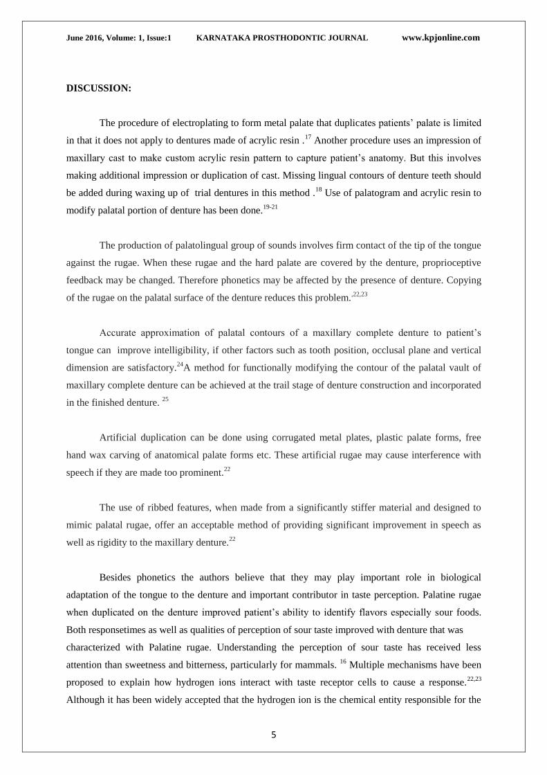
June 2016 Volume 1 Issue1 KARNATAKA PROSTHODONTIC JOURNAL wwwkpjonlinecom
9
Rehabilitation of such patients may be accomplished surgically or prosthetically Surgical
reconstruction includes microvascular flap techniques using vascularized or nonvascularized soft
tissue flaps4 However in many instances these flaps may unsuccessfully obturate the nasopharyngeal
port and patients may then be referred to the maxillofacial prosthodontist for evaluation and
treatment The presence of the flap may complicate the successful prosthetic obturation of these
surgically reconstructed defects
This article describes a stage by stage rehabilitation of an acquired lateral soft palate defect
with an interim pharyngeal obturator and a speech bulb This prosthesis made the rehabilitation
comfortable and served as a transitional and training denture prior to insertion of the more definitive
prosthesis The prosthesis helped to alleviate speech problems and assisted in the masticatory
function The speech bulb was easy to insert and remove for the patient It was also easy to fabricate
and adjust to the denture base
CLINICAL REPORT
A 29-year-old man diagnosed with adenoid cystic carcinoma of the minor palatal salivary
glands had undergone a partial maxillectomy and excision of the soft palate (Fig1) and was referred
to the Department of Prosthodontics Manipal College of Dental Sciences Mangalore India
Immediate surgical reconstruction was not recommended due to the need for further treatment with
radiation therapy The patient received postoperative external beam radiation therapy by anterior
direct beam on a telecobalt machine with a total dose of 60 Gy in 30 fractions over a period of 6
weeks The patient tolerated the radiation well and was subsequently referred for possible prosthetic
restoration of the oral defect after radiation therapy On examination of the defect laterally resected
and dysfunctional soft palate along with partial maxillectomy on the right side was noted Various
modalities of prosthetic reconstruction were discussed with the patient and the patient indicated a
June 2016 Volume 1 Issue1 KARNATAKA PROSTHODONTIC JOURNAL wwwkpjonlinecom
10
desire for an economical solution Hence heat-polymerizing interim acrylic resin prosthesis was
planned and the expectations of this prosthesis were explained to the patient
To improve patient adaptation the speech-aid device was constructed stage by stage In the
first stage the impression of the defect was obtained with irreversible hydrocolloid (Imprint Dental
Products of India Ltd) The impression was removed and poured in Type III dental stone (Dentstone
Pankaj Industries Mumbai Maharashtra India) Undercuts were blocked in Type II dental stone (Fig
2) Maxillomandibular jaw relations were obtained and prosthesis was waxed to form A heat
polymerized clasp retained acrylic-resin maxillary prosthesis was delivered to the patient (Fig 3)
After 3 weeks an acrylic-resin extension was added to the posterior border of this prosthesis This was
extended posteriorly to the intact residual soft palate and parallel to the soft tissue in the nasopharynx
approximately 3 to 4 mm short of the adjacent tissues at the maximum level of contraction Some
pressure was exerted to slightly elevate the remaining soft palate to compensate for the thickness of
the material and not to encroach on the tongue space The contours of the defect and velopharyngeal
musculature were functionally recorded with modeling compound to form the speech bulb The bulb
was placed within the nasopharynx at the plane of velopharyngeal (VP) closure The patient was
instructed in repeated swallowing so that the bulb was grossly molded but still underextended The
bulb was designed to be slightly superior to the level of the VP closure and to approximate the
pharyngeal walls so as to allow competent VP valving during speech but leave free nasal breathing
and production of nasalized speech sounds The denture base with the extension was chilled in cold
water and preparations were made for conversion of the bulb into acrylic resin Type II stone was
placed around the obturator impression to include the intaglio side of the denture base (Fig 4) The
impression material was replaced with heat-polymerizing acrylic resin (Fig 5) The patient was
instructed to wear the prosthesis at all times during the day including at meals and to remove it at
night The adaptation of the patient to the prosthesis was prompt and good although he reported
impeded breathing during strenuous activities
June 2016 Volume 1 Issue1 KARNATAKA PROSTHODONTIC JOURNAL wwwkpjonlinecom
11
The patient was scheduled for the first post-insertion adjustment 3 days after the insertion At
the first post-insertion appointment the surgical wound was observed to ensure health of the tissues to
relieve the prosthesis for pressure areas on the tissues to compensate for processing changes and to
emphasize hygiene and home care The patient was placed on a 3-month recall for evaluation and
observation of any recurrence
SUMMARY
This clinical report describes a multistep procedure for prosthetic rehabilitation of a soft
palate defect with an interim pharyngeal obturator and speech bulb The advantages of this prosthesis
are that the technique is noninvasive cost-effective tissue tolerant comfortable to use and easy to
fabricate and clean The prosthesis coupled with the patients compensatory phenomenon improved
the quality of life and provided appropriate and effective nasopharyngeal obturation
FIGURES
Fig 1 Preoperative view of the defect
Fig 2 Blocked out cast
June 2016 Volume 1 Issue1 KARNATAKA PROSTHODONTIC JOURNAL wwwkpjonlinecom
12
REFERENCES
1 Parker S Tong T Bolden S Wingo PA Cancer statistics 1997 CA Cancer J Clin 1997475-
27
2 Cancer Incidence by site Age-Standardized Rate per 100000 Ottawa Statistics Canada and
the Canadian Council of Cancer Registries Health protection Branch- Laboratory Centre for
Disease Control 2000
3 WHO Mortality Database Age- Standardized Rate per 100000 Geneva WHO Databank
1999
4 Chambers MS Lemon JC Martin JW Obturation of the partial soft palate defect J Prosthet
Dent 20049175-9
5 Curtis TA Beumer J Speech velopharyngeal function and restoration of soft palate defects
In Maxillofacial rehabilitation prosthodontic and surgical considerations eds Beumer J Curtis TA
and Marunick MT Isbiyaku EuroAmerica St Louis 1996 304-319
Fig 3 Clasp retained acrylic removable
partial denture
Fig 4 Cast with stone placed around the
obturator impression
Fig 5 Prosthesis in situ
Fig 6 Postoperative view in occusion
June 2016 Volume 1 Issue1 KARNATAKA PROSTHODONTIC JOURNAL wwwkpjonlinecom
13
Prosthetic Rehabilitation of a Patient with Atrophic
Ridges A Clinical Report
Vaibhav Gupta Dr Shobha Rodrigues
1 Dr Vidya K Shenoy
2
Dr Thilak Shetty3 Dr Sharon Saldanha
4 Dr Mahesh M
4
Dr Puneeth Hegde5
Professoramp HOD Department of Prosthodontics MCODSMangalore1
Professor amp HOD Department of Prosthodontics A J Institute Of Dental Sciences
Mangalore2
Professor Department of Prosthodontics MCODSMangalore3
Associate Professor Department of Prosthodontics MCODSMangalore4
Assistant Professor Department of Prosthodontics MCODSMangalore5
Intern Department of Prosthodontics MCODS Mangalore
ABSTRACT
Extreme resorption of the maxillary and mandibular denture-bearing area may lead to
problems with prosthetic rehabilitation As resorption progresses there is a resultant narrow more
constricted upper residual ridge opposed by a wider lower residual ridge decreased supporting
tissues that results in a large restorative space between the opposing residual ridges
This clinical report describes a method for prostheticrehabilitation of a completely edentulous
patient with hollow maxillary denture and a conventional mandibular denture contours of which are
in harmony with the neutral zone
INTRODUCTION
India has a large geriatric population of 77 million comprising 77 of its total population In
a community based study1 planned to assess the level of edentulousness denture need and denture
wear it was found that although level of edentulousness was high there was a low level of denture
wearingAmong the many factors elucidated for poor denture wear one contributory factor may be
the extremeresorption of the edentulous ridges resulting in dentures functioning as oral
acrobaticsNevertheless treatment options for prosthodontic rehabilitation of completely edentulous
patients include conventional complete dentures and implant supported fixed orremovable prosthesis
June 2016 Volume 1 Issue1 KARNATAKA PROSTHODONTIC JOURNAL wwwkpjonlinecom
14
Complete dentures are mechanical devices that must function in harmony with the
surrounding orofacial musculature In addition they must fulfillthe basic objectives of Prosthodontics
including retention stability support aesthetics and preservation of remaining tissues However
extreme resorption of the maxillaryand mandibular denture-bearing area may lead to problems with
prosthetic rehabilitation As resorption progresses there is a resultant narrow more constricted upper
residual ridge opposed by a wider lower residual ridge decreased supporting tissues that results in a
large restorative space between the opposing residualridges This may result in heavy complete
dentures that may compound to the poor denture-bearing ability of the tissues and lead to decreased
retention and resistance
The extensive volume of the denture base material in prostheses provided to patients with
severe residual ridge resorption necessitates making the denture base hollow to reduce the prosthesis
weight There are numerous references in the literature that propagate the merits of dentures
constructed in harmony with the neuromuscular function as well as describe various materials and
methods for fabrication of hollow prostheses2-18
Although controversial19
it has been suggested that
gravity and the addition of weight to the mandibular complete denture may aid in prosthesis
retention2021
In additionthe coordination of complete dentures with the neuromuscular function and
arrangement of teeth in the neutral zone is highly effective in an atrophic mandible 2andis the
foundation of a successful stable denture
Previously described techniques for weight reduction include using a solid 3-dimensional
spacer including dental stone3-13
cellophane wrapped asbestos14
silicone putty1516
or modelling
clay1718
during laboratory processing to exclude denture base material from the planned hollow cavity
of the prosthesis
Mahdy3
also presented a double flask technique that allows forthe complete fabrication of the
obturator from the waxtry-in stage to completion of the prosthesisThe primary disadvantage of such
techniques is that the long junction between the two previously polymerized portions of the denture
that is luted with autopolymerising resinis apotential site for leakage and discolouration The need for
a lightweight hollow maxillary denture and stable mandibular denture fabricated from a strong
durable material is quite evident
This clinical report describes a method for prostheticrehabilitation of a completely edentulous
patient with hollow maxillary denture and a conventional mandibular denture contours of which are
in harmony with the neutral zone
June 2016 Volume 1 Issue1 KARNATAKA PROSTHODONTIC JOURNAL wwwkpjonlinecom
15
CLINICAL REPORT
A 69-year-old man completely edentulous male patient (Fig 1 amp 2) with a history of denture
wear for the past 10years was referred to the Departmentof Prosthodontics of this Institution for
prosthetic rehabilitation of severely resorbed ridges Past medical history was noncontributory Dental
history revealed unstable and loose maxillary and mandibular dentures Intraoral examination
revealed severely resorbed ridges with increased interridge space (Fig3)Variousmodalities of
prosthetic reconstruction were discussedwith the patient and the patient indicated a desire for
aneconomical solution Hence a heat-polymerized hollow maxillary denture and a weighted
mandibular denture with prosthetic teeth arranged in the neutral zone was planned and the
expectations of this prosthesis were explained to the patient
The traditional sequence of denture construction was followed till the definitive impressions
were made and the master casts were constructed and indexed in the land areaMaxillary occlusal rim
was constructed with modelling wax (Hindustan Modelling wax Hindustan dental products
Hyderabad India) The lower wax rim was constructed on a stabilized record base with low fusing
compound(Pinnacle Dental Products of India Ltd Mumbai India) softened at 135degF and shaped
similar to a wax occlusal rim The tray and modelling compound was placed in the mouth and the
patient was instructed to swallow and purse the lips The modelling compound was hardened in the
mouth sufficiently to prevent distortionJaw relations were recorded and the casts were mounted on a
mean value articulator(Fig 4)The modelling compound was lubricated and encased in a templateof
vinyl polysiloxane putty (Reprosil Dentsply Caulk Milford Del) which serves as an index for future
teeth arrangement(Fig5)The low fusing compound was replaced with modelling wax within the
confines of the prepared index Prosthetic teeth arrangement (Premadent Super Dental Products
Delhi India) was done and the dentureswere fabricated in the conventional manner till the verification
appointmentThe mandibular denture was then processed in the conventional manner as per the
manufacturerrsquos instructions Two identical flasks were used to fabricate a hollow maxillary denture on
lines of a described article 13
For this the trial denture was processed in the standard manner
through the wax elimination stage Two layers of baseplate wax was thenadapted (Supernal SD
Dental Corporation Lucknow India) to the definitive cast in the drag conforming to the border
extensions (Fig6 A) A second identical flask was used to invest the baseplate wax and again the wax
elimination process was completed The cope and second drag was packed with heat-polymerized
acrylic resin (DPI-heat cure Dental Products of India Ltd)Similarly a minimal thickness of acrylic
resin was processed around the teeth using a different cope(Fig6 B) The original cope was seated on
the original drag and complete closure of the flask was ascertained (Fig 6 C) A thin feather edge
margin was created along the visible junction to minimize the thickness of the autopolymerising
June 2016 Volume 1 Issue1 KARNATAKA PROSTHODONTIC JOURNAL wwwkpjonlinecom
16
resinThe visiblejunction between the two previously polymerized portions was luted with
autopolymerising acrylic resin (DPI-RR Dental Products of India Ltd)The whole prosthesis was
recovered and the palatal surface was luted in a similar manner The entire junction was waxed and
reprocessed so that the seam that seals the two sections is completely covered with heat-processed
acrylic resin minimizing the stain and leakage around the area of the seam and increasing the
durability and longevity of the prosthesisThe dentures were deflasked equilibrated and a hollow
maxillary denture and weighted mandibular denture was delivered to the patient (Fig 7amp Fig8)The
patientwas instructed on home care and prosthesis maintenance
SUMMARY
This clinical reportdescribes a method for prosthetic rehabilitation of a completely edentulous
patientwith resorbed ridges and excessiveinterarchspacewith a hollow maxillary denture and a
weighted mandibulardenture contours of which are in harmony with the neutral zoneControlling the
thickness of the hollowportion without the use of any three dimensional spacer and eliminating
leakage and discoloration are severaladvantages of this technique An additional laboratory step is
however required for the final culmination of the prosthesis
FIGURES
Fig 1 Preoperative view of
patient
Fig2 Profile of the patient
June 2016 Volume 1 Issue1 KARNATAKA PROSTHODONTIC JOURNAL wwwkpjonlinecom
17
Fig 3 Intraoral view
Fig 4 Maxillomandibular jaw
relations recorded
Fig 5 Putty index for the
neutral zone
Fig6 A Adaptation of baseplate
wax to the definitive cast
Fig 6 B Acrylic resin processed
around denture teeth
Fig 6 C Denture processing using
three identical sections of two
denture flasks
June 2016 Volume 1 Issue1 KARNATAKA PROSTHODONTIC JOURNAL wwwkpjonlinecom
18
REFERENCES
1 Shah N Parkash H Sundaram KR Edentulousness denture wear and denture needs of
Indian elderly a community based study Journal of Oral Rehabilitation200431467-476
2 Gahan MJ Walmsley AD The neutral zone impression revisited Br Dent J 2005198269-
272
3 El Mahdy AS Processing a hollow obturator J Prosthet Dent 196922682-686
4 Brown KE Fabrication of a hollow-bulb obturator J Prosthet Dent 19692197-103
5 Ackerman AJ Prosthetic management of oral and facial defects following cancer surgery J
Prosthet Dent 19555413-432
6 Nidiffer TJ Shipman TH Hollow bulb obturator for acquired palatal openings J Prosthet
Dent 19577126-134
7 Rahn AO Boucher LJ Maxillofacial prosthetics principles and concepts St Louis Elsevier
1970 p 95
8 Chalian VA Drane JB Standish SM Intraoral prosthetics In Maxillofacial prosthetics
multidisciplinary practice edsChalian VA Drane JB Standish SM Baltimore Williams amp
Wilkins 1971 133-157
9 Buckner H Construction of a denture with hollow obturator lid and soft acrylic lining J
Prosthet Dent 19743195-99
10 Browning JD Kinderknecht J Fabrication of a hollow obturator with fluid resin J Prosthet
Dent 198452891-895
11 Fattore LD Fine L Edmonds DC The hollow denture an alternative treatment for atrophic
maxillae J Prosthet Dent 198859514-516
12 Gardner LK Parr GR Rahn AO Simplified technique for the fabrication of a hollow
obturator prosthesis using vinyl polysiloxane J Prosthet Dent 19916660-62
13 McAndrew KS Rothenberger S Minsley GE An innovative investment method for the
fabrication of a closed hollow obturator prosthesis J Prosthet Dent 199880129-132
Fig 7 Post-operative view of the
patient
Fig8 Hollow maxillary denture and
weighted mandibular denture
June 2016 Volume 1 Issue1 KARNATAKA PROSTHODONTIC JOURNAL wwwkpjonlinecom
19
14 Worley JL Kniejski ME A method for controlling the thickness of hollow obturator
prostheses J Prosthet Dent 198350227-229
15 Holt RA Jr A hollow complete lower denture J Prosthet Dent 198145452-454
16 Jhanji A Stevens ST Fabrication of one-piece hollow obturators J Prosthet Dent
199166136-138
17 Elliott DJ The hollow bulb obturator its fabrication using one denture flask Quintessence
Dent Technol 1983713-14
18 DaBreo EL A light-cured interim obturator prosthesis A clinical report J Proshet Dent
199063371-373
19 Ohkubo C Hosoi T Effect of weight change of mandibular complete dentures on chewing
and stability a pilot study J Prosthet Dent 199982636-642
20 Jacobson TE Krol AJ A contemporary review of the factors involved in complete denture
retention stability and support Part I retention J Prosthet Dent 1983495-15
21 Wormley JH Brunton DA Weighted mandibular dentures J Prosthet Dent 197432101-102
June 2016 Volume 1 Issue1 KARNATAKA PROSTHODONTIC JOURNAL wwwkpjonlinecom
20
Prosthodontic Management of Marginal
Hemimandibulectomy With Surgically Induced Lip Drop
Dr Krishna Prasad D1 Dr Anupama Prasad D
2 Dr Anshul Bardia
3
Department of Prosthodontics and Crown amp Bridge ABShetty Memorial Institute of Dental
Sciences Nitte UniversityDeralakatteMangaluruKarnataka India -575018
ABSTRACT
Loss of continuity of the mandible destroys the balance and symmetry of mandibular
function leading to altered mandibular movements and deviation of the residual fragment towards the
resected side The rehabilitation of these cases must be carefully planned and the treatment requires a
denture construction in such a way to get maximum retention support stability esthetics and
function This case report describes the treatment of a patient with partially resected edentulous
mandible combining functional and esthetic requirements A removable prosthetic appliance was
fabricated to maintain the lip in its normal position thereby helping the patient to perform normal
functions All basic principles of rehabilitation are applied and interpretation was based on altered
anatomic and functional situation An esthetic configuration with ideal function was achieved and the
occlusion showed a satisfactory stability
KEYWORDS Hemimandibulectomy maxillofacial rehabilitation esthetics
INTRODUCTION
One of the most challenging and demanding maxillofacial endeavours is the construction of
functional complete dentures for the edentulous patient who has undergone a mandibular resection
Loss of continuity of the mandible destroys the balance of the mandibular movement and function
leading to altered mandibular movement and deviation of the residual fragment towards the surgical
side The greater the loss of tissues greater will be the deviation of the mandible to the resected side
thus compromising the prognosis of the prosthetic rehabilitation to a greater extent
Professor1 Reader
2 Former Post graduate student
3
Corresponding author DrAnupama Prasad D Reader Department of Prosthodontics
and Crown amp Bridge A B Shetty Memorial Institute of Dental Sciences Nitte University
Deralakatte Mangaluru -575018 Email anupamaprasaddymailcom
Apart from deviation other dysfunctions in such patients are observed in swallowing speech control
of saliva mandibular movements mastication respiration and psychic functioning1
June 2016 Volume 1 Issue1 KARNATAKA PROSTHODONTIC JOURNAL wwwkpjonlinecom
21
Based on the nature of resection Cantor and Curtis (1971) devised a prosthetic classification
that is as follows2
Class I Radical alveolectomy with preservation of mandibular continuity
Class II Lateral resection of the mandible distal to the cuspid
Class III Lateral resection of the mandible and maxilla
Class IV Lateral bone graft surgical reconstruction
Class V Anterior bone graft surgical reconstruction
Class VI Resection of the anterior portion of the mandible without reconstructive surgery to unite the
lateral fragments
In cases with class II III IV V guide flange prosthesis would be a treatment modality One
of the basic objectives in rehabilitation is to retrain the muscles for mandibular denture control and
repeated occlusal approximation
This article highlights prosthetic rehabilitation of a class I hemimandibulectomy patient for
whom a mandibular prosthesis is fabricated with loop on corner of mouth for lip support
CASE REPORT
A 74 year-old completely edentulous female patient was referred to the Department of
Prosthodontics after extraction of remaining natural teeth which were periodontally compromised for
maxillofacial rehabilitation with a chief complaint of difficulty in eating and speaking Her medical
history revealed that she was diagnosed for squamous cell carcinoma on the right side of the
mandible for which she had undergone marginal resection of mandible on right side 3 years back
The patientrsquos history indicated that she had a tobacco-chewing habit since 40 years An extra oral
examination revealed asymmetrical face and a convex profile There was a deviation of the mouth to
the right side that is toward resected side
On intraoral examination it was found that the maxillary and mandibular arches were
completely edentulous On palpation the mandibular ridge was present till first premolar region On
evaluation of pre extraction diagnostic ortho-pantamogram segmental absence of the mandible distal
to the first premolar to the last molar on the same side was noted This particular case represents to
class I Cantor and Curtis classification
June 2016 Volume 1 Issue1 KARNATAKA PROSTHODONTIC JOURNAL wwwkpjonlinecom
22
CLINICAL PROCEDURE
Preliminary impressions were made with irreversible hydrocolloid material (Zelgan Plus
Dentsply Gurgaon India) using stock trays Casts were prepared (Fig 1) and self-cure clear acrylic
resin (RR Dentsply India) custom trays were constructed The tray was border-molded with
modeling plastic (DPI Tracing stick Dental products of India Mumbai India) taking care to avoid
overextension Final impressions were made with light-body vinyl polysiloxane (AquasilDentsply
Caulk Milford DE) While in case of mandibular final impression tray handle was extended with
autopolymerizing resin and a cylindrical mandibular rest is fabricated in the posterior region with an
increased vertical height Then softened impression compound was placed on the top of the
mandibular rests and inserted in the patientrsquos mouth Patient was advised to close her mouth so that
the mandibular rest fit against the maxillary alveolar ridge
This was done to stabilize the tray in position by preventing anterioposterior and mediolateral
displacement of the tray during final impression and which was made using the light body material
and the patient was asked to close the mouth such that cylindrical rest will fit over maxillary ridge
For recording the functional state patient was instructed to run his tongue along her lips suck in her
cheeks pull in her lips and swallow by keeping her mouth closed till the impression material
hardened
Master casts were poured with Type III dental stone (DPI Mumbai India) Stabilized record
bases were made with self-cure acrylic (DPI Mumbai India) using the sprinkle-on technique Wax
rims were adjusted until a tentative occlusal vertical dimension was established Face bow transfer
was made to orient the maxillary cast to the semi-adjustable articulator (Artexsemiadjustable
articulator rotofix face bow) Maxillomandibular relations were recorded with wax interocclusal
records The patient tactile sense and sense of comfort was used to assess the vertical dimension of
occlusion The patient was asked to move her mandible as far possible to the untreated side and then
gently close her jaw into position to record a functional maxillomandibular relationship
The teeth were arranged in the usual manner semi-anatomic posterior teeth (Acryrock Pyrax
polymers Roorkee India) were used Maxillary and mandibular teeth were arranged to achieve
balanced occlusion Occlusal table on resected side was up to the second premolar just to establish
the cross arch stability and balance in the right lateral excursive movements A wax set-up was tried
in the mouth and was checked for esthetics phonetics occlusal vertical dimension and balanced
occlusion The basic objective is to achieve an occlusal scheme which will have a multiplicity of
occlusal contacts in centric position Long centric concept and to a slightly decreased vertical
June 2016 Volume 1 Issue1 KARNATAKA PROSTHODONTIC JOURNAL wwwkpjonlinecom
23
dimension of occlusion in an attempt to decrease occlusal force is given The level of the occlusal
plane especially in edentulous patients should be acceptable to the remaining portion of the tongue to
permit easier distribution and control of food on the occlusal table and control of complete denture
prosthesis3 A posterior palatal seal was recorded and the dentures were waxed processed (DPI RR
heat cure DPI India) and remounted and the occlusion was refined Freedom of movement and lack
of cuspalintercuspation was checked before denture insertion The dentures were evaluated intraorally
and the mandible was manipulated to the static centric position area4 Any interference in normal
movements was corrected During insertion to improve the tissue contact situation resilient liner
(UfigelVOCO gmbh ) was used to reline the mandibular denture by keeping the mandible into the
maximum intercuspation position The sealer was applied once over the polymerized surface of the
resilient liner which prevents water sorption by the liner and helps in maintaining the softness for a
longer period of time The dentures were removed repolished and then reinserted
The prosthesis design is composed of snapfit buttons (Fig 2) which includes male and female
component in which male component is attached to lower mandibular denture in premolar region and
female component in removable segment (Fig 3) to which stainless steel wire is attached curving
out at the anterior end to form a loop supporting the lip extraorally The removable component of
prosthesis was fabricated with self-cure acrylic resin and it was designed such that patient can easily
remove the removal component during mastication
Addition of a 21-gauge stainless steel wire in the form of a J-shaped buccal loop to engage the
corner of the mouth of the unaffected side in order to pull the corner of the mouth and achieve an
esthetically pleasing appearance The wire loop was embedded in the acrylic of the buccal flange of
the removal segment of the denture5 (Fig 4) It was adjusted to ensure that its position provided
circumoral symmetry and esthetics without compromising comfort and simulated functional jaw
movements The extraoral wire components were relined with permanent tissue conditioner to reduce
the shine so as to blend with the skin Follow-up appointments were carried out routinely to ensure
patient comfort and satisfaction No discomfort or any problems in mouth opening or mastication
were noted resulting from the J-shaped loop and the patient was quite happy with the prosthesis
The patient was instructed to chew only on the non-resected side to avoid denture instability
It may be necessary to accept an occlusion that is not bilaterally balanced in eccentric occluding
positions for an edentulous resected maxilla or mandible The patient was given routine post insertion
instructions and was motivated to make efforts to learn to adapt to the new dentures Simple exercises
were suggested to the patient such as repeated opening and closing of mandible This helped the
patient learn to manipulate the lower prosthesis into the proper position Initially retention of the
June 2016 Volume 1 Issue1 KARNATAKA PROSTHODONTIC JOURNAL wwwkpjonlinecom
24
dentures especially of the lower one was a problem but this improved with constant use Within a
week the patient expressed satisfaction in mastication phonetics and esthetics and drastic
improvement is seen from initial stages of prosthesis planning (Fig 5) and after the fit and insertion of
the final prosthesis (Fig 6)
DISCUSSION
The prosthetic rehabilitation of a hemimandibulectomy subject is a difficult task for a
prosthodontist as the normal physiological functions like swallowing speech mandibular movements
mastication control of saliva and respiration are adversely affected by radical mandibular surgery
These dysfunctions radically alter the prosthetic prognosis Surgical reconstruction by implants and
grafts of various types is the ideal treatment when feasible
In the present case the OPG (Fig7) and intra oral pictures (Fig8) revealed the absence of
mandibular segment As the surgical reconstruction is not always feasible in every patient
prosthodontic approach has to be considered to restore the esthetic and function in such subject
Because of the loss of the normal anatomy and physiology of the oral cavity many principles of
complete denture prosthesis must be compromised Since the mandibulectomypatients have reduced
masticatory strength and little soft and hard tissue support it is important to record and utilize as
broad denture base as possible within the physiological limits
Closed-mouth impression techniques have been suggested but these were designed for
making accurate static impressions6 The column trays described in this article are similar in form but
they are used to record the muscular dynamics of the postsurgical lower denture space The reasons
for increasing the height of the lateral columns of the custom trays are as follows-
1 To reduce the amount of force exerted by the remaining muscles of mastication
2 To make swallowing more difficult
This type of ―stress swallowing will cause extreme muscular activity of the residual tongue
and floor of the mouth An impression of this functional activity should help prevent future denture
displacement In the final denture form the tissue conditioner placed on the dentures when they are
first inserted provides comfort during the adjustment period corrects any tissue surface discrepancies
resulting from the impression material and refines the final denture form during function
Lott and Levin stated that retention will increase in proportion to an increase in the area
covered by the denture Boucher states that the amount of biting force tolerated by a denture is
proportional to the size of the tissue-bearing area Since hemimandibulectomy patients have markedly
June 2016 Volume 1 Issue1 KARNATAKA PROSTHODONTIC JOURNAL wwwkpjonlinecom
25
reduced masticatory strength and little soft and hard-tissue support it is important to record and
utilize as broad denture base as is possible The use of a tissue-conditioning agent facilitates the
extension of a functional denture form to the maximum size tolerated by the oral tissues This form
should enhance the patientrsquos ability to manipulate the prosthesis and to realize maximal masticatory
potential2
Facial symmetry could be improved with the use of removable prostheses Esthetics has to be
compromised however because labial commissural sag is necessary if a functional seal is to be
maintained between the lips Without this seal drinking and speaking appear to be much more
difficult7 In this particular case an effort was made to restore the patientrsquos appearance and comfort by
repositioning and supporting the lip in a natural position with the described prosthesis Since the J-
hook was lined with permanent tissue conditioner which does not cause any irritation and it has to be
changed every 6 month patient has to wear for full time except during mastication of hard food
CONCLUSION
The described technique offers an inexpensive simple and expedient approach to manage the
hemimandibulectomy patient The availability of well-formed edentulous ridges and an excellent
peripheral seal permitted excellent retention and stability of the dentures and the presence of the loop
to support oral commisure The philosophical approach to the treatment and rehabilitation of
edentulous patients with resected mandibles is not in concentrating on what has been sacrificed in the
eradication of the disease but rather in taking full advantage of the remaining structures
FIGURES
Figure 1 Maxillary and
mandibular diagnostic cast
Figure 2 Snapfit button include male
and female component
June 2016 Volume 1 Issue1 KARNATAKA PROSTHODONTIC JOURNAL wwwkpjonlinecom
26
Figure 4 Prosthetic design with extroral
loop
Figure 3 Male component in the
mandibular denture and female
component in the removable segment
Figure 5 Preoperative photograph
Figure 6 Postoperative
photograph
Figure 7 Pre extraction
diagnostic OPG
Figure 8 Intra oral view
June 2016 Volume 1 Issue1 KARNATAKA PROSTHODONTIC JOURNAL wwwkpjonlinecom
27
REFERENCES
1 Beumer J Curtis T Firtell D Maxillofacial rehabilitation St Louis Mosby 1979 p 90-169
2 Cantor R Curtis TA Prosthetic management of edentulous mandibulectomy patients Part 1
Anatomic physiologic and psychologic consideration J Prosthet Dent 1971 25446-57
3 Ronald P Desjardins Occlusal considerations for the partial mandibulectomy patient J Prosthet
Dent 1979 41308-315
4 Cantor R Curtis TA Prosthetic management of edentulous mandibulectomy patients Part II
Clinical procedures J Prosthet Dent 1971 25546-55
5 Bagchi G Nath DK Restoration of facial symmetry in a patient with bell palsy using a modified
maxillary complete denture a case report Int J Prosthodont 2012 25(3)290-3
6 MacMillin J J Closed Mouth Technique for Impressions of the Lower Jaw J Amer Dent Ass
34 715 1947
7 Larsen SJ Carter JF Abrahamian HA Prosthetic support for unilateral facial paralysis J Prosthet
Dent 1976 35(2)192-201
June 2016 Volume 1 Issue1 KARNATAKA PROSTHODONTIC JOURNAL wwwkpjonlinecom
28
Short Dental Implants ndash A Review Of Clinical
Performance Biomechanical Aspects And Risk Factors
For Survival
Dr Talreja Karishma S Dr Rodrigues Shobha J2 Dr Pai Umesh Y
3
Department of Prosthodontics Manipal College of Dental Sciences Mangalore Manipal
University
INTRODUCTION
Implant supported prosthesis are gradually becoming the norm for restoration of missing
teeth1
The posterior edentulous arches are a biologically and mechanically challenging area for
rehabilitation with implant supported prostheses These regions have unfavourable bone quality and
lesser bone volume as compared to anterior edentulous sites compelling the operator to place shorter
implants The poor bone quality limits the number of implants placed thus increasing bending forces
on individual implants Furthermore occluding force increases the closer the teeth are placed to the
temporomandibular joint2
The obsolete protocol of placing the longest possible implant within anatomical limitations
has lead authors to employ procedures like distraction osteogenesis bone grafting guided bone
regeneration sinus floor elevation and mandibular nerve repositioning to gain adequate residual ridge
height at these sites These techniques have a variable degree of success and require considerable
dexterity and skill from the operator Short dental implants open up an exciting portal out of
complicated surgical procedures involved in implant site preparation in posterior atrophic arches
Short Dental implants (SDI) are a more cost-effective alternative that reduces treatment time
and rules out complications related to surgical and grafting procedures Authors in their studies have
quoted different lengths however considering 10mm as the standard length an implant less than
10mm in length is considered a Short Dental Implant and is usually applied in alveolar ridges with
decreased bone height3
The biomechanical rationale in support of SDIs is that loading bearing forces are concentrated
on the crestal portion of the implant and an increase of implant length from 7 to 10mm does not
significantly improve its anchorage 4 Instead with an increase of every 1mm in the implant diameter
the functional surface area increases by 30-200 thus improving the load dissipation ability of the
implant5 Recent Finite Element Analyses has demonstrated that implant length had no effect on stress
concentration on crestal bone around an implant hence a SDI may be a sound choice6
BDS 3rd
year Post Graduate Student Head of Department2
Associate Professor3
Corresponding Author Dr Karishma S Talreja Department of Prosthodontics
ManipalCollege of Dental Sciences Light House Hill Rd Mangalore-575001
Contact no- +919930794574 Email address- karishmatalreja90gmailcom
June 2016 Volume 1 Issue1 KARNATAKA PROSTHODONTIC JOURNAL wwwkpjonlinecom
29
Friberg and Jemt 27
were among the early authors to note high failure rates in both arches
with short fixtures (7mm) Early failure rate was pronounced in resorbed arches with poor bone
quality However the implants used in this study were of narrow diameter and had a smooth
machined surface SDIs are designed to provide an increased Bone-to-implant contact by virtue of an
increased diameter Newer SDIs overcome such limitations by incorporation of surface modifications
like acid-etching that increase the surface area for osseointegration
This article is a review of the many aspects of risk factors for success and performance of
SDIs under various clinical scenarios
RISK FACTORS
The risk factors for failure of SDIs may be broadly divided into endogenous (systemic or
local) and exogenous (operator or biomaterial-related) factors8
Endogenous factors-
SMOKING
Mezzomo et al9 in a meta-analysis on success rate of single crowns found a higher failure
percentage in studies wherein smokers were included as compared to studies that excluded them
Strietzel amp Reichert found a significant association between heavy smoking (gt10 cigarettesday) and
frequency of implant loss10
SYSTEMIC DISEASES
Most studies exclude pregnant women immunocompromised patients and those under
medication from their sample size This impairs the assessment of implant survival in such patients
For single crowns supported by SDIs no statistically significant difference was found in the failure
percentage in systemically compromised patients9
BRUXISM
Twail et al11
found more incidences of prosthetic failures like veneer fractures and screw
loosening in bruxer groups however no statistically significant difference was found on inter group
comparison between buxer non-bruxer and occasional bruxer groups
PERIODONTAL DISEASE
The biological failure proportion of studies that included periodontal patients did not show a
statistically significant upward trend as compared to studies that did not include periodontal patients
Marginal bone loss in periodontal groups however was found to be significantly higher
Perimplantitis and persistent periodontal disease are major risk factors for the loss of integration of
SDIs9
BONE QUALITY
Studies have failed to find an association between high failure rates and low quality
bone101218
On the other hand higher failure rates were associated with machines surface implants as
compared to rough surface implants in poor quality bone11
The density of the bone directly correlates
to the strength of the bone with less density demonstrating strength reduction of 50 -80 compared
June 2016 Volume 1 Issue1 KARNATAKA PROSTHODONTIC JOURNAL wwwkpjonlinecom
30
to high density bone types13
Weng et al14
noted a 25 failure rate of SDI (machined surface)
supported prostheses in the posterior maxilla failures occurring within 18 months of loading Hence
rougher surfaces for implants are preferred in poor bone quality Finite element analysis has found
that maximum Von-Mises stress variability was minimal when the diameter of SDIs was within 55
and 71mm Peak stress on the implant-bone interface is seen to increase with reduction in bone
density3 Osteopenic bone has thin cortices and reduced spongiosa hence needs larger diameters for
optimal load bearing capacity Implant diameter in excess of 4mm and length more than 9mm are
optimal properties for screwed implants in type IV bone15
OPERATOR RELATED RISK FACTORS
Operator related risk factors include the surgical technique prosthetic design and loading
protocol undertaken in the placement of the implant
SURGICAL TECHNIQUE
Misch et al16
proposed employing a one stage approach in D2 bone by adding a permucosal
extension at the time of surgery and a two-stage approach in D4 bone While a two-stage implant
placement approach has been suggested by some authors17
no significant difference has been found in
failure rates between single-stage and two-stage implants Also in fully edentulous patients two-stage
implants are preferred4918
Esposito et al 19
concluded that a submerged approach may be preferable in
implants that do not achieve optimal primary stability and in completely edentulous cases
CROWNIMPLANT RATIO
The crown height is a vertical cantilever and when increased from 10 to 20mm the force on
the implant is increased by 100 An angled prosthetic load is also a force magnifier on the implant
Hence detrimental effects of non-axial forces on crestal bone increase with increase in crown
height16
A high crown- to- implant ratio was assumed to have a negative biological effect on crestal
bone loss20
Peri-implant bone resorption is similar in all implant-to-crown ratio groups even when
increased by 2 to 3 times provided non axial forces were controlled11
Rossi Tawil Mertens and
Deporter et al claimed that increased CI ratio placed no deterimental effects on the health of the
implant11202122
Nedir and Birdi et al2324
evaluated crown-implant ratios ranging from 105 to 180
and 09 to 32 respectively to find no detrimental effects on surrounding bone Current research has
rejected crown-implant ratio as a major biomechanical risk factor as long as occlusal contacts are
placed as close as possible to the long axis of the implant and favourable force orientation and load
distribution is maintained11
Crown height space on the other hand is a more reliable indicator of detrimental effects on
marginal bone when crown height spaces exceed 15-mm length25
For each additional millimeter of
crown height stress concentration at the implant neck may increase by 2026
Hof et al 27
observed
greater bone loss in the anterior maxilla with increased crown-to-implant ratio than the posterior
areas This may be possibly explained by off-axis loading at the implant-bone interface
June 2016 Volume 1 Issue1 KARNATAKA PROSTHODONTIC JOURNAL wwwkpjonlinecom
31
PLATFORM SWITCHING
Platform switching shifts the stress concentration zone from the crest bone-implant interface
to the axis of the implant thus reducing stress levels at the cervical bone area28
Telleman et al29
from
the results of a randomized control trial found that 1 year post loading inter proximal bone levels were
better maintained at implants restored according to the platform switching concept
IMPLANT NUMBER AND SPLINTING
Factors contributing to marginal bone loss around dental implants include surgical trauma
faulty implant positioning occlusal overloading or non-axial loading30-32
Stress level in bone around
splinted implants is found to be lower than bone around unsplinted implants by a factor of 933
A
positive influence of splinting and number of splinted implants has been observed on success rate of
SDIs in atrophic posterior arches up to a 10 year follow up period1620273435
Placement of additional
implants increases the effective surface area for stress distribution Hence one implant for each
missing premolar and two for each missing molar were suggested16
To further capitalize on functional
area these must be splinted
WIDTH OF OCCLUSAL TABLE AND TYPE OF OCCLUSION-
Within 54 and 83mm the width of the occlusal table did not significantly affect peri implant
bone loss11
Axial forces distribute stress more evenly throughout the implant as compared to bending
moments Occlusion should be mutually protected and prostheses should be free of non axial
loading1116
CANTILEVER FORCES
The length of the posterior cantilever in the mandible is directly related to complications
andor failure of the prosthesis3637
Romeo et al 38
found no detrimental effects of cantilevers
provided cantilever length was appropriate and occlusal function was under control Mesial and distal
cantilever lengths limited to 275 +-165 and 224+-160mm respectively have found to cause
marginal bone loss within acceptable limits11
LOADING PROTOCOL
Most authors follow and recommend a delayed loading protocol for SDIs Rossi et al21
conducted a study using SLActive straumann 6mm implants that were early loaded (6 weeks after
insertion) These implants yielded high survival rates and moderate loss of bone after two years of
loading However long-term follow-up larger sample size and randomized trials are required to
provide concrete evidence for incorporation of early loading protocols into clinical use
BIOMATERIAL RELATED FACTORS
Implant length implant diameter surface topography and implant thread pitch are important
parameters that influence the selection of the most fitting implant in a given clinical situation
June 2016 Volume 1 Issue1 KARNATAKA PROSTHODONTIC JOURNAL wwwkpjonlinecom
32
IMPLANT LENGTH-
Implant length is defined as the length between the implant neck and the implant apex
Increase in implant length has found to have minimal beneficial effect on load distribution around the
crestal portion of the implant6 Mezzemo et al
9 in a meta-analysis stated that short implants supporting
single crowns obtained similar if not superior survival rates as compared to standard length implants
Few studies exist on implants of 5 and 6mm length thus limiting the data obtained from systematic
reviews A two year trial of implants of four millimeter length with SLActive surfaces has yielded
survival rates of 957 after 1 year and 923 at the end of the trial 34
Ling Sun et al18
have reported
highest survival rate for implant lengths of 75 and 9mm But no statistically significant difference
exists based on length
IMPLANT DIAMETER
For every 1mm increase in diameter functional SA is increased by 30 ndash 200 along with
BIC5 Sato et al
39 on the basis of an in vitro study stated that wide implants are capable of bearing
larger loads and perform better than implants of smaller diameter under tensile forces Wider
diameters of implants are hence referred for reduced bone density This however is limited by the
bucco-lingual width of the residual ridge
IMPLANT SURFACE
Griffin and Cheung40
suggested ―the implant maximized surface area as the main contributing
factor to the high success rate Rougher surfaces offer extensive area for osseointegration and have
better bone-implant-contact as compared to machined or acid ndash etched surfaces16-18
Various surface
modified implants like SLActive surfaces213441
TiOblast implants Astra Tech20
and bioabsorbable
HA blasted implants 16
have shown better results as compared to the poor results seen with machined
surface implants7
IMPLANT THREAD PITCH-
Thread pitch is defined as the distance between adjacent threads or the number of threads per
unit length in the same axial plane and on the same side of the axis5 Hence the greater the implant
pitch the greater the surface area available for osseointegration and load dissipation Another implant
thread geometry parameter worth consideration in this context is thread depth16
Misch16
has suggested a protocol for the reduction of stress at the bone-implant interface for
SDIs they include-
1) no cantilevers on the prostheses
2) no angled forces to the posterior restorations
3) splinting multiple implants together
4) implant surface modification
5) increase implant thread pitch
June 2016 Volume 1 Issue1 KARNATAKA PROSTHODONTIC JOURNAL wwwkpjonlinecom
33
INDICATIONS
Annibali et al4 in a systematic review reported successful results for short dental implants
with a pooled survival rate of 991 and a low incidence of biological and biomechanical
complications after a mean follow-up period of 32+- 17 years
Studies have evaluated the efficiency of 6mm vs 10mm implants supporting fixed partial
dentures in augmented bone41
6mm vs 11mm implants combined with sinus floor elevation
supporting single crowns42
and 6 5mm implants rehabilitating bilateral atrophic posterior arches vs
longer implants in augmented bone 4344
to find similar if not better performance of SDIs with fewer
post-operative complications in comparsion to conventional implants in augmented bone Based on
the results of randomized control trials and clinical studies the following indications of SDIs in
atrophic arches can be put forth41-44
1) implant supported single crowns
2) implant supported fixed partial dentures
3) implant supported overdentures
The need for long term follow-up studies is quintessential to evaluate the effect of bone loss
on the survival of the SDIs While the loss of 2mm of crestal bone has minimal impact on the stability
of a 10 mm or longer implant a similar bone loss pattern on a 7mm implant for example leaves
behind a considerably lesser bone volume for load dissipation
The assessment of failure rates of SDIs should consider the poor quality of bone that is
commonly observed in atrophic arches indicated for SDIs in comparison to bone found in regions
indicated for conventional implants and rather be compared to the outcome of implants placed in
grafted sites5
ADVANTAGES OF SDIs-16
Lack of bone grafting reduces cost and duration of treatment
Surgical risk of sinus perforation mandibular paresthesia is eliminated along with decreased
chances of overheating the osteotomy site or damage to dilacerated adjacent tooth root
No need for additional inventory and decreased surgical complexity
Implant placement in smaller interarch space
LIMITATIONS
The reversed crown to implant ratio may not be an esthetic concern in the posterior
quadrants however it may not be acceptable in the anterior maxilla Here the morbidity related to an
autologous bone graft for reconstruction must be considered20
Other than this there is a draught of
data on results of long term clinical trials of SDIs in poor quality bone Also management of atrophic
ridges that have horizontal ridge insufficiency with SDIs is a question that still remains unanswered
June 2016 Volume 1 Issue1 KARNATAKA PROSTHODONTIC JOURNAL wwwkpjonlinecom
34
REFERENCES
1 Jemt T Lekholm U Adell R Osseointegrated implants in the treatment of partially
edentulous patients A preliminary study on 876 consecutively placed fixtures Int J Oral
Maxillofac Implants 19894211-217
2 Jemt T Lekholm U Oral implant treatment in posterior partially edentulous jaws A 5-year
follow-up report Int J Oral Maxillofac Implants 19938635-640
3 Kang N Wu YY Gong P Yue L O GM A study of force distribution of loading stresses on
implant-bone interface on short implant length using 3-dimensional finite element analysis Oral
Surg Oral Med Oral pathol Oral Radiol 2014 118519-523
4 Annibali S Cristalli MP DellrsquoAquila D Bignozzi I La Monaca G Pilloni A Short dental
implants A Systematic Review J Dent Res 91(1)25-322012
5 Misch C Bidez MW Contemporary Implant Dentistry St Louis Mo CV Mosby 1999
6 Fugazzotto PA Shorter implants in clinical practice Rationale and treatment results Int J
Oral Maxillofac Implants 200823487-496
7 Friberg B Jemt T Lekholm U Early failures in 4641 consecutively placed Branemark dental
implants A Study from stage 1 surgery to the connection of completed prostheses Int J Oral
Maxillofac Implants 19916142-146
8 Esposito M Hirsh J-M Lekholm U Thomsen P Biological factors contributing to failures of
osseointergrated oral implants Euro Oral Sci 1998106721-764
9 Mezzomo et al Meta-analysis of single crowns supported by short (lt10mm) implants in the
posterior region J Clin Periodontol 2014 41 191-213
10 Strietzel F P amp Reichert P A (2007) Oral rehabilitation using Camlog screw-cylinder
implants with a particle-blasted and acid-etched microstructured surface Results from a
prospective study with special consideration of short implants Clinical Oral Implants Research 18
591ndash600
11 Tawil et al Influence of prosthetic parameters on the survival and complication rates of Short
Dental Implants Int J Oral Maxillofac Implants 200621275-282
12 Nicolau P Korostoff J Ganeles J Jackowski J Krafft T Neves M Divi J Rasse M
Guerra F amp Fischer K (2013) Immediate and early loading of chemically modified implants in
the posterior jaws 3-year results from a prospective randomized multicenter study Clinical
Implant Dentistry and Related Research 15 600ndash612
13 Misch CE Qu Z Bidez MW Mechanical properties of trabecular bone in the human
mandible Implications for dental implant treatment planning and surgical placement J Oral
Maxillofac Surg 199957700-706
14 Weng D Jacobson Z Tarnow D et al a prospective multicenter clinical trial of 3i machined-
surface implants Results after 6 years of follow-up Int J Oral Maxillofac Implants 200318417-
423
15 Li T Kong L Wang Y Hu K Song L et al Selection of optimal dental implant diameter and
length in type IV bone a three dimensional finite element analysis Int J Oral Maxillofac Surg
2009381077-1083
16 Misch C Steigenga J Barboza E Misch-Dietsh F Cianciola L Kazor C Short dental
implants in posterior partial edentulism a multicenter retrospective 6-year case series study J
Periodontol 2006771340-1347
17 Gentile MA Chuang SK Dodson TB Survival estimates and risk factors for failure with 6 times
57-mm implants Int J Oral Maxillofac Implants 200520930ndash937
June 2016 Volume 1 Issue1 KARNATAKA PROSTHODONTIC JOURNAL wwwkpjonlinecom
35
18 Sun H Huang C Wu Y Shi B Failure rate of short (lt= 10mm) dental implants and factors
influencing their failure A Systematic Review Int J Oral Maxillofac Implants 200126816-825
19 Esposito M Grusovin MG Chew YS Coulthard P Worthington HV One-stage versus two-
stage implant placement A Cochrane systematic review of randomised controlled clinical trials
Eur J Oral Implantol 2009291ndash99
20 Mertens C Meyer-Baumer A Kappel H Hoffmann J Steveling H Use of 8-mm and 9-mm
Implants in atrophic alveolar rides 10-year results Int J Oral Maxillofac Implants 2012271501-
1508
21 Rossi F Ricci E Marchetti C Lang N P amp Botticelli D (2010) Early loading of single
crowns supported by 6-mm-long implants with a moderately rough surface a prospective 2-year
follow-up cohort study Clinical Oral Implants Research 21 937ndash943
22 Deporter D A Kermalli J Todescan R amp Atenafu E (2012) Performance of sintered
poroussurfaced press-fit implants after 10 years of function in the partially edentulous posterior
mandible International Journal of Periodontics and Restorative Dentistry 32 563ndash570
23 Nedir RBischof MBriaux JMBeyer SBernard JPA 7-year life table analysis from a
prospective study on ITI implants with special emphasis on the use of short implants Clin Oral
Implants Res 200415150ndash157
24 Birdi H Schulte J Kovacs A Weed M Chuang SK Crown-to-Implant ratios of short length
implants J Oral Implant VolXXXVINo Six2010
25 Kim YOh TJMisch CEWang HLOcclusal considerations in implant therapy clinical
guidelines with biomechanical rationale Clin Oral Implants Res 2005 1626ndash35
26 Blanes RJ Bernard JP Blanes ZM Belser UC A 10-year prospective study of ITI dental
implants placed in the posteriorregionIIinfluenceof thecrown-to-implantratioand different
prosthetic treatment modalities on crestal bone loss Clin Oral Implants Res 2007 18707ndash714
27 Hof M Pommer B Zukic N Vasak C Lorenzoni M Zechner W Influence of Prosthetic
Parameters on Peri ndashImplant Bone Resorption in the first year of loading A Multifactorial
Analysis Clinical Implant Dentistry and Related Reasearch Vol 17 Supplement 1 (2015)
28 Maeda Y Miura J Taki I amp Sogo M (2007) Biomechanical analysis on platform
switching is there any biomechanical rationale Clinical Oral Implants Research 18 581ndash584
29 Telleman et al Impact of platform switching on inter-proximal bone levels around short
implants in the posterior region I year results from a randomized clinical trial J Clin Periodontol
2012 39688-697
30 Tomasi C Sanz M Cecchinato D et al Bone dimensional variations at implants placed in
fresh extraction sockets a multilevel multivariate analysis Clin Oral Implants Res 2010 2130ndash
36 6 Nissan J Ghelfan O Gross O Priel I Gross M Chaushu G
31 The effect of crownimplant ratio and crown height space on stress distribution in unsplinted
implant supporting restorations J Oral Maxillofac Surg 2011 691934ndash1939
32 Pozzi A Agliardi E Tallarico M Barlattani A Clinical and radiological outcomes of two
implants with different prosthetic interfaces and neck configurations randomized controlled split-
mouth clinical trial Clin Implant Dent Relat Res 2012 [Epub ahead of print]
33 Bergkvist G Simonsson K Rydberg K Johansson F Derand T A finite element analysis of
stress distribution in bone tissue surrounding uncoupled or splinted dental implants Clin Implant
Dent Relat Res 20081040ndash46
34 Slotte C Gronningsaeter A Halmoy A Ohrnell L Stroh G Isaksson S Johansson L Four-
millimeter implants supporting fixed partial dental prostheses in the severely resorbed posterior
June 2016 Volume 1 Issue1 KARNATAKA PROSTHODONTIC JOURNAL wwwkpjonlinecom
36
mandible two-year results Clinical Implant Dentistry and Related Research Vol 14 Supplement
12012
35 Nissan J Gross O Ghelfan O Priel I Gross M Chaushu G The effect of splinting implant-
supported restorations on stress distribution of different crown-implant ratios and crown height
spaces J Oral Maxillofac Surg 2011 692990ndash 2994
36 White S Caputo AA Anderkvist T Effect of cantilever length on stress transfer by implant
supported prostheses J Prosthet Dent 199471493-499
37 Rangert BEng MSullivan RJemt TLoad factor control for implants in the posterior partially
edentulous segmentInt J Oral Maxillofac Implants 199712360ndash370
38 Romeo ELops DMargutti EGhisolfi MChiapasco MVogel G Implant-supported fixed
cantilever prostheses in partially edentulous archesA seven year prospective studyClin Oral
Implants Res 200314303ndash311
39 Sato Y Shindoi N Hosokawa R Tsuga K Akagawa Y A biomechanical effect of wide
implant placement and offset placement of three implants in the posterior partially edentulous
region J Oral Rehabil 20002715ndash21
40 Griffin TJ Cheung WS (2004) The use of short wide implants in posterior areas with
reduced bone height a retrospective investigation J Prosthet Dent 92139-144
41 Romeo E Storelli S Casano G Scanferla M Botticelli D six-mm versus 10-mm long
implants in the rehabilitation of posterior edentulous jaws a 5-year follow-up of a randomised
control trial
42 Gulje F Raghoebar G Vissink A Meijer H single crowns in the resorbed posterior maxilla
supported by either 6-mm implants or by 11-mm implants combined with sinus floor elevation
surgery a 1-year randomised control trial Eur J Oral Implantol 20147(3)247-255
43 Esposito M Cannizzaro G Soardi E Pistilli R Piattelli M Corvino V Felice P Posterior
atrophic jaws rehabilitated with prostheses supported by 6 mm-long 4 mm-wide implants or by
longer implants in augmented bone Preliminary results from a pilot randomised controlled trial
Eur J Oral Implantol 20125(1)19ndash33
44 Pistilli R Felice P Cannizzaro G Piattelli M Corvino V et al Posterior atrophic jaws
rehabilitated with prostheses supported by 6 mm long 4 mm wide implants or by longer implants
in augmented bone One-year post-loading results from a pilot randomised controlled trial Eur J
Oral Implantol 20136(4)359ndash372
45 Chan M Narhi C Baat T Kalk W Treatment of atrophic edentulous Maxilla with implant
supported overdentures a review of literature Int J Prosthodont 1998117-15
June 2016 Volume 1 Issue1 KARNATAKA PROSTHODONTIC JOURNAL wwwkpjonlinecom
37
Knowledge Attitude and Oral Hygiene Practice Among
Patients Wearing Fixed Partial Dentures In South Coastal
Karnataka Region
Dr Manoj Shetty1 Dr Krishna Prasad D
2Dr Chethan Hegde
3 Dr Nikhila Thulasida
MDSProfessor and HOD Dept of Oral Implantology1
MDS Professor Dept of Prosthodontic2
MDSProfessor and HOD Dept of Prosthodontics3
PG studentDept of Prosthodontics4
A B Shetty Memorial Institute of dental sciencesDeralakatte Mangalore
ABSTRACT
The purpose of this study was to assess the knowledge attitude and the oral hygiene measures
undertaken by the patient wearing fixed partial denture visiting the Department of Prosthodontics in
AB Shetty Memorial Institute of Dental Sciences Deralakatte which is located in the south coastal
Karnataka region To improve the oral hygiene practices and to impart further knowledge among the
patients it is necessary to know the current status The sample size of the present study was 200 and a
written consent form was obtained from the patients before conducting the survey using a
questionnaire
The study showed that 35 of the sample chose fixed prosthesis by their choice 55 as it
was suggested by the dentist and the rest 10 due to othersrsquo suggestions 40 preferred fixed
prosthesis due to esthetic and functional reasons whereas 36 solely because of functional benefits
and 24 for esthetic reasons615 of the population knew the material with which their prosthesis is
fabricated which indicates that majority of the people are aware of prosthesis they have Among
this796 had metal-ceramic crowns 154 had all metal and 48 have all ceramic crowns which
shows that the patients are more inclined towards esthetic restorations From the questions regarding
the fit of prosthesis colour matching and shape of prosthetic teeth 85 patient found the quality of
prosthesis excellent525 good355 average and37 poor Regarding the oral hygiene practices
72 of the sample brushes only once a day 275 twice a day and 05 more than twice Also 80
were unaware of the type of bristles in their brushes
KEYWORDS Fixed partial denture knowledge attitude oral hygiene practices
June 2016 Volume 1 Issue1 KARNATAKA PROSTHODONTIC JOURNAL wwwkpjonlinecom
38
INTRODUCTION
Teeth play an important role in the maintenance of a positive self image1Loss of teeth had
hampered the social life of people But with the advancement of technology in the field of prosthetic
rehabilitation increased awareness among people and improvements in the health care system people
are able to achieve better treatment for lost teeth through fixed dental prosthesis In order to impart
further knowledge among the patients and to improve the oral hygiene practices it is necessary to
access the knowledge attitude and hygiene practices among the patients wearing fixed partial
dentures
The purpose of this study was to assess the knowledge attitude and the oral hygiene measures
undertaken by the patient wearing fixed partial denture visiting the Department of Prosthodontics in
AB Shetty Memorial Institute of Dental Sciences Deralakatte which is located in the south coastal
Karnataka region The study was conducted to evaluate the knowledge or source of knowledge about
the fixed prosthesis to assess the level of satisfaction and efficiency of fixed prosthesis post insertion
and to assess the hygiene practices and maintenance of fixed prosthesis among the patients
MATERIALS AND METHODS
SAMPLE SELECTION 200 patients visiting the Department of Prosthodontics in AB Shetty
Memorial Institute of Dental Sciences Deralakatte were selected A written informed consent was
obtained from each patient prior to starting the survey
After obtaining the consent of the patients who are having a fixed dental prosthesis to
participate in the study patients were asked to fill the questionnaire form which was later evaluated to
assess the knowledge attitude and oral hygiene practices among the patients
RESULTS
Among the 200 fixed partial denture wearers 110 chose fixed prosthesis as it was suggested
by the dentist and 70 by their own choice and 20 due to other reasons(Fig 1) This indicates a lack of
knowledge or decision making ability of the patient
48200 patients preferred fixed prosthesis due to esthetic reasons 72100 due to the functional
benefits and 80100 due to both(Fig 2) Among 615 who knew the material with which their
June 2016 Volume 1 Issue1 KARNATAKA PROSTHODONTIC JOURNAL wwwkpjonlinecom
39
prosthesis is made of 796 have metal ceramic crowns 154 have all metal and 48 have all
ceramic crowns(Fig 3)
Evaluation of the attitude of the patients towards the fixed dental prosthesis was found to be
good 85 patient found the quality of prosthesis excellent 525 good 355 average and37
poor(Fig 4)
Assessment of oral hygiene practices among the fixed partial denture wearers showed that
143200 patients brush once a day 55200 twice a day and 2200 more than twice 16200 patients
uses other aids to clean the prosthetic area whereas the remaining patients do not use any other
aids(Fig 5) Among the 8 who uses additional aids 1416 uses floss and 216 uses interproximal
brushes(Fig 6)
DISCUSSION
Various studies have been carried out in the country and various parts of the world to
determine the prevalence prosthetic status and attitude of patients towards fixed prosthetic treatment
There has been an increased trend towards fixed prosthesis compared to removable prosthesis in
recent years
In this study the knowledge attitude and oral hygiene practices among the fixed prosthesis
wearers in south coastal region were evaluated using a questionnaire The sample size of the present
study is 200 and a written consent form was obtained from the patients before conducting the survey
According to the survey 35 of the sample chose fixed prosthesis by their choice 55 as it was
suggested by the dentist and the rest 10 due to others suggestions This may be due to the lack of
education and decreased accessibility to the internet sources in the rural areas Knowledge among the
rural population should be improved so that patients can have better decision making skills regarding
their prosthesis 40 preferred fixed dental prosthesis due to esthetic and functional reasons whereas
36 solely because of functional benefits and 24 for esthetic reasons615 of the population knew
the material with which their prosthesis is fabricated which indicates that majority of the people are
aware of prosthesis they have Among this796 had metal-ceramic crowns 154 had all metal and
48 have all ceramic crowns which shows that the patients are more inclined towards esthetic
restorations The cost factor was the major reason that prompted few patients to opt for all metal
prosthesis The study conducted by Napankangas et al in Finland showed overall 124 of men and
121 of women had single crowns while 48 of men and 80 of women had FPDs A logistic
regression analysis showed that the presence of crowns and FPDs was significantly associated with a
June 2016 Volume 1 Issue1 KARNATAKA PROSTHODONTIC JOURNAL wwwkpjonlinecom
40
southern place of residence high and middle levels of education and high frequency of dental visits2
The finding from the study conducted by Shigli et al in Belgaum also indicate that awareness need to
be increased regarding esthetics and phonetics3 A higher frequency of removable restorations was
present in older age groups in subjects living in rural areas in those from a lower socio-economic
status and in subjects with less education and lower incomes according to the study by Zitzmann et
al4
From the questions regarding the fit of prosthesis colour matching and shape of prosthetic
teeth 85 patient found the quality of prosthesis excellent525 good355 average and37
poor This data shows a positive attitude towards the fixed prosthetic treatment Imparting knowledge
about the fixed prosthesis will allow the population to make better judgement and decision regarding
their prosthetic rehabilitation
Regarding the oral hygiene practices 72 of the sample brushes only once a day 275
twice a day and 05 more than twice also 80 were unaware of the type of bristles in their brushes
It is mainly due to the ignorance and lack of literacy among the population Only 8 of patients use
other aids to clean the prosthetic area in the oral cavity This indicates a need to increase the
awareness among the population regarding oral hygiene practices so as to increase the longevity of the
prosthesis and improving oral health Even the study by Patil VV et al emphasized the need for
improved dental health awareness and availability of dental facilities to industrial workers in
Belgaum5Cross sectional study by Gutshow et al also found a highly significant association between
level of school education and the need of prosthetic treatment6
CONCLUSION
There has been a dramatic increase in the number of crowns and fixed dental prosthesis
provided according to the studies conducted in Scotland and Finland78
The same trend is seen all over
the world regarding the prosthetic rehabilitation
According to the survey conducted among the patients visiting the Department of
Prosthodontics in AB Shetty memorial institute of Dental Sciences it was found that knowledge
among the patients wearing fixed partial dentures about the prosthesis were good Majority of the
patients were well satisfied with the prosthesis and was having a positive attitude towards the fixed
prosthetic replacement The awareness about oral hygiene practices were comparatively less among
the conducted sample and require further reinforcement regarding the same Though variety of
cultural influences attitudes beliefs educational background and financial status determine the
June 2016 Volume 1 Issue1 KARNATAKA PROSTHODONTIC JOURNAL wwwkpjonlinecom
41
treatment received by the patient increase of awareness among the patients will definitely help in
enhancing the oral health and social attitude of the patients
REFERENCES
1 Roessler D MComlpete denture success for patients and dentistsInt Dent J 200353340-5
2 Napankangas R Haikola B Oikarinen K Soderholm ALPrevalence of single crowns and
fixed partial dentures in elderly citizens in the southern and northern parts of Finland J Oral
Rehabil 2011 38 328-32
3 Shigli K Hebbal M Angadi GSAttitudes Towards Replacement of Teeth Among Patients at
the Institute of Dental Sciences Belgaum India J Dent Educ 2007 71(11) 1467-75
4 Zitzmann NU Hagmann E Weiger RWhat is the prevalence of various types of prosthetic
dental restorations in Europe Clin Oral Impl Res 2007 18(3) 20-33
5 Patil VV Shigli K Hebbal M Agrawal NTooth loss prosthetic status and treatment needs
among industrial workers in Belgaum Karnataka India J Oral Sci 2012 54 285-92
6 Gutschow F Jakstat HCross-sectional study on the prevalence of prosthodontic
reconstructions in young men Deutsche Zahnarztliche Zeitschrift 199156 602ndash605
7 Fyffe HEProvision of crowns in Scotland ndash a ten year longitudinal study Community Dent
Health 19929159ndash16
8 Ranta K Tuominen R Paunio IRehabilitation with fixed prostheses among Finnish adults
Community Dent Health 19864137ndash142
June 2016 Volume 1 Issue1 KARNATAKA PROSTHODONTIC JOURNAL wwwkpjonlinecom
42
Aesthetic And Functional Rehabilitation Of A Severely
Mutilated Dentition
Dr Nirupama R1 Prof (Dr) Manoj Shetty
2
Department of Prosthodontics A B Shetty Memorial Institute of dental sciences
Deralakatte Mangalore
ABSTRACT
Increased life expectancies have led to an increased demand for prosthetic
rehabilitation of the elderly citizens who also are active socially The subjects demand rehabilitation
not only for function but also for aestheticsThe present case report describes treatment modality to
restore a severely compromised dentition with the complete occlusal rehabilitation in a fifty five year
old male patient with missing posterior teeth and fractured anterior fixed partial denture prosthesis
The patients aesthetic and functional expectations were fulfilled by adapting Pankey Mann Schuyler
Philosophy
KEY WORDS Full mouth rehabilitation aesthetics severely compromised dentitionBroadricks
Occlusal Plane Analyzer Occlusal Plane
INTRODUCTION
Tooth surface loss may occur either as a physiological process due to ageing or due to
pathological processes such as caries or non carious lesionsOcclusal disease is the loss of the
anatomical parts of occluding tooth surfaces which ultimately result in functional
impairmentPhysiological wear results in progressive but very slowexcessive wear refers to any level
of occlusal wear that can be expected to require corrective intervention in order to preserve the
dentition The physiological wear of teeth is probably an age-related phenomenon As the teeth
continue to function and be challenged by erosive attritive and abrasive factors there will be change
to the surfaces of teeth
MDS Sr lecturer1
MDS2
June 2016 Volume 1 Issue1 KARNATAKA PROSTHODONTIC JOURNAL wwwkpjonlinecom
43
Patients with severely worn dentition and loss of multiple posterior teeth may result in
reduced vertical dimension and occlusal instabilityRaising the vertical dimension is then required to
correct the occlusal relationship to restore function as well as aesthetics
Reconstruction of a worn dentition is essential In any restorative treatment plan the first
decision to be made is whether or not the restorations are designed to harmonize with the existing
occlusion or make a change towards an ideal occlusionThe treatment can be either active or passive
The passive approach involves monitoring the degree of wear and plansvarious preventive strategies
Monitoring involves taking a series of repetitive examinations and certain measurements over a period
of time in order to asses if a condition is progressive Standardized intra-oral photographs study
models and measuring lesion dimensions are all potential approaches
Performing a successful occlusal rehabilitation is an arduous task that entails meticulous
treatment planning resulting in preservation of remaining natural teeth and healthy maintenance of
supporting structures
The present case report explains a simplified multidisciplinary approach to a functional and
aesthetics restoration of a severely compromised dentition by adapting Pankey Mann Schuyler (PMS)
philosophy with minor modifications
CASE REPORT
A 55 year old male patient who was moderately built reported to the department of
prosthodontics His chief complaint was difficulty in chewing food because of loss of posterior teeth
and poor aesthetics due to loss of old fixed partial denture in relation to maxillary anterior region
Patientrsquos personal and medical history was non contributory
On intra oral examination the maxillary arch showed fractured fixed partial denture in
relation to 11 12 13 21 22 and 23 Remaining teeth present were 11 13 1422232425 and 27 11
13 22 and 23 were subjected for root canal treatment (RCT) later followed by new fixed partial
denture Old crowns present on 14 and 25 were removed and also subjected for RCT along with 24
followed by crowns which were used as abutments for cast partial denture (CPD)
In mandibular arch 35 and 36 were missing and temporary filling in relation to 36 was seen
later subjected to RCT All anterior teeth (31 32 33 41 42 and 43) were attrided and referred for
intentional RCT in order to receive crowns
June 2016 Volume 1 Issue1 KARNATAKA PROSTHODONTIC JOURNAL wwwkpjonlinecom
44
Over all on intra oral examination there was decrease in vertical dimension due to loss of
many posterior teeth and attrition of lower anterior teeth(Figure I)A diagnostic OPG(Figure II) was
made andevaluated
Diagnostic casts were mounted on a semi-adjustable articulator and wax mock up was done
for the purpose of treatment planning It was decided thereafter to adapt and modify PMS philosophy
to rehabilitate the dentition and increase the vertical dimension by 2mm (Figure III)Broadrickrsquos
occlusal plane analysis was then done to establish the occlusal plane (Figure IV)
In the next appointment the lower anteriors were prepared and temporisation was
doneSimultaneously the root canal treated upper anterior teeth were subjected to post and core
treatment in order to minimise the total number of visits In the subsequent visit permanent
cementation of the lower anterior restorations and tooth preparation and temporisation of the upper
anterior teeth were accomplished(Figure V) An interim removable partial denture was also provided
to the patient in order to increase the vertical dimension
After the evaluation of the patientrsquos adaptation and tolerance to the increased VD
cementation of the upper anterior restorations was done and the incisal guidance was established The
lower posterior teeth were then prepared and restored according to Broadrickrsquosocclusal plane
analysis(FigureV)
In the next visit preparation of the upper premolar teeth which were to be used as abutments
for the upper CPD was doneThe crowns with the rest seats were then temporarily cemented onto the
abutments in order to make a pick up impression The master cast thus obtained was surveyed and
metal milling was done(Figure VII) A conventional upper cast partial denture was then fabricated and
inserted(FigureVIII)
CONCLUSION
Managing full mouth rehabilitation cases are most challenging in dental practice Several
decisions have to be made concerning the occlusion The clinician must take in to consideration not
only the aesthetic and functional aspects but also ensure that the physiological restoration is in
harmony with the stomatognathic system We must also remember that not all patients can be
successfully treated with a single preconceived treatment philosophy Thus this report presents an
adaptation of PMS in order to successfully rehabilitate the above severely mutilated dentition case
(Figure IX)
June 2016 Volume 1 Issue1 KARNATAKA PROSTHODONTIC JOURNAL wwwkpjonlinecom
45
Figure I Pre-operative view of maxillary and mandibular arch intra orally
Figure II Pre-operative OPG
veiw
Figure III Diagnostic mounting and mock up done on the
same according to broadricks occlusal plane analysis
June 2016 Volume 1 Issue1 KARNATAKA PROSTHODONTIC JOURNAL wwwkpjonlinecom
46
Figure IV Mandibular arch mock up done according to Broadricks occlusal plane analysis
Figure VII Metal milling on
abutment teeth for maxillary arch
CPD posteriorly cation
Figure V Permanent cementation of the lower anterior restorations and tooth
preparation and temporisation of the upper anterior teeth
Figure VI Analysing Brodricks
Occlusal plane before
cementation of lower posterior
restoration
June 2016 Volume 1 Issue1 KARNATAKA PROSTHODONTIC JOURNAL wwwkpjonlinecom
47
REFERENCES
1 Peter EDawson Evaluation diagnosis and treatment of occlusal problems 2nd Ed Cv
Mosby Company St Louis BaltimoreToronto 1989
2 Bloom DR Padayachy JN Increasing occlusal vertical dimension - Why when amp
howBritish Dental Journal 2006200251-6] Eckert SE Carr AB Single maxillary complete
denture Dent Clin North Am 200448
3 Wassel RW Steele JG Welsh G Considerations when planning occlusal rehabilitation A
review of the literature International Dental Journal 1998 48
4 Paul H Pokorny Jonathan P Wiens and Harold Litvak Occlusion for fixed
prosthodonticsA historical perspective of the gnathological influence
J Prosthet Dent 200899
5 Rivera-Moreles WC Mohl ND Restoration of vertical dimension of occlusion in the
severely worn dentition Dent Clin North Am 199236(3)
Figure VIII Post-operative view of maxillary and mandibular arch intraoraly
FigureIX Pre-operative and post-operative view after final
restoration
June 2016 Volume 1 Issue1 KARNATAKA PROSTHODONTIC JOURNAL wwwkpjonlinecom
48
SynCone- A New Dimension In Implant Overdenture A
Case Report
Prof(Dr) Manoj Shetty1 Dr Azlinder
2 Prof(Dr)Rakshith Hegde
3 Prof (Dr) Chethan
Hegde4 Dr Nivya John
ABSTRACT
AnkylosSynCone concept has become an emerging trend among implantologistsinorder to
provide rehabilitation of edentulous ridges With the luxury of friction fit telescoping crowns and the
immediate loading concept SynCone concept thrives to be the next big thing in the implant
worldhere is a case report on mandibular edentulous rehabilitation with SynConeconceptThe
telescopic crown technique has benefits such as excellent three dimensional immobilization of the
restoration flexibility of design and optimum access for oral hygiene with cost effective
proceduresWith these features we can forsee a new dimension in rehabilitation of missing natural
teeth
KEYWORDS SynCone degunomesimmediateloadingtelescopic copings
INTRODUCTION
Implants have now become the forefront of modern day dentistry when it comes to
rehabilitation of the missing tooth or teethVarious implant concepts provided by various implant
systems gives us a wide range of options for treatment planning
One such newly trending concept is the SynCone conceptconcept is one of the recent trends
in implant dentistry for the rehabilitation of edentulous ridges This concept attributes to
theplacement of implants and immediately loading the prosthesis
The benefit of SynCone concept is its versatilityThey provide fast and cost efficient
restorations of edentulous ridgesMinimally invasive treatment makes it possible to load the
prosthesis under Local anaesthesia on the same dayFor delayed restorations it serves as a
prefabricated retaining element for the maxillary and mandibular ridgeNew abutment angulations
provide improved parallelization1
Here is a case report on rehabilitation of the edentulous mandible with implant placements
interforaminally using the SynCone concept
Professor and Head Of Oral ImplantologyMDS1 BDS
2 ProfessorMDS
3 Head Of The
Department MDS4
Final Year Post Graduate
CASE REPORT
June 2016 Volume 1 Issue1 KARNATAKA PROSTHODONTIC JOURNAL wwwkpjonlinecom
49
A 45 year old female patient(fig 1) reported at the out patient department of Prosthodontics
and Implantology with periodontally damaged lower teeth and maxillary ridge was rehabilitated with
fixed restorations(Fig 2)
Patients chief complaint was poor esthetics and difficulty in chewing due to missinglower
back teethPatient was in a good health condition Blood picture showed normal values for the
surgicaltreatmentIntra oral clinical examination revealed Grade II mobility with respect to all the
teeth of the mandibular arch Bone density bone heightand bone width were analysed using the
preoperative radiograph(fig 3) Implant selection was done accordingly
MATERIAL AND METHODS
TREATMENT PLAN
Extraction of all teeth and immediate implant placement
Placement of 4 implants in the mandibular region with immediate loading-AnkylosSynCone
Concept
The treatment plan was explained to the patient and the consent was taken
Pre surgical antibiotic prophylaxis was commenced one day before surgery and 1hour before the
surgeryThe patient was prescribed (Cap Amox 500Mg Thrice daily )
THE SURGICAL PHASE
The patient was prepared and sterilized surgical instruments were arranged(fig 4)Atruatmatic
extraction was carried out with respect to mandibular anterior teeth under local anaesthesia(fig 5)
Implant osteotomies were carried out with recommended sequence of drills with copious
irrigation Lindermandrill was used to prepare the osteotomy site(Fig 6a) Trispadedrill(35mm) was
used to extend the osteotomy site(fig 6b) Paralleling pins were placed in each osteotomy indicating
their parallelism(fig 7)Bone reamer was then used and implants (ankylos A95A11A11A95 wrt
34334344 ) were placed avoiding the mental foramen(fig 8)
Prefabricated 4o
SynCone abutments were then placed(fig 9)The correct positions of the
implant and the SynCone abutments were then checked with four paralleling pinsVicryl 30 sutures
were then placed
THE PROSTHETIC PHASE
The abutments were then isolated with rubberdam(fig 10) petroleum jelly was applied to the
mandibular ridge inorder to protect the mucosa
SynCone gold degunomeswere then inserted (fig 11)
June 2016 Volume 1 Issue1 KARNATAKA PROSTHODONTIC JOURNAL wwwkpjonlinecom
50
A window was prepared in the mandibular denture that involved the areas with the
degunomes which was later picked up by self cure acrylic resin(fig 12)The denture was then
trimmed of the excess material polished and loaded immediately in the patient(fig 13)
The post op radiograph confirmed ideal placement of the implants(fig 14)
POST SURGICAL TREATMENT
The patient was adviced to follow the prescription below
Cap Amox (500mg)---------------------- 15
Three times daily for 5 days
Tab Imol Plus ------------------------------10
Two times daily for 5 days
Chlorhexidine mouthwash was adviced to be gargled twice daily for 15 days
Patient was given instructions on maintenance of oral hygiene and recalled after one week
one month and three months(fig 15)
DISCUSSION
SynCone Concept has become an emerging trend in implant dentistry Mostly because of its
unique abutment retainer systemBecause the implants have morse taper connection the
SynConeabutment retains a full degree of rotational movement 1In this system pre machined
titanium abutment is used which is available in a 4 5 or a 6 taper The abutments are also fabricated
in a 15 and 225 and 30 angulations when correction of angulations is needed especially in the
maxillary archThe SynCone abutments are available with sulcus heights of 15mm 3 mm 45mm to
accommodate variability in sulcus heights and also to subcrestally place implants1
The long term retentive characteristics of the abutments was assessed by Zhang et al Authors
concluded that inspite of the removal and cleaning pf the denture a constant retentive force was
expected for 5yrs 3Huan and jhu reported no adverse effects on 12 -24 month follow up of
immediately replaced mandibular overdenture 5Marco et al in his studies showed a 989 success
rate of implant supported overdenture using SynCone concept
The precision fit provided by the gold copings and the SynCone abutments in the denture
prevents excessive horizontal forces on the implant which may alter the course of an otherwise
uneventful osseointegration This treatment concept can be applied in significantly non parallel
divergent implant placement due to 4 and 6 degree SynCone abutment taper and the use of angled
abutments
June 2016 Volume 1 Issue1 KARNATAKA PROSTHODONTIC JOURNAL wwwkpjonlinecom
51
The AnkylosSyncone provides an immediately functional overdenture chair side while the
patient is still anesthetized This is one of the most important characteristics of using Syncone
abutments that the patient can leave the clinic with a fully functional and esthetically pleasing
prosthesis
The AnkylosSyncone Concept is gaining popularity because of the possibility of immediately
loading and a new system of telescopic crown techniqueThe friction fit of the degunomes and the
abutments gives it a high end retention The telescopic design of the coping and its ability to be
attached to the SynCone abutment without the use of any cement or a screw gives it a cutting edge
among other treatments These two features enable the prosthodontist to fabricate a restoration that is
extremely stable and performs as well as a fixed restoration yet at the same time can be removed by
the patient for daily maintenance
The concept of immediate loading provides this concept its efficiency6The telescopic crown
technique has benefits such as excellent three dimensional immobilization of the restoration
flexibility of design and optimum access for oral hygiene with cost effective proceduresWith these
features we can forsee a new dimension in rehabilitation of missing natural teeth
CONCLUSION
The rapid technological advances in the field of dentistry have resulted in the wide use of
implants to support and retain fixed and removable prosthesis One of the options in implant ndash
supported removable prosthesis within the Ankylos Dental Implant system is the Syncone overdenture
conceptTheexcellent immediate functional and esthetic result of SynCone concept marks a turning
point in the future of implant dentisty
FIGURES
Fig 1 Showing the patient
June 2016 Volume 1 Issue1 KARNATAKA PROSTHODONTIC JOURNAL wwwkpjonlinecom
52
Fig 2a Showing the mandibular
teeth Fig 2b Showing the maxillary restorations
Fig 3 Showing the pre-op Radiograph
June 2016 Volume 1 Issue1 KARNATAKA PROSTHODONTIC JOURNAL wwwkpjonlinecom
53
Fig 4 Showing the sterilized armamentarium
Fig 5a Showing atruamatic extraction of
mandibular anterior teeth
Fig 5b Showing the extracted teeth
Fig 6a Fig 6b
Fig 6 Osteotomy site showing pilot drill(a) and trispade drill(b)
June 2016 Volume 1 Issue1 KARNATAKA PROSTHODONTIC JOURNAL wwwkpjonlinecom
54
Fig 7 Placement of paralleling pins
Fig 8 Showing ankylos implant
being placed
Fig 9 Showing the SynCone abutments in
placed
Fig 10 Showing the placement of sutures
Fig 11 Showing rubber dam placement Fig 12 Showing the inserted gold
degunomes
June 2016 Volume 1 Issue1 KARNATAKA PROSTHODONTIC JOURNAL wwwkpjonlinecom
55
Fig 13 Showing the pick up
impression with cold cure acrylic
resin
Fig 14 Showing the finished and
polished overdenture
Fig 14 Post op radiograph showing the fit of the abutments and
degunomes
Fig 14 Patient after treatment
June 2016 Volume 1 Issue1 KARNATAKA PROSTHODONTIC JOURNAL wwwkpjonlinecom
56
REFERENCES
1) Syncone concept DentsplyManual of AnkylosSynCone Concept
2) Marco DDiego NImmediate rehabilitation of the edentulaous mandible using the
ankylosteklescopic copings and intra oral welding a pilot study conceptInt J Periodont And
Restor Dent201232189-94
3) Zhang RG Hannak WB Roggensack M Freesmeyer WB Retentive characteristics of
AnkylosSynCone conical crown system over long-term use in vitro Eur J ProsthodontRestor Dent
20081661ndash66 2
4) Huang JS Zhu XB Immediate implantsupport and overdenture retained by conical crowns
Three case reports [in Chinese] Hua Xi Kou Qiang Yi XueZaZhi 200927461ndash464
5) Branemark PI Osseointegration and its experimental background J Prosthet Dent
198350399ndash410
6) Jaffin RA Kumar A BermanCL Immediate loading ofimplants in partially and fully
edentulous jaws a series of 27case reports J Periodontol 200071833ndash838
7) Steenberghe D NaertI Andersson M Brajnovic I VanCleynenbreugel J Suetens P A
]custom template and definitiveprosthesis allowing immediateimplant loading in the maxillaa
clinical report Int J Oral MaxillofacImplants 200217663ndash670
8) Degidi M Piattelli A Immediatefunctional and non-functionalloading of dental implants a 2-
to 60 month follow-up studyof 646 titanium implants J Periodontol200374225ndash241
9) Degidi M Nardi D Piattelli A Immediate rehabilitation of the edentulous mandible with a
definitive prosthesis supported by an intraorally welded titanium bar Int J Oral Maxillofac
Implants 200924 342ndash347
10) Klee de Vasconcellos D Bottino MA Saad PA Faloppa FF A new device in immediately
loaded implant treatment in the edentulous mandible Int J Oral Maxillofac Implants 2006
21615ndash622
June 2016 Volume 1 Issue1 KARNATAKA PROSTHODONTIC JOURNAL wwwkpjonlinecom
10
desire for an economical solution Hence heat-polymerizing interim acrylic resin prosthesis was
planned and the expectations of this prosthesis were explained to the patient
To improve patient adaptation the speech-aid device was constructed stage by stage In the
first stage the impression of the defect was obtained with irreversible hydrocolloid (Imprint Dental
Products of India Ltd) The impression was removed and poured in Type III dental stone (Dentstone
Pankaj Industries Mumbai Maharashtra India) Undercuts were blocked in Type II dental stone (Fig
2) Maxillomandibular jaw relations were obtained and prosthesis was waxed to form A heat
polymerized clasp retained acrylic-resin maxillary prosthesis was delivered to the patient (Fig 3)
After 3 weeks an acrylic-resin extension was added to the posterior border of this prosthesis This was
extended posteriorly to the intact residual soft palate and parallel to the soft tissue in the nasopharynx
approximately 3 to 4 mm short of the adjacent tissues at the maximum level of contraction Some
pressure was exerted to slightly elevate the remaining soft palate to compensate for the thickness of
the material and not to encroach on the tongue space The contours of the defect and velopharyngeal
musculature were functionally recorded with modeling compound to form the speech bulb The bulb
was placed within the nasopharynx at the plane of velopharyngeal (VP) closure The patient was
instructed in repeated swallowing so that the bulb was grossly molded but still underextended The
bulb was designed to be slightly superior to the level of the VP closure and to approximate the
pharyngeal walls so as to allow competent VP valving during speech but leave free nasal breathing
and production of nasalized speech sounds The denture base with the extension was chilled in cold
water and preparations were made for conversion of the bulb into acrylic resin Type II stone was
placed around the obturator impression to include the intaglio side of the denture base (Fig 4) The
impression material was replaced with heat-polymerizing acrylic resin (Fig 5) The patient was
instructed to wear the prosthesis at all times during the day including at meals and to remove it at
night The adaptation of the patient to the prosthesis was prompt and good although he reported
impeded breathing during strenuous activities
June 2016 Volume 1 Issue1 KARNATAKA PROSTHODONTIC JOURNAL wwwkpjonlinecom
11
The patient was scheduled for the first post-insertion adjustment 3 days after the insertion At
the first post-insertion appointment the surgical wound was observed to ensure health of the tissues to
relieve the prosthesis for pressure areas on the tissues to compensate for processing changes and to
emphasize hygiene and home care The patient was placed on a 3-month recall for evaluation and
observation of any recurrence
SUMMARY
This clinical report describes a multistep procedure for prosthetic rehabilitation of a soft
palate defect with an interim pharyngeal obturator and speech bulb The advantages of this prosthesis
are that the technique is noninvasive cost-effective tissue tolerant comfortable to use and easy to
fabricate and clean The prosthesis coupled with the patients compensatory phenomenon improved
the quality of life and provided appropriate and effective nasopharyngeal obturation
FIGURES
Fig 1 Preoperative view of the defect
Fig 2 Blocked out cast
June 2016 Volume 1 Issue1 KARNATAKA PROSTHODONTIC JOURNAL wwwkpjonlinecom
12
REFERENCES
1 Parker S Tong T Bolden S Wingo PA Cancer statistics 1997 CA Cancer J Clin 1997475-
27
2 Cancer Incidence by site Age-Standardized Rate per 100000 Ottawa Statistics Canada and
the Canadian Council of Cancer Registries Health protection Branch- Laboratory Centre for
Disease Control 2000
3 WHO Mortality Database Age- Standardized Rate per 100000 Geneva WHO Databank
1999
4 Chambers MS Lemon JC Martin JW Obturation of the partial soft palate defect J Prosthet
Dent 20049175-9
5 Curtis TA Beumer J Speech velopharyngeal function and restoration of soft palate defects
In Maxillofacial rehabilitation prosthodontic and surgical considerations eds Beumer J Curtis TA
and Marunick MT Isbiyaku EuroAmerica St Louis 1996 304-319
Fig 3 Clasp retained acrylic removable
partial denture
Fig 4 Cast with stone placed around the
obturator impression
Fig 5 Prosthesis in situ
Fig 6 Postoperative view in occusion
June 2016 Volume 1 Issue1 KARNATAKA PROSTHODONTIC JOURNAL wwwkpjonlinecom
13
Prosthetic Rehabilitation of a Patient with Atrophic
Ridges A Clinical Report
Vaibhav Gupta Dr Shobha Rodrigues
1 Dr Vidya K Shenoy
2
Dr Thilak Shetty3 Dr Sharon Saldanha
4 Dr Mahesh M
4
Dr Puneeth Hegde5
Professoramp HOD Department of Prosthodontics MCODSMangalore1
Professor amp HOD Department of Prosthodontics A J Institute Of Dental Sciences
Mangalore2
Professor Department of Prosthodontics MCODSMangalore3
Associate Professor Department of Prosthodontics MCODSMangalore4
Assistant Professor Department of Prosthodontics MCODSMangalore5
Intern Department of Prosthodontics MCODS Mangalore
ABSTRACT
Extreme resorption of the maxillary and mandibular denture-bearing area may lead to
problems with prosthetic rehabilitation As resorption progresses there is a resultant narrow more
constricted upper residual ridge opposed by a wider lower residual ridge decreased supporting
tissues that results in a large restorative space between the opposing residual ridges
This clinical report describes a method for prostheticrehabilitation of a completely edentulous
patient with hollow maxillary denture and a conventional mandibular denture contours of which are
in harmony with the neutral zone
INTRODUCTION
India has a large geriatric population of 77 million comprising 77 of its total population In
a community based study1 planned to assess the level of edentulousness denture need and denture
wear it was found that although level of edentulousness was high there was a low level of denture
wearingAmong the many factors elucidated for poor denture wear one contributory factor may be
the extremeresorption of the edentulous ridges resulting in dentures functioning as oral
acrobaticsNevertheless treatment options for prosthodontic rehabilitation of completely edentulous
patients include conventional complete dentures and implant supported fixed orremovable prosthesis
June 2016 Volume 1 Issue1 KARNATAKA PROSTHODONTIC JOURNAL wwwkpjonlinecom
14
Complete dentures are mechanical devices that must function in harmony with the
surrounding orofacial musculature In addition they must fulfillthe basic objectives of Prosthodontics
including retention stability support aesthetics and preservation of remaining tissues However
extreme resorption of the maxillaryand mandibular denture-bearing area may lead to problems with
prosthetic rehabilitation As resorption progresses there is a resultant narrow more constricted upper
residual ridge opposed by a wider lower residual ridge decreased supporting tissues that results in a
large restorative space between the opposing residualridges This may result in heavy complete
dentures that may compound to the poor denture-bearing ability of the tissues and lead to decreased
retention and resistance
The extensive volume of the denture base material in prostheses provided to patients with
severe residual ridge resorption necessitates making the denture base hollow to reduce the prosthesis
weight There are numerous references in the literature that propagate the merits of dentures
constructed in harmony with the neuromuscular function as well as describe various materials and
methods for fabrication of hollow prostheses2-18
Although controversial19
it has been suggested that
gravity and the addition of weight to the mandibular complete denture may aid in prosthesis
retention2021
In additionthe coordination of complete dentures with the neuromuscular function and
arrangement of teeth in the neutral zone is highly effective in an atrophic mandible 2andis the
foundation of a successful stable denture
Previously described techniques for weight reduction include using a solid 3-dimensional
spacer including dental stone3-13
cellophane wrapped asbestos14
silicone putty1516
or modelling
clay1718
during laboratory processing to exclude denture base material from the planned hollow cavity
of the prosthesis
Mahdy3
also presented a double flask technique that allows forthe complete fabrication of the
obturator from the waxtry-in stage to completion of the prosthesisThe primary disadvantage of such
techniques is that the long junction between the two previously polymerized portions of the denture
that is luted with autopolymerising resinis apotential site for leakage and discolouration The need for
a lightweight hollow maxillary denture and stable mandibular denture fabricated from a strong
durable material is quite evident
This clinical report describes a method for prostheticrehabilitation of a completely edentulous
patient with hollow maxillary denture and a conventional mandibular denture contours of which are
in harmony with the neutral zone
June 2016 Volume 1 Issue1 KARNATAKA PROSTHODONTIC JOURNAL wwwkpjonlinecom
15
CLINICAL REPORT
A 69-year-old man completely edentulous male patient (Fig 1 amp 2) with a history of denture
wear for the past 10years was referred to the Departmentof Prosthodontics of this Institution for
prosthetic rehabilitation of severely resorbed ridges Past medical history was noncontributory Dental
history revealed unstable and loose maxillary and mandibular dentures Intraoral examination
revealed severely resorbed ridges with increased interridge space (Fig3)Variousmodalities of
prosthetic reconstruction were discussedwith the patient and the patient indicated a desire for
aneconomical solution Hence a heat-polymerized hollow maxillary denture and a weighted
mandibular denture with prosthetic teeth arranged in the neutral zone was planned and the
expectations of this prosthesis were explained to the patient
The traditional sequence of denture construction was followed till the definitive impressions
were made and the master casts were constructed and indexed in the land areaMaxillary occlusal rim
was constructed with modelling wax (Hindustan Modelling wax Hindustan dental products
Hyderabad India) The lower wax rim was constructed on a stabilized record base with low fusing
compound(Pinnacle Dental Products of India Ltd Mumbai India) softened at 135degF and shaped
similar to a wax occlusal rim The tray and modelling compound was placed in the mouth and the
patient was instructed to swallow and purse the lips The modelling compound was hardened in the
mouth sufficiently to prevent distortionJaw relations were recorded and the casts were mounted on a
mean value articulator(Fig 4)The modelling compound was lubricated and encased in a templateof
vinyl polysiloxane putty (Reprosil Dentsply Caulk Milford Del) which serves as an index for future
teeth arrangement(Fig5)The low fusing compound was replaced with modelling wax within the
confines of the prepared index Prosthetic teeth arrangement (Premadent Super Dental Products
Delhi India) was done and the dentureswere fabricated in the conventional manner till the verification
appointmentThe mandibular denture was then processed in the conventional manner as per the
manufacturerrsquos instructions Two identical flasks were used to fabricate a hollow maxillary denture on
lines of a described article 13
For this the trial denture was processed in the standard manner
through the wax elimination stage Two layers of baseplate wax was thenadapted (Supernal SD
Dental Corporation Lucknow India) to the definitive cast in the drag conforming to the border
extensions (Fig6 A) A second identical flask was used to invest the baseplate wax and again the wax
elimination process was completed The cope and second drag was packed with heat-polymerized
acrylic resin (DPI-heat cure Dental Products of India Ltd)Similarly a minimal thickness of acrylic
resin was processed around the teeth using a different cope(Fig6 B) The original cope was seated on
the original drag and complete closure of the flask was ascertained (Fig 6 C) A thin feather edge
margin was created along the visible junction to minimize the thickness of the autopolymerising
June 2016 Volume 1 Issue1 KARNATAKA PROSTHODONTIC JOURNAL wwwkpjonlinecom
16
resinThe visiblejunction between the two previously polymerized portions was luted with
autopolymerising acrylic resin (DPI-RR Dental Products of India Ltd)The whole prosthesis was
recovered and the palatal surface was luted in a similar manner The entire junction was waxed and
reprocessed so that the seam that seals the two sections is completely covered with heat-processed
acrylic resin minimizing the stain and leakage around the area of the seam and increasing the
durability and longevity of the prosthesisThe dentures were deflasked equilibrated and a hollow
maxillary denture and weighted mandibular denture was delivered to the patient (Fig 7amp Fig8)The
patientwas instructed on home care and prosthesis maintenance
SUMMARY
This clinical reportdescribes a method for prosthetic rehabilitation of a completely edentulous
patientwith resorbed ridges and excessiveinterarchspacewith a hollow maxillary denture and a
weighted mandibulardenture contours of which are in harmony with the neutral zoneControlling the
thickness of the hollowportion without the use of any three dimensional spacer and eliminating
leakage and discoloration are severaladvantages of this technique An additional laboratory step is
however required for the final culmination of the prosthesis
FIGURES
Fig 1 Preoperative view of
patient
Fig2 Profile of the patient
June 2016 Volume 1 Issue1 KARNATAKA PROSTHODONTIC JOURNAL wwwkpjonlinecom
17
Fig 3 Intraoral view
Fig 4 Maxillomandibular jaw
relations recorded
Fig 5 Putty index for the
neutral zone
Fig6 A Adaptation of baseplate
wax to the definitive cast
Fig 6 B Acrylic resin processed
around denture teeth
Fig 6 C Denture processing using
three identical sections of two
denture flasks
June 2016 Volume 1 Issue1 KARNATAKA PROSTHODONTIC JOURNAL wwwkpjonlinecom
18
REFERENCES
1 Shah N Parkash H Sundaram KR Edentulousness denture wear and denture needs of
Indian elderly a community based study Journal of Oral Rehabilitation200431467-476
2 Gahan MJ Walmsley AD The neutral zone impression revisited Br Dent J 2005198269-
272
3 El Mahdy AS Processing a hollow obturator J Prosthet Dent 196922682-686
4 Brown KE Fabrication of a hollow-bulb obturator J Prosthet Dent 19692197-103
5 Ackerman AJ Prosthetic management of oral and facial defects following cancer surgery J
Prosthet Dent 19555413-432
6 Nidiffer TJ Shipman TH Hollow bulb obturator for acquired palatal openings J Prosthet
Dent 19577126-134
7 Rahn AO Boucher LJ Maxillofacial prosthetics principles and concepts St Louis Elsevier
1970 p 95
8 Chalian VA Drane JB Standish SM Intraoral prosthetics In Maxillofacial prosthetics
multidisciplinary practice edsChalian VA Drane JB Standish SM Baltimore Williams amp
Wilkins 1971 133-157
9 Buckner H Construction of a denture with hollow obturator lid and soft acrylic lining J
Prosthet Dent 19743195-99
10 Browning JD Kinderknecht J Fabrication of a hollow obturator with fluid resin J Prosthet
Dent 198452891-895
11 Fattore LD Fine L Edmonds DC The hollow denture an alternative treatment for atrophic
maxillae J Prosthet Dent 198859514-516
12 Gardner LK Parr GR Rahn AO Simplified technique for the fabrication of a hollow
obturator prosthesis using vinyl polysiloxane J Prosthet Dent 19916660-62
13 McAndrew KS Rothenberger S Minsley GE An innovative investment method for the
fabrication of a closed hollow obturator prosthesis J Prosthet Dent 199880129-132
Fig 7 Post-operative view of the
patient
Fig8 Hollow maxillary denture and
weighted mandibular denture
June 2016 Volume 1 Issue1 KARNATAKA PROSTHODONTIC JOURNAL wwwkpjonlinecom
19
14 Worley JL Kniejski ME A method for controlling the thickness of hollow obturator
prostheses J Prosthet Dent 198350227-229
15 Holt RA Jr A hollow complete lower denture J Prosthet Dent 198145452-454
16 Jhanji A Stevens ST Fabrication of one-piece hollow obturators J Prosthet Dent
199166136-138
17 Elliott DJ The hollow bulb obturator its fabrication using one denture flask Quintessence
Dent Technol 1983713-14
18 DaBreo EL A light-cured interim obturator prosthesis A clinical report J Proshet Dent
199063371-373
19 Ohkubo C Hosoi T Effect of weight change of mandibular complete dentures on chewing
and stability a pilot study J Prosthet Dent 199982636-642
20 Jacobson TE Krol AJ A contemporary review of the factors involved in complete denture
retention stability and support Part I retention J Prosthet Dent 1983495-15
21 Wormley JH Brunton DA Weighted mandibular dentures J Prosthet Dent 197432101-102
June 2016 Volume 1 Issue1 KARNATAKA PROSTHODONTIC JOURNAL wwwkpjonlinecom
20
Prosthodontic Management of Marginal
Hemimandibulectomy With Surgically Induced Lip Drop
Dr Krishna Prasad D1 Dr Anupama Prasad D
2 Dr Anshul Bardia
3
Department of Prosthodontics and Crown amp Bridge ABShetty Memorial Institute of Dental
Sciences Nitte UniversityDeralakatteMangaluruKarnataka India -575018
ABSTRACT
Loss of continuity of the mandible destroys the balance and symmetry of mandibular
function leading to altered mandibular movements and deviation of the residual fragment towards the
resected side The rehabilitation of these cases must be carefully planned and the treatment requires a
denture construction in such a way to get maximum retention support stability esthetics and
function This case report describes the treatment of a patient with partially resected edentulous
mandible combining functional and esthetic requirements A removable prosthetic appliance was
fabricated to maintain the lip in its normal position thereby helping the patient to perform normal
functions All basic principles of rehabilitation are applied and interpretation was based on altered
anatomic and functional situation An esthetic configuration with ideal function was achieved and the
occlusion showed a satisfactory stability
KEYWORDS Hemimandibulectomy maxillofacial rehabilitation esthetics
INTRODUCTION
One of the most challenging and demanding maxillofacial endeavours is the construction of
functional complete dentures for the edentulous patient who has undergone a mandibular resection
Loss of continuity of the mandible destroys the balance of the mandibular movement and function
leading to altered mandibular movement and deviation of the residual fragment towards the surgical
side The greater the loss of tissues greater will be the deviation of the mandible to the resected side
thus compromising the prognosis of the prosthetic rehabilitation to a greater extent
Professor1 Reader
2 Former Post graduate student
3
Corresponding author DrAnupama Prasad D Reader Department of Prosthodontics
and Crown amp Bridge A B Shetty Memorial Institute of Dental Sciences Nitte University
Deralakatte Mangaluru -575018 Email anupamaprasaddymailcom
Apart from deviation other dysfunctions in such patients are observed in swallowing speech control
of saliva mandibular movements mastication respiration and psychic functioning1
June 2016 Volume 1 Issue1 KARNATAKA PROSTHODONTIC JOURNAL wwwkpjonlinecom
21
Based on the nature of resection Cantor and Curtis (1971) devised a prosthetic classification
that is as follows2
Class I Radical alveolectomy with preservation of mandibular continuity
Class II Lateral resection of the mandible distal to the cuspid
Class III Lateral resection of the mandible and maxilla
Class IV Lateral bone graft surgical reconstruction
Class V Anterior bone graft surgical reconstruction
Class VI Resection of the anterior portion of the mandible without reconstructive surgery to unite the
lateral fragments
In cases with class II III IV V guide flange prosthesis would be a treatment modality One
of the basic objectives in rehabilitation is to retrain the muscles for mandibular denture control and
repeated occlusal approximation
This article highlights prosthetic rehabilitation of a class I hemimandibulectomy patient for
whom a mandibular prosthesis is fabricated with loop on corner of mouth for lip support
CASE REPORT
A 74 year-old completely edentulous female patient was referred to the Department of
Prosthodontics after extraction of remaining natural teeth which were periodontally compromised for
maxillofacial rehabilitation with a chief complaint of difficulty in eating and speaking Her medical
history revealed that she was diagnosed for squamous cell carcinoma on the right side of the
mandible for which she had undergone marginal resection of mandible on right side 3 years back
The patientrsquos history indicated that she had a tobacco-chewing habit since 40 years An extra oral
examination revealed asymmetrical face and a convex profile There was a deviation of the mouth to
the right side that is toward resected side
On intraoral examination it was found that the maxillary and mandibular arches were
completely edentulous On palpation the mandibular ridge was present till first premolar region On
evaluation of pre extraction diagnostic ortho-pantamogram segmental absence of the mandible distal
to the first premolar to the last molar on the same side was noted This particular case represents to
class I Cantor and Curtis classification
June 2016 Volume 1 Issue1 KARNATAKA PROSTHODONTIC JOURNAL wwwkpjonlinecom
22
CLINICAL PROCEDURE
Preliminary impressions were made with irreversible hydrocolloid material (Zelgan Plus
Dentsply Gurgaon India) using stock trays Casts were prepared (Fig 1) and self-cure clear acrylic
resin (RR Dentsply India) custom trays were constructed The tray was border-molded with
modeling plastic (DPI Tracing stick Dental products of India Mumbai India) taking care to avoid
overextension Final impressions were made with light-body vinyl polysiloxane (AquasilDentsply
Caulk Milford DE) While in case of mandibular final impression tray handle was extended with
autopolymerizing resin and a cylindrical mandibular rest is fabricated in the posterior region with an
increased vertical height Then softened impression compound was placed on the top of the
mandibular rests and inserted in the patientrsquos mouth Patient was advised to close her mouth so that
the mandibular rest fit against the maxillary alveolar ridge
This was done to stabilize the tray in position by preventing anterioposterior and mediolateral
displacement of the tray during final impression and which was made using the light body material
and the patient was asked to close the mouth such that cylindrical rest will fit over maxillary ridge
For recording the functional state patient was instructed to run his tongue along her lips suck in her
cheeks pull in her lips and swallow by keeping her mouth closed till the impression material
hardened
Master casts were poured with Type III dental stone (DPI Mumbai India) Stabilized record
bases were made with self-cure acrylic (DPI Mumbai India) using the sprinkle-on technique Wax
rims were adjusted until a tentative occlusal vertical dimension was established Face bow transfer
was made to orient the maxillary cast to the semi-adjustable articulator (Artexsemiadjustable
articulator rotofix face bow) Maxillomandibular relations were recorded with wax interocclusal
records The patient tactile sense and sense of comfort was used to assess the vertical dimension of
occlusion The patient was asked to move her mandible as far possible to the untreated side and then
gently close her jaw into position to record a functional maxillomandibular relationship
The teeth were arranged in the usual manner semi-anatomic posterior teeth (Acryrock Pyrax
polymers Roorkee India) were used Maxillary and mandibular teeth were arranged to achieve
balanced occlusion Occlusal table on resected side was up to the second premolar just to establish
the cross arch stability and balance in the right lateral excursive movements A wax set-up was tried
in the mouth and was checked for esthetics phonetics occlusal vertical dimension and balanced
occlusion The basic objective is to achieve an occlusal scheme which will have a multiplicity of
occlusal contacts in centric position Long centric concept and to a slightly decreased vertical
June 2016 Volume 1 Issue1 KARNATAKA PROSTHODONTIC JOURNAL wwwkpjonlinecom
23
dimension of occlusion in an attempt to decrease occlusal force is given The level of the occlusal
plane especially in edentulous patients should be acceptable to the remaining portion of the tongue to
permit easier distribution and control of food on the occlusal table and control of complete denture
prosthesis3 A posterior palatal seal was recorded and the dentures were waxed processed (DPI RR
heat cure DPI India) and remounted and the occlusion was refined Freedom of movement and lack
of cuspalintercuspation was checked before denture insertion The dentures were evaluated intraorally
and the mandible was manipulated to the static centric position area4 Any interference in normal
movements was corrected During insertion to improve the tissue contact situation resilient liner
(UfigelVOCO gmbh ) was used to reline the mandibular denture by keeping the mandible into the
maximum intercuspation position The sealer was applied once over the polymerized surface of the
resilient liner which prevents water sorption by the liner and helps in maintaining the softness for a
longer period of time The dentures were removed repolished and then reinserted
The prosthesis design is composed of snapfit buttons (Fig 2) which includes male and female
component in which male component is attached to lower mandibular denture in premolar region and
female component in removable segment (Fig 3) to which stainless steel wire is attached curving
out at the anterior end to form a loop supporting the lip extraorally The removable component of
prosthesis was fabricated with self-cure acrylic resin and it was designed such that patient can easily
remove the removal component during mastication
Addition of a 21-gauge stainless steel wire in the form of a J-shaped buccal loop to engage the
corner of the mouth of the unaffected side in order to pull the corner of the mouth and achieve an
esthetically pleasing appearance The wire loop was embedded in the acrylic of the buccal flange of
the removal segment of the denture5 (Fig 4) It was adjusted to ensure that its position provided
circumoral symmetry and esthetics without compromising comfort and simulated functional jaw
movements The extraoral wire components were relined with permanent tissue conditioner to reduce
the shine so as to blend with the skin Follow-up appointments were carried out routinely to ensure
patient comfort and satisfaction No discomfort or any problems in mouth opening or mastication
were noted resulting from the J-shaped loop and the patient was quite happy with the prosthesis
The patient was instructed to chew only on the non-resected side to avoid denture instability
It may be necessary to accept an occlusion that is not bilaterally balanced in eccentric occluding
positions for an edentulous resected maxilla or mandible The patient was given routine post insertion
instructions and was motivated to make efforts to learn to adapt to the new dentures Simple exercises
were suggested to the patient such as repeated opening and closing of mandible This helped the
patient learn to manipulate the lower prosthesis into the proper position Initially retention of the
June 2016 Volume 1 Issue1 KARNATAKA PROSTHODONTIC JOURNAL wwwkpjonlinecom
24
dentures especially of the lower one was a problem but this improved with constant use Within a
week the patient expressed satisfaction in mastication phonetics and esthetics and drastic
improvement is seen from initial stages of prosthesis planning (Fig 5) and after the fit and insertion of
the final prosthesis (Fig 6)
DISCUSSION
The prosthetic rehabilitation of a hemimandibulectomy subject is a difficult task for a
prosthodontist as the normal physiological functions like swallowing speech mandibular movements
mastication control of saliva and respiration are adversely affected by radical mandibular surgery
These dysfunctions radically alter the prosthetic prognosis Surgical reconstruction by implants and
grafts of various types is the ideal treatment when feasible
In the present case the OPG (Fig7) and intra oral pictures (Fig8) revealed the absence of
mandibular segment As the surgical reconstruction is not always feasible in every patient
prosthodontic approach has to be considered to restore the esthetic and function in such subject
Because of the loss of the normal anatomy and physiology of the oral cavity many principles of
complete denture prosthesis must be compromised Since the mandibulectomypatients have reduced
masticatory strength and little soft and hard tissue support it is important to record and utilize as
broad denture base as possible within the physiological limits
Closed-mouth impression techniques have been suggested but these were designed for
making accurate static impressions6 The column trays described in this article are similar in form but
they are used to record the muscular dynamics of the postsurgical lower denture space The reasons
for increasing the height of the lateral columns of the custom trays are as follows-
1 To reduce the amount of force exerted by the remaining muscles of mastication
2 To make swallowing more difficult
This type of ―stress swallowing will cause extreme muscular activity of the residual tongue
and floor of the mouth An impression of this functional activity should help prevent future denture
displacement In the final denture form the tissue conditioner placed on the dentures when they are
first inserted provides comfort during the adjustment period corrects any tissue surface discrepancies
resulting from the impression material and refines the final denture form during function
Lott and Levin stated that retention will increase in proportion to an increase in the area
covered by the denture Boucher states that the amount of biting force tolerated by a denture is
proportional to the size of the tissue-bearing area Since hemimandibulectomy patients have markedly
June 2016 Volume 1 Issue1 KARNATAKA PROSTHODONTIC JOURNAL wwwkpjonlinecom
25
reduced masticatory strength and little soft and hard-tissue support it is important to record and
utilize as broad denture base as is possible The use of a tissue-conditioning agent facilitates the
extension of a functional denture form to the maximum size tolerated by the oral tissues This form
should enhance the patientrsquos ability to manipulate the prosthesis and to realize maximal masticatory
potential2
Facial symmetry could be improved with the use of removable prostheses Esthetics has to be
compromised however because labial commissural sag is necessary if a functional seal is to be
maintained between the lips Without this seal drinking and speaking appear to be much more
difficult7 In this particular case an effort was made to restore the patientrsquos appearance and comfort by
repositioning and supporting the lip in a natural position with the described prosthesis Since the J-
hook was lined with permanent tissue conditioner which does not cause any irritation and it has to be
changed every 6 month patient has to wear for full time except during mastication of hard food
CONCLUSION
The described technique offers an inexpensive simple and expedient approach to manage the
hemimandibulectomy patient The availability of well-formed edentulous ridges and an excellent
peripheral seal permitted excellent retention and stability of the dentures and the presence of the loop
to support oral commisure The philosophical approach to the treatment and rehabilitation of
edentulous patients with resected mandibles is not in concentrating on what has been sacrificed in the
eradication of the disease but rather in taking full advantage of the remaining structures
FIGURES
Figure 1 Maxillary and
mandibular diagnostic cast
Figure 2 Snapfit button include male
and female component
June 2016 Volume 1 Issue1 KARNATAKA PROSTHODONTIC JOURNAL wwwkpjonlinecom
26
Figure 4 Prosthetic design with extroral
loop
Figure 3 Male component in the
mandibular denture and female
component in the removable segment
Figure 5 Preoperative photograph
Figure 6 Postoperative
photograph
Figure 7 Pre extraction
diagnostic OPG
Figure 8 Intra oral view
June 2016 Volume 1 Issue1 KARNATAKA PROSTHODONTIC JOURNAL wwwkpjonlinecom
27
REFERENCES
1 Beumer J Curtis T Firtell D Maxillofacial rehabilitation St Louis Mosby 1979 p 90-169
2 Cantor R Curtis TA Prosthetic management of edentulous mandibulectomy patients Part 1
Anatomic physiologic and psychologic consideration J Prosthet Dent 1971 25446-57
3 Ronald P Desjardins Occlusal considerations for the partial mandibulectomy patient J Prosthet
Dent 1979 41308-315
4 Cantor R Curtis TA Prosthetic management of edentulous mandibulectomy patients Part II
Clinical procedures J Prosthet Dent 1971 25546-55
5 Bagchi G Nath DK Restoration of facial symmetry in a patient with bell palsy using a modified
maxillary complete denture a case report Int J Prosthodont 2012 25(3)290-3
6 MacMillin J J Closed Mouth Technique for Impressions of the Lower Jaw J Amer Dent Ass
34 715 1947
7 Larsen SJ Carter JF Abrahamian HA Prosthetic support for unilateral facial paralysis J Prosthet
Dent 1976 35(2)192-201
June 2016 Volume 1 Issue1 KARNATAKA PROSTHODONTIC JOURNAL wwwkpjonlinecom
28
Short Dental Implants ndash A Review Of Clinical
Performance Biomechanical Aspects And Risk Factors
For Survival
Dr Talreja Karishma S Dr Rodrigues Shobha J2 Dr Pai Umesh Y
3
Department of Prosthodontics Manipal College of Dental Sciences Mangalore Manipal
University
INTRODUCTION
Implant supported prosthesis are gradually becoming the norm for restoration of missing
teeth1
The posterior edentulous arches are a biologically and mechanically challenging area for
rehabilitation with implant supported prostheses These regions have unfavourable bone quality and
lesser bone volume as compared to anterior edentulous sites compelling the operator to place shorter
implants The poor bone quality limits the number of implants placed thus increasing bending forces
on individual implants Furthermore occluding force increases the closer the teeth are placed to the
temporomandibular joint2
The obsolete protocol of placing the longest possible implant within anatomical limitations
has lead authors to employ procedures like distraction osteogenesis bone grafting guided bone
regeneration sinus floor elevation and mandibular nerve repositioning to gain adequate residual ridge
height at these sites These techniques have a variable degree of success and require considerable
dexterity and skill from the operator Short dental implants open up an exciting portal out of
complicated surgical procedures involved in implant site preparation in posterior atrophic arches
Short Dental implants (SDI) are a more cost-effective alternative that reduces treatment time
and rules out complications related to surgical and grafting procedures Authors in their studies have
quoted different lengths however considering 10mm as the standard length an implant less than
10mm in length is considered a Short Dental Implant and is usually applied in alveolar ridges with
decreased bone height3
The biomechanical rationale in support of SDIs is that loading bearing forces are concentrated
on the crestal portion of the implant and an increase of implant length from 7 to 10mm does not
significantly improve its anchorage 4 Instead with an increase of every 1mm in the implant diameter
the functional surface area increases by 30-200 thus improving the load dissipation ability of the
implant5 Recent Finite Element Analyses has demonstrated that implant length had no effect on stress
concentration on crestal bone around an implant hence a SDI may be a sound choice6
BDS 3rd
year Post Graduate Student Head of Department2
Associate Professor3
Corresponding Author Dr Karishma S Talreja Department of Prosthodontics
ManipalCollege of Dental Sciences Light House Hill Rd Mangalore-575001
Contact no- +919930794574 Email address- karishmatalreja90gmailcom
June 2016 Volume 1 Issue1 KARNATAKA PROSTHODONTIC JOURNAL wwwkpjonlinecom
29
Friberg and Jemt 27
were among the early authors to note high failure rates in both arches
with short fixtures (7mm) Early failure rate was pronounced in resorbed arches with poor bone
quality However the implants used in this study were of narrow diameter and had a smooth
machined surface SDIs are designed to provide an increased Bone-to-implant contact by virtue of an
increased diameter Newer SDIs overcome such limitations by incorporation of surface modifications
like acid-etching that increase the surface area for osseointegration
This article is a review of the many aspects of risk factors for success and performance of
SDIs under various clinical scenarios
RISK FACTORS
The risk factors for failure of SDIs may be broadly divided into endogenous (systemic or
local) and exogenous (operator or biomaterial-related) factors8
Endogenous factors-
SMOKING
Mezzomo et al9 in a meta-analysis on success rate of single crowns found a higher failure
percentage in studies wherein smokers were included as compared to studies that excluded them
Strietzel amp Reichert found a significant association between heavy smoking (gt10 cigarettesday) and
frequency of implant loss10
SYSTEMIC DISEASES
Most studies exclude pregnant women immunocompromised patients and those under
medication from their sample size This impairs the assessment of implant survival in such patients
For single crowns supported by SDIs no statistically significant difference was found in the failure
percentage in systemically compromised patients9
BRUXISM
Twail et al11
found more incidences of prosthetic failures like veneer fractures and screw
loosening in bruxer groups however no statistically significant difference was found on inter group
comparison between buxer non-bruxer and occasional bruxer groups
PERIODONTAL DISEASE
The biological failure proportion of studies that included periodontal patients did not show a
statistically significant upward trend as compared to studies that did not include periodontal patients
Marginal bone loss in periodontal groups however was found to be significantly higher
Perimplantitis and persistent periodontal disease are major risk factors for the loss of integration of
SDIs9
BONE QUALITY
Studies have failed to find an association between high failure rates and low quality
bone101218
On the other hand higher failure rates were associated with machines surface implants as
compared to rough surface implants in poor quality bone11
The density of the bone directly correlates
to the strength of the bone with less density demonstrating strength reduction of 50 -80 compared
June 2016 Volume 1 Issue1 KARNATAKA PROSTHODONTIC JOURNAL wwwkpjonlinecom
30
to high density bone types13
Weng et al14
noted a 25 failure rate of SDI (machined surface)
supported prostheses in the posterior maxilla failures occurring within 18 months of loading Hence
rougher surfaces for implants are preferred in poor bone quality Finite element analysis has found
that maximum Von-Mises stress variability was minimal when the diameter of SDIs was within 55
and 71mm Peak stress on the implant-bone interface is seen to increase with reduction in bone
density3 Osteopenic bone has thin cortices and reduced spongiosa hence needs larger diameters for
optimal load bearing capacity Implant diameter in excess of 4mm and length more than 9mm are
optimal properties for screwed implants in type IV bone15
OPERATOR RELATED RISK FACTORS
Operator related risk factors include the surgical technique prosthetic design and loading
protocol undertaken in the placement of the implant
SURGICAL TECHNIQUE
Misch et al16
proposed employing a one stage approach in D2 bone by adding a permucosal
extension at the time of surgery and a two-stage approach in D4 bone While a two-stage implant
placement approach has been suggested by some authors17
no significant difference has been found in
failure rates between single-stage and two-stage implants Also in fully edentulous patients two-stage
implants are preferred4918
Esposito et al 19
concluded that a submerged approach may be preferable in
implants that do not achieve optimal primary stability and in completely edentulous cases
CROWNIMPLANT RATIO
The crown height is a vertical cantilever and when increased from 10 to 20mm the force on
the implant is increased by 100 An angled prosthetic load is also a force magnifier on the implant
Hence detrimental effects of non-axial forces on crestal bone increase with increase in crown
height16
A high crown- to- implant ratio was assumed to have a negative biological effect on crestal
bone loss20
Peri-implant bone resorption is similar in all implant-to-crown ratio groups even when
increased by 2 to 3 times provided non axial forces were controlled11
Rossi Tawil Mertens and
Deporter et al claimed that increased CI ratio placed no deterimental effects on the health of the
implant11202122
Nedir and Birdi et al2324
evaluated crown-implant ratios ranging from 105 to 180
and 09 to 32 respectively to find no detrimental effects on surrounding bone Current research has
rejected crown-implant ratio as a major biomechanical risk factor as long as occlusal contacts are
placed as close as possible to the long axis of the implant and favourable force orientation and load
distribution is maintained11
Crown height space on the other hand is a more reliable indicator of detrimental effects on
marginal bone when crown height spaces exceed 15-mm length25
For each additional millimeter of
crown height stress concentration at the implant neck may increase by 2026
Hof et al 27
observed
greater bone loss in the anterior maxilla with increased crown-to-implant ratio than the posterior
areas This may be possibly explained by off-axis loading at the implant-bone interface
June 2016 Volume 1 Issue1 KARNATAKA PROSTHODONTIC JOURNAL wwwkpjonlinecom
31
PLATFORM SWITCHING
Platform switching shifts the stress concentration zone from the crest bone-implant interface
to the axis of the implant thus reducing stress levels at the cervical bone area28
Telleman et al29
from
the results of a randomized control trial found that 1 year post loading inter proximal bone levels were
better maintained at implants restored according to the platform switching concept
IMPLANT NUMBER AND SPLINTING
Factors contributing to marginal bone loss around dental implants include surgical trauma
faulty implant positioning occlusal overloading or non-axial loading30-32
Stress level in bone around
splinted implants is found to be lower than bone around unsplinted implants by a factor of 933
A
positive influence of splinting and number of splinted implants has been observed on success rate of
SDIs in atrophic posterior arches up to a 10 year follow up period1620273435
Placement of additional
implants increases the effective surface area for stress distribution Hence one implant for each
missing premolar and two for each missing molar were suggested16
To further capitalize on functional
area these must be splinted
WIDTH OF OCCLUSAL TABLE AND TYPE OF OCCLUSION-
Within 54 and 83mm the width of the occlusal table did not significantly affect peri implant
bone loss11
Axial forces distribute stress more evenly throughout the implant as compared to bending
moments Occlusion should be mutually protected and prostheses should be free of non axial
loading1116
CANTILEVER FORCES
The length of the posterior cantilever in the mandible is directly related to complications
andor failure of the prosthesis3637
Romeo et al 38
found no detrimental effects of cantilevers
provided cantilever length was appropriate and occlusal function was under control Mesial and distal
cantilever lengths limited to 275 +-165 and 224+-160mm respectively have found to cause
marginal bone loss within acceptable limits11
LOADING PROTOCOL
Most authors follow and recommend a delayed loading protocol for SDIs Rossi et al21
conducted a study using SLActive straumann 6mm implants that were early loaded (6 weeks after
insertion) These implants yielded high survival rates and moderate loss of bone after two years of
loading However long-term follow-up larger sample size and randomized trials are required to
provide concrete evidence for incorporation of early loading protocols into clinical use
BIOMATERIAL RELATED FACTORS
Implant length implant diameter surface topography and implant thread pitch are important
parameters that influence the selection of the most fitting implant in a given clinical situation
June 2016 Volume 1 Issue1 KARNATAKA PROSTHODONTIC JOURNAL wwwkpjonlinecom
32
IMPLANT LENGTH-
Implant length is defined as the length between the implant neck and the implant apex
Increase in implant length has found to have minimal beneficial effect on load distribution around the
crestal portion of the implant6 Mezzemo et al
9 in a meta-analysis stated that short implants supporting
single crowns obtained similar if not superior survival rates as compared to standard length implants
Few studies exist on implants of 5 and 6mm length thus limiting the data obtained from systematic
reviews A two year trial of implants of four millimeter length with SLActive surfaces has yielded
survival rates of 957 after 1 year and 923 at the end of the trial 34
Ling Sun et al18
have reported
highest survival rate for implant lengths of 75 and 9mm But no statistically significant difference
exists based on length
IMPLANT DIAMETER
For every 1mm increase in diameter functional SA is increased by 30 ndash 200 along with
BIC5 Sato et al
39 on the basis of an in vitro study stated that wide implants are capable of bearing
larger loads and perform better than implants of smaller diameter under tensile forces Wider
diameters of implants are hence referred for reduced bone density This however is limited by the
bucco-lingual width of the residual ridge
IMPLANT SURFACE
Griffin and Cheung40
suggested ―the implant maximized surface area as the main contributing
factor to the high success rate Rougher surfaces offer extensive area for osseointegration and have
better bone-implant-contact as compared to machined or acid ndash etched surfaces16-18
Various surface
modified implants like SLActive surfaces213441
TiOblast implants Astra Tech20
and bioabsorbable
HA blasted implants 16
have shown better results as compared to the poor results seen with machined
surface implants7
IMPLANT THREAD PITCH-
Thread pitch is defined as the distance between adjacent threads or the number of threads per
unit length in the same axial plane and on the same side of the axis5 Hence the greater the implant
pitch the greater the surface area available for osseointegration and load dissipation Another implant
thread geometry parameter worth consideration in this context is thread depth16
Misch16
has suggested a protocol for the reduction of stress at the bone-implant interface for
SDIs they include-
1) no cantilevers on the prostheses
2) no angled forces to the posterior restorations
3) splinting multiple implants together
4) implant surface modification
5) increase implant thread pitch
June 2016 Volume 1 Issue1 KARNATAKA PROSTHODONTIC JOURNAL wwwkpjonlinecom
33
INDICATIONS
Annibali et al4 in a systematic review reported successful results for short dental implants
with a pooled survival rate of 991 and a low incidence of biological and biomechanical
complications after a mean follow-up period of 32+- 17 years
Studies have evaluated the efficiency of 6mm vs 10mm implants supporting fixed partial
dentures in augmented bone41
6mm vs 11mm implants combined with sinus floor elevation
supporting single crowns42
and 6 5mm implants rehabilitating bilateral atrophic posterior arches vs
longer implants in augmented bone 4344
to find similar if not better performance of SDIs with fewer
post-operative complications in comparsion to conventional implants in augmented bone Based on
the results of randomized control trials and clinical studies the following indications of SDIs in
atrophic arches can be put forth41-44
1) implant supported single crowns
2) implant supported fixed partial dentures
3) implant supported overdentures
The need for long term follow-up studies is quintessential to evaluate the effect of bone loss
on the survival of the SDIs While the loss of 2mm of crestal bone has minimal impact on the stability
of a 10 mm or longer implant a similar bone loss pattern on a 7mm implant for example leaves
behind a considerably lesser bone volume for load dissipation
The assessment of failure rates of SDIs should consider the poor quality of bone that is
commonly observed in atrophic arches indicated for SDIs in comparison to bone found in regions
indicated for conventional implants and rather be compared to the outcome of implants placed in
grafted sites5
ADVANTAGES OF SDIs-16
Lack of bone grafting reduces cost and duration of treatment
Surgical risk of sinus perforation mandibular paresthesia is eliminated along with decreased
chances of overheating the osteotomy site or damage to dilacerated adjacent tooth root
No need for additional inventory and decreased surgical complexity
Implant placement in smaller interarch space
LIMITATIONS
The reversed crown to implant ratio may not be an esthetic concern in the posterior
quadrants however it may not be acceptable in the anterior maxilla Here the morbidity related to an
autologous bone graft for reconstruction must be considered20
Other than this there is a draught of
data on results of long term clinical trials of SDIs in poor quality bone Also management of atrophic
ridges that have horizontal ridge insufficiency with SDIs is a question that still remains unanswered
June 2016 Volume 1 Issue1 KARNATAKA PROSTHODONTIC JOURNAL wwwkpjonlinecom
34
REFERENCES
1 Jemt T Lekholm U Adell R Osseointegrated implants in the treatment of partially
edentulous patients A preliminary study on 876 consecutively placed fixtures Int J Oral
Maxillofac Implants 19894211-217
2 Jemt T Lekholm U Oral implant treatment in posterior partially edentulous jaws A 5-year
follow-up report Int J Oral Maxillofac Implants 19938635-640
3 Kang N Wu YY Gong P Yue L O GM A study of force distribution of loading stresses on
implant-bone interface on short implant length using 3-dimensional finite element analysis Oral
Surg Oral Med Oral pathol Oral Radiol 2014 118519-523
4 Annibali S Cristalli MP DellrsquoAquila D Bignozzi I La Monaca G Pilloni A Short dental
implants A Systematic Review J Dent Res 91(1)25-322012
5 Misch C Bidez MW Contemporary Implant Dentistry St Louis Mo CV Mosby 1999
6 Fugazzotto PA Shorter implants in clinical practice Rationale and treatment results Int J
Oral Maxillofac Implants 200823487-496
7 Friberg B Jemt T Lekholm U Early failures in 4641 consecutively placed Branemark dental
implants A Study from stage 1 surgery to the connection of completed prostheses Int J Oral
Maxillofac Implants 19916142-146
8 Esposito M Hirsh J-M Lekholm U Thomsen P Biological factors contributing to failures of
osseointergrated oral implants Euro Oral Sci 1998106721-764
9 Mezzomo et al Meta-analysis of single crowns supported by short (lt10mm) implants in the
posterior region J Clin Periodontol 2014 41 191-213
10 Strietzel F P amp Reichert P A (2007) Oral rehabilitation using Camlog screw-cylinder
implants with a particle-blasted and acid-etched microstructured surface Results from a
prospective study with special consideration of short implants Clinical Oral Implants Research 18
591ndash600
11 Tawil et al Influence of prosthetic parameters on the survival and complication rates of Short
Dental Implants Int J Oral Maxillofac Implants 200621275-282
12 Nicolau P Korostoff J Ganeles J Jackowski J Krafft T Neves M Divi J Rasse M
Guerra F amp Fischer K (2013) Immediate and early loading of chemically modified implants in
the posterior jaws 3-year results from a prospective randomized multicenter study Clinical
Implant Dentistry and Related Research 15 600ndash612
13 Misch CE Qu Z Bidez MW Mechanical properties of trabecular bone in the human
mandible Implications for dental implant treatment planning and surgical placement J Oral
Maxillofac Surg 199957700-706
14 Weng D Jacobson Z Tarnow D et al a prospective multicenter clinical trial of 3i machined-
surface implants Results after 6 years of follow-up Int J Oral Maxillofac Implants 200318417-
423
15 Li T Kong L Wang Y Hu K Song L et al Selection of optimal dental implant diameter and
length in type IV bone a three dimensional finite element analysis Int J Oral Maxillofac Surg
2009381077-1083
16 Misch C Steigenga J Barboza E Misch-Dietsh F Cianciola L Kazor C Short dental
implants in posterior partial edentulism a multicenter retrospective 6-year case series study J
Periodontol 2006771340-1347
17 Gentile MA Chuang SK Dodson TB Survival estimates and risk factors for failure with 6 times
57-mm implants Int J Oral Maxillofac Implants 200520930ndash937
June 2016 Volume 1 Issue1 KARNATAKA PROSTHODONTIC JOURNAL wwwkpjonlinecom
35
18 Sun H Huang C Wu Y Shi B Failure rate of short (lt= 10mm) dental implants and factors
influencing their failure A Systematic Review Int J Oral Maxillofac Implants 200126816-825
19 Esposito M Grusovin MG Chew YS Coulthard P Worthington HV One-stage versus two-
stage implant placement A Cochrane systematic review of randomised controlled clinical trials
Eur J Oral Implantol 2009291ndash99
20 Mertens C Meyer-Baumer A Kappel H Hoffmann J Steveling H Use of 8-mm and 9-mm
Implants in atrophic alveolar rides 10-year results Int J Oral Maxillofac Implants 2012271501-
1508
21 Rossi F Ricci E Marchetti C Lang N P amp Botticelli D (2010) Early loading of single
crowns supported by 6-mm-long implants with a moderately rough surface a prospective 2-year
follow-up cohort study Clinical Oral Implants Research 21 937ndash943
22 Deporter D A Kermalli J Todescan R amp Atenafu E (2012) Performance of sintered
poroussurfaced press-fit implants after 10 years of function in the partially edentulous posterior
mandible International Journal of Periodontics and Restorative Dentistry 32 563ndash570
23 Nedir RBischof MBriaux JMBeyer SBernard JPA 7-year life table analysis from a
prospective study on ITI implants with special emphasis on the use of short implants Clin Oral
Implants Res 200415150ndash157
24 Birdi H Schulte J Kovacs A Weed M Chuang SK Crown-to-Implant ratios of short length
implants J Oral Implant VolXXXVINo Six2010
25 Kim YOh TJMisch CEWang HLOcclusal considerations in implant therapy clinical
guidelines with biomechanical rationale Clin Oral Implants Res 2005 1626ndash35
26 Blanes RJ Bernard JP Blanes ZM Belser UC A 10-year prospective study of ITI dental
implants placed in the posteriorregionIIinfluenceof thecrown-to-implantratioand different
prosthetic treatment modalities on crestal bone loss Clin Oral Implants Res 2007 18707ndash714
27 Hof M Pommer B Zukic N Vasak C Lorenzoni M Zechner W Influence of Prosthetic
Parameters on Peri ndashImplant Bone Resorption in the first year of loading A Multifactorial
Analysis Clinical Implant Dentistry and Related Reasearch Vol 17 Supplement 1 (2015)
28 Maeda Y Miura J Taki I amp Sogo M (2007) Biomechanical analysis on platform
switching is there any biomechanical rationale Clinical Oral Implants Research 18 581ndash584
29 Telleman et al Impact of platform switching on inter-proximal bone levels around short
implants in the posterior region I year results from a randomized clinical trial J Clin Periodontol
2012 39688-697
30 Tomasi C Sanz M Cecchinato D et al Bone dimensional variations at implants placed in
fresh extraction sockets a multilevel multivariate analysis Clin Oral Implants Res 2010 2130ndash
36 6 Nissan J Ghelfan O Gross O Priel I Gross M Chaushu G
31 The effect of crownimplant ratio and crown height space on stress distribution in unsplinted
implant supporting restorations J Oral Maxillofac Surg 2011 691934ndash1939
32 Pozzi A Agliardi E Tallarico M Barlattani A Clinical and radiological outcomes of two
implants with different prosthetic interfaces and neck configurations randomized controlled split-
mouth clinical trial Clin Implant Dent Relat Res 2012 [Epub ahead of print]
33 Bergkvist G Simonsson K Rydberg K Johansson F Derand T A finite element analysis of
stress distribution in bone tissue surrounding uncoupled or splinted dental implants Clin Implant
Dent Relat Res 20081040ndash46
34 Slotte C Gronningsaeter A Halmoy A Ohrnell L Stroh G Isaksson S Johansson L Four-
millimeter implants supporting fixed partial dental prostheses in the severely resorbed posterior
June 2016 Volume 1 Issue1 KARNATAKA PROSTHODONTIC JOURNAL wwwkpjonlinecom
36
mandible two-year results Clinical Implant Dentistry and Related Research Vol 14 Supplement
12012
35 Nissan J Gross O Ghelfan O Priel I Gross M Chaushu G The effect of splinting implant-
supported restorations on stress distribution of different crown-implant ratios and crown height
spaces J Oral Maxillofac Surg 2011 692990ndash 2994
36 White S Caputo AA Anderkvist T Effect of cantilever length on stress transfer by implant
supported prostheses J Prosthet Dent 199471493-499
37 Rangert BEng MSullivan RJemt TLoad factor control for implants in the posterior partially
edentulous segmentInt J Oral Maxillofac Implants 199712360ndash370
38 Romeo ELops DMargutti EGhisolfi MChiapasco MVogel G Implant-supported fixed
cantilever prostheses in partially edentulous archesA seven year prospective studyClin Oral
Implants Res 200314303ndash311
39 Sato Y Shindoi N Hosokawa R Tsuga K Akagawa Y A biomechanical effect of wide
implant placement and offset placement of three implants in the posterior partially edentulous
region J Oral Rehabil 20002715ndash21
40 Griffin TJ Cheung WS (2004) The use of short wide implants in posterior areas with
reduced bone height a retrospective investigation J Prosthet Dent 92139-144
41 Romeo E Storelli S Casano G Scanferla M Botticelli D six-mm versus 10-mm long
implants in the rehabilitation of posterior edentulous jaws a 5-year follow-up of a randomised
control trial
42 Gulje F Raghoebar G Vissink A Meijer H single crowns in the resorbed posterior maxilla
supported by either 6-mm implants or by 11-mm implants combined with sinus floor elevation
surgery a 1-year randomised control trial Eur J Oral Implantol 20147(3)247-255
43 Esposito M Cannizzaro G Soardi E Pistilli R Piattelli M Corvino V Felice P Posterior
atrophic jaws rehabilitated with prostheses supported by 6 mm-long 4 mm-wide implants or by
longer implants in augmented bone Preliminary results from a pilot randomised controlled trial
Eur J Oral Implantol 20125(1)19ndash33
44 Pistilli R Felice P Cannizzaro G Piattelli M Corvino V et al Posterior atrophic jaws
rehabilitated with prostheses supported by 6 mm long 4 mm wide implants or by longer implants
in augmented bone One-year post-loading results from a pilot randomised controlled trial Eur J
Oral Implantol 20136(4)359ndash372
45 Chan M Narhi C Baat T Kalk W Treatment of atrophic edentulous Maxilla with implant
supported overdentures a review of literature Int J Prosthodont 1998117-15
June 2016 Volume 1 Issue1 KARNATAKA PROSTHODONTIC JOURNAL wwwkpjonlinecom
37
Knowledge Attitude and Oral Hygiene Practice Among
Patients Wearing Fixed Partial Dentures In South Coastal
Karnataka Region
Dr Manoj Shetty1 Dr Krishna Prasad D
2Dr Chethan Hegde
3 Dr Nikhila Thulasida
MDSProfessor and HOD Dept of Oral Implantology1
MDS Professor Dept of Prosthodontic2
MDSProfessor and HOD Dept of Prosthodontics3
PG studentDept of Prosthodontics4
A B Shetty Memorial Institute of dental sciencesDeralakatte Mangalore
ABSTRACT
The purpose of this study was to assess the knowledge attitude and the oral hygiene measures
undertaken by the patient wearing fixed partial denture visiting the Department of Prosthodontics in
AB Shetty Memorial Institute of Dental Sciences Deralakatte which is located in the south coastal
Karnataka region To improve the oral hygiene practices and to impart further knowledge among the
patients it is necessary to know the current status The sample size of the present study was 200 and a
written consent form was obtained from the patients before conducting the survey using a
questionnaire
The study showed that 35 of the sample chose fixed prosthesis by their choice 55 as it
was suggested by the dentist and the rest 10 due to othersrsquo suggestions 40 preferred fixed
prosthesis due to esthetic and functional reasons whereas 36 solely because of functional benefits
and 24 for esthetic reasons615 of the population knew the material with which their prosthesis is
fabricated which indicates that majority of the people are aware of prosthesis they have Among
this796 had metal-ceramic crowns 154 had all metal and 48 have all ceramic crowns which
shows that the patients are more inclined towards esthetic restorations From the questions regarding
the fit of prosthesis colour matching and shape of prosthetic teeth 85 patient found the quality of
prosthesis excellent525 good355 average and37 poor Regarding the oral hygiene practices
72 of the sample brushes only once a day 275 twice a day and 05 more than twice Also 80
were unaware of the type of bristles in their brushes
KEYWORDS Fixed partial denture knowledge attitude oral hygiene practices
June 2016 Volume 1 Issue1 KARNATAKA PROSTHODONTIC JOURNAL wwwkpjonlinecom
38
INTRODUCTION
Teeth play an important role in the maintenance of a positive self image1Loss of teeth had
hampered the social life of people But with the advancement of technology in the field of prosthetic
rehabilitation increased awareness among people and improvements in the health care system people
are able to achieve better treatment for lost teeth through fixed dental prosthesis In order to impart
further knowledge among the patients and to improve the oral hygiene practices it is necessary to
access the knowledge attitude and hygiene practices among the patients wearing fixed partial
dentures
The purpose of this study was to assess the knowledge attitude and the oral hygiene measures
undertaken by the patient wearing fixed partial denture visiting the Department of Prosthodontics in
AB Shetty Memorial Institute of Dental Sciences Deralakatte which is located in the south coastal
Karnataka region The study was conducted to evaluate the knowledge or source of knowledge about
the fixed prosthesis to assess the level of satisfaction and efficiency of fixed prosthesis post insertion
and to assess the hygiene practices and maintenance of fixed prosthesis among the patients
MATERIALS AND METHODS
SAMPLE SELECTION 200 patients visiting the Department of Prosthodontics in AB Shetty
Memorial Institute of Dental Sciences Deralakatte were selected A written informed consent was
obtained from each patient prior to starting the survey
After obtaining the consent of the patients who are having a fixed dental prosthesis to
participate in the study patients were asked to fill the questionnaire form which was later evaluated to
assess the knowledge attitude and oral hygiene practices among the patients
RESULTS
Among the 200 fixed partial denture wearers 110 chose fixed prosthesis as it was suggested
by the dentist and 70 by their own choice and 20 due to other reasons(Fig 1) This indicates a lack of
knowledge or decision making ability of the patient
48200 patients preferred fixed prosthesis due to esthetic reasons 72100 due to the functional
benefits and 80100 due to both(Fig 2) Among 615 who knew the material with which their
June 2016 Volume 1 Issue1 KARNATAKA PROSTHODONTIC JOURNAL wwwkpjonlinecom
39
prosthesis is made of 796 have metal ceramic crowns 154 have all metal and 48 have all
ceramic crowns(Fig 3)
Evaluation of the attitude of the patients towards the fixed dental prosthesis was found to be
good 85 patient found the quality of prosthesis excellent 525 good 355 average and37
poor(Fig 4)
Assessment of oral hygiene practices among the fixed partial denture wearers showed that
143200 patients brush once a day 55200 twice a day and 2200 more than twice 16200 patients
uses other aids to clean the prosthetic area whereas the remaining patients do not use any other
aids(Fig 5) Among the 8 who uses additional aids 1416 uses floss and 216 uses interproximal
brushes(Fig 6)
DISCUSSION
Various studies have been carried out in the country and various parts of the world to
determine the prevalence prosthetic status and attitude of patients towards fixed prosthetic treatment
There has been an increased trend towards fixed prosthesis compared to removable prosthesis in
recent years
In this study the knowledge attitude and oral hygiene practices among the fixed prosthesis
wearers in south coastal region were evaluated using a questionnaire The sample size of the present
study is 200 and a written consent form was obtained from the patients before conducting the survey
According to the survey 35 of the sample chose fixed prosthesis by their choice 55 as it was
suggested by the dentist and the rest 10 due to others suggestions This may be due to the lack of
education and decreased accessibility to the internet sources in the rural areas Knowledge among the
rural population should be improved so that patients can have better decision making skills regarding
their prosthesis 40 preferred fixed dental prosthesis due to esthetic and functional reasons whereas
36 solely because of functional benefits and 24 for esthetic reasons615 of the population knew
the material with which their prosthesis is fabricated which indicates that majority of the people are
aware of prosthesis they have Among this796 had metal-ceramic crowns 154 had all metal and
48 have all ceramic crowns which shows that the patients are more inclined towards esthetic
restorations The cost factor was the major reason that prompted few patients to opt for all metal
prosthesis The study conducted by Napankangas et al in Finland showed overall 124 of men and
121 of women had single crowns while 48 of men and 80 of women had FPDs A logistic
regression analysis showed that the presence of crowns and FPDs was significantly associated with a
June 2016 Volume 1 Issue1 KARNATAKA PROSTHODONTIC JOURNAL wwwkpjonlinecom
40
southern place of residence high and middle levels of education and high frequency of dental visits2
The finding from the study conducted by Shigli et al in Belgaum also indicate that awareness need to
be increased regarding esthetics and phonetics3 A higher frequency of removable restorations was
present in older age groups in subjects living in rural areas in those from a lower socio-economic
status and in subjects with less education and lower incomes according to the study by Zitzmann et
al4
From the questions regarding the fit of prosthesis colour matching and shape of prosthetic
teeth 85 patient found the quality of prosthesis excellent525 good355 average and37
poor This data shows a positive attitude towards the fixed prosthetic treatment Imparting knowledge
about the fixed prosthesis will allow the population to make better judgement and decision regarding
their prosthetic rehabilitation
Regarding the oral hygiene practices 72 of the sample brushes only once a day 275
twice a day and 05 more than twice also 80 were unaware of the type of bristles in their brushes
It is mainly due to the ignorance and lack of literacy among the population Only 8 of patients use
other aids to clean the prosthetic area in the oral cavity This indicates a need to increase the
awareness among the population regarding oral hygiene practices so as to increase the longevity of the
prosthesis and improving oral health Even the study by Patil VV et al emphasized the need for
improved dental health awareness and availability of dental facilities to industrial workers in
Belgaum5Cross sectional study by Gutshow et al also found a highly significant association between
level of school education and the need of prosthetic treatment6
CONCLUSION
There has been a dramatic increase in the number of crowns and fixed dental prosthesis
provided according to the studies conducted in Scotland and Finland78
The same trend is seen all over
the world regarding the prosthetic rehabilitation
According to the survey conducted among the patients visiting the Department of
Prosthodontics in AB Shetty memorial institute of Dental Sciences it was found that knowledge
among the patients wearing fixed partial dentures about the prosthesis were good Majority of the
patients were well satisfied with the prosthesis and was having a positive attitude towards the fixed
prosthetic replacement The awareness about oral hygiene practices were comparatively less among
the conducted sample and require further reinforcement regarding the same Though variety of
cultural influences attitudes beliefs educational background and financial status determine the
June 2016 Volume 1 Issue1 KARNATAKA PROSTHODONTIC JOURNAL wwwkpjonlinecom
41
treatment received by the patient increase of awareness among the patients will definitely help in
enhancing the oral health and social attitude of the patients
REFERENCES
1 Roessler D MComlpete denture success for patients and dentistsInt Dent J 200353340-5
2 Napankangas R Haikola B Oikarinen K Soderholm ALPrevalence of single crowns and
fixed partial dentures in elderly citizens in the southern and northern parts of Finland J Oral
Rehabil 2011 38 328-32
3 Shigli K Hebbal M Angadi GSAttitudes Towards Replacement of Teeth Among Patients at
the Institute of Dental Sciences Belgaum India J Dent Educ 2007 71(11) 1467-75
4 Zitzmann NU Hagmann E Weiger RWhat is the prevalence of various types of prosthetic
dental restorations in Europe Clin Oral Impl Res 2007 18(3) 20-33
5 Patil VV Shigli K Hebbal M Agrawal NTooth loss prosthetic status and treatment needs
among industrial workers in Belgaum Karnataka India J Oral Sci 2012 54 285-92
6 Gutschow F Jakstat HCross-sectional study on the prevalence of prosthodontic
reconstructions in young men Deutsche Zahnarztliche Zeitschrift 199156 602ndash605
7 Fyffe HEProvision of crowns in Scotland ndash a ten year longitudinal study Community Dent
Health 19929159ndash16
8 Ranta K Tuominen R Paunio IRehabilitation with fixed prostheses among Finnish adults
Community Dent Health 19864137ndash142
June 2016 Volume 1 Issue1 KARNATAKA PROSTHODONTIC JOURNAL wwwkpjonlinecom
42
Aesthetic And Functional Rehabilitation Of A Severely
Mutilated Dentition
Dr Nirupama R1 Prof (Dr) Manoj Shetty
2
Department of Prosthodontics A B Shetty Memorial Institute of dental sciences
Deralakatte Mangalore
ABSTRACT
Increased life expectancies have led to an increased demand for prosthetic
rehabilitation of the elderly citizens who also are active socially The subjects demand rehabilitation
not only for function but also for aestheticsThe present case report describes treatment modality to
restore a severely compromised dentition with the complete occlusal rehabilitation in a fifty five year
old male patient with missing posterior teeth and fractured anterior fixed partial denture prosthesis
The patients aesthetic and functional expectations were fulfilled by adapting Pankey Mann Schuyler
Philosophy
KEY WORDS Full mouth rehabilitation aesthetics severely compromised dentitionBroadricks
Occlusal Plane Analyzer Occlusal Plane
INTRODUCTION
Tooth surface loss may occur either as a physiological process due to ageing or due to
pathological processes such as caries or non carious lesionsOcclusal disease is the loss of the
anatomical parts of occluding tooth surfaces which ultimately result in functional
impairmentPhysiological wear results in progressive but very slowexcessive wear refers to any level
of occlusal wear that can be expected to require corrective intervention in order to preserve the
dentition The physiological wear of teeth is probably an age-related phenomenon As the teeth
continue to function and be challenged by erosive attritive and abrasive factors there will be change
to the surfaces of teeth
MDS Sr lecturer1
MDS2
June 2016 Volume 1 Issue1 KARNATAKA PROSTHODONTIC JOURNAL wwwkpjonlinecom
43
Patients with severely worn dentition and loss of multiple posterior teeth may result in
reduced vertical dimension and occlusal instabilityRaising the vertical dimension is then required to
correct the occlusal relationship to restore function as well as aesthetics
Reconstruction of a worn dentition is essential In any restorative treatment plan the first
decision to be made is whether or not the restorations are designed to harmonize with the existing
occlusion or make a change towards an ideal occlusionThe treatment can be either active or passive
The passive approach involves monitoring the degree of wear and plansvarious preventive strategies
Monitoring involves taking a series of repetitive examinations and certain measurements over a period
of time in order to asses if a condition is progressive Standardized intra-oral photographs study
models and measuring lesion dimensions are all potential approaches
Performing a successful occlusal rehabilitation is an arduous task that entails meticulous
treatment planning resulting in preservation of remaining natural teeth and healthy maintenance of
supporting structures
The present case report explains a simplified multidisciplinary approach to a functional and
aesthetics restoration of a severely compromised dentition by adapting Pankey Mann Schuyler (PMS)
philosophy with minor modifications
CASE REPORT
A 55 year old male patient who was moderately built reported to the department of
prosthodontics His chief complaint was difficulty in chewing food because of loss of posterior teeth
and poor aesthetics due to loss of old fixed partial denture in relation to maxillary anterior region
Patientrsquos personal and medical history was non contributory
On intra oral examination the maxillary arch showed fractured fixed partial denture in
relation to 11 12 13 21 22 and 23 Remaining teeth present were 11 13 1422232425 and 27 11
13 22 and 23 were subjected for root canal treatment (RCT) later followed by new fixed partial
denture Old crowns present on 14 and 25 were removed and also subjected for RCT along with 24
followed by crowns which were used as abutments for cast partial denture (CPD)
In mandibular arch 35 and 36 were missing and temporary filling in relation to 36 was seen
later subjected to RCT All anterior teeth (31 32 33 41 42 and 43) were attrided and referred for
intentional RCT in order to receive crowns
June 2016 Volume 1 Issue1 KARNATAKA PROSTHODONTIC JOURNAL wwwkpjonlinecom
44
Over all on intra oral examination there was decrease in vertical dimension due to loss of
many posterior teeth and attrition of lower anterior teeth(Figure I)A diagnostic OPG(Figure II) was
made andevaluated
Diagnostic casts were mounted on a semi-adjustable articulator and wax mock up was done
for the purpose of treatment planning It was decided thereafter to adapt and modify PMS philosophy
to rehabilitate the dentition and increase the vertical dimension by 2mm (Figure III)Broadrickrsquos
occlusal plane analysis was then done to establish the occlusal plane (Figure IV)
In the next appointment the lower anteriors were prepared and temporisation was
doneSimultaneously the root canal treated upper anterior teeth were subjected to post and core
treatment in order to minimise the total number of visits In the subsequent visit permanent
cementation of the lower anterior restorations and tooth preparation and temporisation of the upper
anterior teeth were accomplished(Figure V) An interim removable partial denture was also provided
to the patient in order to increase the vertical dimension
After the evaluation of the patientrsquos adaptation and tolerance to the increased VD
cementation of the upper anterior restorations was done and the incisal guidance was established The
lower posterior teeth were then prepared and restored according to Broadrickrsquosocclusal plane
analysis(FigureV)
In the next visit preparation of the upper premolar teeth which were to be used as abutments
for the upper CPD was doneThe crowns with the rest seats were then temporarily cemented onto the
abutments in order to make a pick up impression The master cast thus obtained was surveyed and
metal milling was done(Figure VII) A conventional upper cast partial denture was then fabricated and
inserted(FigureVIII)
CONCLUSION
Managing full mouth rehabilitation cases are most challenging in dental practice Several
decisions have to be made concerning the occlusion The clinician must take in to consideration not
only the aesthetic and functional aspects but also ensure that the physiological restoration is in
harmony with the stomatognathic system We must also remember that not all patients can be
successfully treated with a single preconceived treatment philosophy Thus this report presents an
adaptation of PMS in order to successfully rehabilitate the above severely mutilated dentition case
(Figure IX)
June 2016 Volume 1 Issue1 KARNATAKA PROSTHODONTIC JOURNAL wwwkpjonlinecom
45
Figure I Pre-operative view of maxillary and mandibular arch intra orally
Figure II Pre-operative OPG
veiw
Figure III Diagnostic mounting and mock up done on the
same according to broadricks occlusal plane analysis
June 2016 Volume 1 Issue1 KARNATAKA PROSTHODONTIC JOURNAL wwwkpjonlinecom
46
Figure IV Mandibular arch mock up done according to Broadricks occlusal plane analysis
Figure VII Metal milling on
abutment teeth for maxillary arch
CPD posteriorly cation
Figure V Permanent cementation of the lower anterior restorations and tooth
preparation and temporisation of the upper anterior teeth
Figure VI Analysing Brodricks
Occlusal plane before
cementation of lower posterior
restoration
June 2016 Volume 1 Issue1 KARNATAKA PROSTHODONTIC JOURNAL wwwkpjonlinecom
47
REFERENCES
1 Peter EDawson Evaluation diagnosis and treatment of occlusal problems 2nd Ed Cv
Mosby Company St Louis BaltimoreToronto 1989
2 Bloom DR Padayachy JN Increasing occlusal vertical dimension - Why when amp
howBritish Dental Journal 2006200251-6] Eckert SE Carr AB Single maxillary complete
denture Dent Clin North Am 200448
3 Wassel RW Steele JG Welsh G Considerations when planning occlusal rehabilitation A
review of the literature International Dental Journal 1998 48
4 Paul H Pokorny Jonathan P Wiens and Harold Litvak Occlusion for fixed
prosthodonticsA historical perspective of the gnathological influence
J Prosthet Dent 200899
5 Rivera-Moreles WC Mohl ND Restoration of vertical dimension of occlusion in the
severely worn dentition Dent Clin North Am 199236(3)
Figure VIII Post-operative view of maxillary and mandibular arch intraoraly
FigureIX Pre-operative and post-operative view after final
restoration
June 2016 Volume 1 Issue1 KARNATAKA PROSTHODONTIC JOURNAL wwwkpjonlinecom
48
SynCone- A New Dimension In Implant Overdenture A
Case Report
Prof(Dr) Manoj Shetty1 Dr Azlinder
2 Prof(Dr)Rakshith Hegde
3 Prof (Dr) Chethan
Hegde4 Dr Nivya John
ABSTRACT
AnkylosSynCone concept has become an emerging trend among implantologistsinorder to
provide rehabilitation of edentulous ridges With the luxury of friction fit telescoping crowns and the
immediate loading concept SynCone concept thrives to be the next big thing in the implant
worldhere is a case report on mandibular edentulous rehabilitation with SynConeconceptThe
telescopic crown technique has benefits such as excellent three dimensional immobilization of the
restoration flexibility of design and optimum access for oral hygiene with cost effective
proceduresWith these features we can forsee a new dimension in rehabilitation of missing natural
teeth
KEYWORDS SynCone degunomesimmediateloadingtelescopic copings
INTRODUCTION
Implants have now become the forefront of modern day dentistry when it comes to
rehabilitation of the missing tooth or teethVarious implant concepts provided by various implant
systems gives us a wide range of options for treatment planning
One such newly trending concept is the SynCone conceptconcept is one of the recent trends
in implant dentistry for the rehabilitation of edentulous ridges This concept attributes to
theplacement of implants and immediately loading the prosthesis
The benefit of SynCone concept is its versatilityThey provide fast and cost efficient
restorations of edentulous ridgesMinimally invasive treatment makes it possible to load the
prosthesis under Local anaesthesia on the same dayFor delayed restorations it serves as a
prefabricated retaining element for the maxillary and mandibular ridgeNew abutment angulations
provide improved parallelization1
Here is a case report on rehabilitation of the edentulous mandible with implant placements
interforaminally using the SynCone concept
Professor and Head Of Oral ImplantologyMDS1 BDS
2 ProfessorMDS
3 Head Of The
Department MDS4
Final Year Post Graduate
CASE REPORT
June 2016 Volume 1 Issue1 KARNATAKA PROSTHODONTIC JOURNAL wwwkpjonlinecom
49
A 45 year old female patient(fig 1) reported at the out patient department of Prosthodontics
and Implantology with periodontally damaged lower teeth and maxillary ridge was rehabilitated with
fixed restorations(Fig 2)
Patients chief complaint was poor esthetics and difficulty in chewing due to missinglower
back teethPatient was in a good health condition Blood picture showed normal values for the
surgicaltreatmentIntra oral clinical examination revealed Grade II mobility with respect to all the
teeth of the mandibular arch Bone density bone heightand bone width were analysed using the
preoperative radiograph(fig 3) Implant selection was done accordingly
MATERIAL AND METHODS
TREATMENT PLAN
Extraction of all teeth and immediate implant placement
Placement of 4 implants in the mandibular region with immediate loading-AnkylosSynCone
Concept
The treatment plan was explained to the patient and the consent was taken
Pre surgical antibiotic prophylaxis was commenced one day before surgery and 1hour before the
surgeryThe patient was prescribed (Cap Amox 500Mg Thrice daily )
THE SURGICAL PHASE
The patient was prepared and sterilized surgical instruments were arranged(fig 4)Atruatmatic
extraction was carried out with respect to mandibular anterior teeth under local anaesthesia(fig 5)
Implant osteotomies were carried out with recommended sequence of drills with copious
irrigation Lindermandrill was used to prepare the osteotomy site(Fig 6a) Trispadedrill(35mm) was
used to extend the osteotomy site(fig 6b) Paralleling pins were placed in each osteotomy indicating
their parallelism(fig 7)Bone reamer was then used and implants (ankylos A95A11A11A95 wrt
34334344 ) were placed avoiding the mental foramen(fig 8)
Prefabricated 4o
SynCone abutments were then placed(fig 9)The correct positions of the
implant and the SynCone abutments were then checked with four paralleling pinsVicryl 30 sutures
were then placed
THE PROSTHETIC PHASE
The abutments were then isolated with rubberdam(fig 10) petroleum jelly was applied to the
mandibular ridge inorder to protect the mucosa
SynCone gold degunomeswere then inserted (fig 11)
June 2016 Volume 1 Issue1 KARNATAKA PROSTHODONTIC JOURNAL wwwkpjonlinecom
50
A window was prepared in the mandibular denture that involved the areas with the
degunomes which was later picked up by self cure acrylic resin(fig 12)The denture was then
trimmed of the excess material polished and loaded immediately in the patient(fig 13)
The post op radiograph confirmed ideal placement of the implants(fig 14)
POST SURGICAL TREATMENT
The patient was adviced to follow the prescription below
Cap Amox (500mg)---------------------- 15
Three times daily for 5 days
Tab Imol Plus ------------------------------10
Two times daily for 5 days
Chlorhexidine mouthwash was adviced to be gargled twice daily for 15 days
Patient was given instructions on maintenance of oral hygiene and recalled after one week
one month and three months(fig 15)
DISCUSSION
SynCone Concept has become an emerging trend in implant dentistry Mostly because of its
unique abutment retainer systemBecause the implants have morse taper connection the
SynConeabutment retains a full degree of rotational movement 1In this system pre machined
titanium abutment is used which is available in a 4 5 or a 6 taper The abutments are also fabricated
in a 15 and 225 and 30 angulations when correction of angulations is needed especially in the
maxillary archThe SynCone abutments are available with sulcus heights of 15mm 3 mm 45mm to
accommodate variability in sulcus heights and also to subcrestally place implants1
The long term retentive characteristics of the abutments was assessed by Zhang et al Authors
concluded that inspite of the removal and cleaning pf the denture a constant retentive force was
expected for 5yrs 3Huan and jhu reported no adverse effects on 12 -24 month follow up of
immediately replaced mandibular overdenture 5Marco et al in his studies showed a 989 success
rate of implant supported overdenture using SynCone concept
The precision fit provided by the gold copings and the SynCone abutments in the denture
prevents excessive horizontal forces on the implant which may alter the course of an otherwise
uneventful osseointegration This treatment concept can be applied in significantly non parallel
divergent implant placement due to 4 and 6 degree SynCone abutment taper and the use of angled
abutments
June 2016 Volume 1 Issue1 KARNATAKA PROSTHODONTIC JOURNAL wwwkpjonlinecom
51
The AnkylosSyncone provides an immediately functional overdenture chair side while the
patient is still anesthetized This is one of the most important characteristics of using Syncone
abutments that the patient can leave the clinic with a fully functional and esthetically pleasing
prosthesis
The AnkylosSyncone Concept is gaining popularity because of the possibility of immediately
loading and a new system of telescopic crown techniqueThe friction fit of the degunomes and the
abutments gives it a high end retention The telescopic design of the coping and its ability to be
attached to the SynCone abutment without the use of any cement or a screw gives it a cutting edge
among other treatments These two features enable the prosthodontist to fabricate a restoration that is
extremely stable and performs as well as a fixed restoration yet at the same time can be removed by
the patient for daily maintenance
The concept of immediate loading provides this concept its efficiency6The telescopic crown
technique has benefits such as excellent three dimensional immobilization of the restoration
flexibility of design and optimum access for oral hygiene with cost effective proceduresWith these
features we can forsee a new dimension in rehabilitation of missing natural teeth
CONCLUSION
The rapid technological advances in the field of dentistry have resulted in the wide use of
implants to support and retain fixed and removable prosthesis One of the options in implant ndash
supported removable prosthesis within the Ankylos Dental Implant system is the Syncone overdenture
conceptTheexcellent immediate functional and esthetic result of SynCone concept marks a turning
point in the future of implant dentisty
FIGURES
Fig 1 Showing the patient
June 2016 Volume 1 Issue1 KARNATAKA PROSTHODONTIC JOURNAL wwwkpjonlinecom
52
Fig 2a Showing the mandibular
teeth Fig 2b Showing the maxillary restorations
Fig 3 Showing the pre-op Radiograph
June 2016 Volume 1 Issue1 KARNATAKA PROSTHODONTIC JOURNAL wwwkpjonlinecom
53
Fig 4 Showing the sterilized armamentarium
Fig 5a Showing atruamatic extraction of
mandibular anterior teeth
Fig 5b Showing the extracted teeth
Fig 6a Fig 6b
Fig 6 Osteotomy site showing pilot drill(a) and trispade drill(b)
June 2016 Volume 1 Issue1 KARNATAKA PROSTHODONTIC JOURNAL wwwkpjonlinecom
54
Fig 7 Placement of paralleling pins
Fig 8 Showing ankylos implant
being placed
Fig 9 Showing the SynCone abutments in
placed
Fig 10 Showing the placement of sutures
Fig 11 Showing rubber dam placement Fig 12 Showing the inserted gold
degunomes
June 2016 Volume 1 Issue1 KARNATAKA PROSTHODONTIC JOURNAL wwwkpjonlinecom
55
Fig 13 Showing the pick up
impression with cold cure acrylic
resin
Fig 14 Showing the finished and
polished overdenture
Fig 14 Post op radiograph showing the fit of the abutments and
degunomes
Fig 14 Patient after treatment
June 2016 Volume 1 Issue1 KARNATAKA PROSTHODONTIC JOURNAL wwwkpjonlinecom
56
REFERENCES
1) Syncone concept DentsplyManual of AnkylosSynCone Concept
2) Marco DDiego NImmediate rehabilitation of the edentulaous mandible using the
ankylosteklescopic copings and intra oral welding a pilot study conceptInt J Periodont And
Restor Dent201232189-94
3) Zhang RG Hannak WB Roggensack M Freesmeyer WB Retentive characteristics of
AnkylosSynCone conical crown system over long-term use in vitro Eur J ProsthodontRestor Dent
20081661ndash66 2
4) Huang JS Zhu XB Immediate implantsupport and overdenture retained by conical crowns
Three case reports [in Chinese] Hua Xi Kou Qiang Yi XueZaZhi 200927461ndash464
5) Branemark PI Osseointegration and its experimental background J Prosthet Dent
198350399ndash410
6) Jaffin RA Kumar A BermanCL Immediate loading ofimplants in partially and fully
edentulous jaws a series of 27case reports J Periodontol 200071833ndash838
7) Steenberghe D NaertI Andersson M Brajnovic I VanCleynenbreugel J Suetens P A
]custom template and definitiveprosthesis allowing immediateimplant loading in the maxillaa
clinical report Int J Oral MaxillofacImplants 200217663ndash670
8) Degidi M Piattelli A Immediatefunctional and non-functionalloading of dental implants a 2-
to 60 month follow-up studyof 646 titanium implants J Periodontol200374225ndash241
9) Degidi M Nardi D Piattelli A Immediate rehabilitation of the edentulous mandible with a
definitive prosthesis supported by an intraorally welded titanium bar Int J Oral Maxillofac
Implants 200924 342ndash347
10) Klee de Vasconcellos D Bottino MA Saad PA Faloppa FF A new device in immediately
loaded implant treatment in the edentulous mandible Int J Oral Maxillofac Implants 2006
21615ndash622

June 2016 Volume 1 Issue1 KARNATAKA PROSTHODONTIC JOURNAL wwwkpjonlinecom
16
resinThe visiblejunction between the two previously polymerized portions was luted with
autopolymerising acrylic resin (DPI-RR Dental Products of India Ltd)The whole prosthesis was
recovered and the palatal surface was luted in a similar manner The entire junction was waxed and
reprocessed so that the seam that seals the two sections is completely covered with heat-processed
acrylic resin minimizing the stain and leakage around the area of the seam and increasing the
durability and longevity of the prosthesisThe dentures were deflasked equilibrated and a hollow
maxillary denture and weighted mandibular denture was delivered to the patient (Fig 7amp Fig8)The
patientwas instructed on home care and prosthesis maintenance
SUMMARY
This clinical reportdescribes a method for prosthetic rehabilitation of a completely edentulous
patientwith resorbed ridges and excessiveinterarchspacewith a hollow maxillary denture and a
weighted mandibulardenture contours of which are in harmony with the neutral zoneControlling the
thickness of the hollowportion without the use of any three dimensional spacer and eliminating
leakage and discoloration are severaladvantages of this technique An additional laboratory step is
however required for the final culmination of the prosthesis
FIGURES
Fig 1 Preoperative view of
patient
Fig2 Profile of the patient
June 2016 Volume 1 Issue1 KARNATAKA PROSTHODONTIC JOURNAL wwwkpjonlinecom
17
Fig 3 Intraoral view
Fig 4 Maxillomandibular jaw
relations recorded
Fig 5 Putty index for the
neutral zone
Fig6 A Adaptation of baseplate
wax to the definitive cast
Fig 6 B Acrylic resin processed
around denture teeth
Fig 6 C Denture processing using
three identical sections of two
denture flasks
June 2016 Volume 1 Issue1 KARNATAKA PROSTHODONTIC JOURNAL wwwkpjonlinecom
18
REFERENCES
1 Shah N Parkash H Sundaram KR Edentulousness denture wear and denture needs of
Indian elderly a community based study Journal of Oral Rehabilitation200431467-476
2 Gahan MJ Walmsley AD The neutral zone impression revisited Br Dent J 2005198269-
272
3 El Mahdy AS Processing a hollow obturator J Prosthet Dent 196922682-686
4 Brown KE Fabrication of a hollow-bulb obturator J Prosthet Dent 19692197-103
5 Ackerman AJ Prosthetic management of oral and facial defects following cancer surgery J
Prosthet Dent 19555413-432
6 Nidiffer TJ Shipman TH Hollow bulb obturator for acquired palatal openings J Prosthet
Dent 19577126-134
7 Rahn AO Boucher LJ Maxillofacial prosthetics principles and concepts St Louis Elsevier
1970 p 95
8 Chalian VA Drane JB Standish SM Intraoral prosthetics In Maxillofacial prosthetics
multidisciplinary practice edsChalian VA Drane JB Standish SM Baltimore Williams amp
Wilkins 1971 133-157
9 Buckner H Construction of a denture with hollow obturator lid and soft acrylic lining J
Prosthet Dent 19743195-99
10 Browning JD Kinderknecht J Fabrication of a hollow obturator with fluid resin J Prosthet
Dent 198452891-895
11 Fattore LD Fine L Edmonds DC The hollow denture an alternative treatment for atrophic
maxillae J Prosthet Dent 198859514-516
12 Gardner LK Parr GR Rahn AO Simplified technique for the fabrication of a hollow
obturator prosthesis using vinyl polysiloxane J Prosthet Dent 19916660-62
13 McAndrew KS Rothenberger S Minsley GE An innovative investment method for the
fabrication of a closed hollow obturator prosthesis J Prosthet Dent 199880129-132
Fig 7 Post-operative view of the
patient
Fig8 Hollow maxillary denture and
weighted mandibular denture
June 2016 Volume 1 Issue1 KARNATAKA PROSTHODONTIC JOURNAL wwwkpjonlinecom
19
14 Worley JL Kniejski ME A method for controlling the thickness of hollow obturator
prostheses J Prosthet Dent 198350227-229
15 Holt RA Jr A hollow complete lower denture J Prosthet Dent 198145452-454
16 Jhanji A Stevens ST Fabrication of one-piece hollow obturators J Prosthet Dent
199166136-138
17 Elliott DJ The hollow bulb obturator its fabrication using one denture flask Quintessence
Dent Technol 1983713-14
18 DaBreo EL A light-cured interim obturator prosthesis A clinical report J Proshet Dent
199063371-373
19 Ohkubo C Hosoi T Effect of weight change of mandibular complete dentures on chewing
and stability a pilot study J Prosthet Dent 199982636-642
20 Jacobson TE Krol AJ A contemporary review of the factors involved in complete denture
retention stability and support Part I retention J Prosthet Dent 1983495-15
21 Wormley JH Brunton DA Weighted mandibular dentures J Prosthet Dent 197432101-102
June 2016 Volume 1 Issue1 KARNATAKA PROSTHODONTIC JOURNAL wwwkpjonlinecom
20
Prosthodontic Management of Marginal
Hemimandibulectomy With Surgically Induced Lip Drop
Dr Krishna Prasad D1 Dr Anupama Prasad D
2 Dr Anshul Bardia
3
Department of Prosthodontics and Crown amp Bridge ABShetty Memorial Institute of Dental
Sciences Nitte UniversityDeralakatteMangaluruKarnataka India -575018
ABSTRACT
Loss of continuity of the mandible destroys the balance and symmetry of mandibular
function leading to altered mandibular movements and deviation of the residual fragment towards the
resected side The rehabilitation of these cases must be carefully planned and the treatment requires a
denture construction in such a way to get maximum retention support stability esthetics and
function This case report describes the treatment of a patient with partially resected edentulous
mandible combining functional and esthetic requirements A removable prosthetic appliance was
fabricated to maintain the lip in its normal position thereby helping the patient to perform normal
functions All basic principles of rehabilitation are applied and interpretation was based on altered
anatomic and functional situation An esthetic configuration with ideal function was achieved and the
occlusion showed a satisfactory stability
KEYWORDS Hemimandibulectomy maxillofacial rehabilitation esthetics
INTRODUCTION
One of the most challenging and demanding maxillofacial endeavours is the construction of
functional complete dentures for the edentulous patient who has undergone a mandibular resection
Loss of continuity of the mandible destroys the balance of the mandibular movement and function
leading to altered mandibular movement and deviation of the residual fragment towards the surgical
side The greater the loss of tissues greater will be the deviation of the mandible to the resected side
thus compromising the prognosis of the prosthetic rehabilitation to a greater extent
Professor1 Reader
2 Former Post graduate student
3
Corresponding author DrAnupama Prasad D Reader Department of Prosthodontics
and Crown amp Bridge A B Shetty Memorial Institute of Dental Sciences Nitte University
Deralakatte Mangaluru -575018 Email anupamaprasaddymailcom
Apart from deviation other dysfunctions in such patients are observed in swallowing speech control
of saliva mandibular movements mastication respiration and psychic functioning1
June 2016 Volume 1 Issue1 KARNATAKA PROSTHODONTIC JOURNAL wwwkpjonlinecom
21
Based on the nature of resection Cantor and Curtis (1971) devised a prosthetic classification
that is as follows2
Class I Radical alveolectomy with preservation of mandibular continuity
Class II Lateral resection of the mandible distal to the cuspid
Class III Lateral resection of the mandible and maxilla
Class IV Lateral bone graft surgical reconstruction
Class V Anterior bone graft surgical reconstruction
Class VI Resection of the anterior portion of the mandible without reconstructive surgery to unite the
lateral fragments
In cases with class II III IV V guide flange prosthesis would be a treatment modality One
of the basic objectives in rehabilitation is to retrain the muscles for mandibular denture control and
repeated occlusal approximation
This article highlights prosthetic rehabilitation of a class I hemimandibulectomy patient for
whom a mandibular prosthesis is fabricated with loop on corner of mouth for lip support
CASE REPORT
A 74 year-old completely edentulous female patient was referred to the Department of
Prosthodontics after extraction of remaining natural teeth which were periodontally compromised for
maxillofacial rehabilitation with a chief complaint of difficulty in eating and speaking Her medical
history revealed that she was diagnosed for squamous cell carcinoma on the right side of the
mandible for which she had undergone marginal resection of mandible on right side 3 years back
The patientrsquos history indicated that she had a tobacco-chewing habit since 40 years An extra oral
examination revealed asymmetrical face and a convex profile There was a deviation of the mouth to
the right side that is toward resected side
On intraoral examination it was found that the maxillary and mandibular arches were
completely edentulous On palpation the mandibular ridge was present till first premolar region On
evaluation of pre extraction diagnostic ortho-pantamogram segmental absence of the mandible distal
to the first premolar to the last molar on the same side was noted This particular case represents to
class I Cantor and Curtis classification
June 2016 Volume 1 Issue1 KARNATAKA PROSTHODONTIC JOURNAL wwwkpjonlinecom
22
CLINICAL PROCEDURE
Preliminary impressions were made with irreversible hydrocolloid material (Zelgan Plus
Dentsply Gurgaon India) using stock trays Casts were prepared (Fig 1) and self-cure clear acrylic
resin (RR Dentsply India) custom trays were constructed The tray was border-molded with
modeling plastic (DPI Tracing stick Dental products of India Mumbai India) taking care to avoid
overextension Final impressions were made with light-body vinyl polysiloxane (AquasilDentsply
Caulk Milford DE) While in case of mandibular final impression tray handle was extended with
autopolymerizing resin and a cylindrical mandibular rest is fabricated in the posterior region with an
increased vertical height Then softened impression compound was placed on the top of the
mandibular rests and inserted in the patientrsquos mouth Patient was advised to close her mouth so that
the mandibular rest fit against the maxillary alveolar ridge
This was done to stabilize the tray in position by preventing anterioposterior and mediolateral
displacement of the tray during final impression and which was made using the light body material
and the patient was asked to close the mouth such that cylindrical rest will fit over maxillary ridge
For recording the functional state patient was instructed to run his tongue along her lips suck in her
cheeks pull in her lips and swallow by keeping her mouth closed till the impression material
hardened
Master casts were poured with Type III dental stone (DPI Mumbai India) Stabilized record
bases were made with self-cure acrylic (DPI Mumbai India) using the sprinkle-on technique Wax
rims were adjusted until a tentative occlusal vertical dimension was established Face bow transfer
was made to orient the maxillary cast to the semi-adjustable articulator (Artexsemiadjustable
articulator rotofix face bow) Maxillomandibular relations were recorded with wax interocclusal
records The patient tactile sense and sense of comfort was used to assess the vertical dimension of
occlusion The patient was asked to move her mandible as far possible to the untreated side and then
gently close her jaw into position to record a functional maxillomandibular relationship
The teeth were arranged in the usual manner semi-anatomic posterior teeth (Acryrock Pyrax
polymers Roorkee India) were used Maxillary and mandibular teeth were arranged to achieve
balanced occlusion Occlusal table on resected side was up to the second premolar just to establish
the cross arch stability and balance in the right lateral excursive movements A wax set-up was tried
in the mouth and was checked for esthetics phonetics occlusal vertical dimension and balanced
occlusion The basic objective is to achieve an occlusal scheme which will have a multiplicity of
occlusal contacts in centric position Long centric concept and to a slightly decreased vertical
June 2016 Volume 1 Issue1 KARNATAKA PROSTHODONTIC JOURNAL wwwkpjonlinecom
23
dimension of occlusion in an attempt to decrease occlusal force is given The level of the occlusal
plane especially in edentulous patients should be acceptable to the remaining portion of the tongue to
permit easier distribution and control of food on the occlusal table and control of complete denture
prosthesis3 A posterior palatal seal was recorded and the dentures were waxed processed (DPI RR
heat cure DPI India) and remounted and the occlusion was refined Freedom of movement and lack
of cuspalintercuspation was checked before denture insertion The dentures were evaluated intraorally
and the mandible was manipulated to the static centric position area4 Any interference in normal
movements was corrected During insertion to improve the tissue contact situation resilient liner
(UfigelVOCO gmbh ) was used to reline the mandibular denture by keeping the mandible into the
maximum intercuspation position The sealer was applied once over the polymerized surface of the
resilient liner which prevents water sorption by the liner and helps in maintaining the softness for a
longer period of time The dentures were removed repolished and then reinserted
The prosthesis design is composed of snapfit buttons (Fig 2) which includes male and female
component in which male component is attached to lower mandibular denture in premolar region and
female component in removable segment (Fig 3) to which stainless steel wire is attached curving
out at the anterior end to form a loop supporting the lip extraorally The removable component of
prosthesis was fabricated with self-cure acrylic resin and it was designed such that patient can easily
remove the removal component during mastication
Addition of a 21-gauge stainless steel wire in the form of a J-shaped buccal loop to engage the
corner of the mouth of the unaffected side in order to pull the corner of the mouth and achieve an
esthetically pleasing appearance The wire loop was embedded in the acrylic of the buccal flange of
the removal segment of the denture5 (Fig 4) It was adjusted to ensure that its position provided
circumoral symmetry and esthetics without compromising comfort and simulated functional jaw
movements The extraoral wire components were relined with permanent tissue conditioner to reduce
the shine so as to blend with the skin Follow-up appointments were carried out routinely to ensure
patient comfort and satisfaction No discomfort or any problems in mouth opening or mastication
were noted resulting from the J-shaped loop and the patient was quite happy with the prosthesis
The patient was instructed to chew only on the non-resected side to avoid denture instability
It may be necessary to accept an occlusion that is not bilaterally balanced in eccentric occluding
positions for an edentulous resected maxilla or mandible The patient was given routine post insertion
instructions and was motivated to make efforts to learn to adapt to the new dentures Simple exercises
were suggested to the patient such as repeated opening and closing of mandible This helped the
patient learn to manipulate the lower prosthesis into the proper position Initially retention of the
June 2016 Volume 1 Issue1 KARNATAKA PROSTHODONTIC JOURNAL wwwkpjonlinecom
24
dentures especially of the lower one was a problem but this improved with constant use Within a
week the patient expressed satisfaction in mastication phonetics and esthetics and drastic
improvement is seen from initial stages of prosthesis planning (Fig 5) and after the fit and insertion of
the final prosthesis (Fig 6)
DISCUSSION
The prosthetic rehabilitation of a hemimandibulectomy subject is a difficult task for a
prosthodontist as the normal physiological functions like swallowing speech mandibular movements
mastication control of saliva and respiration are adversely affected by radical mandibular surgery
These dysfunctions radically alter the prosthetic prognosis Surgical reconstruction by implants and
grafts of various types is the ideal treatment when feasible
In the present case the OPG (Fig7) and intra oral pictures (Fig8) revealed the absence of
mandibular segment As the surgical reconstruction is not always feasible in every patient
prosthodontic approach has to be considered to restore the esthetic and function in such subject
Because of the loss of the normal anatomy and physiology of the oral cavity many principles of
complete denture prosthesis must be compromised Since the mandibulectomypatients have reduced
masticatory strength and little soft and hard tissue support it is important to record and utilize as
broad denture base as possible within the physiological limits
Closed-mouth impression techniques have been suggested but these were designed for
making accurate static impressions6 The column trays described in this article are similar in form but
they are used to record the muscular dynamics of the postsurgical lower denture space The reasons
for increasing the height of the lateral columns of the custom trays are as follows-
1 To reduce the amount of force exerted by the remaining muscles of mastication
2 To make swallowing more difficult
This type of ―stress swallowing will cause extreme muscular activity of the residual tongue
and floor of the mouth An impression of this functional activity should help prevent future denture
displacement In the final denture form the tissue conditioner placed on the dentures when they are
first inserted provides comfort during the adjustment period corrects any tissue surface discrepancies
resulting from the impression material and refines the final denture form during function
Lott and Levin stated that retention will increase in proportion to an increase in the area
covered by the denture Boucher states that the amount of biting force tolerated by a denture is
proportional to the size of the tissue-bearing area Since hemimandibulectomy patients have markedly
June 2016 Volume 1 Issue1 KARNATAKA PROSTHODONTIC JOURNAL wwwkpjonlinecom
25
reduced masticatory strength and little soft and hard-tissue support it is important to record and
utilize as broad denture base as is possible The use of a tissue-conditioning agent facilitates the
extension of a functional denture form to the maximum size tolerated by the oral tissues This form
should enhance the patientrsquos ability to manipulate the prosthesis and to realize maximal masticatory
potential2
Facial symmetry could be improved with the use of removable prostheses Esthetics has to be
compromised however because labial commissural sag is necessary if a functional seal is to be
maintained between the lips Without this seal drinking and speaking appear to be much more
difficult7 In this particular case an effort was made to restore the patientrsquos appearance and comfort by
repositioning and supporting the lip in a natural position with the described prosthesis Since the J-
hook was lined with permanent tissue conditioner which does not cause any irritation and it has to be
changed every 6 month patient has to wear for full time except during mastication of hard food
CONCLUSION
The described technique offers an inexpensive simple and expedient approach to manage the
hemimandibulectomy patient The availability of well-formed edentulous ridges and an excellent
peripheral seal permitted excellent retention and stability of the dentures and the presence of the loop
to support oral commisure The philosophical approach to the treatment and rehabilitation of
edentulous patients with resected mandibles is not in concentrating on what has been sacrificed in the
eradication of the disease but rather in taking full advantage of the remaining structures
FIGURES
Figure 1 Maxillary and
mandibular diagnostic cast
Figure 2 Snapfit button include male
and female component
June 2016 Volume 1 Issue1 KARNATAKA PROSTHODONTIC JOURNAL wwwkpjonlinecom
26
Figure 4 Prosthetic design with extroral
loop
Figure 3 Male component in the
mandibular denture and female
component in the removable segment
Figure 5 Preoperative photograph
Figure 6 Postoperative
photograph
Figure 7 Pre extraction
diagnostic OPG
Figure 8 Intra oral view
June 2016 Volume 1 Issue1 KARNATAKA PROSTHODONTIC JOURNAL wwwkpjonlinecom
27
REFERENCES
1 Beumer J Curtis T Firtell D Maxillofacial rehabilitation St Louis Mosby 1979 p 90-169
2 Cantor R Curtis TA Prosthetic management of edentulous mandibulectomy patients Part 1
Anatomic physiologic and psychologic consideration J Prosthet Dent 1971 25446-57
3 Ronald P Desjardins Occlusal considerations for the partial mandibulectomy patient J Prosthet
Dent 1979 41308-315
4 Cantor R Curtis TA Prosthetic management of edentulous mandibulectomy patients Part II
Clinical procedures J Prosthet Dent 1971 25546-55
5 Bagchi G Nath DK Restoration of facial symmetry in a patient with bell palsy using a modified
maxillary complete denture a case report Int J Prosthodont 2012 25(3)290-3
6 MacMillin J J Closed Mouth Technique for Impressions of the Lower Jaw J Amer Dent Ass
34 715 1947
7 Larsen SJ Carter JF Abrahamian HA Prosthetic support for unilateral facial paralysis J Prosthet
Dent 1976 35(2)192-201
June 2016 Volume 1 Issue1 KARNATAKA PROSTHODONTIC JOURNAL wwwkpjonlinecom
28
Short Dental Implants ndash A Review Of Clinical
Performance Biomechanical Aspects And Risk Factors
For Survival
Dr Talreja Karishma S Dr Rodrigues Shobha J2 Dr Pai Umesh Y
3
Department of Prosthodontics Manipal College of Dental Sciences Mangalore Manipal
University
INTRODUCTION
Implant supported prosthesis are gradually becoming the norm for restoration of missing
teeth1
The posterior edentulous arches are a biologically and mechanically challenging area for
rehabilitation with implant supported prostheses These regions have unfavourable bone quality and
lesser bone volume as compared to anterior edentulous sites compelling the operator to place shorter
implants The poor bone quality limits the number of implants placed thus increasing bending forces
on individual implants Furthermore occluding force increases the closer the teeth are placed to the
temporomandibular joint2
The obsolete protocol of placing the longest possible implant within anatomical limitations
has lead authors to employ procedures like distraction osteogenesis bone grafting guided bone
regeneration sinus floor elevation and mandibular nerve repositioning to gain adequate residual ridge
height at these sites These techniques have a variable degree of success and require considerable
dexterity and skill from the operator Short dental implants open up an exciting portal out of
complicated surgical procedures involved in implant site preparation in posterior atrophic arches
Short Dental implants (SDI) are a more cost-effective alternative that reduces treatment time
and rules out complications related to surgical and grafting procedures Authors in their studies have
quoted different lengths however considering 10mm as the standard length an implant less than
10mm in length is considered a Short Dental Implant and is usually applied in alveolar ridges with
decreased bone height3
The biomechanical rationale in support of SDIs is that loading bearing forces are concentrated
on the crestal portion of the implant and an increase of implant length from 7 to 10mm does not
significantly improve its anchorage 4 Instead with an increase of every 1mm in the implant diameter
the functional surface area increases by 30-200 thus improving the load dissipation ability of the
implant5 Recent Finite Element Analyses has demonstrated that implant length had no effect on stress
concentration on crestal bone around an implant hence a SDI may be a sound choice6
BDS 3rd
year Post Graduate Student Head of Department2
Associate Professor3
Corresponding Author Dr Karishma S Talreja Department of Prosthodontics
ManipalCollege of Dental Sciences Light House Hill Rd Mangalore-575001
Contact no- +919930794574 Email address- karishmatalreja90gmailcom
June 2016 Volume 1 Issue1 KARNATAKA PROSTHODONTIC JOURNAL wwwkpjonlinecom
29
Friberg and Jemt 27
were among the early authors to note high failure rates in both arches
with short fixtures (7mm) Early failure rate was pronounced in resorbed arches with poor bone
quality However the implants used in this study were of narrow diameter and had a smooth
machined surface SDIs are designed to provide an increased Bone-to-implant contact by virtue of an
increased diameter Newer SDIs overcome such limitations by incorporation of surface modifications
like acid-etching that increase the surface area for osseointegration
This article is a review of the many aspects of risk factors for success and performance of
SDIs under various clinical scenarios
RISK FACTORS
The risk factors for failure of SDIs may be broadly divided into endogenous (systemic or
local) and exogenous (operator or biomaterial-related) factors8
Endogenous factors-
SMOKING
Mezzomo et al9 in a meta-analysis on success rate of single crowns found a higher failure
percentage in studies wherein smokers were included as compared to studies that excluded them
Strietzel amp Reichert found a significant association between heavy smoking (gt10 cigarettesday) and
frequency of implant loss10
SYSTEMIC DISEASES
Most studies exclude pregnant women immunocompromised patients and those under
medication from their sample size This impairs the assessment of implant survival in such patients
For single crowns supported by SDIs no statistically significant difference was found in the failure
percentage in systemically compromised patients9
BRUXISM
Twail et al11
found more incidences of prosthetic failures like veneer fractures and screw
loosening in bruxer groups however no statistically significant difference was found on inter group
comparison between buxer non-bruxer and occasional bruxer groups
PERIODONTAL DISEASE
The biological failure proportion of studies that included periodontal patients did not show a
statistically significant upward trend as compared to studies that did not include periodontal patients
Marginal bone loss in periodontal groups however was found to be significantly higher
Perimplantitis and persistent periodontal disease are major risk factors for the loss of integration of
SDIs9
BONE QUALITY
Studies have failed to find an association between high failure rates and low quality
bone101218
On the other hand higher failure rates were associated with machines surface implants as
compared to rough surface implants in poor quality bone11
The density of the bone directly correlates
to the strength of the bone with less density demonstrating strength reduction of 50 -80 compared
June 2016 Volume 1 Issue1 KARNATAKA PROSTHODONTIC JOURNAL wwwkpjonlinecom
30
to high density bone types13
Weng et al14
noted a 25 failure rate of SDI (machined surface)
supported prostheses in the posterior maxilla failures occurring within 18 months of loading Hence
rougher surfaces for implants are preferred in poor bone quality Finite element analysis has found
that maximum Von-Mises stress variability was minimal when the diameter of SDIs was within 55
and 71mm Peak stress on the implant-bone interface is seen to increase with reduction in bone
density3 Osteopenic bone has thin cortices and reduced spongiosa hence needs larger diameters for
optimal load bearing capacity Implant diameter in excess of 4mm and length more than 9mm are
optimal properties for screwed implants in type IV bone15
OPERATOR RELATED RISK FACTORS
Operator related risk factors include the surgical technique prosthetic design and loading
protocol undertaken in the placement of the implant
SURGICAL TECHNIQUE
Misch et al16
proposed employing a one stage approach in D2 bone by adding a permucosal
extension at the time of surgery and a two-stage approach in D4 bone While a two-stage implant
placement approach has been suggested by some authors17
no significant difference has been found in
failure rates between single-stage and two-stage implants Also in fully edentulous patients two-stage
implants are preferred4918
Esposito et al 19
concluded that a submerged approach may be preferable in
implants that do not achieve optimal primary stability and in completely edentulous cases
CROWNIMPLANT RATIO
The crown height is a vertical cantilever and when increased from 10 to 20mm the force on
the implant is increased by 100 An angled prosthetic load is also a force magnifier on the implant
Hence detrimental effects of non-axial forces on crestal bone increase with increase in crown
height16
A high crown- to- implant ratio was assumed to have a negative biological effect on crestal
bone loss20
Peri-implant bone resorption is similar in all implant-to-crown ratio groups even when
increased by 2 to 3 times provided non axial forces were controlled11
Rossi Tawil Mertens and
Deporter et al claimed that increased CI ratio placed no deterimental effects on the health of the
implant11202122
Nedir and Birdi et al2324
evaluated crown-implant ratios ranging from 105 to 180
and 09 to 32 respectively to find no detrimental effects on surrounding bone Current research has
rejected crown-implant ratio as a major biomechanical risk factor as long as occlusal contacts are
placed as close as possible to the long axis of the implant and favourable force orientation and load
distribution is maintained11
Crown height space on the other hand is a more reliable indicator of detrimental effects on
marginal bone when crown height spaces exceed 15-mm length25
For each additional millimeter of
crown height stress concentration at the implant neck may increase by 2026
Hof et al 27
observed
greater bone loss in the anterior maxilla with increased crown-to-implant ratio than the posterior
areas This may be possibly explained by off-axis loading at the implant-bone interface
June 2016 Volume 1 Issue1 KARNATAKA PROSTHODONTIC JOURNAL wwwkpjonlinecom
31
PLATFORM SWITCHING
Platform switching shifts the stress concentration zone from the crest bone-implant interface
to the axis of the implant thus reducing stress levels at the cervical bone area28
Telleman et al29
from
the results of a randomized control trial found that 1 year post loading inter proximal bone levels were
better maintained at implants restored according to the platform switching concept
IMPLANT NUMBER AND SPLINTING
Factors contributing to marginal bone loss around dental implants include surgical trauma
faulty implant positioning occlusal overloading or non-axial loading30-32
Stress level in bone around
splinted implants is found to be lower than bone around unsplinted implants by a factor of 933
A
positive influence of splinting and number of splinted implants has been observed on success rate of
SDIs in atrophic posterior arches up to a 10 year follow up period1620273435
Placement of additional
implants increases the effective surface area for stress distribution Hence one implant for each
missing premolar and two for each missing molar were suggested16
To further capitalize on functional
area these must be splinted
WIDTH OF OCCLUSAL TABLE AND TYPE OF OCCLUSION-
Within 54 and 83mm the width of the occlusal table did not significantly affect peri implant
bone loss11
Axial forces distribute stress more evenly throughout the implant as compared to bending
moments Occlusion should be mutually protected and prostheses should be free of non axial
loading1116
CANTILEVER FORCES
The length of the posterior cantilever in the mandible is directly related to complications
andor failure of the prosthesis3637
Romeo et al 38
found no detrimental effects of cantilevers
provided cantilever length was appropriate and occlusal function was under control Mesial and distal
cantilever lengths limited to 275 +-165 and 224+-160mm respectively have found to cause
marginal bone loss within acceptable limits11
LOADING PROTOCOL
Most authors follow and recommend a delayed loading protocol for SDIs Rossi et al21
conducted a study using SLActive straumann 6mm implants that were early loaded (6 weeks after
insertion) These implants yielded high survival rates and moderate loss of bone after two years of
loading However long-term follow-up larger sample size and randomized trials are required to
provide concrete evidence for incorporation of early loading protocols into clinical use
BIOMATERIAL RELATED FACTORS
Implant length implant diameter surface topography and implant thread pitch are important
parameters that influence the selection of the most fitting implant in a given clinical situation
June 2016 Volume 1 Issue1 KARNATAKA PROSTHODONTIC JOURNAL wwwkpjonlinecom
32
IMPLANT LENGTH-
Implant length is defined as the length between the implant neck and the implant apex
Increase in implant length has found to have minimal beneficial effect on load distribution around the
crestal portion of the implant6 Mezzemo et al
9 in a meta-analysis stated that short implants supporting
single crowns obtained similar if not superior survival rates as compared to standard length implants
Few studies exist on implants of 5 and 6mm length thus limiting the data obtained from systematic
reviews A two year trial of implants of four millimeter length with SLActive surfaces has yielded
survival rates of 957 after 1 year and 923 at the end of the trial 34
Ling Sun et al18
have reported
highest survival rate for implant lengths of 75 and 9mm But no statistically significant difference
exists based on length
IMPLANT DIAMETER
For every 1mm increase in diameter functional SA is increased by 30 ndash 200 along with
BIC5 Sato et al
39 on the basis of an in vitro study stated that wide implants are capable of bearing
larger loads and perform better than implants of smaller diameter under tensile forces Wider
diameters of implants are hence referred for reduced bone density This however is limited by the
bucco-lingual width of the residual ridge
IMPLANT SURFACE
Griffin and Cheung40
suggested ―the implant maximized surface area as the main contributing
factor to the high success rate Rougher surfaces offer extensive area for osseointegration and have
better bone-implant-contact as compared to machined or acid ndash etched surfaces16-18
Various surface
modified implants like SLActive surfaces213441
TiOblast implants Astra Tech20
and bioabsorbable
HA blasted implants 16
have shown better results as compared to the poor results seen with machined
surface implants7
IMPLANT THREAD PITCH-
Thread pitch is defined as the distance between adjacent threads or the number of threads per
unit length in the same axial plane and on the same side of the axis5 Hence the greater the implant
pitch the greater the surface area available for osseointegration and load dissipation Another implant
thread geometry parameter worth consideration in this context is thread depth16
Misch16
has suggested a protocol for the reduction of stress at the bone-implant interface for
SDIs they include-
1) no cantilevers on the prostheses
2) no angled forces to the posterior restorations
3) splinting multiple implants together
4) implant surface modification
5) increase implant thread pitch
June 2016 Volume 1 Issue1 KARNATAKA PROSTHODONTIC JOURNAL wwwkpjonlinecom
33
INDICATIONS
Annibali et al4 in a systematic review reported successful results for short dental implants
with a pooled survival rate of 991 and a low incidence of biological and biomechanical
complications after a mean follow-up period of 32+- 17 years
Studies have evaluated the efficiency of 6mm vs 10mm implants supporting fixed partial
dentures in augmented bone41
6mm vs 11mm implants combined with sinus floor elevation
supporting single crowns42
and 6 5mm implants rehabilitating bilateral atrophic posterior arches vs
longer implants in augmented bone 4344
to find similar if not better performance of SDIs with fewer
post-operative complications in comparsion to conventional implants in augmented bone Based on
the results of randomized control trials and clinical studies the following indications of SDIs in
atrophic arches can be put forth41-44
1) implant supported single crowns
2) implant supported fixed partial dentures
3) implant supported overdentures
The need for long term follow-up studies is quintessential to evaluate the effect of bone loss
on the survival of the SDIs While the loss of 2mm of crestal bone has minimal impact on the stability
of a 10 mm or longer implant a similar bone loss pattern on a 7mm implant for example leaves
behind a considerably lesser bone volume for load dissipation
The assessment of failure rates of SDIs should consider the poor quality of bone that is
commonly observed in atrophic arches indicated for SDIs in comparison to bone found in regions
indicated for conventional implants and rather be compared to the outcome of implants placed in
grafted sites5
ADVANTAGES OF SDIs-16
Lack of bone grafting reduces cost and duration of treatment
Surgical risk of sinus perforation mandibular paresthesia is eliminated along with decreased
chances of overheating the osteotomy site or damage to dilacerated adjacent tooth root
No need for additional inventory and decreased surgical complexity
Implant placement in smaller interarch space
LIMITATIONS
The reversed crown to implant ratio may not be an esthetic concern in the posterior
quadrants however it may not be acceptable in the anterior maxilla Here the morbidity related to an
autologous bone graft for reconstruction must be considered20
Other than this there is a draught of
data on results of long term clinical trials of SDIs in poor quality bone Also management of atrophic
ridges that have horizontal ridge insufficiency with SDIs is a question that still remains unanswered
June 2016 Volume 1 Issue1 KARNATAKA PROSTHODONTIC JOURNAL wwwkpjonlinecom
34
REFERENCES
1 Jemt T Lekholm U Adell R Osseointegrated implants in the treatment of partially
edentulous patients A preliminary study on 876 consecutively placed fixtures Int J Oral
Maxillofac Implants 19894211-217
2 Jemt T Lekholm U Oral implant treatment in posterior partially edentulous jaws A 5-year
follow-up report Int J Oral Maxillofac Implants 19938635-640
3 Kang N Wu YY Gong P Yue L O GM A study of force distribution of loading stresses on
implant-bone interface on short implant length using 3-dimensional finite element analysis Oral
Surg Oral Med Oral pathol Oral Radiol 2014 118519-523
4 Annibali S Cristalli MP DellrsquoAquila D Bignozzi I La Monaca G Pilloni A Short dental
implants A Systematic Review J Dent Res 91(1)25-322012
5 Misch C Bidez MW Contemporary Implant Dentistry St Louis Mo CV Mosby 1999
6 Fugazzotto PA Shorter implants in clinical practice Rationale and treatment results Int J
Oral Maxillofac Implants 200823487-496
7 Friberg B Jemt T Lekholm U Early failures in 4641 consecutively placed Branemark dental
implants A Study from stage 1 surgery to the connection of completed prostheses Int J Oral
Maxillofac Implants 19916142-146
8 Esposito M Hirsh J-M Lekholm U Thomsen P Biological factors contributing to failures of
osseointergrated oral implants Euro Oral Sci 1998106721-764
9 Mezzomo et al Meta-analysis of single crowns supported by short (lt10mm) implants in the
posterior region J Clin Periodontol 2014 41 191-213
10 Strietzel F P amp Reichert P A (2007) Oral rehabilitation using Camlog screw-cylinder
implants with a particle-blasted and acid-etched microstructured surface Results from a
prospective study with special consideration of short implants Clinical Oral Implants Research 18
591ndash600
11 Tawil et al Influence of prosthetic parameters on the survival and complication rates of Short
Dental Implants Int J Oral Maxillofac Implants 200621275-282
12 Nicolau P Korostoff J Ganeles J Jackowski J Krafft T Neves M Divi J Rasse M
Guerra F amp Fischer K (2013) Immediate and early loading of chemically modified implants in
the posterior jaws 3-year results from a prospective randomized multicenter study Clinical
Implant Dentistry and Related Research 15 600ndash612
13 Misch CE Qu Z Bidez MW Mechanical properties of trabecular bone in the human
mandible Implications for dental implant treatment planning and surgical placement J Oral
Maxillofac Surg 199957700-706
14 Weng D Jacobson Z Tarnow D et al a prospective multicenter clinical trial of 3i machined-
surface implants Results after 6 years of follow-up Int J Oral Maxillofac Implants 200318417-
423
15 Li T Kong L Wang Y Hu K Song L et al Selection of optimal dental implant diameter and
length in type IV bone a three dimensional finite element analysis Int J Oral Maxillofac Surg
2009381077-1083
16 Misch C Steigenga J Barboza E Misch-Dietsh F Cianciola L Kazor C Short dental
implants in posterior partial edentulism a multicenter retrospective 6-year case series study J
Periodontol 2006771340-1347
17 Gentile MA Chuang SK Dodson TB Survival estimates and risk factors for failure with 6 times
57-mm implants Int J Oral Maxillofac Implants 200520930ndash937
June 2016 Volume 1 Issue1 KARNATAKA PROSTHODONTIC JOURNAL wwwkpjonlinecom
35
18 Sun H Huang C Wu Y Shi B Failure rate of short (lt= 10mm) dental implants and factors
influencing their failure A Systematic Review Int J Oral Maxillofac Implants 200126816-825
19 Esposito M Grusovin MG Chew YS Coulthard P Worthington HV One-stage versus two-
stage implant placement A Cochrane systematic review of randomised controlled clinical trials
Eur J Oral Implantol 2009291ndash99
20 Mertens C Meyer-Baumer A Kappel H Hoffmann J Steveling H Use of 8-mm and 9-mm
Implants in atrophic alveolar rides 10-year results Int J Oral Maxillofac Implants 2012271501-
1508
21 Rossi F Ricci E Marchetti C Lang N P amp Botticelli D (2010) Early loading of single
crowns supported by 6-mm-long implants with a moderately rough surface a prospective 2-year
follow-up cohort study Clinical Oral Implants Research 21 937ndash943
22 Deporter D A Kermalli J Todescan R amp Atenafu E (2012) Performance of sintered
poroussurfaced press-fit implants after 10 years of function in the partially edentulous posterior
mandible International Journal of Periodontics and Restorative Dentistry 32 563ndash570
23 Nedir RBischof MBriaux JMBeyer SBernard JPA 7-year life table analysis from a
prospective study on ITI implants with special emphasis on the use of short implants Clin Oral
Implants Res 200415150ndash157
24 Birdi H Schulte J Kovacs A Weed M Chuang SK Crown-to-Implant ratios of short length
implants J Oral Implant VolXXXVINo Six2010
25 Kim YOh TJMisch CEWang HLOcclusal considerations in implant therapy clinical
guidelines with biomechanical rationale Clin Oral Implants Res 2005 1626ndash35
26 Blanes RJ Bernard JP Blanes ZM Belser UC A 10-year prospective study of ITI dental
implants placed in the posteriorregionIIinfluenceof thecrown-to-implantratioand different
prosthetic treatment modalities on crestal bone loss Clin Oral Implants Res 2007 18707ndash714
27 Hof M Pommer B Zukic N Vasak C Lorenzoni M Zechner W Influence of Prosthetic
Parameters on Peri ndashImplant Bone Resorption in the first year of loading A Multifactorial
Analysis Clinical Implant Dentistry and Related Reasearch Vol 17 Supplement 1 (2015)
28 Maeda Y Miura J Taki I amp Sogo M (2007) Biomechanical analysis on platform
switching is there any biomechanical rationale Clinical Oral Implants Research 18 581ndash584
29 Telleman et al Impact of platform switching on inter-proximal bone levels around short
implants in the posterior region I year results from a randomized clinical trial J Clin Periodontol
2012 39688-697
30 Tomasi C Sanz M Cecchinato D et al Bone dimensional variations at implants placed in
fresh extraction sockets a multilevel multivariate analysis Clin Oral Implants Res 2010 2130ndash
36 6 Nissan J Ghelfan O Gross O Priel I Gross M Chaushu G
31 The effect of crownimplant ratio and crown height space on stress distribution in unsplinted
implant supporting restorations J Oral Maxillofac Surg 2011 691934ndash1939
32 Pozzi A Agliardi E Tallarico M Barlattani A Clinical and radiological outcomes of two
implants with different prosthetic interfaces and neck configurations randomized controlled split-
mouth clinical trial Clin Implant Dent Relat Res 2012 [Epub ahead of print]
33 Bergkvist G Simonsson K Rydberg K Johansson F Derand T A finite element analysis of
stress distribution in bone tissue surrounding uncoupled or splinted dental implants Clin Implant
Dent Relat Res 20081040ndash46
34 Slotte C Gronningsaeter A Halmoy A Ohrnell L Stroh G Isaksson S Johansson L Four-
millimeter implants supporting fixed partial dental prostheses in the severely resorbed posterior
June 2016 Volume 1 Issue1 KARNATAKA PROSTHODONTIC JOURNAL wwwkpjonlinecom
36
mandible two-year results Clinical Implant Dentistry and Related Research Vol 14 Supplement
12012
35 Nissan J Gross O Ghelfan O Priel I Gross M Chaushu G The effect of splinting implant-
supported restorations on stress distribution of different crown-implant ratios and crown height
spaces J Oral Maxillofac Surg 2011 692990ndash 2994
36 White S Caputo AA Anderkvist T Effect of cantilever length on stress transfer by implant
supported prostheses J Prosthet Dent 199471493-499
37 Rangert BEng MSullivan RJemt TLoad factor control for implants in the posterior partially
edentulous segmentInt J Oral Maxillofac Implants 199712360ndash370
38 Romeo ELops DMargutti EGhisolfi MChiapasco MVogel G Implant-supported fixed
cantilever prostheses in partially edentulous archesA seven year prospective studyClin Oral
Implants Res 200314303ndash311
39 Sato Y Shindoi N Hosokawa R Tsuga K Akagawa Y A biomechanical effect of wide
implant placement and offset placement of three implants in the posterior partially edentulous
region J Oral Rehabil 20002715ndash21
40 Griffin TJ Cheung WS (2004) The use of short wide implants in posterior areas with
reduced bone height a retrospective investigation J Prosthet Dent 92139-144
41 Romeo E Storelli S Casano G Scanferla M Botticelli D six-mm versus 10-mm long
implants in the rehabilitation of posterior edentulous jaws a 5-year follow-up of a randomised
control trial
42 Gulje F Raghoebar G Vissink A Meijer H single crowns in the resorbed posterior maxilla
supported by either 6-mm implants or by 11-mm implants combined with sinus floor elevation
surgery a 1-year randomised control trial Eur J Oral Implantol 20147(3)247-255
43 Esposito M Cannizzaro G Soardi E Pistilli R Piattelli M Corvino V Felice P Posterior
atrophic jaws rehabilitated with prostheses supported by 6 mm-long 4 mm-wide implants or by
longer implants in augmented bone Preliminary results from a pilot randomised controlled trial
Eur J Oral Implantol 20125(1)19ndash33
44 Pistilli R Felice P Cannizzaro G Piattelli M Corvino V et al Posterior atrophic jaws
rehabilitated with prostheses supported by 6 mm long 4 mm wide implants or by longer implants
in augmented bone One-year post-loading results from a pilot randomised controlled trial Eur J
Oral Implantol 20136(4)359ndash372
45 Chan M Narhi C Baat T Kalk W Treatment of atrophic edentulous Maxilla with implant
supported overdentures a review of literature Int J Prosthodont 1998117-15
June 2016 Volume 1 Issue1 KARNATAKA PROSTHODONTIC JOURNAL wwwkpjonlinecom
37
Knowledge Attitude and Oral Hygiene Practice Among
Patients Wearing Fixed Partial Dentures In South Coastal
Karnataka Region
Dr Manoj Shetty1 Dr Krishna Prasad D
2Dr Chethan Hegde
3 Dr Nikhila Thulasida
MDSProfessor and HOD Dept of Oral Implantology1
MDS Professor Dept of Prosthodontic2
MDSProfessor and HOD Dept of Prosthodontics3
PG studentDept of Prosthodontics4
A B Shetty Memorial Institute of dental sciencesDeralakatte Mangalore
ABSTRACT
The purpose of this study was to assess the knowledge attitude and the oral hygiene measures
undertaken by the patient wearing fixed partial denture visiting the Department of Prosthodontics in
AB Shetty Memorial Institute of Dental Sciences Deralakatte which is located in the south coastal
Karnataka region To improve the oral hygiene practices and to impart further knowledge among the
patients it is necessary to know the current status The sample size of the present study was 200 and a
written consent form was obtained from the patients before conducting the survey using a
questionnaire
The study showed that 35 of the sample chose fixed prosthesis by their choice 55 as it
was suggested by the dentist and the rest 10 due to othersrsquo suggestions 40 preferred fixed
prosthesis due to esthetic and functional reasons whereas 36 solely because of functional benefits
and 24 for esthetic reasons615 of the population knew the material with which their prosthesis is
fabricated which indicates that majority of the people are aware of prosthesis they have Among
this796 had metal-ceramic crowns 154 had all metal and 48 have all ceramic crowns which
shows that the patients are more inclined towards esthetic restorations From the questions regarding
the fit of prosthesis colour matching and shape of prosthetic teeth 85 patient found the quality of
prosthesis excellent525 good355 average and37 poor Regarding the oral hygiene practices
72 of the sample brushes only once a day 275 twice a day and 05 more than twice Also 80
were unaware of the type of bristles in their brushes
KEYWORDS Fixed partial denture knowledge attitude oral hygiene practices
June 2016 Volume 1 Issue1 KARNATAKA PROSTHODONTIC JOURNAL wwwkpjonlinecom
38
INTRODUCTION
Teeth play an important role in the maintenance of a positive self image1Loss of teeth had
hampered the social life of people But with the advancement of technology in the field of prosthetic
rehabilitation increased awareness among people and improvements in the health care system people
are able to achieve better treatment for lost teeth through fixed dental prosthesis In order to impart
further knowledge among the patients and to improve the oral hygiene practices it is necessary to
access the knowledge attitude and hygiene practices among the patients wearing fixed partial
dentures
The purpose of this study was to assess the knowledge attitude and the oral hygiene measures
undertaken by the patient wearing fixed partial denture visiting the Department of Prosthodontics in
AB Shetty Memorial Institute of Dental Sciences Deralakatte which is located in the south coastal
Karnataka region The study was conducted to evaluate the knowledge or source of knowledge about
the fixed prosthesis to assess the level of satisfaction and efficiency of fixed prosthesis post insertion
and to assess the hygiene practices and maintenance of fixed prosthesis among the patients
MATERIALS AND METHODS
SAMPLE SELECTION 200 patients visiting the Department of Prosthodontics in AB Shetty
Memorial Institute of Dental Sciences Deralakatte were selected A written informed consent was
obtained from each patient prior to starting the survey
After obtaining the consent of the patients who are having a fixed dental prosthesis to
participate in the study patients were asked to fill the questionnaire form which was later evaluated to
assess the knowledge attitude and oral hygiene practices among the patients
RESULTS
Among the 200 fixed partial denture wearers 110 chose fixed prosthesis as it was suggested
by the dentist and 70 by their own choice and 20 due to other reasons(Fig 1) This indicates a lack of
knowledge or decision making ability of the patient
48200 patients preferred fixed prosthesis due to esthetic reasons 72100 due to the functional
benefits and 80100 due to both(Fig 2) Among 615 who knew the material with which their
June 2016 Volume 1 Issue1 KARNATAKA PROSTHODONTIC JOURNAL wwwkpjonlinecom
39
prosthesis is made of 796 have metal ceramic crowns 154 have all metal and 48 have all
ceramic crowns(Fig 3)
Evaluation of the attitude of the patients towards the fixed dental prosthesis was found to be
good 85 patient found the quality of prosthesis excellent 525 good 355 average and37
poor(Fig 4)
Assessment of oral hygiene practices among the fixed partial denture wearers showed that
143200 patients brush once a day 55200 twice a day and 2200 more than twice 16200 patients
uses other aids to clean the prosthetic area whereas the remaining patients do not use any other
aids(Fig 5) Among the 8 who uses additional aids 1416 uses floss and 216 uses interproximal
brushes(Fig 6)
DISCUSSION
Various studies have been carried out in the country and various parts of the world to
determine the prevalence prosthetic status and attitude of patients towards fixed prosthetic treatment
There has been an increased trend towards fixed prosthesis compared to removable prosthesis in
recent years
In this study the knowledge attitude and oral hygiene practices among the fixed prosthesis
wearers in south coastal region were evaluated using a questionnaire The sample size of the present
study is 200 and a written consent form was obtained from the patients before conducting the survey
According to the survey 35 of the sample chose fixed prosthesis by their choice 55 as it was
suggested by the dentist and the rest 10 due to others suggestions This may be due to the lack of
education and decreased accessibility to the internet sources in the rural areas Knowledge among the
rural population should be improved so that patients can have better decision making skills regarding
their prosthesis 40 preferred fixed dental prosthesis due to esthetic and functional reasons whereas
36 solely because of functional benefits and 24 for esthetic reasons615 of the population knew
the material with which their prosthesis is fabricated which indicates that majority of the people are
aware of prosthesis they have Among this796 had metal-ceramic crowns 154 had all metal and
48 have all ceramic crowns which shows that the patients are more inclined towards esthetic
restorations The cost factor was the major reason that prompted few patients to opt for all metal
prosthesis The study conducted by Napankangas et al in Finland showed overall 124 of men and
121 of women had single crowns while 48 of men and 80 of women had FPDs A logistic
regression analysis showed that the presence of crowns and FPDs was significantly associated with a
June 2016 Volume 1 Issue1 KARNATAKA PROSTHODONTIC JOURNAL wwwkpjonlinecom
40
southern place of residence high and middle levels of education and high frequency of dental visits2
The finding from the study conducted by Shigli et al in Belgaum also indicate that awareness need to
be increased regarding esthetics and phonetics3 A higher frequency of removable restorations was
present in older age groups in subjects living in rural areas in those from a lower socio-economic
status and in subjects with less education and lower incomes according to the study by Zitzmann et
al4
From the questions regarding the fit of prosthesis colour matching and shape of prosthetic
teeth 85 patient found the quality of prosthesis excellent525 good355 average and37
poor This data shows a positive attitude towards the fixed prosthetic treatment Imparting knowledge
about the fixed prosthesis will allow the population to make better judgement and decision regarding
their prosthetic rehabilitation
Regarding the oral hygiene practices 72 of the sample brushes only once a day 275
twice a day and 05 more than twice also 80 were unaware of the type of bristles in their brushes
It is mainly due to the ignorance and lack of literacy among the population Only 8 of patients use
other aids to clean the prosthetic area in the oral cavity This indicates a need to increase the
awareness among the population regarding oral hygiene practices so as to increase the longevity of the
prosthesis and improving oral health Even the study by Patil VV et al emphasized the need for
improved dental health awareness and availability of dental facilities to industrial workers in
Belgaum5Cross sectional study by Gutshow et al also found a highly significant association between
level of school education and the need of prosthetic treatment6
CONCLUSION
There has been a dramatic increase in the number of crowns and fixed dental prosthesis
provided according to the studies conducted in Scotland and Finland78
The same trend is seen all over
the world regarding the prosthetic rehabilitation
According to the survey conducted among the patients visiting the Department of
Prosthodontics in AB Shetty memorial institute of Dental Sciences it was found that knowledge
among the patients wearing fixed partial dentures about the prosthesis were good Majority of the
patients were well satisfied with the prosthesis and was having a positive attitude towards the fixed
prosthetic replacement The awareness about oral hygiene practices were comparatively less among
the conducted sample and require further reinforcement regarding the same Though variety of
cultural influences attitudes beliefs educational background and financial status determine the
June 2016 Volume 1 Issue1 KARNATAKA PROSTHODONTIC JOURNAL wwwkpjonlinecom
41
treatment received by the patient increase of awareness among the patients will definitely help in
enhancing the oral health and social attitude of the patients
REFERENCES
1 Roessler D MComlpete denture success for patients and dentistsInt Dent J 200353340-5
2 Napankangas R Haikola B Oikarinen K Soderholm ALPrevalence of single crowns and
fixed partial dentures in elderly citizens in the southern and northern parts of Finland J Oral
Rehabil 2011 38 328-32
3 Shigli K Hebbal M Angadi GSAttitudes Towards Replacement of Teeth Among Patients at
the Institute of Dental Sciences Belgaum India J Dent Educ 2007 71(11) 1467-75
4 Zitzmann NU Hagmann E Weiger RWhat is the prevalence of various types of prosthetic
dental restorations in Europe Clin Oral Impl Res 2007 18(3) 20-33
5 Patil VV Shigli K Hebbal M Agrawal NTooth loss prosthetic status and treatment needs
among industrial workers in Belgaum Karnataka India J Oral Sci 2012 54 285-92
6 Gutschow F Jakstat HCross-sectional study on the prevalence of prosthodontic
reconstructions in young men Deutsche Zahnarztliche Zeitschrift 199156 602ndash605
7 Fyffe HEProvision of crowns in Scotland ndash a ten year longitudinal study Community Dent
Health 19929159ndash16
8 Ranta K Tuominen R Paunio IRehabilitation with fixed prostheses among Finnish adults
Community Dent Health 19864137ndash142
June 2016 Volume 1 Issue1 KARNATAKA PROSTHODONTIC JOURNAL wwwkpjonlinecom
42
Aesthetic And Functional Rehabilitation Of A Severely
Mutilated Dentition
Dr Nirupama R1 Prof (Dr) Manoj Shetty
2
Department of Prosthodontics A B Shetty Memorial Institute of dental sciences
Deralakatte Mangalore
ABSTRACT
Increased life expectancies have led to an increased demand for prosthetic
rehabilitation of the elderly citizens who also are active socially The subjects demand rehabilitation
not only for function but also for aestheticsThe present case report describes treatment modality to
restore a severely compromised dentition with the complete occlusal rehabilitation in a fifty five year
old male patient with missing posterior teeth and fractured anterior fixed partial denture prosthesis
The patients aesthetic and functional expectations were fulfilled by adapting Pankey Mann Schuyler
Philosophy
KEY WORDS Full mouth rehabilitation aesthetics severely compromised dentitionBroadricks
Occlusal Plane Analyzer Occlusal Plane
INTRODUCTION
Tooth surface loss may occur either as a physiological process due to ageing or due to
pathological processes such as caries or non carious lesionsOcclusal disease is the loss of the
anatomical parts of occluding tooth surfaces which ultimately result in functional
impairmentPhysiological wear results in progressive but very slowexcessive wear refers to any level
of occlusal wear that can be expected to require corrective intervention in order to preserve the
dentition The physiological wear of teeth is probably an age-related phenomenon As the teeth
continue to function and be challenged by erosive attritive and abrasive factors there will be change
to the surfaces of teeth
MDS Sr lecturer1
MDS2
June 2016 Volume 1 Issue1 KARNATAKA PROSTHODONTIC JOURNAL wwwkpjonlinecom
43
Patients with severely worn dentition and loss of multiple posterior teeth may result in
reduced vertical dimension and occlusal instabilityRaising the vertical dimension is then required to
correct the occlusal relationship to restore function as well as aesthetics
Reconstruction of a worn dentition is essential In any restorative treatment plan the first
decision to be made is whether or not the restorations are designed to harmonize with the existing
occlusion or make a change towards an ideal occlusionThe treatment can be either active or passive
The passive approach involves monitoring the degree of wear and plansvarious preventive strategies
Monitoring involves taking a series of repetitive examinations and certain measurements over a period
of time in order to asses if a condition is progressive Standardized intra-oral photographs study
models and measuring lesion dimensions are all potential approaches
Performing a successful occlusal rehabilitation is an arduous task that entails meticulous
treatment planning resulting in preservation of remaining natural teeth and healthy maintenance of
supporting structures
The present case report explains a simplified multidisciplinary approach to a functional and
aesthetics restoration of a severely compromised dentition by adapting Pankey Mann Schuyler (PMS)
philosophy with minor modifications
CASE REPORT
A 55 year old male patient who was moderately built reported to the department of
prosthodontics His chief complaint was difficulty in chewing food because of loss of posterior teeth
and poor aesthetics due to loss of old fixed partial denture in relation to maxillary anterior region
Patientrsquos personal and medical history was non contributory
On intra oral examination the maxillary arch showed fractured fixed partial denture in
relation to 11 12 13 21 22 and 23 Remaining teeth present were 11 13 1422232425 and 27 11
13 22 and 23 were subjected for root canal treatment (RCT) later followed by new fixed partial
denture Old crowns present on 14 and 25 were removed and also subjected for RCT along with 24
followed by crowns which were used as abutments for cast partial denture (CPD)
In mandibular arch 35 and 36 were missing and temporary filling in relation to 36 was seen
later subjected to RCT All anterior teeth (31 32 33 41 42 and 43) were attrided and referred for
intentional RCT in order to receive crowns
June 2016 Volume 1 Issue1 KARNATAKA PROSTHODONTIC JOURNAL wwwkpjonlinecom
44
Over all on intra oral examination there was decrease in vertical dimension due to loss of
many posterior teeth and attrition of lower anterior teeth(Figure I)A diagnostic OPG(Figure II) was
made andevaluated
Diagnostic casts were mounted on a semi-adjustable articulator and wax mock up was done
for the purpose of treatment planning It was decided thereafter to adapt and modify PMS philosophy
to rehabilitate the dentition and increase the vertical dimension by 2mm (Figure III)Broadrickrsquos
occlusal plane analysis was then done to establish the occlusal plane (Figure IV)
In the next appointment the lower anteriors were prepared and temporisation was
doneSimultaneously the root canal treated upper anterior teeth were subjected to post and core
treatment in order to minimise the total number of visits In the subsequent visit permanent
cementation of the lower anterior restorations and tooth preparation and temporisation of the upper
anterior teeth were accomplished(Figure V) An interim removable partial denture was also provided
to the patient in order to increase the vertical dimension
After the evaluation of the patientrsquos adaptation and tolerance to the increased VD
cementation of the upper anterior restorations was done and the incisal guidance was established The
lower posterior teeth were then prepared and restored according to Broadrickrsquosocclusal plane
analysis(FigureV)
In the next visit preparation of the upper premolar teeth which were to be used as abutments
for the upper CPD was doneThe crowns with the rest seats were then temporarily cemented onto the
abutments in order to make a pick up impression The master cast thus obtained was surveyed and
metal milling was done(Figure VII) A conventional upper cast partial denture was then fabricated and
inserted(FigureVIII)
CONCLUSION
Managing full mouth rehabilitation cases are most challenging in dental practice Several
decisions have to be made concerning the occlusion The clinician must take in to consideration not
only the aesthetic and functional aspects but also ensure that the physiological restoration is in
harmony with the stomatognathic system We must also remember that not all patients can be
successfully treated with a single preconceived treatment philosophy Thus this report presents an
adaptation of PMS in order to successfully rehabilitate the above severely mutilated dentition case
(Figure IX)
June 2016 Volume 1 Issue1 KARNATAKA PROSTHODONTIC JOURNAL wwwkpjonlinecom
45
Figure I Pre-operative view of maxillary and mandibular arch intra orally
Figure II Pre-operative OPG
veiw
Figure III Diagnostic mounting and mock up done on the
same according to broadricks occlusal plane analysis
June 2016 Volume 1 Issue1 KARNATAKA PROSTHODONTIC JOURNAL wwwkpjonlinecom
46
Figure IV Mandibular arch mock up done according to Broadricks occlusal plane analysis
Figure VII Metal milling on
abutment teeth for maxillary arch
CPD posteriorly cation
Figure V Permanent cementation of the lower anterior restorations and tooth
preparation and temporisation of the upper anterior teeth
Figure VI Analysing Brodricks
Occlusal plane before
cementation of lower posterior
restoration
June 2016 Volume 1 Issue1 KARNATAKA PROSTHODONTIC JOURNAL wwwkpjonlinecom
47
REFERENCES
1 Peter EDawson Evaluation diagnosis and treatment of occlusal problems 2nd Ed Cv
Mosby Company St Louis BaltimoreToronto 1989
2 Bloom DR Padayachy JN Increasing occlusal vertical dimension - Why when amp
howBritish Dental Journal 2006200251-6] Eckert SE Carr AB Single maxillary complete
denture Dent Clin North Am 200448
3 Wassel RW Steele JG Welsh G Considerations when planning occlusal rehabilitation A
review of the literature International Dental Journal 1998 48
4 Paul H Pokorny Jonathan P Wiens and Harold Litvak Occlusion for fixed
prosthodonticsA historical perspective of the gnathological influence
J Prosthet Dent 200899
5 Rivera-Moreles WC Mohl ND Restoration of vertical dimension of occlusion in the
severely worn dentition Dent Clin North Am 199236(3)
Figure VIII Post-operative view of maxillary and mandibular arch intraoraly
FigureIX Pre-operative and post-operative view after final
restoration
June 2016 Volume 1 Issue1 KARNATAKA PROSTHODONTIC JOURNAL wwwkpjonlinecom
48
SynCone- A New Dimension In Implant Overdenture A
Case Report
Prof(Dr) Manoj Shetty1 Dr Azlinder
2 Prof(Dr)Rakshith Hegde
3 Prof (Dr) Chethan
Hegde4 Dr Nivya John
ABSTRACT
AnkylosSynCone concept has become an emerging trend among implantologistsinorder to
provide rehabilitation of edentulous ridges With the luxury of friction fit telescoping crowns and the
immediate loading concept SynCone concept thrives to be the next big thing in the implant
worldhere is a case report on mandibular edentulous rehabilitation with SynConeconceptThe
telescopic crown technique has benefits such as excellent three dimensional immobilization of the
restoration flexibility of design and optimum access for oral hygiene with cost effective
proceduresWith these features we can forsee a new dimension in rehabilitation of missing natural
teeth
KEYWORDS SynCone degunomesimmediateloadingtelescopic copings
INTRODUCTION
Implants have now become the forefront of modern day dentistry when it comes to
rehabilitation of the missing tooth or teethVarious implant concepts provided by various implant
systems gives us a wide range of options for treatment planning
One such newly trending concept is the SynCone conceptconcept is one of the recent trends
in implant dentistry for the rehabilitation of edentulous ridges This concept attributes to
theplacement of implants and immediately loading the prosthesis
The benefit of SynCone concept is its versatilityThey provide fast and cost efficient
restorations of edentulous ridgesMinimally invasive treatment makes it possible to load the
prosthesis under Local anaesthesia on the same dayFor delayed restorations it serves as a
prefabricated retaining element for the maxillary and mandibular ridgeNew abutment angulations
provide improved parallelization1
Here is a case report on rehabilitation of the edentulous mandible with implant placements
interforaminally using the SynCone concept
Professor and Head Of Oral ImplantologyMDS1 BDS
2 ProfessorMDS
3 Head Of The
Department MDS4
Final Year Post Graduate
CASE REPORT
June 2016 Volume 1 Issue1 KARNATAKA PROSTHODONTIC JOURNAL wwwkpjonlinecom
49
A 45 year old female patient(fig 1) reported at the out patient department of Prosthodontics
and Implantology with periodontally damaged lower teeth and maxillary ridge was rehabilitated with
fixed restorations(Fig 2)
Patients chief complaint was poor esthetics and difficulty in chewing due to missinglower
back teethPatient was in a good health condition Blood picture showed normal values for the
surgicaltreatmentIntra oral clinical examination revealed Grade II mobility with respect to all the
teeth of the mandibular arch Bone density bone heightand bone width were analysed using the
preoperative radiograph(fig 3) Implant selection was done accordingly
MATERIAL AND METHODS
TREATMENT PLAN
Extraction of all teeth and immediate implant placement
Placement of 4 implants in the mandibular region with immediate loading-AnkylosSynCone
Concept
The treatment plan was explained to the patient and the consent was taken
Pre surgical antibiotic prophylaxis was commenced one day before surgery and 1hour before the
surgeryThe patient was prescribed (Cap Amox 500Mg Thrice daily )
THE SURGICAL PHASE
The patient was prepared and sterilized surgical instruments were arranged(fig 4)Atruatmatic
extraction was carried out with respect to mandibular anterior teeth under local anaesthesia(fig 5)
Implant osteotomies were carried out with recommended sequence of drills with copious
irrigation Lindermandrill was used to prepare the osteotomy site(Fig 6a) Trispadedrill(35mm) was
used to extend the osteotomy site(fig 6b) Paralleling pins were placed in each osteotomy indicating
their parallelism(fig 7)Bone reamer was then used and implants (ankylos A95A11A11A95 wrt
34334344 ) were placed avoiding the mental foramen(fig 8)
Prefabricated 4o
SynCone abutments were then placed(fig 9)The correct positions of the
implant and the SynCone abutments were then checked with four paralleling pinsVicryl 30 sutures
were then placed
THE PROSTHETIC PHASE
The abutments were then isolated with rubberdam(fig 10) petroleum jelly was applied to the
mandibular ridge inorder to protect the mucosa
SynCone gold degunomeswere then inserted (fig 11)
June 2016 Volume 1 Issue1 KARNATAKA PROSTHODONTIC JOURNAL wwwkpjonlinecom
50
A window was prepared in the mandibular denture that involved the areas with the
degunomes which was later picked up by self cure acrylic resin(fig 12)The denture was then
trimmed of the excess material polished and loaded immediately in the patient(fig 13)
The post op radiograph confirmed ideal placement of the implants(fig 14)
POST SURGICAL TREATMENT
The patient was adviced to follow the prescription below
Cap Amox (500mg)---------------------- 15
Three times daily for 5 days
Tab Imol Plus ------------------------------10
Two times daily for 5 days
Chlorhexidine mouthwash was adviced to be gargled twice daily for 15 days
Patient was given instructions on maintenance of oral hygiene and recalled after one week
one month and three months(fig 15)
DISCUSSION
SynCone Concept has become an emerging trend in implant dentistry Mostly because of its
unique abutment retainer systemBecause the implants have morse taper connection the
SynConeabutment retains a full degree of rotational movement 1In this system pre machined
titanium abutment is used which is available in a 4 5 or a 6 taper The abutments are also fabricated
in a 15 and 225 and 30 angulations when correction of angulations is needed especially in the
maxillary archThe SynCone abutments are available with sulcus heights of 15mm 3 mm 45mm to
accommodate variability in sulcus heights and also to subcrestally place implants1
The long term retentive characteristics of the abutments was assessed by Zhang et al Authors
concluded that inspite of the removal and cleaning pf the denture a constant retentive force was
expected for 5yrs 3Huan and jhu reported no adverse effects on 12 -24 month follow up of
immediately replaced mandibular overdenture 5Marco et al in his studies showed a 989 success
rate of implant supported overdenture using SynCone concept
The precision fit provided by the gold copings and the SynCone abutments in the denture
prevents excessive horizontal forces on the implant which may alter the course of an otherwise
uneventful osseointegration This treatment concept can be applied in significantly non parallel
divergent implant placement due to 4 and 6 degree SynCone abutment taper and the use of angled
abutments
June 2016 Volume 1 Issue1 KARNATAKA PROSTHODONTIC JOURNAL wwwkpjonlinecom
51
The AnkylosSyncone provides an immediately functional overdenture chair side while the
patient is still anesthetized This is one of the most important characteristics of using Syncone
abutments that the patient can leave the clinic with a fully functional and esthetically pleasing
prosthesis
The AnkylosSyncone Concept is gaining popularity because of the possibility of immediately
loading and a new system of telescopic crown techniqueThe friction fit of the degunomes and the
abutments gives it a high end retention The telescopic design of the coping and its ability to be
attached to the SynCone abutment without the use of any cement or a screw gives it a cutting edge
among other treatments These two features enable the prosthodontist to fabricate a restoration that is
extremely stable and performs as well as a fixed restoration yet at the same time can be removed by
the patient for daily maintenance
The concept of immediate loading provides this concept its efficiency6The telescopic crown
technique has benefits such as excellent three dimensional immobilization of the restoration
flexibility of design and optimum access for oral hygiene with cost effective proceduresWith these
features we can forsee a new dimension in rehabilitation of missing natural teeth
CONCLUSION
The rapid technological advances in the field of dentistry have resulted in the wide use of
implants to support and retain fixed and removable prosthesis One of the options in implant ndash
supported removable prosthesis within the Ankylos Dental Implant system is the Syncone overdenture
conceptTheexcellent immediate functional and esthetic result of SynCone concept marks a turning
point in the future of implant dentisty
FIGURES
Fig 1 Showing the patient
June 2016 Volume 1 Issue1 KARNATAKA PROSTHODONTIC JOURNAL wwwkpjonlinecom
52
Fig 2a Showing the mandibular
teeth Fig 2b Showing the maxillary restorations
Fig 3 Showing the pre-op Radiograph
June 2016 Volume 1 Issue1 KARNATAKA PROSTHODONTIC JOURNAL wwwkpjonlinecom
53
Fig 4 Showing the sterilized armamentarium
Fig 5a Showing atruamatic extraction of
mandibular anterior teeth
Fig 5b Showing the extracted teeth
Fig 6a Fig 6b
Fig 6 Osteotomy site showing pilot drill(a) and trispade drill(b)
June 2016 Volume 1 Issue1 KARNATAKA PROSTHODONTIC JOURNAL wwwkpjonlinecom
54
Fig 7 Placement of paralleling pins
Fig 8 Showing ankylos implant
being placed
Fig 9 Showing the SynCone abutments in
placed
Fig 10 Showing the placement of sutures
Fig 11 Showing rubber dam placement Fig 12 Showing the inserted gold
degunomes
June 2016 Volume 1 Issue1 KARNATAKA PROSTHODONTIC JOURNAL wwwkpjonlinecom
55
Fig 13 Showing the pick up
impression with cold cure acrylic
resin
Fig 14 Showing the finished and
polished overdenture
Fig 14 Post op radiograph showing the fit of the abutments and
degunomes
Fig 14 Patient after treatment
June 2016 Volume 1 Issue1 KARNATAKA PROSTHODONTIC JOURNAL wwwkpjonlinecom
56
REFERENCES
1) Syncone concept DentsplyManual of AnkylosSynCone Concept
2) Marco DDiego NImmediate rehabilitation of the edentulaous mandible using the
ankylosteklescopic copings and intra oral welding a pilot study conceptInt J Periodont And
Restor Dent201232189-94
3) Zhang RG Hannak WB Roggensack M Freesmeyer WB Retentive characteristics of
AnkylosSynCone conical crown system over long-term use in vitro Eur J ProsthodontRestor Dent
20081661ndash66 2
4) Huang JS Zhu XB Immediate implantsupport and overdenture retained by conical crowns
Three case reports [in Chinese] Hua Xi Kou Qiang Yi XueZaZhi 200927461ndash464
5) Branemark PI Osseointegration and its experimental background J Prosthet Dent
198350399ndash410
6) Jaffin RA Kumar A BermanCL Immediate loading ofimplants in partially and fully
edentulous jaws a series of 27case reports J Periodontol 200071833ndash838
7) Steenberghe D NaertI Andersson M Brajnovic I VanCleynenbreugel J Suetens P A
]custom template and definitiveprosthesis allowing immediateimplant loading in the maxillaa
clinical report Int J Oral MaxillofacImplants 200217663ndash670
8) Degidi M Piattelli A Immediatefunctional and non-functionalloading of dental implants a 2-
to 60 month follow-up studyof 646 titanium implants J Periodontol200374225ndash241
9) Degidi M Nardi D Piattelli A Immediate rehabilitation of the edentulous mandible with a
definitive prosthesis supported by an intraorally welded titanium bar Int J Oral Maxillofac
Implants 200924 342ndash347
10) Klee de Vasconcellos D Bottino MA Saad PA Faloppa FF A new device in immediately
loaded implant treatment in the edentulous mandible Int J Oral Maxillofac Implants 2006
21615ndash622
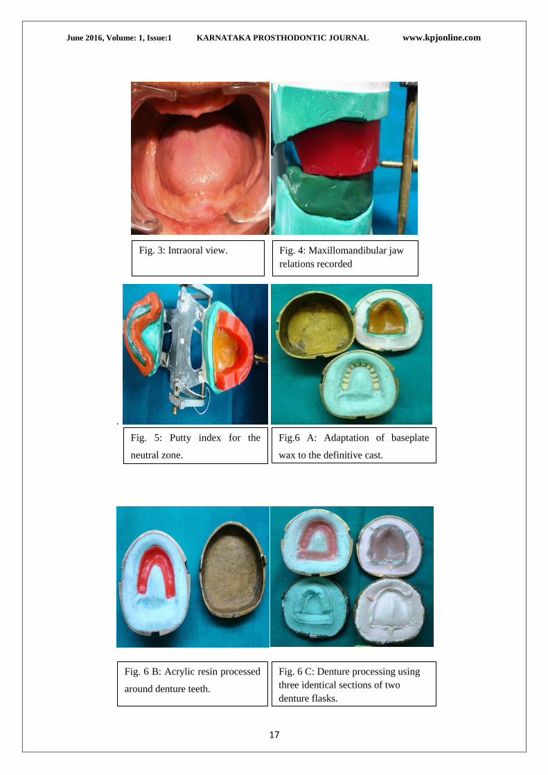
June 2016 Volume 1 Issue1 KARNATAKA PROSTHODONTIC JOURNAL wwwkpjonlinecom
20
Prosthodontic Management of Marginal
Hemimandibulectomy With Surgically Induced Lip Drop
Dr Krishna Prasad D1 Dr Anupama Prasad D
2 Dr Anshul Bardia
3
Department of Prosthodontics and Crown amp Bridge ABShetty Memorial Institute of Dental
Sciences Nitte UniversityDeralakatteMangaluruKarnataka India -575018
ABSTRACT
Loss of continuity of the mandible destroys the balance and symmetry of mandibular
function leading to altered mandibular movements and deviation of the residual fragment towards the
resected side The rehabilitation of these cases must be carefully planned and the treatment requires a
denture construction in such a way to get maximum retention support stability esthetics and
function This case report describes the treatment of a patient with partially resected edentulous
mandible combining functional and esthetic requirements A removable prosthetic appliance was
fabricated to maintain the lip in its normal position thereby helping the patient to perform normal
functions All basic principles of rehabilitation are applied and interpretation was based on altered
anatomic and functional situation An esthetic configuration with ideal function was achieved and the
occlusion showed a satisfactory stability
KEYWORDS Hemimandibulectomy maxillofacial rehabilitation esthetics
INTRODUCTION
One of the most challenging and demanding maxillofacial endeavours is the construction of
functional complete dentures for the edentulous patient who has undergone a mandibular resection
Loss of continuity of the mandible destroys the balance of the mandibular movement and function
leading to altered mandibular movement and deviation of the residual fragment towards the surgical
side The greater the loss of tissues greater will be the deviation of the mandible to the resected side
thus compromising the prognosis of the prosthetic rehabilitation to a greater extent
Professor1 Reader
2 Former Post graduate student
3
Corresponding author DrAnupama Prasad D Reader Department of Prosthodontics
and Crown amp Bridge A B Shetty Memorial Institute of Dental Sciences Nitte University
Deralakatte Mangaluru -575018 Email anupamaprasaddymailcom
Apart from deviation other dysfunctions in such patients are observed in swallowing speech control
of saliva mandibular movements mastication respiration and psychic functioning1
June 2016 Volume 1 Issue1 KARNATAKA PROSTHODONTIC JOURNAL wwwkpjonlinecom
21
Based on the nature of resection Cantor and Curtis (1971) devised a prosthetic classification
that is as follows2
Class I Radical alveolectomy with preservation of mandibular continuity
Class II Lateral resection of the mandible distal to the cuspid
Class III Lateral resection of the mandible and maxilla
Class IV Lateral bone graft surgical reconstruction
Class V Anterior bone graft surgical reconstruction
Class VI Resection of the anterior portion of the mandible without reconstructive surgery to unite the
lateral fragments
In cases with class II III IV V guide flange prosthesis would be a treatment modality One
of the basic objectives in rehabilitation is to retrain the muscles for mandibular denture control and
repeated occlusal approximation
This article highlights prosthetic rehabilitation of a class I hemimandibulectomy patient for
whom a mandibular prosthesis is fabricated with loop on corner of mouth for lip support
CASE REPORT
A 74 year-old completely edentulous female patient was referred to the Department of
Prosthodontics after extraction of remaining natural teeth which were periodontally compromised for
maxillofacial rehabilitation with a chief complaint of difficulty in eating and speaking Her medical
history revealed that she was diagnosed for squamous cell carcinoma on the right side of the
mandible for which she had undergone marginal resection of mandible on right side 3 years back
The patientrsquos history indicated that she had a tobacco-chewing habit since 40 years An extra oral
examination revealed asymmetrical face and a convex profile There was a deviation of the mouth to
the right side that is toward resected side
On intraoral examination it was found that the maxillary and mandibular arches were
completely edentulous On palpation the mandibular ridge was present till first premolar region On
evaluation of pre extraction diagnostic ortho-pantamogram segmental absence of the mandible distal
to the first premolar to the last molar on the same side was noted This particular case represents to
class I Cantor and Curtis classification
June 2016 Volume 1 Issue1 KARNATAKA PROSTHODONTIC JOURNAL wwwkpjonlinecom
22
CLINICAL PROCEDURE
Preliminary impressions were made with irreversible hydrocolloid material (Zelgan Plus
Dentsply Gurgaon India) using stock trays Casts were prepared (Fig 1) and self-cure clear acrylic
resin (RR Dentsply India) custom trays were constructed The tray was border-molded with
modeling plastic (DPI Tracing stick Dental products of India Mumbai India) taking care to avoid
overextension Final impressions were made with light-body vinyl polysiloxane (AquasilDentsply
Caulk Milford DE) While in case of mandibular final impression tray handle was extended with
autopolymerizing resin and a cylindrical mandibular rest is fabricated in the posterior region with an
increased vertical height Then softened impression compound was placed on the top of the
mandibular rests and inserted in the patientrsquos mouth Patient was advised to close her mouth so that
the mandibular rest fit against the maxillary alveolar ridge
This was done to stabilize the tray in position by preventing anterioposterior and mediolateral
displacement of the tray during final impression and which was made using the light body material
and the patient was asked to close the mouth such that cylindrical rest will fit over maxillary ridge
For recording the functional state patient was instructed to run his tongue along her lips suck in her
cheeks pull in her lips and swallow by keeping her mouth closed till the impression material
hardened
Master casts were poured with Type III dental stone (DPI Mumbai India) Stabilized record
bases were made with self-cure acrylic (DPI Mumbai India) using the sprinkle-on technique Wax
rims were adjusted until a tentative occlusal vertical dimension was established Face bow transfer
was made to orient the maxillary cast to the semi-adjustable articulator (Artexsemiadjustable
articulator rotofix face bow) Maxillomandibular relations were recorded with wax interocclusal
records The patient tactile sense and sense of comfort was used to assess the vertical dimension of
occlusion The patient was asked to move her mandible as far possible to the untreated side and then
gently close her jaw into position to record a functional maxillomandibular relationship
The teeth were arranged in the usual manner semi-anatomic posterior teeth (Acryrock Pyrax
polymers Roorkee India) were used Maxillary and mandibular teeth were arranged to achieve
balanced occlusion Occlusal table on resected side was up to the second premolar just to establish
the cross arch stability and balance in the right lateral excursive movements A wax set-up was tried
in the mouth and was checked for esthetics phonetics occlusal vertical dimension and balanced
occlusion The basic objective is to achieve an occlusal scheme which will have a multiplicity of
occlusal contacts in centric position Long centric concept and to a slightly decreased vertical
June 2016 Volume 1 Issue1 KARNATAKA PROSTHODONTIC JOURNAL wwwkpjonlinecom
23
dimension of occlusion in an attempt to decrease occlusal force is given The level of the occlusal
plane especially in edentulous patients should be acceptable to the remaining portion of the tongue to
permit easier distribution and control of food on the occlusal table and control of complete denture
prosthesis3 A posterior palatal seal was recorded and the dentures were waxed processed (DPI RR
heat cure DPI India) and remounted and the occlusion was refined Freedom of movement and lack
of cuspalintercuspation was checked before denture insertion The dentures were evaluated intraorally
and the mandible was manipulated to the static centric position area4 Any interference in normal
movements was corrected During insertion to improve the tissue contact situation resilient liner
(UfigelVOCO gmbh ) was used to reline the mandibular denture by keeping the mandible into the
maximum intercuspation position The sealer was applied once over the polymerized surface of the
resilient liner which prevents water sorption by the liner and helps in maintaining the softness for a
longer period of time The dentures were removed repolished and then reinserted
The prosthesis design is composed of snapfit buttons (Fig 2) which includes male and female
component in which male component is attached to lower mandibular denture in premolar region and
female component in removable segment (Fig 3) to which stainless steel wire is attached curving
out at the anterior end to form a loop supporting the lip extraorally The removable component of
prosthesis was fabricated with self-cure acrylic resin and it was designed such that patient can easily
remove the removal component during mastication
Addition of a 21-gauge stainless steel wire in the form of a J-shaped buccal loop to engage the
corner of the mouth of the unaffected side in order to pull the corner of the mouth and achieve an
esthetically pleasing appearance The wire loop was embedded in the acrylic of the buccal flange of
the removal segment of the denture5 (Fig 4) It was adjusted to ensure that its position provided
circumoral symmetry and esthetics without compromising comfort and simulated functional jaw
movements The extraoral wire components were relined with permanent tissue conditioner to reduce
the shine so as to blend with the skin Follow-up appointments were carried out routinely to ensure
patient comfort and satisfaction No discomfort or any problems in mouth opening or mastication
were noted resulting from the J-shaped loop and the patient was quite happy with the prosthesis
The patient was instructed to chew only on the non-resected side to avoid denture instability
It may be necessary to accept an occlusion that is not bilaterally balanced in eccentric occluding
positions for an edentulous resected maxilla or mandible The patient was given routine post insertion
instructions and was motivated to make efforts to learn to adapt to the new dentures Simple exercises
were suggested to the patient such as repeated opening and closing of mandible This helped the
patient learn to manipulate the lower prosthesis into the proper position Initially retention of the
June 2016 Volume 1 Issue1 KARNATAKA PROSTHODONTIC JOURNAL wwwkpjonlinecom
24
dentures especially of the lower one was a problem but this improved with constant use Within a
week the patient expressed satisfaction in mastication phonetics and esthetics and drastic
improvement is seen from initial stages of prosthesis planning (Fig 5) and after the fit and insertion of
the final prosthesis (Fig 6)
DISCUSSION
The prosthetic rehabilitation of a hemimandibulectomy subject is a difficult task for a
prosthodontist as the normal physiological functions like swallowing speech mandibular movements
mastication control of saliva and respiration are adversely affected by radical mandibular surgery
These dysfunctions radically alter the prosthetic prognosis Surgical reconstruction by implants and
grafts of various types is the ideal treatment when feasible
In the present case the OPG (Fig7) and intra oral pictures (Fig8) revealed the absence of
mandibular segment As the surgical reconstruction is not always feasible in every patient
prosthodontic approach has to be considered to restore the esthetic and function in such subject
Because of the loss of the normal anatomy and physiology of the oral cavity many principles of
complete denture prosthesis must be compromised Since the mandibulectomypatients have reduced
masticatory strength and little soft and hard tissue support it is important to record and utilize as
broad denture base as possible within the physiological limits
Closed-mouth impression techniques have been suggested but these were designed for
making accurate static impressions6 The column trays described in this article are similar in form but
they are used to record the muscular dynamics of the postsurgical lower denture space The reasons
for increasing the height of the lateral columns of the custom trays are as follows-
1 To reduce the amount of force exerted by the remaining muscles of mastication
2 To make swallowing more difficult
This type of ―stress swallowing will cause extreme muscular activity of the residual tongue
and floor of the mouth An impression of this functional activity should help prevent future denture
displacement In the final denture form the tissue conditioner placed on the dentures when they are
first inserted provides comfort during the adjustment period corrects any tissue surface discrepancies
resulting from the impression material and refines the final denture form during function
Lott and Levin stated that retention will increase in proportion to an increase in the area
covered by the denture Boucher states that the amount of biting force tolerated by a denture is
proportional to the size of the tissue-bearing area Since hemimandibulectomy patients have markedly
June 2016 Volume 1 Issue1 KARNATAKA PROSTHODONTIC JOURNAL wwwkpjonlinecom
25
reduced masticatory strength and little soft and hard-tissue support it is important to record and
utilize as broad denture base as is possible The use of a tissue-conditioning agent facilitates the
extension of a functional denture form to the maximum size tolerated by the oral tissues This form
should enhance the patientrsquos ability to manipulate the prosthesis and to realize maximal masticatory
potential2
Facial symmetry could be improved with the use of removable prostheses Esthetics has to be
compromised however because labial commissural sag is necessary if a functional seal is to be
maintained between the lips Without this seal drinking and speaking appear to be much more
difficult7 In this particular case an effort was made to restore the patientrsquos appearance and comfort by
repositioning and supporting the lip in a natural position with the described prosthesis Since the J-
hook was lined with permanent tissue conditioner which does not cause any irritation and it has to be
changed every 6 month patient has to wear for full time except during mastication of hard food
CONCLUSION
The described technique offers an inexpensive simple and expedient approach to manage the
hemimandibulectomy patient The availability of well-formed edentulous ridges and an excellent
peripheral seal permitted excellent retention and stability of the dentures and the presence of the loop
to support oral commisure The philosophical approach to the treatment and rehabilitation of
edentulous patients with resected mandibles is not in concentrating on what has been sacrificed in the
eradication of the disease but rather in taking full advantage of the remaining structures
FIGURES
Figure 1 Maxillary and
mandibular diagnostic cast
Figure 2 Snapfit button include male
and female component
June 2016 Volume 1 Issue1 KARNATAKA PROSTHODONTIC JOURNAL wwwkpjonlinecom
26
Figure 4 Prosthetic design with extroral
loop
Figure 3 Male component in the
mandibular denture and female
component in the removable segment
Figure 5 Preoperative photograph
Figure 6 Postoperative
photograph
Figure 7 Pre extraction
diagnostic OPG
Figure 8 Intra oral view
June 2016 Volume 1 Issue1 KARNATAKA PROSTHODONTIC JOURNAL wwwkpjonlinecom
27
REFERENCES
1 Beumer J Curtis T Firtell D Maxillofacial rehabilitation St Louis Mosby 1979 p 90-169
2 Cantor R Curtis TA Prosthetic management of edentulous mandibulectomy patients Part 1
Anatomic physiologic and psychologic consideration J Prosthet Dent 1971 25446-57
3 Ronald P Desjardins Occlusal considerations for the partial mandibulectomy patient J Prosthet
Dent 1979 41308-315
4 Cantor R Curtis TA Prosthetic management of edentulous mandibulectomy patients Part II
Clinical procedures J Prosthet Dent 1971 25546-55
5 Bagchi G Nath DK Restoration of facial symmetry in a patient with bell palsy using a modified
maxillary complete denture a case report Int J Prosthodont 2012 25(3)290-3
6 MacMillin J J Closed Mouth Technique for Impressions of the Lower Jaw J Amer Dent Ass
34 715 1947
7 Larsen SJ Carter JF Abrahamian HA Prosthetic support for unilateral facial paralysis J Prosthet
Dent 1976 35(2)192-201
June 2016 Volume 1 Issue1 KARNATAKA PROSTHODONTIC JOURNAL wwwkpjonlinecom
28
Short Dental Implants ndash A Review Of Clinical
Performance Biomechanical Aspects And Risk Factors
For Survival
Dr Talreja Karishma S Dr Rodrigues Shobha J2 Dr Pai Umesh Y
3
Department of Prosthodontics Manipal College of Dental Sciences Mangalore Manipal
University
INTRODUCTION
Implant supported prosthesis are gradually becoming the norm for restoration of missing
teeth1
The posterior edentulous arches are a biologically and mechanically challenging area for
rehabilitation with implant supported prostheses These regions have unfavourable bone quality and
lesser bone volume as compared to anterior edentulous sites compelling the operator to place shorter
implants The poor bone quality limits the number of implants placed thus increasing bending forces
on individual implants Furthermore occluding force increases the closer the teeth are placed to the
temporomandibular joint2
The obsolete protocol of placing the longest possible implant within anatomical limitations
has lead authors to employ procedures like distraction osteogenesis bone grafting guided bone
regeneration sinus floor elevation and mandibular nerve repositioning to gain adequate residual ridge
height at these sites These techniques have a variable degree of success and require considerable
dexterity and skill from the operator Short dental implants open up an exciting portal out of
complicated surgical procedures involved in implant site preparation in posterior atrophic arches
Short Dental implants (SDI) are a more cost-effective alternative that reduces treatment time
and rules out complications related to surgical and grafting procedures Authors in their studies have
quoted different lengths however considering 10mm as the standard length an implant less than
10mm in length is considered a Short Dental Implant and is usually applied in alveolar ridges with
decreased bone height3
The biomechanical rationale in support of SDIs is that loading bearing forces are concentrated
on the crestal portion of the implant and an increase of implant length from 7 to 10mm does not
significantly improve its anchorage 4 Instead with an increase of every 1mm in the implant diameter
the functional surface area increases by 30-200 thus improving the load dissipation ability of the
implant5 Recent Finite Element Analyses has demonstrated that implant length had no effect on stress
concentration on crestal bone around an implant hence a SDI may be a sound choice6
BDS 3rd
year Post Graduate Student Head of Department2
Associate Professor3
Corresponding Author Dr Karishma S Talreja Department of Prosthodontics
ManipalCollege of Dental Sciences Light House Hill Rd Mangalore-575001
Contact no- +919930794574 Email address- karishmatalreja90gmailcom
June 2016 Volume 1 Issue1 KARNATAKA PROSTHODONTIC JOURNAL wwwkpjonlinecom
29
Friberg and Jemt 27
were among the early authors to note high failure rates in both arches
with short fixtures (7mm) Early failure rate was pronounced in resorbed arches with poor bone
quality However the implants used in this study were of narrow diameter and had a smooth
machined surface SDIs are designed to provide an increased Bone-to-implant contact by virtue of an
increased diameter Newer SDIs overcome such limitations by incorporation of surface modifications
like acid-etching that increase the surface area for osseointegration
This article is a review of the many aspects of risk factors for success and performance of
SDIs under various clinical scenarios
RISK FACTORS
The risk factors for failure of SDIs may be broadly divided into endogenous (systemic or
local) and exogenous (operator or biomaterial-related) factors8
Endogenous factors-
SMOKING
Mezzomo et al9 in a meta-analysis on success rate of single crowns found a higher failure
percentage in studies wherein smokers were included as compared to studies that excluded them
Strietzel amp Reichert found a significant association between heavy smoking (gt10 cigarettesday) and
frequency of implant loss10
SYSTEMIC DISEASES
Most studies exclude pregnant women immunocompromised patients and those under
medication from their sample size This impairs the assessment of implant survival in such patients
For single crowns supported by SDIs no statistically significant difference was found in the failure
percentage in systemically compromised patients9
BRUXISM
Twail et al11
found more incidences of prosthetic failures like veneer fractures and screw
loosening in bruxer groups however no statistically significant difference was found on inter group
comparison between buxer non-bruxer and occasional bruxer groups
PERIODONTAL DISEASE
The biological failure proportion of studies that included periodontal patients did not show a
statistically significant upward trend as compared to studies that did not include periodontal patients
Marginal bone loss in periodontal groups however was found to be significantly higher
Perimplantitis and persistent periodontal disease are major risk factors for the loss of integration of
SDIs9
BONE QUALITY
Studies have failed to find an association between high failure rates and low quality
bone101218
On the other hand higher failure rates were associated with machines surface implants as
compared to rough surface implants in poor quality bone11
The density of the bone directly correlates
to the strength of the bone with less density demonstrating strength reduction of 50 -80 compared
June 2016 Volume 1 Issue1 KARNATAKA PROSTHODONTIC JOURNAL wwwkpjonlinecom
30
to high density bone types13
Weng et al14
noted a 25 failure rate of SDI (machined surface)
supported prostheses in the posterior maxilla failures occurring within 18 months of loading Hence
rougher surfaces for implants are preferred in poor bone quality Finite element analysis has found
that maximum Von-Mises stress variability was minimal when the diameter of SDIs was within 55
and 71mm Peak stress on the implant-bone interface is seen to increase with reduction in bone
density3 Osteopenic bone has thin cortices and reduced spongiosa hence needs larger diameters for
optimal load bearing capacity Implant diameter in excess of 4mm and length more than 9mm are
optimal properties for screwed implants in type IV bone15
OPERATOR RELATED RISK FACTORS
Operator related risk factors include the surgical technique prosthetic design and loading
protocol undertaken in the placement of the implant
SURGICAL TECHNIQUE
Misch et al16
proposed employing a one stage approach in D2 bone by adding a permucosal
extension at the time of surgery and a two-stage approach in D4 bone While a two-stage implant
placement approach has been suggested by some authors17
no significant difference has been found in
failure rates between single-stage and two-stage implants Also in fully edentulous patients two-stage
implants are preferred4918
Esposito et al 19
concluded that a submerged approach may be preferable in
implants that do not achieve optimal primary stability and in completely edentulous cases
CROWNIMPLANT RATIO
The crown height is a vertical cantilever and when increased from 10 to 20mm the force on
the implant is increased by 100 An angled prosthetic load is also a force magnifier on the implant
Hence detrimental effects of non-axial forces on crestal bone increase with increase in crown
height16
A high crown- to- implant ratio was assumed to have a negative biological effect on crestal
bone loss20
Peri-implant bone resorption is similar in all implant-to-crown ratio groups even when
increased by 2 to 3 times provided non axial forces were controlled11
Rossi Tawil Mertens and
Deporter et al claimed that increased CI ratio placed no deterimental effects on the health of the
implant11202122
Nedir and Birdi et al2324
evaluated crown-implant ratios ranging from 105 to 180
and 09 to 32 respectively to find no detrimental effects on surrounding bone Current research has
rejected crown-implant ratio as a major biomechanical risk factor as long as occlusal contacts are
placed as close as possible to the long axis of the implant and favourable force orientation and load
distribution is maintained11
Crown height space on the other hand is a more reliable indicator of detrimental effects on
marginal bone when crown height spaces exceed 15-mm length25
For each additional millimeter of
crown height stress concentration at the implant neck may increase by 2026
Hof et al 27
observed
greater bone loss in the anterior maxilla with increased crown-to-implant ratio than the posterior
areas This may be possibly explained by off-axis loading at the implant-bone interface
June 2016 Volume 1 Issue1 KARNATAKA PROSTHODONTIC JOURNAL wwwkpjonlinecom
31
PLATFORM SWITCHING
Platform switching shifts the stress concentration zone from the crest bone-implant interface
to the axis of the implant thus reducing stress levels at the cervical bone area28
Telleman et al29
from
the results of a randomized control trial found that 1 year post loading inter proximal bone levels were
better maintained at implants restored according to the platform switching concept
IMPLANT NUMBER AND SPLINTING
Factors contributing to marginal bone loss around dental implants include surgical trauma
faulty implant positioning occlusal overloading or non-axial loading30-32
Stress level in bone around
splinted implants is found to be lower than bone around unsplinted implants by a factor of 933
A
positive influence of splinting and number of splinted implants has been observed on success rate of
SDIs in atrophic posterior arches up to a 10 year follow up period1620273435
Placement of additional
implants increases the effective surface area for stress distribution Hence one implant for each
missing premolar and two for each missing molar were suggested16
To further capitalize on functional
area these must be splinted
WIDTH OF OCCLUSAL TABLE AND TYPE OF OCCLUSION-
Within 54 and 83mm the width of the occlusal table did not significantly affect peri implant
bone loss11
Axial forces distribute stress more evenly throughout the implant as compared to bending
moments Occlusion should be mutually protected and prostheses should be free of non axial
loading1116
CANTILEVER FORCES
The length of the posterior cantilever in the mandible is directly related to complications
andor failure of the prosthesis3637
Romeo et al 38
found no detrimental effects of cantilevers
provided cantilever length was appropriate and occlusal function was under control Mesial and distal
cantilever lengths limited to 275 +-165 and 224+-160mm respectively have found to cause
marginal bone loss within acceptable limits11
LOADING PROTOCOL
Most authors follow and recommend a delayed loading protocol for SDIs Rossi et al21
conducted a study using SLActive straumann 6mm implants that were early loaded (6 weeks after
insertion) These implants yielded high survival rates and moderate loss of bone after two years of
loading However long-term follow-up larger sample size and randomized trials are required to
provide concrete evidence for incorporation of early loading protocols into clinical use
BIOMATERIAL RELATED FACTORS
Implant length implant diameter surface topography and implant thread pitch are important
parameters that influence the selection of the most fitting implant in a given clinical situation
June 2016 Volume 1 Issue1 KARNATAKA PROSTHODONTIC JOURNAL wwwkpjonlinecom
32
IMPLANT LENGTH-
Implant length is defined as the length between the implant neck and the implant apex
Increase in implant length has found to have minimal beneficial effect on load distribution around the
crestal portion of the implant6 Mezzemo et al
9 in a meta-analysis stated that short implants supporting
single crowns obtained similar if not superior survival rates as compared to standard length implants
Few studies exist on implants of 5 and 6mm length thus limiting the data obtained from systematic
reviews A two year trial of implants of four millimeter length with SLActive surfaces has yielded
survival rates of 957 after 1 year and 923 at the end of the trial 34
Ling Sun et al18
have reported
highest survival rate for implant lengths of 75 and 9mm But no statistically significant difference
exists based on length
IMPLANT DIAMETER
For every 1mm increase in diameter functional SA is increased by 30 ndash 200 along with
BIC5 Sato et al
39 on the basis of an in vitro study stated that wide implants are capable of bearing
larger loads and perform better than implants of smaller diameter under tensile forces Wider
diameters of implants are hence referred for reduced bone density This however is limited by the
bucco-lingual width of the residual ridge
IMPLANT SURFACE
Griffin and Cheung40
suggested ―the implant maximized surface area as the main contributing
factor to the high success rate Rougher surfaces offer extensive area for osseointegration and have
better bone-implant-contact as compared to machined or acid ndash etched surfaces16-18
Various surface
modified implants like SLActive surfaces213441
TiOblast implants Astra Tech20
and bioabsorbable
HA blasted implants 16
have shown better results as compared to the poor results seen with machined
surface implants7
IMPLANT THREAD PITCH-
Thread pitch is defined as the distance between adjacent threads or the number of threads per
unit length in the same axial plane and on the same side of the axis5 Hence the greater the implant
pitch the greater the surface area available for osseointegration and load dissipation Another implant
thread geometry parameter worth consideration in this context is thread depth16
Misch16
has suggested a protocol for the reduction of stress at the bone-implant interface for
SDIs they include-
1) no cantilevers on the prostheses
2) no angled forces to the posterior restorations
3) splinting multiple implants together
4) implant surface modification
5) increase implant thread pitch
June 2016 Volume 1 Issue1 KARNATAKA PROSTHODONTIC JOURNAL wwwkpjonlinecom
33
INDICATIONS
Annibali et al4 in a systematic review reported successful results for short dental implants
with a pooled survival rate of 991 and a low incidence of biological and biomechanical
complications after a mean follow-up period of 32+- 17 years
Studies have evaluated the efficiency of 6mm vs 10mm implants supporting fixed partial
dentures in augmented bone41
6mm vs 11mm implants combined with sinus floor elevation
supporting single crowns42
and 6 5mm implants rehabilitating bilateral atrophic posterior arches vs
longer implants in augmented bone 4344
to find similar if not better performance of SDIs with fewer
post-operative complications in comparsion to conventional implants in augmented bone Based on
the results of randomized control trials and clinical studies the following indications of SDIs in
atrophic arches can be put forth41-44
1) implant supported single crowns
2) implant supported fixed partial dentures
3) implant supported overdentures
The need for long term follow-up studies is quintessential to evaluate the effect of bone loss
on the survival of the SDIs While the loss of 2mm of crestal bone has minimal impact on the stability
of a 10 mm or longer implant a similar bone loss pattern on a 7mm implant for example leaves
behind a considerably lesser bone volume for load dissipation
The assessment of failure rates of SDIs should consider the poor quality of bone that is
commonly observed in atrophic arches indicated for SDIs in comparison to bone found in regions
indicated for conventional implants and rather be compared to the outcome of implants placed in
grafted sites5
ADVANTAGES OF SDIs-16
Lack of bone grafting reduces cost and duration of treatment
Surgical risk of sinus perforation mandibular paresthesia is eliminated along with decreased
chances of overheating the osteotomy site or damage to dilacerated adjacent tooth root
No need for additional inventory and decreased surgical complexity
Implant placement in smaller interarch space
LIMITATIONS
The reversed crown to implant ratio may not be an esthetic concern in the posterior
quadrants however it may not be acceptable in the anterior maxilla Here the morbidity related to an
autologous bone graft for reconstruction must be considered20
Other than this there is a draught of
data on results of long term clinical trials of SDIs in poor quality bone Also management of atrophic
ridges that have horizontal ridge insufficiency with SDIs is a question that still remains unanswered
June 2016 Volume 1 Issue1 KARNATAKA PROSTHODONTIC JOURNAL wwwkpjonlinecom
34
REFERENCES
1 Jemt T Lekholm U Adell R Osseointegrated implants in the treatment of partially
edentulous patients A preliminary study on 876 consecutively placed fixtures Int J Oral
Maxillofac Implants 19894211-217
2 Jemt T Lekholm U Oral implant treatment in posterior partially edentulous jaws A 5-year
follow-up report Int J Oral Maxillofac Implants 19938635-640
3 Kang N Wu YY Gong P Yue L O GM A study of force distribution of loading stresses on
implant-bone interface on short implant length using 3-dimensional finite element analysis Oral
Surg Oral Med Oral pathol Oral Radiol 2014 118519-523
4 Annibali S Cristalli MP DellrsquoAquila D Bignozzi I La Monaca G Pilloni A Short dental
implants A Systematic Review J Dent Res 91(1)25-322012
5 Misch C Bidez MW Contemporary Implant Dentistry St Louis Mo CV Mosby 1999
6 Fugazzotto PA Shorter implants in clinical practice Rationale and treatment results Int J
Oral Maxillofac Implants 200823487-496
7 Friberg B Jemt T Lekholm U Early failures in 4641 consecutively placed Branemark dental
implants A Study from stage 1 surgery to the connection of completed prostheses Int J Oral
Maxillofac Implants 19916142-146
8 Esposito M Hirsh J-M Lekholm U Thomsen P Biological factors contributing to failures of
osseointergrated oral implants Euro Oral Sci 1998106721-764
9 Mezzomo et al Meta-analysis of single crowns supported by short (lt10mm) implants in the
posterior region J Clin Periodontol 2014 41 191-213
10 Strietzel F P amp Reichert P A (2007) Oral rehabilitation using Camlog screw-cylinder
implants with a particle-blasted and acid-etched microstructured surface Results from a
prospective study with special consideration of short implants Clinical Oral Implants Research 18
591ndash600
11 Tawil et al Influence of prosthetic parameters on the survival and complication rates of Short
Dental Implants Int J Oral Maxillofac Implants 200621275-282
12 Nicolau P Korostoff J Ganeles J Jackowski J Krafft T Neves M Divi J Rasse M
Guerra F amp Fischer K (2013) Immediate and early loading of chemically modified implants in
the posterior jaws 3-year results from a prospective randomized multicenter study Clinical
Implant Dentistry and Related Research 15 600ndash612
13 Misch CE Qu Z Bidez MW Mechanical properties of trabecular bone in the human
mandible Implications for dental implant treatment planning and surgical placement J Oral
Maxillofac Surg 199957700-706
14 Weng D Jacobson Z Tarnow D et al a prospective multicenter clinical trial of 3i machined-
surface implants Results after 6 years of follow-up Int J Oral Maxillofac Implants 200318417-
423
15 Li T Kong L Wang Y Hu K Song L et al Selection of optimal dental implant diameter and
length in type IV bone a three dimensional finite element analysis Int J Oral Maxillofac Surg
2009381077-1083
16 Misch C Steigenga J Barboza E Misch-Dietsh F Cianciola L Kazor C Short dental
implants in posterior partial edentulism a multicenter retrospective 6-year case series study J
Periodontol 2006771340-1347
17 Gentile MA Chuang SK Dodson TB Survival estimates and risk factors for failure with 6 times
57-mm implants Int J Oral Maxillofac Implants 200520930ndash937
June 2016 Volume 1 Issue1 KARNATAKA PROSTHODONTIC JOURNAL wwwkpjonlinecom
35
18 Sun H Huang C Wu Y Shi B Failure rate of short (lt= 10mm) dental implants and factors
influencing their failure A Systematic Review Int J Oral Maxillofac Implants 200126816-825
19 Esposito M Grusovin MG Chew YS Coulthard P Worthington HV One-stage versus two-
stage implant placement A Cochrane systematic review of randomised controlled clinical trials
Eur J Oral Implantol 2009291ndash99
20 Mertens C Meyer-Baumer A Kappel H Hoffmann J Steveling H Use of 8-mm and 9-mm
Implants in atrophic alveolar rides 10-year results Int J Oral Maxillofac Implants 2012271501-
1508
21 Rossi F Ricci E Marchetti C Lang N P amp Botticelli D (2010) Early loading of single
crowns supported by 6-mm-long implants with a moderately rough surface a prospective 2-year
follow-up cohort study Clinical Oral Implants Research 21 937ndash943
22 Deporter D A Kermalli J Todescan R amp Atenafu E (2012) Performance of sintered
poroussurfaced press-fit implants after 10 years of function in the partially edentulous posterior
mandible International Journal of Periodontics and Restorative Dentistry 32 563ndash570
23 Nedir RBischof MBriaux JMBeyer SBernard JPA 7-year life table analysis from a
prospective study on ITI implants with special emphasis on the use of short implants Clin Oral
Implants Res 200415150ndash157
24 Birdi H Schulte J Kovacs A Weed M Chuang SK Crown-to-Implant ratios of short length
implants J Oral Implant VolXXXVINo Six2010
25 Kim YOh TJMisch CEWang HLOcclusal considerations in implant therapy clinical
guidelines with biomechanical rationale Clin Oral Implants Res 2005 1626ndash35
26 Blanes RJ Bernard JP Blanes ZM Belser UC A 10-year prospective study of ITI dental
implants placed in the posteriorregionIIinfluenceof thecrown-to-implantratioand different
prosthetic treatment modalities on crestal bone loss Clin Oral Implants Res 2007 18707ndash714
27 Hof M Pommer B Zukic N Vasak C Lorenzoni M Zechner W Influence of Prosthetic
Parameters on Peri ndashImplant Bone Resorption in the first year of loading A Multifactorial
Analysis Clinical Implant Dentistry and Related Reasearch Vol 17 Supplement 1 (2015)
28 Maeda Y Miura J Taki I amp Sogo M (2007) Biomechanical analysis on platform
switching is there any biomechanical rationale Clinical Oral Implants Research 18 581ndash584
29 Telleman et al Impact of platform switching on inter-proximal bone levels around short
implants in the posterior region I year results from a randomized clinical trial J Clin Periodontol
2012 39688-697
30 Tomasi C Sanz M Cecchinato D et al Bone dimensional variations at implants placed in
fresh extraction sockets a multilevel multivariate analysis Clin Oral Implants Res 2010 2130ndash
36 6 Nissan J Ghelfan O Gross O Priel I Gross M Chaushu G
31 The effect of crownimplant ratio and crown height space on stress distribution in unsplinted
implant supporting restorations J Oral Maxillofac Surg 2011 691934ndash1939
32 Pozzi A Agliardi E Tallarico M Barlattani A Clinical and radiological outcomes of two
implants with different prosthetic interfaces and neck configurations randomized controlled split-
mouth clinical trial Clin Implant Dent Relat Res 2012 [Epub ahead of print]
33 Bergkvist G Simonsson K Rydberg K Johansson F Derand T A finite element analysis of
stress distribution in bone tissue surrounding uncoupled or splinted dental implants Clin Implant
Dent Relat Res 20081040ndash46
34 Slotte C Gronningsaeter A Halmoy A Ohrnell L Stroh G Isaksson S Johansson L Four-
millimeter implants supporting fixed partial dental prostheses in the severely resorbed posterior
June 2016 Volume 1 Issue1 KARNATAKA PROSTHODONTIC JOURNAL wwwkpjonlinecom
36
mandible two-year results Clinical Implant Dentistry and Related Research Vol 14 Supplement
12012
35 Nissan J Gross O Ghelfan O Priel I Gross M Chaushu G The effect of splinting implant-
supported restorations on stress distribution of different crown-implant ratios and crown height
spaces J Oral Maxillofac Surg 2011 692990ndash 2994
36 White S Caputo AA Anderkvist T Effect of cantilever length on stress transfer by implant
supported prostheses J Prosthet Dent 199471493-499
37 Rangert BEng MSullivan RJemt TLoad factor control for implants in the posterior partially
edentulous segmentInt J Oral Maxillofac Implants 199712360ndash370
38 Romeo ELops DMargutti EGhisolfi MChiapasco MVogel G Implant-supported fixed
cantilever prostheses in partially edentulous archesA seven year prospective studyClin Oral
Implants Res 200314303ndash311
39 Sato Y Shindoi N Hosokawa R Tsuga K Akagawa Y A biomechanical effect of wide
implant placement and offset placement of three implants in the posterior partially edentulous
region J Oral Rehabil 20002715ndash21
40 Griffin TJ Cheung WS (2004) The use of short wide implants in posterior areas with
reduced bone height a retrospective investigation J Prosthet Dent 92139-144
41 Romeo E Storelli S Casano G Scanferla M Botticelli D six-mm versus 10-mm long
implants in the rehabilitation of posterior edentulous jaws a 5-year follow-up of a randomised
control trial
42 Gulje F Raghoebar G Vissink A Meijer H single crowns in the resorbed posterior maxilla
supported by either 6-mm implants or by 11-mm implants combined with sinus floor elevation
surgery a 1-year randomised control trial Eur J Oral Implantol 20147(3)247-255
43 Esposito M Cannizzaro G Soardi E Pistilli R Piattelli M Corvino V Felice P Posterior
atrophic jaws rehabilitated with prostheses supported by 6 mm-long 4 mm-wide implants or by
longer implants in augmented bone Preliminary results from a pilot randomised controlled trial
Eur J Oral Implantol 20125(1)19ndash33
44 Pistilli R Felice P Cannizzaro G Piattelli M Corvino V et al Posterior atrophic jaws
rehabilitated with prostheses supported by 6 mm long 4 mm wide implants or by longer implants
in augmented bone One-year post-loading results from a pilot randomised controlled trial Eur J
Oral Implantol 20136(4)359ndash372
45 Chan M Narhi C Baat T Kalk W Treatment of atrophic edentulous Maxilla with implant
supported overdentures a review of literature Int J Prosthodont 1998117-15
June 2016 Volume 1 Issue1 KARNATAKA PROSTHODONTIC JOURNAL wwwkpjonlinecom
37
Knowledge Attitude and Oral Hygiene Practice Among
Patients Wearing Fixed Partial Dentures In South Coastal
Karnataka Region
Dr Manoj Shetty1 Dr Krishna Prasad D
2Dr Chethan Hegde
3 Dr Nikhila Thulasida
MDSProfessor and HOD Dept of Oral Implantology1
MDS Professor Dept of Prosthodontic2
MDSProfessor and HOD Dept of Prosthodontics3
PG studentDept of Prosthodontics4
A B Shetty Memorial Institute of dental sciencesDeralakatte Mangalore
ABSTRACT
The purpose of this study was to assess the knowledge attitude and the oral hygiene measures
undertaken by the patient wearing fixed partial denture visiting the Department of Prosthodontics in
AB Shetty Memorial Institute of Dental Sciences Deralakatte which is located in the south coastal
Karnataka region To improve the oral hygiene practices and to impart further knowledge among the
patients it is necessary to know the current status The sample size of the present study was 200 and a
written consent form was obtained from the patients before conducting the survey using a
questionnaire
The study showed that 35 of the sample chose fixed prosthesis by their choice 55 as it
was suggested by the dentist and the rest 10 due to othersrsquo suggestions 40 preferred fixed
prosthesis due to esthetic and functional reasons whereas 36 solely because of functional benefits
and 24 for esthetic reasons615 of the population knew the material with which their prosthesis is
fabricated which indicates that majority of the people are aware of prosthesis they have Among
this796 had metal-ceramic crowns 154 had all metal and 48 have all ceramic crowns which
shows that the patients are more inclined towards esthetic restorations From the questions regarding
the fit of prosthesis colour matching and shape of prosthetic teeth 85 patient found the quality of
prosthesis excellent525 good355 average and37 poor Regarding the oral hygiene practices
72 of the sample brushes only once a day 275 twice a day and 05 more than twice Also 80
were unaware of the type of bristles in their brushes
KEYWORDS Fixed partial denture knowledge attitude oral hygiene practices
June 2016 Volume 1 Issue1 KARNATAKA PROSTHODONTIC JOURNAL wwwkpjonlinecom
38
INTRODUCTION
Teeth play an important role in the maintenance of a positive self image1Loss of teeth had
hampered the social life of people But with the advancement of technology in the field of prosthetic
rehabilitation increased awareness among people and improvements in the health care system people
are able to achieve better treatment for lost teeth through fixed dental prosthesis In order to impart
further knowledge among the patients and to improve the oral hygiene practices it is necessary to
access the knowledge attitude and hygiene practices among the patients wearing fixed partial
dentures
The purpose of this study was to assess the knowledge attitude and the oral hygiene measures
undertaken by the patient wearing fixed partial denture visiting the Department of Prosthodontics in
AB Shetty Memorial Institute of Dental Sciences Deralakatte which is located in the south coastal
Karnataka region The study was conducted to evaluate the knowledge or source of knowledge about
the fixed prosthesis to assess the level of satisfaction and efficiency of fixed prosthesis post insertion
and to assess the hygiene practices and maintenance of fixed prosthesis among the patients
MATERIALS AND METHODS
SAMPLE SELECTION 200 patients visiting the Department of Prosthodontics in AB Shetty
Memorial Institute of Dental Sciences Deralakatte were selected A written informed consent was
obtained from each patient prior to starting the survey
After obtaining the consent of the patients who are having a fixed dental prosthesis to
participate in the study patients were asked to fill the questionnaire form which was later evaluated to
assess the knowledge attitude and oral hygiene practices among the patients
RESULTS
Among the 200 fixed partial denture wearers 110 chose fixed prosthesis as it was suggested
by the dentist and 70 by their own choice and 20 due to other reasons(Fig 1) This indicates a lack of
knowledge or decision making ability of the patient
48200 patients preferred fixed prosthesis due to esthetic reasons 72100 due to the functional
benefits and 80100 due to both(Fig 2) Among 615 who knew the material with which their
June 2016 Volume 1 Issue1 KARNATAKA PROSTHODONTIC JOURNAL wwwkpjonlinecom
39
prosthesis is made of 796 have metal ceramic crowns 154 have all metal and 48 have all
ceramic crowns(Fig 3)
Evaluation of the attitude of the patients towards the fixed dental prosthesis was found to be
good 85 patient found the quality of prosthesis excellent 525 good 355 average and37
poor(Fig 4)
Assessment of oral hygiene practices among the fixed partial denture wearers showed that
143200 patients brush once a day 55200 twice a day and 2200 more than twice 16200 patients
uses other aids to clean the prosthetic area whereas the remaining patients do not use any other
aids(Fig 5) Among the 8 who uses additional aids 1416 uses floss and 216 uses interproximal
brushes(Fig 6)
DISCUSSION
Various studies have been carried out in the country and various parts of the world to
determine the prevalence prosthetic status and attitude of patients towards fixed prosthetic treatment
There has been an increased trend towards fixed prosthesis compared to removable prosthesis in
recent years
In this study the knowledge attitude and oral hygiene practices among the fixed prosthesis
wearers in south coastal region were evaluated using a questionnaire The sample size of the present
study is 200 and a written consent form was obtained from the patients before conducting the survey
According to the survey 35 of the sample chose fixed prosthesis by their choice 55 as it was
suggested by the dentist and the rest 10 due to others suggestions This may be due to the lack of
education and decreased accessibility to the internet sources in the rural areas Knowledge among the
rural population should be improved so that patients can have better decision making skills regarding
their prosthesis 40 preferred fixed dental prosthesis due to esthetic and functional reasons whereas
36 solely because of functional benefits and 24 for esthetic reasons615 of the population knew
the material with which their prosthesis is fabricated which indicates that majority of the people are
aware of prosthesis they have Among this796 had metal-ceramic crowns 154 had all metal and
48 have all ceramic crowns which shows that the patients are more inclined towards esthetic
restorations The cost factor was the major reason that prompted few patients to opt for all metal
prosthesis The study conducted by Napankangas et al in Finland showed overall 124 of men and
121 of women had single crowns while 48 of men and 80 of women had FPDs A logistic
regression analysis showed that the presence of crowns and FPDs was significantly associated with a
June 2016 Volume 1 Issue1 KARNATAKA PROSTHODONTIC JOURNAL wwwkpjonlinecom
40
southern place of residence high and middle levels of education and high frequency of dental visits2
The finding from the study conducted by Shigli et al in Belgaum also indicate that awareness need to
be increased regarding esthetics and phonetics3 A higher frequency of removable restorations was
present in older age groups in subjects living in rural areas in those from a lower socio-economic
status and in subjects with less education and lower incomes according to the study by Zitzmann et
al4
From the questions regarding the fit of prosthesis colour matching and shape of prosthetic
teeth 85 patient found the quality of prosthesis excellent525 good355 average and37
poor This data shows a positive attitude towards the fixed prosthetic treatment Imparting knowledge
about the fixed prosthesis will allow the population to make better judgement and decision regarding
their prosthetic rehabilitation
Regarding the oral hygiene practices 72 of the sample brushes only once a day 275
twice a day and 05 more than twice also 80 were unaware of the type of bristles in their brushes
It is mainly due to the ignorance and lack of literacy among the population Only 8 of patients use
other aids to clean the prosthetic area in the oral cavity This indicates a need to increase the
awareness among the population regarding oral hygiene practices so as to increase the longevity of the
prosthesis and improving oral health Even the study by Patil VV et al emphasized the need for
improved dental health awareness and availability of dental facilities to industrial workers in
Belgaum5Cross sectional study by Gutshow et al also found a highly significant association between
level of school education and the need of prosthetic treatment6
CONCLUSION
There has been a dramatic increase in the number of crowns and fixed dental prosthesis
provided according to the studies conducted in Scotland and Finland78
The same trend is seen all over
the world regarding the prosthetic rehabilitation
According to the survey conducted among the patients visiting the Department of
Prosthodontics in AB Shetty memorial institute of Dental Sciences it was found that knowledge
among the patients wearing fixed partial dentures about the prosthesis were good Majority of the
patients were well satisfied with the prosthesis and was having a positive attitude towards the fixed
prosthetic replacement The awareness about oral hygiene practices were comparatively less among
the conducted sample and require further reinforcement regarding the same Though variety of
cultural influences attitudes beliefs educational background and financial status determine the
June 2016 Volume 1 Issue1 KARNATAKA PROSTHODONTIC JOURNAL wwwkpjonlinecom
41
treatment received by the patient increase of awareness among the patients will definitely help in
enhancing the oral health and social attitude of the patients
REFERENCES
1 Roessler D MComlpete denture success for patients and dentistsInt Dent J 200353340-5
2 Napankangas R Haikola B Oikarinen K Soderholm ALPrevalence of single crowns and
fixed partial dentures in elderly citizens in the southern and northern parts of Finland J Oral
Rehabil 2011 38 328-32
3 Shigli K Hebbal M Angadi GSAttitudes Towards Replacement of Teeth Among Patients at
the Institute of Dental Sciences Belgaum India J Dent Educ 2007 71(11) 1467-75
4 Zitzmann NU Hagmann E Weiger RWhat is the prevalence of various types of prosthetic
dental restorations in Europe Clin Oral Impl Res 2007 18(3) 20-33
5 Patil VV Shigli K Hebbal M Agrawal NTooth loss prosthetic status and treatment needs
among industrial workers in Belgaum Karnataka India J Oral Sci 2012 54 285-92
6 Gutschow F Jakstat HCross-sectional study on the prevalence of prosthodontic
reconstructions in young men Deutsche Zahnarztliche Zeitschrift 199156 602ndash605
7 Fyffe HEProvision of crowns in Scotland ndash a ten year longitudinal study Community Dent
Health 19929159ndash16
8 Ranta K Tuominen R Paunio IRehabilitation with fixed prostheses among Finnish adults
Community Dent Health 19864137ndash142
June 2016 Volume 1 Issue1 KARNATAKA PROSTHODONTIC JOURNAL wwwkpjonlinecom
42
Aesthetic And Functional Rehabilitation Of A Severely
Mutilated Dentition
Dr Nirupama R1 Prof (Dr) Manoj Shetty
2
Department of Prosthodontics A B Shetty Memorial Institute of dental sciences
Deralakatte Mangalore
ABSTRACT
Increased life expectancies have led to an increased demand for prosthetic
rehabilitation of the elderly citizens who also are active socially The subjects demand rehabilitation
not only for function but also for aestheticsThe present case report describes treatment modality to
restore a severely compromised dentition with the complete occlusal rehabilitation in a fifty five year
old male patient with missing posterior teeth and fractured anterior fixed partial denture prosthesis
The patients aesthetic and functional expectations were fulfilled by adapting Pankey Mann Schuyler
Philosophy
KEY WORDS Full mouth rehabilitation aesthetics severely compromised dentitionBroadricks
Occlusal Plane Analyzer Occlusal Plane
INTRODUCTION
Tooth surface loss may occur either as a physiological process due to ageing or due to
pathological processes such as caries or non carious lesionsOcclusal disease is the loss of the
anatomical parts of occluding tooth surfaces which ultimately result in functional
impairmentPhysiological wear results in progressive but very slowexcessive wear refers to any level
of occlusal wear that can be expected to require corrective intervention in order to preserve the
dentition The physiological wear of teeth is probably an age-related phenomenon As the teeth
continue to function and be challenged by erosive attritive and abrasive factors there will be change
to the surfaces of teeth
MDS Sr lecturer1
MDS2
June 2016 Volume 1 Issue1 KARNATAKA PROSTHODONTIC JOURNAL wwwkpjonlinecom
43
Patients with severely worn dentition and loss of multiple posterior teeth may result in
reduced vertical dimension and occlusal instabilityRaising the vertical dimension is then required to
correct the occlusal relationship to restore function as well as aesthetics
Reconstruction of a worn dentition is essential In any restorative treatment plan the first
decision to be made is whether or not the restorations are designed to harmonize with the existing
occlusion or make a change towards an ideal occlusionThe treatment can be either active or passive
The passive approach involves monitoring the degree of wear and plansvarious preventive strategies
Monitoring involves taking a series of repetitive examinations and certain measurements over a period
of time in order to asses if a condition is progressive Standardized intra-oral photographs study
models and measuring lesion dimensions are all potential approaches
Performing a successful occlusal rehabilitation is an arduous task that entails meticulous
treatment planning resulting in preservation of remaining natural teeth and healthy maintenance of
supporting structures
The present case report explains a simplified multidisciplinary approach to a functional and
aesthetics restoration of a severely compromised dentition by adapting Pankey Mann Schuyler (PMS)
philosophy with minor modifications
CASE REPORT
A 55 year old male patient who was moderately built reported to the department of
prosthodontics His chief complaint was difficulty in chewing food because of loss of posterior teeth
and poor aesthetics due to loss of old fixed partial denture in relation to maxillary anterior region
Patientrsquos personal and medical history was non contributory
On intra oral examination the maxillary arch showed fractured fixed partial denture in
relation to 11 12 13 21 22 and 23 Remaining teeth present were 11 13 1422232425 and 27 11
13 22 and 23 were subjected for root canal treatment (RCT) later followed by new fixed partial
denture Old crowns present on 14 and 25 were removed and also subjected for RCT along with 24
followed by crowns which were used as abutments for cast partial denture (CPD)
In mandibular arch 35 and 36 were missing and temporary filling in relation to 36 was seen
later subjected to RCT All anterior teeth (31 32 33 41 42 and 43) were attrided and referred for
intentional RCT in order to receive crowns
June 2016 Volume 1 Issue1 KARNATAKA PROSTHODONTIC JOURNAL wwwkpjonlinecom
44
Over all on intra oral examination there was decrease in vertical dimension due to loss of
many posterior teeth and attrition of lower anterior teeth(Figure I)A diagnostic OPG(Figure II) was
made andevaluated
Diagnostic casts were mounted on a semi-adjustable articulator and wax mock up was done
for the purpose of treatment planning It was decided thereafter to adapt and modify PMS philosophy
to rehabilitate the dentition and increase the vertical dimension by 2mm (Figure III)Broadrickrsquos
occlusal plane analysis was then done to establish the occlusal plane (Figure IV)
In the next appointment the lower anteriors were prepared and temporisation was
doneSimultaneously the root canal treated upper anterior teeth were subjected to post and core
treatment in order to minimise the total number of visits In the subsequent visit permanent
cementation of the lower anterior restorations and tooth preparation and temporisation of the upper
anterior teeth were accomplished(Figure V) An interim removable partial denture was also provided
to the patient in order to increase the vertical dimension
After the evaluation of the patientrsquos adaptation and tolerance to the increased VD
cementation of the upper anterior restorations was done and the incisal guidance was established The
lower posterior teeth were then prepared and restored according to Broadrickrsquosocclusal plane
analysis(FigureV)
In the next visit preparation of the upper premolar teeth which were to be used as abutments
for the upper CPD was doneThe crowns with the rest seats were then temporarily cemented onto the
abutments in order to make a pick up impression The master cast thus obtained was surveyed and
metal milling was done(Figure VII) A conventional upper cast partial denture was then fabricated and
inserted(FigureVIII)
CONCLUSION
Managing full mouth rehabilitation cases are most challenging in dental practice Several
decisions have to be made concerning the occlusion The clinician must take in to consideration not
only the aesthetic and functional aspects but also ensure that the physiological restoration is in
harmony with the stomatognathic system We must also remember that not all patients can be
successfully treated with a single preconceived treatment philosophy Thus this report presents an
adaptation of PMS in order to successfully rehabilitate the above severely mutilated dentition case
(Figure IX)
June 2016 Volume 1 Issue1 KARNATAKA PROSTHODONTIC JOURNAL wwwkpjonlinecom
45
Figure I Pre-operative view of maxillary and mandibular arch intra orally
Figure II Pre-operative OPG
veiw
Figure III Diagnostic mounting and mock up done on the
same according to broadricks occlusal plane analysis
June 2016 Volume 1 Issue1 KARNATAKA PROSTHODONTIC JOURNAL wwwkpjonlinecom
46
Figure IV Mandibular arch mock up done according to Broadricks occlusal plane analysis
Figure VII Metal milling on
abutment teeth for maxillary arch
CPD posteriorly cation
Figure V Permanent cementation of the lower anterior restorations and tooth
preparation and temporisation of the upper anterior teeth
Figure VI Analysing Brodricks
Occlusal plane before
cementation of lower posterior
restoration
June 2016 Volume 1 Issue1 KARNATAKA PROSTHODONTIC JOURNAL wwwkpjonlinecom
47
REFERENCES
1 Peter EDawson Evaluation diagnosis and treatment of occlusal problems 2nd Ed Cv
Mosby Company St Louis BaltimoreToronto 1989
2 Bloom DR Padayachy JN Increasing occlusal vertical dimension - Why when amp
howBritish Dental Journal 2006200251-6] Eckert SE Carr AB Single maxillary complete
denture Dent Clin North Am 200448
3 Wassel RW Steele JG Welsh G Considerations when planning occlusal rehabilitation A
review of the literature International Dental Journal 1998 48
4 Paul H Pokorny Jonathan P Wiens and Harold Litvak Occlusion for fixed
prosthodonticsA historical perspective of the gnathological influence
J Prosthet Dent 200899
5 Rivera-Moreles WC Mohl ND Restoration of vertical dimension of occlusion in the
severely worn dentition Dent Clin North Am 199236(3)
Figure VIII Post-operative view of maxillary and mandibular arch intraoraly
FigureIX Pre-operative and post-operative view after final
restoration
June 2016 Volume 1 Issue1 KARNATAKA PROSTHODONTIC JOURNAL wwwkpjonlinecom
48
SynCone- A New Dimension In Implant Overdenture A
Case Report
Prof(Dr) Manoj Shetty1 Dr Azlinder
2 Prof(Dr)Rakshith Hegde
3 Prof (Dr) Chethan
Hegde4 Dr Nivya John
ABSTRACT
AnkylosSynCone concept has become an emerging trend among implantologistsinorder to
provide rehabilitation of edentulous ridges With the luxury of friction fit telescoping crowns and the
immediate loading concept SynCone concept thrives to be the next big thing in the implant
worldhere is a case report on mandibular edentulous rehabilitation with SynConeconceptThe
telescopic crown technique has benefits such as excellent three dimensional immobilization of the
restoration flexibility of design and optimum access for oral hygiene with cost effective
proceduresWith these features we can forsee a new dimension in rehabilitation of missing natural
teeth
KEYWORDS SynCone degunomesimmediateloadingtelescopic copings
INTRODUCTION
Implants have now become the forefront of modern day dentistry when it comes to
rehabilitation of the missing tooth or teethVarious implant concepts provided by various implant
systems gives us a wide range of options for treatment planning
One such newly trending concept is the SynCone conceptconcept is one of the recent trends
in implant dentistry for the rehabilitation of edentulous ridges This concept attributes to
theplacement of implants and immediately loading the prosthesis
The benefit of SynCone concept is its versatilityThey provide fast and cost efficient
restorations of edentulous ridgesMinimally invasive treatment makes it possible to load the
prosthesis under Local anaesthesia on the same dayFor delayed restorations it serves as a
prefabricated retaining element for the maxillary and mandibular ridgeNew abutment angulations
provide improved parallelization1
Here is a case report on rehabilitation of the edentulous mandible with implant placements
interforaminally using the SynCone concept
Professor and Head Of Oral ImplantologyMDS1 BDS
2 ProfessorMDS
3 Head Of The
Department MDS4
Final Year Post Graduate
CASE REPORT
June 2016 Volume 1 Issue1 KARNATAKA PROSTHODONTIC JOURNAL wwwkpjonlinecom
49
A 45 year old female patient(fig 1) reported at the out patient department of Prosthodontics
and Implantology with periodontally damaged lower teeth and maxillary ridge was rehabilitated with
fixed restorations(Fig 2)
Patients chief complaint was poor esthetics and difficulty in chewing due to missinglower
back teethPatient was in a good health condition Blood picture showed normal values for the
surgicaltreatmentIntra oral clinical examination revealed Grade II mobility with respect to all the
teeth of the mandibular arch Bone density bone heightand bone width were analysed using the
preoperative radiograph(fig 3) Implant selection was done accordingly
MATERIAL AND METHODS
TREATMENT PLAN
Extraction of all teeth and immediate implant placement
Placement of 4 implants in the mandibular region with immediate loading-AnkylosSynCone
Concept
The treatment plan was explained to the patient and the consent was taken
Pre surgical antibiotic prophylaxis was commenced one day before surgery and 1hour before the
surgeryThe patient was prescribed (Cap Amox 500Mg Thrice daily )
THE SURGICAL PHASE
The patient was prepared and sterilized surgical instruments were arranged(fig 4)Atruatmatic
extraction was carried out with respect to mandibular anterior teeth under local anaesthesia(fig 5)
Implant osteotomies were carried out with recommended sequence of drills with copious
irrigation Lindermandrill was used to prepare the osteotomy site(Fig 6a) Trispadedrill(35mm) was
used to extend the osteotomy site(fig 6b) Paralleling pins were placed in each osteotomy indicating
their parallelism(fig 7)Bone reamer was then used and implants (ankylos A95A11A11A95 wrt
34334344 ) were placed avoiding the mental foramen(fig 8)
Prefabricated 4o
SynCone abutments were then placed(fig 9)The correct positions of the
implant and the SynCone abutments were then checked with four paralleling pinsVicryl 30 sutures
were then placed
THE PROSTHETIC PHASE
The abutments were then isolated with rubberdam(fig 10) petroleum jelly was applied to the
mandibular ridge inorder to protect the mucosa
SynCone gold degunomeswere then inserted (fig 11)
June 2016 Volume 1 Issue1 KARNATAKA PROSTHODONTIC JOURNAL wwwkpjonlinecom
50
A window was prepared in the mandibular denture that involved the areas with the
degunomes which was later picked up by self cure acrylic resin(fig 12)The denture was then
trimmed of the excess material polished and loaded immediately in the patient(fig 13)
The post op radiograph confirmed ideal placement of the implants(fig 14)
POST SURGICAL TREATMENT
The patient was adviced to follow the prescription below
Cap Amox (500mg)---------------------- 15
Three times daily for 5 days
Tab Imol Plus ------------------------------10
Two times daily for 5 days
Chlorhexidine mouthwash was adviced to be gargled twice daily for 15 days
Patient was given instructions on maintenance of oral hygiene and recalled after one week
one month and three months(fig 15)
DISCUSSION
SynCone Concept has become an emerging trend in implant dentistry Mostly because of its
unique abutment retainer systemBecause the implants have morse taper connection the
SynConeabutment retains a full degree of rotational movement 1In this system pre machined
titanium abutment is used which is available in a 4 5 or a 6 taper The abutments are also fabricated
in a 15 and 225 and 30 angulations when correction of angulations is needed especially in the
maxillary archThe SynCone abutments are available with sulcus heights of 15mm 3 mm 45mm to
accommodate variability in sulcus heights and also to subcrestally place implants1
The long term retentive characteristics of the abutments was assessed by Zhang et al Authors
concluded that inspite of the removal and cleaning pf the denture a constant retentive force was
expected for 5yrs 3Huan and jhu reported no adverse effects on 12 -24 month follow up of
immediately replaced mandibular overdenture 5Marco et al in his studies showed a 989 success
rate of implant supported overdenture using SynCone concept
The precision fit provided by the gold copings and the SynCone abutments in the denture
prevents excessive horizontal forces on the implant which may alter the course of an otherwise
uneventful osseointegration This treatment concept can be applied in significantly non parallel
divergent implant placement due to 4 and 6 degree SynCone abutment taper and the use of angled
abutments
June 2016 Volume 1 Issue1 KARNATAKA PROSTHODONTIC JOURNAL wwwkpjonlinecom
51
The AnkylosSyncone provides an immediately functional overdenture chair side while the
patient is still anesthetized This is one of the most important characteristics of using Syncone
abutments that the patient can leave the clinic with a fully functional and esthetically pleasing
prosthesis
The AnkylosSyncone Concept is gaining popularity because of the possibility of immediately
loading and a new system of telescopic crown techniqueThe friction fit of the degunomes and the
abutments gives it a high end retention The telescopic design of the coping and its ability to be
attached to the SynCone abutment without the use of any cement or a screw gives it a cutting edge
among other treatments These two features enable the prosthodontist to fabricate a restoration that is
extremely stable and performs as well as a fixed restoration yet at the same time can be removed by
the patient for daily maintenance
The concept of immediate loading provides this concept its efficiency6The telescopic crown
technique has benefits such as excellent three dimensional immobilization of the restoration
flexibility of design and optimum access for oral hygiene with cost effective proceduresWith these
features we can forsee a new dimension in rehabilitation of missing natural teeth
CONCLUSION
The rapid technological advances in the field of dentistry have resulted in the wide use of
implants to support and retain fixed and removable prosthesis One of the options in implant ndash
supported removable prosthesis within the Ankylos Dental Implant system is the Syncone overdenture
conceptTheexcellent immediate functional and esthetic result of SynCone concept marks a turning
point in the future of implant dentisty
FIGURES
Fig 1 Showing the patient
June 2016 Volume 1 Issue1 KARNATAKA PROSTHODONTIC JOURNAL wwwkpjonlinecom
52
Fig 2a Showing the mandibular
teeth Fig 2b Showing the maxillary restorations
Fig 3 Showing the pre-op Radiograph
June 2016 Volume 1 Issue1 KARNATAKA PROSTHODONTIC JOURNAL wwwkpjonlinecom
53
Fig 4 Showing the sterilized armamentarium
Fig 5a Showing atruamatic extraction of
mandibular anterior teeth
Fig 5b Showing the extracted teeth
Fig 6a Fig 6b
Fig 6 Osteotomy site showing pilot drill(a) and trispade drill(b)
June 2016 Volume 1 Issue1 KARNATAKA PROSTHODONTIC JOURNAL wwwkpjonlinecom
54
Fig 7 Placement of paralleling pins
Fig 8 Showing ankylos implant
being placed
Fig 9 Showing the SynCone abutments in
placed
Fig 10 Showing the placement of sutures
Fig 11 Showing rubber dam placement Fig 12 Showing the inserted gold
degunomes
June 2016 Volume 1 Issue1 KARNATAKA PROSTHODONTIC JOURNAL wwwkpjonlinecom
55
Fig 13 Showing the pick up
impression with cold cure acrylic
resin
Fig 14 Showing the finished and
polished overdenture
Fig 14 Post op radiograph showing the fit of the abutments and
degunomes
Fig 14 Patient after treatment
June 2016 Volume 1 Issue1 KARNATAKA PROSTHODONTIC JOURNAL wwwkpjonlinecom
56
REFERENCES
1) Syncone concept DentsplyManual of AnkylosSynCone Concept
2) Marco DDiego NImmediate rehabilitation of the edentulaous mandible using the
ankylosteklescopic copings and intra oral welding a pilot study conceptInt J Periodont And
Restor Dent201232189-94
3) Zhang RG Hannak WB Roggensack M Freesmeyer WB Retentive characteristics of
AnkylosSynCone conical crown system over long-term use in vitro Eur J ProsthodontRestor Dent
20081661ndash66 2
4) Huang JS Zhu XB Immediate implantsupport and overdenture retained by conical crowns
Three case reports [in Chinese] Hua Xi Kou Qiang Yi XueZaZhi 200927461ndash464
5) Branemark PI Osseointegration and its experimental background J Prosthet Dent
198350399ndash410
6) Jaffin RA Kumar A BermanCL Immediate loading ofimplants in partially and fully
edentulous jaws a series of 27case reports J Periodontol 200071833ndash838
7) Steenberghe D NaertI Andersson M Brajnovic I VanCleynenbreugel J Suetens P A
]custom template and definitiveprosthesis allowing immediateimplant loading in the maxillaa
clinical report Int J Oral MaxillofacImplants 200217663ndash670
8) Degidi M Piattelli A Immediatefunctional and non-functionalloading of dental implants a 2-
to 60 month follow-up studyof 646 titanium implants J Periodontol200374225ndash241
9) Degidi M Nardi D Piattelli A Immediate rehabilitation of the edentulous mandible with a
definitive prosthesis supported by an intraorally welded titanium bar Int J Oral Maxillofac
Implants 200924 342ndash347
10) Klee de Vasconcellos D Bottino MA Saad PA Faloppa FF A new device in immediately
loaded implant treatment in the edentulous mandible Int J Oral Maxillofac Implants 2006
21615ndash622
June 2016 Volume 1 Issue1 KARNATAKA PROSTHODONTIC JOURNAL wwwkpjonlinecom
21
Based on the nature of resection Cantor and Curtis (1971) devised a prosthetic classification
that is as follows2
Class I Radical alveolectomy with preservation of mandibular continuity
Class II Lateral resection of the mandible distal to the cuspid
Class III Lateral resection of the mandible and maxilla
Class IV Lateral bone graft surgical reconstruction
Class V Anterior bone graft surgical reconstruction
Class VI Resection of the anterior portion of the mandible without reconstructive surgery to unite the
lateral fragments
In cases with class II III IV V guide flange prosthesis would be a treatment modality One
of the basic objectives in rehabilitation is to retrain the muscles for mandibular denture control and
repeated occlusal approximation
This article highlights prosthetic rehabilitation of a class I hemimandibulectomy patient for
whom a mandibular prosthesis is fabricated with loop on corner of mouth for lip support
CASE REPORT
A 74 year-old completely edentulous female patient was referred to the Department of
Prosthodontics after extraction of remaining natural teeth which were periodontally compromised for
maxillofacial rehabilitation with a chief complaint of difficulty in eating and speaking Her medical
history revealed that she was diagnosed for squamous cell carcinoma on the right side of the
mandible for which she had undergone marginal resection of mandible on right side 3 years back
The patientrsquos history indicated that she had a tobacco-chewing habit since 40 years An extra oral
examination revealed asymmetrical face and a convex profile There was a deviation of the mouth to
the right side that is toward resected side
On intraoral examination it was found that the maxillary and mandibular arches were
completely edentulous On palpation the mandibular ridge was present till first premolar region On
evaluation of pre extraction diagnostic ortho-pantamogram segmental absence of the mandible distal
to the first premolar to the last molar on the same side was noted This particular case represents to
class I Cantor and Curtis classification
June 2016 Volume 1 Issue1 KARNATAKA PROSTHODONTIC JOURNAL wwwkpjonlinecom
22
CLINICAL PROCEDURE
Preliminary impressions were made with irreversible hydrocolloid material (Zelgan Plus
Dentsply Gurgaon India) using stock trays Casts were prepared (Fig 1) and self-cure clear acrylic
resin (RR Dentsply India) custom trays were constructed The tray was border-molded with
modeling plastic (DPI Tracing stick Dental products of India Mumbai India) taking care to avoid
overextension Final impressions were made with light-body vinyl polysiloxane (AquasilDentsply
Caulk Milford DE) While in case of mandibular final impression tray handle was extended with
autopolymerizing resin and a cylindrical mandibular rest is fabricated in the posterior region with an
increased vertical height Then softened impression compound was placed on the top of the
mandibular rests and inserted in the patientrsquos mouth Patient was advised to close her mouth so that
the mandibular rest fit against the maxillary alveolar ridge
This was done to stabilize the tray in position by preventing anterioposterior and mediolateral
displacement of the tray during final impression and which was made using the light body material
and the patient was asked to close the mouth such that cylindrical rest will fit over maxillary ridge
For recording the functional state patient was instructed to run his tongue along her lips suck in her
cheeks pull in her lips and swallow by keeping her mouth closed till the impression material
hardened
Master casts were poured with Type III dental stone (DPI Mumbai India) Stabilized record
bases were made with self-cure acrylic (DPI Mumbai India) using the sprinkle-on technique Wax
rims were adjusted until a tentative occlusal vertical dimension was established Face bow transfer
was made to orient the maxillary cast to the semi-adjustable articulator (Artexsemiadjustable
articulator rotofix face bow) Maxillomandibular relations were recorded with wax interocclusal
records The patient tactile sense and sense of comfort was used to assess the vertical dimension of
occlusion The patient was asked to move her mandible as far possible to the untreated side and then
gently close her jaw into position to record a functional maxillomandibular relationship
The teeth were arranged in the usual manner semi-anatomic posterior teeth (Acryrock Pyrax
polymers Roorkee India) were used Maxillary and mandibular teeth were arranged to achieve
balanced occlusion Occlusal table on resected side was up to the second premolar just to establish
the cross arch stability and balance in the right lateral excursive movements A wax set-up was tried
in the mouth and was checked for esthetics phonetics occlusal vertical dimension and balanced
occlusion The basic objective is to achieve an occlusal scheme which will have a multiplicity of
occlusal contacts in centric position Long centric concept and to a slightly decreased vertical
June 2016 Volume 1 Issue1 KARNATAKA PROSTHODONTIC JOURNAL wwwkpjonlinecom
23
dimension of occlusion in an attempt to decrease occlusal force is given The level of the occlusal
plane especially in edentulous patients should be acceptable to the remaining portion of the tongue to
permit easier distribution and control of food on the occlusal table and control of complete denture
prosthesis3 A posterior palatal seal was recorded and the dentures were waxed processed (DPI RR
heat cure DPI India) and remounted and the occlusion was refined Freedom of movement and lack
of cuspalintercuspation was checked before denture insertion The dentures were evaluated intraorally
and the mandible was manipulated to the static centric position area4 Any interference in normal
movements was corrected During insertion to improve the tissue contact situation resilient liner
(UfigelVOCO gmbh ) was used to reline the mandibular denture by keeping the mandible into the
maximum intercuspation position The sealer was applied once over the polymerized surface of the
resilient liner which prevents water sorption by the liner and helps in maintaining the softness for a
longer period of time The dentures were removed repolished and then reinserted
The prosthesis design is composed of snapfit buttons (Fig 2) which includes male and female
component in which male component is attached to lower mandibular denture in premolar region and
female component in removable segment (Fig 3) to which stainless steel wire is attached curving
out at the anterior end to form a loop supporting the lip extraorally The removable component of
prosthesis was fabricated with self-cure acrylic resin and it was designed such that patient can easily
remove the removal component during mastication
Addition of a 21-gauge stainless steel wire in the form of a J-shaped buccal loop to engage the
corner of the mouth of the unaffected side in order to pull the corner of the mouth and achieve an
esthetically pleasing appearance The wire loop was embedded in the acrylic of the buccal flange of
the removal segment of the denture5 (Fig 4) It was adjusted to ensure that its position provided
circumoral symmetry and esthetics without compromising comfort and simulated functional jaw
movements The extraoral wire components were relined with permanent tissue conditioner to reduce
the shine so as to blend with the skin Follow-up appointments were carried out routinely to ensure
patient comfort and satisfaction No discomfort or any problems in mouth opening or mastication
were noted resulting from the J-shaped loop and the patient was quite happy with the prosthesis
The patient was instructed to chew only on the non-resected side to avoid denture instability
It may be necessary to accept an occlusion that is not bilaterally balanced in eccentric occluding
positions for an edentulous resected maxilla or mandible The patient was given routine post insertion
instructions and was motivated to make efforts to learn to adapt to the new dentures Simple exercises
were suggested to the patient such as repeated opening and closing of mandible This helped the
patient learn to manipulate the lower prosthesis into the proper position Initially retention of the
June 2016 Volume 1 Issue1 KARNATAKA PROSTHODONTIC JOURNAL wwwkpjonlinecom
24
dentures especially of the lower one was a problem but this improved with constant use Within a
week the patient expressed satisfaction in mastication phonetics and esthetics and drastic
improvement is seen from initial stages of prosthesis planning (Fig 5) and after the fit and insertion of
the final prosthesis (Fig 6)
DISCUSSION
The prosthetic rehabilitation of a hemimandibulectomy subject is a difficult task for a
prosthodontist as the normal physiological functions like swallowing speech mandibular movements
mastication control of saliva and respiration are adversely affected by radical mandibular surgery
These dysfunctions radically alter the prosthetic prognosis Surgical reconstruction by implants and
grafts of various types is the ideal treatment when feasible
In the present case the OPG (Fig7) and intra oral pictures (Fig8) revealed the absence of
mandibular segment As the surgical reconstruction is not always feasible in every patient
prosthodontic approach has to be considered to restore the esthetic and function in such subject
Because of the loss of the normal anatomy and physiology of the oral cavity many principles of
complete denture prosthesis must be compromised Since the mandibulectomypatients have reduced
masticatory strength and little soft and hard tissue support it is important to record and utilize as
broad denture base as possible within the physiological limits
Closed-mouth impression techniques have been suggested but these were designed for
making accurate static impressions6 The column trays described in this article are similar in form but
they are used to record the muscular dynamics of the postsurgical lower denture space The reasons
for increasing the height of the lateral columns of the custom trays are as follows-
1 To reduce the amount of force exerted by the remaining muscles of mastication
2 To make swallowing more difficult
This type of ―stress swallowing will cause extreme muscular activity of the residual tongue
and floor of the mouth An impression of this functional activity should help prevent future denture
displacement In the final denture form the tissue conditioner placed on the dentures when they are
first inserted provides comfort during the adjustment period corrects any tissue surface discrepancies
resulting from the impression material and refines the final denture form during function
Lott and Levin stated that retention will increase in proportion to an increase in the area
covered by the denture Boucher states that the amount of biting force tolerated by a denture is
proportional to the size of the tissue-bearing area Since hemimandibulectomy patients have markedly
June 2016 Volume 1 Issue1 KARNATAKA PROSTHODONTIC JOURNAL wwwkpjonlinecom
25
reduced masticatory strength and little soft and hard-tissue support it is important to record and
utilize as broad denture base as is possible The use of a tissue-conditioning agent facilitates the
extension of a functional denture form to the maximum size tolerated by the oral tissues This form
should enhance the patientrsquos ability to manipulate the prosthesis and to realize maximal masticatory
potential2
Facial symmetry could be improved with the use of removable prostheses Esthetics has to be
compromised however because labial commissural sag is necessary if a functional seal is to be
maintained between the lips Without this seal drinking and speaking appear to be much more
difficult7 In this particular case an effort was made to restore the patientrsquos appearance and comfort by
repositioning and supporting the lip in a natural position with the described prosthesis Since the J-
hook was lined with permanent tissue conditioner which does not cause any irritation and it has to be
changed every 6 month patient has to wear for full time except during mastication of hard food
CONCLUSION
The described technique offers an inexpensive simple and expedient approach to manage the
hemimandibulectomy patient The availability of well-formed edentulous ridges and an excellent
peripheral seal permitted excellent retention and stability of the dentures and the presence of the loop
to support oral commisure The philosophical approach to the treatment and rehabilitation of
edentulous patients with resected mandibles is not in concentrating on what has been sacrificed in the
eradication of the disease but rather in taking full advantage of the remaining structures
FIGURES
Figure 1 Maxillary and
mandibular diagnostic cast
Figure 2 Snapfit button include male
and female component
June 2016 Volume 1 Issue1 KARNATAKA PROSTHODONTIC JOURNAL wwwkpjonlinecom
26
Figure 4 Prosthetic design with extroral
loop
Figure 3 Male component in the
mandibular denture and female
component in the removable segment
Figure 5 Preoperative photograph
Figure 6 Postoperative
photograph
Figure 7 Pre extraction
diagnostic OPG
Figure 8 Intra oral view
June 2016 Volume 1 Issue1 KARNATAKA PROSTHODONTIC JOURNAL wwwkpjonlinecom
27
REFERENCES
1 Beumer J Curtis T Firtell D Maxillofacial rehabilitation St Louis Mosby 1979 p 90-169
2 Cantor R Curtis TA Prosthetic management of edentulous mandibulectomy patients Part 1
Anatomic physiologic and psychologic consideration J Prosthet Dent 1971 25446-57
3 Ronald P Desjardins Occlusal considerations for the partial mandibulectomy patient J Prosthet
Dent 1979 41308-315
4 Cantor R Curtis TA Prosthetic management of edentulous mandibulectomy patients Part II
Clinical procedures J Prosthet Dent 1971 25546-55
5 Bagchi G Nath DK Restoration of facial symmetry in a patient with bell palsy using a modified
maxillary complete denture a case report Int J Prosthodont 2012 25(3)290-3
6 MacMillin J J Closed Mouth Technique for Impressions of the Lower Jaw J Amer Dent Ass
34 715 1947
7 Larsen SJ Carter JF Abrahamian HA Prosthetic support for unilateral facial paralysis J Prosthet
Dent 1976 35(2)192-201
June 2016 Volume 1 Issue1 KARNATAKA PROSTHODONTIC JOURNAL wwwkpjonlinecom
28
Short Dental Implants ndash A Review Of Clinical
Performance Biomechanical Aspects And Risk Factors
For Survival
Dr Talreja Karishma S Dr Rodrigues Shobha J2 Dr Pai Umesh Y
3
Department of Prosthodontics Manipal College of Dental Sciences Mangalore Manipal
University
INTRODUCTION
Implant supported prosthesis are gradually becoming the norm for restoration of missing
teeth1
The posterior edentulous arches are a biologically and mechanically challenging area for
rehabilitation with implant supported prostheses These regions have unfavourable bone quality and
lesser bone volume as compared to anterior edentulous sites compelling the operator to place shorter
implants The poor bone quality limits the number of implants placed thus increasing bending forces
on individual implants Furthermore occluding force increases the closer the teeth are placed to the
temporomandibular joint2
The obsolete protocol of placing the longest possible implant within anatomical limitations
has lead authors to employ procedures like distraction osteogenesis bone grafting guided bone
regeneration sinus floor elevation and mandibular nerve repositioning to gain adequate residual ridge
height at these sites These techniques have a variable degree of success and require considerable
dexterity and skill from the operator Short dental implants open up an exciting portal out of
complicated surgical procedures involved in implant site preparation in posterior atrophic arches
Short Dental implants (SDI) are a more cost-effective alternative that reduces treatment time
and rules out complications related to surgical and grafting procedures Authors in their studies have
quoted different lengths however considering 10mm as the standard length an implant less than
10mm in length is considered a Short Dental Implant and is usually applied in alveolar ridges with
decreased bone height3
The biomechanical rationale in support of SDIs is that loading bearing forces are concentrated
on the crestal portion of the implant and an increase of implant length from 7 to 10mm does not
significantly improve its anchorage 4 Instead with an increase of every 1mm in the implant diameter
the functional surface area increases by 30-200 thus improving the load dissipation ability of the
implant5 Recent Finite Element Analyses has demonstrated that implant length had no effect on stress
concentration on crestal bone around an implant hence a SDI may be a sound choice6
BDS 3rd
year Post Graduate Student Head of Department2
Associate Professor3
Corresponding Author Dr Karishma S Talreja Department of Prosthodontics
ManipalCollege of Dental Sciences Light House Hill Rd Mangalore-575001
Contact no- +919930794574 Email address- karishmatalreja90gmailcom
June 2016 Volume 1 Issue1 KARNATAKA PROSTHODONTIC JOURNAL wwwkpjonlinecom
29
Friberg and Jemt 27
were among the early authors to note high failure rates in both arches
with short fixtures (7mm) Early failure rate was pronounced in resorbed arches with poor bone
quality However the implants used in this study were of narrow diameter and had a smooth
machined surface SDIs are designed to provide an increased Bone-to-implant contact by virtue of an
increased diameter Newer SDIs overcome such limitations by incorporation of surface modifications
like acid-etching that increase the surface area for osseointegration
This article is a review of the many aspects of risk factors for success and performance of
SDIs under various clinical scenarios
RISK FACTORS
The risk factors for failure of SDIs may be broadly divided into endogenous (systemic or
local) and exogenous (operator or biomaterial-related) factors8
Endogenous factors-
SMOKING
Mezzomo et al9 in a meta-analysis on success rate of single crowns found a higher failure
percentage in studies wherein smokers were included as compared to studies that excluded them
Strietzel amp Reichert found a significant association between heavy smoking (gt10 cigarettesday) and
frequency of implant loss10
SYSTEMIC DISEASES
Most studies exclude pregnant women immunocompromised patients and those under
medication from their sample size This impairs the assessment of implant survival in such patients
For single crowns supported by SDIs no statistically significant difference was found in the failure
percentage in systemically compromised patients9
BRUXISM
Twail et al11
found more incidences of prosthetic failures like veneer fractures and screw
loosening in bruxer groups however no statistically significant difference was found on inter group
comparison between buxer non-bruxer and occasional bruxer groups
PERIODONTAL DISEASE
The biological failure proportion of studies that included periodontal patients did not show a
statistically significant upward trend as compared to studies that did not include periodontal patients
Marginal bone loss in periodontal groups however was found to be significantly higher
Perimplantitis and persistent periodontal disease are major risk factors for the loss of integration of
SDIs9
BONE QUALITY
Studies have failed to find an association between high failure rates and low quality
bone101218
On the other hand higher failure rates were associated with machines surface implants as
compared to rough surface implants in poor quality bone11
The density of the bone directly correlates
to the strength of the bone with less density demonstrating strength reduction of 50 -80 compared
June 2016 Volume 1 Issue1 KARNATAKA PROSTHODONTIC JOURNAL wwwkpjonlinecom
30
to high density bone types13
Weng et al14
noted a 25 failure rate of SDI (machined surface)
supported prostheses in the posterior maxilla failures occurring within 18 months of loading Hence
rougher surfaces for implants are preferred in poor bone quality Finite element analysis has found
that maximum Von-Mises stress variability was minimal when the diameter of SDIs was within 55
and 71mm Peak stress on the implant-bone interface is seen to increase with reduction in bone
density3 Osteopenic bone has thin cortices and reduced spongiosa hence needs larger diameters for
optimal load bearing capacity Implant diameter in excess of 4mm and length more than 9mm are
optimal properties for screwed implants in type IV bone15
OPERATOR RELATED RISK FACTORS
Operator related risk factors include the surgical technique prosthetic design and loading
protocol undertaken in the placement of the implant
SURGICAL TECHNIQUE
Misch et al16
proposed employing a one stage approach in D2 bone by adding a permucosal
extension at the time of surgery and a two-stage approach in D4 bone While a two-stage implant
placement approach has been suggested by some authors17
no significant difference has been found in
failure rates between single-stage and two-stage implants Also in fully edentulous patients two-stage
implants are preferred4918
Esposito et al 19
concluded that a submerged approach may be preferable in
implants that do not achieve optimal primary stability and in completely edentulous cases
CROWNIMPLANT RATIO
The crown height is a vertical cantilever and when increased from 10 to 20mm the force on
the implant is increased by 100 An angled prosthetic load is also a force magnifier on the implant
Hence detrimental effects of non-axial forces on crestal bone increase with increase in crown
height16
A high crown- to- implant ratio was assumed to have a negative biological effect on crestal
bone loss20
Peri-implant bone resorption is similar in all implant-to-crown ratio groups even when
increased by 2 to 3 times provided non axial forces were controlled11
Rossi Tawil Mertens and
Deporter et al claimed that increased CI ratio placed no deterimental effects on the health of the
implant11202122
Nedir and Birdi et al2324
evaluated crown-implant ratios ranging from 105 to 180
and 09 to 32 respectively to find no detrimental effects on surrounding bone Current research has
rejected crown-implant ratio as a major biomechanical risk factor as long as occlusal contacts are
placed as close as possible to the long axis of the implant and favourable force orientation and load
distribution is maintained11
Crown height space on the other hand is a more reliable indicator of detrimental effects on
marginal bone when crown height spaces exceed 15-mm length25
For each additional millimeter of
crown height stress concentration at the implant neck may increase by 2026
Hof et al 27
observed
greater bone loss in the anterior maxilla with increased crown-to-implant ratio than the posterior
areas This may be possibly explained by off-axis loading at the implant-bone interface
June 2016 Volume 1 Issue1 KARNATAKA PROSTHODONTIC JOURNAL wwwkpjonlinecom
31
PLATFORM SWITCHING
Platform switching shifts the stress concentration zone from the crest bone-implant interface
to the axis of the implant thus reducing stress levels at the cervical bone area28
Telleman et al29
from
the results of a randomized control trial found that 1 year post loading inter proximal bone levels were
better maintained at implants restored according to the platform switching concept
IMPLANT NUMBER AND SPLINTING
Factors contributing to marginal bone loss around dental implants include surgical trauma
faulty implant positioning occlusal overloading or non-axial loading30-32
Stress level in bone around
splinted implants is found to be lower than bone around unsplinted implants by a factor of 933
A
positive influence of splinting and number of splinted implants has been observed on success rate of
SDIs in atrophic posterior arches up to a 10 year follow up period1620273435
Placement of additional
implants increases the effective surface area for stress distribution Hence one implant for each
missing premolar and two for each missing molar were suggested16
To further capitalize on functional
area these must be splinted
WIDTH OF OCCLUSAL TABLE AND TYPE OF OCCLUSION-
Within 54 and 83mm the width of the occlusal table did not significantly affect peri implant
bone loss11
Axial forces distribute stress more evenly throughout the implant as compared to bending
moments Occlusion should be mutually protected and prostheses should be free of non axial
loading1116
CANTILEVER FORCES
The length of the posterior cantilever in the mandible is directly related to complications
andor failure of the prosthesis3637
Romeo et al 38
found no detrimental effects of cantilevers
provided cantilever length was appropriate and occlusal function was under control Mesial and distal
cantilever lengths limited to 275 +-165 and 224+-160mm respectively have found to cause
marginal bone loss within acceptable limits11
LOADING PROTOCOL
Most authors follow and recommend a delayed loading protocol for SDIs Rossi et al21
conducted a study using SLActive straumann 6mm implants that were early loaded (6 weeks after
insertion) These implants yielded high survival rates and moderate loss of bone after two years of
loading However long-term follow-up larger sample size and randomized trials are required to
provide concrete evidence for incorporation of early loading protocols into clinical use
BIOMATERIAL RELATED FACTORS
Implant length implant diameter surface topography and implant thread pitch are important
parameters that influence the selection of the most fitting implant in a given clinical situation
June 2016 Volume 1 Issue1 KARNATAKA PROSTHODONTIC JOURNAL wwwkpjonlinecom
32
IMPLANT LENGTH-
Implant length is defined as the length between the implant neck and the implant apex
Increase in implant length has found to have minimal beneficial effect on load distribution around the
crestal portion of the implant6 Mezzemo et al
9 in a meta-analysis stated that short implants supporting
single crowns obtained similar if not superior survival rates as compared to standard length implants
Few studies exist on implants of 5 and 6mm length thus limiting the data obtained from systematic
reviews A two year trial of implants of four millimeter length with SLActive surfaces has yielded
survival rates of 957 after 1 year and 923 at the end of the trial 34
Ling Sun et al18
have reported
highest survival rate for implant lengths of 75 and 9mm But no statistically significant difference
exists based on length
IMPLANT DIAMETER
For every 1mm increase in diameter functional SA is increased by 30 ndash 200 along with
BIC5 Sato et al
39 on the basis of an in vitro study stated that wide implants are capable of bearing
larger loads and perform better than implants of smaller diameter under tensile forces Wider
diameters of implants are hence referred for reduced bone density This however is limited by the
bucco-lingual width of the residual ridge
IMPLANT SURFACE
Griffin and Cheung40
suggested ―the implant maximized surface area as the main contributing
factor to the high success rate Rougher surfaces offer extensive area for osseointegration and have
better bone-implant-contact as compared to machined or acid ndash etched surfaces16-18
Various surface
modified implants like SLActive surfaces213441
TiOblast implants Astra Tech20
and bioabsorbable
HA blasted implants 16
have shown better results as compared to the poor results seen with machined
surface implants7
IMPLANT THREAD PITCH-
Thread pitch is defined as the distance between adjacent threads or the number of threads per
unit length in the same axial plane and on the same side of the axis5 Hence the greater the implant
pitch the greater the surface area available for osseointegration and load dissipation Another implant
thread geometry parameter worth consideration in this context is thread depth16
Misch16
has suggested a protocol for the reduction of stress at the bone-implant interface for
SDIs they include-
1) no cantilevers on the prostheses
2) no angled forces to the posterior restorations
3) splinting multiple implants together
4) implant surface modification
5) increase implant thread pitch
June 2016 Volume 1 Issue1 KARNATAKA PROSTHODONTIC JOURNAL wwwkpjonlinecom
33
INDICATIONS
Annibali et al4 in a systematic review reported successful results for short dental implants
with a pooled survival rate of 991 and a low incidence of biological and biomechanical
complications after a mean follow-up period of 32+- 17 years
Studies have evaluated the efficiency of 6mm vs 10mm implants supporting fixed partial
dentures in augmented bone41
6mm vs 11mm implants combined with sinus floor elevation
supporting single crowns42
and 6 5mm implants rehabilitating bilateral atrophic posterior arches vs
longer implants in augmented bone 4344
to find similar if not better performance of SDIs with fewer
post-operative complications in comparsion to conventional implants in augmented bone Based on
the results of randomized control trials and clinical studies the following indications of SDIs in
atrophic arches can be put forth41-44
1) implant supported single crowns
2) implant supported fixed partial dentures
3) implant supported overdentures
The need for long term follow-up studies is quintessential to evaluate the effect of bone loss
on the survival of the SDIs While the loss of 2mm of crestal bone has minimal impact on the stability
of a 10 mm or longer implant a similar bone loss pattern on a 7mm implant for example leaves
behind a considerably lesser bone volume for load dissipation
The assessment of failure rates of SDIs should consider the poor quality of bone that is
commonly observed in atrophic arches indicated for SDIs in comparison to bone found in regions
indicated for conventional implants and rather be compared to the outcome of implants placed in
grafted sites5
ADVANTAGES OF SDIs-16
Lack of bone grafting reduces cost and duration of treatment
Surgical risk of sinus perforation mandibular paresthesia is eliminated along with decreased
chances of overheating the osteotomy site or damage to dilacerated adjacent tooth root
No need for additional inventory and decreased surgical complexity
Implant placement in smaller interarch space
LIMITATIONS
The reversed crown to implant ratio may not be an esthetic concern in the posterior
quadrants however it may not be acceptable in the anterior maxilla Here the morbidity related to an
autologous bone graft for reconstruction must be considered20
Other than this there is a draught of
data on results of long term clinical trials of SDIs in poor quality bone Also management of atrophic
ridges that have horizontal ridge insufficiency with SDIs is a question that still remains unanswered
June 2016 Volume 1 Issue1 KARNATAKA PROSTHODONTIC JOURNAL wwwkpjonlinecom
34
REFERENCES
1 Jemt T Lekholm U Adell R Osseointegrated implants in the treatment of partially
edentulous patients A preliminary study on 876 consecutively placed fixtures Int J Oral
Maxillofac Implants 19894211-217
2 Jemt T Lekholm U Oral implant treatment in posterior partially edentulous jaws A 5-year
follow-up report Int J Oral Maxillofac Implants 19938635-640
3 Kang N Wu YY Gong P Yue L O GM A study of force distribution of loading stresses on
implant-bone interface on short implant length using 3-dimensional finite element analysis Oral
Surg Oral Med Oral pathol Oral Radiol 2014 118519-523
4 Annibali S Cristalli MP DellrsquoAquila D Bignozzi I La Monaca G Pilloni A Short dental
implants A Systematic Review J Dent Res 91(1)25-322012
5 Misch C Bidez MW Contemporary Implant Dentistry St Louis Mo CV Mosby 1999
6 Fugazzotto PA Shorter implants in clinical practice Rationale and treatment results Int J
Oral Maxillofac Implants 200823487-496
7 Friberg B Jemt T Lekholm U Early failures in 4641 consecutively placed Branemark dental
implants A Study from stage 1 surgery to the connection of completed prostheses Int J Oral
Maxillofac Implants 19916142-146
8 Esposito M Hirsh J-M Lekholm U Thomsen P Biological factors contributing to failures of
osseointergrated oral implants Euro Oral Sci 1998106721-764
9 Mezzomo et al Meta-analysis of single crowns supported by short (lt10mm) implants in the
posterior region J Clin Periodontol 2014 41 191-213
10 Strietzel F P amp Reichert P A (2007) Oral rehabilitation using Camlog screw-cylinder
implants with a particle-blasted and acid-etched microstructured surface Results from a
prospective study with special consideration of short implants Clinical Oral Implants Research 18
591ndash600
11 Tawil et al Influence of prosthetic parameters on the survival and complication rates of Short
Dental Implants Int J Oral Maxillofac Implants 200621275-282
12 Nicolau P Korostoff J Ganeles J Jackowski J Krafft T Neves M Divi J Rasse M
Guerra F amp Fischer K (2013) Immediate and early loading of chemically modified implants in
the posterior jaws 3-year results from a prospective randomized multicenter study Clinical
Implant Dentistry and Related Research 15 600ndash612
13 Misch CE Qu Z Bidez MW Mechanical properties of trabecular bone in the human
mandible Implications for dental implant treatment planning and surgical placement J Oral
Maxillofac Surg 199957700-706
14 Weng D Jacobson Z Tarnow D et al a prospective multicenter clinical trial of 3i machined-
surface implants Results after 6 years of follow-up Int J Oral Maxillofac Implants 200318417-
423
15 Li T Kong L Wang Y Hu K Song L et al Selection of optimal dental implant diameter and
length in type IV bone a three dimensional finite element analysis Int J Oral Maxillofac Surg
2009381077-1083
16 Misch C Steigenga J Barboza E Misch-Dietsh F Cianciola L Kazor C Short dental
implants in posterior partial edentulism a multicenter retrospective 6-year case series study J
Periodontol 2006771340-1347
17 Gentile MA Chuang SK Dodson TB Survival estimates and risk factors for failure with 6 times
57-mm implants Int J Oral Maxillofac Implants 200520930ndash937
June 2016 Volume 1 Issue1 KARNATAKA PROSTHODONTIC JOURNAL wwwkpjonlinecom
35
18 Sun H Huang C Wu Y Shi B Failure rate of short (lt= 10mm) dental implants and factors
influencing their failure A Systematic Review Int J Oral Maxillofac Implants 200126816-825
19 Esposito M Grusovin MG Chew YS Coulthard P Worthington HV One-stage versus two-
stage implant placement A Cochrane systematic review of randomised controlled clinical trials
Eur J Oral Implantol 2009291ndash99
20 Mertens C Meyer-Baumer A Kappel H Hoffmann J Steveling H Use of 8-mm and 9-mm
Implants in atrophic alveolar rides 10-year results Int J Oral Maxillofac Implants 2012271501-
1508
21 Rossi F Ricci E Marchetti C Lang N P amp Botticelli D (2010) Early loading of single
crowns supported by 6-mm-long implants with a moderately rough surface a prospective 2-year
follow-up cohort study Clinical Oral Implants Research 21 937ndash943
22 Deporter D A Kermalli J Todescan R amp Atenafu E (2012) Performance of sintered
poroussurfaced press-fit implants after 10 years of function in the partially edentulous posterior
mandible International Journal of Periodontics and Restorative Dentistry 32 563ndash570
23 Nedir RBischof MBriaux JMBeyer SBernard JPA 7-year life table analysis from a
prospective study on ITI implants with special emphasis on the use of short implants Clin Oral
Implants Res 200415150ndash157
24 Birdi H Schulte J Kovacs A Weed M Chuang SK Crown-to-Implant ratios of short length
implants J Oral Implant VolXXXVINo Six2010
25 Kim YOh TJMisch CEWang HLOcclusal considerations in implant therapy clinical
guidelines with biomechanical rationale Clin Oral Implants Res 2005 1626ndash35
26 Blanes RJ Bernard JP Blanes ZM Belser UC A 10-year prospective study of ITI dental
implants placed in the posteriorregionIIinfluenceof thecrown-to-implantratioand different
prosthetic treatment modalities on crestal bone loss Clin Oral Implants Res 2007 18707ndash714
27 Hof M Pommer B Zukic N Vasak C Lorenzoni M Zechner W Influence of Prosthetic
Parameters on Peri ndashImplant Bone Resorption in the first year of loading A Multifactorial
Analysis Clinical Implant Dentistry and Related Reasearch Vol 17 Supplement 1 (2015)
28 Maeda Y Miura J Taki I amp Sogo M (2007) Biomechanical analysis on platform
switching is there any biomechanical rationale Clinical Oral Implants Research 18 581ndash584
29 Telleman et al Impact of platform switching on inter-proximal bone levels around short
implants in the posterior region I year results from a randomized clinical trial J Clin Periodontol
2012 39688-697
30 Tomasi C Sanz M Cecchinato D et al Bone dimensional variations at implants placed in
fresh extraction sockets a multilevel multivariate analysis Clin Oral Implants Res 2010 2130ndash
36 6 Nissan J Ghelfan O Gross O Priel I Gross M Chaushu G
31 The effect of crownimplant ratio and crown height space on stress distribution in unsplinted
implant supporting restorations J Oral Maxillofac Surg 2011 691934ndash1939
32 Pozzi A Agliardi E Tallarico M Barlattani A Clinical and radiological outcomes of two
implants with different prosthetic interfaces and neck configurations randomized controlled split-
mouth clinical trial Clin Implant Dent Relat Res 2012 [Epub ahead of print]
33 Bergkvist G Simonsson K Rydberg K Johansson F Derand T A finite element analysis of
stress distribution in bone tissue surrounding uncoupled or splinted dental implants Clin Implant
Dent Relat Res 20081040ndash46
34 Slotte C Gronningsaeter A Halmoy A Ohrnell L Stroh G Isaksson S Johansson L Four-
millimeter implants supporting fixed partial dental prostheses in the severely resorbed posterior
June 2016 Volume 1 Issue1 KARNATAKA PROSTHODONTIC JOURNAL wwwkpjonlinecom
36
mandible two-year results Clinical Implant Dentistry and Related Research Vol 14 Supplement
12012
35 Nissan J Gross O Ghelfan O Priel I Gross M Chaushu G The effect of splinting implant-
supported restorations on stress distribution of different crown-implant ratios and crown height
spaces J Oral Maxillofac Surg 2011 692990ndash 2994
36 White S Caputo AA Anderkvist T Effect of cantilever length on stress transfer by implant
supported prostheses J Prosthet Dent 199471493-499
37 Rangert BEng MSullivan RJemt TLoad factor control for implants in the posterior partially
edentulous segmentInt J Oral Maxillofac Implants 199712360ndash370
38 Romeo ELops DMargutti EGhisolfi MChiapasco MVogel G Implant-supported fixed
cantilever prostheses in partially edentulous archesA seven year prospective studyClin Oral
Implants Res 200314303ndash311
39 Sato Y Shindoi N Hosokawa R Tsuga K Akagawa Y A biomechanical effect of wide
implant placement and offset placement of three implants in the posterior partially edentulous
region J Oral Rehabil 20002715ndash21
40 Griffin TJ Cheung WS (2004) The use of short wide implants in posterior areas with
reduced bone height a retrospective investigation J Prosthet Dent 92139-144
41 Romeo E Storelli S Casano G Scanferla M Botticelli D six-mm versus 10-mm long
implants in the rehabilitation of posterior edentulous jaws a 5-year follow-up of a randomised
control trial
42 Gulje F Raghoebar G Vissink A Meijer H single crowns in the resorbed posterior maxilla
supported by either 6-mm implants or by 11-mm implants combined with sinus floor elevation
surgery a 1-year randomised control trial Eur J Oral Implantol 20147(3)247-255
43 Esposito M Cannizzaro G Soardi E Pistilli R Piattelli M Corvino V Felice P Posterior
atrophic jaws rehabilitated with prostheses supported by 6 mm-long 4 mm-wide implants or by
longer implants in augmented bone Preliminary results from a pilot randomised controlled trial
Eur J Oral Implantol 20125(1)19ndash33
44 Pistilli R Felice P Cannizzaro G Piattelli M Corvino V et al Posterior atrophic jaws
rehabilitated with prostheses supported by 6 mm long 4 mm wide implants or by longer implants
in augmented bone One-year post-loading results from a pilot randomised controlled trial Eur J
Oral Implantol 20136(4)359ndash372
45 Chan M Narhi C Baat T Kalk W Treatment of atrophic edentulous Maxilla with implant
supported overdentures a review of literature Int J Prosthodont 1998117-15
June 2016 Volume 1 Issue1 KARNATAKA PROSTHODONTIC JOURNAL wwwkpjonlinecom
37
Knowledge Attitude and Oral Hygiene Practice Among
Patients Wearing Fixed Partial Dentures In South Coastal
Karnataka Region
Dr Manoj Shetty1 Dr Krishna Prasad D
2Dr Chethan Hegde
3 Dr Nikhila Thulasida
MDSProfessor and HOD Dept of Oral Implantology1
MDS Professor Dept of Prosthodontic2
MDSProfessor and HOD Dept of Prosthodontics3
PG studentDept of Prosthodontics4
A B Shetty Memorial Institute of dental sciencesDeralakatte Mangalore
ABSTRACT
The purpose of this study was to assess the knowledge attitude and the oral hygiene measures
undertaken by the patient wearing fixed partial denture visiting the Department of Prosthodontics in
AB Shetty Memorial Institute of Dental Sciences Deralakatte which is located in the south coastal
Karnataka region To improve the oral hygiene practices and to impart further knowledge among the
patients it is necessary to know the current status The sample size of the present study was 200 and a
written consent form was obtained from the patients before conducting the survey using a
questionnaire
The study showed that 35 of the sample chose fixed prosthesis by their choice 55 as it
was suggested by the dentist and the rest 10 due to othersrsquo suggestions 40 preferred fixed
prosthesis due to esthetic and functional reasons whereas 36 solely because of functional benefits
and 24 for esthetic reasons615 of the population knew the material with which their prosthesis is
fabricated which indicates that majority of the people are aware of prosthesis they have Among
this796 had metal-ceramic crowns 154 had all metal and 48 have all ceramic crowns which
shows that the patients are more inclined towards esthetic restorations From the questions regarding
the fit of prosthesis colour matching and shape of prosthetic teeth 85 patient found the quality of
prosthesis excellent525 good355 average and37 poor Regarding the oral hygiene practices
72 of the sample brushes only once a day 275 twice a day and 05 more than twice Also 80
were unaware of the type of bristles in their brushes
KEYWORDS Fixed partial denture knowledge attitude oral hygiene practices
June 2016 Volume 1 Issue1 KARNATAKA PROSTHODONTIC JOURNAL wwwkpjonlinecom
38
INTRODUCTION
Teeth play an important role in the maintenance of a positive self image1Loss of teeth had
hampered the social life of people But with the advancement of technology in the field of prosthetic
rehabilitation increased awareness among people and improvements in the health care system people
are able to achieve better treatment for lost teeth through fixed dental prosthesis In order to impart
further knowledge among the patients and to improve the oral hygiene practices it is necessary to
access the knowledge attitude and hygiene practices among the patients wearing fixed partial
dentures
The purpose of this study was to assess the knowledge attitude and the oral hygiene measures
undertaken by the patient wearing fixed partial denture visiting the Department of Prosthodontics in
AB Shetty Memorial Institute of Dental Sciences Deralakatte which is located in the south coastal
Karnataka region The study was conducted to evaluate the knowledge or source of knowledge about
the fixed prosthesis to assess the level of satisfaction and efficiency of fixed prosthesis post insertion
and to assess the hygiene practices and maintenance of fixed prosthesis among the patients
MATERIALS AND METHODS
SAMPLE SELECTION 200 patients visiting the Department of Prosthodontics in AB Shetty
Memorial Institute of Dental Sciences Deralakatte were selected A written informed consent was
obtained from each patient prior to starting the survey
After obtaining the consent of the patients who are having a fixed dental prosthesis to
participate in the study patients were asked to fill the questionnaire form which was later evaluated to
assess the knowledge attitude and oral hygiene practices among the patients
RESULTS
Among the 200 fixed partial denture wearers 110 chose fixed prosthesis as it was suggested
by the dentist and 70 by their own choice and 20 due to other reasons(Fig 1) This indicates a lack of
knowledge or decision making ability of the patient
48200 patients preferred fixed prosthesis due to esthetic reasons 72100 due to the functional
benefits and 80100 due to both(Fig 2) Among 615 who knew the material with which their
June 2016 Volume 1 Issue1 KARNATAKA PROSTHODONTIC JOURNAL wwwkpjonlinecom
39
prosthesis is made of 796 have metal ceramic crowns 154 have all metal and 48 have all
ceramic crowns(Fig 3)
Evaluation of the attitude of the patients towards the fixed dental prosthesis was found to be
good 85 patient found the quality of prosthesis excellent 525 good 355 average and37
poor(Fig 4)
Assessment of oral hygiene practices among the fixed partial denture wearers showed that
143200 patients brush once a day 55200 twice a day and 2200 more than twice 16200 patients
uses other aids to clean the prosthetic area whereas the remaining patients do not use any other
aids(Fig 5) Among the 8 who uses additional aids 1416 uses floss and 216 uses interproximal
brushes(Fig 6)
DISCUSSION
Various studies have been carried out in the country and various parts of the world to
determine the prevalence prosthetic status and attitude of patients towards fixed prosthetic treatment
There has been an increased trend towards fixed prosthesis compared to removable prosthesis in
recent years
In this study the knowledge attitude and oral hygiene practices among the fixed prosthesis
wearers in south coastal region were evaluated using a questionnaire The sample size of the present
study is 200 and a written consent form was obtained from the patients before conducting the survey
According to the survey 35 of the sample chose fixed prosthesis by their choice 55 as it was
suggested by the dentist and the rest 10 due to others suggestions This may be due to the lack of
education and decreased accessibility to the internet sources in the rural areas Knowledge among the
rural population should be improved so that patients can have better decision making skills regarding
their prosthesis 40 preferred fixed dental prosthesis due to esthetic and functional reasons whereas
36 solely because of functional benefits and 24 for esthetic reasons615 of the population knew
the material with which their prosthesis is fabricated which indicates that majority of the people are
aware of prosthesis they have Among this796 had metal-ceramic crowns 154 had all metal and
48 have all ceramic crowns which shows that the patients are more inclined towards esthetic
restorations The cost factor was the major reason that prompted few patients to opt for all metal
prosthesis The study conducted by Napankangas et al in Finland showed overall 124 of men and
121 of women had single crowns while 48 of men and 80 of women had FPDs A logistic
regression analysis showed that the presence of crowns and FPDs was significantly associated with a
June 2016 Volume 1 Issue1 KARNATAKA PROSTHODONTIC JOURNAL wwwkpjonlinecom
40
southern place of residence high and middle levels of education and high frequency of dental visits2
The finding from the study conducted by Shigli et al in Belgaum also indicate that awareness need to
be increased regarding esthetics and phonetics3 A higher frequency of removable restorations was
present in older age groups in subjects living in rural areas in those from a lower socio-economic
status and in subjects with less education and lower incomes according to the study by Zitzmann et
al4
From the questions regarding the fit of prosthesis colour matching and shape of prosthetic
teeth 85 patient found the quality of prosthesis excellent525 good355 average and37
poor This data shows a positive attitude towards the fixed prosthetic treatment Imparting knowledge
about the fixed prosthesis will allow the population to make better judgement and decision regarding
their prosthetic rehabilitation
Regarding the oral hygiene practices 72 of the sample brushes only once a day 275
twice a day and 05 more than twice also 80 were unaware of the type of bristles in their brushes
It is mainly due to the ignorance and lack of literacy among the population Only 8 of patients use
other aids to clean the prosthetic area in the oral cavity This indicates a need to increase the
awareness among the population regarding oral hygiene practices so as to increase the longevity of the
prosthesis and improving oral health Even the study by Patil VV et al emphasized the need for
improved dental health awareness and availability of dental facilities to industrial workers in
Belgaum5Cross sectional study by Gutshow et al also found a highly significant association between
level of school education and the need of prosthetic treatment6
CONCLUSION
There has been a dramatic increase in the number of crowns and fixed dental prosthesis
provided according to the studies conducted in Scotland and Finland78
The same trend is seen all over
the world regarding the prosthetic rehabilitation
According to the survey conducted among the patients visiting the Department of
Prosthodontics in AB Shetty memorial institute of Dental Sciences it was found that knowledge
among the patients wearing fixed partial dentures about the prosthesis were good Majority of the
patients were well satisfied with the prosthesis and was having a positive attitude towards the fixed
prosthetic replacement The awareness about oral hygiene practices were comparatively less among
the conducted sample and require further reinforcement regarding the same Though variety of
cultural influences attitudes beliefs educational background and financial status determine the
June 2016 Volume 1 Issue1 KARNATAKA PROSTHODONTIC JOURNAL wwwkpjonlinecom
41
treatment received by the patient increase of awareness among the patients will definitely help in
enhancing the oral health and social attitude of the patients
REFERENCES
1 Roessler D MComlpete denture success for patients and dentistsInt Dent J 200353340-5
2 Napankangas R Haikola B Oikarinen K Soderholm ALPrevalence of single crowns and
fixed partial dentures in elderly citizens in the southern and northern parts of Finland J Oral
Rehabil 2011 38 328-32
3 Shigli K Hebbal M Angadi GSAttitudes Towards Replacement of Teeth Among Patients at
the Institute of Dental Sciences Belgaum India J Dent Educ 2007 71(11) 1467-75
4 Zitzmann NU Hagmann E Weiger RWhat is the prevalence of various types of prosthetic
dental restorations in Europe Clin Oral Impl Res 2007 18(3) 20-33
5 Patil VV Shigli K Hebbal M Agrawal NTooth loss prosthetic status and treatment needs
among industrial workers in Belgaum Karnataka India J Oral Sci 2012 54 285-92
6 Gutschow F Jakstat HCross-sectional study on the prevalence of prosthodontic
reconstructions in young men Deutsche Zahnarztliche Zeitschrift 199156 602ndash605
7 Fyffe HEProvision of crowns in Scotland ndash a ten year longitudinal study Community Dent
Health 19929159ndash16
8 Ranta K Tuominen R Paunio IRehabilitation with fixed prostheses among Finnish adults
Community Dent Health 19864137ndash142
June 2016 Volume 1 Issue1 KARNATAKA PROSTHODONTIC JOURNAL wwwkpjonlinecom
42
Aesthetic And Functional Rehabilitation Of A Severely
Mutilated Dentition
Dr Nirupama R1 Prof (Dr) Manoj Shetty
2
Department of Prosthodontics A B Shetty Memorial Institute of dental sciences
Deralakatte Mangalore
ABSTRACT
Increased life expectancies have led to an increased demand for prosthetic
rehabilitation of the elderly citizens who also are active socially The subjects demand rehabilitation
not only for function but also for aestheticsThe present case report describes treatment modality to
restore a severely compromised dentition with the complete occlusal rehabilitation in a fifty five year
old male patient with missing posterior teeth and fractured anterior fixed partial denture prosthesis
The patients aesthetic and functional expectations were fulfilled by adapting Pankey Mann Schuyler
Philosophy
KEY WORDS Full mouth rehabilitation aesthetics severely compromised dentitionBroadricks
Occlusal Plane Analyzer Occlusal Plane
INTRODUCTION
Tooth surface loss may occur either as a physiological process due to ageing or due to
pathological processes such as caries or non carious lesionsOcclusal disease is the loss of the
anatomical parts of occluding tooth surfaces which ultimately result in functional
impairmentPhysiological wear results in progressive but very slowexcessive wear refers to any level
of occlusal wear that can be expected to require corrective intervention in order to preserve the
dentition The physiological wear of teeth is probably an age-related phenomenon As the teeth
continue to function and be challenged by erosive attritive and abrasive factors there will be change
to the surfaces of teeth
MDS Sr lecturer1
MDS2
June 2016 Volume 1 Issue1 KARNATAKA PROSTHODONTIC JOURNAL wwwkpjonlinecom
43
Patients with severely worn dentition and loss of multiple posterior teeth may result in
reduced vertical dimension and occlusal instabilityRaising the vertical dimension is then required to
correct the occlusal relationship to restore function as well as aesthetics
Reconstruction of a worn dentition is essential In any restorative treatment plan the first
decision to be made is whether or not the restorations are designed to harmonize with the existing
occlusion or make a change towards an ideal occlusionThe treatment can be either active or passive
The passive approach involves monitoring the degree of wear and plansvarious preventive strategies
Monitoring involves taking a series of repetitive examinations and certain measurements over a period
of time in order to asses if a condition is progressive Standardized intra-oral photographs study
models and measuring lesion dimensions are all potential approaches
Performing a successful occlusal rehabilitation is an arduous task that entails meticulous
treatment planning resulting in preservation of remaining natural teeth and healthy maintenance of
supporting structures
The present case report explains a simplified multidisciplinary approach to a functional and
aesthetics restoration of a severely compromised dentition by adapting Pankey Mann Schuyler (PMS)
philosophy with minor modifications
CASE REPORT
A 55 year old male patient who was moderately built reported to the department of
prosthodontics His chief complaint was difficulty in chewing food because of loss of posterior teeth
and poor aesthetics due to loss of old fixed partial denture in relation to maxillary anterior region
Patientrsquos personal and medical history was non contributory
On intra oral examination the maxillary arch showed fractured fixed partial denture in
relation to 11 12 13 21 22 and 23 Remaining teeth present were 11 13 1422232425 and 27 11
13 22 and 23 were subjected for root canal treatment (RCT) later followed by new fixed partial
denture Old crowns present on 14 and 25 were removed and also subjected for RCT along with 24
followed by crowns which were used as abutments for cast partial denture (CPD)
In mandibular arch 35 and 36 were missing and temporary filling in relation to 36 was seen
later subjected to RCT All anterior teeth (31 32 33 41 42 and 43) were attrided and referred for
intentional RCT in order to receive crowns
June 2016 Volume 1 Issue1 KARNATAKA PROSTHODONTIC JOURNAL wwwkpjonlinecom
44
Over all on intra oral examination there was decrease in vertical dimension due to loss of
many posterior teeth and attrition of lower anterior teeth(Figure I)A diagnostic OPG(Figure II) was
made andevaluated
Diagnostic casts were mounted on a semi-adjustable articulator and wax mock up was done
for the purpose of treatment planning It was decided thereafter to adapt and modify PMS philosophy
to rehabilitate the dentition and increase the vertical dimension by 2mm (Figure III)Broadrickrsquos
occlusal plane analysis was then done to establish the occlusal plane (Figure IV)
In the next appointment the lower anteriors were prepared and temporisation was
doneSimultaneously the root canal treated upper anterior teeth were subjected to post and core
treatment in order to minimise the total number of visits In the subsequent visit permanent
cementation of the lower anterior restorations and tooth preparation and temporisation of the upper
anterior teeth were accomplished(Figure V) An interim removable partial denture was also provided
to the patient in order to increase the vertical dimension
After the evaluation of the patientrsquos adaptation and tolerance to the increased VD
cementation of the upper anterior restorations was done and the incisal guidance was established The
lower posterior teeth were then prepared and restored according to Broadrickrsquosocclusal plane
analysis(FigureV)
In the next visit preparation of the upper premolar teeth which were to be used as abutments
for the upper CPD was doneThe crowns with the rest seats were then temporarily cemented onto the
abutments in order to make a pick up impression The master cast thus obtained was surveyed and
metal milling was done(Figure VII) A conventional upper cast partial denture was then fabricated and
inserted(FigureVIII)
CONCLUSION
Managing full mouth rehabilitation cases are most challenging in dental practice Several
decisions have to be made concerning the occlusion The clinician must take in to consideration not
only the aesthetic and functional aspects but also ensure that the physiological restoration is in
harmony with the stomatognathic system We must also remember that not all patients can be
successfully treated with a single preconceived treatment philosophy Thus this report presents an
adaptation of PMS in order to successfully rehabilitate the above severely mutilated dentition case
(Figure IX)
June 2016 Volume 1 Issue1 KARNATAKA PROSTHODONTIC JOURNAL wwwkpjonlinecom
45
Figure I Pre-operative view of maxillary and mandibular arch intra orally
Figure II Pre-operative OPG
veiw
Figure III Diagnostic mounting and mock up done on the
same according to broadricks occlusal plane analysis
June 2016 Volume 1 Issue1 KARNATAKA PROSTHODONTIC JOURNAL wwwkpjonlinecom
46
Figure IV Mandibular arch mock up done according to Broadricks occlusal plane analysis
Figure VII Metal milling on
abutment teeth for maxillary arch
CPD posteriorly cation
Figure V Permanent cementation of the lower anterior restorations and tooth
preparation and temporisation of the upper anterior teeth
Figure VI Analysing Brodricks
Occlusal plane before
cementation of lower posterior
restoration
June 2016 Volume 1 Issue1 KARNATAKA PROSTHODONTIC JOURNAL wwwkpjonlinecom
47
REFERENCES
1 Peter EDawson Evaluation diagnosis and treatment of occlusal problems 2nd Ed Cv
Mosby Company St Louis BaltimoreToronto 1989
2 Bloom DR Padayachy JN Increasing occlusal vertical dimension - Why when amp
howBritish Dental Journal 2006200251-6] Eckert SE Carr AB Single maxillary complete
denture Dent Clin North Am 200448
3 Wassel RW Steele JG Welsh G Considerations when planning occlusal rehabilitation A
review of the literature International Dental Journal 1998 48
4 Paul H Pokorny Jonathan P Wiens and Harold Litvak Occlusion for fixed
prosthodonticsA historical perspective of the gnathological influence
J Prosthet Dent 200899
5 Rivera-Moreles WC Mohl ND Restoration of vertical dimension of occlusion in the
severely worn dentition Dent Clin North Am 199236(3)
Figure VIII Post-operative view of maxillary and mandibular arch intraoraly
FigureIX Pre-operative and post-operative view after final
restoration
June 2016 Volume 1 Issue1 KARNATAKA PROSTHODONTIC JOURNAL wwwkpjonlinecom
48
SynCone- A New Dimension In Implant Overdenture A
Case Report
Prof(Dr) Manoj Shetty1 Dr Azlinder
2 Prof(Dr)Rakshith Hegde
3 Prof (Dr) Chethan
Hegde4 Dr Nivya John
ABSTRACT
AnkylosSynCone concept has become an emerging trend among implantologistsinorder to
provide rehabilitation of edentulous ridges With the luxury of friction fit telescoping crowns and the
immediate loading concept SynCone concept thrives to be the next big thing in the implant
worldhere is a case report on mandibular edentulous rehabilitation with SynConeconceptThe
telescopic crown technique has benefits such as excellent three dimensional immobilization of the
restoration flexibility of design and optimum access for oral hygiene with cost effective
proceduresWith these features we can forsee a new dimension in rehabilitation of missing natural
teeth
KEYWORDS SynCone degunomesimmediateloadingtelescopic copings
INTRODUCTION
Implants have now become the forefront of modern day dentistry when it comes to
rehabilitation of the missing tooth or teethVarious implant concepts provided by various implant
systems gives us a wide range of options for treatment planning
One such newly trending concept is the SynCone conceptconcept is one of the recent trends
in implant dentistry for the rehabilitation of edentulous ridges This concept attributes to
theplacement of implants and immediately loading the prosthesis
The benefit of SynCone concept is its versatilityThey provide fast and cost efficient
restorations of edentulous ridgesMinimally invasive treatment makes it possible to load the
prosthesis under Local anaesthesia on the same dayFor delayed restorations it serves as a
prefabricated retaining element for the maxillary and mandibular ridgeNew abutment angulations
provide improved parallelization1
Here is a case report on rehabilitation of the edentulous mandible with implant placements
interforaminally using the SynCone concept
Professor and Head Of Oral ImplantologyMDS1 BDS
2 ProfessorMDS
3 Head Of The
Department MDS4
Final Year Post Graduate
CASE REPORT
June 2016 Volume 1 Issue1 KARNATAKA PROSTHODONTIC JOURNAL wwwkpjonlinecom
49
A 45 year old female patient(fig 1) reported at the out patient department of Prosthodontics
and Implantology with periodontally damaged lower teeth and maxillary ridge was rehabilitated with
fixed restorations(Fig 2)
Patients chief complaint was poor esthetics and difficulty in chewing due to missinglower
back teethPatient was in a good health condition Blood picture showed normal values for the
surgicaltreatmentIntra oral clinical examination revealed Grade II mobility with respect to all the
teeth of the mandibular arch Bone density bone heightand bone width were analysed using the
preoperative radiograph(fig 3) Implant selection was done accordingly
MATERIAL AND METHODS
TREATMENT PLAN
Extraction of all teeth and immediate implant placement
Placement of 4 implants in the mandibular region with immediate loading-AnkylosSynCone
Concept
The treatment plan was explained to the patient and the consent was taken
Pre surgical antibiotic prophylaxis was commenced one day before surgery and 1hour before the
surgeryThe patient was prescribed (Cap Amox 500Mg Thrice daily )
THE SURGICAL PHASE
The patient was prepared and sterilized surgical instruments were arranged(fig 4)Atruatmatic
extraction was carried out with respect to mandibular anterior teeth under local anaesthesia(fig 5)
Implant osteotomies were carried out with recommended sequence of drills with copious
irrigation Lindermandrill was used to prepare the osteotomy site(Fig 6a) Trispadedrill(35mm) was
used to extend the osteotomy site(fig 6b) Paralleling pins were placed in each osteotomy indicating
their parallelism(fig 7)Bone reamer was then used and implants (ankylos A95A11A11A95 wrt
34334344 ) were placed avoiding the mental foramen(fig 8)
Prefabricated 4o
SynCone abutments were then placed(fig 9)The correct positions of the
implant and the SynCone abutments were then checked with four paralleling pinsVicryl 30 sutures
were then placed
THE PROSTHETIC PHASE
The abutments were then isolated with rubberdam(fig 10) petroleum jelly was applied to the
mandibular ridge inorder to protect the mucosa
SynCone gold degunomeswere then inserted (fig 11)
June 2016 Volume 1 Issue1 KARNATAKA PROSTHODONTIC JOURNAL wwwkpjonlinecom
50
A window was prepared in the mandibular denture that involved the areas with the
degunomes which was later picked up by self cure acrylic resin(fig 12)The denture was then
trimmed of the excess material polished and loaded immediately in the patient(fig 13)
The post op radiograph confirmed ideal placement of the implants(fig 14)
POST SURGICAL TREATMENT
The patient was adviced to follow the prescription below
Cap Amox (500mg)---------------------- 15
Three times daily for 5 days
Tab Imol Plus ------------------------------10
Two times daily for 5 days
Chlorhexidine mouthwash was adviced to be gargled twice daily for 15 days
Patient was given instructions on maintenance of oral hygiene and recalled after one week
one month and three months(fig 15)
DISCUSSION
SynCone Concept has become an emerging trend in implant dentistry Mostly because of its
unique abutment retainer systemBecause the implants have morse taper connection the
SynConeabutment retains a full degree of rotational movement 1In this system pre machined
titanium abutment is used which is available in a 4 5 or a 6 taper The abutments are also fabricated
in a 15 and 225 and 30 angulations when correction of angulations is needed especially in the
maxillary archThe SynCone abutments are available with sulcus heights of 15mm 3 mm 45mm to
accommodate variability in sulcus heights and also to subcrestally place implants1
The long term retentive characteristics of the abutments was assessed by Zhang et al Authors
concluded that inspite of the removal and cleaning pf the denture a constant retentive force was
expected for 5yrs 3Huan and jhu reported no adverse effects on 12 -24 month follow up of
immediately replaced mandibular overdenture 5Marco et al in his studies showed a 989 success
rate of implant supported overdenture using SynCone concept
The precision fit provided by the gold copings and the SynCone abutments in the denture
prevents excessive horizontal forces on the implant which may alter the course of an otherwise
uneventful osseointegration This treatment concept can be applied in significantly non parallel
divergent implant placement due to 4 and 6 degree SynCone abutment taper and the use of angled
abutments
June 2016 Volume 1 Issue1 KARNATAKA PROSTHODONTIC JOURNAL wwwkpjonlinecom
51
The AnkylosSyncone provides an immediately functional overdenture chair side while the
patient is still anesthetized This is one of the most important characteristics of using Syncone
abutments that the patient can leave the clinic with a fully functional and esthetically pleasing
prosthesis
The AnkylosSyncone Concept is gaining popularity because of the possibility of immediately
loading and a new system of telescopic crown techniqueThe friction fit of the degunomes and the
abutments gives it a high end retention The telescopic design of the coping and its ability to be
attached to the SynCone abutment without the use of any cement or a screw gives it a cutting edge
among other treatments These two features enable the prosthodontist to fabricate a restoration that is
extremely stable and performs as well as a fixed restoration yet at the same time can be removed by
the patient for daily maintenance
The concept of immediate loading provides this concept its efficiency6The telescopic crown
technique has benefits such as excellent three dimensional immobilization of the restoration
flexibility of design and optimum access for oral hygiene with cost effective proceduresWith these
features we can forsee a new dimension in rehabilitation of missing natural teeth
CONCLUSION
The rapid technological advances in the field of dentistry have resulted in the wide use of
implants to support and retain fixed and removable prosthesis One of the options in implant ndash
supported removable prosthesis within the Ankylos Dental Implant system is the Syncone overdenture
conceptTheexcellent immediate functional and esthetic result of SynCone concept marks a turning
point in the future of implant dentisty
FIGURES
Fig 1 Showing the patient
June 2016 Volume 1 Issue1 KARNATAKA PROSTHODONTIC JOURNAL wwwkpjonlinecom
52
Fig 2a Showing the mandibular
teeth Fig 2b Showing the maxillary restorations
Fig 3 Showing the pre-op Radiograph
June 2016 Volume 1 Issue1 KARNATAKA PROSTHODONTIC JOURNAL wwwkpjonlinecom
53
Fig 4 Showing the sterilized armamentarium
Fig 5a Showing atruamatic extraction of
mandibular anterior teeth
Fig 5b Showing the extracted teeth
Fig 6a Fig 6b
Fig 6 Osteotomy site showing pilot drill(a) and trispade drill(b)
June 2016 Volume 1 Issue1 KARNATAKA PROSTHODONTIC JOURNAL wwwkpjonlinecom
54
Fig 7 Placement of paralleling pins
Fig 8 Showing ankylos implant
being placed
Fig 9 Showing the SynCone abutments in
placed
Fig 10 Showing the placement of sutures
Fig 11 Showing rubber dam placement Fig 12 Showing the inserted gold
degunomes
June 2016 Volume 1 Issue1 KARNATAKA PROSTHODONTIC JOURNAL wwwkpjonlinecom
55
Fig 13 Showing the pick up
impression with cold cure acrylic
resin
Fig 14 Showing the finished and
polished overdenture
Fig 14 Post op radiograph showing the fit of the abutments and
degunomes
Fig 14 Patient after treatment
June 2016 Volume 1 Issue1 KARNATAKA PROSTHODONTIC JOURNAL wwwkpjonlinecom
56
REFERENCES
1) Syncone concept DentsplyManual of AnkylosSynCone Concept
2) Marco DDiego NImmediate rehabilitation of the edentulaous mandible using the
ankylosteklescopic copings and intra oral welding a pilot study conceptInt J Periodont And
Restor Dent201232189-94
3) Zhang RG Hannak WB Roggensack M Freesmeyer WB Retentive characteristics of
AnkylosSynCone conical crown system over long-term use in vitro Eur J ProsthodontRestor Dent
20081661ndash66 2
4) Huang JS Zhu XB Immediate implantsupport and overdenture retained by conical crowns
Three case reports [in Chinese] Hua Xi Kou Qiang Yi XueZaZhi 200927461ndash464
5) Branemark PI Osseointegration and its experimental background J Prosthet Dent
198350399ndash410
6) Jaffin RA Kumar A BermanCL Immediate loading ofimplants in partially and fully
edentulous jaws a series of 27case reports J Periodontol 200071833ndash838
7) Steenberghe D NaertI Andersson M Brajnovic I VanCleynenbreugel J Suetens P A
]custom template and definitiveprosthesis allowing immediateimplant loading in the maxillaa
clinical report Int J Oral MaxillofacImplants 200217663ndash670
8) Degidi M Piattelli A Immediatefunctional and non-functionalloading of dental implants a 2-
to 60 month follow-up studyof 646 titanium implants J Periodontol200374225ndash241
9) Degidi M Nardi D Piattelli A Immediate rehabilitation of the edentulous mandible with a
definitive prosthesis supported by an intraorally welded titanium bar Int J Oral Maxillofac
Implants 200924 342ndash347
10) Klee de Vasconcellos D Bottino MA Saad PA Faloppa FF A new device in immediately
loaded implant treatment in the edentulous mandible Int J Oral Maxillofac Implants 2006
21615ndash622

June 2016 Volume 1 Issue1 KARNATAKA PROSTHODONTIC JOURNAL wwwkpjonlinecom
31
PLATFORM SWITCHING
Platform switching shifts the stress concentration zone from the crest bone-implant interface
to the axis of the implant thus reducing stress levels at the cervical bone area28
Telleman et al29
from
the results of a randomized control trial found that 1 year post loading inter proximal bone levels were
better maintained at implants restored according to the platform switching concept
IMPLANT NUMBER AND SPLINTING
Factors contributing to marginal bone loss around dental implants include surgical trauma
faulty implant positioning occlusal overloading or non-axial loading30-32
Stress level in bone around
splinted implants is found to be lower than bone around unsplinted implants by a factor of 933
A
positive influence of splinting and number of splinted implants has been observed on success rate of
SDIs in atrophic posterior arches up to a 10 year follow up period1620273435
Placement of additional
implants increases the effective surface area for stress distribution Hence one implant for each
missing premolar and two for each missing molar were suggested16
To further capitalize on functional
area these must be splinted
WIDTH OF OCCLUSAL TABLE AND TYPE OF OCCLUSION-
Within 54 and 83mm the width of the occlusal table did not significantly affect peri implant
bone loss11
Axial forces distribute stress more evenly throughout the implant as compared to bending
moments Occlusion should be mutually protected and prostheses should be free of non axial
loading1116
CANTILEVER FORCES
The length of the posterior cantilever in the mandible is directly related to complications
andor failure of the prosthesis3637
Romeo et al 38
found no detrimental effects of cantilevers
provided cantilever length was appropriate and occlusal function was under control Mesial and distal
cantilever lengths limited to 275 +-165 and 224+-160mm respectively have found to cause
marginal bone loss within acceptable limits11
LOADING PROTOCOL
Most authors follow and recommend a delayed loading protocol for SDIs Rossi et al21
conducted a study using SLActive straumann 6mm implants that were early loaded (6 weeks after
insertion) These implants yielded high survival rates and moderate loss of bone after two years of
loading However long-term follow-up larger sample size and randomized trials are required to
provide concrete evidence for incorporation of early loading protocols into clinical use
BIOMATERIAL RELATED FACTORS
Implant length implant diameter surface topography and implant thread pitch are important
parameters that influence the selection of the most fitting implant in a given clinical situation
June 2016 Volume 1 Issue1 KARNATAKA PROSTHODONTIC JOURNAL wwwkpjonlinecom
32
IMPLANT LENGTH-
Implant length is defined as the length between the implant neck and the implant apex
Increase in implant length has found to have minimal beneficial effect on load distribution around the
crestal portion of the implant6 Mezzemo et al
9 in a meta-analysis stated that short implants supporting
single crowns obtained similar if not superior survival rates as compared to standard length implants
Few studies exist on implants of 5 and 6mm length thus limiting the data obtained from systematic
reviews A two year trial of implants of four millimeter length with SLActive surfaces has yielded
survival rates of 957 after 1 year and 923 at the end of the trial 34
Ling Sun et al18
have reported
highest survival rate for implant lengths of 75 and 9mm But no statistically significant difference
exists based on length
IMPLANT DIAMETER
For every 1mm increase in diameter functional SA is increased by 30 ndash 200 along with
BIC5 Sato et al
39 on the basis of an in vitro study stated that wide implants are capable of bearing
larger loads and perform better than implants of smaller diameter under tensile forces Wider
diameters of implants are hence referred for reduced bone density This however is limited by the
bucco-lingual width of the residual ridge
IMPLANT SURFACE
Griffin and Cheung40
suggested ―the implant maximized surface area as the main contributing
factor to the high success rate Rougher surfaces offer extensive area for osseointegration and have
better bone-implant-contact as compared to machined or acid ndash etched surfaces16-18
Various surface
modified implants like SLActive surfaces213441
TiOblast implants Astra Tech20
and bioabsorbable
HA blasted implants 16
have shown better results as compared to the poor results seen with machined
surface implants7
IMPLANT THREAD PITCH-
Thread pitch is defined as the distance between adjacent threads or the number of threads per
unit length in the same axial plane and on the same side of the axis5 Hence the greater the implant
pitch the greater the surface area available for osseointegration and load dissipation Another implant
thread geometry parameter worth consideration in this context is thread depth16
Misch16
has suggested a protocol for the reduction of stress at the bone-implant interface for
SDIs they include-
1) no cantilevers on the prostheses
2) no angled forces to the posterior restorations
3) splinting multiple implants together
4) implant surface modification
5) increase implant thread pitch
June 2016 Volume 1 Issue1 KARNATAKA PROSTHODONTIC JOURNAL wwwkpjonlinecom
33
INDICATIONS
Annibali et al4 in a systematic review reported successful results for short dental implants
with a pooled survival rate of 991 and a low incidence of biological and biomechanical
complications after a mean follow-up period of 32+- 17 years
Studies have evaluated the efficiency of 6mm vs 10mm implants supporting fixed partial
dentures in augmented bone41
6mm vs 11mm implants combined with sinus floor elevation
supporting single crowns42
and 6 5mm implants rehabilitating bilateral atrophic posterior arches vs
longer implants in augmented bone 4344
to find similar if not better performance of SDIs with fewer
post-operative complications in comparsion to conventional implants in augmented bone Based on
the results of randomized control trials and clinical studies the following indications of SDIs in
atrophic arches can be put forth41-44
1) implant supported single crowns
2) implant supported fixed partial dentures
3) implant supported overdentures
The need for long term follow-up studies is quintessential to evaluate the effect of bone loss
on the survival of the SDIs While the loss of 2mm of crestal bone has minimal impact on the stability
of a 10 mm or longer implant a similar bone loss pattern on a 7mm implant for example leaves
behind a considerably lesser bone volume for load dissipation
The assessment of failure rates of SDIs should consider the poor quality of bone that is
commonly observed in atrophic arches indicated for SDIs in comparison to bone found in regions
indicated for conventional implants and rather be compared to the outcome of implants placed in
grafted sites5
ADVANTAGES OF SDIs-16
Lack of bone grafting reduces cost and duration of treatment
Surgical risk of sinus perforation mandibular paresthesia is eliminated along with decreased
chances of overheating the osteotomy site or damage to dilacerated adjacent tooth root
No need for additional inventory and decreased surgical complexity
Implant placement in smaller interarch space
LIMITATIONS
The reversed crown to implant ratio may not be an esthetic concern in the posterior
quadrants however it may not be acceptable in the anterior maxilla Here the morbidity related to an
autologous bone graft for reconstruction must be considered20
Other than this there is a draught of
data on results of long term clinical trials of SDIs in poor quality bone Also management of atrophic
ridges that have horizontal ridge insufficiency with SDIs is a question that still remains unanswered
June 2016 Volume 1 Issue1 KARNATAKA PROSTHODONTIC JOURNAL wwwkpjonlinecom
34
REFERENCES
1 Jemt T Lekholm U Adell R Osseointegrated implants in the treatment of partially
edentulous patients A preliminary study on 876 consecutively placed fixtures Int J Oral
Maxillofac Implants 19894211-217
2 Jemt T Lekholm U Oral implant treatment in posterior partially edentulous jaws A 5-year
follow-up report Int J Oral Maxillofac Implants 19938635-640
3 Kang N Wu YY Gong P Yue L O GM A study of force distribution of loading stresses on
implant-bone interface on short implant length using 3-dimensional finite element analysis Oral
Surg Oral Med Oral pathol Oral Radiol 2014 118519-523
4 Annibali S Cristalli MP DellrsquoAquila D Bignozzi I La Monaca G Pilloni A Short dental
implants A Systematic Review J Dent Res 91(1)25-322012
5 Misch C Bidez MW Contemporary Implant Dentistry St Louis Mo CV Mosby 1999
6 Fugazzotto PA Shorter implants in clinical practice Rationale and treatment results Int J
Oral Maxillofac Implants 200823487-496
7 Friberg B Jemt T Lekholm U Early failures in 4641 consecutively placed Branemark dental
implants A Study from stage 1 surgery to the connection of completed prostheses Int J Oral
Maxillofac Implants 19916142-146
8 Esposito M Hirsh J-M Lekholm U Thomsen P Biological factors contributing to failures of
osseointergrated oral implants Euro Oral Sci 1998106721-764
9 Mezzomo et al Meta-analysis of single crowns supported by short (lt10mm) implants in the
posterior region J Clin Periodontol 2014 41 191-213
10 Strietzel F P amp Reichert P A (2007) Oral rehabilitation using Camlog screw-cylinder
implants with a particle-blasted and acid-etched microstructured surface Results from a
prospective study with special consideration of short implants Clinical Oral Implants Research 18
591ndash600
11 Tawil et al Influence of prosthetic parameters on the survival and complication rates of Short
Dental Implants Int J Oral Maxillofac Implants 200621275-282
12 Nicolau P Korostoff J Ganeles J Jackowski J Krafft T Neves M Divi J Rasse M
Guerra F amp Fischer K (2013) Immediate and early loading of chemically modified implants in
the posterior jaws 3-year results from a prospective randomized multicenter study Clinical
Implant Dentistry and Related Research 15 600ndash612
13 Misch CE Qu Z Bidez MW Mechanical properties of trabecular bone in the human
mandible Implications for dental implant treatment planning and surgical placement J Oral
Maxillofac Surg 199957700-706
14 Weng D Jacobson Z Tarnow D et al a prospective multicenter clinical trial of 3i machined-
surface implants Results after 6 years of follow-up Int J Oral Maxillofac Implants 200318417-
423
15 Li T Kong L Wang Y Hu K Song L et al Selection of optimal dental implant diameter and
length in type IV bone a three dimensional finite element analysis Int J Oral Maxillofac Surg
2009381077-1083
16 Misch C Steigenga J Barboza E Misch-Dietsh F Cianciola L Kazor C Short dental
implants in posterior partial edentulism a multicenter retrospective 6-year case series study J
Periodontol 2006771340-1347
17 Gentile MA Chuang SK Dodson TB Survival estimates and risk factors for failure with 6 times
57-mm implants Int J Oral Maxillofac Implants 200520930ndash937
June 2016 Volume 1 Issue1 KARNATAKA PROSTHODONTIC JOURNAL wwwkpjonlinecom
35
18 Sun H Huang C Wu Y Shi B Failure rate of short (lt= 10mm) dental implants and factors
influencing their failure A Systematic Review Int J Oral Maxillofac Implants 200126816-825
19 Esposito M Grusovin MG Chew YS Coulthard P Worthington HV One-stage versus two-
stage implant placement A Cochrane systematic review of randomised controlled clinical trials
Eur J Oral Implantol 2009291ndash99
20 Mertens C Meyer-Baumer A Kappel H Hoffmann J Steveling H Use of 8-mm and 9-mm
Implants in atrophic alveolar rides 10-year results Int J Oral Maxillofac Implants 2012271501-
1508
21 Rossi F Ricci E Marchetti C Lang N P amp Botticelli D (2010) Early loading of single
crowns supported by 6-mm-long implants with a moderately rough surface a prospective 2-year
follow-up cohort study Clinical Oral Implants Research 21 937ndash943
22 Deporter D A Kermalli J Todescan R amp Atenafu E (2012) Performance of sintered
poroussurfaced press-fit implants after 10 years of function in the partially edentulous posterior
mandible International Journal of Periodontics and Restorative Dentistry 32 563ndash570
23 Nedir RBischof MBriaux JMBeyer SBernard JPA 7-year life table analysis from a
prospective study on ITI implants with special emphasis on the use of short implants Clin Oral
Implants Res 200415150ndash157
24 Birdi H Schulte J Kovacs A Weed M Chuang SK Crown-to-Implant ratios of short length
implants J Oral Implant VolXXXVINo Six2010
25 Kim YOh TJMisch CEWang HLOcclusal considerations in implant therapy clinical
guidelines with biomechanical rationale Clin Oral Implants Res 2005 1626ndash35
26 Blanes RJ Bernard JP Blanes ZM Belser UC A 10-year prospective study of ITI dental
implants placed in the posteriorregionIIinfluenceof thecrown-to-implantratioand different
prosthetic treatment modalities on crestal bone loss Clin Oral Implants Res 2007 18707ndash714
27 Hof M Pommer B Zukic N Vasak C Lorenzoni M Zechner W Influence of Prosthetic
Parameters on Peri ndashImplant Bone Resorption in the first year of loading A Multifactorial
Analysis Clinical Implant Dentistry and Related Reasearch Vol 17 Supplement 1 (2015)
28 Maeda Y Miura J Taki I amp Sogo M (2007) Biomechanical analysis on platform
switching is there any biomechanical rationale Clinical Oral Implants Research 18 581ndash584
29 Telleman et al Impact of platform switching on inter-proximal bone levels around short
implants in the posterior region I year results from a randomized clinical trial J Clin Periodontol
2012 39688-697
30 Tomasi C Sanz M Cecchinato D et al Bone dimensional variations at implants placed in
fresh extraction sockets a multilevel multivariate analysis Clin Oral Implants Res 2010 2130ndash
36 6 Nissan J Ghelfan O Gross O Priel I Gross M Chaushu G
31 The effect of crownimplant ratio and crown height space on stress distribution in unsplinted
implant supporting restorations J Oral Maxillofac Surg 2011 691934ndash1939
32 Pozzi A Agliardi E Tallarico M Barlattani A Clinical and radiological outcomes of two
implants with different prosthetic interfaces and neck configurations randomized controlled split-
mouth clinical trial Clin Implant Dent Relat Res 2012 [Epub ahead of print]
33 Bergkvist G Simonsson K Rydberg K Johansson F Derand T A finite element analysis of
stress distribution in bone tissue surrounding uncoupled or splinted dental implants Clin Implant
Dent Relat Res 20081040ndash46
34 Slotte C Gronningsaeter A Halmoy A Ohrnell L Stroh G Isaksson S Johansson L Four-
millimeter implants supporting fixed partial dental prostheses in the severely resorbed posterior
June 2016 Volume 1 Issue1 KARNATAKA PROSTHODONTIC JOURNAL wwwkpjonlinecom
36
mandible two-year results Clinical Implant Dentistry and Related Research Vol 14 Supplement
12012
35 Nissan J Gross O Ghelfan O Priel I Gross M Chaushu G The effect of splinting implant-
supported restorations on stress distribution of different crown-implant ratios and crown height
spaces J Oral Maxillofac Surg 2011 692990ndash 2994
36 White S Caputo AA Anderkvist T Effect of cantilever length on stress transfer by implant
supported prostheses J Prosthet Dent 199471493-499
37 Rangert BEng MSullivan RJemt TLoad factor control for implants in the posterior partially
edentulous segmentInt J Oral Maxillofac Implants 199712360ndash370
38 Romeo ELops DMargutti EGhisolfi MChiapasco MVogel G Implant-supported fixed
cantilever prostheses in partially edentulous archesA seven year prospective studyClin Oral
Implants Res 200314303ndash311
39 Sato Y Shindoi N Hosokawa R Tsuga K Akagawa Y A biomechanical effect of wide
implant placement and offset placement of three implants in the posterior partially edentulous
region J Oral Rehabil 20002715ndash21
40 Griffin TJ Cheung WS (2004) The use of short wide implants in posterior areas with
reduced bone height a retrospective investigation J Prosthet Dent 92139-144
41 Romeo E Storelli S Casano G Scanferla M Botticelli D six-mm versus 10-mm long
implants in the rehabilitation of posterior edentulous jaws a 5-year follow-up of a randomised
control trial
42 Gulje F Raghoebar G Vissink A Meijer H single crowns in the resorbed posterior maxilla
supported by either 6-mm implants or by 11-mm implants combined with sinus floor elevation
surgery a 1-year randomised control trial Eur J Oral Implantol 20147(3)247-255
43 Esposito M Cannizzaro G Soardi E Pistilli R Piattelli M Corvino V Felice P Posterior
atrophic jaws rehabilitated with prostheses supported by 6 mm-long 4 mm-wide implants or by
longer implants in augmented bone Preliminary results from a pilot randomised controlled trial
Eur J Oral Implantol 20125(1)19ndash33
44 Pistilli R Felice P Cannizzaro G Piattelli M Corvino V et al Posterior atrophic jaws
rehabilitated with prostheses supported by 6 mm long 4 mm wide implants or by longer implants
in augmented bone One-year post-loading results from a pilot randomised controlled trial Eur J
Oral Implantol 20136(4)359ndash372
45 Chan M Narhi C Baat T Kalk W Treatment of atrophic edentulous Maxilla with implant
supported overdentures a review of literature Int J Prosthodont 1998117-15
June 2016 Volume 1 Issue1 KARNATAKA PROSTHODONTIC JOURNAL wwwkpjonlinecom
37
Knowledge Attitude and Oral Hygiene Practice Among
Patients Wearing Fixed Partial Dentures In South Coastal
Karnataka Region
Dr Manoj Shetty1 Dr Krishna Prasad D
2Dr Chethan Hegde
3 Dr Nikhila Thulasida
MDSProfessor and HOD Dept of Oral Implantology1
MDS Professor Dept of Prosthodontic2
MDSProfessor and HOD Dept of Prosthodontics3
PG studentDept of Prosthodontics4
A B Shetty Memorial Institute of dental sciencesDeralakatte Mangalore
ABSTRACT
The purpose of this study was to assess the knowledge attitude and the oral hygiene measures
undertaken by the patient wearing fixed partial denture visiting the Department of Prosthodontics in
AB Shetty Memorial Institute of Dental Sciences Deralakatte which is located in the south coastal
Karnataka region To improve the oral hygiene practices and to impart further knowledge among the
patients it is necessary to know the current status The sample size of the present study was 200 and a
written consent form was obtained from the patients before conducting the survey using a
questionnaire
The study showed that 35 of the sample chose fixed prosthesis by their choice 55 as it
was suggested by the dentist and the rest 10 due to othersrsquo suggestions 40 preferred fixed
prosthesis due to esthetic and functional reasons whereas 36 solely because of functional benefits
and 24 for esthetic reasons615 of the population knew the material with which their prosthesis is
fabricated which indicates that majority of the people are aware of prosthesis they have Among
this796 had metal-ceramic crowns 154 had all metal and 48 have all ceramic crowns which
shows that the patients are more inclined towards esthetic restorations From the questions regarding
the fit of prosthesis colour matching and shape of prosthetic teeth 85 patient found the quality of
prosthesis excellent525 good355 average and37 poor Regarding the oral hygiene practices
72 of the sample brushes only once a day 275 twice a day and 05 more than twice Also 80
were unaware of the type of bristles in their brushes
KEYWORDS Fixed partial denture knowledge attitude oral hygiene practices
June 2016 Volume 1 Issue1 KARNATAKA PROSTHODONTIC JOURNAL wwwkpjonlinecom
38
INTRODUCTION
Teeth play an important role in the maintenance of a positive self image1Loss of teeth had
hampered the social life of people But with the advancement of technology in the field of prosthetic
rehabilitation increased awareness among people and improvements in the health care system people
are able to achieve better treatment for lost teeth through fixed dental prosthesis In order to impart
further knowledge among the patients and to improve the oral hygiene practices it is necessary to
access the knowledge attitude and hygiene practices among the patients wearing fixed partial
dentures
The purpose of this study was to assess the knowledge attitude and the oral hygiene measures
undertaken by the patient wearing fixed partial denture visiting the Department of Prosthodontics in
AB Shetty Memorial Institute of Dental Sciences Deralakatte which is located in the south coastal
Karnataka region The study was conducted to evaluate the knowledge or source of knowledge about
the fixed prosthesis to assess the level of satisfaction and efficiency of fixed prosthesis post insertion
and to assess the hygiene practices and maintenance of fixed prosthesis among the patients
MATERIALS AND METHODS
SAMPLE SELECTION 200 patients visiting the Department of Prosthodontics in AB Shetty
Memorial Institute of Dental Sciences Deralakatte were selected A written informed consent was
obtained from each patient prior to starting the survey
After obtaining the consent of the patients who are having a fixed dental prosthesis to
participate in the study patients were asked to fill the questionnaire form which was later evaluated to
assess the knowledge attitude and oral hygiene practices among the patients
RESULTS
Among the 200 fixed partial denture wearers 110 chose fixed prosthesis as it was suggested
by the dentist and 70 by their own choice and 20 due to other reasons(Fig 1) This indicates a lack of
knowledge or decision making ability of the patient
48200 patients preferred fixed prosthesis due to esthetic reasons 72100 due to the functional
benefits and 80100 due to both(Fig 2) Among 615 who knew the material with which their
June 2016 Volume 1 Issue1 KARNATAKA PROSTHODONTIC JOURNAL wwwkpjonlinecom
39
prosthesis is made of 796 have metal ceramic crowns 154 have all metal and 48 have all
ceramic crowns(Fig 3)
Evaluation of the attitude of the patients towards the fixed dental prosthesis was found to be
good 85 patient found the quality of prosthesis excellent 525 good 355 average and37
poor(Fig 4)
Assessment of oral hygiene practices among the fixed partial denture wearers showed that
143200 patients brush once a day 55200 twice a day and 2200 more than twice 16200 patients
uses other aids to clean the prosthetic area whereas the remaining patients do not use any other
aids(Fig 5) Among the 8 who uses additional aids 1416 uses floss and 216 uses interproximal
brushes(Fig 6)
DISCUSSION
Various studies have been carried out in the country and various parts of the world to
determine the prevalence prosthetic status and attitude of patients towards fixed prosthetic treatment
There has been an increased trend towards fixed prosthesis compared to removable prosthesis in
recent years
In this study the knowledge attitude and oral hygiene practices among the fixed prosthesis
wearers in south coastal region were evaluated using a questionnaire The sample size of the present
study is 200 and a written consent form was obtained from the patients before conducting the survey
According to the survey 35 of the sample chose fixed prosthesis by their choice 55 as it was
suggested by the dentist and the rest 10 due to others suggestions This may be due to the lack of
education and decreased accessibility to the internet sources in the rural areas Knowledge among the
rural population should be improved so that patients can have better decision making skills regarding
their prosthesis 40 preferred fixed dental prosthesis due to esthetic and functional reasons whereas
36 solely because of functional benefits and 24 for esthetic reasons615 of the population knew
the material with which their prosthesis is fabricated which indicates that majority of the people are
aware of prosthesis they have Among this796 had metal-ceramic crowns 154 had all metal and
48 have all ceramic crowns which shows that the patients are more inclined towards esthetic
restorations The cost factor was the major reason that prompted few patients to opt for all metal
prosthesis The study conducted by Napankangas et al in Finland showed overall 124 of men and
121 of women had single crowns while 48 of men and 80 of women had FPDs A logistic
regression analysis showed that the presence of crowns and FPDs was significantly associated with a
June 2016 Volume 1 Issue1 KARNATAKA PROSTHODONTIC JOURNAL wwwkpjonlinecom
40
southern place of residence high and middle levels of education and high frequency of dental visits2
The finding from the study conducted by Shigli et al in Belgaum also indicate that awareness need to
be increased regarding esthetics and phonetics3 A higher frequency of removable restorations was
present in older age groups in subjects living in rural areas in those from a lower socio-economic
status and in subjects with less education and lower incomes according to the study by Zitzmann et
al4
From the questions regarding the fit of prosthesis colour matching and shape of prosthetic
teeth 85 patient found the quality of prosthesis excellent525 good355 average and37
poor This data shows a positive attitude towards the fixed prosthetic treatment Imparting knowledge
about the fixed prosthesis will allow the population to make better judgement and decision regarding
their prosthetic rehabilitation
Regarding the oral hygiene practices 72 of the sample brushes only once a day 275
twice a day and 05 more than twice also 80 were unaware of the type of bristles in their brushes
It is mainly due to the ignorance and lack of literacy among the population Only 8 of patients use
other aids to clean the prosthetic area in the oral cavity This indicates a need to increase the
awareness among the population regarding oral hygiene practices so as to increase the longevity of the
prosthesis and improving oral health Even the study by Patil VV et al emphasized the need for
improved dental health awareness and availability of dental facilities to industrial workers in
Belgaum5Cross sectional study by Gutshow et al also found a highly significant association between
level of school education and the need of prosthetic treatment6
CONCLUSION
There has been a dramatic increase in the number of crowns and fixed dental prosthesis
provided according to the studies conducted in Scotland and Finland78
The same trend is seen all over
the world regarding the prosthetic rehabilitation
According to the survey conducted among the patients visiting the Department of
Prosthodontics in AB Shetty memorial institute of Dental Sciences it was found that knowledge
among the patients wearing fixed partial dentures about the prosthesis were good Majority of the
patients were well satisfied with the prosthesis and was having a positive attitude towards the fixed
prosthetic replacement The awareness about oral hygiene practices were comparatively less among
the conducted sample and require further reinforcement regarding the same Though variety of
cultural influences attitudes beliefs educational background and financial status determine the
June 2016 Volume 1 Issue1 KARNATAKA PROSTHODONTIC JOURNAL wwwkpjonlinecom
41
treatment received by the patient increase of awareness among the patients will definitely help in
enhancing the oral health and social attitude of the patients
REFERENCES
1 Roessler D MComlpete denture success for patients and dentistsInt Dent J 200353340-5
2 Napankangas R Haikola B Oikarinen K Soderholm ALPrevalence of single crowns and
fixed partial dentures in elderly citizens in the southern and northern parts of Finland J Oral
Rehabil 2011 38 328-32
3 Shigli K Hebbal M Angadi GSAttitudes Towards Replacement of Teeth Among Patients at
the Institute of Dental Sciences Belgaum India J Dent Educ 2007 71(11) 1467-75
4 Zitzmann NU Hagmann E Weiger RWhat is the prevalence of various types of prosthetic
dental restorations in Europe Clin Oral Impl Res 2007 18(3) 20-33
5 Patil VV Shigli K Hebbal M Agrawal NTooth loss prosthetic status and treatment needs
among industrial workers in Belgaum Karnataka India J Oral Sci 2012 54 285-92
6 Gutschow F Jakstat HCross-sectional study on the prevalence of prosthodontic
reconstructions in young men Deutsche Zahnarztliche Zeitschrift 199156 602ndash605
7 Fyffe HEProvision of crowns in Scotland ndash a ten year longitudinal study Community Dent
Health 19929159ndash16
8 Ranta K Tuominen R Paunio IRehabilitation with fixed prostheses among Finnish adults
Community Dent Health 19864137ndash142
June 2016 Volume 1 Issue1 KARNATAKA PROSTHODONTIC JOURNAL wwwkpjonlinecom
42
Aesthetic And Functional Rehabilitation Of A Severely
Mutilated Dentition
Dr Nirupama R1 Prof (Dr) Manoj Shetty
2
Department of Prosthodontics A B Shetty Memorial Institute of dental sciences
Deralakatte Mangalore
ABSTRACT
Increased life expectancies have led to an increased demand for prosthetic
rehabilitation of the elderly citizens who also are active socially The subjects demand rehabilitation
not only for function but also for aestheticsThe present case report describes treatment modality to
restore a severely compromised dentition with the complete occlusal rehabilitation in a fifty five year
old male patient with missing posterior teeth and fractured anterior fixed partial denture prosthesis
The patients aesthetic and functional expectations were fulfilled by adapting Pankey Mann Schuyler
Philosophy
KEY WORDS Full mouth rehabilitation aesthetics severely compromised dentitionBroadricks
Occlusal Plane Analyzer Occlusal Plane
INTRODUCTION
Tooth surface loss may occur either as a physiological process due to ageing or due to
pathological processes such as caries or non carious lesionsOcclusal disease is the loss of the
anatomical parts of occluding tooth surfaces which ultimately result in functional
impairmentPhysiological wear results in progressive but very slowexcessive wear refers to any level
of occlusal wear that can be expected to require corrective intervention in order to preserve the
dentition The physiological wear of teeth is probably an age-related phenomenon As the teeth
continue to function and be challenged by erosive attritive and abrasive factors there will be change
to the surfaces of teeth
MDS Sr lecturer1
MDS2
June 2016 Volume 1 Issue1 KARNATAKA PROSTHODONTIC JOURNAL wwwkpjonlinecom
43
Patients with severely worn dentition and loss of multiple posterior teeth may result in
reduced vertical dimension and occlusal instabilityRaising the vertical dimension is then required to
correct the occlusal relationship to restore function as well as aesthetics
Reconstruction of a worn dentition is essential In any restorative treatment plan the first
decision to be made is whether or not the restorations are designed to harmonize with the existing
occlusion or make a change towards an ideal occlusionThe treatment can be either active or passive
The passive approach involves monitoring the degree of wear and plansvarious preventive strategies
Monitoring involves taking a series of repetitive examinations and certain measurements over a period
of time in order to asses if a condition is progressive Standardized intra-oral photographs study
models and measuring lesion dimensions are all potential approaches
Performing a successful occlusal rehabilitation is an arduous task that entails meticulous
treatment planning resulting in preservation of remaining natural teeth and healthy maintenance of
supporting structures
The present case report explains a simplified multidisciplinary approach to a functional and
aesthetics restoration of a severely compromised dentition by adapting Pankey Mann Schuyler (PMS)
philosophy with minor modifications
CASE REPORT
A 55 year old male patient who was moderately built reported to the department of
prosthodontics His chief complaint was difficulty in chewing food because of loss of posterior teeth
and poor aesthetics due to loss of old fixed partial denture in relation to maxillary anterior region
Patientrsquos personal and medical history was non contributory
On intra oral examination the maxillary arch showed fractured fixed partial denture in
relation to 11 12 13 21 22 and 23 Remaining teeth present were 11 13 1422232425 and 27 11
13 22 and 23 were subjected for root canal treatment (RCT) later followed by new fixed partial
denture Old crowns present on 14 and 25 were removed and also subjected for RCT along with 24
followed by crowns which were used as abutments for cast partial denture (CPD)
In mandibular arch 35 and 36 were missing and temporary filling in relation to 36 was seen
later subjected to RCT All anterior teeth (31 32 33 41 42 and 43) were attrided and referred for
intentional RCT in order to receive crowns
June 2016 Volume 1 Issue1 KARNATAKA PROSTHODONTIC JOURNAL wwwkpjonlinecom
44
Over all on intra oral examination there was decrease in vertical dimension due to loss of
many posterior teeth and attrition of lower anterior teeth(Figure I)A diagnostic OPG(Figure II) was
made andevaluated
Diagnostic casts were mounted on a semi-adjustable articulator and wax mock up was done
for the purpose of treatment planning It was decided thereafter to adapt and modify PMS philosophy
to rehabilitate the dentition and increase the vertical dimension by 2mm (Figure III)Broadrickrsquos
occlusal plane analysis was then done to establish the occlusal plane (Figure IV)
In the next appointment the lower anteriors were prepared and temporisation was
doneSimultaneously the root canal treated upper anterior teeth were subjected to post and core
treatment in order to minimise the total number of visits In the subsequent visit permanent
cementation of the lower anterior restorations and tooth preparation and temporisation of the upper
anterior teeth were accomplished(Figure V) An interim removable partial denture was also provided
to the patient in order to increase the vertical dimension
After the evaluation of the patientrsquos adaptation and tolerance to the increased VD
cementation of the upper anterior restorations was done and the incisal guidance was established The
lower posterior teeth were then prepared and restored according to Broadrickrsquosocclusal plane
analysis(FigureV)
In the next visit preparation of the upper premolar teeth which were to be used as abutments
for the upper CPD was doneThe crowns with the rest seats were then temporarily cemented onto the
abutments in order to make a pick up impression The master cast thus obtained was surveyed and
metal milling was done(Figure VII) A conventional upper cast partial denture was then fabricated and
inserted(FigureVIII)
CONCLUSION
Managing full mouth rehabilitation cases are most challenging in dental practice Several
decisions have to be made concerning the occlusion The clinician must take in to consideration not
only the aesthetic and functional aspects but also ensure that the physiological restoration is in
harmony with the stomatognathic system We must also remember that not all patients can be
successfully treated with a single preconceived treatment philosophy Thus this report presents an
adaptation of PMS in order to successfully rehabilitate the above severely mutilated dentition case
(Figure IX)
June 2016 Volume 1 Issue1 KARNATAKA PROSTHODONTIC JOURNAL wwwkpjonlinecom
45
Figure I Pre-operative view of maxillary and mandibular arch intra orally
Figure II Pre-operative OPG
veiw
Figure III Diagnostic mounting and mock up done on the
same according to broadricks occlusal plane analysis
June 2016 Volume 1 Issue1 KARNATAKA PROSTHODONTIC JOURNAL wwwkpjonlinecom
46
Figure IV Mandibular arch mock up done according to Broadricks occlusal plane analysis
Figure VII Metal milling on
abutment teeth for maxillary arch
CPD posteriorly cation
Figure V Permanent cementation of the lower anterior restorations and tooth
preparation and temporisation of the upper anterior teeth
Figure VI Analysing Brodricks
Occlusal plane before
cementation of lower posterior
restoration
June 2016 Volume 1 Issue1 KARNATAKA PROSTHODONTIC JOURNAL wwwkpjonlinecom
47
REFERENCES
1 Peter EDawson Evaluation diagnosis and treatment of occlusal problems 2nd Ed Cv
Mosby Company St Louis BaltimoreToronto 1989
2 Bloom DR Padayachy JN Increasing occlusal vertical dimension - Why when amp
howBritish Dental Journal 2006200251-6] Eckert SE Carr AB Single maxillary complete
denture Dent Clin North Am 200448
3 Wassel RW Steele JG Welsh G Considerations when planning occlusal rehabilitation A
review of the literature International Dental Journal 1998 48
4 Paul H Pokorny Jonathan P Wiens and Harold Litvak Occlusion for fixed
prosthodonticsA historical perspective of the gnathological influence
J Prosthet Dent 200899
5 Rivera-Moreles WC Mohl ND Restoration of vertical dimension of occlusion in the
severely worn dentition Dent Clin North Am 199236(3)
Figure VIII Post-operative view of maxillary and mandibular arch intraoraly
FigureIX Pre-operative and post-operative view after final
restoration
June 2016 Volume 1 Issue1 KARNATAKA PROSTHODONTIC JOURNAL wwwkpjonlinecom
48
SynCone- A New Dimension In Implant Overdenture A
Case Report
Prof(Dr) Manoj Shetty1 Dr Azlinder
2 Prof(Dr)Rakshith Hegde
3 Prof (Dr) Chethan
Hegde4 Dr Nivya John
ABSTRACT
AnkylosSynCone concept has become an emerging trend among implantologistsinorder to
provide rehabilitation of edentulous ridges With the luxury of friction fit telescoping crowns and the
immediate loading concept SynCone concept thrives to be the next big thing in the implant
worldhere is a case report on mandibular edentulous rehabilitation with SynConeconceptThe
telescopic crown technique has benefits such as excellent three dimensional immobilization of the
restoration flexibility of design and optimum access for oral hygiene with cost effective
proceduresWith these features we can forsee a new dimension in rehabilitation of missing natural
teeth
KEYWORDS SynCone degunomesimmediateloadingtelescopic copings
INTRODUCTION
Implants have now become the forefront of modern day dentistry when it comes to
rehabilitation of the missing tooth or teethVarious implant concepts provided by various implant
systems gives us a wide range of options for treatment planning
One such newly trending concept is the SynCone conceptconcept is one of the recent trends
in implant dentistry for the rehabilitation of edentulous ridges This concept attributes to
theplacement of implants and immediately loading the prosthesis
The benefit of SynCone concept is its versatilityThey provide fast and cost efficient
restorations of edentulous ridgesMinimally invasive treatment makes it possible to load the
prosthesis under Local anaesthesia on the same dayFor delayed restorations it serves as a
prefabricated retaining element for the maxillary and mandibular ridgeNew abutment angulations
provide improved parallelization1
Here is a case report on rehabilitation of the edentulous mandible with implant placements
interforaminally using the SynCone concept
Professor and Head Of Oral ImplantologyMDS1 BDS
2 ProfessorMDS
3 Head Of The
Department MDS4
Final Year Post Graduate
CASE REPORT
June 2016 Volume 1 Issue1 KARNATAKA PROSTHODONTIC JOURNAL wwwkpjonlinecom
49
A 45 year old female patient(fig 1) reported at the out patient department of Prosthodontics
and Implantology with periodontally damaged lower teeth and maxillary ridge was rehabilitated with
fixed restorations(Fig 2)
Patients chief complaint was poor esthetics and difficulty in chewing due to missinglower
back teethPatient was in a good health condition Blood picture showed normal values for the
surgicaltreatmentIntra oral clinical examination revealed Grade II mobility with respect to all the
teeth of the mandibular arch Bone density bone heightand bone width were analysed using the
preoperative radiograph(fig 3) Implant selection was done accordingly
MATERIAL AND METHODS
TREATMENT PLAN
Extraction of all teeth and immediate implant placement
Placement of 4 implants in the mandibular region with immediate loading-AnkylosSynCone
Concept
The treatment plan was explained to the patient and the consent was taken
Pre surgical antibiotic prophylaxis was commenced one day before surgery and 1hour before the
surgeryThe patient was prescribed (Cap Amox 500Mg Thrice daily )
THE SURGICAL PHASE
The patient was prepared and sterilized surgical instruments were arranged(fig 4)Atruatmatic
extraction was carried out with respect to mandibular anterior teeth under local anaesthesia(fig 5)
Implant osteotomies were carried out with recommended sequence of drills with copious
irrigation Lindermandrill was used to prepare the osteotomy site(Fig 6a) Trispadedrill(35mm) was
used to extend the osteotomy site(fig 6b) Paralleling pins were placed in each osteotomy indicating
their parallelism(fig 7)Bone reamer was then used and implants (ankylos A95A11A11A95 wrt
34334344 ) were placed avoiding the mental foramen(fig 8)
Prefabricated 4o
SynCone abutments were then placed(fig 9)The correct positions of the
implant and the SynCone abutments were then checked with four paralleling pinsVicryl 30 sutures
were then placed
THE PROSTHETIC PHASE
The abutments were then isolated with rubberdam(fig 10) petroleum jelly was applied to the
mandibular ridge inorder to protect the mucosa
SynCone gold degunomeswere then inserted (fig 11)
June 2016 Volume 1 Issue1 KARNATAKA PROSTHODONTIC JOURNAL wwwkpjonlinecom
50
A window was prepared in the mandibular denture that involved the areas with the
degunomes which was later picked up by self cure acrylic resin(fig 12)The denture was then
trimmed of the excess material polished and loaded immediately in the patient(fig 13)
The post op radiograph confirmed ideal placement of the implants(fig 14)
POST SURGICAL TREATMENT
The patient was adviced to follow the prescription below
Cap Amox (500mg)---------------------- 15
Three times daily for 5 days
Tab Imol Plus ------------------------------10
Two times daily for 5 days
Chlorhexidine mouthwash was adviced to be gargled twice daily for 15 days
Patient was given instructions on maintenance of oral hygiene and recalled after one week
one month and three months(fig 15)
DISCUSSION
SynCone Concept has become an emerging trend in implant dentistry Mostly because of its
unique abutment retainer systemBecause the implants have morse taper connection the
SynConeabutment retains a full degree of rotational movement 1In this system pre machined
titanium abutment is used which is available in a 4 5 or a 6 taper The abutments are also fabricated
in a 15 and 225 and 30 angulations when correction of angulations is needed especially in the
maxillary archThe SynCone abutments are available with sulcus heights of 15mm 3 mm 45mm to
accommodate variability in sulcus heights and also to subcrestally place implants1
The long term retentive characteristics of the abutments was assessed by Zhang et al Authors
concluded that inspite of the removal and cleaning pf the denture a constant retentive force was
expected for 5yrs 3Huan and jhu reported no adverse effects on 12 -24 month follow up of
immediately replaced mandibular overdenture 5Marco et al in his studies showed a 989 success
rate of implant supported overdenture using SynCone concept
The precision fit provided by the gold copings and the SynCone abutments in the denture
prevents excessive horizontal forces on the implant which may alter the course of an otherwise
uneventful osseointegration This treatment concept can be applied in significantly non parallel
divergent implant placement due to 4 and 6 degree SynCone abutment taper and the use of angled
abutments
June 2016 Volume 1 Issue1 KARNATAKA PROSTHODONTIC JOURNAL wwwkpjonlinecom
51
The AnkylosSyncone provides an immediately functional overdenture chair side while the
patient is still anesthetized This is one of the most important characteristics of using Syncone
abutments that the patient can leave the clinic with a fully functional and esthetically pleasing
prosthesis
The AnkylosSyncone Concept is gaining popularity because of the possibility of immediately
loading and a new system of telescopic crown techniqueThe friction fit of the degunomes and the
abutments gives it a high end retention The telescopic design of the coping and its ability to be
attached to the SynCone abutment without the use of any cement or a screw gives it a cutting edge
among other treatments These two features enable the prosthodontist to fabricate a restoration that is
extremely stable and performs as well as a fixed restoration yet at the same time can be removed by
the patient for daily maintenance
The concept of immediate loading provides this concept its efficiency6The telescopic crown
technique has benefits such as excellent three dimensional immobilization of the restoration
flexibility of design and optimum access for oral hygiene with cost effective proceduresWith these
features we can forsee a new dimension in rehabilitation of missing natural teeth
CONCLUSION
The rapid technological advances in the field of dentistry have resulted in the wide use of
implants to support and retain fixed and removable prosthesis One of the options in implant ndash
supported removable prosthesis within the Ankylos Dental Implant system is the Syncone overdenture
conceptTheexcellent immediate functional and esthetic result of SynCone concept marks a turning
point in the future of implant dentisty
FIGURES
Fig 1 Showing the patient
June 2016 Volume 1 Issue1 KARNATAKA PROSTHODONTIC JOURNAL wwwkpjonlinecom
52
Fig 2a Showing the mandibular
teeth Fig 2b Showing the maxillary restorations
Fig 3 Showing the pre-op Radiograph
June 2016 Volume 1 Issue1 KARNATAKA PROSTHODONTIC JOURNAL wwwkpjonlinecom
53
Fig 4 Showing the sterilized armamentarium
Fig 5a Showing atruamatic extraction of
mandibular anterior teeth
Fig 5b Showing the extracted teeth
Fig 6a Fig 6b
Fig 6 Osteotomy site showing pilot drill(a) and trispade drill(b)
June 2016 Volume 1 Issue1 KARNATAKA PROSTHODONTIC JOURNAL wwwkpjonlinecom
54
Fig 7 Placement of paralleling pins
Fig 8 Showing ankylos implant
being placed
Fig 9 Showing the SynCone abutments in
placed
Fig 10 Showing the placement of sutures
Fig 11 Showing rubber dam placement Fig 12 Showing the inserted gold
degunomes
June 2016 Volume 1 Issue1 KARNATAKA PROSTHODONTIC JOURNAL wwwkpjonlinecom
55
Fig 13 Showing the pick up
impression with cold cure acrylic
resin
Fig 14 Showing the finished and
polished overdenture
Fig 14 Post op radiograph showing the fit of the abutments and
degunomes
Fig 14 Patient after treatment
June 2016 Volume 1 Issue1 KARNATAKA PROSTHODONTIC JOURNAL wwwkpjonlinecom
56
REFERENCES
1) Syncone concept DentsplyManual of AnkylosSynCone Concept
2) Marco DDiego NImmediate rehabilitation of the edentulaous mandible using the
ankylosteklescopic copings and intra oral welding a pilot study conceptInt J Periodont And
Restor Dent201232189-94
3) Zhang RG Hannak WB Roggensack M Freesmeyer WB Retentive characteristics of
AnkylosSynCone conical crown system over long-term use in vitro Eur J ProsthodontRestor Dent
20081661ndash66 2
4) Huang JS Zhu XB Immediate implantsupport and overdenture retained by conical crowns
Three case reports [in Chinese] Hua Xi Kou Qiang Yi XueZaZhi 200927461ndash464
5) Branemark PI Osseointegration and its experimental background J Prosthet Dent
198350399ndash410
6) Jaffin RA Kumar A BermanCL Immediate loading ofimplants in partially and fully
edentulous jaws a series of 27case reports J Periodontol 200071833ndash838
7) Steenberghe D NaertI Andersson M Brajnovic I VanCleynenbreugel J Suetens P A
]custom template and definitiveprosthesis allowing immediateimplant loading in the maxillaa
clinical report Int J Oral MaxillofacImplants 200217663ndash670
8) Degidi M Piattelli A Immediatefunctional and non-functionalloading of dental implants a 2-
to 60 month follow-up studyof 646 titanium implants J Periodontol200374225ndash241
9) Degidi M Nardi D Piattelli A Immediate rehabilitation of the edentulous mandible with a
definitive prosthesis supported by an intraorally welded titanium bar Int J Oral Maxillofac
Implants 200924 342ndash347
10) Klee de Vasconcellos D Bottino MA Saad PA Faloppa FF A new device in immediately
loaded implant treatment in the edentulous mandible Int J Oral Maxillofac Implants 2006
21615ndash622
June 2016 Volume 1 Issue1 KARNATAKA PROSTHODONTIC JOURNAL wwwkpjonlinecom
33
INDICATIONS
Annibali et al4 in a systematic review reported successful results for short dental implants
with a pooled survival rate of 991 and a low incidence of biological and biomechanical
complications after a mean follow-up period of 32+- 17 years
Studies have evaluated the efficiency of 6mm vs 10mm implants supporting fixed partial
dentures in augmented bone41
6mm vs 11mm implants combined with sinus floor elevation
supporting single crowns42
and 6 5mm implants rehabilitating bilateral atrophic posterior arches vs
longer implants in augmented bone 4344
to find similar if not better performance of SDIs with fewer
post-operative complications in comparsion to conventional implants in augmented bone Based on
the results of randomized control trials and clinical studies the following indications of SDIs in
atrophic arches can be put forth41-44
1) implant supported single crowns
2) implant supported fixed partial dentures
3) implant supported overdentures
The need for long term follow-up studies is quintessential to evaluate the effect of bone loss
on the survival of the SDIs While the loss of 2mm of crestal bone has minimal impact on the stability
of a 10 mm or longer implant a similar bone loss pattern on a 7mm implant for example leaves
behind a considerably lesser bone volume for load dissipation
The assessment of failure rates of SDIs should consider the poor quality of bone that is
commonly observed in atrophic arches indicated for SDIs in comparison to bone found in regions
indicated for conventional implants and rather be compared to the outcome of implants placed in
grafted sites5
ADVANTAGES OF SDIs-16
Lack of bone grafting reduces cost and duration of treatment
Surgical risk of sinus perforation mandibular paresthesia is eliminated along with decreased
chances of overheating the osteotomy site or damage to dilacerated adjacent tooth root
No need for additional inventory and decreased surgical complexity
Implant placement in smaller interarch space
LIMITATIONS
The reversed crown to implant ratio may not be an esthetic concern in the posterior
quadrants however it may not be acceptable in the anterior maxilla Here the morbidity related to an
autologous bone graft for reconstruction must be considered20
Other than this there is a draught of
data on results of long term clinical trials of SDIs in poor quality bone Also management of atrophic
ridges that have horizontal ridge insufficiency with SDIs is a question that still remains unanswered
June 2016 Volume 1 Issue1 KARNATAKA PROSTHODONTIC JOURNAL wwwkpjonlinecom
34
REFERENCES
1 Jemt T Lekholm U Adell R Osseointegrated implants in the treatment of partially
edentulous patients A preliminary study on 876 consecutively placed fixtures Int J Oral
Maxillofac Implants 19894211-217
2 Jemt T Lekholm U Oral implant treatment in posterior partially edentulous jaws A 5-year
follow-up report Int J Oral Maxillofac Implants 19938635-640
3 Kang N Wu YY Gong P Yue L O GM A study of force distribution of loading stresses on
implant-bone interface on short implant length using 3-dimensional finite element analysis Oral
Surg Oral Med Oral pathol Oral Radiol 2014 118519-523
4 Annibali S Cristalli MP DellrsquoAquila D Bignozzi I La Monaca G Pilloni A Short dental
implants A Systematic Review J Dent Res 91(1)25-322012
5 Misch C Bidez MW Contemporary Implant Dentistry St Louis Mo CV Mosby 1999
6 Fugazzotto PA Shorter implants in clinical practice Rationale and treatment results Int J
Oral Maxillofac Implants 200823487-496
7 Friberg B Jemt T Lekholm U Early failures in 4641 consecutively placed Branemark dental
implants A Study from stage 1 surgery to the connection of completed prostheses Int J Oral
Maxillofac Implants 19916142-146
8 Esposito M Hirsh J-M Lekholm U Thomsen P Biological factors contributing to failures of
osseointergrated oral implants Euro Oral Sci 1998106721-764
9 Mezzomo et al Meta-analysis of single crowns supported by short (lt10mm) implants in the
posterior region J Clin Periodontol 2014 41 191-213
10 Strietzel F P amp Reichert P A (2007) Oral rehabilitation using Camlog screw-cylinder
implants with a particle-blasted and acid-etched microstructured surface Results from a
prospective study with special consideration of short implants Clinical Oral Implants Research 18
591ndash600
11 Tawil et al Influence of prosthetic parameters on the survival and complication rates of Short
Dental Implants Int J Oral Maxillofac Implants 200621275-282
12 Nicolau P Korostoff J Ganeles J Jackowski J Krafft T Neves M Divi J Rasse M
Guerra F amp Fischer K (2013) Immediate and early loading of chemically modified implants in
the posterior jaws 3-year results from a prospective randomized multicenter study Clinical
Implant Dentistry and Related Research 15 600ndash612
13 Misch CE Qu Z Bidez MW Mechanical properties of trabecular bone in the human
mandible Implications for dental implant treatment planning and surgical placement J Oral
Maxillofac Surg 199957700-706
14 Weng D Jacobson Z Tarnow D et al a prospective multicenter clinical trial of 3i machined-
surface implants Results after 6 years of follow-up Int J Oral Maxillofac Implants 200318417-
423
15 Li T Kong L Wang Y Hu K Song L et al Selection of optimal dental implant diameter and
length in type IV bone a three dimensional finite element analysis Int J Oral Maxillofac Surg
2009381077-1083
16 Misch C Steigenga J Barboza E Misch-Dietsh F Cianciola L Kazor C Short dental
implants in posterior partial edentulism a multicenter retrospective 6-year case series study J
Periodontol 2006771340-1347
17 Gentile MA Chuang SK Dodson TB Survival estimates and risk factors for failure with 6 times
57-mm implants Int J Oral Maxillofac Implants 200520930ndash937
June 2016 Volume 1 Issue1 KARNATAKA PROSTHODONTIC JOURNAL wwwkpjonlinecom
35
18 Sun H Huang C Wu Y Shi B Failure rate of short (lt= 10mm) dental implants and factors
influencing their failure A Systematic Review Int J Oral Maxillofac Implants 200126816-825
19 Esposito M Grusovin MG Chew YS Coulthard P Worthington HV One-stage versus two-
stage implant placement A Cochrane systematic review of randomised controlled clinical trials
Eur J Oral Implantol 2009291ndash99
20 Mertens C Meyer-Baumer A Kappel H Hoffmann J Steveling H Use of 8-mm and 9-mm
Implants in atrophic alveolar rides 10-year results Int J Oral Maxillofac Implants 2012271501-
1508
21 Rossi F Ricci E Marchetti C Lang N P amp Botticelli D (2010) Early loading of single
crowns supported by 6-mm-long implants with a moderately rough surface a prospective 2-year
follow-up cohort study Clinical Oral Implants Research 21 937ndash943
22 Deporter D A Kermalli J Todescan R amp Atenafu E (2012) Performance of sintered
poroussurfaced press-fit implants after 10 years of function in the partially edentulous posterior
mandible International Journal of Periodontics and Restorative Dentistry 32 563ndash570
23 Nedir RBischof MBriaux JMBeyer SBernard JPA 7-year life table analysis from a
prospective study on ITI implants with special emphasis on the use of short implants Clin Oral
Implants Res 200415150ndash157
24 Birdi H Schulte J Kovacs A Weed M Chuang SK Crown-to-Implant ratios of short length
implants J Oral Implant VolXXXVINo Six2010
25 Kim YOh TJMisch CEWang HLOcclusal considerations in implant therapy clinical
guidelines with biomechanical rationale Clin Oral Implants Res 2005 1626ndash35
26 Blanes RJ Bernard JP Blanes ZM Belser UC A 10-year prospective study of ITI dental
implants placed in the posteriorregionIIinfluenceof thecrown-to-implantratioand different
prosthetic treatment modalities on crestal bone loss Clin Oral Implants Res 2007 18707ndash714
27 Hof M Pommer B Zukic N Vasak C Lorenzoni M Zechner W Influence of Prosthetic
Parameters on Peri ndashImplant Bone Resorption in the first year of loading A Multifactorial
Analysis Clinical Implant Dentistry and Related Reasearch Vol 17 Supplement 1 (2015)
28 Maeda Y Miura J Taki I amp Sogo M (2007) Biomechanical analysis on platform
switching is there any biomechanical rationale Clinical Oral Implants Research 18 581ndash584
29 Telleman et al Impact of platform switching on inter-proximal bone levels around short
implants in the posterior region I year results from a randomized clinical trial J Clin Periodontol
2012 39688-697
30 Tomasi C Sanz M Cecchinato D et al Bone dimensional variations at implants placed in
fresh extraction sockets a multilevel multivariate analysis Clin Oral Implants Res 2010 2130ndash
36 6 Nissan J Ghelfan O Gross O Priel I Gross M Chaushu G
31 The effect of crownimplant ratio and crown height space on stress distribution in unsplinted
implant supporting restorations J Oral Maxillofac Surg 2011 691934ndash1939
32 Pozzi A Agliardi E Tallarico M Barlattani A Clinical and radiological outcomes of two
implants with different prosthetic interfaces and neck configurations randomized controlled split-
mouth clinical trial Clin Implant Dent Relat Res 2012 [Epub ahead of print]
33 Bergkvist G Simonsson K Rydberg K Johansson F Derand T A finite element analysis of
stress distribution in bone tissue surrounding uncoupled or splinted dental implants Clin Implant
Dent Relat Res 20081040ndash46
34 Slotte C Gronningsaeter A Halmoy A Ohrnell L Stroh G Isaksson S Johansson L Four-
millimeter implants supporting fixed partial dental prostheses in the severely resorbed posterior
June 2016 Volume 1 Issue1 KARNATAKA PROSTHODONTIC JOURNAL wwwkpjonlinecom
36
mandible two-year results Clinical Implant Dentistry and Related Research Vol 14 Supplement
12012
35 Nissan J Gross O Ghelfan O Priel I Gross M Chaushu G The effect of splinting implant-
supported restorations on stress distribution of different crown-implant ratios and crown height
spaces J Oral Maxillofac Surg 2011 692990ndash 2994
36 White S Caputo AA Anderkvist T Effect of cantilever length on stress transfer by implant
supported prostheses J Prosthet Dent 199471493-499
37 Rangert BEng MSullivan RJemt TLoad factor control for implants in the posterior partially
edentulous segmentInt J Oral Maxillofac Implants 199712360ndash370
38 Romeo ELops DMargutti EGhisolfi MChiapasco MVogel G Implant-supported fixed
cantilever prostheses in partially edentulous archesA seven year prospective studyClin Oral
Implants Res 200314303ndash311
39 Sato Y Shindoi N Hosokawa R Tsuga K Akagawa Y A biomechanical effect of wide
implant placement and offset placement of three implants in the posterior partially edentulous
region J Oral Rehabil 20002715ndash21
40 Griffin TJ Cheung WS (2004) The use of short wide implants in posterior areas with
reduced bone height a retrospective investigation J Prosthet Dent 92139-144
41 Romeo E Storelli S Casano G Scanferla M Botticelli D six-mm versus 10-mm long
implants in the rehabilitation of posterior edentulous jaws a 5-year follow-up of a randomised
control trial
42 Gulje F Raghoebar G Vissink A Meijer H single crowns in the resorbed posterior maxilla
supported by either 6-mm implants or by 11-mm implants combined with sinus floor elevation
surgery a 1-year randomised control trial Eur J Oral Implantol 20147(3)247-255
43 Esposito M Cannizzaro G Soardi E Pistilli R Piattelli M Corvino V Felice P Posterior
atrophic jaws rehabilitated with prostheses supported by 6 mm-long 4 mm-wide implants or by
longer implants in augmented bone Preliminary results from a pilot randomised controlled trial
Eur J Oral Implantol 20125(1)19ndash33
44 Pistilli R Felice P Cannizzaro G Piattelli M Corvino V et al Posterior atrophic jaws
rehabilitated with prostheses supported by 6 mm long 4 mm wide implants or by longer implants
in augmented bone One-year post-loading results from a pilot randomised controlled trial Eur J
Oral Implantol 20136(4)359ndash372
45 Chan M Narhi C Baat T Kalk W Treatment of atrophic edentulous Maxilla with implant
supported overdentures a review of literature Int J Prosthodont 1998117-15
June 2016 Volume 1 Issue1 KARNATAKA PROSTHODONTIC JOURNAL wwwkpjonlinecom
37
Knowledge Attitude and Oral Hygiene Practice Among
Patients Wearing Fixed Partial Dentures In South Coastal
Karnataka Region
Dr Manoj Shetty1 Dr Krishna Prasad D
2Dr Chethan Hegde
3 Dr Nikhila Thulasida
MDSProfessor and HOD Dept of Oral Implantology1
MDS Professor Dept of Prosthodontic2
MDSProfessor and HOD Dept of Prosthodontics3
PG studentDept of Prosthodontics4
A B Shetty Memorial Institute of dental sciencesDeralakatte Mangalore
ABSTRACT
The purpose of this study was to assess the knowledge attitude and the oral hygiene measures
undertaken by the patient wearing fixed partial denture visiting the Department of Prosthodontics in
AB Shetty Memorial Institute of Dental Sciences Deralakatte which is located in the south coastal
Karnataka region To improve the oral hygiene practices and to impart further knowledge among the
patients it is necessary to know the current status The sample size of the present study was 200 and a
written consent form was obtained from the patients before conducting the survey using a
questionnaire
The study showed that 35 of the sample chose fixed prosthesis by their choice 55 as it
was suggested by the dentist and the rest 10 due to othersrsquo suggestions 40 preferred fixed
prosthesis due to esthetic and functional reasons whereas 36 solely because of functional benefits
and 24 for esthetic reasons615 of the population knew the material with which their prosthesis is
fabricated which indicates that majority of the people are aware of prosthesis they have Among
this796 had metal-ceramic crowns 154 had all metal and 48 have all ceramic crowns which
shows that the patients are more inclined towards esthetic restorations From the questions regarding
the fit of prosthesis colour matching and shape of prosthetic teeth 85 patient found the quality of
prosthesis excellent525 good355 average and37 poor Regarding the oral hygiene practices
72 of the sample brushes only once a day 275 twice a day and 05 more than twice Also 80
were unaware of the type of bristles in their brushes
KEYWORDS Fixed partial denture knowledge attitude oral hygiene practices
June 2016 Volume 1 Issue1 KARNATAKA PROSTHODONTIC JOURNAL wwwkpjonlinecom
38
INTRODUCTION
Teeth play an important role in the maintenance of a positive self image1Loss of teeth had
hampered the social life of people But with the advancement of technology in the field of prosthetic
rehabilitation increased awareness among people and improvements in the health care system people
are able to achieve better treatment for lost teeth through fixed dental prosthesis In order to impart
further knowledge among the patients and to improve the oral hygiene practices it is necessary to
access the knowledge attitude and hygiene practices among the patients wearing fixed partial
dentures
The purpose of this study was to assess the knowledge attitude and the oral hygiene measures
undertaken by the patient wearing fixed partial denture visiting the Department of Prosthodontics in
AB Shetty Memorial Institute of Dental Sciences Deralakatte which is located in the south coastal
Karnataka region The study was conducted to evaluate the knowledge or source of knowledge about
the fixed prosthesis to assess the level of satisfaction and efficiency of fixed prosthesis post insertion
and to assess the hygiene practices and maintenance of fixed prosthesis among the patients
MATERIALS AND METHODS
SAMPLE SELECTION 200 patients visiting the Department of Prosthodontics in AB Shetty
Memorial Institute of Dental Sciences Deralakatte were selected A written informed consent was
obtained from each patient prior to starting the survey
After obtaining the consent of the patients who are having a fixed dental prosthesis to
participate in the study patients were asked to fill the questionnaire form which was later evaluated to
assess the knowledge attitude and oral hygiene practices among the patients
RESULTS
Among the 200 fixed partial denture wearers 110 chose fixed prosthesis as it was suggested
by the dentist and 70 by their own choice and 20 due to other reasons(Fig 1) This indicates a lack of
knowledge or decision making ability of the patient
48200 patients preferred fixed prosthesis due to esthetic reasons 72100 due to the functional
benefits and 80100 due to both(Fig 2) Among 615 who knew the material with which their
June 2016 Volume 1 Issue1 KARNATAKA PROSTHODONTIC JOURNAL wwwkpjonlinecom
39
prosthesis is made of 796 have metal ceramic crowns 154 have all metal and 48 have all
ceramic crowns(Fig 3)
Evaluation of the attitude of the patients towards the fixed dental prosthesis was found to be
good 85 patient found the quality of prosthesis excellent 525 good 355 average and37
poor(Fig 4)
Assessment of oral hygiene practices among the fixed partial denture wearers showed that
143200 patients brush once a day 55200 twice a day and 2200 more than twice 16200 patients
uses other aids to clean the prosthetic area whereas the remaining patients do not use any other
aids(Fig 5) Among the 8 who uses additional aids 1416 uses floss and 216 uses interproximal
brushes(Fig 6)
DISCUSSION
Various studies have been carried out in the country and various parts of the world to
determine the prevalence prosthetic status and attitude of patients towards fixed prosthetic treatment
There has been an increased trend towards fixed prosthesis compared to removable prosthesis in
recent years
In this study the knowledge attitude and oral hygiene practices among the fixed prosthesis
wearers in south coastal region were evaluated using a questionnaire The sample size of the present
study is 200 and a written consent form was obtained from the patients before conducting the survey
According to the survey 35 of the sample chose fixed prosthesis by their choice 55 as it was
suggested by the dentist and the rest 10 due to others suggestions This may be due to the lack of
education and decreased accessibility to the internet sources in the rural areas Knowledge among the
rural population should be improved so that patients can have better decision making skills regarding
their prosthesis 40 preferred fixed dental prosthesis due to esthetic and functional reasons whereas
36 solely because of functional benefits and 24 for esthetic reasons615 of the population knew
the material with which their prosthesis is fabricated which indicates that majority of the people are
aware of prosthesis they have Among this796 had metal-ceramic crowns 154 had all metal and
48 have all ceramic crowns which shows that the patients are more inclined towards esthetic
restorations The cost factor was the major reason that prompted few patients to opt for all metal
prosthesis The study conducted by Napankangas et al in Finland showed overall 124 of men and
121 of women had single crowns while 48 of men and 80 of women had FPDs A logistic
regression analysis showed that the presence of crowns and FPDs was significantly associated with a
June 2016 Volume 1 Issue1 KARNATAKA PROSTHODONTIC JOURNAL wwwkpjonlinecom
40
southern place of residence high and middle levels of education and high frequency of dental visits2
The finding from the study conducted by Shigli et al in Belgaum also indicate that awareness need to
be increased regarding esthetics and phonetics3 A higher frequency of removable restorations was
present in older age groups in subjects living in rural areas in those from a lower socio-economic
status and in subjects with less education and lower incomes according to the study by Zitzmann et
al4
From the questions regarding the fit of prosthesis colour matching and shape of prosthetic
teeth 85 patient found the quality of prosthesis excellent525 good355 average and37
poor This data shows a positive attitude towards the fixed prosthetic treatment Imparting knowledge
about the fixed prosthesis will allow the population to make better judgement and decision regarding
their prosthetic rehabilitation
Regarding the oral hygiene practices 72 of the sample brushes only once a day 275
twice a day and 05 more than twice also 80 were unaware of the type of bristles in their brushes
It is mainly due to the ignorance and lack of literacy among the population Only 8 of patients use
other aids to clean the prosthetic area in the oral cavity This indicates a need to increase the
awareness among the population regarding oral hygiene practices so as to increase the longevity of the
prosthesis and improving oral health Even the study by Patil VV et al emphasized the need for
improved dental health awareness and availability of dental facilities to industrial workers in
Belgaum5Cross sectional study by Gutshow et al also found a highly significant association between
level of school education and the need of prosthetic treatment6
CONCLUSION
There has been a dramatic increase in the number of crowns and fixed dental prosthesis
provided according to the studies conducted in Scotland and Finland78
The same trend is seen all over
the world regarding the prosthetic rehabilitation
According to the survey conducted among the patients visiting the Department of
Prosthodontics in AB Shetty memorial institute of Dental Sciences it was found that knowledge
among the patients wearing fixed partial dentures about the prosthesis were good Majority of the
patients were well satisfied with the prosthesis and was having a positive attitude towards the fixed
prosthetic replacement The awareness about oral hygiene practices were comparatively less among
the conducted sample and require further reinforcement regarding the same Though variety of
cultural influences attitudes beliefs educational background and financial status determine the
June 2016 Volume 1 Issue1 KARNATAKA PROSTHODONTIC JOURNAL wwwkpjonlinecom
41
treatment received by the patient increase of awareness among the patients will definitely help in
enhancing the oral health and social attitude of the patients
REFERENCES
1 Roessler D MComlpete denture success for patients and dentistsInt Dent J 200353340-5
2 Napankangas R Haikola B Oikarinen K Soderholm ALPrevalence of single crowns and
fixed partial dentures in elderly citizens in the southern and northern parts of Finland J Oral
Rehabil 2011 38 328-32
3 Shigli K Hebbal M Angadi GSAttitudes Towards Replacement of Teeth Among Patients at
the Institute of Dental Sciences Belgaum India J Dent Educ 2007 71(11) 1467-75
4 Zitzmann NU Hagmann E Weiger RWhat is the prevalence of various types of prosthetic
dental restorations in Europe Clin Oral Impl Res 2007 18(3) 20-33
5 Patil VV Shigli K Hebbal M Agrawal NTooth loss prosthetic status and treatment needs
among industrial workers in Belgaum Karnataka India J Oral Sci 2012 54 285-92
6 Gutschow F Jakstat HCross-sectional study on the prevalence of prosthodontic
reconstructions in young men Deutsche Zahnarztliche Zeitschrift 199156 602ndash605
7 Fyffe HEProvision of crowns in Scotland ndash a ten year longitudinal study Community Dent
Health 19929159ndash16
8 Ranta K Tuominen R Paunio IRehabilitation with fixed prostheses among Finnish adults
Community Dent Health 19864137ndash142
June 2016 Volume 1 Issue1 KARNATAKA PROSTHODONTIC JOURNAL wwwkpjonlinecom
42
Aesthetic And Functional Rehabilitation Of A Severely
Mutilated Dentition
Dr Nirupama R1 Prof (Dr) Manoj Shetty
2
Department of Prosthodontics A B Shetty Memorial Institute of dental sciences
Deralakatte Mangalore
ABSTRACT
Increased life expectancies have led to an increased demand for prosthetic
rehabilitation of the elderly citizens who also are active socially The subjects demand rehabilitation
not only for function but also for aestheticsThe present case report describes treatment modality to
restore a severely compromised dentition with the complete occlusal rehabilitation in a fifty five year
old male patient with missing posterior teeth and fractured anterior fixed partial denture prosthesis
The patients aesthetic and functional expectations were fulfilled by adapting Pankey Mann Schuyler
Philosophy
KEY WORDS Full mouth rehabilitation aesthetics severely compromised dentitionBroadricks
Occlusal Plane Analyzer Occlusal Plane
INTRODUCTION
Tooth surface loss may occur either as a physiological process due to ageing or due to
pathological processes such as caries or non carious lesionsOcclusal disease is the loss of the
anatomical parts of occluding tooth surfaces which ultimately result in functional
impairmentPhysiological wear results in progressive but very slowexcessive wear refers to any level
of occlusal wear that can be expected to require corrective intervention in order to preserve the
dentition The physiological wear of teeth is probably an age-related phenomenon As the teeth
continue to function and be challenged by erosive attritive and abrasive factors there will be change
to the surfaces of teeth
MDS Sr lecturer1
MDS2
June 2016 Volume 1 Issue1 KARNATAKA PROSTHODONTIC JOURNAL wwwkpjonlinecom
43
Patients with severely worn dentition and loss of multiple posterior teeth may result in
reduced vertical dimension and occlusal instabilityRaising the vertical dimension is then required to
correct the occlusal relationship to restore function as well as aesthetics
Reconstruction of a worn dentition is essential In any restorative treatment plan the first
decision to be made is whether or not the restorations are designed to harmonize with the existing
occlusion or make a change towards an ideal occlusionThe treatment can be either active or passive
The passive approach involves monitoring the degree of wear and plansvarious preventive strategies
Monitoring involves taking a series of repetitive examinations and certain measurements over a period
of time in order to asses if a condition is progressive Standardized intra-oral photographs study
models and measuring lesion dimensions are all potential approaches
Performing a successful occlusal rehabilitation is an arduous task that entails meticulous
treatment planning resulting in preservation of remaining natural teeth and healthy maintenance of
supporting structures
The present case report explains a simplified multidisciplinary approach to a functional and
aesthetics restoration of a severely compromised dentition by adapting Pankey Mann Schuyler (PMS)
philosophy with minor modifications
CASE REPORT
A 55 year old male patient who was moderately built reported to the department of
prosthodontics His chief complaint was difficulty in chewing food because of loss of posterior teeth
and poor aesthetics due to loss of old fixed partial denture in relation to maxillary anterior region
Patientrsquos personal and medical history was non contributory
On intra oral examination the maxillary arch showed fractured fixed partial denture in
relation to 11 12 13 21 22 and 23 Remaining teeth present were 11 13 1422232425 and 27 11
13 22 and 23 were subjected for root canal treatment (RCT) later followed by new fixed partial
denture Old crowns present on 14 and 25 were removed and also subjected for RCT along with 24
followed by crowns which were used as abutments for cast partial denture (CPD)
In mandibular arch 35 and 36 were missing and temporary filling in relation to 36 was seen
later subjected to RCT All anterior teeth (31 32 33 41 42 and 43) were attrided and referred for
intentional RCT in order to receive crowns
June 2016 Volume 1 Issue1 KARNATAKA PROSTHODONTIC JOURNAL wwwkpjonlinecom
44
Over all on intra oral examination there was decrease in vertical dimension due to loss of
many posterior teeth and attrition of lower anterior teeth(Figure I)A diagnostic OPG(Figure II) was
made andevaluated
Diagnostic casts were mounted on a semi-adjustable articulator and wax mock up was done
for the purpose of treatment planning It was decided thereafter to adapt and modify PMS philosophy
to rehabilitate the dentition and increase the vertical dimension by 2mm (Figure III)Broadrickrsquos
occlusal plane analysis was then done to establish the occlusal plane (Figure IV)
In the next appointment the lower anteriors were prepared and temporisation was
doneSimultaneously the root canal treated upper anterior teeth were subjected to post and core
treatment in order to minimise the total number of visits In the subsequent visit permanent
cementation of the lower anterior restorations and tooth preparation and temporisation of the upper
anterior teeth were accomplished(Figure V) An interim removable partial denture was also provided
to the patient in order to increase the vertical dimension
After the evaluation of the patientrsquos adaptation and tolerance to the increased VD
cementation of the upper anterior restorations was done and the incisal guidance was established The
lower posterior teeth were then prepared and restored according to Broadrickrsquosocclusal plane
analysis(FigureV)
In the next visit preparation of the upper premolar teeth which were to be used as abutments
for the upper CPD was doneThe crowns with the rest seats were then temporarily cemented onto the
abutments in order to make a pick up impression The master cast thus obtained was surveyed and
metal milling was done(Figure VII) A conventional upper cast partial denture was then fabricated and
inserted(FigureVIII)
CONCLUSION
Managing full mouth rehabilitation cases are most challenging in dental practice Several
decisions have to be made concerning the occlusion The clinician must take in to consideration not
only the aesthetic and functional aspects but also ensure that the physiological restoration is in
harmony with the stomatognathic system We must also remember that not all patients can be
successfully treated with a single preconceived treatment philosophy Thus this report presents an
adaptation of PMS in order to successfully rehabilitate the above severely mutilated dentition case
(Figure IX)
June 2016 Volume 1 Issue1 KARNATAKA PROSTHODONTIC JOURNAL wwwkpjonlinecom
45
Figure I Pre-operative view of maxillary and mandibular arch intra orally
Figure II Pre-operative OPG
veiw
Figure III Diagnostic mounting and mock up done on the
same according to broadricks occlusal plane analysis
June 2016 Volume 1 Issue1 KARNATAKA PROSTHODONTIC JOURNAL wwwkpjonlinecom
46
Figure IV Mandibular arch mock up done according to Broadricks occlusal plane analysis
Figure VII Metal milling on
abutment teeth for maxillary arch
CPD posteriorly cation
Figure V Permanent cementation of the lower anterior restorations and tooth
preparation and temporisation of the upper anterior teeth
Figure VI Analysing Brodricks
Occlusal plane before
cementation of lower posterior
restoration
June 2016 Volume 1 Issue1 KARNATAKA PROSTHODONTIC JOURNAL wwwkpjonlinecom
47
REFERENCES
1 Peter EDawson Evaluation diagnosis and treatment of occlusal problems 2nd Ed Cv
Mosby Company St Louis BaltimoreToronto 1989
2 Bloom DR Padayachy JN Increasing occlusal vertical dimension - Why when amp
howBritish Dental Journal 2006200251-6] Eckert SE Carr AB Single maxillary complete
denture Dent Clin North Am 200448
3 Wassel RW Steele JG Welsh G Considerations when planning occlusal rehabilitation A
review of the literature International Dental Journal 1998 48
4 Paul H Pokorny Jonathan P Wiens and Harold Litvak Occlusion for fixed
prosthodonticsA historical perspective of the gnathological influence
J Prosthet Dent 200899
5 Rivera-Moreles WC Mohl ND Restoration of vertical dimension of occlusion in the
severely worn dentition Dent Clin North Am 199236(3)
Figure VIII Post-operative view of maxillary and mandibular arch intraoraly
FigureIX Pre-operative and post-operative view after final
restoration
June 2016 Volume 1 Issue1 KARNATAKA PROSTHODONTIC JOURNAL wwwkpjonlinecom
48
SynCone- A New Dimension In Implant Overdenture A
Case Report
Prof(Dr) Manoj Shetty1 Dr Azlinder
2 Prof(Dr)Rakshith Hegde
3 Prof (Dr) Chethan
Hegde4 Dr Nivya John
ABSTRACT
AnkylosSynCone concept has become an emerging trend among implantologistsinorder to
provide rehabilitation of edentulous ridges With the luxury of friction fit telescoping crowns and the
immediate loading concept SynCone concept thrives to be the next big thing in the implant
worldhere is a case report on mandibular edentulous rehabilitation with SynConeconceptThe
telescopic crown technique has benefits such as excellent three dimensional immobilization of the
restoration flexibility of design and optimum access for oral hygiene with cost effective
proceduresWith these features we can forsee a new dimension in rehabilitation of missing natural
teeth
KEYWORDS SynCone degunomesimmediateloadingtelescopic copings
INTRODUCTION
Implants have now become the forefront of modern day dentistry when it comes to
rehabilitation of the missing tooth or teethVarious implant concepts provided by various implant
systems gives us a wide range of options for treatment planning
One such newly trending concept is the SynCone conceptconcept is one of the recent trends
in implant dentistry for the rehabilitation of edentulous ridges This concept attributes to
theplacement of implants and immediately loading the prosthesis
The benefit of SynCone concept is its versatilityThey provide fast and cost efficient
restorations of edentulous ridgesMinimally invasive treatment makes it possible to load the
prosthesis under Local anaesthesia on the same dayFor delayed restorations it serves as a
prefabricated retaining element for the maxillary and mandibular ridgeNew abutment angulations
provide improved parallelization1
Here is a case report on rehabilitation of the edentulous mandible with implant placements
interforaminally using the SynCone concept
Professor and Head Of Oral ImplantologyMDS1 BDS
2 ProfessorMDS
3 Head Of The
Department MDS4
Final Year Post Graduate
CASE REPORT
June 2016 Volume 1 Issue1 KARNATAKA PROSTHODONTIC JOURNAL wwwkpjonlinecom
49
A 45 year old female patient(fig 1) reported at the out patient department of Prosthodontics
and Implantology with periodontally damaged lower teeth and maxillary ridge was rehabilitated with
fixed restorations(Fig 2)
Patients chief complaint was poor esthetics and difficulty in chewing due to missinglower
back teethPatient was in a good health condition Blood picture showed normal values for the
surgicaltreatmentIntra oral clinical examination revealed Grade II mobility with respect to all the
teeth of the mandibular arch Bone density bone heightand bone width were analysed using the
preoperative radiograph(fig 3) Implant selection was done accordingly
MATERIAL AND METHODS
TREATMENT PLAN
Extraction of all teeth and immediate implant placement
Placement of 4 implants in the mandibular region with immediate loading-AnkylosSynCone
Concept
The treatment plan was explained to the patient and the consent was taken
Pre surgical antibiotic prophylaxis was commenced one day before surgery and 1hour before the
surgeryThe patient was prescribed (Cap Amox 500Mg Thrice daily )
THE SURGICAL PHASE
The patient was prepared and sterilized surgical instruments were arranged(fig 4)Atruatmatic
extraction was carried out with respect to mandibular anterior teeth under local anaesthesia(fig 5)
Implant osteotomies were carried out with recommended sequence of drills with copious
irrigation Lindermandrill was used to prepare the osteotomy site(Fig 6a) Trispadedrill(35mm) was
used to extend the osteotomy site(fig 6b) Paralleling pins were placed in each osteotomy indicating
their parallelism(fig 7)Bone reamer was then used and implants (ankylos A95A11A11A95 wrt
34334344 ) were placed avoiding the mental foramen(fig 8)
Prefabricated 4o
SynCone abutments were then placed(fig 9)The correct positions of the
implant and the SynCone abutments were then checked with four paralleling pinsVicryl 30 sutures
were then placed
THE PROSTHETIC PHASE
The abutments were then isolated with rubberdam(fig 10) petroleum jelly was applied to the
mandibular ridge inorder to protect the mucosa
SynCone gold degunomeswere then inserted (fig 11)
June 2016 Volume 1 Issue1 KARNATAKA PROSTHODONTIC JOURNAL wwwkpjonlinecom
50
A window was prepared in the mandibular denture that involved the areas with the
degunomes which was later picked up by self cure acrylic resin(fig 12)The denture was then
trimmed of the excess material polished and loaded immediately in the patient(fig 13)
The post op radiograph confirmed ideal placement of the implants(fig 14)
POST SURGICAL TREATMENT
The patient was adviced to follow the prescription below
Cap Amox (500mg)---------------------- 15
Three times daily for 5 days
Tab Imol Plus ------------------------------10
Two times daily for 5 days
Chlorhexidine mouthwash was adviced to be gargled twice daily for 15 days
Patient was given instructions on maintenance of oral hygiene and recalled after one week
one month and three months(fig 15)
DISCUSSION
SynCone Concept has become an emerging trend in implant dentistry Mostly because of its
unique abutment retainer systemBecause the implants have morse taper connection the
SynConeabutment retains a full degree of rotational movement 1In this system pre machined
titanium abutment is used which is available in a 4 5 or a 6 taper The abutments are also fabricated
in a 15 and 225 and 30 angulations when correction of angulations is needed especially in the
maxillary archThe SynCone abutments are available with sulcus heights of 15mm 3 mm 45mm to
accommodate variability in sulcus heights and also to subcrestally place implants1
The long term retentive characteristics of the abutments was assessed by Zhang et al Authors
concluded that inspite of the removal and cleaning pf the denture a constant retentive force was
expected for 5yrs 3Huan and jhu reported no adverse effects on 12 -24 month follow up of
immediately replaced mandibular overdenture 5Marco et al in his studies showed a 989 success
rate of implant supported overdenture using SynCone concept
The precision fit provided by the gold copings and the SynCone abutments in the denture
prevents excessive horizontal forces on the implant which may alter the course of an otherwise
uneventful osseointegration This treatment concept can be applied in significantly non parallel
divergent implant placement due to 4 and 6 degree SynCone abutment taper and the use of angled
abutments
June 2016 Volume 1 Issue1 KARNATAKA PROSTHODONTIC JOURNAL wwwkpjonlinecom
51
The AnkylosSyncone provides an immediately functional overdenture chair side while the
patient is still anesthetized This is one of the most important characteristics of using Syncone
abutments that the patient can leave the clinic with a fully functional and esthetically pleasing
prosthesis
The AnkylosSyncone Concept is gaining popularity because of the possibility of immediately
loading and a new system of telescopic crown techniqueThe friction fit of the degunomes and the
abutments gives it a high end retention The telescopic design of the coping and its ability to be
attached to the SynCone abutment without the use of any cement or a screw gives it a cutting edge
among other treatments These two features enable the prosthodontist to fabricate a restoration that is
extremely stable and performs as well as a fixed restoration yet at the same time can be removed by
the patient for daily maintenance
The concept of immediate loading provides this concept its efficiency6The telescopic crown
technique has benefits such as excellent three dimensional immobilization of the restoration
flexibility of design and optimum access for oral hygiene with cost effective proceduresWith these
features we can forsee a new dimension in rehabilitation of missing natural teeth
CONCLUSION
The rapid technological advances in the field of dentistry have resulted in the wide use of
implants to support and retain fixed and removable prosthesis One of the options in implant ndash
supported removable prosthesis within the Ankylos Dental Implant system is the Syncone overdenture
conceptTheexcellent immediate functional and esthetic result of SynCone concept marks a turning
point in the future of implant dentisty
FIGURES
Fig 1 Showing the patient
June 2016 Volume 1 Issue1 KARNATAKA PROSTHODONTIC JOURNAL wwwkpjonlinecom
52
Fig 2a Showing the mandibular
teeth Fig 2b Showing the maxillary restorations
Fig 3 Showing the pre-op Radiograph
June 2016 Volume 1 Issue1 KARNATAKA PROSTHODONTIC JOURNAL wwwkpjonlinecom
53
Fig 4 Showing the sterilized armamentarium
Fig 5a Showing atruamatic extraction of
mandibular anterior teeth
Fig 5b Showing the extracted teeth
Fig 6a Fig 6b
Fig 6 Osteotomy site showing pilot drill(a) and trispade drill(b)
June 2016 Volume 1 Issue1 KARNATAKA PROSTHODONTIC JOURNAL wwwkpjonlinecom
54
Fig 7 Placement of paralleling pins
Fig 8 Showing ankylos implant
being placed
Fig 9 Showing the SynCone abutments in
placed
Fig 10 Showing the placement of sutures
Fig 11 Showing rubber dam placement Fig 12 Showing the inserted gold
degunomes
June 2016 Volume 1 Issue1 KARNATAKA PROSTHODONTIC JOURNAL wwwkpjonlinecom
55
Fig 13 Showing the pick up
impression with cold cure acrylic
resin
Fig 14 Showing the finished and
polished overdenture
Fig 14 Post op radiograph showing the fit of the abutments and
degunomes
Fig 14 Patient after treatment
June 2016 Volume 1 Issue1 KARNATAKA PROSTHODONTIC JOURNAL wwwkpjonlinecom
56
REFERENCES
1) Syncone concept DentsplyManual of AnkylosSynCone Concept
2) Marco DDiego NImmediate rehabilitation of the edentulaous mandible using the
ankylosteklescopic copings and intra oral welding a pilot study conceptInt J Periodont And
Restor Dent201232189-94
3) Zhang RG Hannak WB Roggensack M Freesmeyer WB Retentive characteristics of
AnkylosSynCone conical crown system over long-term use in vitro Eur J ProsthodontRestor Dent
20081661ndash66 2
4) Huang JS Zhu XB Immediate implantsupport and overdenture retained by conical crowns
Three case reports [in Chinese] Hua Xi Kou Qiang Yi XueZaZhi 200927461ndash464
5) Branemark PI Osseointegration and its experimental background J Prosthet Dent
198350399ndash410
6) Jaffin RA Kumar A BermanCL Immediate loading ofimplants in partially and fully
edentulous jaws a series of 27case reports J Periodontol 200071833ndash838
7) Steenberghe D NaertI Andersson M Brajnovic I VanCleynenbreugel J Suetens P A
]custom template and definitiveprosthesis allowing immediateimplant loading in the maxillaa
clinical report Int J Oral MaxillofacImplants 200217663ndash670
8) Degidi M Piattelli A Immediatefunctional and non-functionalloading of dental implants a 2-
to 60 month follow-up studyof 646 titanium implants J Periodontol200374225ndash241
9) Degidi M Nardi D Piattelli A Immediate rehabilitation of the edentulous mandible with a
definitive prosthesis supported by an intraorally welded titanium bar Int J Oral Maxillofac
Implants 200924 342ndash347
10) Klee de Vasconcellos D Bottino MA Saad PA Faloppa FF A new device in immediately
loaded implant treatment in the edentulous mandible Int J Oral Maxillofac Implants 2006
21615ndash622

June 2016 Volume 1 Issue1 KARNATAKA PROSTHODONTIC JOURNAL wwwkpjonlinecom
40
southern place of residence high and middle levels of education and high frequency of dental visits2
The finding from the study conducted by Shigli et al in Belgaum also indicate that awareness need to
be increased regarding esthetics and phonetics3 A higher frequency of removable restorations was
present in older age groups in subjects living in rural areas in those from a lower socio-economic
status and in subjects with less education and lower incomes according to the study by Zitzmann et
al4
From the questions regarding the fit of prosthesis colour matching and shape of prosthetic
teeth 85 patient found the quality of prosthesis excellent525 good355 average and37
poor This data shows a positive attitude towards the fixed prosthetic treatment Imparting knowledge
about the fixed prosthesis will allow the population to make better judgement and decision regarding
their prosthetic rehabilitation
Regarding the oral hygiene practices 72 of the sample brushes only once a day 275
twice a day and 05 more than twice also 80 were unaware of the type of bristles in their brushes
It is mainly due to the ignorance and lack of literacy among the population Only 8 of patients use
other aids to clean the prosthetic area in the oral cavity This indicates a need to increase the
awareness among the population regarding oral hygiene practices so as to increase the longevity of the
prosthesis and improving oral health Even the study by Patil VV et al emphasized the need for
improved dental health awareness and availability of dental facilities to industrial workers in
Belgaum5Cross sectional study by Gutshow et al also found a highly significant association between
level of school education and the need of prosthetic treatment6
CONCLUSION
There has been a dramatic increase in the number of crowns and fixed dental prosthesis
provided according to the studies conducted in Scotland and Finland78
The same trend is seen all over
the world regarding the prosthetic rehabilitation
According to the survey conducted among the patients visiting the Department of
Prosthodontics in AB Shetty memorial institute of Dental Sciences it was found that knowledge
among the patients wearing fixed partial dentures about the prosthesis were good Majority of the
patients were well satisfied with the prosthesis and was having a positive attitude towards the fixed
prosthetic replacement The awareness about oral hygiene practices were comparatively less among
the conducted sample and require further reinforcement regarding the same Though variety of
cultural influences attitudes beliefs educational background and financial status determine the
June 2016 Volume 1 Issue1 KARNATAKA PROSTHODONTIC JOURNAL wwwkpjonlinecom
41
treatment received by the patient increase of awareness among the patients will definitely help in
enhancing the oral health and social attitude of the patients
REFERENCES
1 Roessler D MComlpete denture success for patients and dentistsInt Dent J 200353340-5
2 Napankangas R Haikola B Oikarinen K Soderholm ALPrevalence of single crowns and
fixed partial dentures in elderly citizens in the southern and northern parts of Finland J Oral
Rehabil 2011 38 328-32
3 Shigli K Hebbal M Angadi GSAttitudes Towards Replacement of Teeth Among Patients at
the Institute of Dental Sciences Belgaum India J Dent Educ 2007 71(11) 1467-75
4 Zitzmann NU Hagmann E Weiger RWhat is the prevalence of various types of prosthetic
dental restorations in Europe Clin Oral Impl Res 2007 18(3) 20-33
5 Patil VV Shigli K Hebbal M Agrawal NTooth loss prosthetic status and treatment needs
among industrial workers in Belgaum Karnataka India J Oral Sci 2012 54 285-92
6 Gutschow F Jakstat HCross-sectional study on the prevalence of prosthodontic
reconstructions in young men Deutsche Zahnarztliche Zeitschrift 199156 602ndash605
7 Fyffe HEProvision of crowns in Scotland ndash a ten year longitudinal study Community Dent
Health 19929159ndash16
8 Ranta K Tuominen R Paunio IRehabilitation with fixed prostheses among Finnish adults
Community Dent Health 19864137ndash142
June 2016 Volume 1 Issue1 KARNATAKA PROSTHODONTIC JOURNAL wwwkpjonlinecom
42
Aesthetic And Functional Rehabilitation Of A Severely
Mutilated Dentition
Dr Nirupama R1 Prof (Dr) Manoj Shetty
2
Department of Prosthodontics A B Shetty Memorial Institute of dental sciences
Deralakatte Mangalore
ABSTRACT
Increased life expectancies have led to an increased demand for prosthetic
rehabilitation of the elderly citizens who also are active socially The subjects demand rehabilitation
not only for function but also for aestheticsThe present case report describes treatment modality to
restore a severely compromised dentition with the complete occlusal rehabilitation in a fifty five year
old male patient with missing posterior teeth and fractured anterior fixed partial denture prosthesis
The patients aesthetic and functional expectations were fulfilled by adapting Pankey Mann Schuyler
Philosophy
KEY WORDS Full mouth rehabilitation aesthetics severely compromised dentitionBroadricks
Occlusal Plane Analyzer Occlusal Plane
INTRODUCTION
Tooth surface loss may occur either as a physiological process due to ageing or due to
pathological processes such as caries or non carious lesionsOcclusal disease is the loss of the
anatomical parts of occluding tooth surfaces which ultimately result in functional
impairmentPhysiological wear results in progressive but very slowexcessive wear refers to any level
of occlusal wear that can be expected to require corrective intervention in order to preserve the
dentition The physiological wear of teeth is probably an age-related phenomenon As the teeth
continue to function and be challenged by erosive attritive and abrasive factors there will be change
to the surfaces of teeth
MDS Sr lecturer1
MDS2
June 2016 Volume 1 Issue1 KARNATAKA PROSTHODONTIC JOURNAL wwwkpjonlinecom
43
Patients with severely worn dentition and loss of multiple posterior teeth may result in
reduced vertical dimension and occlusal instabilityRaising the vertical dimension is then required to
correct the occlusal relationship to restore function as well as aesthetics
Reconstruction of a worn dentition is essential In any restorative treatment plan the first
decision to be made is whether or not the restorations are designed to harmonize with the existing
occlusion or make a change towards an ideal occlusionThe treatment can be either active or passive
The passive approach involves monitoring the degree of wear and plansvarious preventive strategies
Monitoring involves taking a series of repetitive examinations and certain measurements over a period
of time in order to asses if a condition is progressive Standardized intra-oral photographs study
models and measuring lesion dimensions are all potential approaches
Performing a successful occlusal rehabilitation is an arduous task that entails meticulous
treatment planning resulting in preservation of remaining natural teeth and healthy maintenance of
supporting structures
The present case report explains a simplified multidisciplinary approach to a functional and
aesthetics restoration of a severely compromised dentition by adapting Pankey Mann Schuyler (PMS)
philosophy with minor modifications
CASE REPORT
A 55 year old male patient who was moderately built reported to the department of
prosthodontics His chief complaint was difficulty in chewing food because of loss of posterior teeth
and poor aesthetics due to loss of old fixed partial denture in relation to maxillary anterior region
Patientrsquos personal and medical history was non contributory
On intra oral examination the maxillary arch showed fractured fixed partial denture in
relation to 11 12 13 21 22 and 23 Remaining teeth present were 11 13 1422232425 and 27 11
13 22 and 23 were subjected for root canal treatment (RCT) later followed by new fixed partial
denture Old crowns present on 14 and 25 were removed and also subjected for RCT along with 24
followed by crowns which were used as abutments for cast partial denture (CPD)
In mandibular arch 35 and 36 were missing and temporary filling in relation to 36 was seen
later subjected to RCT All anterior teeth (31 32 33 41 42 and 43) were attrided and referred for
intentional RCT in order to receive crowns
June 2016 Volume 1 Issue1 KARNATAKA PROSTHODONTIC JOURNAL wwwkpjonlinecom
44
Over all on intra oral examination there was decrease in vertical dimension due to loss of
many posterior teeth and attrition of lower anterior teeth(Figure I)A diagnostic OPG(Figure II) was
made andevaluated
Diagnostic casts were mounted on a semi-adjustable articulator and wax mock up was done
for the purpose of treatment planning It was decided thereafter to adapt and modify PMS philosophy
to rehabilitate the dentition and increase the vertical dimension by 2mm (Figure III)Broadrickrsquos
occlusal plane analysis was then done to establish the occlusal plane (Figure IV)
In the next appointment the lower anteriors were prepared and temporisation was
doneSimultaneously the root canal treated upper anterior teeth were subjected to post and core
treatment in order to minimise the total number of visits In the subsequent visit permanent
cementation of the lower anterior restorations and tooth preparation and temporisation of the upper
anterior teeth were accomplished(Figure V) An interim removable partial denture was also provided
to the patient in order to increase the vertical dimension
After the evaluation of the patientrsquos adaptation and tolerance to the increased VD
cementation of the upper anterior restorations was done and the incisal guidance was established The
lower posterior teeth were then prepared and restored according to Broadrickrsquosocclusal plane
analysis(FigureV)
In the next visit preparation of the upper premolar teeth which were to be used as abutments
for the upper CPD was doneThe crowns with the rest seats were then temporarily cemented onto the
abutments in order to make a pick up impression The master cast thus obtained was surveyed and
metal milling was done(Figure VII) A conventional upper cast partial denture was then fabricated and
inserted(FigureVIII)
CONCLUSION
Managing full mouth rehabilitation cases are most challenging in dental practice Several
decisions have to be made concerning the occlusion The clinician must take in to consideration not
only the aesthetic and functional aspects but also ensure that the physiological restoration is in
harmony with the stomatognathic system We must also remember that not all patients can be
successfully treated with a single preconceived treatment philosophy Thus this report presents an
adaptation of PMS in order to successfully rehabilitate the above severely mutilated dentition case
(Figure IX)
June 2016 Volume 1 Issue1 KARNATAKA PROSTHODONTIC JOURNAL wwwkpjonlinecom
45
Figure I Pre-operative view of maxillary and mandibular arch intra orally
Figure II Pre-operative OPG
veiw
Figure III Diagnostic mounting and mock up done on the
same according to broadricks occlusal plane analysis
June 2016 Volume 1 Issue1 KARNATAKA PROSTHODONTIC JOURNAL wwwkpjonlinecom
46
Figure IV Mandibular arch mock up done according to Broadricks occlusal plane analysis
Figure VII Metal milling on
abutment teeth for maxillary arch
CPD posteriorly cation
Figure V Permanent cementation of the lower anterior restorations and tooth
preparation and temporisation of the upper anterior teeth
Figure VI Analysing Brodricks
Occlusal plane before
cementation of lower posterior
restoration
June 2016 Volume 1 Issue1 KARNATAKA PROSTHODONTIC JOURNAL wwwkpjonlinecom
47
REFERENCES
1 Peter EDawson Evaluation diagnosis and treatment of occlusal problems 2nd Ed Cv
Mosby Company St Louis BaltimoreToronto 1989
2 Bloom DR Padayachy JN Increasing occlusal vertical dimension - Why when amp
howBritish Dental Journal 2006200251-6] Eckert SE Carr AB Single maxillary complete
denture Dent Clin North Am 200448
3 Wassel RW Steele JG Welsh G Considerations when planning occlusal rehabilitation A
review of the literature International Dental Journal 1998 48
4 Paul H Pokorny Jonathan P Wiens and Harold Litvak Occlusion for fixed
prosthodonticsA historical perspective of the gnathological influence
J Prosthet Dent 200899
5 Rivera-Moreles WC Mohl ND Restoration of vertical dimension of occlusion in the
severely worn dentition Dent Clin North Am 199236(3)
Figure VIII Post-operative view of maxillary and mandibular arch intraoraly
FigureIX Pre-operative and post-operative view after final
restoration
June 2016 Volume 1 Issue1 KARNATAKA PROSTHODONTIC JOURNAL wwwkpjonlinecom
48
SynCone- A New Dimension In Implant Overdenture A
Case Report
Prof(Dr) Manoj Shetty1 Dr Azlinder
2 Prof(Dr)Rakshith Hegde
3 Prof (Dr) Chethan
Hegde4 Dr Nivya John
ABSTRACT
AnkylosSynCone concept has become an emerging trend among implantologistsinorder to
provide rehabilitation of edentulous ridges With the luxury of friction fit telescoping crowns and the
immediate loading concept SynCone concept thrives to be the next big thing in the implant
worldhere is a case report on mandibular edentulous rehabilitation with SynConeconceptThe
telescopic crown technique has benefits such as excellent three dimensional immobilization of the
restoration flexibility of design and optimum access for oral hygiene with cost effective
proceduresWith these features we can forsee a new dimension in rehabilitation of missing natural
teeth
KEYWORDS SynCone degunomesimmediateloadingtelescopic copings
INTRODUCTION
Implants have now become the forefront of modern day dentistry when it comes to
rehabilitation of the missing tooth or teethVarious implant concepts provided by various implant
systems gives us a wide range of options for treatment planning
One such newly trending concept is the SynCone conceptconcept is one of the recent trends
in implant dentistry for the rehabilitation of edentulous ridges This concept attributes to
theplacement of implants and immediately loading the prosthesis
The benefit of SynCone concept is its versatilityThey provide fast and cost efficient
restorations of edentulous ridgesMinimally invasive treatment makes it possible to load the
prosthesis under Local anaesthesia on the same dayFor delayed restorations it serves as a
prefabricated retaining element for the maxillary and mandibular ridgeNew abutment angulations
provide improved parallelization1
Here is a case report on rehabilitation of the edentulous mandible with implant placements
interforaminally using the SynCone concept
Professor and Head Of Oral ImplantologyMDS1 BDS
2 ProfessorMDS
3 Head Of The
Department MDS4
Final Year Post Graduate
CASE REPORT
June 2016 Volume 1 Issue1 KARNATAKA PROSTHODONTIC JOURNAL wwwkpjonlinecom
49
A 45 year old female patient(fig 1) reported at the out patient department of Prosthodontics
and Implantology with periodontally damaged lower teeth and maxillary ridge was rehabilitated with
fixed restorations(Fig 2)
Patients chief complaint was poor esthetics and difficulty in chewing due to missinglower
back teethPatient was in a good health condition Blood picture showed normal values for the
surgicaltreatmentIntra oral clinical examination revealed Grade II mobility with respect to all the
teeth of the mandibular arch Bone density bone heightand bone width were analysed using the
preoperative radiograph(fig 3) Implant selection was done accordingly
MATERIAL AND METHODS
TREATMENT PLAN
Extraction of all teeth and immediate implant placement
Placement of 4 implants in the mandibular region with immediate loading-AnkylosSynCone
Concept
The treatment plan was explained to the patient and the consent was taken
Pre surgical antibiotic prophylaxis was commenced one day before surgery and 1hour before the
surgeryThe patient was prescribed (Cap Amox 500Mg Thrice daily )
THE SURGICAL PHASE
The patient was prepared and sterilized surgical instruments were arranged(fig 4)Atruatmatic
extraction was carried out with respect to mandibular anterior teeth under local anaesthesia(fig 5)
Implant osteotomies were carried out with recommended sequence of drills with copious
irrigation Lindermandrill was used to prepare the osteotomy site(Fig 6a) Trispadedrill(35mm) was
used to extend the osteotomy site(fig 6b) Paralleling pins were placed in each osteotomy indicating
their parallelism(fig 7)Bone reamer was then used and implants (ankylos A95A11A11A95 wrt
34334344 ) were placed avoiding the mental foramen(fig 8)
Prefabricated 4o
SynCone abutments were then placed(fig 9)The correct positions of the
implant and the SynCone abutments were then checked with four paralleling pinsVicryl 30 sutures
were then placed
THE PROSTHETIC PHASE
The abutments were then isolated with rubberdam(fig 10) petroleum jelly was applied to the
mandibular ridge inorder to protect the mucosa
SynCone gold degunomeswere then inserted (fig 11)
June 2016 Volume 1 Issue1 KARNATAKA PROSTHODONTIC JOURNAL wwwkpjonlinecom
50
A window was prepared in the mandibular denture that involved the areas with the
degunomes which was later picked up by self cure acrylic resin(fig 12)The denture was then
trimmed of the excess material polished and loaded immediately in the patient(fig 13)
The post op radiograph confirmed ideal placement of the implants(fig 14)
POST SURGICAL TREATMENT
The patient was adviced to follow the prescription below
Cap Amox (500mg)---------------------- 15
Three times daily for 5 days
Tab Imol Plus ------------------------------10
Two times daily for 5 days
Chlorhexidine mouthwash was adviced to be gargled twice daily for 15 days
Patient was given instructions on maintenance of oral hygiene and recalled after one week
one month and three months(fig 15)
DISCUSSION
SynCone Concept has become an emerging trend in implant dentistry Mostly because of its
unique abutment retainer systemBecause the implants have morse taper connection the
SynConeabutment retains a full degree of rotational movement 1In this system pre machined
titanium abutment is used which is available in a 4 5 or a 6 taper The abutments are also fabricated
in a 15 and 225 and 30 angulations when correction of angulations is needed especially in the
maxillary archThe SynCone abutments are available with sulcus heights of 15mm 3 mm 45mm to
accommodate variability in sulcus heights and also to subcrestally place implants1
The long term retentive characteristics of the abutments was assessed by Zhang et al Authors
concluded that inspite of the removal and cleaning pf the denture a constant retentive force was
expected for 5yrs 3Huan and jhu reported no adverse effects on 12 -24 month follow up of
immediately replaced mandibular overdenture 5Marco et al in his studies showed a 989 success
rate of implant supported overdenture using SynCone concept
The precision fit provided by the gold copings and the SynCone abutments in the denture
prevents excessive horizontal forces on the implant which may alter the course of an otherwise
uneventful osseointegration This treatment concept can be applied in significantly non parallel
divergent implant placement due to 4 and 6 degree SynCone abutment taper and the use of angled
abutments
June 2016 Volume 1 Issue1 KARNATAKA PROSTHODONTIC JOURNAL wwwkpjonlinecom
51
The AnkylosSyncone provides an immediately functional overdenture chair side while the
patient is still anesthetized This is one of the most important characteristics of using Syncone
abutments that the patient can leave the clinic with a fully functional and esthetically pleasing
prosthesis
The AnkylosSyncone Concept is gaining popularity because of the possibility of immediately
loading and a new system of telescopic crown techniqueThe friction fit of the degunomes and the
abutments gives it a high end retention The telescopic design of the coping and its ability to be
attached to the SynCone abutment without the use of any cement or a screw gives it a cutting edge
among other treatments These two features enable the prosthodontist to fabricate a restoration that is
extremely stable and performs as well as a fixed restoration yet at the same time can be removed by
the patient for daily maintenance
The concept of immediate loading provides this concept its efficiency6The telescopic crown
technique has benefits such as excellent three dimensional immobilization of the restoration
flexibility of design and optimum access for oral hygiene with cost effective proceduresWith these
features we can forsee a new dimension in rehabilitation of missing natural teeth
CONCLUSION
The rapid technological advances in the field of dentistry have resulted in the wide use of
implants to support and retain fixed and removable prosthesis One of the options in implant ndash
supported removable prosthesis within the Ankylos Dental Implant system is the Syncone overdenture
conceptTheexcellent immediate functional and esthetic result of SynCone concept marks a turning
point in the future of implant dentisty
FIGURES
Fig 1 Showing the patient
June 2016 Volume 1 Issue1 KARNATAKA PROSTHODONTIC JOURNAL wwwkpjonlinecom
52
Fig 2a Showing the mandibular
teeth Fig 2b Showing the maxillary restorations
Fig 3 Showing the pre-op Radiograph
June 2016 Volume 1 Issue1 KARNATAKA PROSTHODONTIC JOURNAL wwwkpjonlinecom
53
Fig 4 Showing the sterilized armamentarium
Fig 5a Showing atruamatic extraction of
mandibular anterior teeth
Fig 5b Showing the extracted teeth
Fig 6a Fig 6b
Fig 6 Osteotomy site showing pilot drill(a) and trispade drill(b)
June 2016 Volume 1 Issue1 KARNATAKA PROSTHODONTIC JOURNAL wwwkpjonlinecom
54
Fig 7 Placement of paralleling pins
Fig 8 Showing ankylos implant
being placed
Fig 9 Showing the SynCone abutments in
placed
Fig 10 Showing the placement of sutures
Fig 11 Showing rubber dam placement Fig 12 Showing the inserted gold
degunomes
June 2016 Volume 1 Issue1 KARNATAKA PROSTHODONTIC JOURNAL wwwkpjonlinecom
55
Fig 13 Showing the pick up
impression with cold cure acrylic
resin
Fig 14 Showing the finished and
polished overdenture
Fig 14 Post op radiograph showing the fit of the abutments and
degunomes
Fig 14 Patient after treatment
June 2016 Volume 1 Issue1 KARNATAKA PROSTHODONTIC JOURNAL wwwkpjonlinecom
56
REFERENCES
1) Syncone concept DentsplyManual of AnkylosSynCone Concept
2) Marco DDiego NImmediate rehabilitation of the edentulaous mandible using the
ankylosteklescopic copings and intra oral welding a pilot study conceptInt J Periodont And
Restor Dent201232189-94
3) Zhang RG Hannak WB Roggensack M Freesmeyer WB Retentive characteristics of
AnkylosSynCone conical crown system over long-term use in vitro Eur J ProsthodontRestor Dent
20081661ndash66 2
4) Huang JS Zhu XB Immediate implantsupport and overdenture retained by conical crowns
Three case reports [in Chinese] Hua Xi Kou Qiang Yi XueZaZhi 200927461ndash464
5) Branemark PI Osseointegration and its experimental background J Prosthet Dent
198350399ndash410
6) Jaffin RA Kumar A BermanCL Immediate loading ofimplants in partially and fully
edentulous jaws a series of 27case reports J Periodontol 200071833ndash838
7) Steenberghe D NaertI Andersson M Brajnovic I VanCleynenbreugel J Suetens P A
]custom template and definitiveprosthesis allowing immediateimplant loading in the maxillaa
clinical report Int J Oral MaxillofacImplants 200217663ndash670
8) Degidi M Piattelli A Immediatefunctional and non-functionalloading of dental implants a 2-
to 60 month follow-up studyof 646 titanium implants J Periodontol200374225ndash241
9) Degidi M Nardi D Piattelli A Immediate rehabilitation of the edentulous mandible with a
definitive prosthesis supported by an intraorally welded titanium bar Int J Oral Maxillofac
Implants 200924 342ndash347
10) Klee de Vasconcellos D Bottino MA Saad PA Faloppa FF A new device in immediately
loaded implant treatment in the edentulous mandible Int J Oral Maxillofac Implants 2006
21615ndash622
June 2016 Volume 1 Issue1 KARNATAKA PROSTHODONTIC JOURNAL wwwkpjonlinecom
42
Aesthetic And Functional Rehabilitation Of A Severely
Mutilated Dentition
Dr Nirupama R1 Prof (Dr) Manoj Shetty
2
Department of Prosthodontics A B Shetty Memorial Institute of dental sciences
Deralakatte Mangalore
ABSTRACT
Increased life expectancies have led to an increased demand for prosthetic
rehabilitation of the elderly citizens who also are active socially The subjects demand rehabilitation
not only for function but also for aestheticsThe present case report describes treatment modality to
restore a severely compromised dentition with the complete occlusal rehabilitation in a fifty five year
old male patient with missing posterior teeth and fractured anterior fixed partial denture prosthesis
The patients aesthetic and functional expectations were fulfilled by adapting Pankey Mann Schuyler
Philosophy
KEY WORDS Full mouth rehabilitation aesthetics severely compromised dentitionBroadricks
Occlusal Plane Analyzer Occlusal Plane
INTRODUCTION
Tooth surface loss may occur either as a physiological process due to ageing or due to
pathological processes such as caries or non carious lesionsOcclusal disease is the loss of the
anatomical parts of occluding tooth surfaces which ultimately result in functional
impairmentPhysiological wear results in progressive but very slowexcessive wear refers to any level
of occlusal wear that can be expected to require corrective intervention in order to preserve the
dentition The physiological wear of teeth is probably an age-related phenomenon As the teeth
continue to function and be challenged by erosive attritive and abrasive factors there will be change
to the surfaces of teeth
MDS Sr lecturer1
MDS2
June 2016 Volume 1 Issue1 KARNATAKA PROSTHODONTIC JOURNAL wwwkpjonlinecom
43
Patients with severely worn dentition and loss of multiple posterior teeth may result in
reduced vertical dimension and occlusal instabilityRaising the vertical dimension is then required to
correct the occlusal relationship to restore function as well as aesthetics
Reconstruction of a worn dentition is essential In any restorative treatment plan the first
decision to be made is whether or not the restorations are designed to harmonize with the existing
occlusion or make a change towards an ideal occlusionThe treatment can be either active or passive
The passive approach involves monitoring the degree of wear and plansvarious preventive strategies
Monitoring involves taking a series of repetitive examinations and certain measurements over a period
of time in order to asses if a condition is progressive Standardized intra-oral photographs study
models and measuring lesion dimensions are all potential approaches
Performing a successful occlusal rehabilitation is an arduous task that entails meticulous
treatment planning resulting in preservation of remaining natural teeth and healthy maintenance of
supporting structures
The present case report explains a simplified multidisciplinary approach to a functional and
aesthetics restoration of a severely compromised dentition by adapting Pankey Mann Schuyler (PMS)
philosophy with minor modifications
CASE REPORT
A 55 year old male patient who was moderately built reported to the department of
prosthodontics His chief complaint was difficulty in chewing food because of loss of posterior teeth
and poor aesthetics due to loss of old fixed partial denture in relation to maxillary anterior region
Patientrsquos personal and medical history was non contributory
On intra oral examination the maxillary arch showed fractured fixed partial denture in
relation to 11 12 13 21 22 and 23 Remaining teeth present were 11 13 1422232425 and 27 11
13 22 and 23 were subjected for root canal treatment (RCT) later followed by new fixed partial
denture Old crowns present on 14 and 25 were removed and also subjected for RCT along with 24
followed by crowns which were used as abutments for cast partial denture (CPD)
In mandibular arch 35 and 36 were missing and temporary filling in relation to 36 was seen
later subjected to RCT All anterior teeth (31 32 33 41 42 and 43) were attrided and referred for
intentional RCT in order to receive crowns
June 2016 Volume 1 Issue1 KARNATAKA PROSTHODONTIC JOURNAL wwwkpjonlinecom
44
Over all on intra oral examination there was decrease in vertical dimension due to loss of
many posterior teeth and attrition of lower anterior teeth(Figure I)A diagnostic OPG(Figure II) was
made andevaluated
Diagnostic casts were mounted on a semi-adjustable articulator and wax mock up was done
for the purpose of treatment planning It was decided thereafter to adapt and modify PMS philosophy
to rehabilitate the dentition and increase the vertical dimension by 2mm (Figure III)Broadrickrsquos
occlusal plane analysis was then done to establish the occlusal plane (Figure IV)
In the next appointment the lower anteriors were prepared and temporisation was
doneSimultaneously the root canal treated upper anterior teeth were subjected to post and core
treatment in order to minimise the total number of visits In the subsequent visit permanent
cementation of the lower anterior restorations and tooth preparation and temporisation of the upper
anterior teeth were accomplished(Figure V) An interim removable partial denture was also provided
to the patient in order to increase the vertical dimension
After the evaluation of the patientrsquos adaptation and tolerance to the increased VD
cementation of the upper anterior restorations was done and the incisal guidance was established The
lower posterior teeth were then prepared and restored according to Broadrickrsquosocclusal plane
analysis(FigureV)
In the next visit preparation of the upper premolar teeth which were to be used as abutments
for the upper CPD was doneThe crowns with the rest seats were then temporarily cemented onto the
abutments in order to make a pick up impression The master cast thus obtained was surveyed and
metal milling was done(Figure VII) A conventional upper cast partial denture was then fabricated and
inserted(FigureVIII)
CONCLUSION
Managing full mouth rehabilitation cases are most challenging in dental practice Several
decisions have to be made concerning the occlusion The clinician must take in to consideration not
only the aesthetic and functional aspects but also ensure that the physiological restoration is in
harmony with the stomatognathic system We must also remember that not all patients can be
successfully treated with a single preconceived treatment philosophy Thus this report presents an
adaptation of PMS in order to successfully rehabilitate the above severely mutilated dentition case
(Figure IX)
June 2016 Volume 1 Issue1 KARNATAKA PROSTHODONTIC JOURNAL wwwkpjonlinecom
45
Figure I Pre-operative view of maxillary and mandibular arch intra orally
Figure II Pre-operative OPG
veiw
Figure III Diagnostic mounting and mock up done on the
same according to broadricks occlusal plane analysis
June 2016 Volume 1 Issue1 KARNATAKA PROSTHODONTIC JOURNAL wwwkpjonlinecom
46
Figure IV Mandibular arch mock up done according to Broadricks occlusal plane analysis
Figure VII Metal milling on
abutment teeth for maxillary arch
CPD posteriorly cation
Figure V Permanent cementation of the lower anterior restorations and tooth
preparation and temporisation of the upper anterior teeth
Figure VI Analysing Brodricks
Occlusal plane before
cementation of lower posterior
restoration
June 2016 Volume 1 Issue1 KARNATAKA PROSTHODONTIC JOURNAL wwwkpjonlinecom
47
REFERENCES
1 Peter EDawson Evaluation diagnosis and treatment of occlusal problems 2nd Ed Cv
Mosby Company St Louis BaltimoreToronto 1989
2 Bloom DR Padayachy JN Increasing occlusal vertical dimension - Why when amp
howBritish Dental Journal 2006200251-6] Eckert SE Carr AB Single maxillary complete
denture Dent Clin North Am 200448
3 Wassel RW Steele JG Welsh G Considerations when planning occlusal rehabilitation A
review of the literature International Dental Journal 1998 48
4 Paul H Pokorny Jonathan P Wiens and Harold Litvak Occlusion for fixed
prosthodonticsA historical perspective of the gnathological influence
J Prosthet Dent 200899
5 Rivera-Moreles WC Mohl ND Restoration of vertical dimension of occlusion in the
severely worn dentition Dent Clin North Am 199236(3)
Figure VIII Post-operative view of maxillary and mandibular arch intraoraly
FigureIX Pre-operative and post-operative view after final
restoration
June 2016 Volume 1 Issue1 KARNATAKA PROSTHODONTIC JOURNAL wwwkpjonlinecom
48
SynCone- A New Dimension In Implant Overdenture A
Case Report
Prof(Dr) Manoj Shetty1 Dr Azlinder
2 Prof(Dr)Rakshith Hegde
3 Prof (Dr) Chethan
Hegde4 Dr Nivya John
ABSTRACT
AnkylosSynCone concept has become an emerging trend among implantologistsinorder to
provide rehabilitation of edentulous ridges With the luxury of friction fit telescoping crowns and the
immediate loading concept SynCone concept thrives to be the next big thing in the implant
worldhere is a case report on mandibular edentulous rehabilitation with SynConeconceptThe
telescopic crown technique has benefits such as excellent three dimensional immobilization of the
restoration flexibility of design and optimum access for oral hygiene with cost effective
proceduresWith these features we can forsee a new dimension in rehabilitation of missing natural
teeth
KEYWORDS SynCone degunomesimmediateloadingtelescopic copings
INTRODUCTION
Implants have now become the forefront of modern day dentistry when it comes to
rehabilitation of the missing tooth or teethVarious implant concepts provided by various implant
systems gives us a wide range of options for treatment planning
One such newly trending concept is the SynCone conceptconcept is one of the recent trends
in implant dentistry for the rehabilitation of edentulous ridges This concept attributes to
theplacement of implants and immediately loading the prosthesis
The benefit of SynCone concept is its versatilityThey provide fast and cost efficient
restorations of edentulous ridgesMinimally invasive treatment makes it possible to load the
prosthesis under Local anaesthesia on the same dayFor delayed restorations it serves as a
prefabricated retaining element for the maxillary and mandibular ridgeNew abutment angulations
provide improved parallelization1
Here is a case report on rehabilitation of the edentulous mandible with implant placements
interforaminally using the SynCone concept
Professor and Head Of Oral ImplantologyMDS1 BDS
2 ProfessorMDS
3 Head Of The
Department MDS4
Final Year Post Graduate
CASE REPORT
June 2016 Volume 1 Issue1 KARNATAKA PROSTHODONTIC JOURNAL wwwkpjonlinecom
49
A 45 year old female patient(fig 1) reported at the out patient department of Prosthodontics
and Implantology with periodontally damaged lower teeth and maxillary ridge was rehabilitated with
fixed restorations(Fig 2)
Patients chief complaint was poor esthetics and difficulty in chewing due to missinglower
back teethPatient was in a good health condition Blood picture showed normal values for the
surgicaltreatmentIntra oral clinical examination revealed Grade II mobility with respect to all the
teeth of the mandibular arch Bone density bone heightand bone width were analysed using the
preoperative radiograph(fig 3) Implant selection was done accordingly
MATERIAL AND METHODS
TREATMENT PLAN
Extraction of all teeth and immediate implant placement
Placement of 4 implants in the mandibular region with immediate loading-AnkylosSynCone
Concept
The treatment plan was explained to the patient and the consent was taken
Pre surgical antibiotic prophylaxis was commenced one day before surgery and 1hour before the
surgeryThe patient was prescribed (Cap Amox 500Mg Thrice daily )
THE SURGICAL PHASE
The patient was prepared and sterilized surgical instruments were arranged(fig 4)Atruatmatic
extraction was carried out with respect to mandibular anterior teeth under local anaesthesia(fig 5)
Implant osteotomies were carried out with recommended sequence of drills with copious
irrigation Lindermandrill was used to prepare the osteotomy site(Fig 6a) Trispadedrill(35mm) was
used to extend the osteotomy site(fig 6b) Paralleling pins were placed in each osteotomy indicating
their parallelism(fig 7)Bone reamer was then used and implants (ankylos A95A11A11A95 wrt
34334344 ) were placed avoiding the mental foramen(fig 8)
Prefabricated 4o
SynCone abutments were then placed(fig 9)The correct positions of the
implant and the SynCone abutments were then checked with four paralleling pinsVicryl 30 sutures
were then placed
THE PROSTHETIC PHASE
The abutments were then isolated with rubberdam(fig 10) petroleum jelly was applied to the
mandibular ridge inorder to protect the mucosa
SynCone gold degunomeswere then inserted (fig 11)
June 2016 Volume 1 Issue1 KARNATAKA PROSTHODONTIC JOURNAL wwwkpjonlinecom
50
A window was prepared in the mandibular denture that involved the areas with the
degunomes which was later picked up by self cure acrylic resin(fig 12)The denture was then
trimmed of the excess material polished and loaded immediately in the patient(fig 13)
The post op radiograph confirmed ideal placement of the implants(fig 14)
POST SURGICAL TREATMENT
The patient was adviced to follow the prescription below
Cap Amox (500mg)---------------------- 15
Three times daily for 5 days
Tab Imol Plus ------------------------------10
Two times daily for 5 days
Chlorhexidine mouthwash was adviced to be gargled twice daily for 15 days
Patient was given instructions on maintenance of oral hygiene and recalled after one week
one month and three months(fig 15)
DISCUSSION
SynCone Concept has become an emerging trend in implant dentistry Mostly because of its
unique abutment retainer systemBecause the implants have morse taper connection the
SynConeabutment retains a full degree of rotational movement 1In this system pre machined
titanium abutment is used which is available in a 4 5 or a 6 taper The abutments are also fabricated
in a 15 and 225 and 30 angulations when correction of angulations is needed especially in the
maxillary archThe SynCone abutments are available with sulcus heights of 15mm 3 mm 45mm to
accommodate variability in sulcus heights and also to subcrestally place implants1
The long term retentive characteristics of the abutments was assessed by Zhang et al Authors
concluded that inspite of the removal and cleaning pf the denture a constant retentive force was
expected for 5yrs 3Huan and jhu reported no adverse effects on 12 -24 month follow up of
immediately replaced mandibular overdenture 5Marco et al in his studies showed a 989 success
rate of implant supported overdenture using SynCone concept
The precision fit provided by the gold copings and the SynCone abutments in the denture
prevents excessive horizontal forces on the implant which may alter the course of an otherwise
uneventful osseointegration This treatment concept can be applied in significantly non parallel
divergent implant placement due to 4 and 6 degree SynCone abutment taper and the use of angled
abutments
June 2016 Volume 1 Issue1 KARNATAKA PROSTHODONTIC JOURNAL wwwkpjonlinecom
51
The AnkylosSyncone provides an immediately functional overdenture chair side while the
patient is still anesthetized This is one of the most important characteristics of using Syncone
abutments that the patient can leave the clinic with a fully functional and esthetically pleasing
prosthesis
The AnkylosSyncone Concept is gaining popularity because of the possibility of immediately
loading and a new system of telescopic crown techniqueThe friction fit of the degunomes and the
abutments gives it a high end retention The telescopic design of the coping and its ability to be
attached to the SynCone abutment without the use of any cement or a screw gives it a cutting edge
among other treatments These two features enable the prosthodontist to fabricate a restoration that is
extremely stable and performs as well as a fixed restoration yet at the same time can be removed by
the patient for daily maintenance
The concept of immediate loading provides this concept its efficiency6The telescopic crown
technique has benefits such as excellent three dimensional immobilization of the restoration
flexibility of design and optimum access for oral hygiene with cost effective proceduresWith these
features we can forsee a new dimension in rehabilitation of missing natural teeth
CONCLUSION
The rapid technological advances in the field of dentistry have resulted in the wide use of
implants to support and retain fixed and removable prosthesis One of the options in implant ndash
supported removable prosthesis within the Ankylos Dental Implant system is the Syncone overdenture
conceptTheexcellent immediate functional and esthetic result of SynCone concept marks a turning
point in the future of implant dentisty
FIGURES
Fig 1 Showing the patient
June 2016 Volume 1 Issue1 KARNATAKA PROSTHODONTIC JOURNAL wwwkpjonlinecom
52
Fig 2a Showing the mandibular
teeth Fig 2b Showing the maxillary restorations
Fig 3 Showing the pre-op Radiograph
June 2016 Volume 1 Issue1 KARNATAKA PROSTHODONTIC JOURNAL wwwkpjonlinecom
53
Fig 4 Showing the sterilized armamentarium
Fig 5a Showing atruamatic extraction of
mandibular anterior teeth
Fig 5b Showing the extracted teeth
Fig 6a Fig 6b
Fig 6 Osteotomy site showing pilot drill(a) and trispade drill(b)
June 2016 Volume 1 Issue1 KARNATAKA PROSTHODONTIC JOURNAL wwwkpjonlinecom
54
Fig 7 Placement of paralleling pins
Fig 8 Showing ankylos implant
being placed
Fig 9 Showing the SynCone abutments in
placed
Fig 10 Showing the placement of sutures
Fig 11 Showing rubber dam placement Fig 12 Showing the inserted gold
degunomes
June 2016 Volume 1 Issue1 KARNATAKA PROSTHODONTIC JOURNAL wwwkpjonlinecom
55
Fig 13 Showing the pick up
impression with cold cure acrylic
resin
Fig 14 Showing the finished and
polished overdenture
Fig 14 Post op radiograph showing the fit of the abutments and
degunomes
Fig 14 Patient after treatment
June 2016 Volume 1 Issue1 KARNATAKA PROSTHODONTIC JOURNAL wwwkpjonlinecom
56
REFERENCES
1) Syncone concept DentsplyManual of AnkylosSynCone Concept
2) Marco DDiego NImmediate rehabilitation of the edentulaous mandible using the
ankylosteklescopic copings and intra oral welding a pilot study conceptInt J Periodont And
Restor Dent201232189-94
3) Zhang RG Hannak WB Roggensack M Freesmeyer WB Retentive characteristics of
AnkylosSynCone conical crown system over long-term use in vitro Eur J ProsthodontRestor Dent
20081661ndash66 2
4) Huang JS Zhu XB Immediate implantsupport and overdenture retained by conical crowns
Three case reports [in Chinese] Hua Xi Kou Qiang Yi XueZaZhi 200927461ndash464
5) Branemark PI Osseointegration and its experimental background J Prosthet Dent
198350399ndash410
6) Jaffin RA Kumar A BermanCL Immediate loading ofimplants in partially and fully
edentulous jaws a series of 27case reports J Periodontol 200071833ndash838
7) Steenberghe D NaertI Andersson M Brajnovic I VanCleynenbreugel J Suetens P A
]custom template and definitiveprosthesis allowing immediateimplant loading in the maxillaa
clinical report Int J Oral MaxillofacImplants 200217663ndash670
8) Degidi M Piattelli A Immediatefunctional and non-functionalloading of dental implants a 2-
to 60 month follow-up studyof 646 titanium implants J Periodontol200374225ndash241
9) Degidi M Nardi D Piattelli A Immediate rehabilitation of the edentulous mandible with a
definitive prosthesis supported by an intraorally welded titanium bar Int J Oral Maxillofac
Implants 200924 342ndash347
10) Klee de Vasconcellos D Bottino MA Saad PA Faloppa FF A new device in immediately
loaded implant treatment in the edentulous mandible Int J Oral Maxillofac Implants 2006
21615ndash622

June 2016 Volume 1 Issue1 KARNATAKA PROSTHODONTIC JOURNAL wwwkpjonlinecom
44
Over all on intra oral examination there was decrease in vertical dimension due to loss of
many posterior teeth and attrition of lower anterior teeth(Figure I)A diagnostic OPG(Figure II) was
made andevaluated
Diagnostic casts were mounted on a semi-adjustable articulator and wax mock up was done
for the purpose of treatment planning It was decided thereafter to adapt and modify PMS philosophy
to rehabilitate the dentition and increase the vertical dimension by 2mm (Figure III)Broadrickrsquos
occlusal plane analysis was then done to establish the occlusal plane (Figure IV)
In the next appointment the lower anteriors were prepared and temporisation was
doneSimultaneously the root canal treated upper anterior teeth were subjected to post and core
treatment in order to minimise the total number of visits In the subsequent visit permanent
cementation of the lower anterior restorations and tooth preparation and temporisation of the upper
anterior teeth were accomplished(Figure V) An interim removable partial denture was also provided
to the patient in order to increase the vertical dimension
After the evaluation of the patientrsquos adaptation and tolerance to the increased VD
cementation of the upper anterior restorations was done and the incisal guidance was established The
lower posterior teeth were then prepared and restored according to Broadrickrsquosocclusal plane
analysis(FigureV)
In the next visit preparation of the upper premolar teeth which were to be used as abutments
for the upper CPD was doneThe crowns with the rest seats were then temporarily cemented onto the
abutments in order to make a pick up impression The master cast thus obtained was surveyed and
metal milling was done(Figure VII) A conventional upper cast partial denture was then fabricated and
inserted(FigureVIII)
CONCLUSION
Managing full mouth rehabilitation cases are most challenging in dental practice Several
decisions have to be made concerning the occlusion The clinician must take in to consideration not
only the aesthetic and functional aspects but also ensure that the physiological restoration is in
harmony with the stomatognathic system We must also remember that not all patients can be
successfully treated with a single preconceived treatment philosophy Thus this report presents an
adaptation of PMS in order to successfully rehabilitate the above severely mutilated dentition case
(Figure IX)
June 2016 Volume 1 Issue1 KARNATAKA PROSTHODONTIC JOURNAL wwwkpjonlinecom
45
Figure I Pre-operative view of maxillary and mandibular arch intra orally
Figure II Pre-operative OPG
veiw
Figure III Diagnostic mounting and mock up done on the
same according to broadricks occlusal plane analysis
June 2016 Volume 1 Issue1 KARNATAKA PROSTHODONTIC JOURNAL wwwkpjonlinecom
46
Figure IV Mandibular arch mock up done according to Broadricks occlusal plane analysis
Figure VII Metal milling on
abutment teeth for maxillary arch
CPD posteriorly cation
Figure V Permanent cementation of the lower anterior restorations and tooth
preparation and temporisation of the upper anterior teeth
Figure VI Analysing Brodricks
Occlusal plane before
cementation of lower posterior
restoration
June 2016 Volume 1 Issue1 KARNATAKA PROSTHODONTIC JOURNAL wwwkpjonlinecom
47
REFERENCES
1 Peter EDawson Evaluation diagnosis and treatment of occlusal problems 2nd Ed Cv
Mosby Company St Louis BaltimoreToronto 1989
2 Bloom DR Padayachy JN Increasing occlusal vertical dimension - Why when amp
howBritish Dental Journal 2006200251-6] Eckert SE Carr AB Single maxillary complete
denture Dent Clin North Am 200448
3 Wassel RW Steele JG Welsh G Considerations when planning occlusal rehabilitation A
review of the literature International Dental Journal 1998 48
4 Paul H Pokorny Jonathan P Wiens and Harold Litvak Occlusion for fixed
prosthodonticsA historical perspective of the gnathological influence
J Prosthet Dent 200899
5 Rivera-Moreles WC Mohl ND Restoration of vertical dimension of occlusion in the
severely worn dentition Dent Clin North Am 199236(3)
Figure VIII Post-operative view of maxillary and mandibular arch intraoraly
FigureIX Pre-operative and post-operative view after final
restoration
June 2016 Volume 1 Issue1 KARNATAKA PROSTHODONTIC JOURNAL wwwkpjonlinecom
48
SynCone- A New Dimension In Implant Overdenture A
Case Report
Prof(Dr) Manoj Shetty1 Dr Azlinder
2 Prof(Dr)Rakshith Hegde
3 Prof (Dr) Chethan
Hegde4 Dr Nivya John
ABSTRACT
AnkylosSynCone concept has become an emerging trend among implantologistsinorder to
provide rehabilitation of edentulous ridges With the luxury of friction fit telescoping crowns and the
immediate loading concept SynCone concept thrives to be the next big thing in the implant
worldhere is a case report on mandibular edentulous rehabilitation with SynConeconceptThe
telescopic crown technique has benefits such as excellent three dimensional immobilization of the
restoration flexibility of design and optimum access for oral hygiene with cost effective
proceduresWith these features we can forsee a new dimension in rehabilitation of missing natural
teeth
KEYWORDS SynCone degunomesimmediateloadingtelescopic copings
INTRODUCTION
Implants have now become the forefront of modern day dentistry when it comes to
rehabilitation of the missing tooth or teethVarious implant concepts provided by various implant
systems gives us a wide range of options for treatment planning
One such newly trending concept is the SynCone conceptconcept is one of the recent trends
in implant dentistry for the rehabilitation of edentulous ridges This concept attributes to
theplacement of implants and immediately loading the prosthesis
The benefit of SynCone concept is its versatilityThey provide fast and cost efficient
restorations of edentulous ridgesMinimally invasive treatment makes it possible to load the
prosthesis under Local anaesthesia on the same dayFor delayed restorations it serves as a
prefabricated retaining element for the maxillary and mandibular ridgeNew abutment angulations
provide improved parallelization1
Here is a case report on rehabilitation of the edentulous mandible with implant placements
interforaminally using the SynCone concept
Professor and Head Of Oral ImplantologyMDS1 BDS
2 ProfessorMDS
3 Head Of The
Department MDS4
Final Year Post Graduate
CASE REPORT
June 2016 Volume 1 Issue1 KARNATAKA PROSTHODONTIC JOURNAL wwwkpjonlinecom
49
A 45 year old female patient(fig 1) reported at the out patient department of Prosthodontics
and Implantology with periodontally damaged lower teeth and maxillary ridge was rehabilitated with
fixed restorations(Fig 2)
Patients chief complaint was poor esthetics and difficulty in chewing due to missinglower
back teethPatient was in a good health condition Blood picture showed normal values for the
surgicaltreatmentIntra oral clinical examination revealed Grade II mobility with respect to all the
teeth of the mandibular arch Bone density bone heightand bone width were analysed using the
preoperative radiograph(fig 3) Implant selection was done accordingly
MATERIAL AND METHODS
TREATMENT PLAN
Extraction of all teeth and immediate implant placement
Placement of 4 implants in the mandibular region with immediate loading-AnkylosSynCone
Concept
The treatment plan was explained to the patient and the consent was taken
Pre surgical antibiotic prophylaxis was commenced one day before surgery and 1hour before the
surgeryThe patient was prescribed (Cap Amox 500Mg Thrice daily )
THE SURGICAL PHASE
The patient was prepared and sterilized surgical instruments were arranged(fig 4)Atruatmatic
extraction was carried out with respect to mandibular anterior teeth under local anaesthesia(fig 5)
Implant osteotomies were carried out with recommended sequence of drills with copious
irrigation Lindermandrill was used to prepare the osteotomy site(Fig 6a) Trispadedrill(35mm) was
used to extend the osteotomy site(fig 6b) Paralleling pins were placed in each osteotomy indicating
their parallelism(fig 7)Bone reamer was then used and implants (ankylos A95A11A11A95 wrt
34334344 ) were placed avoiding the mental foramen(fig 8)
Prefabricated 4o
SynCone abutments were then placed(fig 9)The correct positions of the
implant and the SynCone abutments were then checked with four paralleling pinsVicryl 30 sutures
were then placed
THE PROSTHETIC PHASE
The abutments were then isolated with rubberdam(fig 10) petroleum jelly was applied to the
mandibular ridge inorder to protect the mucosa
SynCone gold degunomeswere then inserted (fig 11)
June 2016 Volume 1 Issue1 KARNATAKA PROSTHODONTIC JOURNAL wwwkpjonlinecom
50
A window was prepared in the mandibular denture that involved the areas with the
degunomes which was later picked up by self cure acrylic resin(fig 12)The denture was then
trimmed of the excess material polished and loaded immediately in the patient(fig 13)
The post op radiograph confirmed ideal placement of the implants(fig 14)
POST SURGICAL TREATMENT
The patient was adviced to follow the prescription below
Cap Amox (500mg)---------------------- 15
Three times daily for 5 days
Tab Imol Plus ------------------------------10
Two times daily for 5 days
Chlorhexidine mouthwash was adviced to be gargled twice daily for 15 days
Patient was given instructions on maintenance of oral hygiene and recalled after one week
one month and three months(fig 15)
DISCUSSION
SynCone Concept has become an emerging trend in implant dentistry Mostly because of its
unique abutment retainer systemBecause the implants have morse taper connection the
SynConeabutment retains a full degree of rotational movement 1In this system pre machined
titanium abutment is used which is available in a 4 5 or a 6 taper The abutments are also fabricated
in a 15 and 225 and 30 angulations when correction of angulations is needed especially in the
maxillary archThe SynCone abutments are available with sulcus heights of 15mm 3 mm 45mm to
accommodate variability in sulcus heights and also to subcrestally place implants1
The long term retentive characteristics of the abutments was assessed by Zhang et al Authors
concluded that inspite of the removal and cleaning pf the denture a constant retentive force was
expected for 5yrs 3Huan and jhu reported no adverse effects on 12 -24 month follow up of
immediately replaced mandibular overdenture 5Marco et al in his studies showed a 989 success
rate of implant supported overdenture using SynCone concept
The precision fit provided by the gold copings and the SynCone abutments in the denture
prevents excessive horizontal forces on the implant which may alter the course of an otherwise
uneventful osseointegration This treatment concept can be applied in significantly non parallel
divergent implant placement due to 4 and 6 degree SynCone abutment taper and the use of angled
abutments
June 2016 Volume 1 Issue1 KARNATAKA PROSTHODONTIC JOURNAL wwwkpjonlinecom
51
The AnkylosSyncone provides an immediately functional overdenture chair side while the
patient is still anesthetized This is one of the most important characteristics of using Syncone
abutments that the patient can leave the clinic with a fully functional and esthetically pleasing
prosthesis
The AnkylosSyncone Concept is gaining popularity because of the possibility of immediately
loading and a new system of telescopic crown techniqueThe friction fit of the degunomes and the
abutments gives it a high end retention The telescopic design of the coping and its ability to be
attached to the SynCone abutment without the use of any cement or a screw gives it a cutting edge
among other treatments These two features enable the prosthodontist to fabricate a restoration that is
extremely stable and performs as well as a fixed restoration yet at the same time can be removed by
the patient for daily maintenance
The concept of immediate loading provides this concept its efficiency6The telescopic crown
technique has benefits such as excellent three dimensional immobilization of the restoration
flexibility of design and optimum access for oral hygiene with cost effective proceduresWith these
features we can forsee a new dimension in rehabilitation of missing natural teeth
CONCLUSION
The rapid technological advances in the field of dentistry have resulted in the wide use of
implants to support and retain fixed and removable prosthesis One of the options in implant ndash
supported removable prosthesis within the Ankylos Dental Implant system is the Syncone overdenture
conceptTheexcellent immediate functional and esthetic result of SynCone concept marks a turning
point in the future of implant dentisty
FIGURES
Fig 1 Showing the patient
June 2016 Volume 1 Issue1 KARNATAKA PROSTHODONTIC JOURNAL wwwkpjonlinecom
52
Fig 2a Showing the mandibular
teeth Fig 2b Showing the maxillary restorations
Fig 3 Showing the pre-op Radiograph
June 2016 Volume 1 Issue1 KARNATAKA PROSTHODONTIC JOURNAL wwwkpjonlinecom
53
Fig 4 Showing the sterilized armamentarium
Fig 5a Showing atruamatic extraction of
mandibular anterior teeth
Fig 5b Showing the extracted teeth
Fig 6a Fig 6b
Fig 6 Osteotomy site showing pilot drill(a) and trispade drill(b)
June 2016 Volume 1 Issue1 KARNATAKA PROSTHODONTIC JOURNAL wwwkpjonlinecom
54
Fig 7 Placement of paralleling pins
Fig 8 Showing ankylos implant
being placed
Fig 9 Showing the SynCone abutments in
placed
Fig 10 Showing the placement of sutures
Fig 11 Showing rubber dam placement Fig 12 Showing the inserted gold
degunomes
June 2016 Volume 1 Issue1 KARNATAKA PROSTHODONTIC JOURNAL wwwkpjonlinecom
55
Fig 13 Showing the pick up
impression with cold cure acrylic
resin
Fig 14 Showing the finished and
polished overdenture
Fig 14 Post op radiograph showing the fit of the abutments and
degunomes
Fig 14 Patient after treatment
June 2016 Volume 1 Issue1 KARNATAKA PROSTHODONTIC JOURNAL wwwkpjonlinecom
56
REFERENCES
1) Syncone concept DentsplyManual of AnkylosSynCone Concept
2) Marco DDiego NImmediate rehabilitation of the edentulaous mandible using the
ankylosteklescopic copings and intra oral welding a pilot study conceptInt J Periodont And
Restor Dent201232189-94
3) Zhang RG Hannak WB Roggensack M Freesmeyer WB Retentive characteristics of
AnkylosSynCone conical crown system over long-term use in vitro Eur J ProsthodontRestor Dent
20081661ndash66 2
4) Huang JS Zhu XB Immediate implantsupport and overdenture retained by conical crowns
Three case reports [in Chinese] Hua Xi Kou Qiang Yi XueZaZhi 200927461ndash464
5) Branemark PI Osseointegration and its experimental background J Prosthet Dent
198350399ndash410
6) Jaffin RA Kumar A BermanCL Immediate loading ofimplants in partially and fully
edentulous jaws a series of 27case reports J Periodontol 200071833ndash838
7) Steenberghe D NaertI Andersson M Brajnovic I VanCleynenbreugel J Suetens P A
]custom template and definitiveprosthesis allowing immediateimplant loading in the maxillaa
clinical report Int J Oral MaxillofacImplants 200217663ndash670
8) Degidi M Piattelli A Immediatefunctional and non-functionalloading of dental implants a 2-
to 60 month follow-up studyof 646 titanium implants J Periodontol200374225ndash241
9) Degidi M Nardi D Piattelli A Immediate rehabilitation of the edentulous mandible with a
definitive prosthesis supported by an intraorally welded titanium bar Int J Oral Maxillofac
Implants 200924 342ndash347
10) Klee de Vasconcellos D Bottino MA Saad PA Faloppa FF A new device in immediately
loaded implant treatment in the edentulous mandible Int J Oral Maxillofac Implants 2006
21615ndash622
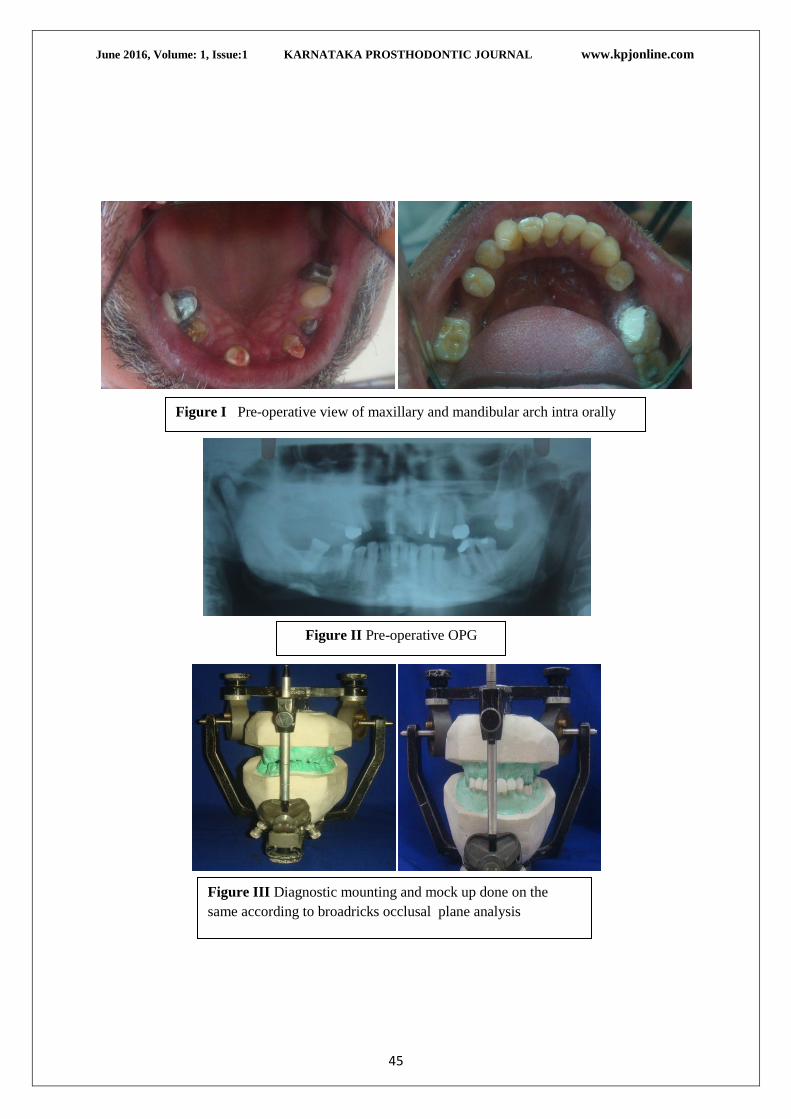
June 2016 Volume 1 Issue1 KARNATAKA PROSTHODONTIC JOURNAL wwwkpjonlinecom
56
REFERENCES
1) Syncone concept DentsplyManual of AnkylosSynCone Concept
2) Marco DDiego NImmediate rehabilitation of the edentulaous mandible using the
ankylosteklescopic copings and intra oral welding a pilot study conceptInt J Periodont And
Restor Dent201232189-94
3) Zhang RG Hannak WB Roggensack M Freesmeyer WB Retentive characteristics of
AnkylosSynCone conical crown system over long-term use in vitro Eur J ProsthodontRestor Dent
20081661ndash66 2
4) Huang JS Zhu XB Immediate implantsupport and overdenture retained by conical crowns
Three case reports [in Chinese] Hua Xi Kou Qiang Yi XueZaZhi 200927461ndash464
5) Branemark PI Osseointegration and its experimental background J Prosthet Dent
198350399ndash410
6) Jaffin RA Kumar A BermanCL Immediate loading ofimplants in partially and fully
edentulous jaws a series of 27case reports J Periodontol 200071833ndash838
7) Steenberghe D NaertI Andersson M Brajnovic I VanCleynenbreugel J Suetens P A
]custom template and definitiveprosthesis allowing immediateimplant loading in the maxillaa
clinical report Int J Oral MaxillofacImplants 200217663ndash670
8) Degidi M Piattelli A Immediatefunctional and non-functionalloading of dental implants a 2-
to 60 month follow-up studyof 646 titanium implants J Periodontol200374225ndash241
9) Degidi M Nardi D Piattelli A Immediate rehabilitation of the edentulous mandible with a
definitive prosthesis supported by an intraorally welded titanium bar Int J Oral Maxillofac
Implants 200924 342ndash347
10) Klee de Vasconcellos D Bottino MA Saad PA Faloppa FF A new device in immediately
loaded implant treatment in the edentulous mandible Int J Oral Maxillofac Implants 2006
21615ndash622


