THE JOURNAL OF BIOLOGICAL CHEMISTRY Vol. 265, No. 16, Issue of June 5, pp. 9452-9457,199O 0 1990 by The American Society for Biochemistry and Molecular Biology, Inc. Printed in U.S. A. Juvenile GM2 Gangliosidosis Caused by Substitution of Histidine for Arginine at Position 499 or 504 of the a-Subunit of &Hexosaminidase* (Received for publication, January 10, 1990) Barry H. Paw, Samuel M. Moskowitz, Nancy Uhrhammer, Neil1 Wright, Michael M. Kabackl, and Elizabeth F. NeufeldQ From the Department of Biological Chemistry and Brain Research Institute, School of Medicine and Molecular Biology Institute, University of California, Los Angeles, California 90024 and the *Department of Pediatrics, School of Medicine, University of Cdiforniu, Sun Diego, California 92103 Juvenile GM2 gangliosidosis is a rare neurodegener- ative disorder closely related to Tay-Sachs disease but of later onset and more protracted course. The bio- chemical defect lies in the a-subunit of the lysosomal enzyme &hexosaminidase. Cultured fibroblasts de- rived from patient A synthesized an a-subunit which could acquire mannose 6-phosphate and be secreted, but which failed to associate with the B-subunit to form the enzymatically active heterodimer. By contrast, fi- broblasts from patient B synthesized an a-subunit that was retained in the endoplasmic reticulum. To identify the molecular basis of the disorder, RNA from fibro- blasts of these two patients was reverse-transcribed, and the cDNA encoding the a-subunit of @-hexosamin- idase was amplified by the polymerase chain reaction (PCR) in four overlapping fragments. The PCR frag- ments were subcloned and shown by sequence analysis to contain a G to A transition corresponding to substi- tution of histidine for arginine at position 504 in the case of patient A and at position 499 in the case of patient B. The mutations were confirmed by hybridi- zation of allele-specific oligonucleotides to PCR-ampli- fied fragments of DNA corresponding to exon 13 of the a-subunit gene. The ArgSo4 + His mutation was found on both alleles of patient A as well as of another unre- lated patient; the homozygosity of this mutant allele is attributable to consanguinity in the two families. The Arg4” + His mutation was found in patient B in com- pound heterozygosity with a common infantile Tay- Sachs allele. There is additional heterogeneity in ju- venile Gmz gangliosidosis, as neither mutation was found in the DNA of a fourth patient. The Arg -) His mutations at positions 499 and 504 are located at CpG dinucleotides, which are known to be mutagenic “hot SpOtS.n * This work was suuDorted in part by Grant NS22376 from the National Institutes of-Health (to-E. F. N.); by a Medical Scientist Training Program Traineeshin GM 08042 from the National Insti- tutes of-Health and Fellowship 18-88-27 from the March of Dimes Birth Defects Foundation (to B. H. P.); by a summer fellowship from the National Tay-Sachs and Allied Diseases Association, Inc. (to S. M. M.); by a contract from the Genetic Disease Section, Maternal and Child Health Branch, Department of Health, State of California; and a grant from the National Tay-Sachs and Allied Diseases Asso- ciation, Inc., (to M. M. K.). The costs of publication of this article were defrayed in part by the payment of page charges. This article must therefore be hereby marked “oduertisement” in accordance with 18 USC. Section 1734 solely to indicate this fact. § To whom correspondence should be addressed: Dept. of Biological Chemistry, UCLA School of Medicine, Los Angeles, CA 90024-1737. The GMZ gangliosidoses are autosomal recessive disorders caused by mutations in any of the three genes required for lysosomal degradation of the GMZganglioside’: the CPand /3- subunits of fi-hexosaminidase’ and the activator protein. The subunits must be associated into the heterodimeric (cx~)is- oenzyme, @-hexosaminidase A, to catalyze the removal of the /3-GalNAc residue from the glycolipid; this function cannot be performed by the homodimeric isoenzymes B (p/3) or S ((Y(U). Deficiency of the /3-hexosaminidase A isoenzyme results in lysosomal accumulation of the undegraded GMzganglioside, particularly in the nervous system. Tay-Sachs disease, the infantile form of /3-hexosaminidase A deficiency, is the best known and most common form of GM2 gangliosidosis; its neurologic manifestations become evident in infancy and lead to death in early childhood. Other forms of fi-hexosaminidase deficiency are classified as “juvenile,” “chronic,” or “adult- onset” depending on age of onset and clinical course. The clinical, pathological, biochemical, and genetic aspects of the GM2 gangliosidoses have been summarized in a recent review (1). Recent studies have shown molecular heterogeneity within the P-hexosaminidase A deficiency diseases, the consequence of mutations in the a-subunit gene (reviewed in Ref. 2). An insertion of 4 nucleotides in exon 11 (3) is the most common mutation among Ashkenazi Jews, accounting for over two- thirds of the carriers3 (3,4) or about 2% of that population as a whole. The same population also harbors a splice site mutation in intron 12, but at a lower frequency (5-7). Deletion of exon 1 and its flanking sequences has been found among some French-Canadians (8, 9). These are null mutations, resulting in absence of mature a-subunit mRNA and lack of synthesis of a-subunit polypeptide and cause the infantile form of Tay-Sachs disease. Other less common allelic muta- tions that cause infantile Tay-Sachs disease encode defective polypeptides; they are not associated with any particular demographic group. These mutations include a substitution of lysine for glutamic acid at position 482 (10) and a deletion of cytosine in codon 504 resulting in premature termination (11); in both cases, the a-subunit is defective in its intracel- lular transport from the endoplasmic reticulum to the Golgi (11-13). In contrast, the “Bl” mutation allows normal proc- essing and transport of the u-subunit and association into the heterodimeric A isoenzyme; however, a substitution of histi- 1 The abbreviations used are: GM2 ganglioside, N-acetylgalactosa- minyl-~l~-(N-acetylneuraminyl-a2~3)galactosyl-~l~-glucosyl- /31-+1-ceramide; PCR, polymerase chain reaction. ’ Listed as P-N-acetylhexosaminidase (EC 3.2.1.52) and also as N- acetyl+glucosaminidase (EC 3.2.1.30). 3 B. H. Paw, P. Tieu, M. M. Kaback, andE. F. Neufeld, manuscript in preparation. 9452 by guest on February 3, 2019 http://www.jbc.org/ Downloaded from
Transcript
THE JOURNAL OF BIOLOGICAL CHEMISTRY Vol. 265, No. 16, Issue of June 5, pp. 9452-9457,199O 0 1990 by The American Society for Biochemistry and Molecular Biology, Inc. Printed in U.S. A.
Juvenile GM2 Gangliosidosis Caused by Substitution of Histidine for Arginine at Position 499 or 504 of the a-Subunit of &Hexosaminidase*
(Received for publication, January 10, 1990)
Barry H. Paw, Samuel M. Moskowitz, Nancy Uhrhammer, Neil1 Wright, Michael M. Kabackl, and Elizabeth F. NeufeldQ From the Department of Biological Chemistry and Brain Research Institute, School of Medicine and Molecular Biology Institute, University of California, Los Angeles, California 90024 and the *Department of Pediatrics, School of Medicine, University of Cdiforniu, Sun Diego, California 92103
Juvenile GM2 gangliosidosis is a rare neurodegener- ative disorder closely related to Tay-Sachs disease but of later onset and more protracted course. The bio- chemical defect lies in the a-subunit of the lysosomal enzyme &hexosaminidase. Cultured fibroblasts de- rived from patient A synthesized an a-subunit which could acquire mannose 6-phosphate and be secreted, but which failed to associate with the B-subunit to form the enzymatically active heterodimer. By contrast, fi- broblasts from patient B synthesized an a-subunit that was retained in the endoplasmic reticulum. To identify the molecular basis of the disorder, RNA from fibro- blasts of these two patients was reverse-transcribed, and the cDNA encoding the a-subunit of @-hexosamin- idase was amplified by the polymerase chain reaction (PCR) in four overlapping fragments. The PCR frag- ments were subcloned and shown by sequence analysis to contain a G to A transition corresponding to substi- tution of histidine for arginine at position 504 in the case of patient A and at position 499 in the case of patient B. The mutations were confirmed by hybridi- zation of allele-specific oligonucleotides to PCR-ampli- fied fragments of DNA corresponding to exon 13 of the a-subunit gene. The ArgSo4 + His mutation was found on both alleles of patient A as well as of another unre- lated patient; the homozygosity of this mutant allele is attributable to consanguinity in the two families. The Arg4” + His mutation was found in patient B in com- pound heterozygosity with a common infantile Tay- Sachs allele. There is additional heterogeneity in ju- venile Gmz gangliosidosis, as neither mutation was found in the DNA of a fourth patient. The Arg -) His mutations at positions 499 and 504 are located at CpG dinucleotides, which are known to be mutagenic “hot SpOtS.n
* This work was suuDorted in part by Grant NS22376 from the National Institutes of-Health (to-E. F. N.); by a Medical Scientist Training Program Traineeshin GM 08042 from the National Insti- tutes of-Health and Fellowship 18-88-27 from the March of Dimes Birth Defects Foundation (to B. H. P.); by a summer fellowship from the National Tay-Sachs and Allied Diseases Association, Inc. (to S. M. M.); by a contract from the Genetic Disease Section, Maternal and Child Health Branch, Department of Health, State of California; and a grant from the National Tay-Sachs and Allied Diseases Asso- ciation, Inc., (to M. M. K.). The costs of publication of this article were defrayed in part by the payment of page charges. This article must therefore be hereby marked “oduertisement” in accordance with 18 USC. Section 1734 solely to indicate this fact.
§ To whom correspondence should be addressed: Dept. of Biological Chemistry, UCLA School of Medicine, Los Angeles, CA 90024-1737.
The GMZ gangliosidoses are autosomal recessive disorders caused by mutations in any of the three genes required for lysosomal degradation of the GMZ ganglioside’: the CP and /3- subunits of fi-hexosaminidase’ and the activator protein. The subunits must be associated into the heterodimeric (cx~) is- oenzyme, @-hexosaminidase A, to catalyze the removal of the /3-GalNAc residue from the glycolipid; this function cannot be performed by the homodimeric isoenzymes B (p/3) or S ((Y(U). Deficiency of the /3-hexosaminidase A isoenzyme results in lysosomal accumulation of the undegraded GMz ganglioside, particularly in the nervous system. Tay-Sachs disease, the infantile form of /3-hexosaminidase A deficiency, is the best known and most common form of GM2 gangliosidosis; its neurologic manifestations become evident in infancy and lead to death in early childhood. Other forms of fi-hexosaminidase deficiency are classified as “juvenile,” “chronic,” or “adult- onset” depending on age of onset and clinical course. The clinical, pathological, biochemical, and genetic aspects of the GM2 gangliosidoses have been summarized in a recent review (1).
Recent studies have shown molecular heterogeneity within the P-hexosaminidase A deficiency diseases, the consequence of mutations in the a-subunit gene (reviewed in Ref. 2). An insertion of 4 nucleotides in exon 11 (3) is the most common mutation among Ashkenazi Jews, accounting for over two- thirds of the carriers3 (3,4) or about 2% of that population as a whole. The same population also harbors a splice site mutation in intron 12, but at a lower frequency (5-7). Deletion of exon 1 and its flanking sequences has been found among some French-Canadians (8, 9). These are null mutations, resulting in absence of mature a-subunit mRNA and lack of synthesis of a-subunit polypeptide and cause the infantile form of Tay-Sachs disease. Other less common allelic muta- tions that cause infantile Tay-Sachs disease encode defective polypeptides; they are not associated with any particular demographic group. These mutations include a substitution of lysine for glutamic acid at position 482 (10) and a deletion of cytosine in codon 504 resulting in premature termination (11); in both cases, the a-subunit is defective in its intracel- lular transport from the endoplasmic reticulum to the Golgi (11-13). In contrast, the “Bl” mutation allows normal proc- essing and transport of the u-subunit and association into the heterodimeric A isoenzyme; however, a substitution of histi-
1 The abbreviations used are: GM2 ganglioside, N-acetylgalactosa- minyl-~l~-(N-acetylneuraminyl-a2~3)galactosyl-~l~-glucosyl- /31-+1-ceramide; PCR, polymerase chain reaction.
’ Listed as P-N-acetylhexosaminidase (EC 3.2.1.52) and also as N- acetyl+glucosaminidase (EC 3.2.1.30).
3 B. H. Paw, P. Tieu, M. M. Kaback, andE. F. Neufeld, manuscript in preparation.
Molecular Basis of Juvenile GM2 Gangliosidosis 9453
dine for arginine at position 178 (14) abolishes its catalytic activity toward the GM2 ganglioside (15). Finally, a substitu- tion of serine for glycine at position 269 (16, 17) causes the synthesis of a defective a-subunit that can be transported through the biosynthetic organelles and be secreted, but which fails to associate with the P-subunit (18); this mutation has been found in patients with adult-onset/chronic GMM2 ganglio- sidosis, usually in compound heterozygosity with one of the common Ashkenazi infantile mutations (16,17), but occasion- ally in homozygous form (19).
In the present study, we have characterized at the level of polypeptide, mRNA, and DNA the mutations of two patients, Juvenile A and Juvenile B, with enzymatically diagnosed p- hexosaminidase A deficiency and clinically diagnosed GM~ gangliosidosis of the juvenile type; for two additional patients, the mutations were examined at the level of DNA only. Data on the biosynthesis of the Juvenile B a-subunit were included in a previous study (18) where her fibroblasts were identified as “California Juvenile.” Identification of the molecular de- fects was achieved by amplification of the entire a-subunit cDNA (after reverse transcription) by the polymerase chain reaction (20) and sequence analysis of the subcloned PCR fragments. Hybridization with allele-specific oligonucleotide probes confirmed the respective mutations in the genomic DNA.
MATERIALS AND METHODS
Clinical Summary-The patient identified here as Juvenile A is of Assyrian origin, and her parents are related as first cousins. She had already developed progressive ataxia, spastic paraplegia, dysarthria, and cherry-red macula at the time of referral at age 10. Juvenile B, who is of mixed Jewish and Scottish-Irish origin, manifested a pro- gressively severe neurologic deterioration, similar to that of her affected brother (21), that began at age 3-5 and progressed until her death at age 26. A third patient, a 12-year-old male, for whom only genomic DNA was examined, is of Armenian extraction. His parents are first cousins; symptoms began at age 4 with clumsiness of hands, followed by progressive ataxia, spasticity, and dementia. A fourth patient is a I-year-old non-Jewish Caucasian female, with onset of tremors and psychomotor retardation beginning at age 2.
Reagents-Restriction and modifying enzymes were purchased from Promega Biotec or Strategene, Thermus aquaticus (Taq) and modified T7 (Sequenase) DNA polymerases were from United States Biochemical, and avian myeloblastosis virus reverse transcriptase was from Seikagaku. Deoxynucleotides were from Pharmacia LKB Biotechnology Inc. [a-?S]dATP (1000 Ci/mmol), [a-32P]dCTP (6000 Ci/mmol), [32P]phosphate (carrier-free), and L-[3,4,5-3H]leucine (150 Ci/mmol) were obtained from Amersham Corp., and [-y-32P]ATP (7000 Ci/mmol) was from ICN. Nitrocellulose membranes were from Schleicher and Schuell and Hybond nylon membranes were from Amersham Corp. The plasmid pGEM3Z was from Promega Biotec. Oligonucleotide primers were synthesized by Dr. D. Glitz (UCLA) on a Du Pant/Vega Coder 300 oligonucleotide synthesizer.
Antisera against the P-hexosaminidase A and B isoenzymes and against the monomeric a-subunit had been raised (22) and character- ized (23) previously. Fixed protein A-bearing Staphylococcus aureus cells (Pansorbin) and goat antiserum against fibronectin were pur- chased from Calbiochem. Fetal bovine serum was from Irvine Scien- tific and other cell culture reagents were from GIBCO.
Cells and Cell Culture-The strain of normal human fetal luna fibroblasts, IMR 90, was obtained from the Corriell Institute for Medical Research, Camden, NJ. The strain of skin fibroblasts from a patient with juvenile GM* gangliosidosis, designated Juvenile A, was kindly nrovided bv Dr. R. Gatti (UCLA). The strain of fibroblasts from the patient designated Juvenile B was initiated in the laboratory of one of the authors (M. M. K.). Fibroblasts were cultured at 35 “C in 5% CO, in Eagle’s minimal essential medium supplemented with 15% fetal bovine serum, pyruvate, nonessential amino acids, and antibiotics.
Peripheral blood leukocytes from two additional juvenile GM, gan- gliosidosis patients and their parents, as well as from enzymatically proven noncarriers, were prepared by standard methods (24).
Biosynthesis Studies-Radiolabeling of fibroblasts, immunoprecip-
itation of fl-hexosaminidase and its subunits, electrophoresis, and fluorographic visualization of the polypeptides were carried out as described (23) except for a change in the labeling medium. The medium used was Eagle’s minimal essential medium with Earle salts, prepared from a GIBCO Selectamine kit to be free of either leucine or phosphate for labeling with [3H]leucine or [32P]phosphate, respec- tively: it was supplemented with antibiotics, pyruvate, nonessential amino acids, and 5% dialyzed fetal bovine ser&n.
RNA PreDaration and Northern Blot Analvsis-Total RNA was isolated from cultured fibroblasts by a pubhshed procedure (25). About 20 fig of total RNA was electrophoresed in a 1% agarose, 0.6 M formaldehyde gel and transferred onto a nitrocellulose filter. Ra- diolabeling (26), blot hybridization (27, 28) with a full-length w subunit (29), or @-subunit (30) cDNA probe and washing (28) were performed according to standard procedures.
Reverse Transcription and PCR Amplification of cDNA-The ex- onic sequence was reverse-transcribed and amplified in four overlap- ping pieces, using the four sets of oligonucleotide primers identified in group 1, Table I. About 20 fig of total RNA was incubated for 1 h at 42 “C with 40 units of reverse transcriptase and one set of primers (250 pmol each of sense and antisense primers) in 50 ~1 of 50 mM Tris. DH 8.3. 50 mM KCl. 10 mM M&l,. 1 mM dithiothreitol. 10 ne/ ml bovine serum albumin; 0.2 pmol e&of dNTP. The RNA ternplaTe was digested with 1.5 pg of RNase A at 37 “C for 30 min. Taq polymerase (4 units) was added in 50 ~1 of PCR amplification buffer (42 mM Tris, pH 8.7, 3.7 mM MgCl,, 17 mM (NH&SOs, 10 mM 2- mercaptoethanol, 0.17 mg/ml bovine serum albumin, 10% dimethyl sulfoxide). PCR amplification (20) of the cDNA was performed in a Perkin Elmer-Cetus DNA Thermal Cycler using the following profile: l-min denaturation at 94 “C, L-min annealing at 48 “C, 4-min exten- sion at 72 “C for 40 cycles.
Subcloning and Plasmid Sequencing-The products of PCR ampli- fication were purified by electrophoresis in a 5% nondenaturing polyacrylamide gel. Eluted DNA fragments were sequentially 5’- phosphorylated with T4 polynucleotide kinase, treated with T4 DNA polymerase, and blunt-end-ligated into the dephosphorylated SmaI restriction site of pGEM3Z. The plasmid DNA was sequenced by the dideoxy method (31) with Sequenase as recommended by the manu- facturer.
Isolation and PCR Amplification of Genomic DNA-High molecular weight genomic DNA was isolated from fibroblasts and leukocytes as described (32). The DNA from relatives of Juvenile A was provided by Dr. R. Gatti. Genomic DNA (1 rg) was annealed to 500 pmol each of sense and antisense primers (Table I, group 2) and amplified with 2.5 units of Taq polymerase in 100 ~1 of 67 mM Tris, pH 8.7, 6.7 mM MgCl?, 17 mM (NH&SOa, 10 mM 2-mercaptoethanol, 0.17 mg/ml bovine serum albumin, 10% dimethyl sulfoxide, 0.2 mM each dNTP. Amplification was carried out for 35 cycles as follows: 2-min denatur- ation at 94 “C, P-min annealing at 48 “C, and 3-min extension at 72 “C. The PCR products were purified by phenol/chloroform extrac- tion and ethanol precipitation.
Allele-specific Oligonucleotide Hybridization-A minor modification of a published procedure (33) was used. About 10 ng of genomic PCR products were dotted onto Hybond nylon membrane in a Schleicher and Schuell dot blot manifold and hybridized with oligonucleotide probes listed in Table I, group 3. Filters were washed as described (33) at 54 “C and subjected to autoradiography.
RESULTS
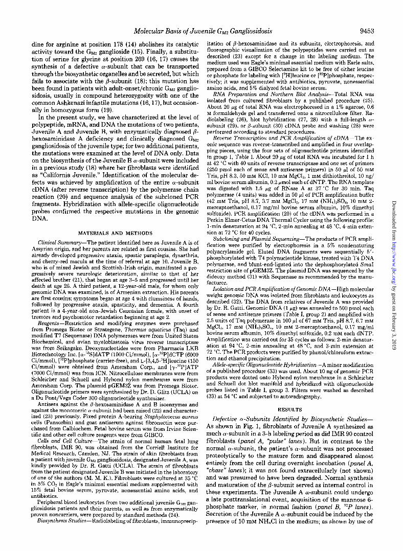
Defective cr-Subunits Identified by Biosynthetic Studies- As shown in Fig. 1, fibroblasts of Juvenile A synthesized as much a-subunit in a 3-h labeling period as did IMR 90 control fibroblasts (panel A, Pulse” lanes). But in contrast to the normal a-subunit, the patient’s a-subunit was not processed proteolytically to the mature form and disappeared almost entirely from the cell during overnight incubation (panel A, “chase” lanes); it was not found extracellularly (not shown) and was presumed to have been degraded. Normal synthesis and maturation of the @-subunit served as internal control in these experiments. The Juvenile A a-subunit could undergo a late posttranslational event, acquisition of the mannose 6- phosphate marker, in normal fashion (panel B, 32P lanes). Secretion of the Juvenile A a-subunit could be induced by the presence of 10 mM NH&l in the medium; as shown by use of
FIG. 1. Synthesis of the a-subunit by fibroblasts derived from juvenile GM2 gangliosidosis patient A. Normal (IMR 90) and Juvenile A confluent fibroblast cultures in loo-mm Petri dishes were labeled with 0.25 mCi of [“H]leucine/dish for a 3-h pulse, followed by an 18-h chase (panel A) and for the same period of pulse and chase in the presence of 10 mM NH,Cl (panel C). In panel B, the cells were labeled for 3 h with 0.25 mCi of [“Hlleucine or 1.0 mCi of [ “PIphosphate, as indicated. Antiserum raised against P-hexosamin- idase A was used for immunoprecipitation from cell extracts, shown in panels A and B. Panel C shows immunoprecipitation from NH:- induced secretions using three different antisera, anti-A, anti-B, and anti-n, as indicated. The antisera precipitate: A, all forms of the cy- and /Y-subunits; R, R-subunit that is associated with p-, as well as all forms of the &subunit; N, only monomeric n-subunit. Abbreviations: q,, & = precursor (Y-, p-subunit; N,, /‘J’., = mature (Y-, p-subunit.
specific antisera, the secreted Juvenile A a-subunit was a monomer, in contrast to the normal a-subunit which was associated with the @-subunit (panel C).
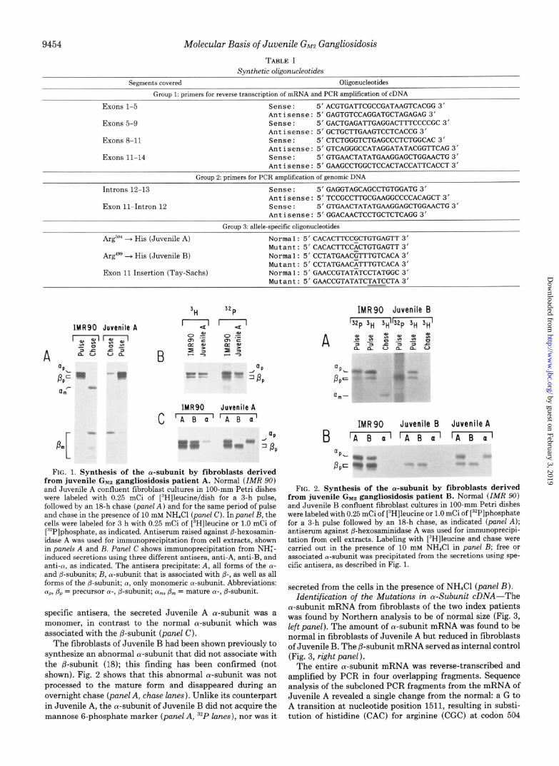
The fibroblasts of Juvenile B had been shown previously to synthesize an abnormal a-subunit that did not associate with the P-subunit (18); this finding has been confirmed (not shown). Fig. 2 shows that this abnormal a-subunit was not processed to the mature form and disappeared during an overnight chase (panel A, chase lanes). Unlike its counterpart in Juvenile A, the a-subunit of Juvenile B did not acquire the mannose 6-phosphate marker (panel A, “‘P lanes), nor was it
IMRSO Juvenile B '32p 3~ 3,,'132p 3,., 3,,'
A ub$ji
':
QP L"-
P PC ._ j# -7
IMRSO Juvenile B Juvenile A
B IA B al IA B al [A B al
(IP.-
PpC : ;;; " -
FIG. 2. Synthesis of the a-subunit by fibroblasts derived from juvenile GM2 gangliosidosis patient B. Normal (IMR 901 and Juvenile B confluent fibroblast cultures in loo-mm Petri dishes were labeled with 0.25 mCi of [“Hlleucine or 1.0 mCi of [:“P]phosphate for a 3-h pulse followed by an 18-h chase, as indicated (panel A); antiserum against fl-hexosaminidase A was used for immunoprecipi- tation from cell extracts. Labeling with [“Hlleucine and chase were carried out in the presence of 10 mM NH&I in panel B; free or associated a-subunit was precipitated from the secretions using spe- cific antisera, as described in Fig. 1.
secreted from the cells in the presence of NH&l (panel B). Identification of the Mutations in a-Subunit cDNA-The
a-subunit mRNA from fibroblasts of the two index patients was found by Northern analysis to be of normal size (Fig. 3, left panel). The amount of a-subunit mRNA was found to be normal in fibroblasts of Juvenile A but reduced in fibroblasts of Juvenile B. The p-subunit mRNA served as internal control (Fig. 3, right panel).
The entire n-subunit mRNA was reverse-transcribed and amplified by PCR in four overlapping fragments. Sequence analysis of the subcloned PCR fragments from the mRNA of Juvenile A revealed a single change from the normal: a G to A transition at nucleotide position 1511, resulting in substi- tution of histidine (CAC) for arginine (CGC) at codon 504
Molecular Basis of Juvenile GM2 Gangliosidosis 9455
mRNA : a-subunit b-subunit
FIG. 3. a-Subunit mRNA in fibroblasts from juvenile GMZ gangliosidosis patients. Total RNA (20 pg) from the normal (IMR 90) and patients’ fibroblasts was subjected to Northern blot analysis. The RNA blot was first hybridized with an o-subunit cDNA probe (left) and then stripped and rehybridized with a @-subunit cDNA probe (right)
Arg :
499 F--- C-
: f F
3’ c
NORMAL
=
NORMAL
A GCT
JUVENILE A fT 5’
JUVENILE B
-- - - Z= AGCT 3’
FIG. 4. Identification of the juvenile Gwz gangliosidosis mu- tations in cDNA. Total RNA from fibroblasts was reverse tran- scribed and the resulting n-subunit cDNA was amplified in overlap- ping segments and subcloned. The sequences of a portion of exon 13 derived from IMR 90 and Juvenile A are compared in the upper panel and from IMR 90 and Juvenile B in the lower panel. Arrows point to the two G to A transitions, resulting in Arg to His substitutions.
(Fig. 4, upper panel). This mutation was found in five inde- pendently isolated clones. The PCR products derived from mRNA of Juvenile B had a G to A transition at nucleotide position 1496, resulting in substitution of histidine (CAT) for arginine (CGT) at codon 499 (Fig. 4, lower panel). This mutation was found in three independently isolated clones. The nucleotide changes were confirmed by sequencing the opposite strand (not shown). Additional base changes were observed in some of the clones derived from Juvenile B, but since each one was found in a solitary clone, they were attributed to errors introduced during amplification by Taq polymerase; this was later confirmed by oligonucleotide hy- bridization (see below).
Identification of the Mutations in Genomic DNA-To dem- onstrate the nucleotide substitutions in genomic DNA isolated from the probands and their relatives, the exon 13 region encompassing the mutations was amplified by PCR, and the
amplified DNA was analyzed by allele-specific oligonucleotide hybridization. As shown in Fig. 5, Juvenile A, whose parents are related to each other, had inherited the Arg504 --f His mutation in homozygous fashion. The identical mutation was found in an unrelated juvenile GM2 gangliosidosis patient, whose parents are also consanguinous. The amplified DNA from the parents of both patients hybridized to both normal and mutant probes as expected for obligate heterozygotes (Fig. 5). Amplified DNA from 63 enzymatically proven non- carriers of /3-hexosaminidase deficiency hybridized solely to the normal oligonucleotide probe, confirming that this muta- tion was not a neutral polymorphism (data not shown).
The PCR fragment from the DNA of Juvenile B hybridized to both the normal and mutant oligonucleotides for codon 499, showing her to be a compound heterozygote for the Arg““” + His mutation (Fig. 6). This mutation was inherited from her mother; the other allele, inherited from her Jewish father, carried the more common 4-base pair insertion in exon 11 that underlies infantile Tay-Sachs disease. The reduced amount of a-subunit mRNA (Fig. 3) is consistent with com- pound heterozygosity with a null allele. Amplified DNA from 63 noncarriers did not hybridize to the Arg4” + His oligo- nucleotide probe, indicating that this change likewise is not a polymorphism (not shown). The genomic DNA of Juvenile B was also tested for base changes observed in some of the subcloned PCR fragments (see above), using oligonucleotides designed specifically for that purpose. In all instances, the genomic DNA was found to hybridize only to the normal oligonucleotide, confirming that the changes which had been observed in occasional clones were the result of Taq polym- erase errors (not shown). Amplified DNA from the fourth juvenile GM2 gangliosidosis patient failed to hybridize to mu- tant probes at codon 499 or 504 (data not shown).
Normal ()@@ 00 l @@ 0.0 Mutant 0.0. 0.0
FIG. 5. Identification of the ArgGo” to His mutation in ge- nomic DNA by allele-specific oligonucleotides. Genomic DNA preparations from IMR 90, from Juvenile A (arrow) and her relatives, as well as from another unrelated patient and his parents were amplified by PCR in the region of exon 13. The PCR products were dotted onto nylon membranes. ‘“P-labeled 19-mer oligonucleotide probes corresponding to normal or mutant sequence at codon 504 were used to hybridize to the amplified DNA.
FIG. 6. Identification of the Arg4”9 to His mutation in ge- nomic DNA by allele-specific oligonucleotides. Genomic DNA preparations from IMR 90, from a Tay-Sachs disease fibroblast strain homozygous for the exon 11 insertion, and from fibroblasts of Juvenile B (arrow) and her parents were amplified separately in the regions of exon 11 and exon 13. The PCR products were dotted onto nylon membranes. ‘rYP-Labeled oligonucleotide probes for normal and mu- tant sequences at codon 499 (juvenile) and exon 11 (infantile) were used to hybridize to the amplified DNA.
9456 Molecular Basis of Juvenile GM2 Gangliosidosis
DISCUSSION
We have identified mutations of the o-subunit gene of p- hexosaminidase, guanine to adenine transitions in exon 13, that are present on one or both alleles of patients with juvenile GM~ gangliosidosis. Two unrelated patients were shown to be homozygous for the Argo4 --$ His mutation. In both families, the parents are consanguinous. Another patient was shown to be a compound heterozygote for the Arg499 + His allele with the exon 11 infantile Tay-Sachs allele that is common in the Ashkenazi Jewish population. The absence of either substi- tution in a fourth patient shows that yet other mutations may give rise to juvenile GM2 gangliosidosis.
The defective o-subunit of Juvenile A (Arg504 --, His) ac- quires the mannose 6-phosphate recognition marker but fails to associate with the p-subunit and is not processed to the mature lysosomal form (presumably because in the absence of association, it is not transported to lysosomes (23)). In the presence of NH:, which diverts newly made hydrolases from the lysosomal to the secretory pathway (22, 34), the mono- meric a-subunit of Juvenile A is secreted. This shows that it can be transported out of the endoplasmic reticulum and Golgi complex. Overall, the abnormal o-subunit of Juvenile A resembles the association-defective a-subunit (18) caused by the Gly*” + Ser substitution found in patients with chronic/adult-onset GM2 gangliosidosis (16, 17).
Because the a-subunit synthesized by fibroblasts of Juve- nile B (identified in the earlier publication as “California Juvenile”) was previously found not to be associated with the &subunit, it had been grouped together with the association- defective o-subunit of chronic and adult-onset GM2 ganglios- idosis (18). However, the Juvenile B a-subunit (Arg499 + His) is not secreted in the presence of NH:, indicating that it is retained in an early biosynthetic compartment, probably the endoplasmic reticulum. Therefore it resembles more closely the defective o-subunits resulting from some infantile Tay- Sachs disease mutations, deletion of cytosine in codon 504 (11,13), and substitution of lysine for glutamic acid in position 482 (10, 12). Retention in the endoplasmic reticulum is thought to be due to misfolding of the mutant a-subunits, as has been observed for other defective proteins (reviewed in Ref. 35). It is clear that the effect of amino acid substitutions on the fate of the a-subunit is not correlated with their position in the linear sequence and will have to be interpreted in terms of the three-dimensional configuration of the poly- peptides. Unfortunately, the requisite x-ray crystallographic information is not available at the present time. Despite the lack of knowledge regarding secondary and tertiary structure of the protein, the importance of arginine residues at positions 499 and 504 can be inferred from their evolutionary conser- vation. The arginine at position 504 in the or-subunit is conserved in the homologous P-subunit of human (30,36) and murine (37) origin; the arginine at position 499 is even more highly conserved, as it is found in the human (30, 36) and murine (37) p-subunit, and also in the P-hexosaminidase of the slime mold, Dictyostelium discoideum (38).
The mutations described here give rise to a juvenile form of GM2 gangliosidosis, which is intermediate in clinical course between the catastrophic infantile form (Tay-Sachs disease) and the relatively mild adult-onset form. The clinical pheno- type cannot be correlated with the linear position of the mutation nor with the properties of the polypeptide observed in radiolabeling studies. It is generally believed that clinical severity is inversely related to the ability of the mutant p- hexosaminidase to degrade GM2 ganglioside in uiuo (1). We have been unable to demonstrate residual activity in fibroblast homogenates of the juvenile GMMQ gangliosidosis patients, using
a fluorogenic substrate, 4-methylumbelliferyl 6-sulfo-P-N- acetylglucosaminide (not shown). However, a mutant enzyme might not survive the standard extraction or assay conditions developed for the normal enzyme, or it might not function with a synthetic substrate. The fibroblasts of patient B had been shown previously to have 3% the normal activity with the physiological substrate, namely, GMz ganglioside com- plexed to the activator protein.4
The two G to A transitions identified in this study have occurred at CpG sites. Other G to A transitions at CpG sites have been found in exon 5 (the “Bl” mutation, Arg17* + His (14)) and in exon 13 (G~u~‘~ + Lys (10)) of the p-hexosamin- idase a-subunit gene and in intron I2 of the P-subunit gene (39). CpG sites are known to be mutagenic “hot spots,” accounting for a disproportionate number of human polymor- phisms (40) as well as of disease-producing mutations (e.g. of hemophilia due to deficiency of Factor VIII (41-43) or Factor IX (44)). Such mutations reflect the tendency of 5-methyl- cytosine, frequently found at CpG sites, to deaminate spon- taneously to thymidine (45). Depending on the DNA strand on which the deamination occurs, the result is either a C to T or a G to A transition on the sense strand. Such mutations occur sporadically, and the same base change in different individuals can be the result of separate mutational events, as has been observed in Factor VIII (43); thus the Arg5°4 + His mutation might have occurred independently in the two kindreds in which it was observed. A two-step repair system that excises the mismatched TMP and fills the gap with CMP has recently been described in mammalian cells (46); the deletion of cytosine at the CpG site in codon 504 (11) could have resulted from an error in the second phase of this repair process. Since the coding sequences of the two subunits of /3- hexosaminidase each contain about three dozen CpG dinucle- otide sites, additional mutations can be anticipated, even though not all the sites would be methylated and many of the mutations would be silent.
Acknowledgments-We thank Dr. Richard Gatti (University of California, Los Angeles) for the tibroblast culture of patient Juvenile A and DNA samples from the family, Dr. Elena Boder (University of California, Los Angeles) for information about that patient, Dr. Robert Carrel (Santa Barbara Regional Center) for referral of patient Juvenile B and her family, Dr. Ira Lott (University of California, Irvine) for referral of the Armenian familv. Drs. Rachel Myerowitz and Richard Proia (National Institutes of Health) for recombinant plasmids containing /3-hexosaminidase a-subunit and P-subunit cDNA, Dr. Dohn Glitz (University of California, Los Angeles) for oligonucleotide synthesis, Doris Quon for help with some of the biosynthetic studies, and Larry Tabata for illustrations.
Note Added in Proof-The A&“” + His mutation was independ- ently discovered by R. M. Boustany and K. Suzuki (personal com- munication) in a Juvenile Gwz gangliosidosis patient from a consan- guinous Lebanese Christian family.
REFERENCES
1. Sandhoff, K., Conzelmann, E., Neufeld, E. F., Kaback, M. M., and Suzuki, K. (1989) in The Metabolic Basis of hherited Disease (Striver, C. R., Beaudet, A. L., Sly, W. S., and Valle, D., eds) pp. 1807-1842, McGraw-Hill, Inc., New York
2. Neufeld, E. F. (1989) J. Biol. Chem. 264, 10927-10930 3. Myerowitz, R., and Costigan, F. C. (1988) J. Biol. Chem. 263,
18587-18589 4. Triggs-Raine, B. L., Feigenbaum, A. S. J., Skomorowski, M. A.,
Natowicz. M.. Kolodnv. E. H.. Clarke. J. T. R.. Mahuran, D. J., and Gravel, R. A.-(1989) km. J. &rz Gekt. 45, A226 (abstr.)
5. Myerowitz, R. (1988) Proc. Nutl. Acad. Sci. U. S. A. 85, 3955- 3959
4 K. Sandhoff, and M. M. Kaback, unpublished results.
Molecular Basis of Juvenile GM2 Gangliosidosis 9457
6.
7.
8.
9.
10.
11.
12.
13.
14. 15.
16. 17.
18.
19.
20.
21.
22.
23.
24.
25.
Arpaia, E., Dumbrille-Ross, A., Maler, T., Neote, K., Tropak, M., Troxel, C., Stirling, J. L., Pitts, J. S., Bapat, B., Lamhonwah, A. M., Mahuran,-D. J., Schuster, S. M., Clarke, J. T. R., Lowden. J. A.. and Gravel. R. A. (1988) Nature 333. 85-86
Ohno, K., and Suzuki, K. (1988) Biochem’Biophys. Res.~Commun. 153,463-469
Myerowitz, R., and Hogikyan, N. D. (1986) Science 232, 1646- 1648
Myerowitz, R., and Hogikyan, N. D. (1987) J. Biol. Chem. 262, 15396-15399
Nakano, T., Muscillo, M., Ohno, K., Hoffman, A. J., and Suzuki, K. (1988) J. Neurochem. 51,984-987
Lau, M. M. H., and Neufeld, E. F. (1989) J. Biol. Chem. 264, 21376-21380
Proia, R. L., and Neufeld, E. F. (1982) Proc. Natl. Acad. Sci. U. S.A. 79,6360-6364
Zokaeem, G., Bayleran, J., Kaplan, P., Hechtman, P., and Neu- feld, E. F. (1987) Am. J. Hum. Genet. 40,537-547
Ohno, K., and Suzuki, K. (1988) J. Neurochem. 50,316-318 Kytzia, H. J., Hinrichs, U., Maire, I., Suzuki, K., and Sandhoff,
K. (1983) EMBO J. 2, 1201-1205 Navon, R., and Proia, R. L. (1989) Science 243,1471-1474 Paw, B. H., Kaback, M. M., and Neufeld, E. F. (1989) Proc. Natl.
Acad. Sci. U. S. A. 86, 2413-2417 d’Azzo, A., Proia, R. L., Kolodny, E. H., Kaback, M. M., and
Neufeld, E. F. (1984) J. Biol. Chem. 259, 11070-11074 Navon, R., Kolodny, E. H., Mitsumoto, H., Thomas, G. H., and
Proia, R. L. (1989) Am. J. Hum. Genet. 46, A209 (abstr.) Saiki, R. K., Gelfand, D. H., Stoffel, S., Scharf, S. J., Higuchi,
R., Horn, G. T., Mullis, K. B., and Erlich, H. A. (1988) Science 239,487-491
Philippart, M., Carrel, R. E., and Landing, B. (1983) J. Neuro- &em. 4 1, S56 (abstr.)
Hasilik, A., and Neufeld, E. F. (1980) J. Biol. Chem. 255, 4937- 4945
Proia, R. L., d’Azzo, A., and Neufeld, E. F. (1984) J. Biol. Chem. 259,3350-3354
Kaback, M. M. (1977) in Tay-Sachs Disease, Screening and Prevention (Kaback, M. M., Rimoin, D., and O’Brien, J. S., eds) pp. 276-277, Alan R. Liss, New York
Chomczynski, P., and Sacchi, N. (1987) Anal. Biochem. 162, 156-159
26. Feinberg, A. P., and Vogelstein, B. (1983) Anal. Biochem. 132, 6-13
27. Thomas. P. S. (1980) Proc. Natl. Acad. Sci. U. S. A. 77. 5201-
28.
29.
30. 31.
32.
33.
34.
35. 36.
37.
38.
39.
40. 41.
42.
43.
44.
45.
46.
5205 Wahl, G. M., Stern, M., and Stark, G. (1979) Proc. Natl. Acad.
Sci. U. S. A. 76,3683-3687 Myerowitz, R., Piekarz, R., Neufeld, E. F., Shows, T. B., and
Suzuki. K. (1985) Proc. Natl. Acad. Sci. U. S. A. 82. 7830-7834 Proia, R.‘L. (i988)'Proc. Natl. Acad. Sci. U. S. A. 85; 1883-1887 Sanger, F., Nicklen, S., and Coulson, A. R. (1977) Proc. Natl.
Acad. Sci. U. S. A. 74,5463-5467 Bell, G. I., Karam, J. H., and Rutter, W. J. (1981) Proc. Natl.
Acad. Sci. U. S. A. 78.5759-5763 Farr, C. J., Saiki, R. K., Erlich, H. A., McCormick, F., and
Marshall, C. J. (1988) Proc. Natl. Acad. Sci. U. S. A. 85, 1629- 1633
Gonzalez-Noriega, A., Grubb, J. H., Talkad, V., and Sly, W. S. (1980) J. Cell Biol. 85, 839-852
Lodish, H. F. (1988) J. Biol. C&m. 263, 2107-2110 Korneluk, R. G., Mahuran, D. J., Neote, K., Klavins, M. H.,
O’Dowd, B. F., Tropak, M., Willard, H. F., Anderson, M.-J., Lowden. J. A.. and Gravel. R. A. (1986) J. Biol. Chem. 261. 8407-8413 ’
Bapat, B., Ethier, M., Neote, K., Mahuran, D., and Gravel, R. A. (1988) FEBS Mt. 237, 191-195
Graham, T. R., Zassenhaus, H. P., and Kaplan, A. (1988) J. Biol. Chem. 263,16823-16829
Nakano, T., and Suzuki, K. (1989) J. Biol. Chem. 264, 5155- 5158
Barker, D., Schafer, M., and White, R. (1984) Cell 36, 131-138 Gitschier, J., Wood; W..I., Tuddenham, E. G. D., Shuman, M. A.,
Goralka. T. M.. Chen. E. Y.. and Lawn. R. M. (1985) Nature 315,42?-430
Youssoufian, H., Kazazian, H. H., Jr., Phillips, D. G., Aronis, S., Tsiftis, G., Brown, V. A., and Antonarakis, S. E. (1986) Nature 324,380-382
Youssoufian, H., Antonarakis, S. E.. Bell. W.. Griffin, A. M.. and Kazazian,H. H. (1988) Am: J. Hum. Gene; 42, 718-725
Koeberl. D. D.. Bottema. C. D. K.. Buerstedde. J. M.. and Som- mer, S. S. (1989) Am. k Hum. &net. 45, 448-457
Coulondre, C., Miller, J. H., Farabaugh, P. J., and Gilbert, W. (1978) Nature 274, 775-780
Wiebauer, K., and Jiricny, J. (1989) Nature 339, 234-236