+ All Categories
Home > Documents > Kanai Original article -...

Kanai Original article -...

Date post: 28-Apr-2018
Category:
Upload: lamphuc
View: 216 times
Download: 1 times
Share this document with a friend
28
1 Original article Prediction of skin permeation by chemical compounds using the artificial membrane, Strat-M TM Takashi Uchida, Wesam R. Kadhum, Sayumi Kanai, Hiroaki Todo, Takeshi Oshizaka, Kenji Sugibayashi* Faculty of Pharmaceutical Sciences, Josai University, 1-1 Keyakidai, Sakado, Saitama 350-0295, Japan. To whom correspondence should be addressed: Kenji Sugibayashi, Ph.D. Faculty of Pharmaceutical Sciences, Josai University, Keyakidai, Sakado, Saitama 350-0295, Japan. Tel.: 049-271-7367 Fax: 049-271-8137 E-mail: [email protected]
Transcript

1

Original article

Prediction of skin permeation by chemical compounds using the artificial membrane,

Strat-MTM

Takashi Uchida, Wesam R. Kadhum, Sayumi Kanai, Hiroaki Todo, Takeshi Oshizaka,

Kenji Sugibayashi*

Faculty of Pharmaceutical Sciences, Josai University, 1-1 Keyakidai, Sakado, Saitama

350-0295, Japan.

To whom correspondence should be addressed:

Kenji Sugibayashi, Ph.D.

Faculty of Pharmaceutical Sciences, Josai University,

Keyakidai, Sakado, Saitama 350-0295, Japan.

Tel.: 049-271-7367

Fax: 049-271-8137

E-mail: [email protected]

2

1. Introduction

In the past 20 years, more than 35 transdermal delivery systems (TDDSs) have

been approved in the US, EU, and Japan(Murthy, 2012). In vitro skin permeation

experiments are considered to be useful in the development of new TDDSs. Artificial

membranes using a parallel artificial membrane permeability assay, PAMPA

(Akamatsu et al., 2009; Avdeef et al., 2007), and skin-PVPA model (Palac et al., 2014)

are currently used to select orally administered drug candidates in a short experimental

period. However, the selection of compound candidates from a large chemical library is

time-consuming because the skin permeation rates of chemical compounds are known to

be markedly lower than those through other mucosal membranes. Therefore,

synthesized artificial membranes (Hatanaka et al., 1990; Sugibayashi et al., 2010) and

three-dimensional cultured human skin models (Kano et al., 2010; Poumay et al., 2004)

have been used as alternatives to human and animal skins in the development of

TDDSs. Previous studies examined the flux or permeability coefficients, P, of chemical

compounds through alternative membranes and those through excised human or

animal skin (Kano et al., 2010; Morimoto et al., 1992; Scott et al., 1986). Sinkó et al.,

recently described (Sinkà et al., 2012) PAMPA incorporated with human ceramide

analogues and fatty acids in the stratum corneum. Although PAMPA could roughly

predict human skin permeability, a 1:1 correlation was not obtained between the P

values of chemicals through the synthetic membrane and human skin. This finding

suggested that this membrane may not be sufficiently sensitive to reflect the

physicochemical properties of penetrants.

The physicochemical properties of drugs, such as their molecular weights and

the lipophilic/hydrophilic balance (i.e., octanol/ buffer apparent distribution ratio at a

3

particular pH, log Ko/w), have been shown to markedly affect permeability through skin

(Potts and Guy, 1992; Lian et al., 2008; Riviere and Brooks, 2011). Most drugs utilized

in TDDSs have a molecular weight under 500 Daltons (Bos and Meinardi, 2000), and

the P value through skin was previously reported to markedly decrease with an

increase in molecular weight over 500 Daltons. Furthermore, elevations were

observed in the P values of drugs with an increase in the log Ko/w. Thus, the P value

can be further divided into two parameters; diffusion (DL-2) and partition (KL)

parameters (Okamoto et al., 1988), where D, K and L are the diffusion coefficient,

partition coefficient, and thickness of the membrane, respectively. These parameters

can be easily obtained from in vitro skin permeation experiments, the values of which

depend on the characteristics as well as penetrant properties of the membrane.

Therefore, the acquisition of these parameters for alternative membranes may prove

very useful in understanding membrane characteristics and their similarities (or

difference) to human and animal skins.

An in silico approach has also been attempted in order to predict the P values

of chemical compounds through skin with their physicochemical properties (Potts and

Guy, 1992); (Lian et al., 2008; Karadzovska et al., 2013). The predicted P values could

be used to more easily understand and evaluate the usefulness and safety of topically

applied chemical compounds. Thus, similarities between the lipophilicity characteristics

or solubility parameters of alternative membranes and human/animal skins may be

needed in order to establish a good prediction from various formulations and applied

conditions (finite or infinite volume condition).

Strat-M ™ (Merck Millipore, USA) was recently launched and is now

commercially available as a skin-mimic artificial membrane. This membrane is

4

composed of multiple layers of polyester sulfone. However, its potential as an

alternative membrane for estimating skin permeation has not yet been clarified.

In the present study, a permeation experiment was performed with Strat-M™,

human skin, or hairless rat skin in order to obtain P, DL-2, and KL of the applied

compounds from aqueous solution. The usefulness and characteristics of Strat-M™

were investigated by comparing these parameters between human skin, hairless rat

skin, and Strat-M ™.

2. Experiment

2.1. Regent and materials

Methyl paraben (MP), ethyl paraben (EP), n-propyl paraben (PP), n-butyl

paraben (BP), isosorbide 5-mononitrate (ISMN), n-propyl p-aminobenzoate (P-PABA),

and n-butyl p-aminobenzoate (B-PABA) were purchased from Tokyo Chemical Industry

Co., Ltd. (Tokyo, Japan). Lidocaine hydrochloride (LID•HCl) was purchased from

Sigma Aldrich (St. Louis, MO, U.S.A.). Antipyrine (ANP), aminopyrine (AMP), methyl

p-aminobenzoate (M-PABA), and diisopropyl fluorophosphate (DFP) as an ester

inhibitor were purchased from Wako Pure Chemical (Osaka, Japan). Ethyl

p-aminobenzoate (E-PABA) was purchased from Kanto Chemical Co., Ltd. (Tokyo,

Japan). Isosorbide dinitrate (ISDN) was kindly gifted from Tokyo Pharmaceutical

Industries Industry Co., Ltd. (Tokyo, Japan). The molecular weights (M.W.) and

octanol/water coefficients (log Ko/w) of these chemicals are shown in Table 1. Strat-MTM

was purchased from Merck Millipore (Billerica, MA, U.S.A.). All other reagents were

of HPLC grade and were used without further purification.

5

Table 1

2.2. Experimental animals

Male hairless rats (WBM/ILA-Ht, 230-280 g) were obtained from the Life

Science Research Center, Josai University (Sakado, Saitama, Japan) or Ishikawa

Experimental Animal Laboratories (Fukaya, Saitama, Japan). All animal experiments

were performed according to the Ethics Committee of Josai University.

2.3. Excised human skin

Excised abdominal human skin (60-year-old Caucasian female) with a

thickness of 545 µm was purchased from Biopredic International (Rennes, France)

through KAC (Kyoto, Japan). The use of excised human skin was approved by the

KAC Ethics Committee for human-derived products.

2.4. Scanning and transmission electron microscopic observations of Strat-M TM

The cross-sectional structure of Strat-M TM was observed using a scanning

electron microscope (SEM) (S-3000N; Jeol Ltd., Tokyo, Japan) operating under a high

vacuum condition after being coated with gold. Prior to transmission electron

microscopic observations, Strat-M TM was fixed with 2% osmium tetroxide solution and

embedded in epoxy resin. Ultra-thin sections were then prepared with an

ultramicrotome. The sections obtained were contrasted with 4% uranyl acetate and

observed with a transmission electron microscope (JEM1200-Ex; Jeol Ltd.). The

thickness of the skin sections was measured using DP2-BSW software (Olympus Co.,

Tokyo, Japan).

6

2.5. Permeation experiments

Strat-M TM: Strat-M TM was set in a Franz-type diffusion cell (effective diffusion

area, 1.77 cm2) and the receiver chamber was maintained at 32°C. Saturated chemical

compounds dissolved in pH7.4 phosphate-buffered saline (PBS), pH 5.0 citrate-buffered

saline, or pH 9.0 carbonate-buffered saline (1.0 mL) and 6.0 mL of the corresponding

buffer were added to the donor and receiver sides, respectively, in all permeation

experiments. The receiver solution was agitated using a stirrer bar and magnetic

stirrer throughout the experiments. An aliquot (500 µL) was withdrawn from the

receiver chamber and the same volume of fresh buffer was added to the chamber to keep

the volume constant. The penetrant concentration in the receiver chamber was

determined by HPLC.

Human skin: Frozen human skin was thawed at room temperature and

mounted on a Franz-type diffusion cell. In the case of the skin permeation

experiments conducted for ester compounds, DFP in PBS at a concentration of 2.7

µmol/mL (6 mL) was added to the receiver chamber for 11 h to prevent metabolism after

skin hydration with PBS. The skin permeation experiments were then started. The

absence of an effect by DFP on the skin permeation of parabens and other compounds

was reported previously (Ahmed et al., 1996; Sugibayashi et al., 2004; Tamura et al.,

1995). Permeation experiments were carried out for the other chemical compounds after

skin had been hydrated for 12 h with an appropriate buffer at 32°C. A penetrant

solution in buffer was added to the donor chamber, whereas DFP in PBS (0.54 µmol/mL)

or buffer alone (6 mL each) was added to the receiver chamber to start the permeation

experiments. An aliquot (500 µL) was withdrawn from the receiver chamber and the

same volume of PBS containing DFP or buffer alone was added to the chamber to keep

7

the volume constant. Other procedures were followed according to the method

described for Strat-MTM.

Hairless rat skin: Abdominal skin was cleaned using a wet Kimwipe and

excised from hairless rats under anesthesia with an i.p. injection of pentobarbital (50

mg/kg). Stripped skin was obtained by stripping the stratum corneum off 20-times

with adhesive tape. To decrease variabilities in skin permeability due to the

abdominal sites of skin, only skin from the right and left upper abdominal areas were

used. Excess fat was trimmed from the excised skin, and the skin sample was set in a

Franz-type diffusion cell. The skin permeation experiments were conducted after 60

min of hydration with the corresponding buffer at 32°C. The other procedures were

performed with the same experimental method to Strat-M TM.

2.6. Determination of chemicals

The same volume of acetonitrile containing parabens was added to the MP, EP,

PP, BP, AMP, ISMN, ISDN, LID, E-PABA, M-PABA, P-PABA, and B-PABA samples.

After gentle mixing, the sample was centrifuged for 5 min at 21,500 ×g and 4°C to

remove proteins and contaminants. The supernatant was injected onto HPLC. The

HPLC system consisted of a pump (LC-10AD; Shimadzu, Kyoto, Japan), Chromatopac

(C-R6A; Shimadzu), UV detector (SPD-6A; Shimadzu), system controller (SCL-6B;

Shimadzu), and auto-injector (SIL-7A; Shimadzu). The LiChroCART®250-4 column

(KGaA·64271; Merck, Darmstadt, Germany) was maintained at 40°C during the eluting

mobile phase, 0.1% phosphoric acid : acetonitrile = 75 : 25 for MP and EP, 0.1%

phosphoric acid : acetonitrile = 55 : 45 for PP and BP, 0.1% phosphoric acid containing 5

mM sodium dodecyl sulfate : acetonitrile = 50 : 50 for AMP, water : acetonitrile = 55 : 45

8

for ISDN, 0.1% phosphoric acid containing 5 mM sodium 1-heptanesulfonate :

acetonitrile = 70 : 30 for LID, water : acetonitrile = 90 : 10 for ISMN and water :

acetonitrile = 80 : 20 for ANP. The flow rate was adjusted to 1.0 mL/min. The

injection volume was 20 µL, and detection was performed at 220 nm for ISMN and

ISDN, 230 nm for LID, 245 nm for AMP, 254 nm for ANP, and 260 nm for MP, EP, PP

and BP. Analyses of E-PABA, M-PABA, P-PABA and B-PABA were carried out with

gradient elution systems. A solvent program that initially started with 30% acetonitrile

and 70% phosphoric acid solution (0.1%) was changed linearly (13 min) to acetonitrile

and phosphoric acid (1 : 1) and held for an additional 10.5 min. The flow rate was

maintained at 1.0 mL/min. The system was gradually returned to the starting

condition for the next sample. Peaks were detected at 280 nm. The Inertsil® ODS-3

column (4.6 × 150 mm, 5 µm, GL Sciences Inc., Tokyo, Japan) was kept at 40°C.

2.7. Calculation of membrane permeation parameters

Permeation parameters, such as the permeability coefficient and partition and

diffusion coefficients, were calculated from the time course of the cumulative amount of

chemicals that permeated through membrane by the following equations (Flynn et al.,

1974).

                                 𝐹𝑙𝑢𝑥 = 𝐶𝑣𝐾𝐷𝐿 = 𝑃 ∙ 𝐶𝑣                                                                                                                                                                                                 1

                             𝐷𝐿!! =1

6𝑇!"#                                                                                                                                                                                                                         2

                             𝐾𝐿 = 6𝑇!"#𝑃                                                                                                                                                                                                                               3

9

where Cv is the applied concentration of the chemical. The lag time, Tlag, was

calculated from the x-axis (time-axis) intercept of the slope at a steady-state flux for the

permeation profile of chemical compounds through the membrane.

2.8. Statistical analysis

Pearson's correlation coefficient was used to characterize the relationship

between logP values in human and rat skins and Strat-MTM. Significance was to 5% in

all evaluations.

3. Results

Figure 1 shows an SEM image of a vertical section of Strat-M TM. Three

layers; i, ii, and iii, were confirmed from the figure. The density of each layer differed

and gradually decreased from the top of the membrane. The thicknesses of these

layers were 52.3 ± 0.5, 76.7 ± 5.07 and 196 ± 7.37 µm (mean ± S.D.) respectively, from

the top, while that of the whole layer was 324.6 ± 4.6 µm.

Figure 1

Figures 2 a, b and c show TEM images of the 1st, 2nd, and 3rd layers of

Strat-MTM, respectively. Staining was observed in the 1st and 2nd layers, but was

absent in the 3rd layer, suggesting that lipids existed in the top two layers.

Figure 2

10

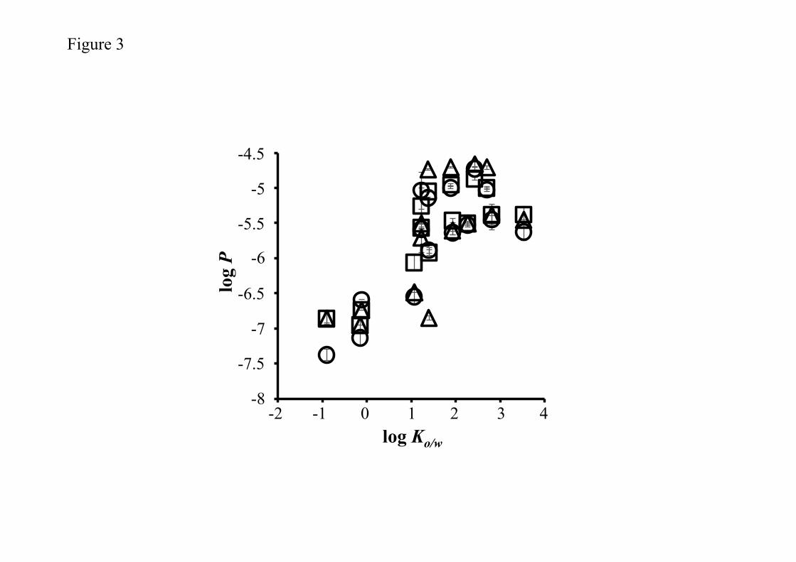
Figure 3 shows the relationships between logKo/w and log P obtained from

permeation experiments through human skin, rat skin, or Strat-MTM. Elevations were

observed in the log P of all membranes with an increase in lipophilicities of the chemical

compounds applied. The relationship obtained between log P and logKo/w could be

represented by a sigmoidal curve. The log P values obtained in Strat-MTM were very

similar to those in human and rat skins.

Figure 3

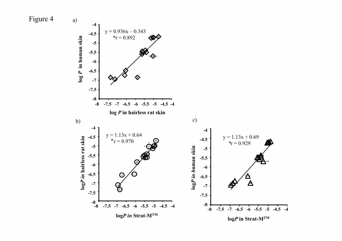
Figures 4 a, b, and c show the relationship between log P in human and rat

skins, in rat skin and Strat-MTM, and in human skin and Strat-MTM, respectively and

permeability coefficients of chemicals through the membranes were shown in Table 2.

A good relationship was observed between the human and rat skin data, as shown in

Figure 4a. Furthermore, the almost 1:1 relationship (slope ≈ 1.0) observed between rat

and human skin strongly indicated that excised hairless rat could be used as an

alternative to human skin. In addition, a good relationship was observed between

Strat-MTM and human or rat skin; however, the slopes were slightly greater than unity

because the permeation of chemical compounds through Strat-MTM was slightly faster

than that through human and hairless rat skins.

Figure 4

Each membrane was characterized using permeation parameters such as

11

logKL and logDL-2. Figure 5a and b shows the relationship between logKo/w and log KL

or logDL-2 in Strat-MTM, human and rat skins. Log KL in the membranes was

increased with an increase of logKo/w of chemicals and these values were almost the

same among the membranes, whereas log DL-2 in the membranes had almost the

constant value despite of different logKo/w of chemicals.

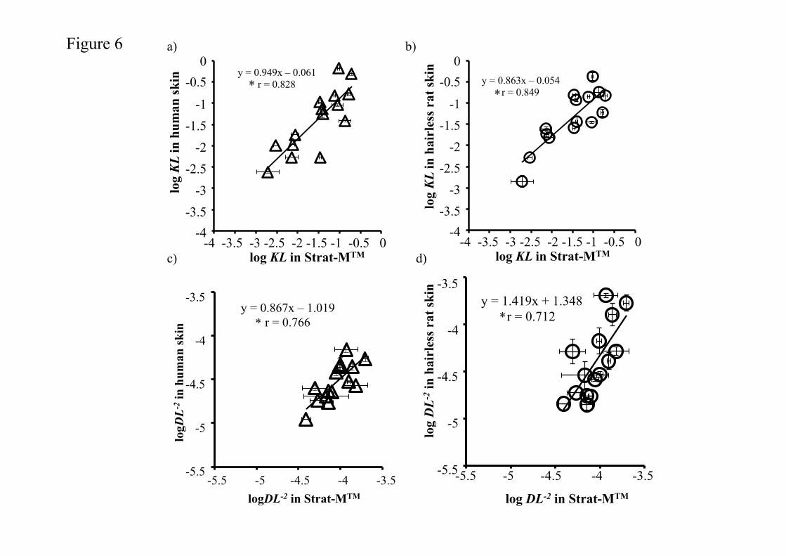
Figure 6a-d shows the relationship for log KL or log DL-2 between Strat-MTM

and human or rat skin. Log KL in Strat-MTM was similar to that in human or rat skin,

and almost 1:1 slopes were observed between Strat-MTM and human or rat skin. On

the other hand, logDL-2 in Strat-MTM shows liner relationships to those in human or rat

skin with higher logDL-2 values than those in human and rat skins.

Figure 6

4. Discussion

In vitro permeation experiments using excised human and animal skins are

very useful for understanding the skin permeation profiles and skin concentrations of

topically applied chemicals. However, in vitro experiments using human skin are

expensive and sometimes limited due to the low availability of samples and ethical

issues. Furthermore, animal testing to evaluate the safety and efficacy of cosmetic

ingredients has been banned by Cosmetic Directive 76/768/EEC.

The outmost layer, the stratum corneum, has the largest barrier function

against skin permeation by chemicals (Prausnitz and Langer, 2008; Naik et al., 2000;

Flynn et al., 1974). In the present study, the rate-limiting step in the permeation of

chemicals through Strat-MTM remains unknown because the permeation experiment

12

could not be performed with each separated layer of Strat-MTM. Membrane

permeation profiles can generally be expressed by partition and diffusion phenomena

(Okamoto et al., 1988). Chemical compounds are immediately distributed or

partitioned into the membrane surface following the application of formulations. Thus,

the diffusion and partition coefficients of a chemical into a membrane should be

investigated to clarify the usefulness of the membrane as an alternative to human skin

in permeation experiment.

Elevations were observed in the log P of chemicals through rat and human

skins and Strat-M TM with an increase in their log Ko/w (Fig. 3), and 1:1 relationships

were detected between human and rat skins or Strat-M TM (Fig. 4). Furthermore, log

KL and log P were similar among membranes at the same log Ko/w of chemicals,

suggesting that the lipophilicity characteristics or solubility parameters of Strat-M TM

were close to those of human and rat skins. These results suggested that skin

permeation by chemicals may be predicted according to their permeability coefficients

through Strat-M TM.

To further investigate the characteristics of Strat-M TM, the permeability

coefficients of ionized (Pi) and non-ionized (Pu) lidocaines were compared with those

through human skin (Kitamura et al., 2009; Hayashi et al., 1992). These values were

calculated using skin permeability coefficients at different ratios of the ionized and

non-ionized forms of the molecule at pH5.0 and pH7.4 in the present study. Pu and Pi

obtained from excised human skin were 7.1×10⁻⁶ and 4.0×10⁻⁸ cm/s, respectively. Pu

was 177-fold higher than Pi. On the other hand, Pu and Pi obtained from Strat-MTM

were 5.6×10⁻⁶ and 1.3×10⁻⁷ cm/s, respectively. Although Pu in Strat-MTM was higher

than Pi in Strat-MTM, Pu in Strat-MTM was only approximately 43-fold higher than Pu in

13

Strat-MTM. Pu in human skin was nearly 1.3-fold higher than that in Strat-MTM,

whereas Pi in Strat-M TM was nearly 3.2-fold higher than that in human skin. Recent

studies have described the main permeation pathway of ionized and large molecular

weight compounds. However, the contribution of the hydrophilic pathway such as hair

follicles and sweat ducts to total skin permeation by chemical compounds remains

unclear. The main permeation route of ionized compounds may be an aqueous-filled

pore route due to their high solubility or partition into the aqueous phase. Higher log

DL-2 in Strat-M TM was observed in hydrophilic chemicals (log Ko/w<0). Since,

diffusivity of chemicals in a membrane is related to its permeation route, low-tortuosity

of hydrophilic pathway in Strat-MTM might be a reason for higher permeability of

hydrophilic compounds. To confirm this assumption, further in vitro permeation experiment was

performed with sodium calcein {M.W.; 644.5, log Ko/w; -3.5 (at pH7.4), pKa; 5.5} and sodium

fluorescein {M.W.; 376.3, log Ko/w; -0.61 (at pH7.4), pKa; 6.4}. These permeations through

Strat-MTM were, however, not observed over 8 h experiment, whereas these were observed in human

and rat skins (data not shown). The reason for the non-permeation of these chemicals through

Strat-M TM might be related to their large molecular sizes. Therefore, physicochemical

characteristics of applied chemicals should be considered to use Strat-M TM in a permeation

experiment. Further experiment should be performed to reveal the characteristics of aqueous

pathway in Strat-M TM.

5. Conclusion

The permeability coefficients of chemical compounds through Strat-MTM can be

used to predict those through excised human and rat skins, especially for chemical

compounds with molecular weights between 151 to 288 and logKo/w from -0.90 to 3.53.

14

Thus, Strat-MTM can be utilized in screening tests to estimate the permeability of

chemicals through human skin. Further experiments need to be conducted with

various formulations such as patches, emulsions, and ointments in order to clarify that

Strat-MTM can be used to determine formulation designs for the topical and/or

transdermal delivery of compounds.

6. Conflict of interest

There is no conflict of interest with any commercial or other associations in this

study.

7. Abbreviations

Ko/w; Octanol-water coefficients P; Permeability coefficient of chemicals KL; Partition parameter into membrane DL-2; Diffusion parameter in membrane Pu; Permeability coefficient of ionize form of chemicals Pi; Permeability coefficient of non-ionize form of chemicals

15

6. References

Ahmed, S., Imai, T., Otagiri, M., 1996. Evaluation of Stereoselective Transdermal

Transport and Concurrent Cutaneous Hydrolysis of Several Ester Prodrugs of

Propranolol: Mechanism of Stereoselective Permeation. Pharm Res 13, 1524–1529.

Akamatsu, M., Fujikawa, M., Nakao, K., Shimizu, R., 2009. In silico prediction of

human oral absorption based on QSAR analyses of PAMPA permeability. Chem.

Biodivers. 6, 1845–1866.

Avdeef, A., Bendels, S., Di, L., Faller, B., Kansy, M., Sugano, K., Yamauchi, Y., 2007.

PAMPA--critical factors for better predictions of absorption. J. Pharm. Sci. 96, 2893–

2909.

Bos, J.D., Meinardi, M., 2000. The 500 Dalton rule for the skin penetration of chemical

compounds and drugs. Experimental dermatology.

Flynn GL, Yalkowsky SH, Roseman TJ., 1974. Mass transport phenomena and models:

theoretical concepts. J. Pharm. Sci.63, 479–510.

Hatanaka, T., Inuma, M., Sugibayashi, K., Morimoto, Y., 1990. Prediction of skin

permeability of drugs. I. Comparison with artificial membrane. Chem. Pharm. Bull. 38,

3452–3459.

Hayashi, T., Sugibayashi, K., Morimoto, Y., 1992. Calculation of skin permeability

coefficient for ionized and unionized species of indomethacin. Chem. Pharm. Bull. 40,

3090–3093.

Imokawa, G., Abe, A., Jin, K., Higaki, Y., Kawashima, M., Hidano, A., 1991. Decreased

level of ceramides in stratum corneum of atopic dermatitis: an etiologic factor in atopic

16

dry skin? J. Invest. Dermatol. 96, 523–526.

Kano, S., Todo, H., Sugie, K., FUJIMOTO, H., NAKADA, K., Tokudome, Y., Hashimoto,

F., Sugibayashi, K., 2010. Utilization of Reconstructed Cultured Human Skin Models as

an Alternative Skin for Permeation Studies of Chemical Compounds. Alternatives to

animal testing and experimentation : AATEX 15, 61–70.

Karadzovska, D., Brooks, J.D., Monteiro-Riviere, N.A., Riviere, J.E., 2013. Predicting

skin permeability from complex vehicles. Advanced Drug Delivery Reviews 65, 265–277.

Keldenich, J., 2009. Measurement and prediction of oral absorption. Chem. Biodivers. 6,

2000–2013.

Kitamura, T., Todo, H., Sugibayashi, K., 2009. Effect of several electrolyzed waters on

the skin permeation of lidocaine, benzoic Acid, and isosorbide mononitrate. Drug Dev

Ind Pharm 35, 145–153.

Lian, G., Chen, L., Han, L., 2008. An evaluation of mathematical models for predicting

skin permeability. J. Pharm. Sci. 97, 584–598.

Morimoto, Y., Hatanaka, T., Sugibayashi, K., Omiya, H., 1992. Prediction of skin

permeability of drugs: comparison of human and hairless rat skin. J. Pharm. Pharmacol.

44, 634–639.

Murthy, 2012. Transdermal drug delivery: approaches and significance. RRTD 1-2.

Naik, A., Kalia, Y., Guy, R., 2000. Transdermal drug delivery: overcoming the skin's

barrier function. Pharm. Sci. Technol. Today 3, 318–326.

Okamoto, H., Hashida, M., Sezaki, H., 1988. Structure-activity relationship of 1-alkyl-

or 1-alkenylazacycloalkanone derivatives as percutaneous penetration enhancers. J.

Pharm. Sci. 77, 418–424.

Palac, Z., Engesland, A., Flaten, G.E., Skalko-Basnet, N., 2014. Filipović-Grčić J, Vanić

17

Z. Liposomes for (trans)dermal drug delivery: the skin-PVPA as a novel in vitro stratum

corneum model in formulation development. J Liposome Res. Early Online, 1-10.

Potts, R.O., Guy, R.H., 1992. Predicting skin permeability. Pharm Res 9, 663–669.

Poumay, Y., Dupont, F., Marcoux, S., Leclercq-Smekens, M., H rin, M., Coquette, A.,

2004. A simple reconstructed human epidermis: preparation of the culture model and

utilization in in vitro studies. Arch Dermatol Res 296, 203–211.

Prausnitz, M.R., Langer, R., 2008. Transdermal drug delivery. Nat Biotechnol 26, 1261–

1268.

Riviere, J.E., Brooks, J.D., 2011. Predicting skin permeability from complex chemical

mixtures: dependency of quantitative structure permeation relationships on biology of

skin model used. Toxicol. Sci. 119, 224–232.

Scott, R.C., Walker, M., Dugard, P.H., 1986. A comparison of the in vitro permeability

properties of human and some laboratory animal skins. Int J Cosmet Sci 8, 189–194.

Sinkó, B.L., Garrigues, T.M., Balogh, G.T., Nagy, Z.K., Tsinman, O., Avdeef, A.,

Takács-Novák, K., 2012. European Journal of Pharmaceutical Sciences. Eur J Pharm

Sci 45, 698–707.

Sugibayashi, K., Hayashi, T., Matsumoto, K., Hasegawa, T., 2004. Utility of a

three-dimensional cultured human skin model as a tool to evaluate the simultaneous

diffusion and metabolism of ethyl nicotinate in skin. Drug Metab. Pharmacokinet. 19,

352–362.

Sugibayashi, K., Todo, H., Oshizaka, T., Owada, Y., 2010. Mathematical model to

predict skin concentration of drugs: toward utilization of silicone membrane to predict

skin concentration of drugs as an animal testing alternative. Pharm Res 27, 134–142.

Tamura, M., Sueishi, T., Sugibayashi, K., Morimoto, Y., Juni, K., Hasegawa, T.,

18

Kawaguchi, T., 1995. Metabolism of testosterone and its ester derivatives in

organotypic coculture of human dermal fibroblasts with differentiated epidermis. Int J

Pharm 131, 263–271.

Tehan, B.G., Lloyd, E.J., Wong, M.G., Pitt, W.R., Gancia, E., Manallack, D.T., 2002. Estimation of

pKa Using Semiempirical Molecular Orbital Methods. Part 2: Application to Amines, Anilines and

Various Nitrogen Containing Heterocyclic Compounds. Quantitative Structure - Activity

Relationships. Wiley Online Library 21, 473–485.

Todo, H.H., Kimura, E.E., Yasuno, H.H., Tokudome, Y.Y., Hashimoto, F.F., Ikarashi,

Y.Y., Sugibayashi, K.K., 2010. Permeation pathway of macromolecules and

nanospheres through skin. Biol Pharm Bull 33, 1394–1399.

Tokudome, Y., Jinno, M., Todo, H., Kon, T., Sugibayashi, K., Hashimoto, F., 2011.

Increase in ceramide level after application of various sizes of sphingomyelin liposomes

to a cultured human skin model. Skin Pharmacol Physiol 24, 218–223.

Yamashita, S., Tanaka, Y., Endoh, Y., Taki, Y., Sakane, T., Nadai, T., Sezaki, H., 1997.

Analysis of drug permeation across Caco-2 monolayer: implication for predicting in vivo

drug absorption. Pharm Res 14, 486–491.

19

Figure captions.

Figure 1 Scanning electron microscopic image of a cross-section of Strat-MTM. The first

i), second ii), and third layer iii) of Strat-MTM.

Figure 2 Transmission scanning electron microscopic observation of a cross-section of

Strat-MTM. The first i), second ii), and third layer iii) of Strat-MTM. Lipids in the

layer were stained with a black color. Arrows show the lipid region.

Figure 3 Relationships between log Ko/w and log P of applied chemical compounds.

Symbols; Excised human skin (△), excised hairless rat skin (○), and Strat-M TM (□). All

results are expressed as the means ± S.E. (n=3).

Figure 4 Relationships between log P values of human skin and rat skin or Strat-M TM.

All results are expressed as the means ± S.E. (n=3). Pearson's Correlation Coefficient

showed significance (P <0.05).

Figure 5 Relationships between log Ko/w and log KL, or log DL-2 of applied chemical

compounds through human skin and rat skin or Strat-M TM.. Symbols; Excised human

skin (△), excised hairless rat skin (○), and Strat-M TM (□). All results are expressed as

the means ± S.E. (n=3).

Figure 6 Relationships in logKL (a, b) or log D (c, d) between Strat-MTM and human

skin or rat skin. (a, c); Strat-MTM vs human skin, (b,d); Strat-MTM vs human skin. All

20

results are expressed as the means ± S.E. (n=3). Pearson's Correlation Coefficient

showed significance (P <0.05).

21

Table 1 Physicochemical properties of the compounds used in the present study

Abbreviati

on M.W. logKo/w*

Lidocaine LID 288.8

-0.9 (pH5.0)

1.3 (pH7.9)

1.4 (pH10)

Isosorbide 5-mononitrate ISMN 191.1 -0.2 (pH7.4)

Caffeine CAF 194.2 -0.12 (pH7.4)

Aminopyrine AMP 231.3 1.1 (pH7.4)

Isosorbide dinitrate ISDN 236.1 1.2 (pH7.4)

Methyl paraben MP 152.2 1.9 (pH7.4)

Ethyl paraben EP 166.1 2.3 (pH7.4)

n-Propyl paraben PP 180.2 2.8 (pH7.4)

n-Butyl paraben BP 194.2 3.5 (pH7.4)

Methyl p-amimobenzoate M-PABA 151.6 1.38 (pH7.4)

Ethyl p-amimobenzoate E-PABA 165.2 1.89 (pH7.4)

n-Propyl p-amimobenzoate P-PABA 179.2 2.43 (pH7.4)

n-Butyl p-amimobenzoate B-PABA 193.2 2.70 (pH7.4)

*Octanol/buffer partition coefficient of compounds at 32°C.

pH was adjusted with pH 7.4 phosphate-buffered saline, pH 5.0 citrate-buffered saline, or pH

9.0 carbonate-buffered saline.

22

Tabl

e 2

Per

mea

bilit

y co

effic

ient

s of c

hem

ical

thro

ugh

Stra

t-MTM

, hai

rless

rat s

kin

and

hum

an sk

in

Com

poun

ds

P (c

m/s

) ×10

-6

P (c

m/s

) ×10

-6

P (c

m/s

) ×10

-6

Stra

t-MTM

H

airle

ss ra

t ski

n H

uman

skin

MP

4.02

± 0

.60

2.39

± 0

.31

2.49

± 0

.03

EP

4.21

± 0

.20

2.96

± 0

.06

3.86

± 0

.49

PP

4.01

± 0

.16

3.63

± 0

.15

3.63

± 0

.15

BP

4.24

± 0

.34

2.31

± 0

.26

3.54

± 0

.18

ISM

N

0.15

± 0

.02

0.07

± 0

.01

0.11

± 0

.01

AM

P 0.

87 ±

0.2

5 0.

29 ±

0.1

1 0.

33 ±

0.0

2

ISD

N

3.14

± 0

.01

2.57

± 0

.15

3.14

± 0

.17

LCH

(pH

5.0)

0.

14 ±

0.0

2 0.

04 ±

0.0

1 0.

05 ±

0.0

2

LCH

(pH

10)

5.55

± 0

.53

9.21

± 3

.68

1.97

± 0

.15

LCH

(pH

7.9)

1.

20 ±

0.1

2 1.

30 ±

0.1

1 0.

14 ±

0.0

1

CA

F 0.

47 ±

0.0

7 0.

26 ±

0.0

2 0.

18 ±

0.0

2

M-P

AB

A

8.95

± 0

.26

7.22

± 0

.17

18.4

± 0

.83

E-PA

BA

11

.25

± 0.

41

10. 6

± 0

.25

20.1

± 2

.24

P-PA

BA

13

.55

± 0.

58

19.6

± 1

.61

22.3

± 3

.03

B-P

AB

A

10.1

4 ±

0.53

9.

49 ±

0.7

9 20

.1 ±

3.0

2

Mea

n ±

S.E.

(n=3

)

200 µm

Figure 1

i)

ii)

iii)

10 µm

1層目

800 nm 800 nm

i) ii) iii)

Figure 2

Figure 3

-­‐8

-­‐7.5

-­‐7

-­‐6.5

-­‐6

-­‐5.5

-­‐5

-­‐4.5

-­‐2 -­‐1 0 1 2 3 4

-4.5

-5

-5.5

-6

-6.5

-7

-7.5

-8 -2 -1 0 1 2 3 4

log Ko/w

log P

b) c)

Figure 4

y = 1.127x + 0.640r = 0.970

!8

!7.5

!7

!6.5

!6

!5.5

!5

!4.5

!4

!8 !7.5 !7 !6.5 !6 !5.5 !5 !4.5 !4-8 -7.5 -7 -6.5 -6 -5.5 -5 -4.5 -4�

logP in Strat-MTM�

logP

in h

airl

ess

rat s

kin� y = 1.13x + 0.64

r = 0.970 *

-4

-4.5

-5

-5.5

-6

-6.5 -7

-7.5

-8�

y = 1.133x + 0.695r = 0.929

!8

!7.5

!7

!6.5

!6

!5.5

!5

!4.5

!4

!8 !7.5 !7 !6.5 !6 !5.5 !5 !4.5 !4

-4

-4.5

-5

-5.5

-6

-6.5 -7

-7.5

-8�

logP

in h

uman

ski

n�

logP in Strat-MTM�

-8 -7.5 -7 -6.5 -6 -5.5 -5 -4.5 -4�

y = 1.13x + 0.69 r = 0.929 *

a)

y = 0.936x - 0.343r = 0.892

!8

!7.5

!7

!6.5

!6

!5.5

!5

!4.5

!4

!8 !7.5 !7 !6.5 !6 !5.5 !5 !4.5 !4

-4

-4.5

-5

-5.5

-6

-6.5 -7

-7.5

-8�-8 -7.5 -7 -6.5 -6 -5.5 -5 -4.5 -4�

log P in hairless rat skin�

log

P in

hum

an s

kin�

y = 0.936x – 0.343 r = 0.892 *

!4

!3

!2

!1

0

!2 !1 0 1 2 3 4-2 -1 0 1 2 3 4

log Ko/w�

log KL�

0

-1

-2

-3

-4

Figure 5

logD

L-2 �

logKo/w�

-2

-3

-4

-5

-6 -2 -1 0 1 2 3 4

!6

!5

!4

!3

!2

!2 !1 0 1 2 3 4

Figure 6 a) b)

c) d)

log

KL in

hum

an s

kin�

log KL in Strat-MTM�

0 -0.5

-1

-1.5

-2

-2.5

-3

-3.5 -4

y = 0.949x + 0.0608r = 0.828

!4

!3.5

!3

!2.5

!2

!1.5

!1

!0.5

0

!4 !3.5 !3 !2.5 !2 !1.5 !1 !0.5 0-4 -3.5 -3 -2.5 -2 -1.5 -1 -0.5 0

y = 0.949x – 0.061 r = 0.828 * y = 0.863x - 0.054

r = 0.849

!4

!3.5

!3

!2.5

!2

!1.5

!1

!0.5

0

!4 !3.5 !3 !2.5 !2 !1.5 !1 !0.5 0

log KL in Strat-MTM�

log

KL in

hai

rles

s ra

t ski

n� 0 -0.5

-1

-1.5

-2

-2.5

-3

-3.5 -4

-4 -3.5 -3 -2.5 -2 -1.5 -1 -0.5 0

y = 0.863x – 0.054 r = 0.849 *

y = 1.419x + 1.348r = 0.712

!5.5

!5

!4.5

!4

!3.5

!5.5 !5 !4.5 !4 !3.5

log DL-2 in Strat-MTM�

log

DL-2

in h

airl

ess

rat s

kin� -3.5

-4

-4.5

-5

-5.5 -5.5 -5 -4.5 -4 -3.5

y = 1.419x + 1.348 r = 0.712 *

y = 0.867x - 1.019R² = 0.766

!5.5

!5

!4.5

!4

!3.5

!5.5 !5 !4.5 !4 !3.5

logD

L-2 in

hum

an s

kin�

logDL-2 in Strat-MTM�

-3.5

-4

-4.5

-5

-5.5 -5.5 -5 -4.5 -4 -3.5

y = 0.867x – 1.019 r = 0.766 *


Recommended