Guidelines for medical treatment of acute Kawasaki disease: Report ofthe Research Committee of the Japanese Society of PediatricCardiology and Cardiac Surgery (2012 revised version)
Research Committee of the Japanese Society of Pediatric Cardiology andCardiac Surgery Committee for Development of Guidelines for Medical Treatment of Acute Kawasaki Disease
The primary purpose of practical guidelines is to contribute totimely and appropriate diagnosis and treatment of a given diseaseor condition, in addition to providing current medical informa-tion on pathogenesis and treatment, as determined by specialistsin the field. Guidelines, however, should not be considered pro-cedure manuals that limit the treatment options of practitioners,because treatment modalities other than those recommended insuch guidelines are often required. Such treatment choices are theresult of comprehensive analysis of all medical circumstances,including patient condition, treatment option, and disease sever-ity. Furthermore, certain drugs shown to be useful in studiesconducted in other countries may not yet have been approved for
use here in Japan. The results of clinical research (includingrandomized controlled trials) must be verified in subsequentresearch, and the safety and effectiveness of a particular treat-ment may take several months to confirm.
Evidence classification
Recent clinical guidelines typically provide evidence levels basedon study design and reported effectiveness.
Level (class) based on study design
These are defined as follows: class Ia, systematic reviews, meta-analyses; class Ib, randomized controlled trials; class IIa, non-randomized controlled trials; class IIb, other quasi-experimentalstudies; class III, non-experimental reports (comparative studies,correlation studies, case studies); and class IV, opinions of com-mittees of experts and authorities.
Classification (grade) based on efficacy
These are given as follows: grade A, highly recommended; gradeB, recommended; grade C, recommended, but evidence is uncer-tain; and grade D, contraindicated.
The present guidelines will use these classification systems inreviewing the available evidence for the various treatments.
Background of the present revision of thetreatment guidelines
In July 2003, the Scientific Committee of the Japanese Society ofPediatric Cardiology and Cardiac Surgery published its Treat-ment Guidelines for Acute Kawasaki Disease (KD). These guide-lines were designed to present, in a clinically relevant manner, thefindings of Ministry of Health research done from 1998 through2000 by the Onishi group at Kagawa Medical University(working under the official title, “The Pediatric PharmaceuticalInvestigation Research Group”). This research had been pub-lished as “Research designed to identify and solve problems inthe suitable use of pharmaceuticals for pediatric medical treat-ment: pharmaceuticals in cardiology” and had originally beenconducted to provide clinical data for the approval of single-usei.v. immunoglobulin (IVIG).
During the 9 years that have passed since the publication ofthe previous guideline, new data have been collected, and reportson new drug treatments have been published. Members of theInternational Kawasaki disease Symposium have been waiting
Correspondence: Tsutomu Saji, MD, First Department of Pediatrics,Toho University, 6-11-1 Omori-Nishi, Ota-Ku, Tokyo 143-8541,Japan. Email: [email protected]
Principal AuthorTsutomu Saji, Department of Pediatrics, Toho University OmoriMedical Center, Tokyo, Japan
Section AuthorsMamoru Ayusawa, Department of Pediatrics and Child Health, NihonUniversity School of Medicine, Tokyo, JapanMasaru Miura, Division of Cardiology, Tokyo Metropolitan Children’sMedical Center, Tokyo, JapanTohru Kobayashi, Department of Pediatrics, Gunma University Gradu-ate School of Medicine, Maebashi, Gunma, JapanHiroyuki Suzuki, Department of Pediatrics, Wakayama Medical Uni-versity, Wakayama, JapanMasaaki Mori, Department of Pediatrics, Yokohama City UniversityMedical Center, Yokohama, Kanagawa, JapanMasaru Terai, Department of Pediatrics, Yachiyo Medical Center,Tokyo Women’s Medical University, Tokyo, JapanShunichi Ogawa, Department of Pediatrics, Nippon Medical School,Tokyo, Japan
Associate MemberHiroyuki Matsuura, Department of Pediatrics, Toho University OmoriMedical Center, Tokyo, Japan
External Evaluation CommitteeTomoyoshi Sonobe, Department of Pediatrics, Japan Red CrossMedical Center, Tokyo, JapanShigeru Uemura, Cardiovascular Center, Showa University NorthernYokohama Hospital, Yokohama, Kanagawa, JapanKenji Hamaoka, Pediatric Cardiology and Nephrology, Kyoto Prefec-tural University of Medicine Graduate School of Medical Science,Kyoto, JapanHirotaro Ogino, Department of Pediatrics, Kansai Medical University,Osaka, JapanMasahiro Ishii, Department of Pediatrics, Kitasato University Schoolof Medicine, Tokyo, Japan
Received 10 October 2013; accepted 4 February 2014.
bs_bs_banner
Pediatrics International (2014) 56, 135–158 doi: 10.1111/ped.12317
for a revision of the previous Japanese guideline. Thus, the Sci-entific Committee was restructured and assigned the task of revis-ing the guideline.
Purpose and methods
Data on IVIG that have accumulated since it was approved andfirst marketed have confirmed the efficacy and safety of single-use IVIG therapy. In addition, the incidence of coronary arterylesions (CAL) has gradually decreased every year since IVIGtreatment was introduced in Japan.1 The incidence of giant coro-nary artery aneurysms (CAA), however, has remained almostunchanged, which highlights the importance of timely use ofsecond- and third-line treatments for IVIG-resistant patients.
In developing the present guideline, we carefully reviewedthe most recent available literature, classified evidence and effi-cacy, and revised suggested treatment methods, including pro-cedures for selecting first-, second- and third-line medications,with a special focus on off-label uses. For example, the previousguideline did not mention new therapeutic agents such asinfliximab (IFX), cyclosporin A (CsA), or methotrexate (MTX).In the present edition, risk/benefit considerations are also clearlypresented, based on data collected in and outside Japan. Despitethe publication of almost 200 reports every year on KD, there isstill no universally accepted treatment for IVIG resistance. Thisis also the case, however, for many other disorders, such asautoimmune disease and rheumatoid conditions, given that nosingle medication will benefit all patients in the same way. Thus,to ensure optimal outcome, physicians must treat each patientindividually.
Diagnosis and treatment of incomplete KD
In the published results of the 21st Nationwide Survey of KD byJichi Medical School a total of 23 730 cases of KD were reportedin Japan during the 2 year period 2009–2010.1 Diagnosis of KDfollows the criteria outlined in the fifth edition of the diagnosisguidelines for KD,2 which requires that at least five of the fol-lowing six principal symptoms are present: (i) fever persisting ≥5days (including fever that subsides before the fifth day inresponse to therapy); (ii) bilateral conjunctival congestion; (iii)changes in lips and oral cavity: reddening of lips, strawberrytongue, diffuse injection of oral and pharyngeal mucosa; (iv)polymorphous exanthema; (v) changes in peripheral extremities:reddening of palms and soles, indurative edema (initial stage);membranous desquamation from fingertips (convalescent stage);and (vi) acute non-purulent cervical lymphadenopathy.
Kawasaki disease, however, may also be diagnosed when onlyfour of the aforementioned symptoms are present, if during theperiod of illness either 2-D echocardiography or coronary angi-ography shows CAA, including dilation of coronary artery, andother causes of CAA can be excluded. A diagnosis of KD ispossible even if five or more of the principal symptoms are notpresent, if other conditions can be excluded and KD is suspected– a condition known as incomplete KD. Indeed, approximately15–20% of KD patients have incomplete KD. But, even if apatient has four or fewer of the principal symptoms, the illnessshould not be regarded as less severe, because cardiovascular
abnormalities are not rare in patients with incomplete KD. Forthis reason, even patients with fewer than five of the aforemen-tioned symptoms should be evaluated for KD. Early treatment isessential, particularly when fever is present, because CAL devel-opment in such cases is not uncommon. Diagnosis of incompleteKD is not a simple matter of adding up the number of overt KDsymptoms: the importance and individual characteristics of eachsymptom of the illness must be correctly assessed. For example,redness and crusting at a bacille Calmette–Guérin (BCG) inocu-lation site in infants younger than 1 year and multilocular cervi-cal lymphadenopathy in children aged ≥4 years are characteristicfeatures of KD.
Basic pathology
The 2012 Revised International Chapel Hill Consensus Confer-ence Nomenclature of Vasculitides defines KD as an arteritisassociated with mucocutaneous lymph node syndrome, pre-dominantly affecting medium and small arteries.3 There is verylittle damage to veins. The location of pathological changesclearly differentiates KD from other vasculitis syndromes, giventhat the principal danger of KD is inflammatory vasculitis ofthe coronary arteries. Edematous lesions develop in the intimamedia, and vascular fragility increases due to partial rupture ofthe internal and external elastic lamina. As a result, the arterialwall can no longer withstand its internal pressure, particularlydiastolic pressure, and becomes distended and deformed,leading in severe cases to aneurysm formation. Only a fewother diseases cause distension of coronary arteries. Theseinclude vasculitis resulting from Epstein–Barr virus infection,lupus, classical periarteritis nodosa, and atherosclerotic lesions.Calcification can also occur in rare cases and affect coronaryarteries, for example in cases of renal dialysis in adults andherpes infection in newborns.
Patients with KD may develop multiple lesions in the proxi-mal region and vessels branching out from it. As aneurysms beginto calcify, further pathological distension or development ofaneurysms and intimal thickening may develop 2–3 years later inareas with previously disrupted internal and external elasticlamina.4
Coronary artery lesions
The principal characteristics of KD are dilation of coronaryarteries and CAA. Most CAA occur in the proximal region and itsbranches, and arteries with a CAA measuring ≥8 mm in diameterare very unlikely to regain their normal morphology. Right CAAmay lead to occlusion or recanalization, and left CAA may pro-gress to stenotic lesions.
Rupture of the internal elastic lamina in the intima media ofthe dilated area weakens the artery wall, and coronary arterialpressure then becomes the direct mechanical cause of disten-sion. In rare cases, aneurysms may develop in branches of theaxillary or celiac arteries. During acute KD, vasculitis worsensduring the first 7 days after disease onset. In patients with mildillness, the vasculature returns to normal by the second or thirdweek.
The principal objective in treating acute KD is minimizing therisk of developing CAL. In practice, this means quickly suppress-ing the acute-phase inflammatory reaction caused by KD. Exceptin cases of very mild KD, IVIG should be started before illnessday 7. Histological studies have shown that arteritis typicallydevelops by 8 or 9 days after KD onset. Therefore, treatmentshould begin before this point, to suppress arteritis and hastenresolution of fever and normalization of inflammation markers.In patients with incomplete KD, IVIG should also be begun assoon as possible after a diagnosis of KD, especially if fever ispresent. In approximately 80% of cases, fever should be loweredto ≤37.5°C within 48 h of starting IVIG. In 40% of IVIG-resistant patients, fever can be reduced to ≤37.5°C with addi-tional IVIG of 1 g/kg. Persistent fever after 48 h of starting IVIGshould be regarded as evidence of IVIG-resistant KD. Preventionof CAA in such patients may largely depend on the selection ofsubsequent treatment.
In addition to CAL, other cardiovascular complications maydevelop in patients with acute KD, including myocarditis,pericardial effusion, valvular regurgitation, and, rarely, arrhyth-mia. Specific treatment may be required for these sequelae, aswell as for cardiac dysfunction or heart failure. Furthermore,other symptom-specific treatment may be required for systemiccomplications such as edema, hypoalbuminemia, electrolyteimbalances (i.e. hyponatremia), paralytic ileus, hepatic dysfunc-tion, cholecystitis, impaired consciousness, convulsions, anemia,diarrhea, vomiting, and dehydration. Particularly during high-dose IVIG infusion, care must be taken to prevent volume over-load so as to protect the patient from complications such as heartfailure.
There is currently no universally accepted classification systemto evaluate KD severity and need for IVIG use, although manysuch scoring systems have been proposed. Initial attempts weremade by Asai and Kusakawa,5 which were followed by the Iwasascore6 and Harada score.7 More recently, predictive modelsdesigned to evaluate the possibility of IVIG resistance were pro-posed, including the Kobayashi score,8 Egami score,9 and Sanoscore.10 In general, such predictive models consider factors such asage, gender, days of illness, white blood cell count, %neutrophils,hematocrit, platelet count, C-reactive protein (CRP), aspartateaminotransferase (AST), alanine aminotransferase (ALT), totalbilirubin, sodium, and albumin. Recently, a randomized controlledtrial found that IVIG plus steroid as initial therapy for patientspredicted to be at high risk for IVIG resistance improved clinicaland coronary arterial outcomes.11–13 The effectiveness of suchpredictive models, however, has not been confirmed in large-scaleprospective cohort studies or meta-analyses, and controversyremains as to whether initial therapy with IVIG plus steroids is theoptimal treatment.
Choice of treatment for IVIG-resistant patients
Several second-line treatment options are available if fever per-sists or has reappeared at 24 h after first-line treatment. The
efficacy of these second-line treatments for resistance to first-linetreatment is currently being investigated by researchers in manycountries, but evidence remains limited due to the lack ofrandomized controlled trials.
Options for second-line treatment include additional IVIG,i.v. methylprednisolone pulse (IVMP), prednisolone (PSL), IFX,ulinastatin (UTI), CsA, MTX, and plasma exchange (PE). Thedecision to use any of these treatments requires careful consid-eration of patient characteristics. At present, the most commonlyused second-line treatment is additional IVIG,1 which is some-times given in combination with other medications. As for ster-oids, a retrospective study noted a high incidence of giantaneurysms.14 That small uncontrolled case study reported thatseveral patients had received steroids before rupture of coronaryarteries, which suggests that physicians should carefully considerthe decision to use steroids for patients with KD if CAA arealready present. When steroids, biologics, or immunosuppres-sants are given to infants, there is also a risk of long-term side-effects, and questions remain regarding the general safety of suchmedications. Thus, a careful risk/benefit evaluation should bedone to consider the likelihood of such adverse effects versus thepossibility of CAA formation.
Algorithm for selecting optimal treatment
To decrease the risks of first-line IVIG resistance and CAA, itseems reasonable to consider risk stratification using predictivemodels and to select more-aggressive initial treatment forpatients at high risk of IVIG resistance. Such patients should betreated with 2 g/kg of IVIG in combination with either 2 mg/kgper day PSL or 30 mg/kg per day IVMP. If the patients fail torespond to these treatments, a third-line treatment will beupgraded to a second-line treatment.
Because few studies have assessed the efficacy of medicationsother than IVIG retreatment, it is impossible at this time to assignan objective order of these treatment options. The present guide-lines, however, offer evidence levels and grades to assist physi-cians in selecting appropriate alternatives. Various methods ofcalculating KD patient risk scores and, thereby, estimating KDseverity have been developed at a number of institutions bydifferent physicians, based on their particular experience withKD.8–10 The Japanese Society of Pediatric Cardiology andCardiac Surgery does not intend to limit the treatment optionsavailable to clinicians, especially when such options have alreadyreceived ethics committee approval at their institution. Instead,the judgment of physicians in selecting treatments should berespected, for practical reasons as well. Such treatments may begiven after a physician has established a sufficient basis forselecting a given treatment and received informed consent/assentfrom the family/patient (Fig. 1).
Immunoglobulin
Purpose
Currently, the most effective anti-inflammatory treatment for KDis early IVIG.15–17 The latest systematic review by the CochraneCollaboration states that CAL development can be reduced by asingle dose of 2 g/kg IVIG given before the 10th day after onset.18
Because the causes of KD are unknown, the mechanisms under-lying the therapeutic benefits of IVIG remain speculative. Table 1lists the hypothesized mechanisms of action.19–22
Indications
I.v. immunoglobulin is suitable for almost all cases of typicalacute KD, that is, when KD is diagnosed based on the presence ofthe principal symptoms specified in the criteria of the diagnosticguideline for KD2 and the patient is at risk for CAL. For patientswith symptoms that only partially fulfill the diagnostic criteria,incomplete KD may be diagnosed – if other diseases or condi-tions can be excluded – after which IVIG should be started asquickly as possible due to the risk of CAL.17
In cases of less severe KD or spontaneous defervescence,clinicians may refrain from IVIG, in accordance with the consid-erations detailed in the Ministry of Health Group Committeeguidelines for IVIG (Harada score)7 and disease severity stand-ards established at the physician’s institution.
Data from the 21st Nationwide Survey of KD show that IVIGwas given to 89.5% of patients.1
Treatment method and dosage
Period of treatment
I.v. immunoglobulin should be started on or before the seventhday after KD onset. It is essential to quickly reduce inflammationand duration of fever, definitely before illness day 8 or 9, whenCAL begin to appear. Markers of systemic inflammation, forexample CRP and neutrophil count, should be lowered as well.
One study compared patients receiving IVIG on the fifth ofillness day or earlier with those who received IVIG on the sixththrough ninth days of illness. Although duration from treatmentonset to defervescence was slightly longer overall among thosereceiving IVIG earlier, total duration of fever was shorter. More-over, the groups did not differ in incidence of fever recurrence oradditional IVIG treatment, or in number of days of hospitaliza-tion. Furthermore, 1 year after appearance of symptoms, thosewho had received IVIG earlier had a lower incidence of CAL.23
Dosage
The suggested IVIG dosage for acute KD is 2 g/kg per day(single use), 1 g/kg per day for 1 or 2 days continuously (modi-fied single use), or 200–400 mg/kg per day, over 3–5 days(divided dosing).
Studies in a number of countries have shown that, as com-pared with divided-dose regimens, a single dose of 2 g/kg per daysignificantly reduced CAL incidence, more quickly normalizedinflammation markers, and was more effective in reducingfever.4,5 As for 1 g/kg/day use, if clinical efficacy is seen on thefirst day, it might not be necessary to continue treatment into thesecond day.
The 21st Nationwide Survey of KD found that a single dose of2 g/kg per day IVIG was used in 85% of reported cases and that1 g/kg per day was given for 1 or 2 days in 6.2% and in 7.7% ofcases, respectively.1
There is no consensus in Japan as to whether older/largerchildren should be treated with 2 g/kg IVIG or a lowerdose.
As for 2 g/kg regimen, the treatment rate varies slightly fordifferent products, although IVIG is typically given over a periodof approximately 12 h in North America. In Japan, one productpermits use within a similar 12 h period, but the total volume of2 g/kg IVIG is usually given over a period of 24 h. Becausevolume overload might occur when the treatment rate is too fast,which can lead to cardiac dysfunction, it is important to adhere tothe recommended treatment rate and carefully observe patienthemodynamics.
Product types and directions for use
At present, four brands of IVIG are approved for KD in Japan(Table 2): two are processed with polyethylene glycol (PEG), oneis sulfonated, and one is processed to ensure a pH of 4 (acidic).No major differences in efficacy have been reported. Table 2 liststhe characteristics of these products, as described in their respec-tive product inserts.
The principal differences are as follows.
Table 1 Immunoregulatory effects of IVIG19–22
I. Fc receptor-mediated effectsBlockade of Fc receptors on macrophages and effector cellsAntibody-dependent cellular cytotoxicityInduction of inhibitory FcγRIIB receptorsPromotes clearance of antibodies that block FcRn
II. Anti-inflammatory effectsAttenuation of complement-mediated damageDecrease in immune complex-mediated inflammationInduction of anti-inflammatory cytokinesInhibition of activation of endothelial cellsNeutralization of microbial toxinsReduction in steroid requirementsModulation of matrix metalloproteinases
III. Effect on B cells and antibodiesControl of emergent bone marrow B-cell repertoiresNegative signaling through Fcγ receptorSelective downregulation/upregulation of antibody productionNeutralization of circulating autoantibodies by anti-idiotypes
IV. Effect on T cellsRegulation of T-helper cell cytokine productionNeutralization of T-cell superantigensRegulation of apoptosis
V. Effect on dendritic cellsInhibition of differentiation and maturationRegulation of inflammatory cytokine production
VI. OtherMutually interacts with immunological moleculesSuppression of autoantibody production against vascular
endothelial cellsAcceleration of phagocytosis arising from binding of
neutrophils and macrophages (opsonin effect)Suppression of inflammation-related gene S100 mRNASuppression of MCP-1 receptor CCR2 gene expression
produced by macrophages
CCR2, C-C chemokine receptor type 2; FcγRIIB, Fc gamma recep-tor IIB; FcRn, neonatal Fc receptor; IVIG, i.v. immunoglobulin;MCP-1, monocyte chemotactic protein-1.
(1) The sulfonated product (Kenketsu Venilon-I; Teijin, Tokyo,Japan) contains serum albumin, and its sodium concentrationis identical to that of saline (154 mEq/L).
(2) The two products processed with PEG come in freeze-dried(Kenketsu Glovenin-I; Nihon Shinyaku, Kyoto, Japan) andliquid (Venoglobulin IH; Japan Blood Products Organiza-tion, Tokyo, Japan) form. The suggested infusion rate forPEG-processed IG is slightly slower than that of thesulfonated product. Kenketsu Glovenin-I has a sodium con-centration of 154 mEq/L. Because liquid preparations areusually refrigerated until use, they must be warmed to at leastroom temperature beforehand.
(3) The pH 4-processed IG (Nisseki Polyglobin-N; Japan RedCross Society, Tokyo, Japan) comes in liquid form andshould also be warmed to at least room temperature beforeuse. During injection, it is essential that the liquid does notleak out of the vein, because this may cause necrosis of theskin. Furthermore, because the preparation contains maltose,the plasma glucose dehydrogenase method should not beused to measure blood sugar after injection, given that thismethod can be affected by the presence of maltose.
Close monitoring and a slower infusion rate are required duringthe first 30–60 min, given that all the aforementioned productsmight result in anaphylaxis during treatment. If no adverse reac-tions occur during the first hour of treatment (rate, 0.01 mg/kgper min), the maximum rate (<0.03 mg/kg per min) of 2 g/kgmay then be used over a course of 12–20 h.
IVIG retreatment for IVIG-resistant patients
Although IVIG is the established first-line treatment for KD,approximately 15–20% of all KD patients (16.6% of patients inthe 21st Nationwide Survey of KD1) have persistent or recrudes-cent fever after 2 g/kg of IVIG, and there has been considerabledebate regarding the optimal second-line treatment for suchpatients. The 21st Nationwide Survey of KD reported that addi-tional IVIG was given to a large majority (91.5%) of the 3231IVIG-resistant patients reported during the survey period. Steroidwas given together with IVIG in 29.0% of patients, IFX in 4.3%,immunosuppressants in 3.7%, and PE in 2.2% of patients. IVIGretreatment alone was effective in approximately half of thepatients.24
In recent years, various scoring systems have been developedto evaluate the likelihood of IVIG resistance at the time of diag-nosis. Representative scoring systems are listed in Table 3.8–10 Ifsuch scores suggest that patients are at high risk of IVIG resist-ance, more aggressive primary therapy in combination with theusual first-line treatment of 2 g/kg IVIG plus aspirin can beconsidered. In the RAISE study, Kobayashi et al. found thatIVIG plus PSL, started at 2 mg/kg per day and halved every 5days, was effective in preventing CAL formation and initial treat-ment failure.8,13 In addition, Egami et al. and Ogata et al. as wellas Sano et al. and Okada et al. reported the effectiveness ofmethylprednisolone (MP; 1–3 doses of 30 mg/kg of IVMP) incombination with IVIG.9–12 As compared with patients receivingonly IVIG plus aspirin, defervescence was significantly more
likely, and the incidence of CAL was significantly lower, amongpatients receiving IVIG plus steroids. Although further researchis necessary, it seems advisable to adapt this risk-stratified strat-egy for severe cases so as to reduce the number of IVIG-resistantpatients and further lower the incidence of CAL.
Effectiveness
I.v. immunoglobulin was found to be quite safe and, at present,has the greatest effectiveness. For these reasons, its effectivenesshas been widely recognized both in Japan and in other countries,and it is also included in the recommendations of many relevanttextbooks.
The incidence of cardiac complications reported in the latestNationwide KD survey decreased to approximately half that in1997–1998, when patients only rarely received 2 g/kg IVIG.During the acute phase of the illness, that is, until approximately1 month after disease onset, the incidence of cardiac complica-tions was 9.3%, including dilation, 7.26%; valvular insufficiency,1.19%; coronary aneurysm, 1.04%; giant coronary aneurysm,0.24%; coronary artery stenosis, 0.03%; and myocardial infarc-tion, 0.01%. Even during the convalescent phase, that is, >28days after disease onset, complications persisted in 3.0% ofpatients, including dilation, 1.90%; aneurysm, 0.78%; valvularinsufficiency, 0.29%; giant aneurysm, 0.22%; stenosis, 0.03%;and myocardial infarction, 0.02%. Furthermore, the number ofdeaths in Japan within 2 years of KD onset was 51 during the 10year period 1991–2000, which decreased by more than 60% to 19cases with the introduction of 2 g/kg IVIG during the subsequent10 year period, 2001–2010 (Fig. 2).1
Table 3 Representative scoring systems for evaluating potentialIVIG resistance
Cut-off point PointsKobayashi score8 (≥5 points; 76%
sensitivity, 80% specificity)Sodium ≤133 mmol/L 2Day of illness at initial IVIG
I.v. immunoglobulin is derived from human plasma and is con-sidered to have very few adverse effects and a high level of safety(Table 4). It is necessary, however, to carefully explain the pos-sibilities of rare side-effects to patients and/or their families andto obtain their informed consent before treatment.
In Japan, there have been no reports of viral contamination ofany IVIG product. Donated blood is carefully screened toconfirm the absence of HBs antigens, anti-HCV antibodies, anti-HIV-1 antibodies, anti-HIV-2 antibodies, and anti-HTLV-1 anti-bodies and to verify normal ALT. Furthermore, when plasma ispooled, the nucleic acid amplification testing (NAT) is used totest for HIV, hepatitis B virus (HBV), hepatitis C virus (HCV),hepatitis A virus, and human parvovirus B19, and only plasmathat tests negative for all these infections is used. Using presentpharmaceutical production processes, the absence of viruses thatare undetectable even by NAT (e.g. abnormal prion proteins andhuman parvovirus B19), cannot be determined with 100% cer-tainty, but there have been no reports of viral infection due toIVIG. Side-effects are infrequent but include post-treatmentchills and shivering, shock (such as cyanosis and hypotension),anaphylactic reactions, aseptic meningitis,25 hemolytic anemia,26
hepatic dysfunction, jaundice, acute renal failure, thrombocyto-penia, and pulmonary edema. Thus, patients should be careful
monitored for these side-effects. Particularly immediately afterthe start of i.v. treatment and when the infusion rate is increased,the physician should monitor for coldness and shivering, alteredconsciousness, discomfort, trembling, cyanosis, hypotension,and shock. Finally, cardiac dysfunction or even acute heart failuremay develop during acute KD, so close attention should be paidto patient vital signs, and to preventing sudden increasesin circulating blood volume, throughout the duration of i.v.treatment.27,28
Other considerations when using IVIG are as follows.
(1) Patients with IgA deficiency: allergic reactions may occur inresponse to IVIG in patients with anti-IgA antibodies.
(2) Patients with renal damage: risk of further impairment ofrenal function.
(3) Patients with cerebral or cardiovascular damage or a historyof these conditions: blood viscosity may increase when high-dose IVIG is given rapidly, thus leading to thromboembolicevents such as cerebral or myocardial infarction.
(4) Patients at risk for thromboembolism: rapid use of high-dose IVIG could increase blood viscosity and lead tothromboembolic events.
(5) Patients with hemolytic anemia, blood loss anemia, immunedeficiencies, or immunosuppressive disorders: the possibilityof human parvovirus B19 infection cannot be completelyexcluded. If such infection occurs, severe systemic effectssuch as fever and sudden or persistent anemia may result.
(6) Patients with reduced cardiac function: high-dose IVIG maylead to cardiac dysfunction or could worsen existing heartfailure.
A post-marketing survey of IVIG for KD noted that among 7259patients who received IVIG treatment, 484 had a total of 697adverse events (9.6%) and only 68 patients experienced 78 severeadverse events (1.1%; Table 5).29
Evidence levels
First-line IVIG treatment: class Ia, grade A.Additional IVIG treatment in IVIG-resistant patients: class
III, grade B.Combined therapy with IVIG and steroid as first-line treat-
ment for suspected IVIG-resistant patients: class Ib, grade B.
I.v. methylprednisolone pulse is usually given because of itspowerful and rapid immunosuppressive effect (Table 6). Amongavailable steroids, MP treatment is often selected for high-dosei.v. infusion because it is less likely to disrupt electrolyte balance.IVMP is widely used in treating severe pediatric illnesses such asrheumatic disease and kidney disease and is also used in treatingconfirmed and suspected IVIG-resistant KD.
Mechanism of action
Steroids bind with glucocorticoid receptors in cytoplasm andregulate nuclear expression of proteins such as NF-κB, whichproduces an anti-inflammatory effect referred to as genomicaction.30 When high-dose MP is given i.v., however, the saturationpoint of these glucocorticoid receptors is greatly exceeded; thus,mechanisms other than genomic action are thought to contributeto its efficacy. Such mechanisms may include acting throughproteins that dissociate from complexes with cytosolicglucocorticoid receptors, membrane-bound glucocorticoid recep-tors, and functional modification of membrane-bound proteinafter interlocation of the cell membrane. These mechanismsprecede genomic action.30,31
When used for KD patients, the effects of IVMP are veryrapid, which suggests that non-genomic mechanisms stimu-late immunocytological activity and suppress inflammatorycytokines. In confirmed and suspected IVIG-resistant patients,IVMP was reported to limit production of cytokines involved ininflammation and CAL,32 and to reduce transcription at thegenetic level.33
Indications
Patients suspected of being IVIG resistant on the basis of clinicalsymptoms and laboratory findings.
Patients found to be IVIG resistant after first-line IVIGtreatment.
It should be noted that IVMP treatment for KD is an off-labeluse.
Treatment method and dosage
In patients with kidney disease or connective-tissue disease, thestandard dose of IVMP is 20–30 mg/kg IVMP, given once a dayover a period of 2–3 h, for 1–3 consecutive days.31 For KDpatients, studies of IVMP in combination with first-line IVIGinvestigated a single dose of 30 mg/kg IVMP.11,12,34 Studies ofsecond-line IVIG treatment in IVIG-resistant patients investi-gated the same IVMP dose given once a day, for 1–3 days.32,33,35–39
Because the half-life of IVMP is only 3 h,31 some studies usedadditional therapy with PSL started at 1–2 mg/kg per day andgradually tapered over a period of 1–3 weeks.38,39
Effectiveness
First-line therapy with IVIG plus IVMP for all KD patients hasnot been proven to prevent CAL.40 There is, however, no evidencethat IVMP increases CAL incidence. In a double-blindrandomized controlled trial comparing IVIG plus IVMP withIVIG plus placebo, no significant differences were found infactors such as duration of fever, incidence of additional treat-ment, incidence of CAL, and coronary artery diameter, as indi-cated by Z score.34 A post-hoc analysis of patients requiringadditional treatment, however, found that the incidence of CALwas significantly lower among those who had received IVIG plusIVMP, which suggests that the combined regimen had been effec-tive among IVIG-resistant patients. Studies have also reportedthat suspected IVIG-resistant patients (as determined by Egamiscore or Sano score) who received first-line IVIG plus IVMP hadearlier defervescence and a significantly lower rate of CAL thandid those who had received IVIG alone.11,12
For patients resistant to initial IVIG, some studies comparedIVMP as a second-line treatment to additional treatment withIVIG and found that duration of fever was shorter after IVMP butthat CAL incidence was similar.32,36–40 The researchers, however,highlighted the fact that IVMP therapy was less expensive thanretreatment of IVIG.36,37 Nevertheless, the finding of equal effi-cacy for IVIG and IVMP has not been shown in non-inferioritytrials and requires confirmation. One study reported that IVIG-resistant patients who did not respond to additional IVIG had alower rate of CAL after subsequent IVMP followed by PSLtreatment.39
Side-effects
The reported side-effects of IVMP treatment for KD patientsinclude sinus bradycardia (6–82%), hypertension (10–91%),hyperglycemia (6–55%), and hypothermia (6–9%).39,41 There-fore, patient vital signs must be very carefully monitored duringIVMP, including monitoring of electrocardiogram and bloodpressure.
To avoid development of gastrointestinal ulcer, patients can begiven H2 blockers and/or other antacid agents. Additionalheparin can also be given as thrombosis prophylaxis.38,39 Never-theless, the necessity of these medications has not been proven.
Evidence levels
Initial IVIG plus IVMP for all KD patients: class Ib, grade C.
Table 5 Post-marketing survey of adverse effects of Ig for KD (no.treatments, 7259)
Initial IVIG plus IVMP for suspected IVIG-resistant patients:class Ib, grade B.
Second-line IVMP use for IVIG-resistant patients: class IIb,grade B.
Prednisolone
Purpose
The primary purpose of PSL therapy is to take advantage of itspowerful anti-inflammatory effects (Table 6). PSL may quicklyresolve KD vasculitis and suppress the potential risk forremodeling of coronary arteries.
Mechanism of action
Prednisolone is the most widely used synthetic corticosteroidhormone, and its glucocorticoid action is stronger than that ofcortisol. Through cytoplasmic steroid receptors, PSL inhibitsgene transcription of inflammatory cytokines and promotes genetranscription of anti-inflammatory cytokines.30 PSL also sup-presses inflammation by inhibiting production of inflammatorycytokines (e.g. tumor necrosis factor-α [TNF-α], interleukin[IL]-6, IL-8), chemokines, and cell adhesion molecules. In addi-tion, PSL stimulates production of anti-inflammatory proteinssuch as lipocortin, IL-1 receptor antagonists, β-2 adrenergicreceptors, and IκB kinase.
Indications
Patients suspected of being IVIG resistant, based on evaluation ofclinical symptoms and laboratory findings.
Patients found to be IVIG resistant after first-line IVIGtreatment.
PSL treatment for KD is an off-label use.
Treatment method and dosage
When used in combination with initial IVIG, 2 mg/kg per dayof PSL is given i.v. in three divided doses.13 After defervescenceand improvement in the patient’s general condition, PSL can begiven orally. After CRP normalizes, the patient is continued for5 days on the same dosage in three divided doses of 2 mg/kgper day. Thereafter, if fever does not recur, the dosage of PSLis decreased to 1 mg/kg per day in two divided doses on thesubsequent 5 days and then a single dose of 0.5 mg/kg per dayon the final 5 days. If fever recurs after dose reduction, addi-tional treatment should be considered, including an increase inPSL dose, IVIG retreatment, or other treatments. The mostcommon periods for relapse are 4–5 days after thestart of PSL and after the dose reduction from 2 mg/kg to1 mg/kg.
For patients resistant to initial IVIG, the regimen for second-line PSL should, in principle, involve the same dosages andtimings as specified for first-line PSL therapy.
Effectiveness
Although corticosteroids are the treatment of choice for otherforms of vasculitis, their use has been limited in KD. In 1975, acase–control study showed that fatal cases were more frequentlytreated with PSL as compared with matched non-fatal cases.42 In
addition, a retrospective study found that PSL had a detrimentaleffect when used as initial therapy.14 Finally, a prospectiverandomized controlled trial of three groups (receiving eitheraspirin, flurbiprofen, or PSL plus dipyridamole) did not confirmthe efficacy of PSL. These results led to PSL beingcontraindicated for KD in the 1980s.43 A retrospective study,however, in the 1990s of a PSL plus aspirin regimen found thiscombination to be useful in preventing CAL and shortening dura-tion of fever,44 which led to a reconsideration of PSL therapy. In2006, a prospective randomized controlled trial comparing initialIVIG plus PSL to initial IVIG alone reported a significantly lowerincidence of CAL in the IVIG plus PSL group.45 A subsequentretrospective study suggested that risk stratification of initialtreatment might be possible using the Kobayashi score;8,46 there-fore, a randomized controlled trial to assess immunoglobulin plussteroid efficacy for KD (RAISE study) was carried out. TheRAISE Study showed that among patients with a Kobayashiscore ≥5, initial treatment with IVIG plus PSL significantlydecreased the incidence of CAL and rate of resistance to initialtreatment.13 Although its external validity remains unproven,initial therapy with IVIG plus PSL for patients at high risk ofIVIG resistance could become the standard therapy for severeKD.
Reports have also shown the effectiveness of PSL as a second-line therapy for IVIG-resistant patients.47 One study however,reported that PSL therapy might induce CAL formation in IVIG-resistant patients, given that more days have elapsed since theonset of illness.48 No randomized controlled trials have assessedPSL therapy for IVIG-resistant patients; thus, the efficacy of PSLfor this subgroup is unknown.
Side-effects
According to the product labeling, PSL may lead to side-effectssuch as shock (0.08%), infection (2.54%), Legg-Calvé-Perthesdisease (0.36%), gastrointestinal perforation (0.02%), gastroin-testinal hemorrhage (0.80%), gastrointestinal ulcer (0.02%),diabetes (3.95%), posterior subcapsular cataract (0.09%), pan-creatitis (0.03%), congestive heart failure (0.02%), and impairedhepatic function (1.21%), as well as circulatory collapse, arrhyth-mia, secondary adrenocortical insufficiency, osteoporosis, myo-pathy, thrombosis, increased intracranial pressure, seizure,abnormal mental function, glaucoma, central serouschorioretinopathy, esophagitis, and jaundice (incidencesunknown).
Prednisolone is contraindicated for patients with (i) infectionsfor which there is no effective antimicrobial agent, such as sys-temic mycoses; (ii) severe infections accompanied by reducedrenal function or chronic renal failure; or (iii) a history of acutemyocardial infarction.
Evidence levels
Initial IVIG plus PSL for suspected IVIG-resistant patients: classIb, grade B.
Second-line treatment for IVIG-resistant patients: class IIb,grade C.
The serum concentration of TNF-α is elevated in KD patients,and several reports have shown a significant association betweenKD severity and incidence of CAA. IFX suppresses inflammationby blocking the action of TNF-α (Table 6).
Mechanism of action
Infliximab was originally developed in mice as a mouse antibodywith human TNF-α. IFX is a chimeric monoclonal antibody andis produced by bonding 25% V-region – a specific antibodyderived from mice – with 75% C-region of the human immuno-globulin G1 κ-chain. Because each IFX molecule contains 25%mouse protein, anti-chimeric antibodies (neutralizing antibodies)develop in approximately 40% of patients; thus, among patientsundergoing repeated use, its efficacy decreases and allergic reac-tions might occur. Production of neutralizing antibodies is inhib-ited in patients with rheumatoid arthritis (RA) who receive IFX incombination with MTX. IFX binds specifically to TNF-α, not toTNF-β. The mechanisms of action are believed to be as follows:(i) neutralize soluble TNF-α and block binding of TNF-α to TNFreceptors (p55 and p75); (ii) bind membrane-associated TNF-αexpressed on the surface of TNF-α-producing cells, inducingapoptosis through complement-dependent cytotoxicity andantibody-dependent cellular cytotoxicity and inhibiting produc-tion of TNF-α; and (iii) dissociate TNF-α bound to receptors. Asa result of these mechanisms, IFX suppresses activation ofinflammatory cells and production of inflammatory cytokinessuch as IL-1 and IL-6.
Indications
IVIG-resistant patients.The use of IFX for treating KD is off-label.
Treatment method and dosage
In Japan, IFX is presently approved for use in adults with (i) RA;(ii) inflammatory bowel disease (IBD; Crohn’s disease, ulcera-tive colitis); (iii) intractable uveitis accompanying Behçetdisease; (iv) pruritus; and (v) ankylosing spondylitis (AS). InEurope and the USA, it has also been approved for use in treatingCrohn’s disease in children aged ≥6 years.49
Children treated with IFX usually receive one dose of 5 mg/kg. In patients with Crohn’s disease, however, there are reports ofother dosages such as 3 mg/kg or 6 mg/kg. For adults with RA,3–10 mg/kg IFX is given i.v. once every 8 weeks. IFX has ahalf-life of approximately 9.5 days and is usually given by i.v.drip infusion mixed in 200–500 mL of saline, over a period of atleast 2 h. Unlike RA, KD is an acute disease, and MTX andsteroids are not usually given as they would be for RA. A single-dose IFX regimen is recommended because KD is an acutedisease, unlike RA, and MTX or steroids are not usually con-comitantly used. Studies in the USA have not established a lowerage limit for IFX use, but there is no assurance of complete safetywhen IFX is given to newborns and infants.
Effectiveness
The first experience of the effectiveness of IFX for treating KDwas reported in 2004 by Weiss et al., who used it with positiveresults to treat a 3-year-old patient who had not responded totreatment with IVIG and IVMP at the 45th day of illness.50 Later,several reports confirmed the effectiveness of IFX in suppressinginflammation among patients resistant to both IVIG and IVMP.These reports suggested that IFX is safe and effective within arelatively short time.51–61 IFX lowered serum levels of inflamma-tory markers such as IL-6, CRP, and soluble TNF-α receptor1.52,62 By 2009, a total of 39 cases (patient age range, 1 month–13years; CAA development, 22 of 39) of IFX use in treating KDthat did not respond to IVIG and/or IVMP had been reported.58 Inthe USA, IFX was used in approximately 1% of the 4811 IVIG-resistant cases, and its use had increased from 0% in 2001 to2.3% by 2006.63 In a recent review of additional treatment forIVIG-resistant patients, either additional IVIG, 3 days of IVMP,or IFX was recommended.64 The effectiveness of anti-TNF-αantibody in reducing vasculitis severity was demonstrated in ananimal model of KD vasculitis.65
In Japan, 6 years have passed since IFX was first used as anoff-label treatment for a patient who failed to respond to IVIG.52
The Japanese Society of Kawasaki Disease surveyed the use ofIFX during 2006–2011 and found a total of 192 patients treatedwith IFX during that period. It was effective in around 80% ofcases but was unsuccessful in reducing fever in 10–15% of cases.Experimental studies have not reported any severe side-effects;thus, IFX appears to be relatively safe for use in most patients. Ingeneral, the incidence of CAA is lower when IFX is used beforethe 10th day after onset.
Side-effects
After IFX had been approved for RA, it was given to >5000 adultpatients with RA in Japan. Adverse events were reported in 28%of these patients within 6 months of first use; 6.2% of these weresevere adverse events, including bacterial pneumonia (2.2%, 108patients), Pneumocystis pneumonia (0.4%, 22 patients), sepsis(0.2%, 10 patients), tuberculosis (0.3%, 14 patients), and severeinfusion reaction (0.5%, 24 patients; Table 7).66–76 As for patientswith juvenile idiopathic arthritis (JIA), there is a report thatadverse events were more frequent at lower doses (3 mg/kg) thanat higher doses (6 mg/kg).69 There are limited data, however, onthe safety of IFX in children. Therefore, the indication of IFX forKD should be determined only after carefully assessing the risk–benefit balance on a case-by-case basis.
Infusion-associated reaction
Because IFX is a chimeric monoclonal antibody, it might induceanaphylactic reactions. For this reason, patients receiving IFXshould be carefully observed for symptoms such as fever, rash,pruritus, and headache, along with regular monitoring of vitalsigns. The patient should also be carefully monitored for otherside-effects, such as respiratory distress, bronchial spasms,angioedema, cyanosis, hypoxia, and urticaria.70
Premedication with acetaminophen and/or antihistamines isconsidered ineffective for preventing anaphylactic symptoms.70
As for long-term IFX treatment, in a study of 163 patients withJIA (68 receiving IFX and 95 receiving etanercept; mean age, 17years; mean treatment period, 22.9 months), there were 71adverse events, and 62.9% of the events occurred in patientstreated with IFX. In contrast, another report found IFX to be safeand well-tolerated, with few side-effects.73 Among patients withJIA who had been receiving IFX for 1 year, the incidence ofinfusion reaction was 3.3% among those who had been receivinga dose of 3 mg/kg and 7% among those receiving 6 mg/kg.74,75 Inaddition, neutralizing human antichimeric antibodies (HACA)were found in many patients who developed an infusion-associated reaction. HACA was also found in 7.1–12.1% ofpediatric patients with Crohn’s disease.76
Delayed hypersensitivity symptoms were seen ≥3 days afterrepeated use of IFX (24 h–3 weeks after treatment), includingmyalgia, rash, fever, fatigue, arthralgia, pruritus, edema of thehands and face, dysphagia, urticaria, pharyngeal pain, and head-ache. Table 7 lists the points of concern when giving IFX topediatric patients. For these reasons, additional use of IFX inpatients with acute KD is not recommended.
Exacerbation of heart failure
Infliximab worsened symptoms of heart failure in adults withNew York Heart Association (NYHA) class III or IV disease andleft ventricular ejection fraction <50%. Even among NYHA classII patients, IFX should be used with caution because serum brainnatriuretic peptide is elevated in acute KD, which suggestsasymptomatic cardiac impairment, including subclinical myocar-ditis, cardiac hypofunction, pericardial effusion, and atrioven-tricular valvular regurgitation.70
Exacerbation of infectious diseases
The possibility of worsening of infectious disease is especiallyimportant for infants who have not yet been vaccinated againstBCG. QuantiFERON (QFT-TB Gold; Japan BCG Laboratory,Tokyo, Japan) testing is not affected by BCG vaccination ormycobacterial infection, but a false-positive result may occur if apatient has a history of past infection. Although pediatric patientssometimes show false-negative results, QuantiFERON testingmay nevertheless be useful. It is essential to conduct a carefuldiagnostic interview, including questions on infections in familymembers and the patient’s BCG vaccination status. Findings fromchest radiography or computed tomography, if required, are alsoimportant.
As for live vaccines other than BCG, such as the rotavirusvaccine, use of IFX should be postponed if the patient has hadsuch a vaccination <2 months previously or has had vaccines formeasles–rubella, mumps, or chickenpox <1 month previously.IFX is contraindicated if any active infection is present.
Unfortunately, evidence is limited regarding the interval nec-essary between inoculation with a live vaccine and IFX treat-ment. Some specialists suggest an interval of 2–3 months toensure patient safety.
Development of malignant tumors
When etanercept was used to treat 1200 patients with JIA, fivepatients developed malignancies, including Hodgkin lymphoma,non-Hodgkin lymphoma, thyroid carcinoma, yolk-sac cancer, andcervical dysplasia of the uterus. All these patients, however, hadalso been treated with other immunosuppressants, and two hadreceived adalimumab and IFX as well. Before IFX is given, thepossible side-effects should be carefully explained to the patientand/or family, and written informed consent should be obtained.71
The US Food and Drug Administration reported that 48 patientsdeveloped malignant carcinomas (of which half were lymphomas)after receiving anti-TNF-α agents, and 11 patients died. Amongthe patients, IFX was given to 31, etanercept to 15, andadalimumab to two patients; 88% of the patients developingmalignant carcinomas had also received other immunosuppres-sants (e.g. azathioprine and MTX).72 The present data do not showa conclusive association between IFX and malignant disease.
Carriers of hepatitis B and C
Among adult patients with rheumatic diseases, asymptomaticcarriers of HBV or chronic hepatitis may experience reactivationof HBV or de novo hepatitis.77,78 Thus, testing for HBs antigensand HBs and HBc antibodies is necessary before IFX treatment.Because HBV carrier status and presence of chronic viral hepa-titis are associated with higher risk of activation of these virusesand exacerbation of existing hepatitis, IFX use in such patientsshould be avoided, as recommended by the Japan College ofRheumatology.78
Screening for HCV infection should be done before IFX treat-ment. IFX is also contraindicated for patients with active hepa-titis C. Patients who are positive for HCV but do not have activehepatitis should be carefully monitored if IFX is used. Althoughthe safety of IFX for hepatitis C patients has not been confirmed,
Table 7 Severe adverse effects and contraindications of anti-TNF-α treatment for children 66–76
Active infectionsRecurrent infections and history of chronic infectionsExisting untreated tuberculosisMultiple sclerosis, optic neuritisCombined use with anakinra (anti-IL-1 receptor antagonist)Active or recent (previous 10 years) malignant tumor (except
skin tumors)Relative contraindications
Pregnancy, breastfeedingHIV, HBV, or HCV infection
there are no reports in Japan or other countries of IFX worseninghepatitis C. Nevertheless, consultation with a pediatric liver spe-cialist is recommended before beginning IFX treatment.
Other
Infliximab is contraindicated in patients with demyelination dis-orders or allergy to IFX. For patients with KD, severe complica-tions due to IFX are likely to be uncommon because IFX ismostly given as one dose and because KD patients usually haveno other chronic active infectious disease. Many children,however, become susceptible to acute infectious disease at earlyinfancy thus, IFX should be used only after careful examinationfor active infections such as pneumonia, otitis media, and urinarytract infections. In addition, long-term follow up of possibleside-effects is required.
Evidence levels
When used for IVIG-resistant patients: class IIb, grade C.
Ulinastatin
Purpose
The principal action of UTI is to reduce inflammatory vascularlesions caused by proteolysis, edema, necrosis, and hemorrhage(Table 6).79
Mechanism of action
Ulinastatin is a human urinary trypsin inhibitor, purified fromhuman urine. UTI is a polyvalent enzyme inhibitor – a serineprotease inhibitor – with a molecular weight of 67 000 kDa andblocks various protein-degrading pancreatic enzymes, includingtrypsin. UTI is produced by many organs, including liver, kidney,pancreas, lungs, heart, adrenals, stomach, large intestine, brain,and testes.
Suppression of TNF-α
Ulinastatin suppresses production and secretion of inflammatorycytokines, for example TNF-α, IL-6, and IL-8 from neutrophilsor TNF-α from monocytes.80 It also inhibits expression ofintercellular adhesion molecule-1 on the surface of vascularendothelial cells activated by TNF-α, thereby playing a protec-tive role with regard to endothelial cells.
Blocking of neutrophil elastase
Ulinastatin has a dual action, first blocking elastase release, espe-cially from neutrophils and platelets, and then deactivatingelastase as it is released. UTI removes oxygen radicals andreduces the activity of cytokines and cell adhesion factors. Bystabilizing lysosome membranes, UTI suppresses the release ofvarious protein-degrading enzymes. Finally, it also blocks therelease of inflammatory cytokines of myocardial inhibitory factorcontaining TNF-α and hypercoagulopathy.81
Indications
IVIG-resistant patients.Initial treatment in combination with IVIG.Its use in KD treatment is off-label.
Treatment method and dosage
Although optimal dosage has not been determined for pediatricpatients, several reports show that a dose of 5000 U/kg given 3–6times/day, not exceeding 50 000 units/dose, is suitable for KDpatients. UTI has a half-life of only 40 min when given i.v. at300 000 U/10 mL. UTI is officially approved to treat two condi-tions: (i) acute pancreatitis in the earlier phase (adult dosage,25 000–50 000 units i.v. 1–3 times/day with dose tapering there-after); and (ii) acute circulatory collapse (adult dosage,100 000 units i.v. 1–3 times/day).
Effectiveness
Ulinastatin has been reported to inhibit mRNA transcriptionof prostaglandin H2 and thromboxane A2 in polynuclearleukocytes.82 It also prevented neutrophil-induced damage toendothelial cells.83 The first use of UTI was reported in 1993,after which several case studies were reported. These reportsappeared to support the effectiveness and safety of UTI treatmentunder certain conditions, such as (i) when given as a single doseto patients with clinically mild disease; (ii) when it allowed areduction in IVIG dose in the context of combination therapy;and (iii) when IVIG was ineffective due to non-response or resist-ance to IVIG.84,85 Although these studies enrolled only a smallnumber of patients, and there have been no well-designed clinicalstudies of UTI, it has been recognized and used as an additionaloption for treating IVIG-resistant patients.86 Recent retrospectivecohort studies showed that as a first-line treatment UTI in com-bination with IVIG plus aspirin was less likely to require second-line treatment and had a lower risk of CAA among patients athigh risk for IVIG resistance, as defined by Kobayashi score.87
Side-effects
The most important side-effect of UTI is anaphylactic shock. UTIshould be used carefully if the patient has a history of drugallergies or allergic reactions to products containing gelatin or apast history of UTI use. Other side-effects include liver dysfunc-tion (0.5%), leukopenia (0.2%), rash, pruritus (0.1%), diarrhea(0.1%), angialgia (0.1%), increased AST and/or ALT, eosino-philia, and vascular pain at the injection site. Also, if UTI is givenalong the same route as IVIG and the medications are thus mixed,the drug will become white and turbid. To avoid this, a differenti.v. route can be selected. Alternatively, IVIG may be paused andthe i.v. route can be flushed with saline before and after UTIinfusion, after which IVIG infusion can continue.
Evidence level
First-line treatment with IVIG plus UTI: class IIa, grade B.IVIG-resistant patients: class IIb, grade C.
Immunosuppressants
Cyclosporin A
Purpose
In 2008, Onouchi et al. reported a susceptibility gene of KD:inositol 1,4,5-trisphosphate 3-kinase C (ITPKC), composed of
inositol triphosphate (Table 6).88 ITPKC suppresses T-cell activ-ity through the calcineurin/nuclear factor of activated T-cells(calcineurin/NFAT) cascade. Patients with suppressed ITPKCfunction may produce more cytokines, such as IL-2. For thisreason, ITPKC was thought to be a critical gene contributing toIVIG resistance and development of CAA. CsA is used to blockcalcineurin function and suppress cytokine production.
Several studies evaluated the efficacy of CsA in IVIG-resistantpatients.89–91 Accumulating evidence of its effectiveness spurredmulticenter observational studies in Japan and other countries,and the results of these studies indicate that CsA is safe andwell-tolerated.90,91
Mechanism of action
Cyclosporin A binds and inhibits calcineurin, which has a majorrole in signal transduction that results in increased T-cell activity.By dephosphorylating NFAT, the transcription factor for IL-2genes, the nuclear import of NFAT is blocked, and production ofcytokines such as IL-2 is inhibited.92
Indications
IVIG-resistant patients.Its use in treating KD is off-label.
Treatment method and dosage
Usually, 4 mg/kg per day of Neoral® (Novartis PharmaceuticalsUK, Surrey, UK) is given orally in two divided doses beforemeals.90 The required dose is drawn into a 1 mL syringe and canbe given to infants. Outside Japan, some researchers believe thatthe absorption of CsA is reduced during acute KD. Thus, theystart patients on i.v. 3–5 mg/kg per day. After resolution of fever,10 mg/kg per day of Neoral is given orally in two divided dosesof 5 mg/kg.91 In principle, before the fifth dose on the third day,the trough level of CsA should be monitored to confirm that it iswithin the therapeutic range (60–200 ng/mL). If it is not withinthe therapeutic range and fever remains, the dose may beincreased by 5–8 mg/kg per day.90 There is no established dura-tion of treatment, but CsA is usually given until CRP againnormalizes, or for a period of 10–14 days. This period may beextended if the dose is tapered.91 Therapeutic doses of aspirin30–50 mg/kg per day should be given in combination with CsAuntil defervescence is confirmed.
Effectiveness
Cyclosporin A has not been evaluated in prospective randomizedtrials, but observational studies of its use as a third-line treatmentin IVIG-resistant patients showed that fever was reduced within72 h in most patients receiving CsA, and CRP returned tonormal.90,91 Additional IVIG, however, was occasionally requiredfor cases in which CsA was ineffective.90 It should be noted thatthere are no reports of its use in infants younger than 4months.90,91
Side-effects
There have been no reports of severe side-effects in treating KD.In approximately 40% of patients, asymptomatic hyperkalemia
was observed in serum samples 3–7 days after treatment.Because plasma samples did not show evidence of hyperkalemia,these may have been cases of pseudohyperkalemia.90 There havealso been reports of hypomagnesemia,91 but no reports have notedarrhythmias due to electrolyte imbalances. Other side-effectsreported in patients receiving long-term CsA include hirsutismand hypertension in a few patients.
Evidence level
Class III, grade C.
Methotrexate
Purpose
In 2008, Lee et al. reported that MTX reduced fever and sup-pressed inflammation in IVIG-resistant patients.93
Mechanism of action
Methotrexate (4-amino-N10-methylpteroyl glutamic acid) is afolic acid antagonist. Pharmacologically, MTX (i) inhibits syn-thesis of purine bodies; (ii) increases adenosine release; (iii)inhibits production of inflammatory cytokines; (iv) suppresseslymphoproliferation; and (v) suppresses migration and adheringof neutrophils; and (vi) suppresses serum immunoglobulin. Themechanism by which low-dose MTX suppresses inflammation,however, has not been confirmed.
Indications
IVIG-resistant patients.Use of MTX in treating KD is off-label.
Treatment method and dosage
Dosage: 10 mg/m2, given orally once a week. Do not providefolic acid supplements. MTX is given until defervescence. In thereport by Lee et al. describing the use of MTX, the median totaldosage was 20 mg/m2(range, 10–50) given in two divideddoses.93
Effectiveness
Although there have been no prospective randomized trials ofMTX, in a case series describing 17 IVIG-resistant patients whoreceived MTX, fever recurred 7 days after the start of MTX inthree patients and 14 days after the start of MTX in one patient.Fever resolved, however, in all four of these patients after theyreceived their second or third dose of MTX. Finally, there was nofever recurrence after MTX was discontinued.
Side-effects
The side-effects of MTX at standard doses include gastrointesti-nal disturbances, hair loss, and myelosuppression, but these side-effects were not seen at low doses.93 In general, side-effects couldinclude shock or anaphylaxis, myelosuppression, infection,hepatic dysfunction, and acute renal failure.
Plasma exchange directly removes cytokines and chemokinesfrom blood and induces quick recovery from cytokine storm(Table 6).
Mechanism of action
Cytokine storm is thought to be a major contributor to KD pathol-ogy. PE might reduce this inflammatory reaction by removingsoluble cytokines, even in IVIG-resistant patients. After PE, theserum level of cytokines and chemokines, especially IL-6 andsoluble TNF receptor, is markedly reduced.
Indications
IVIG-resistant patients.
Treatment method and dosage
The replacement solution is 5% albumin, and the total volume tobe exchanged is approximately 1–1.5-fold the circulating plasmavolume (mL), calculated as follows: [bodyweight (kg)/13 ×(1–Hct/100) × 1000] (Hct, hematocrit [%]). Treatment is via thefemoral vein, subclavian vein, or internal or external jugularveins, using a 6–7 Fr pediatric dialysis double-lumen catheter.During treatment, heparin 15–30 U/kg, first as a bolus i.v. infu-sion and 15–30 U/kg per h thereafter, may also be given for itsanticoagulant effect, with the activated clotting time adjusted to180–250 s. It is also necessary to keep the patient sedated.
Effectiveness
There have been no prospective randomized trials of PE fortreatment of pediatric diseases, including KD. Two retrospectivestudies assessed the effectiveness of PE.94,95 One compared PEwith IVIG given to 20 patients within 15 days of KD onset.96
Although the findings were not statistically significant, nopatients developed CAL, and there were no adverse effects.
In studies of the safety and efficacy of PE, multivariate analy-sis comparing PE with additional IVIG yielded an odds ratio of0.052 and showed a significant reduction in CAL incidenceamong PE patients.97,98 Among PE-resistant patients, somealready had CAL. Thus, to ensure optimal outcome PE shouldprobably be started before development of CAL.99
Side-effects
In general, the side-effects of PE include hypotension,hypovolemia, and shock. In addition, the replacement solution (inthe case of fresh frozen plasma) might induce urticaria, allergicreactions, anaphylactic reactions, and hypocalcemia, as well asfever, chills, shivering, nausea, vomiting, and coagulopathies.100
Because the volume of extracorporeal circulation will exceedcirculating blood volume in pediatric patients, it may be neces-sary to reduce the volume to lower the risk of hypotension.
Evidence level
Class III, grade C.
Antiplatelets/anticoagulants
Aspirin
Purpose
Because the mechanism of action of aspirin differs by dosage,medium–high doses are usually given to treat KD in the febrilephase, due to decreased absorption and hypoalbuminemia, toobtain the expected anti-inflammatory benefits (Tables 6, 8). Lowdoses, however, are usually given to inhibit platelet aggregationafter the febrile phase, when the risk of CAA is much lower.
Mechanism of action
Aspirin irreversibly inhibits platelet aggregation to block synthe-sis of thromboxane A2 by cyclooxygenase-1 activity. It alsoexerts an anti-inflammatory effect by blocking synthesisof prostaglandin E2 from arachidonic acid during prostaglandinsynthesis.
Indications
Approved for all patients.
Treatment method and dosage
Aspirin is given orally. In the USA, high-dose aspirin80–100 mg/kg per day is usually given in combination with IVIGas an initial treatment.101 In Japan, a moderate dose of30–50 mg/kg per day is usually given in three divided doses perday, together with IVIG. Thereafter, 48–72 h after defervescence,dosage can be reduced to one dose of 3–5 mg/kg per day. Evenamong patients without CAA, aspirin is typically continued for6–8 weeks after onset of symptoms.
Effectiveness
Two meta-analyses in the late 1990s showed that CAA incidencewas not associated with aspirin dose, although it was associatedwith IVIG dose and IVIG effectiveness.102,103
Side-effects
High-dose aspirin is associated with hemorrhage, asthma attacks,impaired liver function, and gastrointestinal ulcers (incidencerates unknown). Other side-effects include hematemesis,urticarial, rash (incidence rates unknown), loss of appetite (0.1 to<5%), and renal impairment (<0.1%). Hepatic dysfunction iscommon, so routine testing of liver enzymes is necessary. Ifabnormalities are found, it is necessary to reduce the dose ortemporarily cease treatment. In children with chickenpox orinfluenza, it is important to be aware of the possible developmentof Reye syndrome. Current evidence does not indicate anincreased risk of Reye syndrome among children receiving long-term low-dose aspirin after acute KD, but these patients shouldreceive influenza vaccinations to ensure safety.
Evidence level
Initial therapy with IVIG plus aspirin: class Ia, grade A.
Other antiplatelet medications
Flurbiprofen (Froben®)
A total of 3–5 mg/kg per day, in three divided doses.
Flurbiprofen is sometimes given instead of aspirin for patientswith severely impaired hepatic function, but there is insufficientevidence of its effectiveness. Furthermore, in patients withhepatic dysfunction related to onset of acute KD, such dysfunc-tion often resolves after IVIG treatment.
Side-effects include epigastric discomfort (1.56%), loss ofappetite (1.03%), rash (0.24%), and, rarely, thrombopenia.
Dipyridamole (Persantin® tablets, Anginal®)
A total of 2–5 mg/kg per day, in three divided doses.Dipyridamole is sometimes given in combination with aspirin
for patients with CAA. Its adverse events include headache(0.91–4.37%) and tachycardia (0.43–0.56%); more severe side-effects include worsening of angina symptoms (<0.1%) and hem-orrhage (incidence unknown).
Ticlopidine (Panaldine®)
A total of 2–5 mg/kg, in three divided doses.Ticlopidine is sometimes used to treat patients with CAA.
The incidence of side-effects is unknown, but reported adverseevents include thrombotic thrombocytopenic purpura andagranulocytosis, and severe liver damage may develop up to 2months after treatment and is sometimes fatal. Therefore, indi-cations should be carefully examined before use. During treat-ment, patients should undergo blood testing at least every 2weeks.
Clopidogrel (Plavix®)
A total of 1.0 mg/kg per day, as a single dose (for patients aged0–24 months, 0.2 mg/kg per day).
Clopidogrel is sometimes used in treating patients withCAA. The mechanism of action is similar to that of ticlopidine,although the incidence of liver damage is lower for clopidogrel.Sufficient antiplatelet action is achieved at a dose of only0.2 mg/kg per day in patients aged 0–24 months.104 Unfortu-nately, there are no data for patients aged ≥25 months;some centers use a dose of 1.0 mg/kg per day for thesepatients.
Use of the antiplatelet medications flurbiprofen, dipyri-damole, ticlopidine and clopidogrel for treating KD is off-label.
Other cardiovascular agents
Anticoagulants
The coagulation/fibrinolytic systems are activated during theacute phase of KD. Therefore, patients with CAA require someform of anticoagulant to counteract this, although patientswithout CAL usually do not require anticoagulant treatment inthe convalescent phase. Warfarin is widely used as an oral anti-coagulant but, among patients requiring urgent treatment, i.v.unfractionated heparin (UFH) later switched to warfarin is thetreatment of choice.
Warfarin
Warfarin prevents formation of intra-aneurysmal thrombicaused by increased activity in the coagulation/fibrinolyticsystem.
Mechanism of action
Warfarin blocks synthesis of vitamin K-dependent blood coagu-lation factors II, VII, IX, and X in liver.
Recent comprehensive genetic studies of the warfarin meta-bolic enzyme found that stable dosing of warfarin is related togenetic polymorphisms, including 30 different alleles, such asCYP2C9. Of these, the genotypes of CYP2C9*2 and *3 seem tobe most affected by warfarin. CYP2C9*3, a poor metabolizergenotype, is prevalent among Japanese people; thus, warfarindosage may need to be reduced in Japanese patients.105
Indications
Patients with medium–giant CAA.Patients with a history of acute myocardial infarction.Patients with a history of thrombogenesis in a CAA.
Treatment method and dosage
To achieve stable dosing, the patient can be started on 0.05–0.12 mg/kg per day o.d., which is increased to the optimal dosagein 4–5 days. Prothrombin time (PT) and the international normal-ized ratio (PT-INR) screens for coagulant factors II, V, VII, and Xare useful for estimating the optimal dose of warfarin. In patientswith KD, warfarin dosage should be adjusted so that the PT-INRis 1.6–2.5 (Thrombotest values: 10–25%).106 In addition, theAmerican Heart Association (AHA) KD Guidelines recommenda dose of 0.05–0.34 mg/kg warfarin, which is then adjusted tomaintain PT-INR between 2.0 and 2.5.101
Usefulness
There have been no large-scale studies of the efficacy of warfarin.In CAA, and particularly in giant CAA, thrombi frequently formbecause of reduced shear stress, due to impaired vascularendothelial function and increased platelet count and aggrega-tion.107 In such cases, oral warfarin treatment is sometimesimpossible because the patient’s general status is unfavorable.These patients may require continuous infusion of UFH. Afterthe anticoagulant effect induced by UFH has been confirmed,patients can be switched to oral warfarin. Natto (Japanese fer-mented soybeans), chlorella, and green and yellow vegetablescontain significant amounts of vitamin K and may decrease theeffectiveness of warfarin, as may commercial infant formulafortified with vitamin K. Breast-fed infants require specialattention because of overdosing. Other medications may alsoinfluence the effectiveness of warfarin. Trimethoprim–sulfamethoxazole combinations, acetaminophen, antimicrobialssuch as erythromycin, antifungals such as fluconazole, anabolicsteroids, amiodarone, and statins enhance the effect of warfarin.In contrast, the effects of warfarin may be reduced in patientstaking phenobarbital, carbamazepine, or rifampicin.
Side-effects
The major side-effect of warfarin is hemorrhage. Epistaxis andgingival hemorrhage are common. The patient should also becarefully monitored for intracranial and intraperitonealhemorrhage. Warfarin, which passes through placenta, iscontraindicated for use in pregnant women due to the possibility
of embryopathies such as dysostosis/dyschondroplasia, centralnervous system disorders, and microcephaly. The incidence ofembryopathies is reported to be around 5%, and the risk is evenlower at a dose of ≤5 mg/day.108
Evidence level
Class IIb, grade C.
Unfractionated heparin
Unfractionated heparin is obtained from the intestinal mucosa,liver, and lungs of healthy animals. It achieves its anticoagulanteffect by binding to anti-thrombin III (AT-III), a physiologicalinhibitor of many clotting factors (II, VII, IX, X, XI, XII). Theeffective half-life of UFH is 1–2 h. An initial dose of 50 U/kgshould be given i.v. over a period of 10 min or longer, which maybe followed by a dose of 20–25 U/kg per h, to maintain anactivated partial thromboplastin time (APTT) of 60–85 s (1.5–2.5-fold the APTT in controls). Infants may need proportionatelylarger doses than older children or adults.
There is insufficient evidence of the effectiveness of UFHwhen given to patients with acute KD. For patients with CAA atvery high risk of thrombus formation, however, UFH should firstbe given as a continuous i.v. infusion, after which it may beswitched to oral warfarin after the anticoagulant effect inducedby UFH has been confirmed. The most significant side-effect ishemorrhage; other side-effects include heparin-induced thrombo-cytopenia (HIT), hepatic dysfunction, rash, diarrhea, and hairloss. Long-term UFH may cause osteoporosis.
Evidence level
Class III, grade C.
Low-molecular-weight heparin
Low-molecular-weight heparin (LMWH) achieves its anticoagu-lant effect along the same pathway as UFH. As compared withUFH, its inhibition of thrombin is weaker. In addition, the inci-dences of side-effects such as HIT and osteoporosis are lower.Enoxaparin, an LMWH, was found to be safe and effective forcoronary intervention/thrombolytic therapy in adult patients withacute coronary syndrome.109
Evidence level
Class III, grade C.
Thrombolytics
Purpose
Patients with large CAA have a higher risk of acute coronarysyndrome. Most KD-related acute myocardial infarctions occurwithin 2 years of KD onset, and most of these events result fromthe formation of new thrombi.
Thrombolytic therapy is indicated when a thrombus isdetected in a CAA or when thrombotic occlusion and myocardialinfarction develop. In adults with acute myocardial infarction, thetreatment of choice is almost always percutaneous coronaryintervention. At present, thrombolytics have an important role in
clinical practice, and earlier treatment is associated with betterresults. American College of Cardiology/AHA guidelines statethat it is best to start the patient on thrombolytic therapy within12 h of thrombotic events.110
Mechanism of action
Thrombolytics are proteins belonging to the plasminogen activa-tors (PA), enzymes that stimulate the activity of the fibrinolyticsystem. Activation of the fibrinolytic system is started by conver-sion of plasminogen to plasmin. Increased plasmin enzyme activ-ity leads to catabolization of fibrin (a component of thrombi) andthrombolysis. Plasmin also catabolizes fibrinogen (the precursorof fibrin), which can induce hemorrhaging. The thrombolytics areclassed as follows.
(1) First-generation thrombolytic: urokinase.(2) Second-generation thrombolytics: tisokinase and its geneti-
cally modified analog alteplase are tissue plasminogen acti-vators (tPA). They have a stronger affinity than first-generation thrombolytics for fibrin (a component of thrombi)and an enhanced thrombolytic effect. This category alsoincludes nasaruplase, the precursor of the fibrinolytic agenturokinase.
(3) Third-generation thrombolytic: the further refined tPAmonteplase has a longer half-life and even greater affinity forfibrin and results in greater plasminogen activation.
Thrombolytics are currently given systemically or forintracoronary thrombolysis (ICT). The research committee rec-ommends systemic treatment with thrombolytics, which may befollowed by ICT if necessary.
Indications
Patients with acute myocardial infarction or intra-aneurysmthrombi.
Patients with sudden enlargement of thrombi in a coronaryartery.
Their use in KD patients is off-label.
Treatment method and dosage
The safety of thrombolytics has not been established in pediatricpatients. Furthermore, because there is insufficient clinical evi-dence to recommend suitable standards, dosages, and treatmentmethods for pediatric patients, the following reference values foradult patients are included.
Urokinase. Covered by the Japanese health insurance systemwhen given to adults as thrombolytic therapy for coronary throm-bosis in cases of acute myocardial infarction. Although urokinaseis the only thrombolytic also covered for use in ICT cases,however, it is almost never used in such cases.
Systemic i.v. treatment: 10 000–16 000 units/kg urokinase;upper limit, 96 000 units i.v. over a period of 30–60 min.
ICT: 4000 units/kg urokinase, injected over a period of10 min. Maximum of four doses.
Alteplase (Activacin®, Grtpa®). Systemic i.v. treatment:290 000–435 000 units/kg, 10% of which should be first given
i.v. over a period of 1–2 min, after which the remaining dose maybe given by i.v. infusion over a 60 min period.
Monteplase (Cleactor®). Systemic i.v. treatment: 27 500 units/kg, i.v. over 2–3 min.
Effectiveness
Evidence of effectiveness is insufficient because no large-scalestudy has evaluated thrombolytic therapy in KD patients. Theo-retically, as in adult patients, thrombolytic therapy should beused as an acute-phase therapy when required, to hastenreperfusion.111–113 After systemic use of thrombolytics,recanalization occurs in 70–80% of patients. When ICT is added,these rates improve by approximately 10%.106
Side-effects
When reperfusion is achieved in cases of acute myocardialinfarction, reported side-effects include arrhythmias such as par-oxysmal ventricular contraction, ventricular tachycardia, andventricular fibrillation, and even cardiac rupture. There is a ten-dency toward bleeding, including hemorrhage from the catheterinsertion point, hematuria, and gingival hemorrhage. Digestivesymptoms such as nausea and vomiting have also been reported.Furthermore, gelatin is used as a stabilizer in the formulation ofurokinase; therefore, shock or anaphylactic symptoms may occur(including during tPA treatment). Before using these drugs, thepatient’s history should be carefully investigated, and his/herprogress carefully monitored after treatment has begun.
When anticoagulants such as heparin and warfarin are givenin combination with antiplatelets such as aspirin, dipyridamole,ticlopidine hydrochloride, or other tPA medications, an additiveeffect may increase bleeding tendency. Thus, in cases of com-bined use, coagulation tests (clotting time, PT) should be per-formed regularly and all clinical data carefully monitored.Conversely, co-treatment with aprotinin and urokinase couldinhibit the fibrinolytic capacity of the latter.
Evidence level
Class IIb, grade C.
Anti-anginals and coronary vasodilators
Angina symptoms are extremely rare during the acute phase ofKD, and patients with such symptoms are typically aged 1–2years and thus cannot easily explain their symptoms to caregiv-ers. In adult patients, the characteristics of angina symptoms mayallow classification of angina as stable or unstable.
The principal therapeutic goal for angina is to reduce heartrate (thereby reducing cardiac workload), decrease preload andafterload, and increase coronary artery flow. For these reasons,beta-blockers, calcium antagonists and nitrovasodilators may beuseful.
(1) Beta-blockers are the first choice for stable effort angina. Toavoid side-effects in other body organs, beta-blockers thatselectively block β-1 are recommended. As well as reducingmyocardial workload and suppressing oxygen consumption,beta-blockers increase coronary blood flow accompanying
bradydiastole, thereby preventing development of myocar-dial ischemia. Although atenolol, bisoprolol, and metoprololhave all been found to be effective,114 beta-blockers mayworsen prognosis in patients with coronary vasospasm,because upregulated α-receptor function may induce exacer-bation of coronary tonus and symptoms of coronary spasticangina.115 Carvedilol is a non-selective beta-blocker that alsoblocks α-1, and it increases coronary flow by loweringperipheral resistance in coronary arteries.116
(2) Calcium antagonists suppress the flow of Ca2+ into vascularsmooth muscle cells. They are therefore extremely useful inpreventing coronary vasospasm and are the first choice intreating coronary spastic angina.117 KD-related myocardialinfarction often occurs during sleep and may be induced bycoronary spasms.118 The ability of calcium antagonists toprotect cardiovascular function seems to be due to stimula-tion of NO production. Because diltiazem blocks the L-typeCa2+ channel in cardiac myocytes, however, it iscontraindicated for use in newborns up to early infancy.
(3) Nitrates exert their effect by dilating coronary arteries andreducing preload. Nitrates increase coronary blood flow andreduce both preload and afterload, which reduces the work-load of the left ventricle, thereby relieving myocardialischemia. Acute KD, however, is characterized by persistentdamage to endothelial cells. Therefore, nitrates may not beeffective in dilating impaired coronary arteries. A sublingualtablet of nitroglycerine or an oral spray of nitroglycerine orisosorbide dinitrate may alleviate angina symptoms.Nitrovasodilators are contraindicated in patients with glau-coma, in those taking phosphodiesterase inhibitors, and inthose with cardiogenic shock, severe hypotension, or severeanemia.
(4) Nicorandil is a hybrid medication (a nitrovasodilator thatopens the ATP-sensitive potassium channel) and can selec-tively dilate coronary arteries and inhibit coronary vasos-pasm.119 It is therefore useful in preventing angina.Nicorandil also affects mitochondria, resulting in pharmaco-logical preconditioning that protects against myocardialischemia.
Evidence level
Class IIb, grade C.
Indication
The use of the aforementioned medications, both in cases of KDand in pediatric patients in general, is off-label.
This article is based on a study first reported in PediatricCardiology and Cardiac Surgery, 2012; 28 (Suppl. 3): 1–s28.120
References
1 Nakamura Y, Yashiro M, Uehara R et al. Epidemiologic featuresof Kawasaki disease in Japan: Results of the 2009–2010 nation-wide survey. J. Epidemiol. 2012; 22: 216–21.
2 Ayusawa M, Sonobe T, Uemura S et al. Revision of diagnosticguidelines for Kawasaki disease (the 5th revised edition). Pediatr.Int. 2005; 47: 232–4.
3 Jennette JC, Falk RJ, Bacon PA et al. 2012 revised InternationalChapel Hill Consensus Conference Nomenclature of Vasculitides.Arthritis Rheum. 2013; 65: 1–11.
4 Naoe S, Takahashi K, Masuda H et al. Kawasaki disease. Withparticular emphasis on arterial lesions. Acta Pathol. Jpn 1991; 41:785–97.
5 Asai T. Diagnosis and prognosis of coronary artery lesions inKawasaki disease. Coronary angiography and the conditions forits application (a score chart). Nihon Rinsho. 1983; 41: 2080–85(in Japanese).
6 Iwasa M, Sugiyama K, Ando T, Nomura H, Katoh T, Wada Y.Selection of high-risk children for immunoglobulin therapy inKawasaki disease. Prog. Clin. Biol. Res. 1987; 250: 543–4.
7 Harada K. Intravenous gamma-globulin treatment in Kawasakidisease. Acta Paediatr Jpn. 1991; 33: 805–10.
8 Kobayashi T, Inoue Y, Takeuchi K et al. Prediction of intra-venous immunoglobulin unresponsiveness in patients with Kawa-saki disease. Circulation 2006; 113: 2606–12.
9 Egami K, Muta H, Ishii M et al. Prediction of resistance to intra-venous immunoglobulin treatment in patients with Kawasakidisease. J. Pediatr. 2006; 149: 237–40.
10 Sano T, Kurotobi S, Matsuzaki K et al. Prediction ofnonresponsiveness to standard high-dose gamma-globulintherapy in patients with acute Kawasaki disease before startinginitial treatment. Eur. J. Pediatr. 2007; 166: 131–7.
11 Okada K, Hara J, Maki I et al. Pulse methylprednisolone withgammaglobulin as an initial treatment for acute Kawasaki disease.Eur. J. Pediatr. 2009; 168: 181–5.
12 Ogata S, Ogihara Y, Honda T et al. Corticosteroid pulse combi-nation therapy for refractory Kawasaki disease: A randomizedtrial. Pediatrics 2012; 129: e17–23.
13 Kobayashi T, Saji T, Otani T et al. Efficacy of immunoglobulinplus prednisolone for prevention of coronary artery abnormalitiesin severe Kawasaki disease: A prospective, randomised, open,blinded-endpoint trial. Lancet 2012; 379: 1613–20.
14 Kato H, Koike S, Yokoyama T. Kawasaki disease: Effect oftreatment on coronary artery involvement. Pediatrics 1979; 63:175–9.
15 Furusho K, Kamiya T, Nakano H et al. High-dose intravenousgammaglobulin for Kawasaki disease. Lancet 1984; 2: 1055–8.
16 Newburger JW, Takahashi M, Burns JC et al. The treatment ofKawasaki syndrome with intravenous gamma globulin. N. Engl.J. Med. 1986; 315: 341–7.
17 Newburger JW, Takahashi M, Beiser AS et al. A single intra-venous infusion of gamma globulin as compared with four infu-sions in the treatment of acute Kawasaki syndrome. N. Engl.J. Med. 1991; 324: 1633–9.
18 Oates-Whitehead RM, Baumer JH, Haines L et al. Intravenousimmunoglobulin for the treatment of Kawasaki disease in chil-dren. Cochrane Database Syst. Rev. 2003; (4): CD004000.
19 Leung DY, Cotran RS, Kurt-Jones E et al. Endothelial cell acti-vation and high interleukin-1 secretion in the pathogenesis ofacute Kawasaki disease. Lancet 1989; 2: 1298–302.
20 Abe J, Jibiki T, Noma S et al. Gene expression profiling of theeffect of high-dose intravenous Ig in patients with Kawasakidisease. J. Immunol. 2005; 174: 5837–45.
21 Terai M, Jibiki T, Harada A et al. Dramatic decrease of circulat-ing levels of monocyte chemoattractant protein-1 in Kawasakidisease after gamma globulin treatment. J. Leukoc. Biol. 1999;65: 566–72.
22 Bayary J, Dasgupta S, Misra N et al. Intravenous immunoglobu-lin in autoimmune disorders; an insight into the immunoregula-tory mechanisms. Int. Immunopharmacol. 2006; 6: 528–34.
23 Tse SM, Silverman ED, McCrindle BW et al. Early treatmentwith intravenous immunoglobulin in patients with Kawasakidisease. J. Pediatr. 2002; 140: 450–55.
24 Uehara R, Yashiro M, Oki I et al. Re-treatment regimens for acutestage of Kawasaki disease patients who failed to respond to initialintravenous immunoglobulin therapy: Analysis from the 17thnationwide survey. Pediatr. Int. 2007; 49: 427–30.
25 Boyce TG, Spearman P. Acute aseptic meningitis secondary tointravenous immunoglobulin in a patient with Kawasaki syn-drome. Pediatr. Infect. Dis. J. 1998; 17: 1054–6.
26 Nakagawa M, Watanabe N, Okuno M et al. Severe hemolyticanemia following high-dose intravenous immunoglobulin admin-istration in a patient with Kawasaki disease. Am. J. Hematol.2000; 63: 160–61.
28 Nimmerjahan F, Ravetch J. Anti-inflammatory actions of intra-venous immunoglobulin. Annu. Rev. Immunol. 2008; 26: 513–33.
29 Saji T, Sonobe T, Hamaoka K et al. Safety and effectiveness ofintravenous immunoglobulin preparations for the treatment ofKawasaki disease. Prog. Med. 2012; 32: 1369–75.
30 Stahn C, Buttgereit F. Genomic and nongenomic effects ofglucocorticoids. Nat. Clin. Pract. Rheumatol. 2008; 4: 525–33.
31 Sinha A, Bagga A. Pulse steroid therapy. Indian J. Pediatr. 2008;75: 1057–66.
32 Miura M, Kohno K, Ohki H et al. Effects of methylprednisolonepulse on cytokine levels in Kawasaki disease patients unrespon-sive to intravenous immunoglobulin. Eur. J. Pediatr. 2008; 167:1119–23.
33 Ogata S, Ogihara Y, Nomoto K et al. Clinical score and transcriptabundance patterns identify Kawasaki disease patients who maybenefit from addition of methylprednisolone. Pediatr. Res. 2009;66: 577–84.
34 Newburger JW, Sleeper LA, McCrindle BW et al. Randomizedtrial of pulsed corticosteroid therapy for primary treatment ofKawasaki disease. N. Engl. J. Med. 2007; 356: 663–75.
35 Wright DA, Newburger JW, Baker A et al. Treatment of immuneglobulin-resistant Kawasaki disease with pulsed dose ofcorticosteroids. J. Pediatr. 1996; 128: 146–9.
36 Hashino K, Ishii M, Iemura M et al. Re-treatment for immuneglobulin-resistant Kawasaki disease: A comparative study of addi-tional immune globulin and steroid pulse therapy. Pediatr. Int.2001; 43: 211–17.
37 Ogata S, Bando Y, Kimura S et al. The strategy of immuneglobulin resistant Kawasaki disease: A comparative study of addi-tional immune globulin and steroid pulse therapy. J. Cardiol.2009; 53: 15–19.
38 Furukawa T, Kishiro M, Akimoto K et al. Effects of steroid pulsetherapy on immunoglobulin-resistant Kawasaki disease. Arch.Dis. Child. 2008; 93: 142–6.
39 Miura M, Tamame T, Naganuma T et al. Steroid pulse therapy forKawasaki disease unresponsive to additional immunoglobulintherapy. Paediatr Child Health. 2011; 16: 479–84.
40 Zhu BH, Lv HT, Sun L et al. A meta-analysis on the effect ofcorticosteroid therapy in Kawasaki disease. Eur. J. Pediatr. 2012;171: 571–8.
41 Miura M, Ohki H, Yoshiba S et al. Adverse effects ofmethylprednisolone pulse therapy in refractory Kawasaki disease.Arch. Dis. Child. 2005; 90: 1096–7.
42 Okawa S, Kawasaki T, Kosaki A et al. Study of deaths from acutemucocutaneous lymph node syndrome (MCLS). Syonika Shinryo1975; 38: 608–14 (in Japanese).
43 Kusakawa S, Tatara K. Research on treatment of acute-stageKawasaki disease (third report): A prospective study of threetreatment options: Aspirin, flurbiprofen, prednisolone+dipyridamole. Nihon Shonika Gakkai Zasshi 1986; 90: 1844–9 (inJapanese).
44 Shinohara M, Sone K, Tomomasa T, Morikawa A.Corticosteroids in the treatment of the acute phase of Kawasakidisease. J. Pediatr. 1999; 135: 465–9.
45 Inoue Y, Okada Y, Shinohara M et al. A multicenter prospectiverandomized trial of corticosteroids in primary therapy for Kawa-saki disease: Clinical course and coronary artery outcome.J. Pediatr. 2006; 149: 336–41.
46 Kobayashi T, Inoue Y, Otani T et al. Risk stratification in thedecision to include prednisolone with intravenous immunoglobu-lin in primary therapy of Kawasaki disease. Pediatr. Infect. Dis.J. 2009; 28: 498–502.
47 Hibino K, Ashida M, Iwashima S et al. A cooperative, multicenterstudy of treatments for Kawasaki disease. Nihon Shonika GakkaiZasshi 2008; 112: 1227–32 (in Japanese).
48 Millar K, Manlhiot C, Yeung RS, Somji Z, McCrindle BW.Corticosteroid administration for patients with coronary arteryaneurysms after Kawasaki disease may be associated withimpaired regression. Int. J. Cardiol. 2012; 154: 9–13.
49 Breda L, Del Torto M, De Sanctis S et al. Biologics in children’sautoimmune disorders: Efficacy and safety. Eur. J. Pediatr. 2010;170: 157–67.
50 Weiss JE, Eberhard A, Chowdhury D et al. Infliximab as a noveltherapy for refractory Kawasaki disease. J. Rheumatol. 2004; 31:808–10.
51 Burns JC, Mason WH, Hauger SB et al. Infliximab treatment forrefractory Kawasaki syndrome. J. Pediatr. 2005; 146: 662–7.
52 Saji T, Kemmotsu Y. Infliximab for Kawasaki syndrome.J. Pediatr. 2006; 149: 426.
53 Stenbog EV, Windelborg B, Horlyck A et al. The effect ofTNFalpha blockade in complicated, refractory Kawasaki disease.Scand. J. Rheumatol. 2006; 35: 318–21.
54 O’Connor MJ, Saulsbury FT. Incomplete and atypical Kawasakidisease in a young infant: Severe, recalcitrant disease responsiveto infliximab. Clin Pediatr 2007; 46: 345–8.
55 Oishi T, Fujieda M, Shiraishi T et al. Infliximab treatment forrefractory Kawasaki disease with coronary artery aneurysm. Circ.J. 2008; 72: 850–52.
56 Girish M, Subramaniam G. Infliximab treatment in refractoryKawasaki syndrome. Indian J. Pediatr. 2008; 75: 521–2.
57 Burns JC, Best BM, Mas PD et al. Infliximab treatment of intra-venous immunoglobulin-resistant Kawasaki disease. J. Pediatr.2008; 153: 833–8.
58 Brogan RJ, Eleftheriou D, Gnanapragasam J et al. Infliximab forthe treatment of intravenous immunoglobulin resistant Kawasakidisease complicated by coronary artery aneurysms: A case report.Pediatr. Rheumatol. 2009; 7: 1–5.
59 Saji T, Nakagawa N, Ogawa S et al. Committee Report: Nation-wide survey report on the use of the biopharmaceutical biologicsinfliximab (Remicade) in treating IVIG resistant cases of acuteKawasaki disease: Safety and usefulness. J. Jpn. Soc. Pediatr.Cardiol. Cardiac Surg. 2009; 25: 268–9 (in Japanese).
60 Mori M, Imagawa T, Hara R et al. Efficacy and limitation ofinfliximab treatment for children with Kawasaki disease intrac-table to intravenous immunoglobulin therapy: Report of an open-label case series. J. Rheumatol. 2012; 39: 864–7.
61 Shirley DA, Stephens I. Primary treatment of incomplete Kawa-saki disease with infliximab and methylpredonisolone in a patientwith a contraindication to intravenous immune globulin. Pediatr.Infect. Dis. J. 2010; 29: 978–9.
62 Hirono K, Kemmotsu Y, Wittkowski H et al. Infliximab reducesthe cytokine-mediated inflammation but does not suppress cellu-lar infiltration of the vessel wall in refractory Kawasaki disease.Pediatr. Res. 2009; 65: 696–701.
63 Son MB, Gauvreau K, Ma L et al. Treatment of Kawasakidisease: Analysis of 27 US pediatric hospitals from 2001 to 2006.Pediatrics 2009; 124: 1–8.
64 Rowley AH, Shulman ST. Pathogenesis and management ofKawasaki disease. Expert Rev. Anti Infect. Ther. 2010; 8: 197–203.
65 Hii-Yuen JS, Duong TT, Yeung RSM. TNF-α is necessary forinduction of coronary artery inflammation and aneurysm forma-tion in an animal model of Kawasaki disease. J. Immunol. 2006;176: 6294–301.
66 Carter JD, Ladhani A, Ricca LR et al. A safety assessment oftumor necrosis factor antagonists during pregnancy: A review ofthe Food and Drug Administration database. J. Rheumatol. 2009;36: 635–41.
67 Molloy ES, Langford CA, Clark TM et al. Anti-tumor necrosisfactor therapy in patients with refractory Takayasu arteritis: Long-term follow-up. Ann. Rheum. Dis. 2008; 67: 1567–9.
68 Koh MJ, Tay YK. An update on Stevens-Johnson syndrome andtoxic epidermal necrolysis in children. Curr. Opin. Pediatr. 2009;21: 505–10.
69 Ruperto N, Lovell DJ, Cuttica R et al. Pediatric RheumatologyInternational Trials Organization; Pediatric Rheumatology Col-laborative Study Group. A randomized, placebo-controlled trial ofinfliximab plus methotrexate for the treatment of polyarticular-course juvenile rheumatoid arthritis. Arthritis Rheum. 2007; 56:3096–106.
70 Saag KG, Teng GG, Patkar M et al. American College of Rheu-matology 2008 recommendations for the use of nonbiologic andbiologic disease-modifying antirheumatic drugs in rheumatoidarthritis. Arthritis Rheum. 2008; 59: 762–84.
71 Horneff G. Malignancy and tumor necrosis factor inhibitors injuvenile idiopathic arthritis. J. Rheumatol. 2010; 69: 516–26.
72 Diak P, Siegel J, Grenade L, Choi L, Lemery S, McMahon A.Malignancy in children and tumor necrosis factor-alpha blockers:Forty-eight cases reported to the Food and Drug Administration.Arthritis Rheum. 2010; 62: 2517–24.
73 Lahdenne P, Wikstrom AM, Aalto K et al. Prevention of acuteadverse events related to infliximab infusions in pediatric patients.Arthritis Care Res. (Hoboken) 2010; 62: 785–90.
74 Gerloni V, Pontikaki I, Gattinnara M et al. Focus on adverseevents of tumour necrosis factor alpha blockade in juvenile idi-opathic arthritis in an open monocentric long-term prospectivestudy of 163 patients. Am. Rheum. Dis. 2008; 67: 1145–52.
75 Ruperto N, Lovell DJ, Cuttica R et al. Long-term efficacy andsafety of infliximab plus methotrexate for the treatment ofpolyarticular course juvenile rheumatoid arthritis: Findings froman open-label treatment extension. Ann. Rheum. Dis. 2010; 69:718–22.
76 de Rodder L, Rings EH, Damen GM et al. Infliximab dependencyin pediatric Crohn’s disease. Long-term follow-up of anunselected cohort. Inflamm. Bowel Dis. 2008; 14: 353–6.
77 Singh JA, Furst DE, Bharat A et al. 2012 update of the 2008American College of Rheumatology: Recommendations for theuse of disease-modifying antirheumatic drugs and biologic agentsin the treatment of rheumatoid arthritis. Arthritis Care Res.(Hoboken) 2012; 64: 625–39.
78 Harigai M, Mochida S, Koike T et al. A proposal for managementof rheumatic disease patients with hepatitis B virus infectionreceiving immunosppressive therapy. Mod. Rheumatol. 2014; 24:1–7.
79 Saji T. Therapy with the protein-degradation enzyme blockerUlinastatin. Shonika Shinryo 2008; 66: 343–8 (in Japanese).
80 Aosasa S, Ono S, Mochizuki H et al. Mechanism of the inhibitoryprotease inhibitor on tumor necrosis factor alpha production ofmonocytes. Shock 2001; 15: 101–5.
81 Aosasa S, Ono S, Seki S et al. Inhibitory effect of protease inhibi-tor on endothelial cell activation. J. Surg. Res. 1998; 80: 182–7.
82 Zaitsu M, Hamasaki Y, Tashiro K et al. Ulinastatin, an elastaseinhibitor, inhibits the increased mRNA expression of prostaglan-din H2 synthase-type 2 in Kawasaki disease. J. Infect. Dis. 2000;181: 1101–9.
83 Nakatani K, Takeshita S, Tsujimoto H et al. Inhibitory effect ofserine protease inhibitors on neutrophil-mediated endothelial cellinjury. J. Leukoc. Biol. 2001; 69: 241–7.
84 Okada M, Nakai S, Ookado K et al. The results of ulinastatin andantithrombin III medications administered to severe Kawasakidisease patients displaying shock symptoms. Nihon ShonikaGakkai Zasshi. 1993; 97: 43–8 (in Japanese).
85 Saji T, Ozawa Y, Takeuchi M et al. Treating Kawasaki diseasewith ulinastatin. Syonika 1999; 40: 1049–54 (in Japanese).
86 Nakatani K, Takeshita S, Kawamura Y. Please tell me the mecha-nism of action of ulinastatin during acute-stage Kawasaki diseaseand the clinical results obtained with it. Syouni Naika 2003; 9:1578–81 (in Japanese).
87 Kanai T, Ishiwata T, Kobayashi T et al. Ulinastatin, a urinarytrypsin inhibitor, for the initial treatment of patients with Kawa-saki disease: As retrospective study. Circulation 2011; 124:2822–8.
88 Onouchi Y, Gunji T, Burns JC et al. ITPKC functional polymor-phism associated with Kawasaki disease susceptibility andformation of coronary artery aneurysms. Nat. Genet. 2008; 40:35–42.
89 Raman V, Kim J, Sharkey A et al. Response of refractory Kawa-saki disease to pulse-steroid and cyclosporine A therapy. Pediatr.Infect. Dis. J. 2001; 20: 635–7.
90 Suzuki H, Terai M, Hamada H et al. Cyclosporin A treatment forKawasaki disease refractory to initial and additional intravenousimmunoglobulin. Pediatr. Infect. Dis. J. 2011; 30: 871–6.
91 Tremolet AH, Pancoast P, Franco A et al. Calcineurin inhibitortreatment of IVIG-resistant Kawasaki disease. J. Pediatr. 2012;161: 506–12.
92 Amazaki Y. The calcineurin and NFAT system and its inhibition.Jpn J. Clin. Immunol. 2010; 33: 249–61.
93 Lee TJ, Kim KH, Chun JK, Kim DS. Low-dose methotrexatetherapy for intravenous immunoglobulin-resistant Kawasakidisease. Yonsei Med. J. 2008; 49: 714–18.
94 Joh K. Effects of plasma exchange in Kawasaki disease. In: OdaT (ed). Therapeutic Plasmapheresis (IV). Schattauer, New York,1985; 519–24.
95 Takagi N, Kihara M, Yamaguchi S et al. Plasma exchange inKawasaki disease. Lancet 1995; 346: 1307.
96 Villain E, Kachaner J, Sidi D et al. Trial of prevention of coronaryaneurysm in Kawasaki’s disease using plasma exchange or infu-sion of immunoglobulins. Arch. Fr. Pediatr. 1987; 44: 79–83 (inFrench).
97 Imagawa T, Mori M, Miyamae T et al. Plasma exchange forrefractory Kawasaki disease. Eur. J. Pediatr. 2004; 163: 263–4.
98 Mori M, Imagawa T, Katakura S et al. Efficacy of plasmaexchange therapy for Kawasaki disease intractable to intravenousgamma-globulin. Mod. Rheumatol. 2004; 14: 43–7.
99 Hokosaki T, Mori M, Nishizawa T et al. Long-term efficacy ofplasma exchange treatment for refractory Kawasaki disease.Pediatr. Int. 2012; 54: 99–103.
100 Japan Apheresis Society Scientific Committee. The present stateof apheresis (results of the 2002 survey). Japan Apher. Soc. 2005;54: 99–103.
101 Newburger JW, Takahashi M, Gerber MA et al. Diagnosis, treat-ment, and long-term management of Kawasaki disease. A State-ment for Health Professionals From the Committee on RheumaticFever, Endocarditis and Kawasaki Disease, Council on Cardio-vascular Disease in the Young, American Heart Association. Cir-culation 2004; 110: 2747–71.
102 Durongpisitkul K, Gururaj VJ, Park JM et al. The prevention ofcoronary artery aneurysm in Kawasaki disease: A meta-analysison the efficacy of aspirin and immunoglobulin treatment. Pediat-rics 1995; 96: 1057–61.
103 Terai M, Shulman ST. Prevalence of coronary artery abnormal-ities in Kawasaki disease is highly dependent on gamma globulindose but independent of salicylate dose. J. Pediatr. 1997; 131:888–93.
104 Li JS, Yow E, Berezny KY et al. Dosing of clopidogrel for plate-let inhibition in infants and young children. Primary results of theplatelet inhibition in children on Clopidogrel (PICOLO) trial.Circulation 2008; 117: 553–9.
105 Takahashi H, Wilkinson GR, Nutescu EA et al. Different contri-butions of polymorphisms in VKORC1 and CYP2C9 to intra- andinter-population differences in maintenance dose of warfarin inJapanese, Caucasians and African-Americans. Pharmacogenet.Genomics 2006; 16: 101–10.
106 JCS Joint Working Group. Guidelines for diagnosis and manage-ment of cardiovascular sequelae in Kawasaki disease (JCS 2008)– digest version. Circ. J. 2010; 74: 1989–2020.
107 Ohkubo T, Fukazawa R, Ikegami E et al. Reduced shear stressand disturbed flow may lead to coronary aneurysm and thrombusformations. Pediatr. Int. 2007; 49: 1–7.
108 Hanania G. Management of anticoagulants during pregnancy.Heart 2001; 86: 125–6.
109 Petersen JL, Mahaffey KW, Hasselblad V et al. Efficacy andbleeding complications among patients randomized to enoxaparinor unfractionated heparin for antithrombin therapy in non-ST-Segment elevation acute coronary syndromes: A systematic over-view. JAMA 2004; 292: 89–96.
110 Smith SC Jr, Allen J, Blair SN et al. AHA/ACC guidelines forsecondary prevention for patients with coronary and other athero-sclerotic vascular disease: 2006 update: endorsed by the NationalHeart, Lung, and Blood Institute. Circulation 2006; 113: 2363–72.
111 Shiraishi J, Sawada T, Tatsumi T et al. Acute myocardial infarc-tion due to a regressed giant coronary aneurysm as possiblesequela of Kawasaki disease. J. Invasive Cardiol. 2001; 13: 569–72.
112 Kato H, Inoue O, Ichinose E et al. Intracoronary urokinase inKawasaki disease: Treatment and prevention of myocardialinfarction. Acta Paediatr. Jpn. 1991; 33: 27–35.
113 Tsubata S, Ichida F, Hamamichi Y, Miyazaki A, Hashimoto I,Okada T. Successful thrombolytic therapy using tissue-typeplasminogen activator in Kawasaki disease. Pediatr. Cardiol.1995; 16: 186–9.
114 Onouchi Z, Hamaoka K, Sakata K et al. Long-term changes incoronary artery aneurysms in patients with Kawasaki disease:Comparison of therapeutic regimens. Circ. J. 2005; 69: 265–72.
115 Ito A, Fukumoto Y, Shimokawa H. Changing characteristics ofpatients with vasospastic angina in the era of new calcium channelblockers. J. Cardiovasc. Pharmacol. 2004; 44: 480–85.
116 Bruns LA, Chrisant MK, Lamour JM et al. Carvedilol as therapyin pediatric heart failure: An initial multicenter experience.J. Pediatr. 2001; 138: 505–11.
117 Kimura E, Kishida H. Treatment of variant angina with drugs: Asurvey of 11 cardiology institutes in Japan. Circulation 1981; 63:844–8.
118 Tsuda E, Yasuda T, Naito H. Vasospastic angina in Kawasakidisease. J. Cardiol. 2008; 51: 65–9.
119 Aizawa T, Ogasawara K, Kato K. Effects of nicorandil on coro-nary circulation in patients with ischemic heart disease: Compari-son with nitroglycerin. J. Cardiovasc. Pharmacol. 1987; 10:S123–129.
120 Saji T, Ayusawa M, Miura M et al. Guidelines for medical treat-ment of acute Kawasaki disease: Report of the Research Com-mittee of the Japanese Society of pediatric cardiology and cardiacsurgery (2012 revised version). Jpn. Soc. Pediatr. Cardiol.Cardiac Surg. 2012; 28 (Suppl. 3): 1–s28.


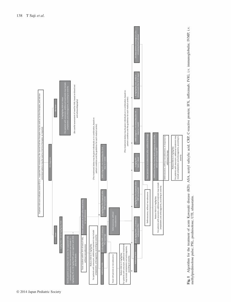


![Kawasaki Robot M series · M series Kawasaki Robot M series Extra large payload robots up to 1,500 kg Cat. No. 3L1750 Nov. ’17 S Printed in Japan Kawasaki Robot] Materials and specifications](https://static.documents.pub/doc/80x56/5d53a61f88c993007d8b677f/kawasaki-robot-m-series-m-series-kawasaki-robot-m-series-extra-large-payload.jpg)


