Keratinocyte-derived Laminin-332 Promotes Adhesion andMigration in Melanocytes and Melanoma*
Received for publication, July 21, 2010, and in revised form, February 11, 2011 Published, JBC Papers in Press, February 24, 2011, DOI 10.1074/jbc.M110.166751
Heesung Chung‡, Eun-Kyung Suh‡, Inn-Oc Han§, and Eok-Soo Oh‡1
From the ‡Division of Life and Pharmaceutical Sciences, Department of Life Sciences, and the Center for Cell Signaling & DrugDiscovery Research, Ewha Womans University, Seoul 120-750, Korea and the §Department of Physiology and Biophysics,College of Medicine, Inha University, Incheon 402-751, Korea
Melanocytes are highly motile cells that play an integral rolein basic skin physiological processes such as wound healing andproper skin pigmentation. It has been postulated that surround-ing keratinocytes contribute to melanocyte migration, butunderlying mechanisms remain rather vague so far. In thisstudy, we set out to analyze the specific potential contribution ofkeratinocyte components to melanocytes and melanoma cellmigration-related processes. Our studies revealed that A375humanmelanomacell attachment, spreading, andmigration areinterestingly better supported by HaCaT keratinocyte extracel-lular matrix (ECM) than by self-derived A375 ECM. Moreover,HaCaT ECM caused increased integrin �6 expression, adhe-sion-mediated focal adhesion kinase phosphorylation, and focaladhesion formations. Similar effects were confirmed in humanmelanocytes. Furthermore, we found that keratinocyte-derivedsoluble factors did not appear to significantly contribute to theseprocesses. Specific extrinsic factors that promoted melanomamigration were attributed to keratinocyte-derived laminin-332,whereas alternative ECM component such as laminin-111 andfibronectin functionsappeared tohave insignificant contributions.Taken together, these studies implicate extrinsic laminin-332 inpromoting the high mobility property and perhaps invasivenessinherently characteristic of, and that are the menace of, melano-cytes andmelanomas, respectively.
Melanocytes, which are present in the skin, hair, eyes, andears, synthesize melanin via a process known as melanogenesis(1), and thus play a key role in the pigmentary systemof the skinin the body. These cells are located in the bottom layer of theskin (basal epidermis), where they comprise 5–10% of the totalcells. The major cells comprising the epidermis are keratino-cytes, which are organized into the basal cell layer, spinous celllayer, granular cell layer, and keratinized squames (2). The kera-tinocytes in the basal cell layer gradually differentiate, prolifer-ate, and migrate upward to form the primary protection of thebody from the outside environment.There is a close and important functional association
between melanocytes and keratinocytes. Melanocytes transfer
maturemelanosomes to neighboring keratinocytes, resulting invisible skin pigmentation (3) and protecting the keratinocytesfrom the deleterious effects of UV light (4). Therefore, melano-cytes play an important role in keratinocyte functions. Recipro-cally, keratinocytes mediate melanocyte functions via severalpathways, including cell-cell adhesion, cell-matrix adhesion,and paracrine signaling (5). Normal melanocytes maintain cell-cell adhesion with keratinocytes by expressing cell-cell adhe-sion proteins such as E-cadherin, desmoglein 1, and connexins(6). In turn, keratinocytes secrete many paracrine factors tomelanocytes, including �-melanocyte-stimulating hormone(�-MSH),2 adrenocorticotrophic hormone, endothelin-1, -2,and -3, FGF-2, and hepatocyte growth factor, all of which reg-ulate the proliferation and differentiation of epidermalmelano-cytes (7). For example, �-MSH, as a ligand of melanocortinreceptor 1, stimulates melanogenesis via the generation ofcAMP. In addition, �-MSH can also enhance melanocyte pro-liferation, confer anti-inflammatory effects, and suppress themigration/invasion of melanoma cells (8–10).Adhesion between skin cells and the surrounding matrix
is regulated by various keratinocyte-produced extracellularmatrix (ECM) factors, including fibronectin, laminin, and col-lagen (11, 12). In particular, the laminins are involved in theregulation of melanocytes. Laminin-332 is known to initiatehemidesmosome formation and support stable attachment ofthe epidermis to the dermis (13). Laminin-332 also enhanceskeratinocyte migration during wound healing (14), stimulatestumor growth and invasion, and enhances the formation oflamellipodia by tumor cells (15). In general, the ECM regulatescell behavior by influencing cell proliferation, survival, mor-phology, migration, and differentiation. In the case of malig-nant cancers, the ECM also regulates invasion and metastasis.Tumor cells from melanoma, a malignant tumor that arisesfrom mutant melanocytes, reportedly use receptor-mediatedrecognition of various ECM molecules to move through thebasement membrane (16). Therefore, both soluble factors andkeratinocyte-derived ECM factors seem to be involved in thefunctional regulation of melanocytes.Because keratinocytes regulate various melanocytes behav-
iors, including proliferation, melanin synthesis, and dendrito-genesis and because the ECM regulates various cell behaviors,we hypothesized that keratinocytes-derived ECM factorsmight* This work was supported by Basic Science Research Program through the
National Research Foundation of Korea funded by Ministry of Education,Science and Technology Grant 2009-0071381 (to E.-S. O.).
1 To whom correspondence should be addressed: Department of Life Sci-ences, Ewha Womans University, Daehyun-dong, Seodaemoon-Gu, Seoul120-750, Korea. Tel.: 82-2-3277-3761; Fax: 82-2-3277-3760; E-mail: [email protected].
2 The abbreviations used are: �-MSH, �-melanocyte-stimulating hormone;ECM, extracellular matrix; FAK, focal adhesion kinase; HaCaT ECM, HaCaT-derived ECM.
act as important regulators of melanocytes. Here, we demon-strate that laminin-332, a component of basement membrane,plays a crucial role in the adhesion and migration of melano-cytes and melanoma.
EXPERIMENTAL PROCEDURES
Materials—The mAb against GAPDH was purchased fromAbFrontier (Korea). The polyclonal antibody against laminin�2 chain and the mAbs against fibronectin and �-actin werepurchased from Santa Cruz (Santa Cruz, CA). The phosphory-lation site-specific polyclonal antibody against FAK (Tyr(P)397)and was purchased from Abcam (Cambridge, UK). The poly-clonal antibody against integrin �2 was purchased from SantaCruz. The polyclonal antibodies against FAK, integrin �4 anda6 were purchased from Cell Signaling (Beverly, MA). Themonoclonal antibody against paxillin was purchased fromUpstate Biotechnology, Inc. (Waltham, MA). Laminin andfibronectin were purchased from Upstate. Collagen type I andlaminin-332 were purchased from Abcam.Cell Culture and Transfection—The A375 and SK-MEL-5
humanmelanoma cell lines, B16F10mousemelanoma cell line,and the HaCaT human keratinocyte cell line were maintainedin DMEM (WelGene, Daegu-si, Korea), supplemented with10% (v/v) FBS (Hyclone) and gentamicin (50 �g/ml; Sigma) at37 °C in a humidified 5%CO2 atmosphere. The primary humanepidermal melanocyte was purchased from Modern Cell &Tissue Technologies, Inc. The human epidermal melanocytewas maintained in melanocyte growth medium-4 (Lonza),supplemented with FBS, rh-Insulin, GA-1000 (gentamicin sul-fate amphotericin-B), calcium chloride, phorbol 12-myristate13-acetate, bovine pituitary extract, hydrocortisone, andrh-FGFB at 37 °C in 5%CO2 in a humidified atmosphere. Tran-sient transfections were carried out using Lipofectamine 2000(Invitrogen) according to the provided instructions.RNA Extraction and RT-PCR—Total RNA was extracted
from cells and analyzed on 1% agarose gel to verify RNAintegrity. Total RNA was reverse transcribed, and aliquots ofthe resulting cDNA were amplified using the following pri-mers: human integrin �2, 5�-GCATTGAAAACACTCGAT-3�(forward) and 5�-TCGGATCCCAAGATTTTCTG-3� (reverse);human integrin �3, 5�-TGGGAGCTGTTTATTGGTCG-3�(forward) and 5�-GGGCCTAGAGGTGGAGTTCT-3� (reverse);human integrin �4, 5�-TCGAATAAAGGATTGGTTTG-CAT-3� (forward) and 5�-AAATGTTGCATGGAATATAC-GGG-3� (reverse); human integrin �5, 5�-CCTCCCAATTTC-AGACTCCC-3� (forward) and 5�-ACAAGGGTCCTTCACA-GTGC-3� (reverse); human integrin �6, 5�-GACTTGAAAGA-AATGGTGAATGC-3� (forward) and 5�-TAGCACCTGTTT-GCTGTTGC-3� (reverse); human LAMC2, 5�-TGGAGAAC-GCTGTGATAGGTGTCG-3� (forward) and 5�-TGTGTAAG-TCTTGGTGAGCCCAC-3� (reverse); human fibronectin,5�-CCGTGGGCAACTCTGTC-3� (forward) and 5�-TGCGG-CAGTTGTCACAG-3� (reverse); and human collagen type I,5�-TCGGCGAGAGCATGACCGATGGAT-3� (forward) and5�-GACGCTGTAGGTGAA GCGGCTGTT-3� (reverse).After an initial denaturation at 94 °C for 5 min, the samples
were subjected to 30 cycles of denaturation at 94 °C for 30 s,
annealing at 55 °C for 30 s, and extension at 72 °C for 60 s. Thereaction products were analyzed on 1% agarose gels.siRNA—Human LAMC2-, FN-, ITGA3-, and ITGA6-spe-
cific siRNA oligonucleotides were designed. The sequenceswere as follows: LAMC2 siRNA, 5�-GCAAAGAGGAUCAAA-CAAAUU-3� (sense) and 5�-UUUGUUUGUAUCCUCUUUG-CUU-3� (antisense); FN siRNA, 5�-GCUGAAGACACAAGG-AAAUUU-3� (sense) and 5�-AUUUCCUUGUGUCUUCAG-CUU-3� (antisense); and ITGA3 siRNA, 5�-CCUACUACUUC-GAGAGGAAUU-3� (sense) and 5�-UUCCUCGAAGUAGUA-GGUU-3� (antisense); and ITGA6 siRNA, 5�-GAGUAUGAA-UUCAGGGUAAUU-3� (sense) and 5�-UUACCCUGAAUUC-AUACUCUU-3� (antisense). Scrambled siRNA (siGENOMEnontargeting siRNA 2) was purchased from Dharmacon(Abbott Park, IL) and used as a control.Immunoblotting—The cultures were washed twice with PBS,
and the cells were lysed in radioimmune precipitation assaybuffer (50 mM Tris, pH 8.0, 150 mM NaCl, 1% Nonidet P-40, 10mM NaF, and 2 mM Na3VO4) containing a protease inhibitormixture (1mg/ml aprotinin, 1mg/ml antipain, 5mg/ml leupep-tin, 1 mg/ml pepstatin A, and 20 mg/ml phenylmethylsulfonylfluoride). The lysates were clarified by centrifugation at13,000 � g for 15 min at 4 °C, denatured with SDS samplebuffer, boiled, and analyzed by SDS-PAGE. The resolved pro-teins were transferred to polyvinylidene difluoride membranes(AmershamBiosciences) and probedwith the appropriate anti-bodies. The signals were detected by enhanced chemilumines-cence (Animal Genetics Inc., Suwon-si, Korea).Cell SpreadingAssay—ECMmolecules (e.g. gelatin, fibronec-
tin, laminin-111, collagen type I, and laminin-332) were dilutedin serum-free medium (1 �g/cm2), added to the above de-scribed ECM-bearing plates, and incubated at 25 °C for 1 h. Theplates were then washed with PBS and blocked with 0.2% heat-inactivated BSA for 1 h. After washing with PBS, the cells wereplated to the ECM-coated plates and incubated for various peri-ods at 37 °C in 5% CO2.Preparation of Keratinocyte-derived ECM—Keratinocyte-
derived ECMwas prepared according to the method of Rodecket al. (17). Briefly, HaCaT cells grown at confluence in tissueculture plates were detached with 0.05% trypsin and 1 mM
EDTA in PBS. The detached cells were removed, and the adher-ent ECM was washed with PBS and treated with 0.1 mg/mlsoybean trypsin inhibitor (Invitrogen). The plates were thenwashed three times with PBS, blocked with 0.2% heat-inacti-vated bovine serum albumin (BSA) for 1 h, and washed threemore times with PBS. Alternatively, HaCaT cells grown in tis-sue culture plates were removed by sequential extraction with1% Triton X-100 (in PBS), 2 M urea (in 1 M NaCl), and 8 M urea(in 1 M NaCl) (18). For cell spreading assays, A375 cells wereplated to the matrix-coated plates and incubated for variousperiods at 37 °C in 5% CO2.Immunofluorescence Analysis—The cells were plated to
12-well plates containing coverslips and fixed with 3.5% para-formaldehyde for 10 min. After being washed with PBS, thecells were blockedwith 0.5% BSA and incubated overnight withan anti-laminin �2, anti-fibronectin, or anti-collagen type Iantibody at 4 °C. After being washed with PBS, the cells wereincubatedwith an FITC-conjugated goat anti-mouse or aTexas
Role of Laminin-332 in Melanocytes and Melanoma
APRIL 15, 2011 • VOLUME 286 • NUMBER 15 JOURNAL OF BIOLOGICAL CHEMISTRY 13439
Red-conjugated goat anti-rabbit antibody for 1 h at 25 °C. Thecoverslips were then mounted on glass slides, and the slideswere observed by fluorescence microscopy.TranswellMigration Assay—Fibronectin or laminin-332 was
coated to each well of a Transwell plate (Costar; 8.0-�m poresize), and then the membranes were allowed to dry at 25 °C for1 h. TheTranswell plates were assembled in a 24-well plate, andthe lower chambers were filled with FBS-containing medium.The cells (1 � 105) were added to each upper chamber withserum-free medium, and the plates were incubated at 37 °C in5%CO2 for 24 h. The cell that hadmigrated to the lower surfaceof the filters were stainedwith 0.6%hematoxylin and 0.5% eosinand counted.Monitoring Cell Adhesion andMigration—Cell adhesion and
migration were monitored using the xCELLigence system(Roche Applied Science). For determination of cell adhesion, Eplate 16 (Roche Applied Science) assemblies were coated withECM molecules and seeded with cells (2.0 � 104 cells/well).Each plate was then assembled on the RTCA DP analyzer, anddata were gathered at 5-min intervals for 5 h at 37 °C in 5%CO2. The data were analyzed using the provided RTCA soft-ware. To examine cell migration, laminin-111, laminin-332 andfibronectin were added to each well of a CIM plate 16 (RocheApplied Science; 8-�m pore size), and the membranes wereallowed to dry at 25 °C for 1 h. The lower chambers were filledwith fresh medium containing 10% FBS or with serum-freemedium. The upper chambers were filled with serum-freemedium (30�l/well), and the plate was incubated at 37 °C in 5%
CO2 for 1 h. The background was measured using a RTCA DPanalyzer. The cells were added to each well, and the plate wasincubated at 25 °C. After 30 min, the CIM plate was assembledonto the RTCADP analyzer, and cell migration was assessed at5-min intervals for 20 h at 37 °C in 5% CO2. The obtained datawere analyzed using the provided RTCA software.Statistical Analysis—The data are represented as the means
from three independent experiments. Statistical analysis wasperformedusing anunpaired Student’s t test. Ap value less than0.01 or 0.05 was considered statistically significant.
RESULTS
Keratinocyte-derived ECM Promotes the Adhesion andSpreading of Melanoma Cells—To investigate the effect ofkeratinocyte-derived ECM onmelanocytes, we first performedspreading assays with A375 human melanoma, a malignanttumor that arises from mutant melanocytes. When detachedand replated, A375 cells attached and spread faster on HaCaT-derived ECM (HaCaT ECM) than on their own ECM (A375ECM). Approximately 60% of A375 cells attached and spreadwithin 30min onHaCaTECM,whereasmore than 90%of thesecells remained unattached after 30min onA375 ECM (Fig. 1A).At 1 h post-plating, the A375 cells incubated on the HaCaTECM had a more cylindrical morphology and flatter, sheet-likespreading compared with those incubated on the A375 ECM;these morphological differences could be seen for up to 10 h.Consistent with our observation of increased cell adhesion,overall tyrosine phosphorylation was much higher in total cell
FIGURE 1. Keratinocyte-derived ECM promotes the spreading of melanoma cells. A, A375 cells (1 � 105 cells/well in 12-well plates) were distributed totissue culture plates without or with ECMs prepared by removing either HaCaT or A375 cells with 0.05% trypsin and 1 mM EDTA in PBS. The cells were incubatedat 37 °C in serum-free DMEM, and digital photographs were taken using a phase contrast microscope at the indicated time points (top panel). Attached andspread cells were counted. The results shown reflect the mean percentages of attached and spread cells per field � S.E. from three independent experiments(bottom panel). *, p � 0.01; **, p � 0.05 versus plate or A375 ECM. B, A375 cells plated on the different ECMs were incubated for the indicated time periods. Totalcell lysates were analyzed by Western blotting with an anti-phosphotyrosine (�-pY) antibody. Each blot was then stripped and reprobed with an anti-GAPDH(�-GAPDH) antibody as a loading control. C, A375 cells plated on the different ECMs were incubated for 60 min. Total cell lysates were analyzed by Westernblotting with either anti-phosphotyrosine (�-pY) or anti-phospho-FAK (�-FAK(pY397)) antibodies; �-actin (�-�-actin) was detected as a loading control. D, A375cells were distributed to tissue culture plates with ECMs prepared by removing either HaCaT or A375 cells with 1% Triton X-100. Digital photographs were takenusing a phase contrast microscope at the indicated time points (left panel). Attached and spread cells were counted. The results shown reflect the meanpercentages of attached and spread cells per field � S.E. from three independent experiments (right panel). *, p � 0.01; **, p � 0.05 versus A375 ECM.
Role of Laminin-332 in Melanocytes and Melanoma
13440 JOURNAL OF BIOLOGICAL CHEMISTRY VOLUME 286 • NUMBER 15 • APRIL 15, 2011
lysates fromA375 cells incubated onHaCaTECMthan in thoseincubated on A375 ECM (Fig. 1B). Notably, phosphorylation ofFAK at tyrosine 397, which is a key signaling event during celladhesion (19), was also increased in total cell lysates fromA375cells onHaCaT ECM (Fig. 1C).We also obtained similar resultsinA375 cells onHaCaTECM,whichwas prepared by removingcells using 1% Triton X-100 (Fig. 1D). These data suggest that
keratinocyte-derived ECM molecules can regulate melanomacell function.Keratinocyte-derived Laminin-332 Promotes the Adhesion
and Spreading of Melanoma Cells—Keratinocytes expressand deposit various ECM proteins, including fibronectin andlaminin. To investigate the difference between A375 ECM andHaCaT ECM, we examined the mRNA expression levels ofsome key molecules in both cell lines (Fig. 2A). Although bothcell lines expressed similar amounts of collagen type I, HaCaTcells expressed much more fibronectin than A375 cells. Inter-estingly, the mRNA for LAMC2, which encodes the laminin �2chain of laminin-332, was expressed only in HaCaT cells. Con-sistent with this finding, immunocytochemical studies revealedthat laminin-332 protein expression was higher in HaCaT cellsversus A375 cells (Fig. 2B). Accordingly, we next investigatedwhich keratinocyte-derived ECM proteins could regulate theadhesion and spreading of melanoma cells (Fig. 2C). Plating ofA375 cells on wells coated with different ECM proteinsrevealed that these cells attached and spread well on platescoated with fibronectin and laminin-332, but not gelatin,collagen type I, or laminin-111. When we compared theadhesion and spreading of A375 cells on these proteins withthat on the HaCaT ECM (Fig. 3A), we obtained broadly sim-ilar results. More than 50% of the A375 cells attached andspread on the HaCaT ECM within 30 min, and most of thecells had attached and spread within 1 h. Although only 50%of the cells spread on wells coated with fibronectin orlaminin-332, and maximal spreading required 5 h, the mor-
FIGURE 2. HaCaT cells produce laminin-332. A, total RNA was extractedfrom exponentially growing A375 and HaCaT cells, and the mRNA expressionlevels of laminin �2 chain (LAMC2), collagen-1 (Col-1), and fibronectin (FN)were analyzed by RT-PCR, using �-actin as a loading control. B, either A375and HaCaT cells were cultured on coverslips and immunostained with eitheranti-fibronectin (FN), anti-laminin �2 chain (LAMC2) or anti-collagen type I(Col-1) antibodies. The results were visualized with either an FITC-conjugatedgoat anti-mouse antibody or a Texas Red-conjugated goat anti-rabbit anti-body. DAPI was used to stain nuclei. C, A375 cells (1 � 105 cells/well in 12-wellplates) were seeded to the indicated ECM-coated plates. After the cells wereincubated at 37 °C for 3 h, digital photographs were taken under a phasecontrast microscope, as described in Fig. 1A. Heat-inactivated BSA was usedas the control.
FIGURE 3. Laminin-332 enhances the adhesion and spreading of melanoma cells. A, A375 cells (1 � 105 cells/well in 12-well plates) were distributed to theindicated ECM-coated plates in serum-free DMEM. After incubation at 37 °C for the indicated periods, the cells were digitally photographed under a phasecontrast microscope (top panel), and attached or spread cells were counted. The results are shown as the mean percentages of attached and spread cells perfield � S.E. from three independent experiments (bottom panel). *, p � 0.01; **, p � 0.05 versus LN-111. B, A375 cells (2.0 � 104 cells/well) were seeded induplicate to the indicated ECM-coated E plates; a noncoated well was used as a control. Cell adhesion curves were monitored using the xCELLigence system(top panel). The rates of cell adhesion over 1 h were analyzed using the RTCA software (bottom panel). C, A375 cells were plated on the different ECMs andincubated for the indicated time periods. Total cell lysates were analyzed by Western blotting with the anti-phosphotyrosine (�-pY) or anti-phospho-FAK(�-FAK(pY397)) antibodies, with �-actin (�-�-actin) detected as the loading control. D, SK-MEL-5 (top panel) and B16F10 (bottom panel) cells (2.0 � 104 cells/well)were seeded in duplicate to the laminin-332-coated E plates; a noncoated well was used as a control. Cell adhesion curves were monitored using thexCELLigence system.
Role of Laminin-332 in Melanocytes and Melanoma
APRIL 15, 2011 • VOLUME 286 • NUMBER 15 JOURNAL OF BIOLOGICAL CHEMISTRY 13441
phologies of the spread cells were very similar to those of thecells spread on the HaCaT ECM. These data suggest thatfibronectin and laminin-332 can regulate the attachmentand spreading of melanoma cells.Next, we monitored the adhesion rates of A375 cells on the
various ECM proteins in real time, using the xCELLigence sys-tem. Our results revealed that the A375 cells attached andspread most effectively on laminin-332 (Fig. 3B). Growth onlaminin-332 also enhanced the tyrosyl phosphorylation ofFAK at 397 (Fig. 3C). Similarly, laminin-332 promoted theadhesion of human SK-MEL-5 and mouse B16F10 mela-noma cells (Fig. 3D).To further investigate the potential involvement of fibronec-
tin and laminin-332 in the regulation of melanoma cell adhe-sion, we used unique 21-bp siRNA sequences targeted againstfibronectin and laminin �2 chain to knock down the expressionlevels of these ECM proteins. HaCaT cells transfected with thesiRNA constructs showed decreased mRNA and protein ex-pression of the targeted proteins (Fig. 4A). Interestingly, A375cells plated on HaCaT ECM derived from laminin-332 knock-down cells showed decreased attachment and spreading. Incontrast, A375 cells attached and spread well on HaCaT ECMderived from fibronectin knockdown cells (Fig. 4B). Consistentwith these findings, tyrosyl-397 phosphorylation of FAK ofA375 cells was reduced in HaCaT ECM derived from laminin-
332 knockdown cells but not fibronectin knockdown cells (Fig.4C). These results suggested that laminin-332 is crucial formel-anoma cell attachment and spreading on keratinocyte-derivedECM.Integrin�6 Regulates the Laminin-332-mediated Adhesion of
Melanoma Cells—Integrins are important cell surface recep-tors that bind to ECM proteins, and integrins �6�1, �6�4, and�3�1 are known receptors for laminin-332 (20–22). Therefore,we hypothesized that integrins could be crucial receptors forlaminin-332-mediated melanoma cell attachment and spread-ing. To examine this possibility, we investigated expression ofintegrin� subunits inA375 cells (Fig. 5). BothA375 andHaCaTcells expressed integrin �2, �3, �4, and �6, and integrin �5 wasexpressed only in A375 cells (data not shown but refer to Fig.5A). Interestingly, adhesion on HaCaT ECM caused alteredmRNAand protein expression of integrin�6 (Fig. 5A). Further-more unique siRNA sequences targeted against integrin�6 thateffectively reduced its mRNA expression significantly blockedthe adhesion and spreading of A375 cells on HaCaT ECM (Fig.5B). However, those effects were not seen in A375 cells trans-fected with siRNA of integrin �3 (si-ITGA3; Fig. 5C). Thesefindings indicate that integrin �6 participates in the laminin-332-directed adhesion of melanoma cells to keratinocyte-de-rived ECM.
FIGURE 4. Keratinocyte-derived laminin-332 is crucial for the adhesion and spreading of melanoma cells. A, HaCaT cells were transfected with siRNAstargeting fibronectin (FN) or laminin �-chain (LAMC2). The expression levels of the target mRNAs were analyzed by RT-PCR, with a control (con) siRNA used asthe siRNA control and �-actin detected as the loading control (left panel). Both cells were cultured on coverslips and immunostained with either anti-fibronectin(FN) or anti-laminin �2 chain (LAMC2) antibodies. The results were visualized with either an FITC-conjugated goat anti-mouse antibody or a Texas Red-conjugated goat anti-rabbit antibody. DAPI was used to stain nuclei (right panel). B, A375, HaCaT, or HaCaT cells transfected with siRNA targeting either laminin�2 chain (HaCaT(�LAMC2)) or fibronectin (HaCaT(�FN)) were cultured to confluence. The cells were then removed, the ECM beds were prepared, and A375cells (1 � 105 cells/well in 12-well plates) were seeded to the ECM-coated plates. After being incubated at 37 °C for the indicated periods, the cells were digitallyphotographed under a phase contrast microscope (top panel), and attached or spread cells were counted. The results are given as mean percentages ofattached and spread cells per field � S.E. from three independent experiments (bottom panel). *, p � 0.01; **, p � 0.05 versus HaCaT ECM. C, A375 cells wereplated on the indicated ECMs as described in B and incubated for the indicated time periods. Total cell lysates were analyzed by Western blotting withanti-phosphotyrosine (�-pY) and anti-phospho-FAK (�-FAK(pY397)) antibodies; �-actin (�-�-actin) was detected as the loading control.
Role of Laminin-332 in Melanocytes and Melanoma
13442 JOURNAL OF BIOLOGICAL CHEMISTRY VOLUME 286 • NUMBER 15 • APRIL 15, 2011
Laminin-332 Promotes Focal Adhesion Formation in Mela-noma Cells—Because focal adhesions serve as the mechanicallinkages to the ECM, we next investigated focal adhesion for-
mation of melanoma cells on laminin-322 (Fig. 6). Although�70% of A375 cells on HaCaT ECM showed focal adhesionformation, focal adhesion formation was significantly reduced
FIGURE 5. Integrin �6 regulates melanoma cell spreading on laminin-332. A, A375 cells were plated on A375 ECM or HaCaT ECM and incubated at 37 °C for5 h, and the mRNA expression levels of the various integrin (Int) subunits were analyzed by RT-PCR. �-Actin was used as the control (left panel). A375 cells wereplated on A375 ECM or HaCaT ECM and incubated at 37 °C. After 7 h, total cell lysates were analyzed by Western blotting with the indicated anti-integrinantibodies (right panel). B, A375 cells were transfected with control or integrin (Int) �6-targeting siRNAs. The mRNA expression level of the target protein wasanalyzed by RT-PCR. �-Actin was used as the control. The protein expression of the integrin �6 was analyzed by Western blotting with anti-integrin �6 (�-Int�6) antibody (top panel). A375 cells transfected with the indicated siRNAs were seeded on HaCaT ECM. After the indicated time periods, the cells were digitallyphotographed under a phase contrast microscope (middle panel), and attached or spread cells were counted. The results are given as the mean percentagesof attached and spread cells per field � S.E. from three independent experiments (bottom panel). *, p � 0.01; **, p � 0.05 versus si-control. C, A375 cells weretransfected with control or integrin �6- or integrin �3-targeting siRNAs. The mRNA expression level of the target protein was analyzed by RT-PCR. �-Actin wasused as the control (top panel). A375 cells transfected with the indicated siRNAs were seeded on HaCaT ECM. After the indicated time periods, the cells weredigitally photographed under a phase contrast microscope (bottom panel).
FIGURE 6. Laminin-332 promotes focal adhesion formation in HaCaT cells. A, either A375 or HaCaT cells transfected with siRNA targeting either laminin �2chain (HaCaT(�LAMC2)) or fibronectin (HaCaT(�FN)) were cultured on coverslips to confluence. The cells were then removed, the ECM beds were prepared,and A375 cells (3 � 105 cells/well in 12-well plates) were seeded to the ECM-coated plates. After being incubated at 37 °C for the 1 h, the cells were fixed andimmunostained with anti-paxillin antibody (Texas Red) and phalloidin (FITC-conjugated) (top panel). The number of cells positive for focal adhesions werecounted (bottom panel). *, p � 0.01; **, p � 0.05 versus A375 ECM. B, A375 cells (3 � 105 cells/well in 12-well plates) transfected with siRNA targeting ITGA6 orITGA3 were plated on HaCaT ECM-coated coverslip. After being incubated at 37 °C for the 1 h, the cells were fixed and immunostained with anti-paxillinantibody (Texas Red) and phalloidin (FITC conjugated) (top panel). The number of cells positive for focal adhesions were counted (bottom panel). *, p � 0.01; **,p � 0.05 versus control (con). The arrowheads point to focal adhesion sites.
Role of Laminin-332 in Melanocytes and Melanoma
APRIL 15, 2011 • VOLUME 286 • NUMBER 15 JOURNAL OF BIOLOGICAL CHEMISTRY 13443
in A375 cells plated on HaCaT ECM derived from laminin-332knockdown cells. In contrast, focal adhesion formation wasslightly decreased in A375 cells on HaCaT ECM derived fromfibronectin knockdown cells (Fig. 6A). Similarly, focal adhesionformation was decreased in integrin �6, but not �3, andknocked down A375 cells plated on HaCaT ECM, comparedwith control (Fig. 6B). These results suggest that keratinocyte-derived laminin-332 promotes focal adhesion formation inmelanoma cells.Laminin-332 Enhances the Migration of Melanoma Cells—
Because cell-ECM adhesion is correlated with cell migration(23), we next used the xCELLigence system to investigatewhether keratinocyte-derived laminin-332 regulates themigra-tory ability of melanoma cells. As expected, A375 cells showedbetter migration on laminin-332 over the other tested ECMproteins (Fig. 7A). Consistently, siRNA sequences targetedagainst integrin �6 significantly reduced migration of A375cells on laminin-332 (Fig. 7B).Laminin-332 Promotes Adhesion and Migration of Melano-
cytes—Wenext investigatedwhether laminin-332was involved inthe regulation of adhesion and migration of human melanocytes(Fig. 8). Like melanoma cells, human melanocytes attached andspread most effectively on laminin-332 among ECMs tested (Fig.8A). In addition, laminin-322 enhanced the tyrosyl phosphoryla-tion of FAK at 397 (Fig. 8B) andmigration of humanmelanocytes(Fig. 8C). Consistently, siRNA sequences targeted against integrin�6 significantly inhibited the migration of A375 cells (Fig. 8D).These results demonstrate that laminin-332 promotes the adhe-sion andmigration of melanocytes.Keratinocyte-derived Soluble Factors AreNot Essential for the
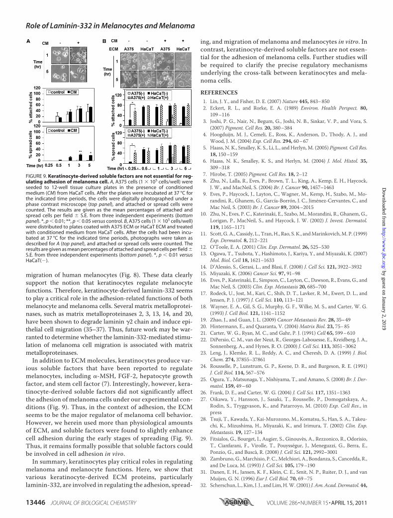
Adhesion of Melanoma Cells—Because both the ECM andsoluble factors are known to regulate cell functions (7, 15), we
investigated whether soluble keratinocyte-derived factorsmight be involved in the adhesion ofmelanoma cells. A375 cellswere plated to tissue culture plates in the presence of condi-tionedmedium fromHaCaT cells. In contrast to ECM, the con-ditioned medium did not significantly affect the adhesion andspreading of A375 cells (Fig. 9A). Similarly, conditionedmedium from HaCaT cells did not affect the adhesion andspreading of A375 cells plated on either A375 ECM or HaCaTECM (Fig. 9B). These results suggest that soluble keratinocyte-derived factors are not essential for the adhesion and spreadingof melanoma cells.
DISCUSSION
In both normal skin andmelanoma, keratinocytes regulatemelanocytes through various means, including cell-cellinteractions, cell-matrix interactions, and paracrine factorproduction. Here, we show that keratinocytes regulate mel-anoma cells through laminin-332. It is known that laminin-332, which is a component of the basement membrane,mediates the firm attachment of basal keratinocytes to thebasement membrane (24–26). Because melanocytes alsoreside within the basal layer of the epidermis and attach tothe basement membrane, it is plausible for laminin-332 toregulate the adhesions of both keratinocytes and melano-cytes. Indeed, because there are relatively fewmelanocytes inthe epidermis, and melanocytes do not make significantamount of laminin-332 (11), the need for laminin-332-me-diated attachment may form part of the basis for the coop-eration between melanocytes and keratinocytes. Here, weshow that keratinocyte-derived laminin-332 can regulatemelanoma cell adhesion. A375 human melanoma cellsattached and spread more effectively on keratinocyte-de-
FIGURE 7. Laminin-332 stimulates the migration of melanoma cells. A, A375 cells (1.5 � 104 cells/well) were seeded in duplicate to the upper chambers ofCIM plates coated with the indicated ECMs; a noncoated well was used as the control (con). The lower chambers were filled with medium containing 10% FBS(�FBS), and migration curves were monitored using the xCELLigence system (top panel). The migration rates over 5 h were analyzed using the RTCA software(bottom panel). B, A375 cells (1 � 105 cells/well) transfected with the indicated siRNAs were seeded in the upper chambers of coated with the indicated ECMs;a noncoated well was used as the control. The lower chambers were filled with medium containing 10% FBS. After 24 h, migrated cells were stained withhematoxylin and eosin. *, p � 0.01; **, p � 0.05 versus si-control.
Role of Laminin-332 in Melanocytes and Melanoma
13444 JOURNAL OF BIOLOGICAL CHEMISTRY VOLUME 286 • NUMBER 15 • APRIL 15, 2011
rived ECM than their own ECM (Fig. 1). Furthermore,laminin-332, which was differentially expressed in keratino-cytes versus melanoma cells (Fig. 2), was found to be crucialfor the attachment and spreading of melanoma cells on thekeratinocyte-derived ECM (Figs. 3 and 4).Laminin-332 is a ligand for a number of receptors, includ-
ing integrins �6�1, �6�4, �3�1, and syndecan (13). Here, weshow that the adhesion on laminin-332 caused alteredexpression of integrin �6 and that siRNA-mediated knock-down of integrin �6 diminished the ability of laminin-332 toenhance the attachment and spreading of A375 cells (Fig. 5).Because integrin �6�4 is known to be down-regulated inmelanoma cells (27), it is likely that integrin �6�1 mediatesthe laminin-332-mediated adhesion of melanoma cells.Oikawa et al. (27) demonstrated that laminin-332 stimulatedmigration of melanoma cells through integrin �3�1 and�6�1, and Tsuji et al. (28) reported that integrin �3mediatedlaminin-332-mediated melanoma cell migration and inva-sion on laminin-332-coated membrane. Because we foundthat integrin �6 regulates laminin-332-mediated cell adhe-sion and migration better than integrin �3 (Figs. 7 and 8),integrin �6 may play a major role in the regulation oflaminin-332-mediated cell adhesion.Previously, laminin-332 was reported to stimulate the
migration of keratinocytes during wound healing (14),tumor growth/invasion, and the formation of lamellipodia in
tumor cells (15). Consistent with a previous report thatlaminin-332 is able to promote melanoma cell migration, asshown in a chemotaxis assay with soluble laminin-332 and ahaptotaxis assay with laminin-332-coated membranes (27,28), we herein show that laminin-332 stimulates the migra-tion of melanoma cells (Fig. 7). Thus, keratinocytes appear toregulate melanoma cell functions. Like melanoma, keratino-cyte-derived laminin-332 plays a crucial role in regulatingmelanocyte functions. Keratinocytes express laminin-332during the human skin wound healing process, and laminin-332 stimulates the migration of keratinocytes (14, 29) andmelanocytes across the wound bed (30, 31). Therefore, kera-tinocyte-derived laminin-332 is expected to enhance mela-nocyte migration for wound repair.Themigration ofmelanocytes is an important event in repig-
mentation of vitiligo, a common skin disorder in which whitespots appear on the skin. Narrow bandUVB light phototherapyhas been widely used for the treatment of vitiligo (32). A previ-ous study showed that conditioned medium from human kera-tinocytes exposed to UVB enhanced proliferation but did notaffect migration of human melanocytes (33). However, UVBstabilizes HIF1-� to increase production of laminin-332 fromkeratinocytes (29, 34). Therefore, it was considered likely thatlaminin-332 derived from keratinocytes regulates melanocytesduring UVB phototherapy treatment. In the present study, weshowed that laminin-332 directly regulated adhesion and
FIGURE 8. Laminin-332 promotes adhesion and migration of melanocytes. A, melanocytes (2.0 � 104 cells/well) were seeded in duplicate to theindicated ECM-coated E plates; a noncoated well was used as a control (Con). Cell adhesion curves were monitored using the xCELLigence system (toppanel). The rates of cell adhesion over 1 h were analyzed using the RTCA software (bottom panel). B, melanocytes (5 � 105 cells/well; six wells) wereplated on the indicated ECMs and incubated for the indicated time periods. Total cell lysates were analyzed by Western blotting with anti-phospho-FAK(�-FAK(pY397)) and anti-FAK (�-FAK) antibodies; �-actin (�-�-actin) was detected as the loading control. C, melanocytes (2 � 104 cells/well) were seededin duplicate to the upper chambers of CIM plates coated with the indicated ECMs; a noncoated well was used as the control. The lower chambers werefilled with medium containing 0.5% FBS, and migration curves were monitored using the xCELLigence system (top panel). The migration rates over 25 hwere analyzed using the RTCA software (bottom panel). D, melanocytes (1 � 105 cells/well) transfected with the indicated siRNAs were seeded in theupper chambers of coated with the LN-332. The lower chambers were filled with medium containing 0.5% FBS. After 24 h, migrated cells were stainedwith hematoxylin and eosin. **, p � 0.05 versus si-control.
Role of Laminin-332 in Melanocytes and Melanoma
APRIL 15, 2011 • VOLUME 286 • NUMBER 15 JOURNAL OF BIOLOGICAL CHEMISTRY 13445
migration of human melanocytes (Fig. 8). These data clearlysupport the notion that keratinocytes regulate melanocytefunctions. Therefore, keratinocyte-derived laminin-332 seemsto play a critical role in the adhesion-related functions of bothmelanocyte and melanoma cells. Several matrix metalloprotei-nases, such as matrix metalloproteinases 2, 3, 13, 14, and 20,have been shown to degrade laminin �2 chain and induce epi-thelial cell migration (35–37). Thus, future work may be war-ranted to determine whether the laminin-332-mediated stimu-lation of melanoma cell migration is associated with matrixmetalloproteinases.In addition to ECMmolecules, keratinocytes produce var-
ious soluble factors that have been reported to regulatemelanocytes, including �-MSH, FGF-2, hepatocyte growthfactor, and stem cell factor (7). Interestingly, however, kera-tinocyte-derived soluble factors did not significantly affectthe adhesion of melanoma cells under our experimental con-ditions (Fig. 9). Thus, in the context of adhesion, the ECMseems to be the major regulator of melanoma cell behavior.However, we herein used more than physiological amountsof ECM, and soluble factors were found to slightly enhancecell adhesion during the early stages of spreading (Fig. 9).Thus, it remains formally possible that soluble factors couldbe involved in cell adhesion in vivo.In summary, keratinocytes play critical roles in regulating
melanoma and melanocyte functions. Here, we show thatvarious keratinocyte-derived ECM proteins, particularlylaminin-332, are involved in regulating the adhesion, spread-
ing, and migration of melanoma and melanocytes in vitro. Incontrast, keratinocyte-derived soluble factors are not essen-tial for the adhesion of melanoma cells. Further studies willbe required to clarify the precise regulatory mechanismsunderlying the cross-talk between keratinocytes and mela-noma cells.
REFERENCES1. Lin, J. Y., and Fisher, D. E. (2007) Nature 445, 843–8502. Eckert, R. L., and Rorke, E. A. (1989) Environ. Health Perspect. 80,
109–1163. Joshi, P. G., Nair, N., Begum, G., Joshi, N. B., Sinkar, V. P., and Vora, S.
(2007) Pigment. Cell Res. 20, 380–3844. Hoogduijn, M. J., Cemeli, E., Ross, K., Anderson, D., Thody, A. J., and
Wood, J. M. (2004) Exp. Cell Res. 294, 60–675. Haass, N. K., Smalley, K. S., Li, L., and Herlyn,M. (2005) Pigment. Cell Res.
18, 150–1596. Haass, N. K., Smalley, K. S., and Herlyn, M. (2004) J. Mol. Histol. 35,
309–3187. Hirobe, T. (2005) Pigment. Cell Res. 18, 2–128. Zhu, N., Lalla, R., Eves, P., Brown, T. L., King, A., Kemp, E. H., Haycock,
J. W., and MacNeil, S. (2004) Br. J. Cancer 90, 1457–14639. Eves, P., Haycock, J., Layton, C., Wagner, M., Kemp, H., Szabo, M., Mo-
randini, R., Ghanem, G., García-Borron, J. C., Jimenez-Cervantes, C., andMac Neil, S. (2003) Br. J. Cancer 89, 2004–2015
10. Zhu, N., Eves, P. C., Katerinaki, E., Szabo, M., Morandini, R., Ghanem, G.,Lorigan, P., MacNeil, S., and Haycock, J. W. (2002) J. Invest. Dermatol.119, 1165–1171
11. Scott, G. A., Cassidy, L., Tran,H., Rao, S. K., andMarinkovich,M. P. (1999)Exp. Dermatol. 8, 212–221
12. O’Toole, E. A. (2001) Clin. Exp. Dermatol. 26, 525–53013. Ogawa, T., Tsubota, Y., Hashimoto, J., Kariya, Y., and Miyazaki, K. (2007)
Mol. Biol. Cell 18, 1621–163314. D’Alessio, S., Gerasi, L., and Blasi, F. (2008) J. Cell Sci. 121, 3922–393215. Miyazaki, K. (2006) Cancer Sci. 97, 91–9816. Eves, P., Katerinaki, E., Simpson, C., Layton, C., Dawson, R., Evans, G., and
Mac Neil, S. (2003) Clin. Exp. Metastasis 20, 685–70017. Rodeck, U., Jost, M., Kari, C., Shih, D. T., Lavker, R. M., Ewert, D. L., and
Jensen, P. J. (1997) J. Cell Sci. 110, 113–12118. Wayner, E. A., Gil, S. G., Murphy, G. F., Wilke, M. S., and Carter, W. G.
(1993) J. Cell Biol. 121, 1141–115219. Zhao, J., and Guan, J. L. (2009) Cancer Metastasis Rev. 28, 35–4920. Hintermann, E., and Quaranta, V. (2004)Matrix Biol. 23, 75–8521. Carter, W. G., Ryan, M. C., and Gahr, P. J. (1991) Cell 65, 599–61022. DiPersio, C. M., van der Neut, R., Georges-Labouesse, E., Kreidberg, J. A.,
Sonnenberg, A., and Hynes, R. O. (2000) J. Cell Sci. 113, 3051–306223. Leng, J., Klemke, R. L., Reddy, A. C., and Cheresh, D. A. (1999) J. Biol.
Chem. 274, 37855–3786124. Rousselle, P., Lunstrum, G. P., Keene, D. R., and Burgeson, R. E. (1991)
J. Cell Biol. 114, 567–57625. Ogura, Y., Matsunaga, Y., Nishiyama, T., and Amano, S. (2008) Br. J. Der-
matol. 159, 49–6026. Frank, D. E., and Carter, W. G. (2004) J. Cell Sci. 117, 1351–136327. Oikawa, Y., Hansson, J., Sasaki, T., Rousselle, P., Domogatskaya, A.,
Rodin, S., Tryggvason, K., and Patarroyo, M. (2010) Exp. Cell Res., inpress
28. Tsuji, T., Kawada, Y., Kai-Murozono, M., Komatsu, S., Han, S. A., Takeu-chi, K., Mizushima, H., Miyazaki, K., and Irimura, T. (2002) Clin. Exp.Metastasis. 19, 127–134
29. Fitsialos, G., Bourget, I., Augier, S., Ginouves, A., Rezzonico, R., Odorisio,T., Cianfarani, F., Virolle, T., Pouyssegur, J., Meneguzzi, G., Berra, E.,Ponzio, G., and Busca, R. (2008) J. Cell Sci. 121, 2992–3001
30. Zambruno,G.,Marchisio, P. C.,Melchiori, A., Bondanza, S., Cancedda, R.,and De Luca, M. (1993) J. Cell Sci. 105, 179–190
31. Danen, E. H., Jansen, K. F., Klein, C. E., Smit, N. P., Ruiter, D. J., and vanMuijen, G. N. (1996) Eur J. Cell Biol. 70, 69–75
32. Scherschun, L., Kim, J. J., and Lim,H.W. (2001) J. Am.Acad. Dermatol. 44,
FIGURE 9. Keratinocyte-derived soluble factors are not essential for reg-ulating adhesion of melanoma cell. A, A375 cells (1 � 105 cells/well) wereseeded to 12-well tissue culture plates in the presence of conditionedmedium (CM) from HaCaT cells. After the plates were incubated at 37 °C forthe indicated time periods, the cells were digitally photographed under aphase contrast microscope (top panel), and attached or spread cells werecounted. The results are given as the mean percentages of attached andspread cells per field � S.E. from three independent experiments (bottompanel). *, p � 0.01; **, p � 0.05 versus control. B, A375 cells (1 � 105 cells/well)were distributed to plates coated with A375 ECM or HaCaT ECM and treatedwith conditioned medium from HaCaT cells. After the cells had been incu-bated at 37 °C for the indicated time periods, photographs were taken asdescribed for A (top panel), and attached or spread cells were counted. Theresults are given as mean percentages of attached and spread cells per field �S.E. from three independent experiments (bottom panel). *, p � 0.01 versusHaCaT(�).
Role of Laminin-332 in Melanocytes and Melanoma
13446 JOURNAL OF BIOLOGICAL CHEMISTRY VOLUME 286 • NUMBER 15 • APRIL 15, 2011
999–100333. Wu, C. S., Yu, C. L., Wu, C. S., Lan, C. C., and Yu, H. S. (2004) Exp.
Dermatol. 13, 755–76334. Li, Y., Bi, Z., Yan, B., and Wan, Y. (2006) Int. J. Mol. Med. 18, 713–71935. Koshikawa, N., Giannelli, G., Cirulli, V., Miyazaki, K., and Quaranta, V.
(2000) J. Cell Biol. 148, 615–624
36. Gilles, C., Polette,M., Coraux, C., Tournier, J.M.,Meneguzzi, G.,Munaut,C., Volders, L., Rousselle, P., Birembaut, P., and Foidart, J. M. (2001) J. CellSci. 114, 2967–2976
37. Pirila, E., Sharabi, A., Salo, T., Quaranta, V., Tu, H., Heljasvaara, R., Ko-shikawa, N., Sorsa, T., and Maisi, P. (2003) Biochem. Biophys. Res. Com-mun. 303, 1012–1017
Role of Laminin-332 in Melanocytes and Melanoma
APRIL 15, 2011 • VOLUME 286 • NUMBER 15 JOURNAL OF BIOLOGICAL CHEMISTRY 13447