Page 1
King’s Research Portal
DOI:10.1016/j.soard.2016.08.007
Document VersionPeer reviewed version
Link to publication record in King's Research Portal
Citation for published version (APA):Sales-Peres, S. H. D. C., Sales-Peres, M. D. C., Passeri, C. R., Ceneviva, R., & Bernabé, E. (2016). WeightLoss after Bariatric Surgery and Periodontal Changes: A 12-Month Prospective Study. Surgery for obesity andrelated diseases. DOI: 10.1016/j.soard.2016.08.007
Citing this paperPlease note that where the full-text provided on King's Research Portal is the Author Accepted Manuscript or Post-Print version this maydiffer from the final Published version. If citing, it is advised that you check and use the publisher's definitive version for pagination,volume/issue, and date of publication details. And where the final published version is provided on the Research Portal, if citing you areagain advised to check the publisher's website for any subsequent corrections.
General rightsCopyright and moral rights for the publications made accessible in the Research Portal are retained by the authors and/or other copyrightowners and it is a condition of accessing publications that users recognize and abide by the legal requirements associated with these rights.
•Users may download and print one copy of any publication from the Research Portal for the purpose of private study or research.•You may not further distribute the material or use it for any profit-making activity or commercial gain•You may freely distribute the URL identifying the publication in the Research Portal
Take down policyIf you believe that this document breaches copyright please contact [email protected] providing details, and we will remove access tothe work immediately and investigate your claim.
Download date: 06. Nov. 2017
Page 2
Author’s Accepted Manuscript
Weight Loss after Bariatric Surgery andPeriodontal Changes: A 12-Month ProspectiveStudy
Silvia Helena de Carvalho Sales-Peres, Matheus deCarvalho Sales-Peres, Celso Roberto Passeri,Reginaldo Ceneviva, Eduardo Bernabé
PII: S1550-7289(16)30168-XDOI: http://dx.doi.org/10.1016/j.soard.2016.08.007Reference: SOARD2708
To appear in: Surgery for Obesity and Related Diseases
Received date: 5 April 2016Revised date: 23 May 2016Accepted date: 4 August 2016
Cite this article as: Silvia Helena de Carvalho Sales-Peres, Matheus de CarvalhoSales-Peres, Celso Roberto Passeri, Reginaldo Ceneviva and Eduardo Bernabé,Weight Loss after Bariatric Surgery and Periodontal Changes: A 12-MonthProspective Study, Surgery for Obesity and Related Diseases,http://dx.doi.org/10.1016/j.soard.2016.08.007
This is a PDF file of an unedited manuscript that has been accepted forpublication. As a service to our customers we are providing this early version ofthe manuscript. The manuscript will undergo copyediting, typesetting, andreview of the resulting galley proof before it is published in its final citable form.Please note that during the production process errors may be discovered whichcould affect the content, and all legal disclaimers that apply to the journal pertain.
www.elsevier.com/locate/buildenv
Page 3
Weight Loss after Bariatric Surgery and Periodontal Changes: A
12-Month Prospective Study
Silvia Helena de Carvalho SALES-PERES,a PhD; Matheus de Carvalho SALES-
PERES,b PhD; Celso Roberto PASSERI,
a PhD; Reginaldo CENEVIVA,
b PhD; Eduardo
BERNABÉ,c PhD
a Department of Pediatric Dentistry, Orthodontics and Public Health, Bauru School of
Dentistry, University of São Paulo, São Paulo, Brazil
b Department of Surgery, Clinical Hospital of Ribeirão Preto, University of São Paulo,
Ribeirão Preto, Brazil.
c Division of Population and Patient Health, King’s College London Dental Institute at Guy’s,
King’s College and St. Thomas’ Hospitals, London, United Kingdom
Source of funding:
This project was funded by the Conselho Nacional de Desenvolvimento Científico e
Tecnológico (CNPq) and Fundação de Amparo à Pesquisa do Estado de São Paulo (FAPESP)
Corresponding author:
Dr. Silvia Helena de Carvalho Sales-Peres, Bauru Dental School, University of São Paulo,
Department of Paediatric Dentistry, Orthodontics and Public Health, Al. Octávio Pinheiro
Brisolla, 9-75, Bauru-SP 17012-901, Brazil, Tel: +55-14-32358260, Email: [email protected]
Running title: weight loss surgery and periodontal disease
Page 4
ABSTRACT
Background: Several longitudinal studies have explored the association of obesity and
weight gain with periodontal disease. However, the effect of weight loss on periodontal
tissues remains unclear.
Objective: To explore whether weight loss after bariatric surgery was associated with
changes in periodontal measures over 12 months.
Setting: Two public hospitals in São Paulo, Brazil.
Methods: We used data from 110 morbidly obese patients (BMI>40 kg/m2 or ≥35 kg/m
2
with comorbid conditions) who underwent bariatric surgery between April 2011 and March
2013. Data on demographic factors, body mass index (BMI), smoking habits and glucose
levels were extracted from medical records pre-operatively and after 6 and 12 months post-
surgery. A full-mouth periodontal examination was conducted by trained examiners to assess
probing pocket depth (PPD), clinical attachment loss (CAL) and bleeding on probing (BOP)
at baseline, 6 and 12 months. Data were analyzed using linear mixed effects (LME) models.
Results: BMI was not significantly related to the proportion of sites with BOP at baseline,
but it was negatively associated with the rate of change in the proportion of sites with BOP.
The greater the BMI loss the higher the proportion of sites with BOP, particularly 6 months
after surgery. However, BMI was not associated with baseline PPD and CAL or rates of
changes in these periodontal outcomes.
Conclusion: The findings suggest that weight loss was associated with increased gingival
bleeding, showing a peak at 6 months after bariatric surgery. Periodontal pocketing and
attachment loss remained unchanged during the study period.
Keywords: body weight; bariatric surgery; periodontal diseases; cohort studies; adults
Page 5
INTRODUCTION
The prevalence of obesity has increased epidemically in both developed and developing
countries over the last few decades [1]. Obesity is a metabolic condition caused by an energy
imbalance (i.e. when energy intake exceeds energy expenditure), which subsequently leads to
an increase in adipose tissue deposits [2]. As adipocytes exert a number of endocrine
functions [3], increased adiposity is associated with a state of low-grade inflammation and
insulin resistance [2, 4]. This obesity-related pro-inflammatory status appears to be involved
in cardiovascular diseases, type 2 diabetes, metabolic disorders and certain cancers [5, 6].
Evidence is growing for a possible link between obesity and periodontal disease [7-9].
Obesity related inflammation may promote periodontitis by secretion of inflammatory
markers by the adipose tissue that may increase gingival inflammation and promote bacterial
proliferation on the tooth root surface [10, 11]. A recent review has also reported that weight
gain was associated with incidence of periodontitis, although only 5 studies were identified
[12]. Since the unfavorable inflammatory profile associated with increased adiposity can be
improved during a period of weight loss [13], what is missing in the literature is an evaluation
of the effect of weight loss on periodontal tissues.
Several methods have been proposed for weight loss in obese patients, like dieting and
physical exercise, pharmacological treatment, and surgical intervention [14]. Bariatric
surgery is an effective therapy for the treatment of obesity compared with non-surgical
interventions [15]. The benefits of bariatric surgery include significant and durable weight
loss, improved or remission of obesity related comorbidities, and improved quality of life [15,
16]. Limited evidence, mainly coming from case reports and cross-sectional studies, suggests
that higher levels of dental caries, periodontal diseases and tooth wear may be found in
patients after bariatric surgery [17, 18]. In a recent prospective study, a resolution of systemic
inflammation after bariatric surgery –i.e. significant decreases in C-reactive protein (CRP)
Page 6
and glucose levels after surgery– did not seem to affect the course of periodontal disease. On
the contrary, the mean periodontal pocket depth and attachment loss increased significantly 6
months after bariatric surgery [19, 20].
The purpose of this study was to explore whether weight loss after bariatric surgery was
associated with changes in periodontal measures over 12 months.
METHODS
This report adheres to the Strengthening the Reporting of Observational Studies (STROBE)
guidelines [21].
Participants
A total of 150 morbidly obese patients (BMI>40.00 Kg/m2 or ≥35.00 Kg/m
2 with comorbid
conditions) were recruited from the patient pool receiving bariatric surgery (Roux-en-Y) in
two public hospitals in São Paulo, Brazil, between April 2011 and March 2013. Patients with
history of any infectious diseases, those who were pregnant or breastfeeding, using anti-
inflammatory agents or antibiotics 3 months prior to the study and those who have fewer than
six teeth were excluded from the study.
The study protocol was approved by the Research Ethics Committees of the two Medical
Schools Hospitals (Ref: 315/08 and 468/08). All patients were informed of the purpose of the
investigation and signed a written informed consent before voluntary participation.
After exclusions, there were 110 patients (aged 20 to 60 years at baseline) who had
periodontal data in at least two of the three examinations (baseline plus 6 or 12 months).
Periodontal data were available for 110, 90 and 110 participants preoperatively and at 6 and
12 months after surgery, respectively. That means 90 patients (82%) contributed to all three
waves of periodontal data whereas the rest (18%) to two waves. A post-hoc calculation
showed that this sample size had a 90% power to identify an 8%-difference in the proportion
Page 7
of sites with bleeding on probing (BOP) before and after surgery, with standard deviation of
25% in each measurement occasion and a correlation of 0.50 between measurements.
Data collection
Data were collected from medical records and through clinical examinations. Demographic
factors (sex and age), anthropometric measures (height and weight), smoking habits, and
fasting blood glucose (FBG) levels were extracted from patients’ medical records
preoperatively and 6 and 12 months after surgery. In line with local protocols, an FBG
greater than 100 mg/dL was used for diagnosis of diabetes. Weight loss after bariatric surgery
was expressed as change in body mass index (BMI). Patients did not receive any dental care
or oral health advice during the hospitalization period.
Participants were also invited to a periodontal examination at baseline and each control visit.
A North Carolina periodontal probe was used for the clinical inspection of all present teeth,
excluding third molars. A full-mouth examination protocol was used, inspecting six sites per
tooth (mesio-facial, mid-facial, disto-facial, mesio-lingual, mid-lingual, and disto-lingual) to
measure probing pocket depth (PPD), clinical attachment loss (CAL) and bleeding on
probing (BOP). PPD was the distance from the gingival margin to the base of the pocket
whereas CAL was the distance between the cement-enamel junction and the base of the
pocket. Three periodontal outcome measures were evaluated, namely the mean PPD and CAL
across all examined sites and the proportion of sites with BOP. Examinations at three times,
namely pre-operatively, 6 and 12 months after bariatric surgery were performed by two
calibrated dentists, with Kappa values for intra- and inter-examiner reliability of 0.82 and
0.80, respectively.
Page 8
Statistical analysis
Linear mixed effects (LME) models were used to estimate the longitudinal association
between change in BMI and changes in BOP, PPD and CAL over 12 months. LME models
use all available outcome data over the follow-up period, handle unequally spaced
observations over time and take into account the fact that repeated measures on the same
individual are correlated [22, 23]. All analyses were run in Stata Statistical Software (Release
13. College Station, TX: StataCorp LP) using the mixed command.
We explored the association of BMI with BOP, PPD and CAL in separate set of models. We
fitted both the intercept and the slope with time as random effects, allowing for individual
differences in periodontal measures at baseline and rates of change in periodontal measures
over the follow-up period. Survey waves (0, 6 and 12 months coded as 0, 1 and 2
respectively) were used as the underlying time scale in all models (fitted as a categorical time
indicator). First we estimated a model without any covariates (null model) to establish the
rate of change in BOP within the observed period. Next, we tested the effect of BMI (for
every 10-unit change) on baseline BOP levels controlling for sex, age, smoking status and
diabetes. We then tested the association of BMI with changes in BOP over time by adding the
interaction between BMI and the time indicator to the main effects model. The main effect
for BMI estimates the effect on BOP at baseline whereas the interaction term between BMI
and time estimates the effect of BMI on change in BOP over 12 months. We presented
significant associations with changes in BOP using line graphs to aid interpretation. The same
steps were followed when testing the association of BMI with PPD and CAL, respectively.
As smoking causes periodontal vasoconstriction [24], we tested whether the effect of BMI on
periodontal measures was different in smokers and non-smokers by testing the significance of
the statistical interaction between BMI and smoking status when added to the model.
Page 9
RESULTS
A total of 110 adult patients (88% women), with a mean age of 38.5 years (Standard
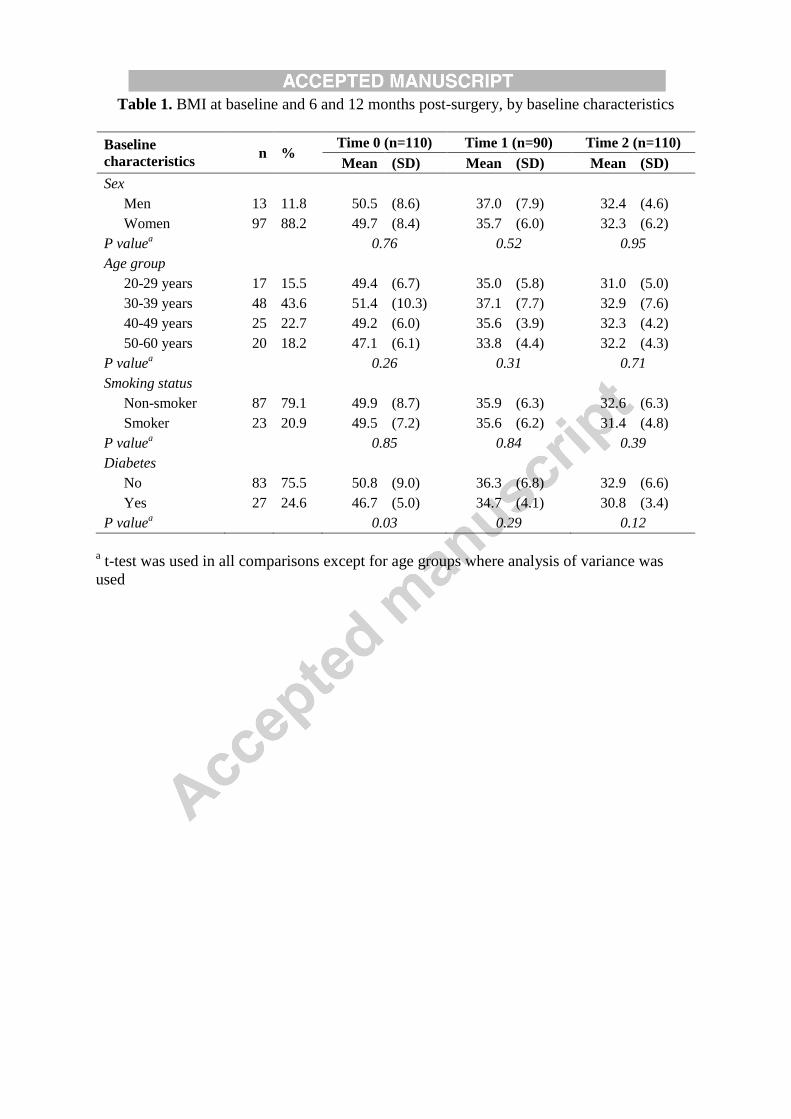
Deviation [SD]: 9.8, range: 20 to 60) were included in this study. The baseline characteristics
of the sample are shown in Table 1. The mean proportion of sites with BOP was 24.6% at
baseline (SD: 23.4; range: 0-100) whereas the mean PPD and CAL were 1.77 mm (SD: 0.47;
range: 1.01-3.10) and 1.86 mm (SD: 0.60; range: 0.58-4.24), respectively.
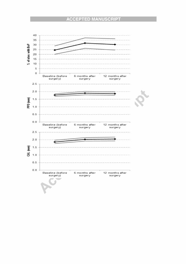
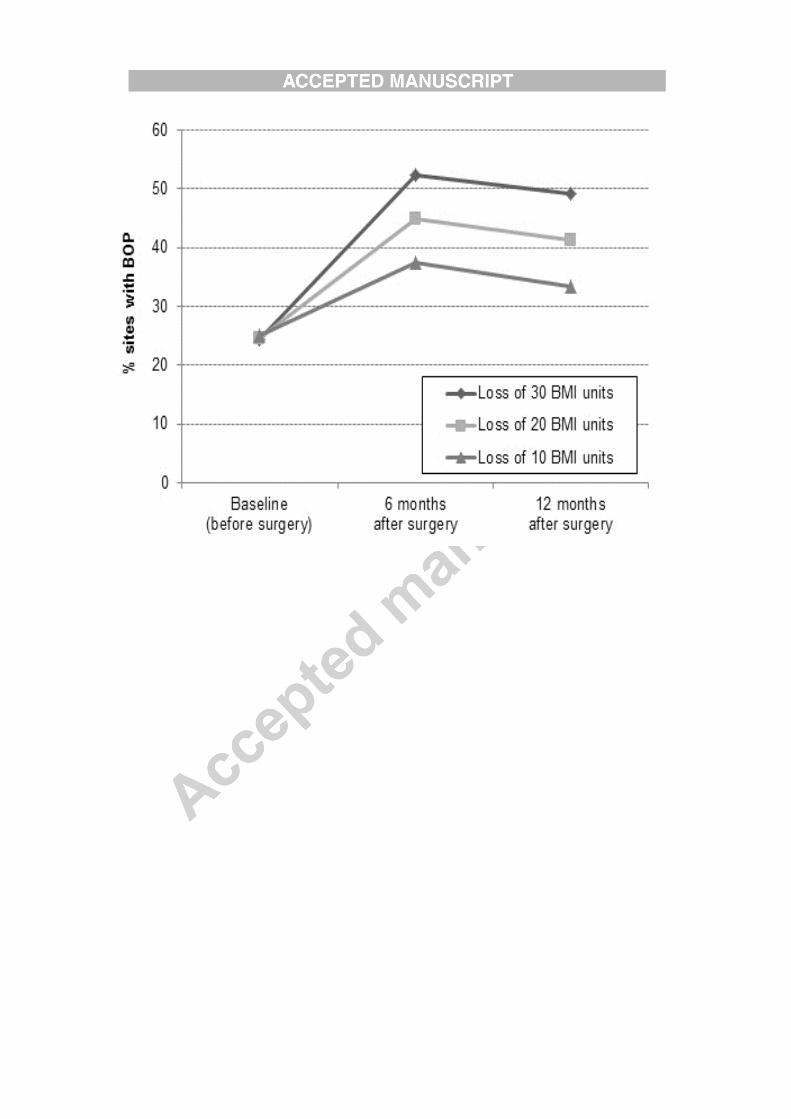
Larger variations were observed for BOP (from 24.6% at baseline to 32.0% at 6 months to
30.8% at 12 months) than for mean PPD (from 1.77mm at baseline to 1.74mm at 6 months to
1.70mm at 12 months) or mean CAL (from 1.86mm at baseline to 1.89mm at 6 months to
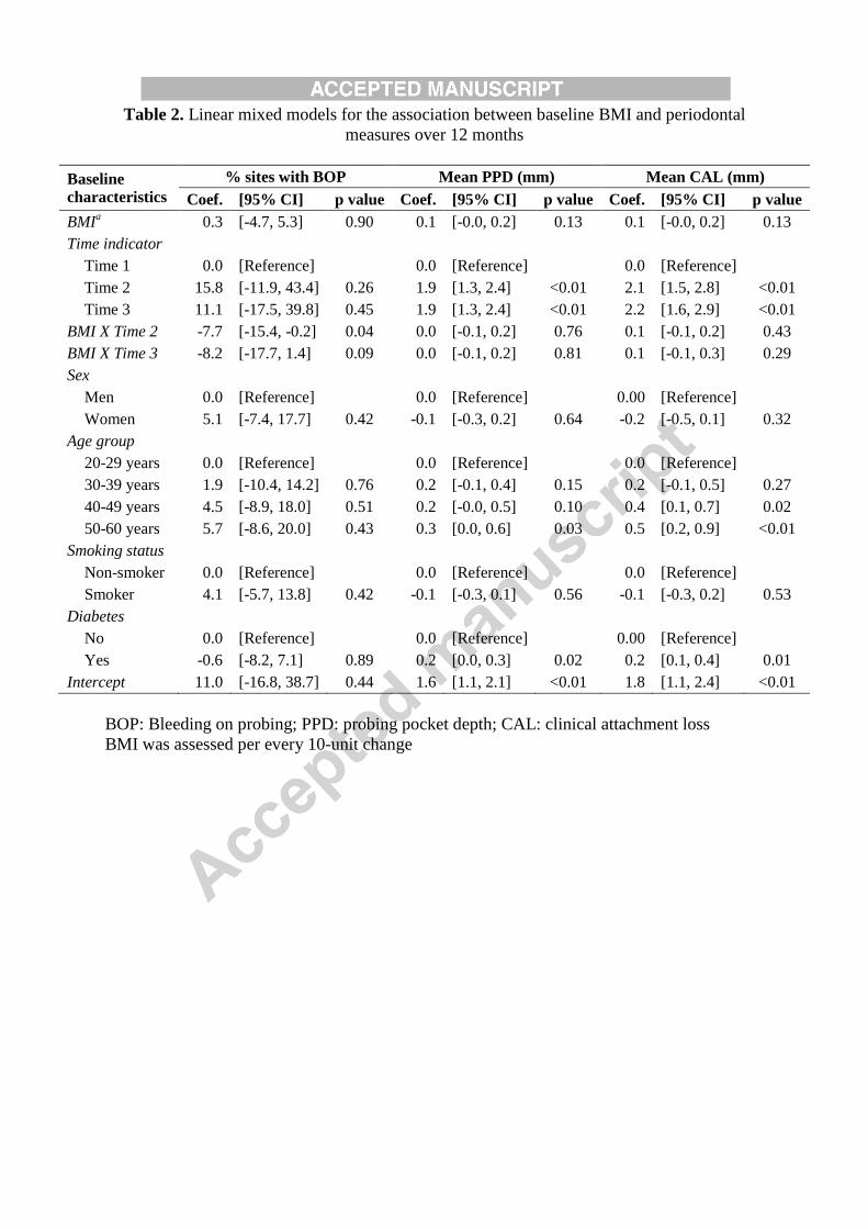
1.88mm at 12 months) after bariatric surgery (Figure 1). BMI was not significantly related to
the proportion of sites with BOP at baseline (Table 2), but it was negatively associated with
the rate of change in the proportion of sites with BOP (p<0.05 for the interaction between
weight and the time indicator). The greater the BMI the higher the proportion of sites with
BOP, particularly at 6 months after surgery. This trend is shown in Figure 2 for different
levels of BMI loss. On the other hand, BMI was not associated with baseline PPD and CAL
or the rates of changes in these periodontal outcomes. Age and diabetes were the only factors
associated with mean PPD and CAL at baseline (Table 2). The interaction term between BMI
and smoking status was not statistically significant (p>0.05).
DISCUSSION
This study shows that weight loss after bariatric surgery was associated with an increase in
the proportion of sites with gingival bleeding, over and above the independent effects of
demographic factors, smoking status and diabetes. Only minor changes in periodontal pocket
depth and attachment loss were noted, which were not associated with weight loss.
Page 10
The present results should be interpreted keeping in mind some study limitations. First, this
study was based on a convenience sample of obese adults, and as such, the results are not
generalizable beyond this group of participants. Second, the inclusion of a control group
(morbidly obese patients that did not undergo bariatric surgery) running in parallel to the
intervention would have strengthened the study design. That said, a control group is
particularly useful to clarify whether improvements in outcome measures (which were not
observed in this study) are due to the intervention being tested. Third, anthropometric
measurements were not purposefully collected for this study but extracted from medical
records. Although this may raise concerns about increased measurement error, there is
evidence that body weights documented in medical records are exchangeable with body
weights recorded in a research setting [25, 26], particularly among women –who represented
88% of our study group– [27, 28]. Fourth, our regression models did not include a control
variable for socioeconomic position, which is strongly related to both obesity [29] and
periodontal disease [30]. Data on socioeconomic circumstances were not routinely collected
as part of patients’ medical records. However, restricting the sample to two neighboring
hospitals serving the same population provided a control for confounders during the study
design as all participants were exposed to similar social and environmental circumstances.
Information on factors such as feeding practices and oral hygiene were not collected either.
This omission does not affect the overall estimate for the association between weight loss and
periodontal disease because the above factors are considered merely intermediates (not
confounders) of the hypothesized association. However, they would have helped clarifying
why weight loss would be associated with increased gingival bleeding.
The present findings suggest that weight loss was related to increased gingival bleeding but
not to periodontal pocketing or attachment loss, offering no support for the hypothesized
inflammatory pathway linking obesity to periodontal disease. If obesity and periodontal
Page 11
disease are causally related, a reduction in body weight should improve periodontal
conditions, at least in terms of signs of gingival inflammation. Improvements in other
common periodontal indicators, such as pocket depth and loss of attachment, may be more
difficult to prove since they may require periodontal treatment after all. Weight loss does
improve inflammation in terms of obesity-related inflammatory markers, specifically
characterized by a decrease in inflammatory markers (CRP, tumor necrosis factor-α,
interleukin-6 and leptin) and an increase in the anti-inflammatory marker, adiponectin [13,
31, 32]. However, it is also worth noticing that the evidence suggests that at least 2 years are
required post-surgery for stabilization of physiological processes and the inflammatory
profile [13]. Thus, it is possible that we were not able to identify changes in other periodontal
measures because of the short follow-up period.
An alternative explanation for these findings has to do with the nutritional and anatomic
changes after bariatric surgery, which may increase the risk of oral complications [17].
Bariatric surgery causes a reduction of gastric capacity and consequently a decrease in food
ingestion [15]. These changes lead to new feeding patterns post-surgery, including frequent
small meals –i.e. grazing– and soft foods [16] that adhere to the tooth surface throughout the
day [20]. This is in addition to a recent report of an increase in Porphyromonas gingivalis 6
months after bariatric surgery [19], suggesting that changes in the amount and microbial
composition of the dental biofilm are a common occurrence among bariatric patients.
Gastroesophageal reflux and vomiting are other common side-effects of bariatric surgery
[33], which may result in erosive lesions of the oral mucosa, including gingival tissues [17].
Finally, bariatric surgery may lead to restriction and/or malabsorption of nutrients, causing
deficiencies in iron, calcium, folate, and vitamins B12, A, D, E and K, some of which are
important to maintain periodontal health [34].
Page 12
This study has some implications for practice and further research. Health professionals
should be aware of possible oral complications of bariatric surgery. Dentists should be part of
the multidisciplinary team taking care of patients undergoing weight loss surgery in order to
monitor their periodontal status throughout the entire process, paying particular attention to
the first 6 months after surgery. From a research perspective, future prospective studies
should explore the long-term effects of weight loss on periodontal conditions and the
interrelationship between weight loss, obesity-related inflammatory markers and periodontal
disease. The role of local factors (such as feeding practices and oral hygiene) in the
relationship between weight loss after surgery and periodontal disease should be assessed.
CONCLUSION
This prospective study shows that weight loss was associated with increased gingival
bleeding, showing a peak at 6 months after bariatric surgery. Periodontal pocketing and
attachment loss remained unchanged during the first 12 months post-surgery.
ACKNOWLEDGMENTS
The authors thank CNPq and FAPESP for financial support and all the volunteers who took
part in the study.
DISCLOSURES
The authors have no commercial associations that might be a conflict of interest in
relation to this article.
Page 13
REFERENCES
1. Ng M, Fleming T, Robinson M, Thomson B, Graetz N, Margono C, Mullany EC,
Biryukov S, Abbafati C, Abera SF et al: Global, regional, and national prevalence
of overweight and obesity in children and adults during 1980-2013: a systematic
analysis for the Global Burden of Disease Study 2013. Lancet (London, England)
2014, 384(9945):766-781.
2. Bray GA: Obesity is a chronic, relapsing neurochemical disease. International
journal of obesity and related metabolic disorders : journal of the International
Association for the Study of Obesity 2004, 28(1):34-38.
3. Galic S, Oakhill JS, Steinberg GR: Adipose tissue as an endocrine organ. Molecular
and cellular endocrinology 2010, 316(2):129-139.
4. Johnson AR, Milner JJ, Makowski L: The inflammation highway: metabolism
accelerates inflammatory traffic in obesity. Immunological reviews 2012,
249(1):218-238.
5. Kopelman P: Health risks associated with overweight and obesity. Obesity reviews
: an official journal of the International Association for the Study of Obesity 2007, 8
Suppl 1:13-17.
6. Brown RE, Kuk JL: Consequences of obesity and weight loss: a devil's advocate
position. Obesity reviews : an official journal of the International Association for the
Study of Obesity 2015, 16(1):77-87.
7. Suvan J, D'Aiuto F, Moles DR, Petrie A, Donos N: Association between
overweight/obesity and periodontitis in adults. A systematic review. Obesity
reviews : an official journal of the International Association for the Study of Obesity
2011, 12(5):e381-404.
8. Keller A, Rohde JF, Raymond K, Heitmann BL: Association between periodontal
disease and overweight and obesity: a systematic review. Journal of
periodontology 2015, 86(6):766-776.
9. Chaffee BW, Weston SJ: Association between chronic periodontal disease and
obesity: a systematic review and meta-analysis. Journal of periodontology 2010,
81(12):1708-1724.
10. Pischon N, Heng N, Bernimoulin JP, Kleber BM, Willich SN, Pischon T: Obesity,
inflammation, and periodontal disease. Journal of dental research 2007, 86(5):400-
409.
11. Sonnenschein SK, Meyle J: Local inflammatory reactions in patients with diabetes
and periodontitis. Periodontology 2000 2015, 69(1):221-254.
12. Nascimento GG, Leite FR, Do LG, Peres KG, Correa MB, Demarco FF, Peres MA: Is
weight gain associated with the incidence of periodontitis? A systematic review
and meta-analysis. Journal of clinical periodontology 2015, 42(6):495-505.
Page 14
13. Forsythe LK, Wallace JM, Livingstone MB: Obesity and inflammation: the effects
of weight loss. Nutrition research reviews 2008, 21(2):117-133.
14. Apovian CM, Aronne LJ: The 2013 American Heart Association/American
College of Cardiology/The Obesity Society Guideline for the Management of
Overweight and Obesity in Adults: What Is New About Diet, Drugs, and Surgery
for Obesity? Circulation 2015, 132(16):1586-1591.
15. Colquitt JL, Pickett K, Loveman E, Frampton GK: Surgery for weight loss in adults.
The Cochrane database of systematic reviews 2014, 8:CD003641.
16. Picot J, Jones J, Colquitt JL, Gospodarevskaya E, Loveman E, Baxter L, Clegg AJ:
The clinical effectiveness and cost-effectiveness of bariatric (weight loss) surgery
for obesity: a systematic review and economic evaluation. Health technology
assessment (Winchester, England) 2009, 13(41):1-190, 215-357, iii-iv.
17. Cummings S, Pratt J: Metabolic and bariatric surgery: Nutrition and dental
considerations. Journal of the American Dental Association (1939) 2015,
146(10):767-772.
18. de Moura-Grec PG, Marsicano JA, Rodrigues LM, de Carvalho Sales-Peres SH:
Alveolar bone loss and periodontal status in a bariatric patient: a brief review
and case report. European journal of gastroenterology & hepatology 2012, 24(1):84-
89.
19. Sales-Peres SH, de Moura-Grec PG, Yamashita JM, Torres EA, Dionisio TJ, Leite
CV, Sales-Peres A, Ceneviva R: Periodontal status and pathogenic bacteria after
gastric bypass: a cohort study. Journal of clinical periodontology 2015, 42(6):530-
536.
20. de Moura-Grec PG, Yamashita JM, Marsicano JA, Ceneviva R, de Souza Leite CV,
de Brito GB, Brienze SL, de Carvalho Sales-Peres SH: Impact of bariatric surgery
on oral health conditions: 6-months cohort study. International dental journal
2014, 64(3):144-149.
21. Vandenbroucke JP, von Elm E, Altman DG, Gotzsche PC, Mulrow CD, Pocock SJ,
Poole C, Schlesselman JJ, Egger M: Strengthening the Reporting of Observational
Studies in Epidemiology (STROBE): explanation and elaboration. Epidemiology
(Cambridge, Mass) 2007, 18(6):805-835.
22. Singer JD, Willett JB: Applied Longitudinal Data Analysis: Modeling Change and
Event Occurrence. Oxford: Oxford University Press; 2003.
23. Twisk JWR: Applied Longitudinal Data Analysis for Epidemiology: A Practical
Guide. New York, NY: Cambridge University Press; 2013.
24. Johnson GK, Guthmiller JM: The impact of cigarette smoking on periodontal
disease and treatment. Periodontology 2000 2007, 44:178-194.
25. Stevens VJ, Wagner EL, Rossner J, Craddick S, Greenlick MR: Validity and
usefulness of medical chart weights in the long-term evaluation of weight loss
programs. Addictive behaviors 1988, 13(2):171-175.
Page 15
26. DiMaria-Ghalili RA: Medical record versus researcher measures of height and
weight. Biological research for nursing 2006, 8(1):15-23.
27. Leo MC, Lindberg NM, Vesco KK, Stevens VJ: Validity of medical chart weights
and heights for obese pregnant women. EGEMS (Washington, DC) 2014,
2(1):1051.
28. Arterburn D, Ichikawa L, Ludman EJ, Operskalski B, Linde JA, Anderson E, Rohde
P, Jeffery RW, Simon GE: Validity of Clinical Body Weight Measures as
Substitutes for Missing Data in a Randomized Trial. Obesity research & clinical
practice 2008, 2(4):277-281.
29. McLaren L: Socioeconomic status and obesity. Epidemiologic reviews 2007, 29:29-
48.
30. Borrell LN, Crawford ND: Socioeconomic position indicators and periodontitis:
examining the evidence. Periodontology 2000 2012, 58(1):69-83.
31. Compher C, Badellino KO: Obesity and inflammation: lessons from bariatric
surgery. JPEN Journal of parenteral and enteral nutrition 2008, 32(6):645-647.
32. Dietrich M, Jialal I: The effect of weight loss on a stable biomarker of
inflammation, C-reactive protein. Nutrition reviews 2005, 63(1):22-28.
33. Tack J, Deloose E: Complications of bariatric surgery: dumping syndrome, reflux
and vitamin deficiencies. Best practice & research Clinical gastroenterology 2014,
28(4):741-749.
34. Genco RJ, Borgnakke WS: Risk factors for periodontal disease. Periodontology
2000 2013, 62(1):59-94.
Page 16
Table 1. BMI at baseline and 6 and 12 months post-surgery, by baseline characteristics
Baseline
characteristics n %
Time 0 (n=110) Time 1 (n=90) Time 2 (n=110)
Mean (SD) Mean (SD) Mean (SD)
Sex
Men 13 11.8 50.5 (8.6) 37.0 (7.9) 32.4 (4.6)
Women 97 88.2 49.7 (8.4) 35.7 (6.0) 32.3 (6.2)
P valuea
0.76 0.52 0.95
Age group
20-29 years 17 15.5 49.4 (6.7) 35.0 (5.8) 31.0 (5.0)
30-39 years 48 43.6 51.4 (10.3) 37.1 (7.7) 32.9 (7.6)
40-49 years 25 22.7 49.2 (6.0) 35.6 (3.9) 32.3 (4.2)
50-60 years 20 18.2 47.1 (6.1) 33.8 (4.4) 32.2 (4.3)
P valuea
0.26 0.31 0.71
Smoking status
Non-smoker 87 79.1 49.9 (8.7) 35.9 (6.3) 32.6 (6.3)
Smoker 23 20.9 49.5 (7.2) 35.6 (6.2) 31.4 (4.8)
P valuea
0.85 0.84 0.39
Diabetes
No 83 75.5 50.8 (9.0) 36.3 (6.8) 32.9 (6.6)
Yes 27 24.6 46.7 (5.0) 34.7 (4.1) 30.8 (3.4)
P valuea 0.03 0.29 0.12
a t-test was used in all comparisons except for age groups where analysis of variance was
used
Page 17
Table 2. Linear mixed models for the association between baseline BMI and periodontal
measures over 12 months
Baseline
characteristics
% sites with BOP Mean PPD (mm) Mean CAL (mm)
Coef. [95% CI] p value Coef. [95% CI] p value Coef. [95% CI] p value
BMIa 0.3 [-4.7, 5.3] 0.90 0.1 [-0.0, 0.2] 0.13 0.1 [-0.0, 0.2] 0.13
Time indicator
Time 1 0.0 [Reference]
0.0 [Reference]
0.0 [Reference]
Time 2 15.8 [-11.9, 43.4] 0.26 1.9 [1.3, 2.4] <0.01 2.1 [1.5, 2.8] <0.01
Time 3 11.1 [-17.5, 39.8] 0.45 1.9 [1.3, 2.4] <0.01 2.2 [1.6, 2.9] <0.01
BMI X Time 2 -7.7 [-15.4, -0.2] 0.04 0.0 [-0.1, 0.2] 0.76 0.1 [-0.1, 0.2] 0.43
BMI X Time 3 -8.2 [-17.7, 1.4] 0.09 0.0 [-0.1, 0.2] 0.81 0.1 [-0.1, 0.3] 0.29
Sex
Men 0.0 [Reference]
0.0 [Reference]
0.00 [Reference]
Women 5.1 [-7.4, 17.7] 0.42 -0.1 [-0.3, 0.2] 0.64 -0.2 [-0.5, 0.1] 0.32
Age group
20-29 years 0.0 [Reference]
0.0 [Reference]
0.0 [Reference]
30-39 years 1.9 [-10.4, 14.2] 0.76 0.2 [-0.1, 0.4] 0.15 0.2 [-0.1, 0.5] 0.27
40-49 years 4.5 [-8.9, 18.0] 0.51 0.2 [-0.0, 0.5] 0.10 0.4 [0.1, 0.7] 0.02
50-60 years 5.7 [-8.6, 20.0] 0.43 0.3 [0.0, 0.6] 0.03 0.5 [0.2, 0.9] <0.01
Smoking status
Non-smoker 0.0 [Reference]
0.0 [Reference]
0.0 [Reference]
Smoker 4.1 [-5.7, 13.8] 0.42 -0.1 [-0.3, 0.1] 0.56 -0.1 [-0.3, 0.2] 0.53
Diabetes
No 0.0 [Reference]
0.0 [Reference]
0.00 [Reference]
Yes -0.6 [-8.2, 7.1] 0.89 0.2 [0.0, 0.3] 0.02 0.2 [0.1, 0.4] 0.01
Intercept 11.0 [-16.8, 38.7] 0.44 1.6 [1.1, 2.1] <0.01 1.8 [1.1, 2.4] <0.01
BOP: Bleeding on probing; PPD: probing pocket depth; CAL: clinical attachment loss
BMI was assessed per every 10-unit change