13
{ Protein synthesis in human body A life blood process
| Date post: | 15-Aug-2015 |
| Category: |
Documents |
| Upload: | ramy-gadalla |
| View: | 18 times |
| Download: | 0 times |
{
Protein synthesis in human body
A life blood process

What’s a chromosome?!!• It’s a structure that’s formed of DNA binding with Histone protein.• It’s consists of two chromatids connected via centromere.• Each chromatid contains a DNA molecule.
Centromere ChromatidDNA
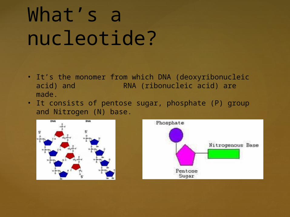
What’s a nucleotide?
• It’s the monomer from which DNA (deoxyribonucleic acid) and RNA (ribonucleic acid) are made.
• It consists of pentose sugar, phosphate (P) group and Nitrogen (N) base.
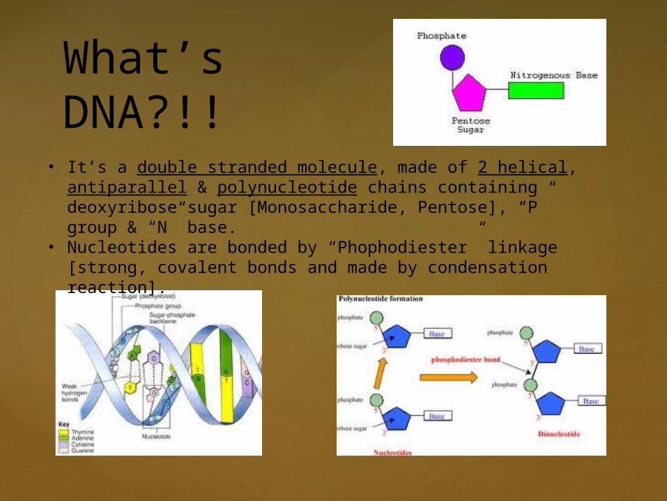
What’s DNA?!!
• It’s a double stranded molecule, made of 2 helical, antiparallel & polynucleotide chains containing deoxyribose sugar [Monosaccharide, Pentose], “P” group & “N” base.
• Nucleotides are bonded by “Phophodiester” linkage [strong, covalent bonds and made by condensation reaction].
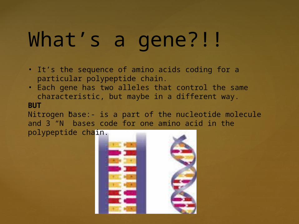
What’s a gene?!!• It’s the sequence of amino acids coding for a particular polypeptide
chain.• Each gene has two alleles that control the same characteristic, but
maybe in a different way.BUTNitrogen base:- is a part of the nucleotide molecule and 3 “N” bases code for one amino acid in the polypeptide chain.

1. Unwinding and unzipping by breaking of “H” bonds.
2. Transcription.
3. DNA rewinds and rezips.
4. Translation [(a) cell protein by free ribosome - (b) secretory protein by attached ribosome].
5. Polypeptide chain reaches Golgi body via shuttle vesicle.
6. Undergoes chemical modifications.
7. Gets packed in secretory (Golgi) vesicles.
8. Golgi vesicles fuses with cell plasma membrane and discharges the secretory protein by “exocytosis”.
Brief Introduction
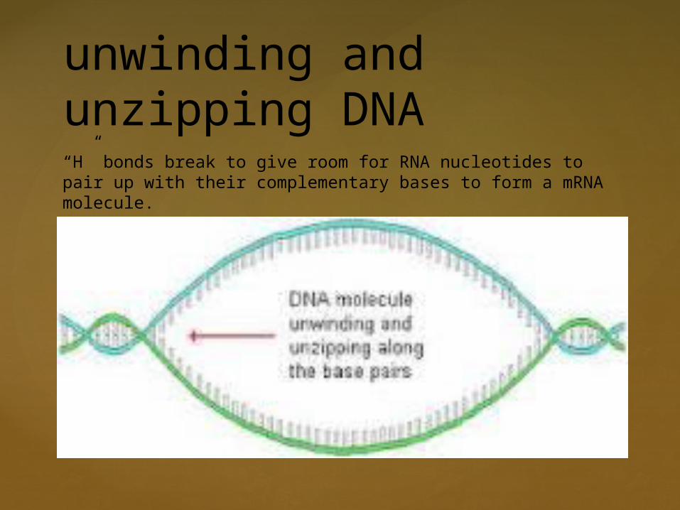
unwinding and unzipping DNA“H” bonds break to give room for RNA nucleotides to pair up with their complementary bases to form a mRNA molecule.
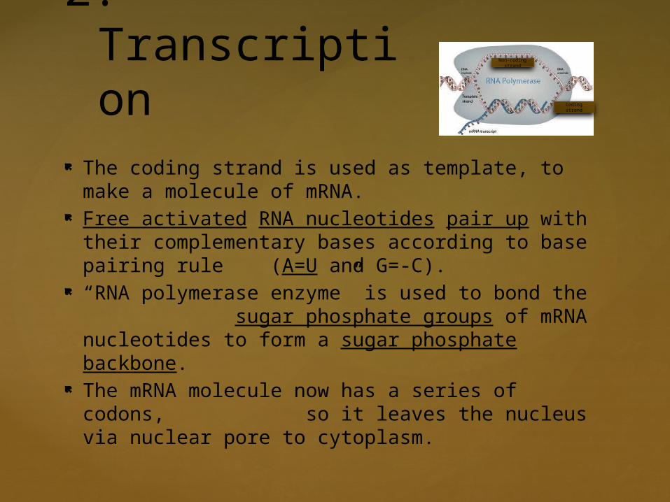
2. Transcription
The coding strand is used as template, to make a molecule of mRNA.
Free activated RNA nucleotides pair up with their complementary bases according to base pairing rule (A=U and G=-C).
“RNA polymerase enzyme” is used to bond the sugar phosphate groups of mRNA nucleotides to form a sugar phosphate backbone.
The mRNA molecule now has a series of codons, so it leaves the nucleus via nuclear pore to cytoplasm.
Non-coding strand
Coding strand
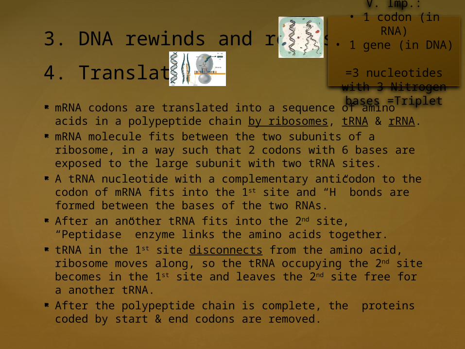
mRNA codons are translated into a sequence of amino acids in a polypeptide chain by ribosomes, tRNA & rRNA.
mRNA molecule fits between the two subunits of a ribosome, in a way such that 2 codons with 6 bases are exposed to the large subunit with two tRNA sites.
A tRNA nucleotide with a complementary anticodon to the codon of mRNA fits into the 1st site and “H” bonds are formed between the bases of the two RNAs.
After an another tRNA fits into the 2nd site, “Peptidase” enzyme links the amino acids together.
tRNA in the 1st site disconnects from the amino acid, ribosome moves along, so the tRNA occupying the 2nd site becomes in the 1st site and leaves the 2nd site free for a another tRNA.
After the polypeptide chain is complete, the proteins coded by start & end codons are removed.
3. DNA rewinds and rezips
4. Translation
V. Imp.:• 1 codon (in RNA)
• 1 gene (in DNA) =3 nucleotides with 3
Nitrogen bases =Triplet
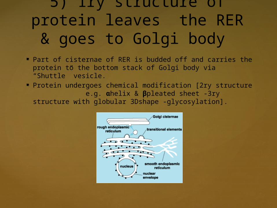
5) 1ry structure of protein leaves the RER & goes to Golgi body
Part of cisternae of RER is budded off and carries the protein to the bottom stack of Golgi body via “Shuttle” vesicle.
Protein undergoes chemical modification [2ry structure e.g. αhelix & βpleated sheet -3ry structure with globular 3Dshape -glycosylation].

7. Secretory vesicles fuse with the cell plasma membrane & expel out the secretory protein by “exocytosis”.
6. Protein gets packed into Golgi vesicles & leave the Golgi body.

Embodiment summary
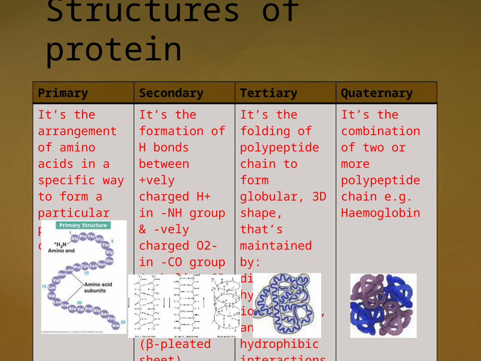
Primary Secondary Tertiary Quaternary
It’s the arrangement of amino acids in a specific way to form a particular polypeptide chain
It’s the formation of H bonds between +vely charged H+ in -NH group & -vely charged O2- in -CO group (α-helix) OR between chains lying side by side (β-pleated sheet)
It’s the folding of polypeptide chain to form globular, 3D shape, that’s maintained by: disulphide, hydrogen, ionic bonds, andhydrophibic interactions
It’s the combination of two or more polypeptide chain e.g. Haemoglobin
Structures of protein


