KSHV Reactivation from Latency Requires Pim-1 and Pim-3 Kinases to Inactivate the Latency-Associated Nuclear Antigen LANA Fang Cheng 1 , Magdalena Weidner-Glunde 2. , Markku Varjosalo 1,3. , Eeva-Marja Rainio 4,5 , Anne Lehtonen 1 , Thomas F. Schulz 2 , Pa ¨ ivi J. Koskinen 4,5 , Jussi Taipale 1,3 , Pa ¨ ivi M. Ojala 1,6 * 1 Genome-Scale Biology Program, Institute of Biomedicine, Biomedicum Helsinki, University of Helsinki, Helsinki, Finland, 2 Institute of Virology, Hannover Medical School, Hannover, Germany, 3 Department of Molecular Medicine, National Public Health Institute (KTL), Helsinki, Finland, 4 Turku Centre for Biotechnology, BioCity, Turku, Finland, 5 Department of Biology, University of Turku, Turku, Finland, 6 The Foundation for the Finnish Cancer Institute, Finland Abstract Host signal-transduction pathways are intimately involved in the switch between latency and productive infection of herpes viruses. As with other herpes viruses, infection by Kaposi’s sarcoma herpesvirus (KSHV) displays these two phases. During latency only few viral genes are expressed, while in the productive infection the virus is reactivated with initiation of extensive viral DNA replication and gene expression, resulting in production of new viral particles. Viral reactivation is crucial for KSHV pathogenesis and contributes to the progression of KS. We have recently identified Pim-1 as a kinase reactivating KSHV upon over-expression. Here we show that another Pim family kinase, Pim-3, also induces viral reactivation. We demonstrate that expression of both Pim-1 and Pim-3 is induced in response to physiological and chemical reactivation in naturally KSHV-infected cells, and we show that they are required for KSHV reactivation under these conditions. Furthermore, our data indicate that Pim-1 and Pim-3 contribute to viral reactivation by phosphorylating the KSHV latency- associated nuclear antigen (LANA) on serine residues 205 and 206. This counteracts the LANA–mediated repression of the KSHV lytic gene transcription. The identification of Pim family kinases as novel cellular regulators of the gammaherpesvirus life cycle facilitates a deeper understanding of virus–host interactions during reactivation and may represent potential novel targets for therapeutic intervention. Citation: Cheng F, Weidner-Glunde M, Varjosalo M, Rainio E-M, Lehtonen A, et al. (2009) KSHV Reactivation from Latency Requires Pim-1 and Pim-3 Kinases to Inactivate the Latency-Associated Nuclear Antigen LANA. PLoS Pathog 5(3): e1000324. doi:10.1371/journal.ppat.1000324 Editor: Klaus Fru ¨ h, Oregon Health & Science University, United States of America Received July 23, 2008; Accepted February 3, 2009; Pulblished March 6, 2009 Copyright: ß 2009 Cheng et al. This is an open-access article distributed under the terms of the Creative Commons Attribution License, which permits unrestricted use, distribution, and reproduction in any medium, provided the original author and source are credited. Funding: This work was supported by grants from the Academy of Finland (P.M.O., P.J.K.), including also the Centre of Excellence in Translational Genome-Scale Biology (P.M.O., J.T.). Additional funds were obtained from Finnish Cancer Foundations (P.M.O., J.T.), Sigrid Juselius Foundation (P.M.O., J.T.), and the European Union (FP6 INCA project LSHC-CT-2005-018704; P.M.O., T.F.S.). Support for M.V. was also provided by Paulo, Finnish Cultural, Emil Aaltonen, Wihuri, and Biomedicum Helsinki Foundations. F.C. and M.V. were supported by the Helsinki Biomedical Graduate School and Helsinki Graduate School of Biotechnology and Molecular Biology, respectively. Competing Interests: The authors have declared that no competing interests exist. * E-mail: [email protected]. These authors contributed equally to this work. Introduction Kaposi’s sarcoma herpesvirus (KSHV) is an etiological agent of three types of malignancies: Kaposi’s sarcoma (KS), multicentric Castleman disease, and primary effusion lymphoma (PEL) [1]. The KSHV genome encodes homologs of cellular proteins, which deregulate signaling pathways, govern cell proliferation, and apoptosis [2]. KSHV infection displays two different phases: latent and lytic. Most tumor cells are latently infected [3,4], and the viral genome remains episomal with only few viral genes expressed [5]. Upon induction of the lytic phase, extensive viral DNA replication is initiated leading to expression of viral lytic genes (reactivation), and production of new infectious viral particles [6]. Viral reactivation is important for spreading of progeny virions between cells and hosts, and is critical for the progression to KS [7,8]. Latency-associated nuclear antigen (LANA) encoded by the open reading frame 73 (ORF73) of the KSHV genome is expressed in all KSHV-infected cells [9,10]. During latency, LANA tethers the viral episomal DNA to the host chromosomes upon cell division [11,12]. Many gammaherpesviruses such as KSHV, Epstein-Barr virus (EBV), rhesus rhadinovirus, herpesvirus saimiri, and murine herpesvirus 68 encode a replication and transcription activator protein (RTA) which plays a critical role in the initiation of viral lytic gene expression (reviewed in [13]). RTA is a transcription factor that activates expression of multiple downstream target genes through the RTA-responsive elements and also autoregulates its own promoter. Expression of RTA is necessary and sufficient to disrupt latency and trigger the complete lytic cascade. LANA also represses expression of RTA and other RTA-responsive lytic genes [14]. Multiple cellular signaling pathways have been shown to regulate KSHV reactivation. Hypoxia and inflammatory cytokines including interferon-c and oncostatin M [15,16,17,18] induce KSHV reactivation, but the underlying mechanisms remain undefined. The primary target of the Notch signaling pathway, RBP-Jk, mediates RTA-dependent activation of KSHV lytic genes [19], and constitutive activation of Notch1 via over-expression of PLoS Pathogens | www.plospathogens.org 1 March 2009 | Volume 5 | Issue 3 | e1000324
Transcript
KSHV Reactivation from Latency Requires Pim-1 andPim-3 Kinases to Inactivate the Latency-AssociatedNuclear Antigen LANAFang Cheng1, Magdalena Weidner-Glunde2., Markku Varjosalo1,3., Eeva-Marja Rainio4,5, Anne
Lehtonen1, Thomas F. Schulz2, Paivi J. Koskinen4,5, Jussi Taipale1,3, Paivi M. Ojala1,6*
1 Genome-Scale Biology Program, Institute of Biomedicine, Biomedicum Helsinki, University of Helsinki, Helsinki, Finland, 2 Institute of Virology, Hannover Medical School,
Hannover, Germany, 3 Department of Molecular Medicine, National Public Health Institute (KTL), Helsinki, Finland, 4 Turku Centre for Biotechnology, BioCity, Turku,
Finland, 5 Department of Biology, University of Turku, Turku, Finland, 6 The Foundation for the Finnish Cancer Institute, Finland
Abstract
Host signal-transduction pathways are intimately involved in the switch between latency and productive infection of herpesviruses. As with other herpes viruses, infection by Kaposi’s sarcoma herpesvirus (KSHV) displays these two phases. Duringlatency only few viral genes are expressed, while in the productive infection the virus is reactivated with initiation ofextensive viral DNA replication and gene expression, resulting in production of new viral particles. Viral reactivation is crucialfor KSHV pathogenesis and contributes to the progression of KS. We have recently identified Pim-1 as a kinase reactivatingKSHV upon over-expression. Here we show that another Pim family kinase, Pim-3, also induces viral reactivation. Wedemonstrate that expression of both Pim-1 and Pim-3 is induced in response to physiological and chemical reactivation innaturally KSHV-infected cells, and we show that they are required for KSHV reactivation under these conditions.Furthermore, our data indicate that Pim-1 and Pim-3 contribute to viral reactivation by phosphorylating the KSHV latency-associated nuclear antigen (LANA) on serine residues 205 and 206. This counteracts the LANA–mediated repression of theKSHV lytic gene transcription. The identification of Pim family kinases as novel cellular regulators of the gammaherpesviruslife cycle facilitates a deeper understanding of virus–host interactions during reactivation and may represent potential noveltargets for therapeutic intervention.
Citation: Cheng F, Weidner-Glunde M, Varjosalo M, Rainio E-M, Lehtonen A, et al. (2009) KSHV Reactivation from Latency Requires Pim-1 and Pim-3 Kinases toInactivate the Latency-Associated Nuclear Antigen LANA. PLoS Pathog 5(3): e1000324. doi:10.1371/journal.ppat.1000324
Editor: Klaus Fruh, Oregon Health & Science University, United States of America
Received July 23, 2008; Accepted February 3, 2009; Pulblished March 6, 2009
Copyright: � 2009 Cheng et al. This is an open-access article distributed under the terms of the Creative Commons Attribution License, which permitsunrestricted use, distribution, and reproduction in any medium, provided the original author and source are credited.
Funding: This work was supported by grants from the Academy of Finland (P.M.O., P.J.K.), including also the Centre of Excellence in Translational Genome-ScaleBiology (P.M.O., J.T.). Additional funds were obtained from Finnish Cancer Foundations (P.M.O., J.T.), Sigrid Juselius Foundation (P.M.O., J.T.), and the EuropeanUnion (FP6 INCA project LSHC-CT-2005-018704; P.M.O., T.F.S.). Support for M.V. was also provided by Paulo, Finnish Cultural, Emil Aaltonen, Wihuri, andBiomedicum Helsinki Foundations. F.C. and M.V. were supported by the Helsinki Biomedical Graduate School and Helsinki Graduate School of Biotechnology andMolecular Biology, respectively.
Competing Interests: The authors have declared that no competing interests exist.
its intracellular domain is sufficient to reactivate KSHV from
latency in PEL cells [20]. Moreover, recent reports further imply
that Ras/Raf/MEK/ERK/Ets-1, JNK and p38 MAPK pathways
mediate TPA-induced KSHV reactivation [21,22,23]. Many
chemicals can also induce reactivation in cell culture. These
include 12-O-tetradecanoyl-phorbol-13-acetate (TPA) and histone
deacetylase inhibitors such as sodium butyrate (NaB) or trichos-
tatin A (reviewed in [24]).
To allow unbiased genome-wide analysis of cross-talk between
cellular kinase pathways and KSHV reactivation, we recently
carried out a gain-of-function screen utilizing a novel expression
library for human protein kinase cDNAs [25]. The screen assessed
the ability of 480 individual, ectopically expressed human kinases
to induce KSHV reactivation, and identified Pim-1 as a novel
kinase involved in KSHV reactivation.
Pim-1 belongs to an oncogenic serine/threonine kinase family
with two other members, Pim-2 and Pim-3, sharing significant
sequence similarities and largely overlapping functions with Pim-1
(reviewed in [26,27]). Pim kinases are overexpressed in various
lymphomas and leukemias [28,29] as well as in prostate cancer
[30]. Recent results have implicated Pim kinases in regulation of
herpesviral oncogenesis. Expression levels of Pim-1 and Pim-2 are
up-regulated upon EBV infection and they in turn enhance the
activity of the viral nuclear antigen EBNA2, suggesting roles in
EBV-induced immortalization and tumorigenesis [31]. In addi-
tion, KSHV infection has been shown to enhance expression of
Pim-2 in CD34+ bone marrow cells [32].
In this report, we have analyzed the requirement of Pim family
kinases in the induction of viral reactivation and investigated the
underlying molecular mechanism. Our results demonstrate that
Pim-1 and -3 are required for KSHV reactivation, and that
phosphorylation of LANA by Pim-1 and -3 counteracts LANA-
dependent repression of viral transcription. These results implicate
Pim-1 and -3 as critical regulators of KSHV reactivation.
Results
Ectopic expression of Pim kinases induces KSHVreactivation
Our recent gain-of function kinome screen identified Pim-1 as a
novel kinase inducing KSHV reactivation upon over-expression
[25]. In the screen, a genome-wide collection of protein-kinase
cDNAs was transfected to Vero cells latently infected with a
recombinant KSHV (rKSHV.219 [33]). The rKSHV.219 is a
double-reporter virus, which expresses the green fluorescent
protein (GFP) from the constitutively active EF-1a-promoter,
and can be induced to express the red fluorescent protein (RFP)
from the KSHV transactivator protein (RTA)-responsive lytic
promoter for polyadenylated nuclear RNA (PAN). PAN is the
most abundant transcript made during the lytic phase [34]. Viral
reactivation was screened by analysis of RFP-positive cells using an
automated high-content microscope. As Pim-1 is not the only
representative of its kinase family, we decided to examine the roles
of other Pim kinase family members in viral reactivation using the
rKSHV.219-Vero cells. The experiments were also performed in
rKSHV.219-infected EA.hy926 endothelial cells, which represent
a biologically relevant KSHV infection model. To this end,
latently infected rKSHV.219-Vero and -EA.hy926 cells were
transiently transfected with V5-tagged Pim-1, -2 or -3 cDNAs or
with an empty vector control. After 48 h, basal reactivation was
induced using a low-titer recombinant baculovirus encoding RTA
(BacK50) as described in Materials and Methods, and RFP
expression was monitored 30 h later (Figure 1A and 1D). The
basal reactivation induces low level of RFP expression (2.5% in
rKSHV Vero, and 1.9% in rKSHV- EA.hy926) in the infected
cells (negative controls), but is necessary for priming the cells for
reactivation by transfected Pim-1 [25]. For positive controls,
maximal viral reactivation was induced by treatment with high-
titer RTA-encoding baculovirus (BacK50) and sodium butyrate
(NaB). Interestingly, all Pim kinases induced RFP expression to at
least 3-fold more as compared to the level obtained by basal
reactivation (negative controls), with Pim-1 inducing the strongest
(4- to 7-fold) increase in both cell lines. Reactivation by Pim
kinases was stronger in Vero than in EA.hy926 cells, which is in
accordance with the ability to obtain higher maximal reactivation
efficiency in these cells by BacK50 and NaB (Figure S1A). Over-
expression of irrelevant kinases CDK7 or LKB1, did not induce
any significant reactivation over the negative control (Figure S1A).
To study the requirement of kinase activity for reactivation, we
used mutant cDNAs of the Pim kinases from the kinome library
[25]. The ATP binding site of the kinases was disrupted by a single
point mutation (K67M in Pim-1, K61M in Pim-2, and K69M in
Pim-3), rendering the kinases inactive. After transfection of the
individual kinase-deficient (KD) mutants, reactivation levels were
close to that of the negative controls (Figure 1A and 1D),
suggesting that kinase activity was required for reactivation.
To analyze whether expression of Pim kinases was sufficient to
trigger the complete lytic replication cascade, we analyzed the
production of progeny virions to the supernatant of the transfected
rKSHV-Vero or -EA.hy926 cells by monitoring GFP expression in
naıve human osteosarcoma (U2OS) cells infected with the
supernatants as described in Materials and Methods (Figure 1B
and 1E). The U2OS cells were chosen as target cells due to their
good susceptibility to KSHV infection, and suitable morphology
for automated microcopy (our unpublished results). All wild-type
(wt) Pim kinases induced a 2- to 5-fold increase in GFP in the
U2OS cells infected with supernatants from the rKSHV-Vero or
-EA.hy926 cell lines when compared to the negative controls,
suggesting that the expression of Pim kinases induced completion
of the full lytic cascade. Importantly, production of infectious
virions was also dependent on kinase activity as expression of the
V5-tagged kinase-deficient Pim kinases, the Pim-1KD, -2KD and
-3KD, in both cell lines led to a clear reduction (maximally 10-
fold) in GFP expression as compared to their wt counterparts.
Over-expression of irrelevant kinases CDK7 or LKB1, did not
induce increase in GFP expression over the negative control in
U2OS target cells (Figure S1B).
To further confirm activation of the full lytic program,
expression of the late lytic protein K8.1 was analyzed in
Author Summary
The switch from latency to productive viral replication(reactivation) is a crucial decision in the viral life cycle, andrecent clinico-epidemiological studies support the impor-tance of lytic replication in the development andprogression of Kaposi’s sarcoma. Hence, cellular signalingpathways operative during viral reactivation could repre-sent potential novel targets for therapeutic intervention.Our work identifies Pim-1 and Pim-3 kinases as essentialkey regulators of the gammaherpesvirus life cycle. Thesekinases target the hallmark of KSHV latency, the LANAprotein, by phosphorylation, which abolishes its ability toact as a transcriptional suppressor of viral lytic replication.This study facilitates a deeper understanding of virus–hostinteractions during reactivation and provides novel op-portunities for pharmacological control and interventionalso in virus-associated cancers.
Figure 1. All three Pim kinases are able to induce KSHV reactivation. (A) rKSHV.219–infected Vero cells (rKSHV-Vero) or (D) EA.hy926 cells(rKSHV-Ea.hy) were transfected with the V5-tagged expression vectors for wt and kinase-deficient (KD) forms of Pim kinases as indicated and infectedwith RTA baculovirus 48 h after transfection (negative control). NaB and infection with RTA baculovirus were used to induce maximal reactivation(positive control). Empty vector transfection was used in the negative and positive controls. 30 h later, the cells were analyzed for RFP expression.96 h after adding RTA or RTA and NaB, the supernatants from KSHV-Vero (B) or KSHV-EA.hy926 (E) cells were collected and used to infect U2OS targetcells. 72 h after infection, U2OS cells were analyzed for GFP expression (a measure of production of infectious virions). Values are means of twoindependent experiments 6SD. 48 h after addition of RTA, cell extracts from rKSHV-Vero (C) or rKSHV-EA.hy926 (F) were analyzed by Westernblotting with antibodies against lytic protein K8.1 and the V5-tag on Pim kinases. Anti-tubulin served as a loading control.doi:10.1371/journal.ppat.1000324.g001
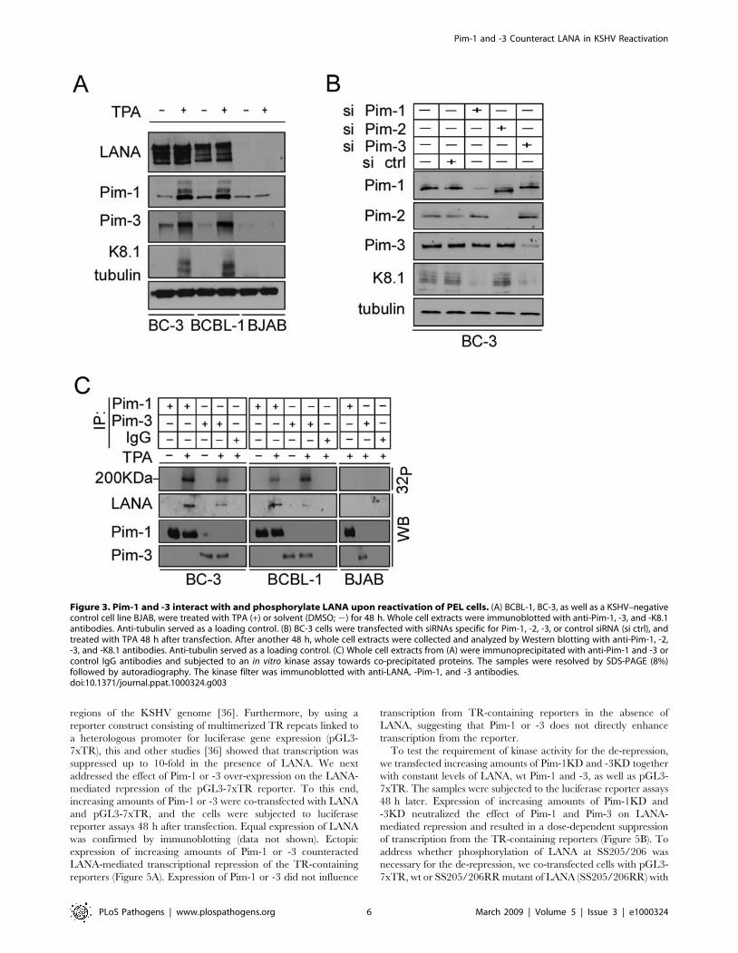
Pim-1, both Pim-1 and -3 phosphorylated GST-N-LANA while no
phosphorylation was observed on GST-C-LANA or with Pim-
1KD or Pim-3KD (Figure 4A). To further map the site of Pim-1
and -3 phosphorylation on the LANA N-terminus, we prepared
truncated versions of the GST-N-LANA as described in the
Materials and methods, and used them as substrates in the in vitro
kinase assay as described above. The results demonstrated that the
critical residues for phosphorylation by Pim-1 and -3 were located
between the amino acids 200 and 340 (Figure 4B). To define the
specific phosphorylation sites on LANA, we prepared a site-
specific LANA phosphomutant identical to the one described
earlier by Bajaj et al [35] where the two critical serines at positions
205 and 206 were mutated into arginines (SS205/206RR). The wt
and two different clones of the SS205/206RR mutant of LANA
(SS205/206RR (1) and SS205/206RR (2)) were then transfected
in the presence or absence of V5-tagged Pim-1 or -3, and cell
extracts were subjected to immunoprecipitation with anti-V5
antibodies. When in vitro kinase assays were performed on the co-
precipitated LANA proteins, we observed that wt LANA was
phosphorylated by both Pim-1 and -3, while the SS205/206RR
mutant of LANA failed to be phosphorylated by neither of them
(Figure 4C). Neither of the LANA proteins was phosphorylated in
the absence of transfected Pim kinases. These data suggest that
serines 205 and 206 on the LANA N-terminus are specifically
phosphorylated by Pim-1 and Pim-3.
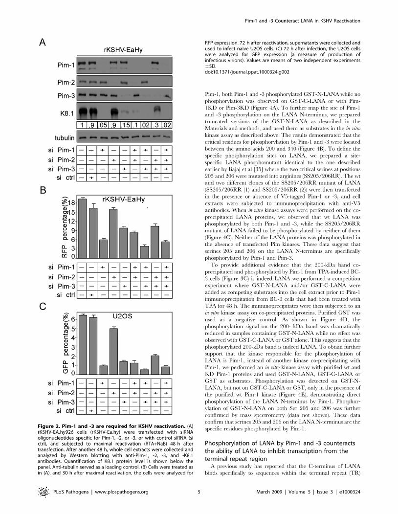
To provide additional evidence that the 200-kDa band co-
precipitated and phosphorylated by Pim-1 from TPA-induced BC-
3 cells (Figure 3C) is indeed LANA we performed a competition
experiment where GST-N-LANA and/or GST-C-LANA were
added as competing substrates into the cell extract prior to Pim-1
immunoprecipitation from BC-3 cells that had been treated with
TPA for 48 h. The immunoprecipitates were then subjected to an
in vitro kinase assay on co-precipitated proteins. Purified GST was
used as a negative control. As shown in Figure 4D, the
phosphorylation signal on the 200- kDa band was dramatically
reduced in samples containing GST-N-LANA while no effect was
observed with GST-C-LANA or GST alone. This suggests that the
phosphorylated 200-kDa band is indeed LANA. To obtain further
support that the kinase responsible for the phosphorylation of
LANA is Pim-1, instead of another kinase co-precipitating with
Pim-1, we performed an in vitro kinase assay with purified wt and
KD Pim-1 proteins and used GST-N-LANA, GST-C-LANA or
GST as substrates. Phosphorylation was detected on GST-N-
LANA, but not on GST-C-LANA or GST, only in the presence of
the purified wt Pim-1 kinase (Figure 4E), demonstrating direct
phosphorylation of the LANA N-terminus by Pim-1. Phosphor-
ylation of GST-N-LANA on both Ser 205 and 206 was further
confirmed by mass spectrometry (data not shown). These data
confirm that serines 205 and 206 on the LANA N-terminus are the
specific residues phosphorylated by Pim-1.
Phosphorylation of LANA by Pim-1 and -3 counteractsthe ability of LANA to inhibit transcription from theterminal repeat region
A previous study has reported that the C-terminus of LANA
binds specifically to sequences within the terminal repeat (TR)
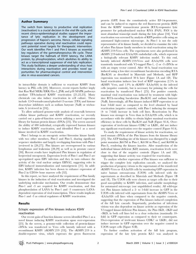
Figure 2. Pim-1 and -3 are required for KSHV reactivation. (A)rKSHV-EA.hy926 cells (rKSHV-Ea.hy) were transfected with siRNAoligonucleotides specific for Pim-1, -2, or -3, or with control siRNA (sictrl), and subjected to maximal reactivation (RTA+NaB) 48 h aftertransfection. After another 48 h, whole cell extracts were collected andanalyzed by Western blotting with anti-Pim-1, -2, -3, and -K8.1antibodies. Quantification of K8.1 protein level is shown below thepanel. Anti-tubulin served as a loading control. (B) Cells were treated asin (A), and 30 h after maximal reactivation, the cells were analyzed for
RFP expression. 72 h after reactivation, supernatants were collected andused to infect naive U2OS cells. (C) 72 h after infection, the U2OS cellswere analyzed for GFP expression (a measure of production ofinfectious virions). Values are means of two independent experiments6SD.doi:10.1371/journal.ppat.1000324.g002
regions of the KSHV genome [36]. Furthermore, by using a
reporter construct consisting of multimerized TR repeats linked to
a heterologous promoter for luciferase gene expression (pGL3-
7xTR), this and other studies [36] showed that transcription was
suppressed up to 10-fold in the presence of LANA. We next
addressed the effect of Pim-1 or -3 over-expression on the LANA-
mediated repression of the pGL3-7xTR reporter. To this end,
increasing amounts of Pim-1 or -3 were co-transfected with LANA
and pGL3-7xTR, and the cells were subjected to luciferase
reporter assays 48 h after transfection. Equal expression of LANA
was confirmed by immunoblotting (data not shown). Ectopic
expression of increasing amounts of Pim-1 or -3 counteracted
LANA-mediated transcriptional repression of the TR-containing
reporters (Figure 5A). Expression of Pim-1 or -3 did not influence
transcription from TR-containing reporters in the absence of
LANA, suggesting that Pim-1 or -3 does not directly enhance
transcription from the reporter.
To test the requirement of kinase activity for the de-repression,
we transfected increasing amounts of Pim-1KD and -3KD together
with constant levels of LANA, wt Pim-1 and -3, as well as pGL3-
7xTR. The samples were subjected to the luciferase reporter assays
48 h later. Expression of increasing amounts of Pim-1KD and
-3KD neutralized the effect of Pim-1 and Pim-3 on LANA-
mediated repression and resulted in a dose-dependent suppression
of transcription from the TR-containing reporters (Figure 5B). To
address whether phosphorylation of LANA at SS205/206 was
necessary for the de-repression, we co-transfected cells with pGL3-
7xTR, wt or SS205/206RR mutant of LANA (SS205/206RR) with
Figure 3. Pim-1 and -3 interact with and phosphorylate LANA upon reactivation of PEL cells. (A) BCBL-1, BC-3, as well as a KSHV–negativecontrol cell line BJAB, were treated with TPA (+) or solvent (DMSO; 2) for 48 h. Whole cell extracts were immunoblotted with anti-Pim-1, -3, and -K8.1antibodies. Anti-tubulin served as a loading control. (B) BC-3 cells were transfected with siRNAs specific for Pim-1, -2, -3, or control siRNA (si ctrl), andtreated with TPA 48 h after transfection. After another 48 h, whole cell extracts were collected and analyzed by Western blotting with anti-Pim-1, -2,-3, and -K8.1 antibodies. Anti-tubulin served as a loading control. (C) Whole cell extracts from (A) were immunoprecipitated with anti-Pim-1 and -3 orcontrol IgG antibodies and subjected to an in vitro kinase assay towards co-precipitated proteins. The samples were resolved by SDS-PAGE (8%)followed by autoradiography. The kinase filter was immunoblotted with anti-LANA, -Pim-1, and -3 antibodies.doi:10.1371/journal.ppat.1000324.g003
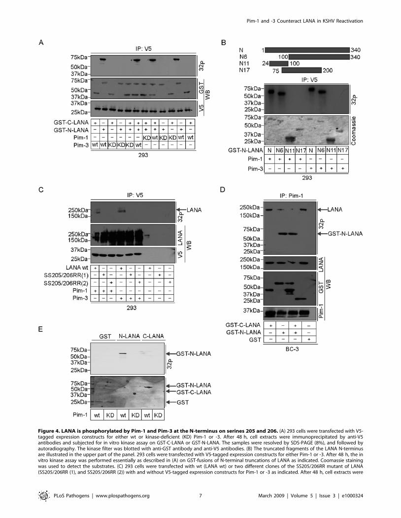
Figure 4. LANA is phosphorylated by Pim-1 and Pim-3 at the N-terminus on serines 205 and 206. (A) 293 cells were transfected with V5-tagged expression constructs for either wt or kinase-deficient (KD) Pim-1 or -3. After 48 h, cell extracts were immunoprecipitated by anti-V5antibodies and subjected for in vitro kinase assay on GST-C-LANA or GST-N-LANA. The samples were resolved by SDS-PAGE (8%), and followed byautoradiography. The kinase filter was blotted with anti-GST antibody and anti-V5 antibodies. (B) The truncated fragments of the LANA N-terminusare illustrated in the upper part of the panel. 293 cells were transfected with V5-tagged expression constructs for either Pim-1 or -3. After 48 h, the invitro kinase assay was performed essentially as described in (A) on GST-fusions of N-terminal truncations of LANA as indicated. Coomassie stainingwas used to detect the substrates. (C) 293 cells were transfected with wt (LANA wt) or two different clones of the SS205/206RR mutant of LANA(SS205/206RR (1), and SS205/206RR (2)) with and without V5-tagged expression constructs for Pim-1 or -3 as indicated. After 48 h, cell extracts were
and without Pim-1 or Pim-3. The samples were subjected to
luciferase reporter assays 48 h after transfection. Expression of the
LANA phosphosite mutant without the Pim kinases induced similar
repression of luciferase activity as the wt LANA, but co-expressed
Pim-1 and -3 were unable to relieve this repression (Figure 5C).
These results indicate that phosphorylation of LANA at serines 205
and 206 by Pim-1 or -3 is needed to counteract LANA-mediated
suppression of transcription from the TR.
Pim-1 and -3 counteract the ability of LANA to inhibitRTA autoactivation
Induction of K8.1 upon Pim-1 and -3 over-expression or in
response to TPA-induced reactivation suggests that RTA responsive
lytic genes are also activated by these kinases in KSHV-infected
cells. Since RTA is not located in the proximity of the TR region in
the KSHV genome we wanted to investigate whether co-expression
of Pim-1 or -3 could affect the ability of LANA to inhibit
transcription from the RTA promoter. Previous studies have
demonstrated that RTA can auto-activate its own viral promoter
[37], and this was shown to be partially repressed by LANA [38].
We therefore addressed the ability of LANA to repress transcription
of luciferase from a RTA-Luc reporter (pGL2-ORF50p) upon co-
expression of RTA, wt Pim-1 or -3. In accordance with Lan et al
[38] expression of LANA led to a partial repression of the RTA-Luc
reporter activity. Moreover, Pim-1 and -3 wt were both able to de-
repress the RTA-Luc activity (Figure 5D). Equal expression of
LANA was confirmed by immunoblotting (data not shown). To
address whether phosphorylation of LANA at SS205/206 was
necessary for the de-repression, we co-transfected cells with pGL2-
ORF50p, wt or SS205/206RR mutant of LANA (SS205/206RR)
with and without Pim-1 or Pim-3. Expression of the LANA
phosphosite mutant without the Pim kinases was able to repress the
RTA-luciferase activity but to a lesser extent as the wt LANA. Yet,
co-expressed Pim-1 and -3 were unable to relieve this repression
(Figure 5D). Taken together, this suggests that expression of Pim-1
and -3 kinases can antagonize the ability of LANA to inhibit
autoactivation of the RTA promoter.
IFN-gamma increases Pim-1 levels and inducesreactivation in PEL cells
To further assess the role of Pim kinases in KSHV reactivation
upon physiological instead of chemical induction, we next analyzed
the expression levels of endogenous Pim-1 in PEL cells upon
cytokine stimulation. To this end, BC-3 cells were first synchronized
as described in Figure 3 and treated with increasing amounts of
interferon-c (IFN-c) for 3 or 16 h. Expression of Pim-1 was
moderately up-regulated in BC-3 cells already 3 h after IFN-ctreatment (Figure 6A). In addition, a slower migrating form of Pim-1
appeared in samples treated with 5–10 IU/ml or 1–10 IU/ml for
3 h or 16 h, respectively, suggesting a post-translational modification
on the protein. Next we examined the effects of IFN-c on the
expression of KSHV lytic genes by quantitative real-time PCR
(qRT-PCR). At 16 h IFN-c treatment with 0.1–10 IU/ml induced
the expression of ORF50/RTA (immediate-early transcript) up to
16.5-fold and ORF57 (delayed-early transcript) up to 19-fold in a
dose-dependent manner (Figure 6D), implying that cytokine
treatment lead to expression of viral lytic genes. We also addressed
if Pim-1 phosphorylated LANA in IFN-c stimulated PEL cells. To
this end, whole cell extracts of untreated, IFN-c- or TPA-treated BC-
3 (16 h stimulation) or control BJAB cells were immunoprecipitated
with anti-Pim-1 antibodies and subjected to in vitro kinase assays on
co-precipitated proteins followed by SDS-PAGE and autoradiogra-
phy. In accordance with the induction of lytic gene expression, an
approximately 200 kDa band co-migrating with LANA in subse-
quent immunoblotting was detected in the autoradiograph of Pim-1
immunoprecipitates after treatment of BC-3 cells with either IFN-cor TPA (Figure 6B, 32P, top panel), further supporting the
physiological role for Pim-1 in KSHV reactivation.
To study the requirement of Pim-1 for the IFN-c induced
KSHV reactivation, we silenced Pim-1 expression by RNA
interference in BC-3 cells as described for Figure 3, and confirmed
the depletion by Western blotting (Figure 6C). 48 h after the cells
were treated with 1 or 5 U/ml of IFN-c for 16 h, and viral
reactivation was analysed by qRT-PCR for the expression of lytic
genes ORF50 and ORF57. Silencing of Pim-1 expression led to a
significant decrease in the expression of lytic genes (66% and 77%
reduction for ORF50, and 38% and 81% reduction for ORF57
with 1 and 5 U/ml IFN-c treatment, respectively) as compared to
cells treated with the control siRNA (Figure 6D and not shown).
Taken together, this data suggests that IFN-c-induced KSHV
reactivation occurs via activation of Pim-1.
Discussion
In this report, we have identified two cellular kinases, Pim-1 and
Pim-3, as critical regulators of KSHV viral reactivation. During
reactivation, Pim-1 and -3 interact with and phosphorylate LANA
in both de novo and naturally KSHV-infected cells. LANA can
repress expression of a subset of lytic genes by specifically binding
to the LANA binding sites 1 and 2 (LBS1 and LBS2) within the
TR region of the KSHV genome [39,40]. Here we demonstrate
that phosphorylation by Pim-1 and -3 counteracts this LANA-
mediated inhibition of transcription from the TR region. Besides
inhibiting the TR-enhancer dependent transcription LANA can
down-modulate the transcriptional activity of the RTA gene
promoter as well as the ability of RTA to autoactivate its own
promoter [38]. Our data shows that co-expression of Pim-1 and -3
with LANA was able to de-repress the LANA-induced partial
inhibition of autoactivation of the RTA-promoter (Figure 5).
However, this Pim kinase-mediated de-repression or the inhibitory
effect of LANA phosphosite mutant was not as dramatic as those
seen in the TR-enhancer dependent transcription assay. This
variation could be due to different mechanisms behind the
repression. While the LANA-induced inhibition of transcription
from the TR region depends on direct binding of LANA to DNA
at LBS1 and LBS2 [39,40], the suppression of RTA autoactivation
occurs through direct binding of LANA to RTA [38]. Taken
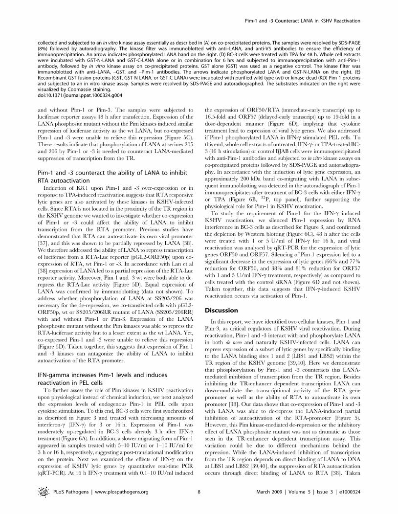
collected and subjected to an in vitro kinase assay essentially as described in (A) on co-precipitated proteins. The samples were resolved by SDS-PAGE(8%) followed by autoradiography. The kinase filter was immunoblotted with anti–LANA, and anti-V5 antibodies to ensure the efficiency ofimmunoprecipitation. An arrow indicates phosphorylated LANA band on the right. (D) BC-3 cells were treated with TPA for 48 h. Whole cell extractswere incubated with GST-N-LANA and GST-C-LANA alone or in combination for 6 hrs and subjected to immunoprecipitation with anti-Pim-1antibody, followed by in vitro kinase assay on co-precipitated proteins. GST alone (GST) was used as a negative control. The kinase filter wasimmunoblotted with anti–LANA, –GST, and –Pim-1 antibodies. The arrows indicate phosphorylated LANA and GST-N-LANA on the right. (E)Recombinant GST-fusion proteins (GST, GST-N-LANA, or GST-C-LANA) were incubated with purified wild-type (wt) or kinase-dead (KD) Pim-1 proteinsand subjected to an in vitro kinase assay. Samples were resolved by SDS-PAGE and autoradiographed. The substrates indicated on the right werevisualized by Coomassie staining.doi:10.1371/journal.ppat.1000324.g004
together, our results suggest that phosphorylation of LANA by Pim
kinases can modulate its ability to interact with the enhancer
elements within the TR region and with the RTA protein itself,
and thereby lead to viral reactivation (Figure 7).
Our KSHV-infected cell models manifest a very low level of
spontaneous lytic replication. However, upon stimulation by low
level of exogenous RTA (using a low-titer BacK50) and co-
expression of Pim kinases, a relatively large population of cells
undergoes reactivation. There are several possible reasons to
explain why priming with exogenous RTA is needed to induce the
Pim kinase-mediated lytic replication. One explanation is that as
the latency program in these infected cell models is very tight, a
threshold level of RTA might be required for the activation of viral
lytic cascade by Pim. Alternatively, Pim kinases may also have an
effect on RTA itself.
Bajaj et al [35] recently demonstrated that Pim-1 phosphory-
lates LANA at either one or both of the serines at positions 205
and 206. We identified Pim-3 as a novel kinase phosphorylating
Figure 5. Phosphorylation of LANA by Pim-1 and -3 antagonizes LANA–mediated transcription inhibition. (A) The reporter plasmidpGL3-7XTR (0.1 mg) was co-transfected with expression vectors for LANA (0.5 mg) with and without increasing concentrations of V5-tagged vectorsfor Pim-1 or -3 (indicated below the graph) into 293 cells. The total transfected DNA concentrations (mg) were kept constant by including an emptycontrol vector. 48 h after transfection, cell extracts were collected for the luciferase assay. (B) Increasing amounts of V5-tagged Pim-1KD or -3KDexpression vectors were co-transfected with constant amount of pGL3-7XTR (0.1 mg), LANA (0.5 mg), and V5-Pim-1 or -3 (0.5 mg each) wt vectors into293 cells. The DNA concentrations (mg) for Pim-1KD and -3KD cDNAs used are indicated below the graph, while the total DNA concentrations (mg)were kept constant by including an empty control vector. Cell extracts were analyzed as in (A). (C) The pGL3-7XTR (0.1 mg), V5-tagged Pim-1 or -3(0.5 mg each) wt vectors were co-transfected with 0.5 mg of wt LANA or SS205/206RR mutant of LANA into 293 cells. At 48 h after transfection, dualluciferase assays were performed with extracts of transfected cells. An empty pcDNA3 was used as a filler plasmid to keep the total DNA constant ineach transfection. Values represent an average of two independent experiments 6SD. (D) The reporter plasmid pGL2-ORF50p (0.1 mg), as well asexpression vectors for RTA (0.5 mg) and wt LANA or SS205/206RR mutant of LANA (0.5 mg) were co-transfected with V5-tagged cDNAs for Pim-1 or -3(indicated below the graph) into 293 cells. The transfected DNA concentrations (mg) were normalized by including an empty control plasmid. 48 hafter transfection, cell extracts were collected for the luciferase assay. Values represent an average of two independent experiments 6SD.doi:10.1371/journal.ppat.1000324.g005
LANA in naturally infected PEL cells, and demonstrated that the
phosphorylation takes place only upon viral reactivation. Our data
confirms Ser 205/206 as the target sites for Pim-1 and
demonstrate that these sites are phosphorylated also by Pim-3.
Pim kinases are involved in partially overlapping survival pathways
and are implicated in a wide variety of human diseases including
cancer, inflammatory disorders, and ischemic diseases [26,41].
Moreover, they have previously been linked to regulation of viral
oncogenesis [31,32,35]. Our data extend these observations by
suggesting a novel link between Pim kinases and KSHV, and
establishes Pim-1 and -3 as key factors in KSHV reactivation.
Our findings provide compelling evidence that Pim-1 and -3, but
not Pim-2, are essential for KSHV reactivation both in de novo
infected endothelial cells and in the naturally infected PEL cells.
Figure 6. IFNc induces reactivation via Pim-1. BC-3 cells were first synchronized to S (Pim-1 si)-phase and treated with increasing amount of IFN-cfor 3 or 16 h. TPA treatment was used as a positive control. (A) Cell extracts were analyzed for the expression of Pim-1 by Western blot, and (D) forexpression of ORF50 and ORF57 lytic transcripts by qRT-PCR. (B) BC-3 and BJAB cells were treated with 1 or 5 U/ml of IFN-c for 16 h, and whole cellextracts were immunoprecipitated with anti-Pim-1 antibodies and subjected to an in vitro kinase assay on co-precipitated proteins. The samples wereresolved by SDS-PAGE (8%) followed by autoradiography. The kinase filter was immunoblotted with anti–LANA and anti–Pim-1 antibodies. Anti-tubulinserved as a loading control. (C) BC-3 cells were transfected with siRNAs specific for Pim-1 (Pim-1 si) or control siRNA (ctrl si) or left untreated (2). 48 hafter transfection, cells were treated for 16 h with IFN-c as described above. Cell extracts were analyzed for the silencing of Pim-1 expression by Westernblot with anti-Pim-1 antibodies, and (D) for expression of ORF50 and ORF57 lytic transcripts by qRT-PCR. Anti-tubulin served as a loading control in C.doi:10.1371/journal.ppat.1000324.g006
Figure 7. A model for Pim-1 and Pim-3 function in KSHV reactivation. In latency, binding of LANA to the LANA binding sites 1 and 2 (LBS 1and LBS2) at the terminal repeat (TR) region represses transcription from the TR region. LANA can also repress transcription from the RTA promoterand inhibit RTA to autoactivate its own promoter. Upon stimulation by cytokines, TPA or NaB, Pim-1 and -3 are up-regulated, and they interact withand phosphorylate LANA. The phosphorylation by Pim-1 and -3 counteracts the LANA–mediated inhibition of transcription from the TR region anddown-modulates autoactivation of the RTA-promoter, leading to viral reactivation.doi:10.1371/journal.ppat.1000324.g007
24. Miller G, ElGuindy A, Countryman J, Ye J, Gradoville L, et al. (2007) Lytic
Cycle Switches of Oncogenic Human Gammaherpesviruses1. Advances in
Cancer Research: Academic Press. pp 81–109.
25. Varjosalo M, Bjorklund M, Cheng F, Syvanen H, Kivioja T, et al. (2008)Application of active and kinase-deficient kinome collection for identification of
that is required for multiple functions of LANA-1. J Virol 79: 13618–13629.47. Viejo-Borbolla A, Ottinger M, Schulz TF (2003) Human Herpesvirus 8: Biology
and Role in the Pathogenesis of Kaposi’s Sarcoma and Other AIDS-relatedMalignancies. Curr Infect Dis Rep 5: 169–175.
48. Fakhari FD, Dittmer DP (2002) Charting latency transcripts in Kaposi’ssarcoma-associated herpesvirus by whole-genome real-time quantitative PCR.