32 International Journal of Scientific Study | January 2014 | Vol 1 | Issue 4 Sonographic Evaluation of Salivary Gland Tumors – A Hospital Based Study Vijai Pratap, S K Jain 1 Associate Pro fessor,Department of Radio-diagnosis, T eerthankar Mahaveer Medical College and Research Centre, Moradabad, India, 1 Professor, Department of Anatomy, Teerthankar Mahaveer Medical College and Research Centre, Moradabad, India Corresponding Author: Dr. S K Jain, Professor, Department of Anatomy, Teerthankar Mahaveer Medical College and Research Centre, Moradabad, India. Phone - +91-9997168754. E-mail: drskjain2005@rediffmail.com imaging modalities are available like Sialography , Computerized T omogr aph y , MRI and Ult ras ound. Ult ras ound is the first imaging modali ty of choice for the saliva ry gland swellings . The advantage of Ultrasound in salivary gland enlargements is that it is comparatively easy to use, non ionizing, & less expensive. In the present study, sonography based differentiation of benign and malignant salivary gland lesions is done. Although benign and malignan t saliv ary g land tumors often have a similar sonographic appearance, several sonographic features, including a heterogeneous echotexture, indistinct margins, regional lymph node enlargement, and absence of distal acoustic enhancement, have been reported to be more frequently associated with malignancy. 5 MATERIAL & METHODS Thi s stu dy is bei ng carr ie d out in the de part men t of Rad io- diagnosis Teerthankar Mahaveer Medical College & its associated INTRODUCTION There are three pairs of salivary glands, namely Parotid, Submandibular and Sublingual. Parotid gland is located in the retro-mandibular fossa, Submandibular under the body of the mandible, & the Sublingual in the sublingual space lying lateral to the genioglossus muscle. Salivary gland tumors are predominantly benign (80%). About 7 0% of the tum ors are l ocate d in the pa roti d gland , 10% in the submandibular gland, and the remainder in the sublingual saliv ary glands. The size of the salivary gland is inversely proportional to the tumor detected being malignant. 1 On histological basis, some benign and malignant salivary gland tumors share overlapping cytological features. 2-4 Identifying the nature of swelling benign or malignant is next to impossible clinically and to rule out any confusion various Original Article Abstract Background: As stated anatomically there are three paired major Sal ivary glands, the P arotid, Submandibular and Sub lingual. Including other diseases salivary glands are also prone for neoplastic involvement though rarely . As a rule smaller the gland the chances of malignancy are more there. Salivary gland tumors mostly emerge in Parotid gland. After clinical evaluation, ultrasound is the most preferred imaging modality to differentiate benign from malignant conditions. The aim of this study is to find out the incidence of salivary gland tumors among various neck pathologies and the most preferred radio-imaging modality to differentiate between benign and neoplastic salivary gland tumors. Methods: This study was carried out in hospital of Teert hankar Mahaveer Medical College & Research Centre, Moradabad, in which all group of patients were included, following total research protocol as admissible in the r esearch and ethical divison of the institute. Ultrasound with frequency of 7 –12 MHz, was employed for the study . Result: Out of 40 patients with lumps in the neck 4 patients (10%) were found to have salivary gland tumors in the neck, out of which 5% were malignant and 5% were benign in nature as demonstrated by ultrasonography . Conclusion: Ultrasonograpy is the most preferred choice of investigation for salivary gland tumors identification, though MRI is the most preferred modality for staging of malignancies of salivary gland tumors. Keywords: Salivary glands, Ultrasonography & Malignancy
Transcript
7/17/2019 kutr
http://slidepdf.com/reader/full/kutr 1/5
32International Journal of Scientific Study | January 2014 | Vol 1 | Issue 4
Sonographic Evaluation of Salivary GlandTumors – A Hospital Based Study
Vijai Pratap, S K Jain1 Associate Professor , Department of Radio-diagnosis, Teerthankar Mahaveer Medical College and
Research Centre, Moradabad, India, 1Professor, Department of Anatomy, Teerthankar Mahaveer
Medical College and Research Centre, Moradabad, India
Corresponding Author: Dr. S K Jain, Professor, Department of Anatomy, Teerthankar Mahaveer
Medical College and Research Centre, Moradabad, India. Phone - +91-9997168754.
imaging modalities are available like Sialography, Computerized Tomography, MRI and Ultrasound. Ultrasound is the firstimaging modality of choice for the salivary gland swellings. Theadvantage of Ultrasound in salivary gland enlargements is that itis comparatively easy to use, non ionizing, & less expensive. Inthe present study, sonography based differentiation of benignand malignant salivary gland lesions is done.
Although benign and malignant salivary gland tumors oftenhave a similar sonographic appearance, several sonographicfeatures, including a heterogeneous echotexture, indistinctmargins, regional lymph node enlargement, and absenceof distal acoustic enhancement, have been reported to bemore frequently associated with malignancy.5
MATERIAL & METHODS
This study is being carried out in the department of Radio-diagnosis Teerthankar Mahaveer Medical College & its associated
INTRODUCTION
There are three pairs of salivary glands, namely Parotid,Submandibular and Sublingual. Parotid gland is located inthe retro-mandibular fossa, Submandibular under the bodyof the mandible, & the Sublingual in the sublingual spacelying lateral to the genioglossus muscle.
Salivary gland tumors are predominantly benign (80%). About 70% of the tumors are located in the parotid gland,10% in the submandibular gland, and the remainder in thesublingual salivary glands. The size of the salivary gland isinversely proportional to the tumor detected being malignant.1
On histological basis, some benign and malignant salivarygland tumors share overlapping cytological features.2-4
Identifying the nature of swelling benign or malignant is nextto impossible clinically and to rule out any confusion various
Original Article
Abstract
Background: As stated anatomically there are three paired major Salivary glands, the Parotid, Submandibular and Sublingual.
Including other diseases salivary glands are also prone for neoplastic involvement though rarely. As a rule smaller the glandthe chances of malignancy are more there. Salivary gland tumors mostly emerge in Parotid gland. After clinical evaluation,
ultrasound is the most preferred imaging modality to differentiate benign from malignant conditions. The aim of this study is to
find out the incidence of salivary gland tumors among various neck pathologies and the most preferred radio-imaging modality
to differentiate between benign and neoplastic salivary gland tumors.
Methods: This study was carried out in hospital of Teerthankar Mahaveer Medical College & Research Centre, Moradabad, in
which all group of patients were included, following total research protocol as admissible in the research and ethical divison of
the institute. Ultrasound with frequency of 7–12 MHz, was employed for the study.
Result: Out of 40 patients with lumps in the neck 4 patients (10%) were found to have salivary gland tumors in the neck, out
of which 5% were malignant and 5% were benign in nature as demonstrated by ultrasonography.
Conclusion: Ultrasonograpy is the most preferred choice of investigation for salivary gland tumors identification, though MRI
is the most preferred modality for staging of malignancies of salivary gland tumors.
Pratap and Jain: Sonographic Evaluation of Salivary Gland Tumors
33 International Journal of Scientific Study | January 2014 | Vol 1 | Issue 4
hospital. Forty patients were evaluated for neck swelling in theneck out of which four patients were identified as havingsalivary gland swelling. A routine protocol was maintained whileevaluating the salivary gland lesions, which included informedconsent (in patients under 18 yrs of age consent was taken fromguardians), presence of female attendant in case of examinationof female subject, Institutional research and ethical committee
approval was taken before hand.
Patients were subjected to routine laboratory investigationsand then taken for Ultrasound examination with the helpof Ultrasound system present in the department.
The ultrasound scanner was placed on the skin immediatelybelow the mandible, allowing the visualization of thesalivary glands.
Out of forty patients in all 22 patients were male and18 females. Age group between 21-30 yrs was found to
be most susceptible for neck swellings. Ultrasound wasperformed using linear-array broadband transducer witha frequency of 7–12 MHz.
Bilateral examination of salivary glands was done as it ismust do protocol.
Sampling MethodConvenience sampling technique was used in this study.
Age and Sex distribution of patients with Neck Masses(Table 1 and Figure 1)
Table 1: Age and Sex distribution of patients withneck masses
Age group (In years) Male Female Total
0-10 2 1 3
11-20 1 3 4
21-30 4 8 12
31-40 3 3 6
41-50 5 2 7
51-60 3 1 4
61-70 3 - 3
71-80 1 - 1
Total 22 18 40
2
1
4
3
5
3 3
11
3
8
3
2
1
0 00
1
2
3
4
5
6
7
89
0-10 11-20 21-30 31-40 41-50 51-60 61-70 71-80
Male
Female
Figure 1: Age and sex distribution of patients with neck
masses
RESULTS
Table 2: Distribution of neck Massses according tothe nature of the lesion
Nature of the lesion No. of cases Percentage of total cases
Inflammatory
Abscess
Adenopathy
1
6
17.5%
2.5%
15%
Developmental
Branchial Cyst
Ranula
Lymphangioma
1
1
1
7.5%
2.5%
2.5%
2.5%
Thyroid Masses
Benign
Malignant
8
4
30%
20%
10%
Mesenchymal
Lipoma
Sarcoma
2
2
10%
5%
5%
Neural
Schwannoma
Neurofibroma
1
1
5%
2.5%
2.5%
Vascular
HemangiomaCarotid body tumor 11
5%
2.5%2.5%
Bone
Osteoma
Metastasis
1
1
5%
2.5%
2.5%
Lymphnode Masses
(non inflammatory)
Lymphoma
Metastasis
1
3
10%
2.5%
7.5%
Salivary Gland Masses
Benign
Malignant
2
2
10%
5%
5%
Total 40 100%
17.50%
7.50%
30%
10%
5% 5% 5%
10% 10%
0.00%
5.00%
10.00%
15.00%
20.00%
25.00%
30.00%
35.00%
Series 1
Figure 2: Distribution of various neck pathologies
Figure 3: Showing occurrence of salivary gland tumors in
range of 10%
7/17/2019 kutr
http://slidepdf.com/reader/full/kutr 3/5
Pratap and Jain: Sonographic Evaluation of Salivary Gland Tumors
34International Journal of Scientific Study | January 2014 | Vol 1 | Issue 4
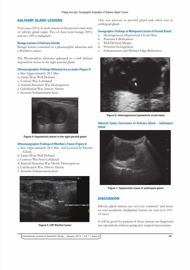
SALIVARY GLAND LESIONS
Four cases (10%) of neck, masses in the present study wereof salivary gland origin. Two of these were benign (50%)and two (50%) malignant.
Benign Lesions of Salivary Glands
Benign lesions consisted of a pleomorphic adenoma anda Warthin’s tumor.
The Pleomorphic adenoma appeared as a well definedhypoechoic lesion in the right parotid gland.
Ultrasonographic Findings Obtained are as under (Figure 4)a. Size Approximately 28.1 Mm.b. Limits Were Well Definedc. Contour Was Lobulatedd. Internal Structure Was Homogenouse. Calcification Was Almost Absentf. Acoustic Enhancement Seen.
Figure 4: Hypoechoic lesion in the right parotid gland
Ultrasonographic Findings of Warthin’s Tumor (Figure 5)a. Size Approximately 28.9 Mm. And Located In Parotid
Glandb. Limits Were Well Definedc. Contour Was Non-Lobulatedd. Internal Structure Was Mostly Heterogenouse. Calcification Was Almost Absentf. Acoustic Enhancement Seen
Figure 5: H/P Warthin tumor
One was present in parotid gland and other was insublingual gland.
Sonographic Findings of Malignant Lesion of Parotid Glanda. Heterogeneous Hypoechoic Ovoid Massb. Punctate Calcificationsc. Well-Defined Margin
d. Posterior Echogenicitye. Enhancement and Distinct Edge Refraction.
Figure 6: Heterogeneous hypoechoic ovoid mass
Adenoid Cystic Carcinoma of Salivary Gland – SublingualGland
Figure 7: Hypoechoic mass of sublingual gland
DISCUSSION
Salivary gland tumors are not very common6 and moreso over neoplastic (malignant) lesions are seen in 5–10%of cases.7
It will be good for patients if these tumors are diagnosedpre-operatively without going into surgical intervention.
7/17/2019 kutr
http://slidepdf.com/reader/full/kutr 4/5
Pratap and Jain: Sonographic Evaluation of Salivary Gland Tumors
35 International Journal of Scientific Study | January 2014 | Vol 1 | Issue 4
Therefore, many clinical researchers have tried to evaluatethe ability of sonography to differentiate benign andmalignant tumors.
Sonography is a powerful tool for characterizing salivarygland tumors. Different imaging techniques are valuable inassessing salivary gland disease, out of which the choice of
modality depends on local protocol, clinical features and,importantly, the site of suspected pathology. Technicaladvances, in many imaging centers have made ultrasoundnowadays the investigation of choice for major salivary glanddisease. It allows a quick, cheap and thorough assessment without the use of ionizing radiation. Ultrasound is able tosimultaneously evaluate gland parenchyma and large ductsas well as demonstrate duct dilatation.
Tumors of the salivary glands are not common, representingabout 3% of all head and neck tumors. Histopathology ofsalivary gland tumors is very varied, with a large number of
both benign and malignant tumors. Out of this Pleomor-phic adenomas are the most common, representing 70-80%of all salivary gland tumors1 most frequently located in theparotid gland. Cytological examination often faces dif ficultyin differentiating adenoid cystic carcinoma from Pleomorphicadenoma.7,8 It is seen histopathologically both lesions containmyxoid material.9-11 A number of ultrasonographic features areconsidered typical for pleomorphic adenomas: sharp borders,lobulations of the contour, homogeneous structure, poor vascularization, acoustic enhancement.12,13 which well correlates with the ultrasonographic pictures of our present study.
Warthin’s tumour is the second common salivary neoplasm,typically occurring in older male patients, with a propensityfor smokers. It arises from parotid intraglandular lymphoidtissue, typically in the tail, and is multiple or bilateral inapproximately 15% cases.
Ultrasound shows an ovoid hypoechoic mass. In our studyit was present unilaterally and patient didn’t give historyof smoking. Sublingual gland tumors are rare and accountfor only 0.4–2.6 of all salivary gland tumors.14,15
However, most of the recorded literature assertthat Pleomorphic adenoma is more common than
Adenolymphoma.7,16 Only Schick et al17 recorded an equalnumber of cases of Pleomorphic adenoma and Warthin’stumour (7:7), which is also seen in our study.
The majority of sublingual gland tumors are malignant18
and ACC is the most common. As can be seen in our studyout of two malignant lesions one is of Adenoid CysticCarcinoma, which very well correlates with the study of Anderson LJ et al.19
CONCLUSION
Before going into any type of radiological investigationhistological grading of salivary gland tumor is a preliminarystep in clinical setting, though not alone.
A variety of radio-imaging modalities may be employed in
salivary gland imaging in which Ultrasound has emerged asthe technique of choice for major salivary gland disease andforms a useful aid for FNA/biopsy. MRI is of particular value for staging salivary gland malignancy.
As a simple guide If ultrasound is able to differentiate asa benign pathology there is no need to go further imaging.
Through our experience we now know that sonographicfeatures are most accurate but we should keep othermodalities in our mind for improving the diagnostic accuracy.
REFERENCES
1. Gritzmann N, Hollerweger A, Macheiner P, Rettenbacher T, Hubner E.
Sonography of the salivary glands. European Radiology 2003; 13:364-375.
2. Elagoz S, Gulluoglu M, Yilmazbayhan D, Ozer H, Arslan I. The value of
fine-needle aspiration cytology in salivary gland lesions, 1994-2004. ORL J