32
La patología del cáncer de mama: novedades Federico Rojo Fundación Jiménez Díaz

La patología del cáncer de mama: novedades
Federico Rojo
Fundación Jiménez Díaz

Lymphocytic infiltration in breast cancer increases postoperative life

Lymphocytic-predominant phenotype is in 20-28% of breast cancers and correlates with outcome

Denkert, C et al. J Clin Oncol 2010
Tumor-associated lymphocytes as a predictor of response on neoadjuvant anthracyclin/taxane CT
pCR 42% vs 3% iTu-Ly p=0.012 (OR 1.38, 95%CI 1.08 to 1.78)
GeparDuo, n=218 GeparTrio, n=840

S Hendry, R Salgado, T Gevaert, PA Russell, T John, B Thapa, M Christie, K van de Vijver, MV Estrada, PI Gonzalez-Ericsson, M Sanders, B Solomon, C Solinas, G Van den Eynden, Y Allory, M Preusser, J Hainfellner, G Pruneri, A Vingiani, S
Demaria, F Symmans, P Nuciforo, L Comerma, EA Thompson, S Lakhani, SR Kim, S Schnitt, C Colpaert, C Sotiriou, SJ Scherer, M Ignatiadis, S Badve, RH Pierce, G Viale, N Sirtaine, F Penault-Llorca, T Sugie, S Fineberg, S Paik, A Srinivasan, A Richardson, Y Wang, E Chmielik, J Brock, DB Johnson, J Balko, S Wienert, V Bossuyt, S Michiels, N Ternes, N Burchardi, SJ Luen, P Savas, F Klauschen, PH Watson, B Nelson, C Criscitiello, S O’Toole, D Larsimont, R de Wind, G Curigliano, F Andre,
M Lacroix-Triki, M van de Vijver, F Rojo, G Floris, S Bedri, J Sparano, D Rimm, T Nielsen, Z Kos, S Hewitt, B Singh, G Farshid, S Loibl, K Allison, N Tung, S Adams, K Willard-Gallo, H Horlings, L Gandhi, A Moreira, F Hirsch, M Dieci, M
Urbanowicz, I Brcic, K Korski, F Gaire, H Koeppen, A Lo, J Giltnane, M Rebelatto, K Steele, J Zha, K Emancipator, J Juco, C Denkert, J Reis-Filho, S Loi and S Fox

Standardized methodology for pathological TILs evaluation
Recommendations for assessing TILs in melanoma, NSCLC, glioma, GU, endometrial, ovarian, GI and HN tumors

S Hendry, R Salgado, T Gevaert, PA Russell, T John, B Thapa, M Christie, K van de Vijver, MV Estrada, PI Gonzalez-Ericsson, M Sanders, B Solomon, C Solinas, G Van den Eynden, Y Allory, M Preusser, J Hainfellner, G Pruneri, A Vingiani, S
Demaria, F Symmans, P Nuciforo, L Comerma, EA Thompson, S Lakhani, SR Kim, S Schnitt, C Colpaert, C Sotiriou, SJ Scherer, M Ignatiadis, S Badve, RH Pierce, G Viale, N Sirtaine, F Penault-Llorca, T Sugie, S Fineberg, S Paik, A Srinivasan, A Richardson, Y Wang, E Chmielik, J Brock, DB Johnson, J Balko, S Wienert, V Bossuyt, S Michiels, N Ternes, N Burchardi, SJ Luen, P Savas, F Klauschen, PH Watson, B Nelson, C Criscitiello, S O’Toole, D Larsimont, R de Wind, G Curigliano, F Andre,
M Lacroix-Triki, M van de Vijver, F Rojo, G Floris, S Bedri, J Sparano, D Rimm, T Nielsen, Z Kos, S Hewitt, B Singh, G Farshid, S Loibl, K Allison, N Tung, S Adams, K Willard-Gallo, H Horlings, L Gandhi, A Moreira, F Hirsch, M Dieci, M
Urbanowicz, I Brcic, K Korski, F Gaire, H Koeppen, A Lo, J Giltnane, M Rebelatto, K Steele, J Zha, K Emancipator, J Juco, C Denkert, J Reis-Filho, S Loi and S Fox

Standardized methodology for pathological TILs evaluation
Recommendations for assessing tumor-infiltrating lymphocytes (TILs) in breast cancer

Standardized methodology for pathological TILs evaluation
Recommendations for assessing tumor-infiltrating lymphocytes (TILs) in breast cancer

Standardized methodology for pathological TILs evaluation
Recommendations for assessing tumor-infiltrating lymphocytes (TILs) in breast cancer
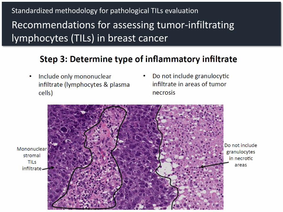
Standardized methodology for pathological TILs evaluation
Recommendations for assessing tumor-infiltrating lymphocytes (TILs) in breast cancer

Denkert, C et al. Mod Pathol 2016
Standardized methodology for pathological TILs evaluation
Ring studies for standardized evaluation of TILs in breast cancer

5. ¿Cuál debe de ser el planteamiento futuro en el diagnóstico morfológico?
Denkert, C et al. SABCS 2016
Metaanalysis of 3,771 patients from 6 neoadjuvant trials
TILs as predictive and prognostic biomarker in breast cancer

5. ¿Cuál debe de ser el planteamiento futuro en el diagnóstico morfológico?
Denkert, C et al. SABCS 2016
Metaanalysis of 3,771 patients from 6 neoadjuvant trials
TILs as predictive and prognostic biomarker in breast cancer

Immune-inflamed phenotype
TILs predict response to pembrolizimab in mTNBC

TILs in residual disease after neoadjuvant therapy

7
Figure 5: Distribution of the categorized %TILs measures for both the stroma and the tumor-cell compartments.
Distribution of TILs in stroma and tumor cell compartments

2. Identify the area for TIL evaluation
Include TILs immediatelyadjacent to the tumor border
Area for TILs evaluation

Exclude TILs closely related to remaining foci of carcinoma in situ or normal lobules within theresidual tumor bed.
3. Areas of the tumor bed to be excludedfrom TIL evaluation
TILs associated with normal lobules
TILs associated with carcinoma in situ
Area for TILs evaluation

Exclude TILs associated withhyalinized and/or edematousvascularized stroma infiltrated bysheets of foamy or hemosiderin-loaded histiocytes.
3. Areas of the tumor bed to be excludedfrom TIL evaluation
EXCEPTION: Assess TILs whentumor cells are embedded withinaggeragates of histiocytes.
Area for TILs evaluation

Exclude TILs associated with necrotic areas.
Exclude TILs in tumor zones with crush artefacts.
3. Areas of the tumor bed to be excludedfrom TIL evaluation
Area for TILs evaluation

If scattered tumor foci are separated widely from each other, then the area around each foci should be considered for TIL-assessment and then averaged.
Estimate average TIL from the different microscopic fields (200-400x magnification)
5. Assess TILs % as continuous parameter
40%
15%5%
5%
Area for TILs evaluation

Estimate average TIL from the different microscopic fields (200-400x magnification)
Example 1
5. Assess TILs % as continuous parameter
Case with low-TILs, no heterogeneity across fields
(<1% in all fields)
TILs assessment

Example 2
Estimate average TIL from the different microscopic fields (200-400x magnification)
5. Assess TILs % as continuous parameter
60%
60%
65%
70%
Case with high TILs (>60%), with slight heterogeneity across fields
TILs assessment

TILs after neoadjuvant treatment predict poor pCR rate in breast cancer
Hamy, AS et al. Ann Oncol 2017
N=175

Residual Cancer Burden assessment after neoadjuvant therapy

Triple-negative (n = 219) HR-positive/HER2-negative (n = 501) HER2-positive (n = 203)
Residual Cancer Burden assessment after neoadjuvant therapy

Comprhensive genomic profiling in circulating tumor DNA

At least 1 genomic alteration in 78% of samples
89% of concordance in detected mutations with tissue
Comprhensive genomic profiling in circulating tumor DNA

Multiple concurrent ESR1 mutations in 40% of cases
Comprhensive genomic profiling in circulating tumor DNA

Comprhensive genomic profiling in circulating tumor DNA

La patología del cáncer de mama: novedades
Federico Rojo
Fundación Jiménez Díaz


