Locate and note the following in Fig 1 (esp. on the Rt side).• splenium of the corpus callosum located dorsally in this figure.• fornix – major hippocampal efferent that arches dorsally along the
lateral ventricle. • alveus – a “capsule” of axons covering the hippocampus and is
continuous with the fornix.(– cornu Ammonis - CA regions of the hippocampus.)
• body of the lateral ventricle – located lateral to the fornix and corpus callosum.
• temporal (inferior) horn of the lateral ventricle – located lateral to the alveus.
• tail of the caudate nucleus – found in the lateral wall of the lateral ventricle
• stria terminalis – small myelinated tract just medial to the tail of the caudate; it connects the amygdala in the temporal lobe with the hypothalamus.
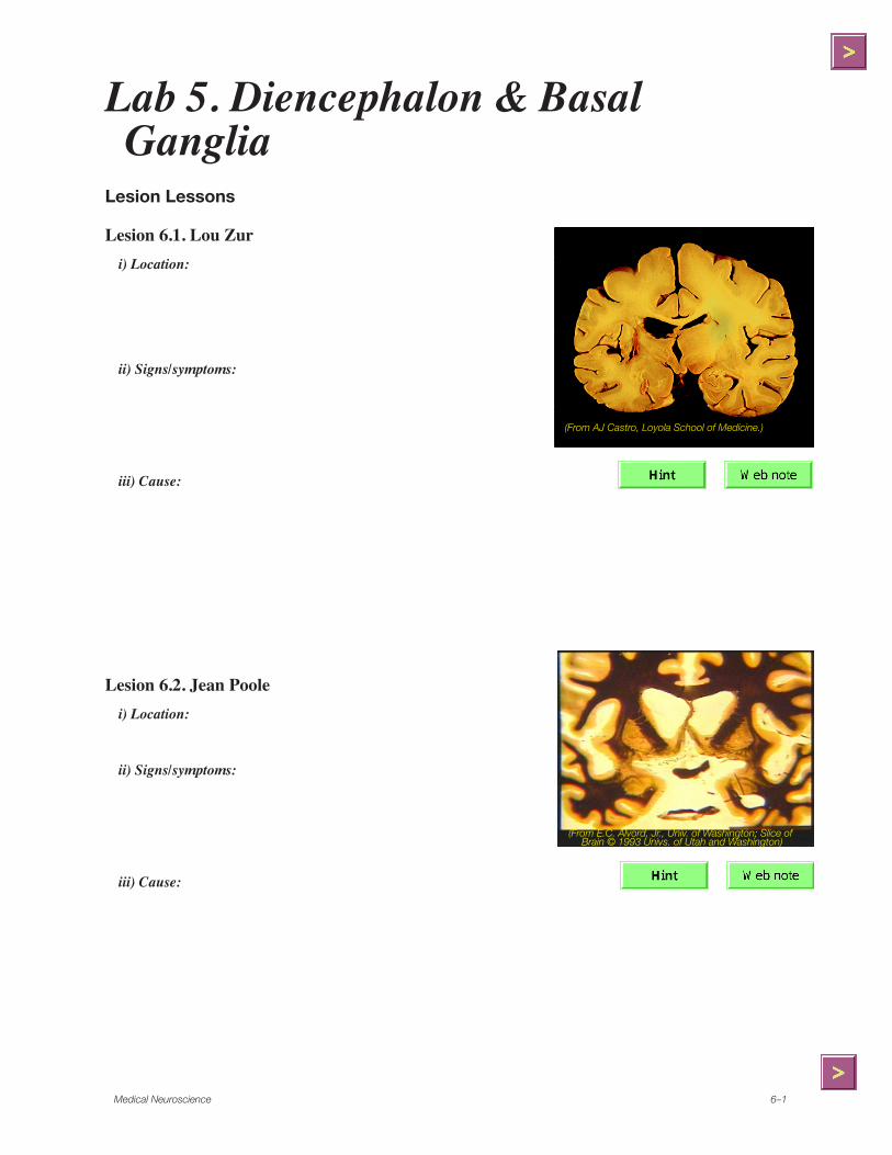
Fig. � . Midbrain-diencephalic transition. (Slide � 3. Univ. of Kansas School of Medicine)
Tail of the caudate and stria terminalis
These two structures follow the shape of the ventricles from the temporal lobe to the frontal lobe.
Also label the following on Figure 1.
• decussation of the sup cerebellar peduncles
• brachium of the sup colliculus
• brachium of the inf colliculus
• periaqueductal gray • superior colliculus • medial lemniscus
• spinothalamic tract • central tegmental tract • MLF
A five year-old child, Billy Club, develops headache and begins to frequently fall. His doctor orders a CT brain scan and subsequently refers the child to Loyola because of a pineal gland tumor.
1. You note the child has trouble looking upward. What structure near the tumor could cause this problem?
2. CSF pathways are obstructed by this tumor on the CT scan. Where would this most likely occur?
3. Which ventricles would abnormally enlarge?
4. What structures could be stretched or compressed by the enlarged ventricles, and cause falling and a gait disorder?
Loyola University6–�
Caudal Thalamus
Note and label the following in Figure 2.• pulvinar – is still present dorsally.• ventral tier thalamic nuclei – are appearing ventrolaterally.
– at this level, these include: – ventral posterior lateral nucleus (VPL). – the caudal end of the ventral posterior medial nucleus (VPM).
• medial lemniscus and spinothalamic tract – at this level are seen as they enter the thalamus, along with the cerebello-thalamic fibers.
Fig. �. Caudal thalamus. (Slide ��. Univ. of Kan-sas School of Medicine)
Also label the following structures on Figure 2.• basilar pons • crus cerebri • substantia nigra
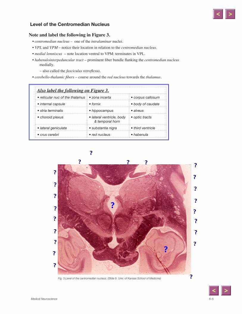
Note and label the following in Figure 3.• centromedian nucleus – one of the intralaminar nuclei. • VPL and VPM – notice their location in relation to the centromedian nucleus.• medial lemniscus – note location ventral to VPM; terminates in VPL.• habenulointerpeduncular tract – prominent fiber bundle flanking the centromedian nucleus
medially.– also called the fasciculus retroflexus.
• cerebello-thalamic fibers – course around the red nucleus towards the thalamus.
Fig. 3.Level of the centromedian nucleus. (Slide 9. Univ. of Kansas School of Medicine)
Also label the following on Figure 3.• reticular nuc of the thalamus • zona incerta • corpus callosum
• internal capsule • fornix • body of caudate
• stria terminalis • hippocampus • alveus
• choroid plexus • lateral ventricle, body & temporal horn
• optic tracts
• lateral geniculate • substantia nigra • third ventricle
• crus cerebri • red nucleus • habenula
x
Polygonal Line
x
Polygonal Line
x
Line
x
Polygonal Line
x
Polygonal Line
x
Polygonal Line
x
Polygonal Line
x
Polygonal Line
x
Line
x
Polygonal Line
x
Line
x
Polygonal Line
x
Polygonal Line
x
Polygonal Line
x
Polygonal Line
x
Polygonal Line
x
Line
x
Line
x
Line
x
Polygonal Line
x
Polygonal Line
x
Polygonal Line
Loyola University6–6
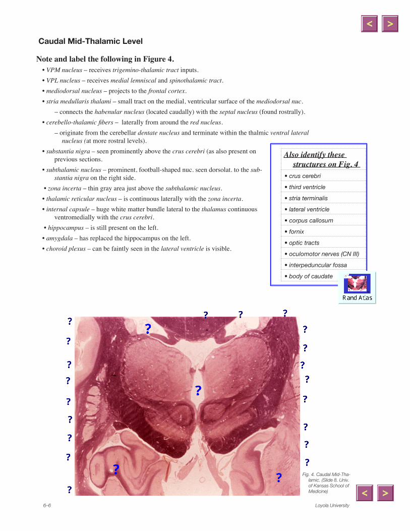
Caudal Mid-Thalamic Level
Note and label the following in Figure 4. • VPM nucleus – receives trigemino-thalamic tract inputs.• VPL nucleus – receives medial lemniscal and spinothalamic tract.• mediodorsal nucleus – projects to the frontal cortex. • stria medullaris thalami – small tract on the medial, ventricular surface of the mediodorsal nuc.
– connects the habenular nucleus (located caudally) with the septal nucleus (found rostrally).• cerebello-thalamic fibers – laterally from around the red nucleus.
– originate from the cerebellar dentate nucleus and terminate within the thalmic ventral lateral nucleus (at more rostral levels).
• substantia nigra – seen prominently above the crus cerebri (as also present on previous sections.
• subthalamic nucleus – prominent, football-shaped nuc. seen dorsolat. to the sub-stantia nigra on the right side.
• zona incerta – thin gray area just above the subthalamic nucleus.• thalamic reticular nucleus – is continuous laterally with the zona incerta.• internal capsule – huge white matter bundle lateral to the thalamus continuous
ventromedially with the crus cerebri. • hippocampus – is still present on the left. • amygdala – has replaced the hippocampus on the left. • choroid plexus – can be faintly seen in the lateral ventricle is visible.
Fig. �. Caudal Mid-Tha-lamic. (Slide 8. Univ. of Kansas School of Medicine)
Also identify these structures on Fig. 4
• crus cerebri
• third ventricle
• stria terminalis
• lateral ventricle
• corpus callosum
• fornix
• optic tracts
• oculomotor nerves (CN III)
• interpeduncular fossa
• body of caudate
x
Polygonal Line
x
Polygonal Line
x
Line
x
Polygonal Line
x
Polygonal Line
x
Polygonal Line
x
Line
x
Polygonal Line
x
Line
x
Polygonal Line
x
Polygonal Line
x
Line
x
Line
x
Polygonal Line
x
Line
x
Polygonal Line
x
Polygonal Line
x
Polygonal Line
x
Polygonal Line
x
Polygonal Line
Medical Neuroscience 6–�
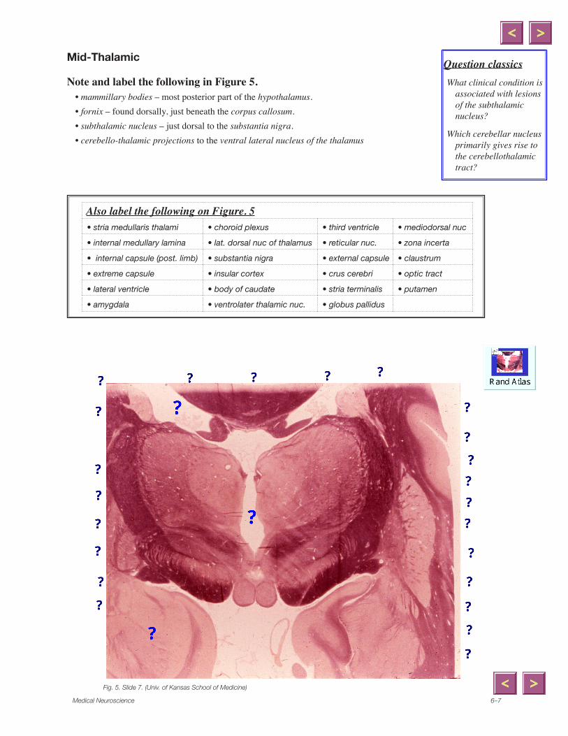
Mid-Thalamic
Note and label the following in Figure 5. • mammillary bodies – most posterior part of the hypothalamus. • fornix – found dorsally, just beneath the corpus callosum.• subthalamic nucleus – just dorsal to the substantia nigra.• cerebello-thalamic projections to the ventral lateral nucleus of the thalamus
. Mid-Tha-lamic. (Slide . (Univ. of Kansas School of Medi-cine)
Also label the following on Figure. 5• stria medullaris thalami • choroid plexus • third ventricle • mediodorsal nuc
• internal medullary lamina • lat. dorsal nuc of thalamus • reticular nuc. • zona incerta
associated with lesions of the subthalamic nucleus?
Which cerebellar nucleus primarily gives rise to the cerebellothalamic tract?
Fig. 5. Slide 7. (Univ. of Kansas School of Medicine)
x
Polygonal Line
x
Polygonal Line
x
Line
x
Polygonal Line
x
Polygonal Line
x
Line
x
Line
x
Line
x
Line
x
Polygonal Line
x
Polygonal Line
x
Polygonal Line
x
Polygonal Line
x
Line
x
Polygonal Line
x
Line
x
Polygonal Line
x
Line
x
Polygonal Line
x
Polygonal Line
x
Polygonal Line
x
Polygonal Line
x
Polygonal Line
x
Polygonal Line
x
Line
Loyola University6–8
Case Break
Right-sided Numbness A 70 year-old hypertensive, diabetic man, Austin Tayshis, wakens one morning to find that his
entire right body feels numb and “asleep.” He sees his doctor at the clinic. Blood pressure is 220/100. He is awake and alert, denies any headache, and otherwise feels fine. Pinprick sensa-tion is decreased over his right head, neck, trunk, and limbs, but normal on the other side. He cannot distinguish between a cold object and a warm one on his right side, and vibration and position sense are likewise impaired. Strength, reflexes, visual function, and the cranial nerves (other than the trigeminal nerve) are normal.
1. Where is the lesion?
2. What type of lesion is most likely?
Medical Neuroscience 6–9
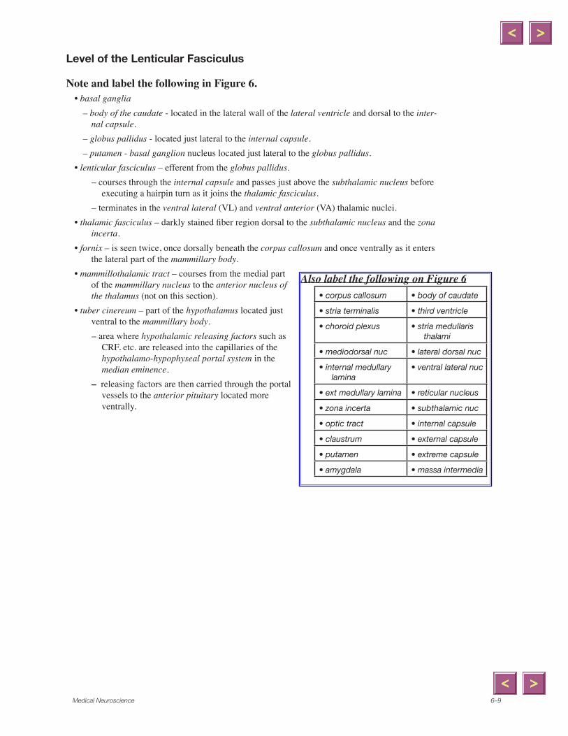
Level of the Lenticular Fasciculus
Note and label the following in Figure 6. • basal ganglia – body of the caudate - located in the lateral wall of the lateral ventricle and dorsal to the inter-
nal capsule. – globus pallidus - located just lateral to the internal capsule. – putamen - basal ganglion nucleus located just lateral to the globus pallidus.• lenticular fasciculus – efferent from the globus pallidus.
– courses through the internal capsule and passes just above the subthalamic nucleus before executing a hairpin turn as it joins the thalamic fasciculus.
– terminates in the ventral lateral (VL) and ventral anterior (VA) thalamic nuclei.• thalamic fasciculus – darkly stained fiber region dorsal to the subthalamic nucleus and the zona
incerta.• fornix – is seen twice, once dorsally beneath the corpus callosum and once ventrally as it enters
the lateral part of the mammillary body.• mammillothalamic tract – courses from the medial part
of the mammillary nucleus to the anterior nucleus of the thalamus (not on this section).
• tuber cinereum – part of the hypothalamus located just ventral to the mammillary body. – area where hypothalamic releasing factors such as
CRF, etc. are released into the capillaries of the hypothalamo-hypophyseal portal system in the median eminence.
– releasing factors are then carried through the portal vessels to the anterior pituitary located more ventrally.
Also label the following on Figure 6• corpus callosum • body of caudate
• stria terminalis • third ventricle
• choroid plexus • stria medullaris thalami
• mediodorsal nuc • lateral dorsal nuc
• internal medullary lamina
• ventral lateral nuc
• ext medullary lamina • reticular nucleus
• zona incerta • subthalamic nuc
• optic tract • internal capsule
• claustrum • external capsule
• putamen • extreme capsule
• amygdala • massa intermedia
Loyola University6–�0
Fig. 6. Level o the Lenticular Fasciculus. (Slide 6. Univ. of Kansas School of Medicine)
x
Polygonal Line
x
Polygonal Line
x
Polygonal Line
x
Polygonal Line
x
Polygonal Line
x
Polygonal Line
x
Polygonal Line
x
Polygonal Line
x
Line
x
Line
x
Line
x
Line
x
Polygonal Line
x
Polygonal Line
x
Polygonal Line
x
Polygonal Line
x
Polygonal Line
x
Polygonal Line
x
Polygonal Line
x
Polygonal Line
x
Line
x
Polygonal Line
x
Polygonal Line
x
Polygonal Line
x
Polygonal Line
x
Polygonal Line
x
Polygonal Line
x
Polygonal Line
Medical Neuroscience 6–��
Level of the Ventral Anterior and Anterior Thalamic Nuclei
Note and label the following in Figure 7. • ansa lenticularis – projection from the globus pallidus that loops beneath the internal capsule
on its way to the ventral anterior nucleus of the thalamus.• anterior commissure – a small round bundle of axons located ventral to the globus pallidus.
– will cross at more rostral levels.• optic tract – very prominent as it exits the optic chiasm (located just rostrally).• fornix – is seen coursing through the hypothalamus ventrally and is also visible dorsally be-
neath the corpus callosum.• mammillothalamic tract – ascends dorsally through the thalamus enroute to the anterior tha-
lamic nucleus.
Fig. �. Level of the Ventral An-terior and Anterior Thalamic NucleiSlide �. (Univ. of Kan-sas School of Medicine)
Also label the following on Figure 7• corpus callosum • body of caudate • stria terminalis
• stria medullaris thalami • third ventricle • mediodorsal nucleus
• mass intermedia • reticular nucleus • zona incerta
• internal capsule (post limb) • basa forebrain • claustrum
Note and label the following in Figure 8.• optic chiasm – located ventrally. • fornix – this curving pathway is cut twice• basal forebrain – several components are located ventral to the globus pallidus including the:
– nucleus basalis of Meynert (NBM) and the ventral pallidum (VP).– anterior perforated substance – comprehensive term for this region as located gross
anatomically.• anterior commissure – located just ventral to the globus pallidus.
Fig. 8. Rostral thala-mus. (Slide
. Univ. of Kansas School of Medicine)
Also label the following on Figure 8• corpus callosum • globus pallidus • body of caudate
cholinergic neurons in the nucleus basalis is thought to be part of the neuropathology in Alzheimer’s disease.
Fig. 8. Slide 4. (Univ. of Kansas School of Medicine)
x
Polygonal Line
x
Polygonal Line
x
Polygonal Line
x
Polygonal Line
x
Polygonal Line
x
Line
x
Line
x
Line
x
Polygonal Line
x
Line
x
Line
x
Polygonal Line
x
Line
x
Polygonal Line
x
Polygonal Line
x
Polygonal Line
x
Polygonal Line
x
Polygonal Line
x
Line
Medical Neuroscience 6–�3
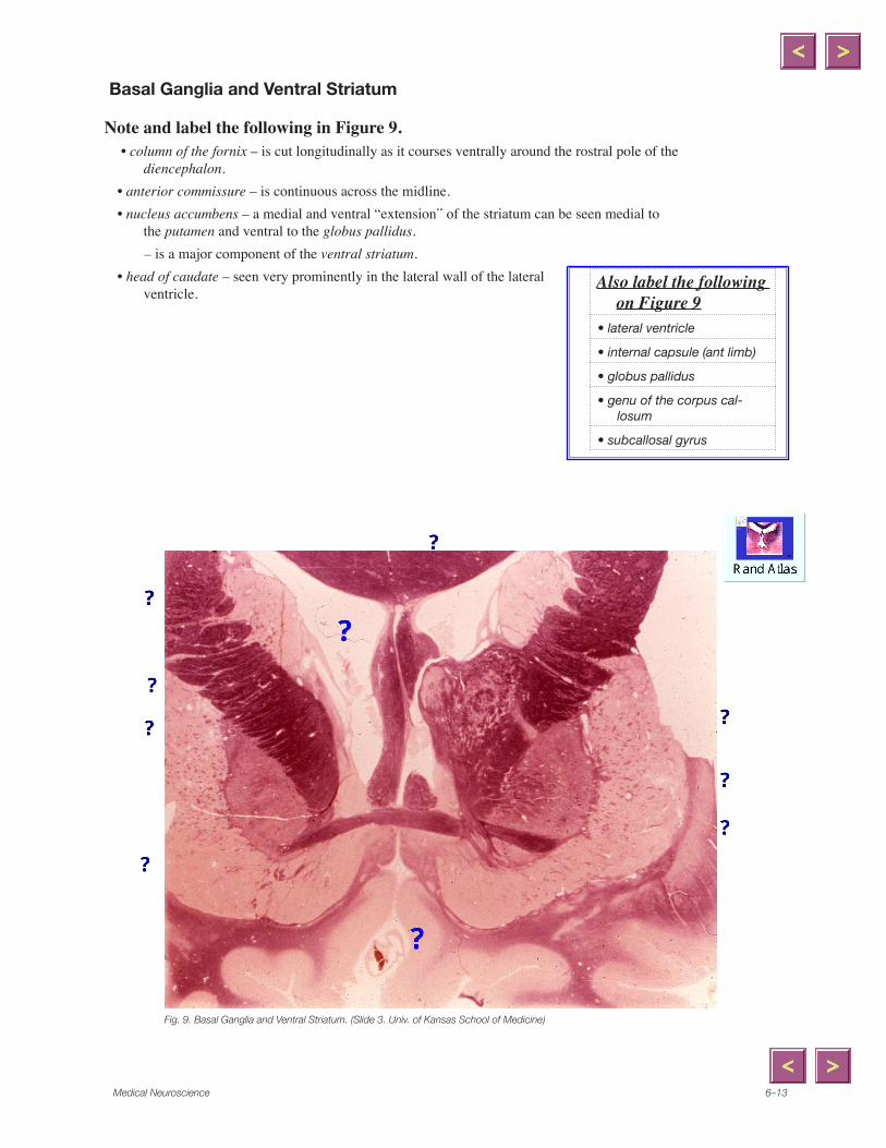
Basal Ganglia and Ventral Striatum
Note and label the following in Figure 9. • column of the fornix – is cut longitudinally as it courses ventrally around the rostral pole of the
diencephalon.• anterior commissure – is continuous across the midline.• nucleus accumbens – a medial and ventral “extension” of the striatum can be seen medial to
the putamen and ventral to the globus pallidus.– is a major component of the ventral striatum.
• head of caudate – seen very prominently in the lateral wall of the lateral ventricle.
Fig. 9. Basal Ganglia and Ventral Striatum. (Slide 3. Univ. of Kansas School of Medicine)
Also label the following on Figure 9
• lateral ventricle
• internal capsule (ant limb)
• globus pallidus
• genu of the corpus cal-losum
• subcallosal gyrus
x
Polygonal Line
x
Line
x
Line
x
Line
x
Line
x
Line
x
Line
x
Line
x
Line
Loyola University6–��
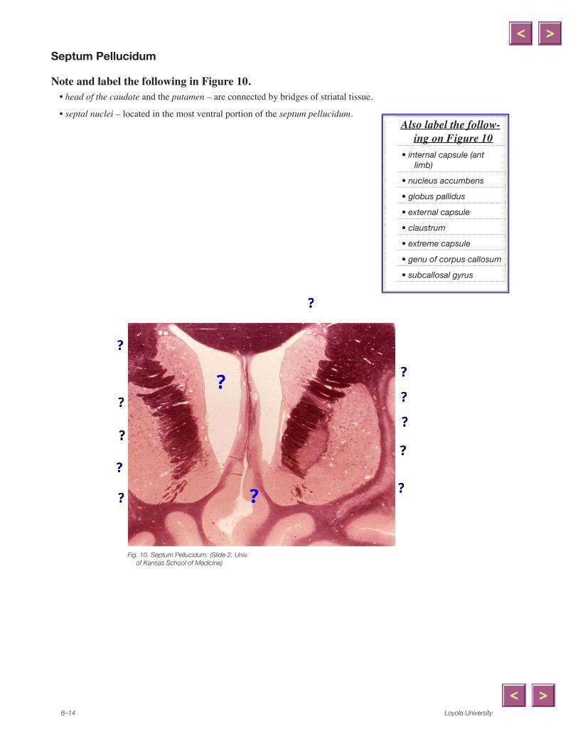
Septum Pellucidum
Note and label the following in Figure 10.• head of the caudate and the putamen – are connected by bridges of striatal tissue.
• septal nuclei – located in the most ventral portion of the septum pellucidum.
Fig. �0. Septum Pellucidum. (Slide �. Univ. of Kansas School of Medicine)
Also label the follow-ing on Figure 10
• internal capsule (ant limb)
• nucleus accumbens
• globus pallidus
• external capsule
• claustrum
• extreme capsule
• genu of corpus callosum
• subcallosal gyrus
x
Line
x
Line
x
Line
x
Line
x
Line
x
Line
x
Line
x
Polygonal Line
x
Polygonal Line
x
Polygonal Line
x
Polygonal Line
x
Polygonal Line
Medical Neuroscience 6–��
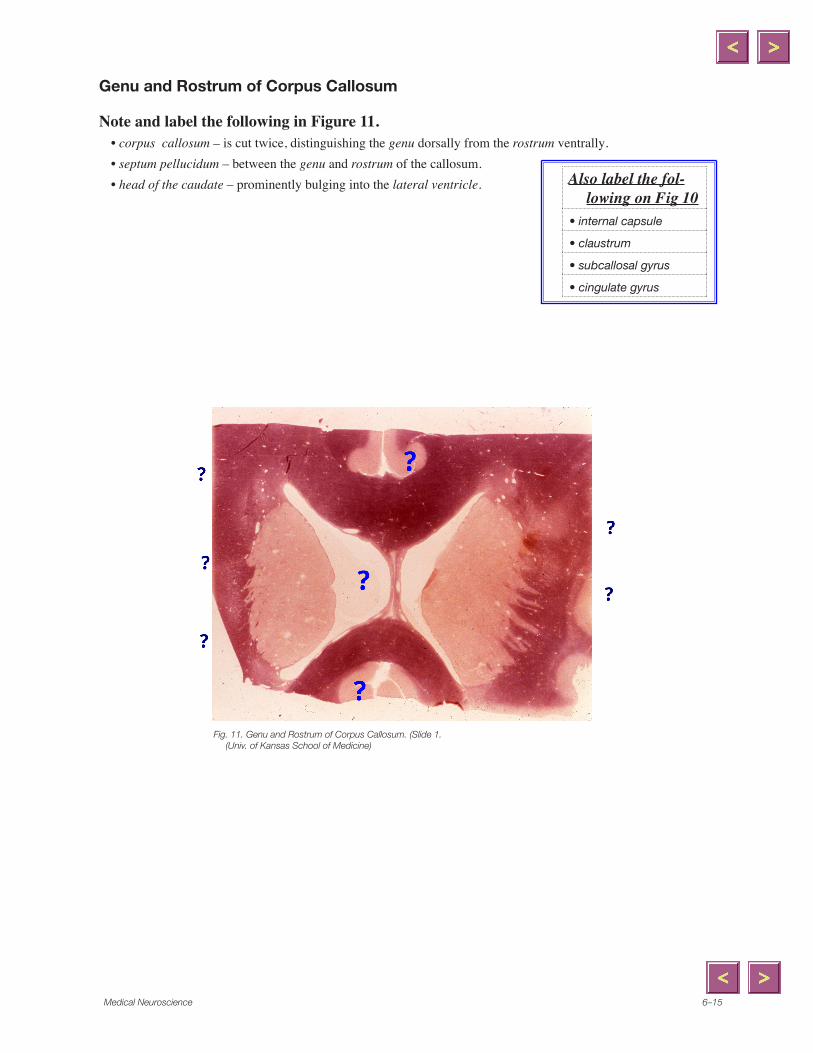
Genu and Rostrum of Corpus Callosum
Note and label the following in Figure 11. • corpus callosum – is cut twice, distinguishing the genu dorsally from the rostrum ventrally.• septum pellucidum – between the genu and rostrum of the callosum.• head of the caudate – prominently bulging into the lateral ventricle.
Fig. ��. Genu and Rostrum of Corpus Callosum. (Slide �. (Univ. of Kansas School of Medicine)
Also label the fol-lowing on Fig 10
• internal capsule
• claustrum
• subcallosal gyrus
• cingulate gyrus
x
Line
x
Line
x
Line
x
Polygonal Line
x
Line
Loyola University6–�6
Review Questions 1. What is the origin, course and termination of the lenticular fasciculus? ...of the ansa
lenticularis?
2. What are the components of the thalamic fasciculus?
3. What are the major afferents and efferents of the ventral posterior thalamic nuclei?
...of the ventral lateral nucleus?
...of the ventral anterior nucleus?
4. What is the origin, course, and termination of the fornix?
...of the mammillothalamic tract?
...of the stria terminalis?
5. Distinguish the anterior and posterior limbs of the internal capsule.
6. What are the afferents and efferents of the medial geniculate nucleus?
...of the lateral geniculate nucleus?
Medical Neuroscience 6–��
7. Which limb of the internal capsule contains the corticospinal fibers?
8. What are the optic radiations?
9. What is the significance of the basal forebrain area?
Loyola University6–�8
MRI Correlation
Label the following structures on figure 12.• corpus callosum • head of the caudate • internal capsule (ant limb)
Patient 6.1. Case of the bump on the headPatient: Ms. Anna Conda: Age: 28 Occupation: Latin dance instructor
Signs and Symptoms:• Ms. Conda was struck by a car that failed to yield to pedestrians in a crosswalk.• After a brief period of unconsciousness and except for a bruise on the side of her head, she
said she was OK and went home.• Three days later she felt confused, had a headache, and was weak and numb on her right
side.• In addition to the weakness, you also find a right-sided hyperreflexia.
Diagnosis:1. What do you think happened to Anna? Did she have a stroke overnight?
2. Would you order an MRI and schedule an appointment for next week?
3. What abnormal reflex is being tested in the movie? Is this reflex always abnormal?
Related questions:1. Here’s Anna’s CT scan. Notice anything?
2. Do the ventricles look normal in this scan? Where’s the midline?
QuickTime™ and aPhoto - JPEG decompressor
are needed to see this picture.
CT scan. (From C. Andrews, Univ. of Utah; Slice of Brain, �993, Univs of Utah and Washington; S.S. Stensaas, Univ. of Utah)