55
Lab Medicine Conference : Amylase, Lipase, Pregnancy Tests, Sedimentation Rate
| Date post: | 29-Dec-2015 |
| Category: |
Documents |
| Upload: | russell-burke |
| View: | 217 times |
| Download: | 2 times |

Lab Medicine Conference :
Amylase, Lipase, Pregnancy Tests,
Sedimentation Rate

Amylase Measurementƒ Named for Greek word for starch
(amylone)ƒ Amylase acts to split starches ; can
cleave amylopectin, amylose, glycogen, & their hydrolyzed products
ƒ 2 types in humans :–P type : in pancreas–S type : mainly in salivary glandsƒ also in lung, fallopian tubes, lactating mammary glands, & sweat glands
ƒ Both have molecular weight 54,000

Netter’s diagram of amylase physiology

Activity of Amylaseƒ Requires calcium for catalytic activityƒ Also nedds a halide (chloride,
bromide, or iodide) for activationƒ Maximum activity at temp. 50 C & pH
6.9 to 7.0ƒ Both P and S types have isoenzymes,
but differentiation not generally clinically useful at present
O

Clearance of Amylaseƒ P type cleared 80 % faster than S
typeƒ Serum half life of P type is 2 hoursƒ Cleared by glomerular filtration
without tubular excretion or reabsorption
ƒ Some limited extrarenal clearance ( ? in reticuloendothelial system)

Methods for Amylase Level Quantification
ƒ Saccharogenic : standard since 1938
ƒ Amyloclastic (Iodometric)ƒ Chromolyticƒ Turbidimetricƒ Nephelometric

Saccharogenic Method for Amylase Quantificationƒ Measures hydrolyzed products of
starch by direct measurement of sugars created by amylase activity
ƒ Measured in Somogyi units–One unit is defined as amount of enzyme in 100 ml of specimen that liberates reducing substances equivalent to 1 mg of glucose from starch in 30 minutes at 40 C
O

Amyloclastic Method for Amylase Quantification
ƒ Measures color change of iodine reaction with starch–When iodine combines with polysaccharides of more than 12 to 18 glucose residues, a brown color appears–No color change occurs when iodine is mixed with polysaccharides of 12 glucose residues or less–Amount of amylase then correlated to amount of color change by photometric analysis

Chromolytic Method for Amylase Quantificationƒ Starch is covalently bonded to a
dye moleculeƒ Hydrolyzed products then
measured photometricallyƒ Very sensitiveƒ Easily performed in lab

Turbidimetric & Nephelometric Methods for Amylase Quantification
ƒ Starch solutions are colloidal by nature
ƒ Amylase action decreases the colloidal nature of a starch solution
ƒ Colloidal status can then be measured photometrically
ƒ Both methods utilize measurement of light reflection–Nephelometric method more accurate–Both methods can be automated

Reporting of Amylase Measurements
ƒ Some current methods reported in International Units (IU)–One IU catalyzes transformation of one micromole of substrate per minute–Some methods using IU may not be as accurate as those using Somogyi units

Macroamylaseƒ Represents P or S type linked to an
immunoglobulin or complex polysaccharides
ƒ Molecular weight 150,000 to 1 millionƒ Not filtered at glomerulus ; stays in
serumƒ Can occur with alcoholism,
malabsorption, & GI tract diseasesƒ Serum amylase levels are 4 to 5
times normal, but urine amylase is low or normal

Direct Pancreatic Causes of Elevated Serum Amylaseƒ Acute or chronic pancreatitisƒ Pancreatic pseudocystsƒ Pancreatic ascitesƒ Mumpsƒ Pancreatic or duodenal trauma

Salivary Causes of Elevated Serum Amylaseƒ Tumorsƒ Salivary gland calculiƒ Sialadenitisƒ Head & neck surgeryƒ Head & neck trauma

Miscellaneous Abdominal Causes of Elevated Serum Amylase
ƒ Renal failure (up to 2X normal)ƒ Perforated peptic ulcerƒ Bowel obstructionƒ Ruptured ectopic pregnancyƒ Mesenteric infarctionƒ Afferent loop syndromeƒ Aortic aneurism / dissectionƒ Cirrhosis

Other Miscellaneous Causes of Elevated Serum Amylaseƒ Macroamylasemiaƒ Cerebral traumaƒ Burnsƒ Generalized shockƒ DKAƒ Renal transplantƒ Pneumoniaƒ Drugs & meds (on a later slide)

Amylase Levels in Acute Pancreatitisƒ Elevation > 5X normal often indicates
acute pancreatitis–sensitivity 70 to 98 %–specificity 70 to 76 %
ƒ Peak levels reached in first 48 hoursƒ Levels return to normal 5 to 7 days
after resolution of inflammationƒ Urine amylase levels peak later & can
remain elevated for days after symptoms are resolved

Clinical Use of Amylase to Creatinine Clearance Ratioƒ Used to help differentiate acute
pancreatitis from other causes of elevated amylase
ƒ (urine amylase X plasma creatinine) divided by (urine creatinine X plasma amylase) X 100
ƒ Normal clearance ratio is < 5 %ƒ Ratios > 10 % suggest acute
pancreatitisƒ Ratio not affected by urine volume or
rate

Limitations of Use of Amylase to Creatinine Clearance Ratio
ƒ 30 % of patients with proven acute pancreatitis may have normal ratio
ƒ Can be elevated in DKA, renal failure, heart disease, peptic ulcer, or postop
ƒ Because of often sporadic secretion of amylase, single measurement may be unreliable

Mechanisms of Amylase Elevation by Drugs and Medications
ƒ Induce spasm of Sphincter of Oddi–e.g., narcotics
ƒ Direct inflammation of parotid gland–e.g., phenylbutazone
ƒ Direct inflammation of pancreas–e.g., alcohol, steroids, thiazides

List of Medications and Drugs Causing Elevated Amylase Levels
ƒ Alcoholsƒ Corticosteroidsƒ Estrogensƒ Thiazide diureticsƒ Sulfonamidesƒ Furosemideƒ Ethacrynic acidƒ Clofibrateƒ Indomethacinƒ Salicylatesƒ Coumadinƒ Asparaginaseƒ Amphetamines
ƒ Narcoticsƒ Phenylbutazoneƒ Rifampinƒ Tetracyclineƒ Vitamin Dƒ Carbon tetrachlorideƒ Histamineƒ Phenforminƒ Acetominophenƒ Propoxypheneƒ Cimetidineƒ Valproic acidƒ Ciproheptadine

Interpretation of Elevated Amylase Levelsƒ Positive predictive value approaches 100 % at
value of 1000 IU/Lƒ At 300 IU/L, total amylase is 90 to 95 % sensitive,
but only 71 to 80 % specificƒ Biliary pancreatitis causes markedly higher initial
serum amylase than for alcoholic or other causes–with biliary : only 11 % of cases < 1000 IU/L–with alcoholic : only 6 % of cases > 1000 IU/L
ƒ If hypertriglyceridemia present, can have pancreatitis with normal amylase (mechanism is uncertain)
ƒ Magnitude of increase does NOT correlate with severity of disease or prognosis
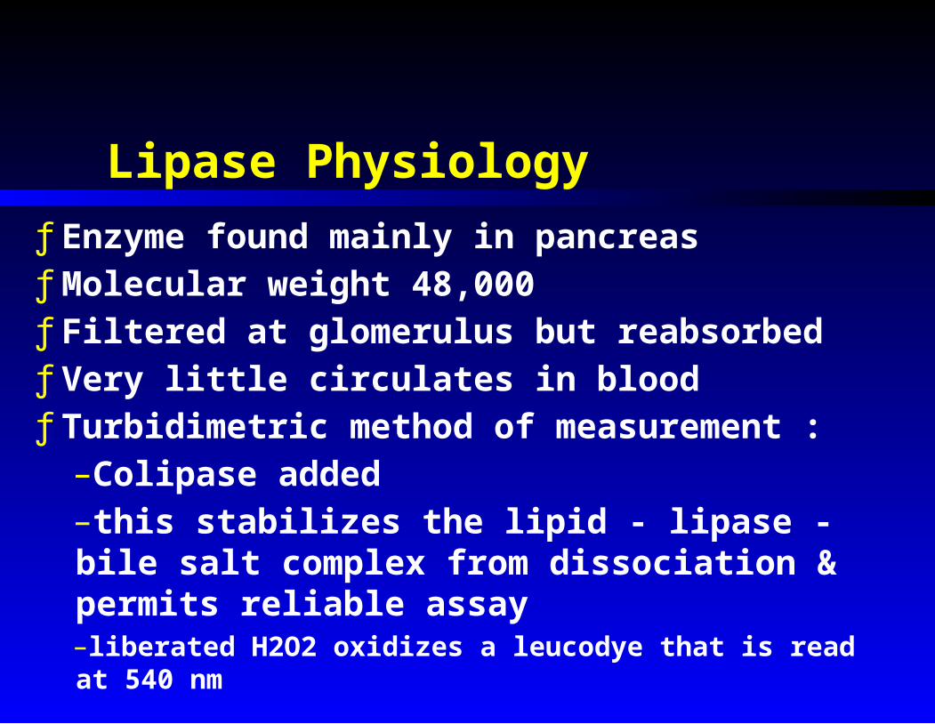
Lipase Physiologyƒ Enzyme found mainly in pancreasƒ Molecular weight 48,000ƒ Filtered at glomerulus but reabsorbedƒ Very little circulates in bloodƒ Turbidimetric method of measurement :–Colipase added–this stabilizes the lipid - lipase - bile salt complex from dissociation & permits reliable assay–liberated H2O2 oxidizes a leucodye that is read at 540 nm

Nonpancreatic Causes of Elevated Serum Lipase (usually < 3X normal)
ƒ Mumpsƒ Types I & IV hyperlipoproteinemiasƒ Peptic ulcerƒ Acute cholecystitisƒ Extrahepatic biliary obstructionƒ Mesenteric infarctionƒ Bowel perforationƒ Acute renal failureƒ Bone fracturesƒ Fat embolism syndromeƒ Crush injuryƒ Post-cholecystectomy syndrome

Lipase Levels in Pancreatitisƒ Parallels amylase in onset &
height of elevationƒ Not elevated with
macroamylasemia & DKAƒ Levels > 3X normal have > 99 %
predictive value for acute pancreatitis
ƒ Generally : lipase assay is as reliable, more specific, almost as sensitive, & about same cost as amylase assay for pancreatitis


Amylase and lipase secretion in acute pancreatitis

Measurement of Immunoreactive Trypsinogen (IRT)ƒ Radioimmunoassay can measure IRT ; is not
affected by serum proteases as are other methodsƒ IRT is elevated in :–Extrahepatic obstructive jaundice–Renal failure–Hypercalcemia–Hypertriglyceridemia–Liver cirrhosis–Chronic pancreatitis
ƒ Can serve to confirm pancreatic origin of an elevated amylase level, but offers no improvement in diagnostic accuracy over amylase or lipase as a single test

Lab Charges at H.M.C. for Pancreatic Lab Tests
Test Routine Stat
Amylase $ 13 $ 39
Lipase $ 22 $ 29
Triglycerides $ 13 $ 29

Simplified Algorithm for Interpretation of Pancreatic Enzyme Levels
ƒ If amylase > 1000, & renal function normal, assume acute pancreatitis
ƒ If amylase < 1000 (even if normal) & pancreatitis suspected, check lipase–If lipase > 3X normal, assume acute pancreatitis–If lipase < 3X normal, consider diagnoses on slide # 23

Categories of Pregnancy Tests
ƒ Bioassays (no longer used)ƒ Immunoassays (variations of
agglutination tests)–Radioimmunoassays (RIA)–Enzyme immunoassays–Radioreceptor assays (RRA)–Each of these is based on detection of Human Chorionic Gonadotropin (HCG) in urine or blood–HCG is measured in International Units (IU)

Bioassay Pregnancy Tests
ƒ Developed in 1920'sƒ Used toads, mice, frogs, or
rabbits for testingƒ Problems :
–High cost–Need for frequent restandardization–High false positive rate (from circulating Leutinizing Hormone)
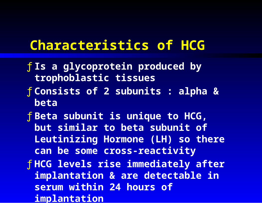
Characteristics of HCGƒ Is a glycoprotein produced by
trophoblastic tissuesƒ Consists of 2 subunits : alpha &
betaƒ Beta subunit is unique to HCG, but
similar to beta subunit of Leutinizing Hormone (LH) so there can be some cross-reactivity
ƒ HCG levels rise immediately after implantation & are detectable in serum within 24 hours of implantation

Patterns of HCG Levels
ƒ During first 6 weeks of normal pregnancy, HCG levels should double every 48 hours
ƒ Mean serum HCG level at 6 weeks (from LMP) is 10,000 mIU/ml
ƒ Detection limit (& definition of nonpregnant level) is 5 mIU/ml
ƒ HCG levels with ectopic pregnancy usually stay < 6000 mIU/ml & do not show 2 day doubling
ƒ Postpartum levels fall to normal in 3 to 5 days

HCG Levels in Normal Pregnancy
Time Post-Conception mIU / ml.
1 week 5 to 50 2 weeks 40 to 1000 3 weeks 100 to 5000 4 weeks 600 to 10,000 5 to 6 weeks 1500 to 100,000 7 to 8 weeks 16,000 to 200,000 2 to 3 months 12,000 to 300,000 Second trimester 24,000 to 55,000 Third trimester 6000 to 48,000

Days from onset of last menstrual period

60 days 120 days Before term

Comparison of Current HCG Tests
Lower Sensitivity Limit (mIU / ml)
Approximate Days Since Last Menses
Beta subunit Radio- Immunoassay
< 5 22
Serum Monoclonal Antibody Test
< 10 22 to 24
Urine Monoclonal Antibody Test
20 24 to 26
Home Monoclonal Antibody Test
200 28

Use of Urine Monoclonal Antibody Tests to Detect Ectopic Pregnancy
ƒ Serum & urine qualitative monoclonal antibody pregnancy tests have almost equivalent ability to detect ectopic pregnancy
ƒ < 3 % of ectopics have serum HCG < 40 mIU/ml
ƒ Ectopics with HCG < 40 mIU/ml are not at imminent risk of rupture & may be undergoing resorption ; repeat test at followup or quantitative HCG (radioimmunoassay for beta HCG) will clarify these cases

Causes of False Positive Pregnancy Test Results
ƒ Increased LH–Midcycle peak–Menopause
ƒ Ectopic HCG production–Trophoblastic tumors (choriocarcinoma or hydatidiform mole)–Small cell lung cancer–Ovarian cancer
ƒ Tubo-ovarian abscess
ƒ Test interface interference–Proteinuria > 1 g / 24 hrs.–Methadone–Phenothiazine–Promethazine
ƒ Missed abortion or miscarriage–Sometimes have levels < 200 IU/ml persistent for up to 90 days
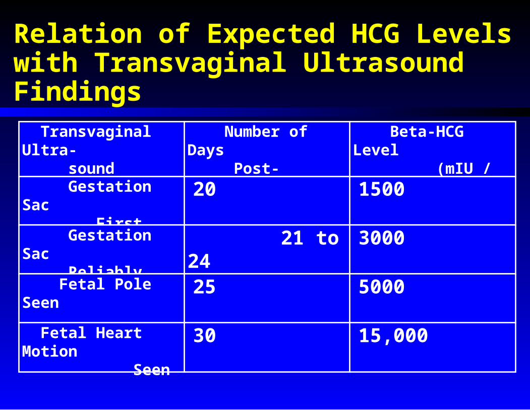
Relation of Expected HCG Levels with Transvaginal Ultrasound Findings Transvaginal Ultra- sound Finding
Number of Days Post-Ovulation
Beta-HCG Level (mIU / ml)
Gestation Sac First Seen
20 1500
Gestation Sac Reliably Seen
21 to 24 3000
Fetal Pole Seen 25 5000
Fetal Heart Motion Seen
30 15,000

Indications for Obtaining Quantitative Serum HCG Level
ƒ Monitor trophoblastic diseaseƒ Positive monoclonal test but no
fetal sac or pole, & no mass in tube on ultrasound
ƒ Uncertain missed abortionƒ Uncertain incomplete abortionƒ Negative monoclonal test but
suspected, and absolutely need to rule out, very early pregnancy
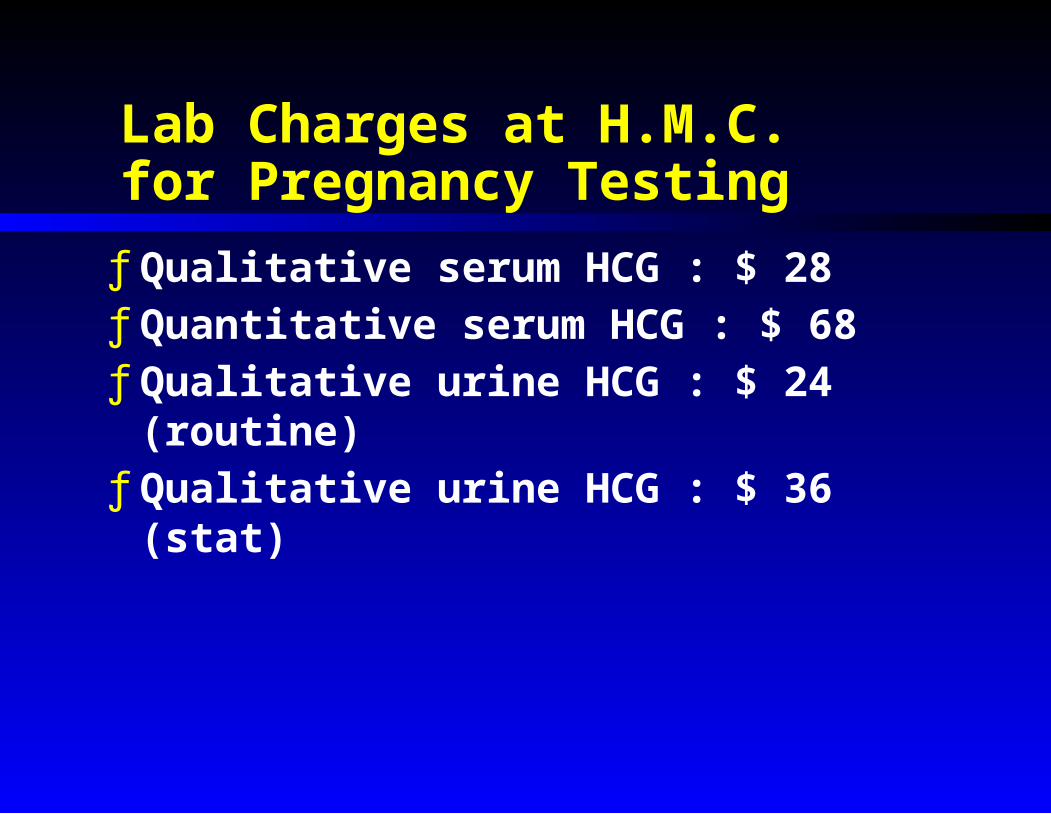
Lab Charges at H.M.C. for Pregnancy Testing
ƒ Qualitative serum HCG : $ 28ƒ Quantitative serum HCG : $ 68ƒ Qualitative urine HCG : $ 24
(routine)ƒ Qualitative urine HCG : $ 36 (stat)

Erythrocyte Sedimentation Rate (ESR)General Principles
ƒ Diagnostically useful indicator of some inflammatory conditions
ƒ Increased ESR due to any condition causing red cell aggregation or rouleaux formation
ƒ Is an index of the suspension stability of RBC's in citrated blood
ƒ Depends on the difference in specific gravity between RBC's & plasma
ƒ A normal value does not exclude organic disease

Factors Affecting the ESR Value
ƒ Depends mainly on concentration of fibrinogen, but to a lesser degree on alpha-2-globulin & gamma globulin
ƒ Is rapid (increased) in disorders where fibrinogen or globulins are increased

Methods of Measurement of ESRƒ Wintrobe or Westergren–Most common methods–Require relatively larger sample
ƒ Microsedimentation–Requires only a few drops of blood–Linzenheimer / Landau / Adler modifications
ƒ Distance from top of tube to RBC column meniscus at 1 hour (mm per hour) reported

Normal Westergren ESR Value Ranges
ƒ Newborn : 0 to 2 mm/hr ƒ Children : 1 to 10 mm/hrƒ Adults
–Males : 0 to 25 mm/hr–Females : 0 to 30 mm/hr
ƒ Micromethods closely correlate, especially at lower values

Zeta Sedimentation Ratio (ZSR)
ƒ Measures ability of RBC's to pack under a standardized stress of alternating dispersion & compaction
ƒ No real advantage over Wintrobe or Westergren methods
ƒ For males, mean ZSR is 44 % & upper limit of normal is 54 %
ƒ For females, mean ZSR is 45 % & upper limit of normal is 56 %

Causes of High (> 100 mm/hr) ESR
ƒ Acute bacterial infections–Meningitis–Pneumonia–Cholangitis–Septic arthritis–Osteomyelitis–Pyelonephritis–Abscesses
ƒ Polymyalgia rheumaticaƒ Rheumatoid arthritisƒ S.L.E.ƒ Viral encephalitis
ƒ Multiple myelomaƒ Leukemiaƒ Lymphomaƒ Carcinomasƒ Drug hypersensitivity
reactionsƒ Pulmonary infarctionƒ Uremiaƒ Open heart surgeryƒ Cerebrovascular
accidentƒ Thrombophlebitisƒ Major orthopedic
surgery

Causes of Moderately Elevated ESR(50 to 100 mm/hr)ƒ Tuberculosisƒ Viral hepatitisƒ Pelvic inflammatory
diseaseƒ Infectious
mononucleosisƒ Acute glmerulonephritisƒ Chronic infectious
diseasesƒ Rheumatic feverƒ Sarcoidosisƒ Rheumatic feverƒ Hypothyroidismƒ Drug fever
ƒ Liver metastasesƒ Atrial myxomaƒ Macroglobulinemiaƒ Regional enteritisƒ Myocardial infarctionƒ Thyroiditisƒ Abdominal surgeryƒ Menstrual periodƒ Pregnancy after first
trimesterƒ Ectopic pregnancy

Causes of Slightly Elevated ESR(25 to 50 mm/hr)
ƒ Uncomplicated viral diseases
ƒ Cholecystitisƒ Malariaƒ Typhoid feverƒ Pertussisƒ Cytomegalovirusƒ Toxoplasmosisƒ Rickettsial
infectionsƒ Digital
osteomyelitisƒ Localized infections
ƒ Osteoarthritisƒ Goutƒ Benign neoplasmsƒ Cirrhosisƒ Peptic ulcer diseaseƒ Acute allergiesƒ Ulcerative colitisƒ Pancreatitisƒ Drug fever

Causes of Very Low ESR (0 to 1 mm/hr)
ƒ High dose steroid or salicylate therapyƒ Severe anemiaƒ Cachexiaƒ Massive hepatic necrosisƒ D.I.C.ƒ Polycythemia veraƒ Trichinosisƒ Chronic lymphocytic or myeloid leukemiaƒ Hypofibrinogenemiaƒ Macroglobulinemia (hyperviscosity
syndrome)ƒ "Chronic mononucleosis syndrome"

ƒ Cholangitis (> 50) vs. Cholecystitis (< 50)ƒ P.I.D. (> 50) vs. Ovarian cyst (< 50)ƒ Strep (< 50) vs. Infectious mono (> 50)ƒ Trichinosis (< 50) vs. Polymyositis (> 50)ƒ Rheumatoid (> 50) vs. Osteo (< 50) arthritisƒ Carcinoma (> 50) vs. Cachexia (< 50)ƒ Temporal arteritis (> 50) vs. tension
headache(< 50)ƒ Synovitis (< 50) vs. Septic arthritis (> 50) of
hipƒ Loose prosthesis (< 50) vs. Infected
prosthesis (> 50)
Situations Where ESR May Be Diagnostically Helpful

Situations in Which ESR Is NOT of Diagnostic Help
ƒ Rheumatoid arthritis vs. sarcoidosisƒ Tubercular arthritis vs. sarcoidosisƒ Rheumatoid arthritis vs. S.L.E.ƒ Early prosthetic valve endocarditis vs. recent open
heart surgeryƒ Inflammatory diskitis vs. septic diskitisƒ Osteogenic sarcoma vs. chronic osteomyelitisƒ Early orthopedic trauma vs. early infectionƒ Drug fever vs. fever due to infectionƒ Regional ileitis vs. ulcerative colitisƒ Appendicitis vs. pyelonephritisƒ Pulmonary infarction vs. pneumonia

Lab Charges at H.M.C. for ESR
ƒ Routine : $ 14ƒ Stat : $ 37ƒ Run off of a purple top tube


