Laboratory Animal Allergy: An Update Robert K. Bush and Gregg M. Stave Abstract Allergic reactions are among the most common conditions affecting the health of workers involved in the care and use of research animals. Between 11 and 44% of the individuals working with laboratory animals report work-related aller- gic symptoms. Of those who become symptomatic, 4 to 22% may eventually develop occupational asthma that can persist even after exposure ceases. Allergic symptoms con- sist of rashes where animals are in contact with the skin, nasal congestion and sneezing, itchy eyes, and asthma (cough, wheezing, and chest tightness). The generation of immunoglobulin E (IgE) antibodies is a prerequisite for the production of allergic symptoms. The mechanism by which IgE antibodies develop is becoming clearer. The propensity to produce IgE is genetically determined, and pre-existing allergy may be a risk factor for the development of labora- tory animal allergy (LAA). However, exposure to animal allergens is the major risk factor for the development of LAA. Techniques to measure the airborne concentration of laboratory animal allergens have been developed. Research on animal allergens themselves indicates that many of the mouse and rat urinary proteins belong to a family of pro- teins called lipocalins, which share sequence homology with antigens of the parasitic agent that causes schistoso- miasis. The fact that parasite infections also trigger IgE antibody responses may account for the development of LAA in persons who have never had any previous allergy. The prevention of LAA should be a major goal of an effec- tive health and safety program in the animal research facil- ity, and it can be accomplished by education and training of employees, reduction of exposure (including the use of per- sonal protective gear), and changes in facility design. Medi- cal surveillance programs can also play a role in improving health of individuals working with laboratory research ani- mals. Early recognition of symptoms and evidence of sen- sitization can lead to interventions to reduce exposure and thereby avoid the long-term health consequences of LAA. Key Words: allergens; allergy; animal allergy; asthma; laboratory animal allergy; laboratory animals; occupational health Epidemiology of Laboratory Animal Allergy (LAA 1 ) E stimates of the number of individuals exposed to labo- ratory animals in their occupation vary considerably. Bland and colleagues (1987) estimated that 90,000 individuals were exposed to laboratory animals in the United States, and 32,000 workers were similarly exposed in the United Kingdom. Seward (1999) estimated that 40,000 to 125,000 individuals are exposed to laboratory animals in the United States. The existence of different definitions of LAA used in published studies (reported symptoms vs. laboratory evi- dence of immunoglobulin E [IgE 1 ]-mediated sensitivity) leads to significant variability in the reported prevalence (percentage of cases in the population) and incidence (per- centage of new cases occurring in the population over a given period of time) of this occupational problem. Preva- lence rates may also be affected if symptomatic workers discontinue work with laboratory animals. In addition, the sample size included in the study influences the results. In the United Kingdom, exposure to laboratory animals has consistently ranked in the top three causes of occupational asthma and comprises 5% of all cases reported to that coun- try’s surveillance of work-related and occupational respira- tory diseases program since 1989 (Gordon 2001). These statistics are striking because laboratory animal workers comprise only a small portion of the total UK work force. In the United States, the National Institute of Occupational Safety and Health has formally recognized LAA as an oc- cupational hazard since 1989. The first reported cases of allergic symptoms due to laboratory animals occurred in the 1950s (Sorrel and Got- tesman 1957). The high prevalence of this condition did not become apparent until cross-sectional epidemiological stud- ies were conducted in the 1970s and -80s (Cockcroft et al. 1981; Gross 1980; Lutsky and Neuman 1975; Schumacher et al. 1981) (Table 1). Robert K. Bush, M.D., is Chief of the Allergy Section of the William S. Middle Veterans Affairs Hospital in Madison, Wisconsin, and Professor of Medicine, University of Wisconsin, Madison. Gregg M. Stave, M.D., J.D., M.P.H., is Director of Strategic Health Planning at GlaxoSmithKline, Re- search Triangle Park, North Carolina, and Consulting Assistant Professor, Division of Occupational and Environmental Medicine, Duke University Medical Center, Durham, North Carolina. 1 Abbreviations used in this article: HEPA, high-efficiency particulate air; IFN, interferon-gamma; IgE, immunoglobulin E; IL, interleukin; LAA, laboratory animal allergy; MHC, major histocompatibility; PPE, personal protective equipment; RAST, radioallergosorbent test. 28 ILAR Journal
Transcript
Laboratory Animal Allergy: An Update
Robert K. Bush and Gregg M. Stave
Abstract
Allergic reactions are among the most common conditionsaffecting the health of workers involved in the care and useof research animals. Between 11 and 44% of the individualsworking with laboratory animals report work-related aller-gic symptoms. Of those who become symptomatic, 4 to22% may eventually develop occupational asthma that canpersist even after exposure ceases. Allergic symptoms con-sist of rashes where animals are in contact with the skin,nasal congestion and sneezing, itchy eyes, and asthma(cough, wheezing, and chest tightness). The generation ofimmunoglobulin E (IgE) antibodies is a prerequisite for theproduction of allergic symptoms. The mechanism by whichIgE antibodies develop is becoming clearer. The propensityto produce IgE is genetically determined, and pre-existingallergy may be a risk factor for the development of labora-tory animal allergy (LAA). However, exposure to animalallergens is the major risk factor for the development ofLAA. Techniques to measure the airborne concentration oflaboratory animal allergens have been developed. Researchon animal allergens themselves indicates that many of themouse and rat urinary proteins belong to a family of pro-teins called lipocalins, which share sequence homologywith antigens of the parasitic agent that causes schistoso-miasis. The fact that parasite infections also trigger IgEantibody responses may account for the development ofLAA in persons who have never had any previous allergy.The prevention of LAA should be a major goal of an effec-tive health and safety program in the animal research facil-ity, and it can be accomplished by education and training ofemployees, reduction of exposure (including the use of per-sonal protective gear), and changes in facility design. Medi-cal surveillance programs can also play a role in improvinghealth of individuals working with laboratory research ani-mals. Early recognition of symptoms and evidence of sen-sitization can lead to interventions to reduce exposure andthereby avoid the long-term health consequences of LAA.
Estimates of the number of individuals exposed to labo-ratory animals in their occupation vary considerably.Bland and colleagues (1987) estimated that 90,000
individuals were exposed to laboratory animals in theUnited States, and 32,000 workers were similarly exposedin the United Kingdom. Seward (1999) estimated that40,000 to 125,000 individuals are exposed to laboratoryanimals in the United States.
The existence of different definitions of LAA used inpublished studies (reported symptoms vs. laboratory evi-dence of immunoglobulin E [IgE1]-mediated sensitivity)leads to significant variability in the reported prevalence(percentage of cases in the population) and incidence (per-centage of new cases occurring in the population over agiven period of time) of this occupational problem. Preva-lence rates may also be affected if symptomatic workersdiscontinue work with laboratory animals. In addition, thesample size included in the study influences the results. Inthe United Kingdom, exposure to laboratory animals hasconsistently ranked in the top three causes of occupationalasthma and comprises 5% of all cases reported to that coun-try’s surveillance of work-related and occupational respira-tory diseases program since 1989 (Gordon 2001). Thesestatistics are striking because laboratory animal workerscomprise only a small portion of the total UK work force. Inthe United States, the National Institute of OccupationalSafety and Health has formally recognized LAA as an oc-cupational hazard since 1989.
The first reported cases of allergic symptoms due tolaboratory animals occurred in the 1950s (Sorrel and Got-tesman 1957). The high prevalence of this condition did notbecome apparent until cross-sectional epidemiological stud-ies were conducted in the 1970s and -80s (Cockcroft et al.1981; Gross 1980; Lutsky and Neuman 1975; Schumacheret al. 1981) (Table 1).
Robert K. Bush, M.D., is Chief of the Allergy Section of the William S.Middle Veterans Affairs Hospital in Madison, Wisconsin, and Professor ofMedicine, University of Wisconsin, Madison. Gregg M. Stave, M.D., J.D.,M.P.H., is Director of Strategic Health Planning at GlaxoSmithKline, Re-search Triangle Park, North Carolina, and Consulting Assistant Professor,Division of Occupational and Environmental Medicine, Duke UniversityMedical Center, Durham, North Carolina.
1Abbreviations used in this article: HEPA, high-efficiency particulate air;IFN�, interferon-gamma; IgE, immunoglobulin E; IL, interleukin; LAA,laboratory animal allergy; MHC, major histocompatibility; PPE, personalprotective equipment; RAST, radioallergosorbent test.
28 ILAR Journal
Gross (1980) observed that symptoms of affected indi-viduals usually began within 6 mo of exposure and rarelyoccurred after 2 to 3 yr of employment. In that study, thepercentages of workers affected by specific species were asfollows: rats, 65%; rabbits, 72%; mice, 66%; and guineapigs, 33%. Nasal symptoms preceded chest symptoms in45% of the individuals, whereas 55% experienced nasal andchest symptoms simultaneously. Chest symptoms never oc-curred in the absence of nasal symptoms.
In Cockcroft and colleagues’ (1981) evaluation, 49 of179 individuals were symptomatic with mainly nasal symp-toms. Skin testing was conducted and revealed a good cor-relation between the presence of rhinitis and positive skintests to relevant laboratory animal allergens. Five individu-als had positive skin tests to laboratory animal allergens butwere asymptomatic.
In Schumacher and colleagues’ (1981) study, of the 39individuals who experienced respiratory symptoms or skinrashes, one third reported severe symptoms. Virtually all ofthese individuals had demonstrable sensitivity by positiveskin tests to laboratory animal allergens.
Slovak and Hill (1981) examined 146 workers of whom48 (30%) had a history of symptoms related to their labo-ratory animal exposure. Of the 48, 22 had positive skin testsand demonstrated rhinitis symptoms that progressed toasthma.
In a similar study of workers in the pharmaceutical in-dustry in the United Kingdom, Beeson et al. (1983) reportedthat of the 15 workers with LAA, slightly more than half (8)had positive skin tests to animal allergens. Sixty-seven per-cent of the individuals were sensitive to allergens other thanlaboratory animals.
In Agrup and colleagues’ (1986) evaluation, there was a
high level of frequency of self-reported symptoms, but thenumber of individuals with symptoms was reduced whenthey were interviewed by clinicians. Nineteen of the 30laboratory technicians with reported symptoms had positiveskin tests or in vitro tests indicating sensitivity to laboratoryanimal allergens.
Laboratory animal exposure results in significant losttime from work. More than one third of individuals workingat the US National Institutes of Health reported lost timefrom work due to their symptoms from laboratory animalallergy sensitivity (Bland et al. 1986). According to com-pleted questionnaires, 131 of 549 individuals (23.9%) re-ported symptoms due to their occupational exposure tolaboratory animals.
In perhaps the highest prevalence rate reported, Ven-ables et al. (1988) found that 44% of 133 pharmaceuticalworkers in the United Kingdom had laboratory animal sen-sitivity. In a review of pooled data from reported studies,Hunskaar and Fosse (1990) found an overall prevalence ofLAA of 20.9% in 4988 individuals. Subsequently, in a largeseries conducted in Japan (Aoyama et al. 1992), 1304 of5641 (23.1%) workers had symptoms related to their labo-ratory exposure. This survey was conducted among 137research facilities, 76 medical schools, 57 research insti-tutes, and four breeding facilities. Rhinitis was the mostcommon symptom. Seventy percent of individuals devel-oped symptoms within 3 yr of exposure. The survey re-vealed sensitivity to a variety of animals for which thepercentages of affected workers were as follows: sensitivityto guinea pigs, 31% of workers; to mice, 26.1%; to rats,24.9%; to cats, 30.1%; to dogs, 24.9%; and to non-human primates, 23.6%. It is important to note that virtuallyany laboratory animal can cause occupational allergy al-
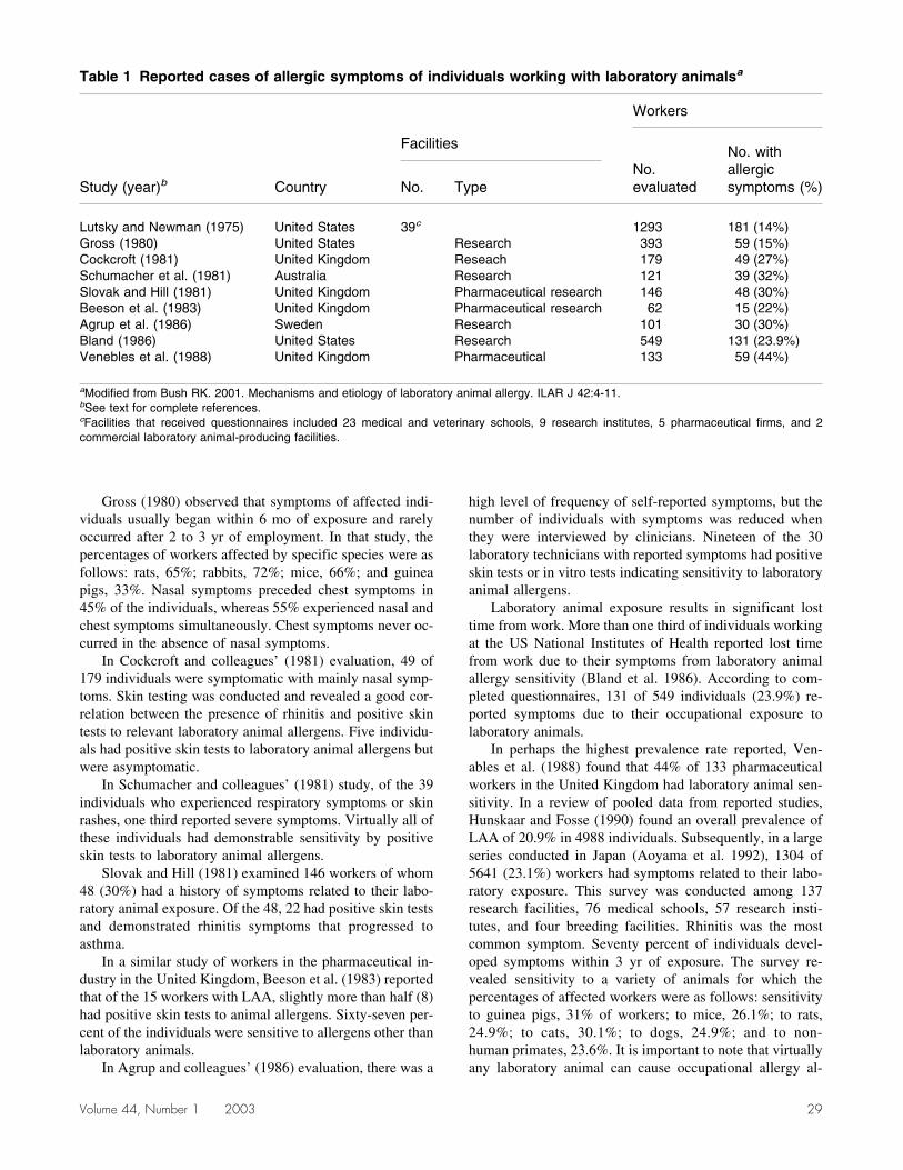
Table 1 Reported cases of allergic symptoms of individuals working with laboratory animalsa
Study (year)b Country
Facilities
Workers
No.evaluated
No. withallergicsymptoms (%)No. Type
Lutsky and Newman (1975) United States 39c 1293 181 (14%)Gross (1980) United States Research 393 59 (15%)Cockcroft (1981) United Kingdom Reseach 179 49 (27%)Schumacher et al. (1981) Australia Research 121 39 (32%)Slovak and Hill (1981) United Kingdom Pharmaceutical research 146 48 (30%)Beeson et al. (1983) United Kingdom Pharmaceutical research 62 15 (22%)Agrup et al. (1986) Sweden Research 101 30 (30%)Bland (1986) United States Research 549 131 (23.9%)Venebles et al. (1988) United Kingdom Pharmaceutical 133 59 (44%)
aModified from Bush RK. 2001. Mechanisms and etiology of laboratory animal allergy. ILAR J 42:4-11.bSee text for complete references.cFacilities that received questionnaires included 23 medical and veterinary schools, 9 research institutes, 5 pharmaceutical firms, and 2commercial laboratory animal-producing facilities.
Volume 44, Number 1 2003 29
though the mammals listed above are the most commonlyinvolved.
In another survey from Australia, Bryant and colleagues(1995) reported that 73 of 138 exposed individuals hadsymptoms of LAA. Of these individuals, 92% had positiveskin tests to laboratory animal allergens; 23% of asymp-tomatic individuals also had positive skin tests to laboratoryanimal allergens. Bronchial hyper-responsiveness, a markerof asthma, was present in 21% of exposed individuals com-pared with 8% in a nonexposed control population.
From the United Kingdom, where good occupationaldisease reporting is available for workers in the laboratoryanimal field, 44% of 32,000 individuals indicated they hadsymptoms related to their work exposure in 1988. By 1994,this number had decreased to 31%. However, when skintesting was performed in those symptomatic individuals in1988, only 13% had positive skin tests. In 1994, 10% hadpositive skin tests (S. Gordon, personal communication,1999). In a recently reported preliminary study from theUniversity of Wisconsin, 29 of 147 (19.7%) workers ex-posed to laboratory animal allergens in a research facilityrelated allergic symptoms to their work exposure (Patel etal. 2000). However, of the 147 workers, only 12% hadpositive skin tests that correlated with histories suggestiveof LAA.
As can be seen, prevalence and incidence rates may varyconsiderably based on whether a questionnaire is used toestablish the presence of LAA or whether laboratory testingis required. The low prevalence of skin tests or in vitro testsshowing IgE sensitivity may be related in part to the poorquality of skin testing and testing reagents available. None-theless, LAA does represent a significant health risk for thepopulation of exposed individuals. Furthermore, few dataare available on the number of individuals who end theiremployment due to LAA. Failure to capture this populationin epidemiological studies could result in a significantlylower estimate of true prevalence or incidence (Monso et al.2000).
The overall prevalence of LAA varies from 11 to 44%(Seward 1999). The prevalence of asthma due to LAAranges from 4 to 22% (Seward 1999). The wide range inthese prevalence figures reflects the vigor with which thediagnosis of LAA was established (positive response toquestionnaires vs. confirmatory medical evaluations).Nonetheless, LAA is common in the workplace where ani-mals are used for research purposes.
In a study at a pharmaceutical company involving work-ers exposed to laboratory animals, the incidence of labora-tory animal allergy was as high as 10.3% (Fisher et al.1998). After the institution of a comprehensive preventionprogram, including environmental control measures and theuse of personal protective equipment (PPE1) to reduce al-lergen exposure, the incidence decreased to 0. This decreasesuggests that LAA is a preventable workplace hazard.
Workers who develop allergies to one animal speciesare at risk of developing allergy to other species (Goodnoand Stave 2002). A work environment that may protect
against the symptoms of an initial or primary allergy to onespecies may still leave workers at risk for subsequent al-lergy to another. Prevention of the initial allergy is the mostsuccessful approach to prevention of sequent allergy toother animals, but all aspects of the worker’s exposureshould be considered.
Symptoms of LLA
Symptoms of LAA are the result of the release of biochemi-cal mediators and the generation of inflammation in thetissues induced by the IgE response. The nature and inten-sity of the symptoms are dependent on the level of exposureto the laboratory animal allergen by the individual. Once theworker has become sensitized (developed IgE antibodies tolaboratory animal allergens), symptoms generally occurrapidly (within minutes) of exposure. Continued daily ex-posure can result in chronic symptoms that may requiredaily treatment. These symptoms can range from mild skinreactions to severe asthma. The most common symptomsare related to allergic reactions involving the nose and eyes(Aoyama et al. 1992; Cullinan et al. 1994) and are known asallergic rhinitis and allergic conjunctivitis, respectively. Na-sal symptoms include congestion, runny nose, sneezing, anditching; ocular symptoms include redness and itchy wateryeyes. Up to 80% of workers with LAA report nasal symp-toms (Bush et al. 1998).
Skin reactions include hives at the site of contact withanimal urine or dander as the result of scratches. Otherrashes include maculopapular (measles-like) rashes, whichare typically quite itchy and occur in about 40% of symp-tomatic individuals (Bush et al. 1998).
Asthma may affect 4 to 22% of symptomatic workersexposed to laboratory animals. Symptoms of asthma consistof cough, wheezing, and shortness of breath. It is importantto recognize that symptoms related to laboratory animalexposure may continue for several hours or longer afterexposure to the animals ceases. In addition, individuals mayexperience symptoms of asthma when exercising and whenexposed to cold air, dust particles, or strong odors. Thisphenomenon, known as nonspecific airway hyper-responsiveness, occurs in other situations of allergen-induced asthma.
Systemic allergic reactions, known as anaphylaxis, canoccur (albeit rarely) as a result of an animal bite (Teasdaleet al. 1993) or from puncture wounds (e.g., needles con-taminated with animal proteins) (Watt and McSharry 1996).These reactions can manifest by generalized itching, hives(urticaria), swelling (angioedema) of the lips, eyes, and/orextremities, respiratory distress due to edema of the larynx,hypotension (shock), or acute asthma attacks. These reac-tions are potentially fatal. Occasionally, a milder form ofsystemic reaction can manifest in which the allergic indi-vidual develops a maculopapular rash or hives under pro-tective clothing as a result of a respiratory exposure tolaboratory animal allergens.
30 ILAR Journal
Time from the onset of exposure to development ofsymptoms is variable but generally is within 3 yr of begin-ning employment. Approximately one third of individualswill develop symptoms in the first year and 70% within 3 yr.Workers who do not devleop symptoms in the first 3 yrremain at risk. In a study from the United Kingdom, themean duration of employment before the onset of nasalsymptoms was 214 days, 335 days for skin symptoms, and365 for the development of chest symptoms (asthma) (Cul-linan et al. 1994). Again, this estimate is quite variabledepending on the individual study reported (Seward 1999).
Risk Factors for the Development of LAA
Epidemiological studies have been useful in determiningfactors that may lead to the development of LAA. The mostimportant risk factor for an individual is the level of expo-sure to laboratory animal allergens. Methods have been de-veloped that allow quantitative estimates of the exposure tolaboratory animal allergens (Gordon 2001; Harrison 2001).
Some questions still exist as to whether individuals withcoexisting allergies to substances outside the laboratoryhave an increased risk of developing LAA, although themajority of reported studies suggests it is an important riskfactor. In the study by Gross (1980), one third of workershad no prior allergic disease before developing LAA. Sch-umacher and colleagues (1981) correlated the developmentof LAA with the presence of atopy (defined as positive skintest to one or more inhalant allergens). Slovak and Hill(1981) documented that atopy predisposes individuals to thedevelopment of asthma related to their animal exposure.Several other studies (e.g., Aoyama et al. 1992; Bland, et al.1986; Botham et al. 1995; Bryant et al. 1995; Cullinan et al.1999; Fisher et al. 1998; Fuortes et al. 1996) indicate thatatopy is a risk factor for the development of LAA. In con-trast, Heederik et al. (1999) found atopy to be a risk factoronly for individuals exposed at low levels; and Renstrom etal. (1994) believe that atopy is not a significant risk factorbut that total IgE level is. These latter investigators are fromthe same research group and share the same subject pool. Ina review (Bush et al. 1998) based on the studies cited above,it was concluded that in individuals with a history of work-related symptoms and objective evidence of allergy as dem-onstrated by a positive skin test or in vitro test, the oddsratio for developing LAA was 3.35 in atopics comparedwith nonatopic workers.
Recent studies from Canada (Gautrin et al. 2000, 2001)showed that apprentices working in animal health technol-ogy facilities were at greater risk for developing LAA ifthey (1) were atopic, (2) had respiratory symptoms in thepollen season, (3) were sensitized to cat or dog allergens, (4)had baseline airway hyper-responsiveness, and/or (5) hadan increasing number of hours of contact with laboratoryanimals.
The level of an individual’s exposure to laboratory ani-mal allergens certainly is a major factor in determining
whether the worker develops LAA. Although most studieshave shown that individuals who have coexisting allergiesto other inhalant allergens are more at risk, some studiesreport different results. Of note is the study by Hollander etal. (1996), who found that sensitivity to mites or pollens wasnot associated with risk for developing LAA; however, sen-sitivity to cats and dogs was associated. Analysis of allavailable data suggests that pre-existing atopy is an impor-tant factor contributing to the development of LAA.
In many of these studies, the relation between atopy andthe development of LAA suggests that a genetic predispo-sition to form IgE antibodies is a significant risk factor. Theexact genetic cause has not yet been identified. Smoking hasalso been associated as a risk factor for the development ofLAA. However, as with atopy, controversial data havearisen. Venables and colleagues (1988) reported a positiveassociation between cigarette smoking and the developmentof LAA, as did Fuortes et al. (1996), and Cullinan et al.(1999). In contrast, Agrup et al. (1986) and Heederik et al.(1999) found no effect. Of note is the study by Fuortes et al.(1997), who reported that individuals with a smoking his-tory had significantly greater declines in pulmonary func-tion compared with nonsmokers. This finding would be anexpected effect of tobacco smoke exposure in addition tothe exposure to the laboratory animals. Tobacco smoke hasbeen shown to elevate serum IgE levels. This increase couldpredispose an individual to an increased risk for LAA, al-though this possibility has not been proven.
Mechanism of LAA
LAA is a form of occupational allergic disease. Such aller-gic diseases are classified as immediate hypersensitivity re-actions, or type 1, according to Gell and Combs (Shearerand Fleischer 1998). Immediate hypersensitivity reactionsinvolve the production of IgE antibodies, which are formedin response to a variety of protein or glycoprotein antigensof which LAA is a typical example. Generation of IgEantibodies requires the central role of CD4+ T-helper lym-phocytes. In the development of LAA, exposure to the al-lergens (antigens capable of eliciting any IgE antibodyresponses), such as mouse or rat urinary proteins, largelyoccurs through inhalation of these proteins into the lung.Some exposure may also occur through skin contact.
Development of IgE Antibodies
The first step in the process of the development of LAAconsists of the production of IgE antibodies to the animalproteins or glycoproteins. This initial step is termed “sen-sitization.” The allergens are taken up by antigen-presentingcells in the lung, which include monocytes, alveolar mac-rophages, and dendritic cells (Kiekhaefer et al. 2001). Den-dritic cells and Langerhans cells in the skin serve a similarfunction and possess the properties necessary for the pre-sentation of antigens to T-lymphocytes. For the antigen to
Volume 44, Number 1 2003 31
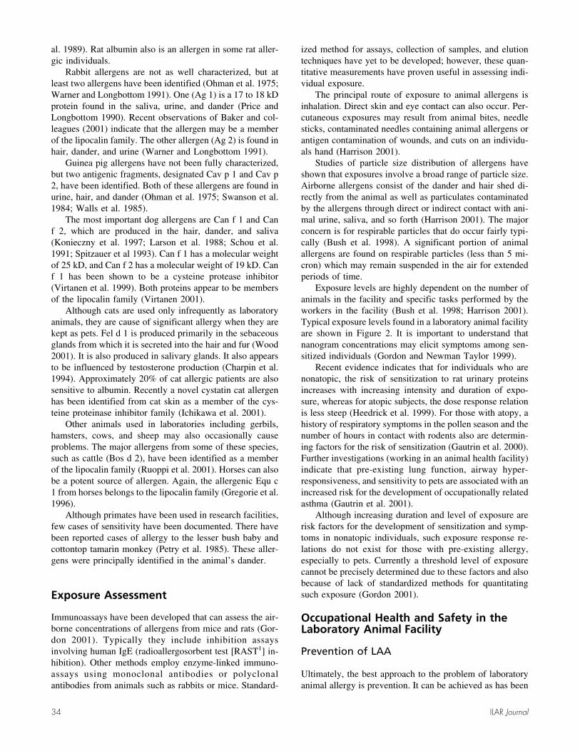
be recognized by the T-cell, it must first be processed intosmall peptide fragments and presented on the surface of theantigen-presenting cell in association with major histocom-patibility (MHC1) class II proteins (Whitton 1998). Anti-gen-presenting cells capture and internalize the protein.They migrate to draining lymph nodes where the processedpeptides are presented on the surface of the cell in associa-tion with the MHC class II molecules. Naive T-cells,through a T-cell receptor that has specificity for a particularantigenic peptide, recognize the complex of the antigen andthe MHC class II molecules. For the naive T-cell to becomeactivated, certain costimulatory signals are also necessary.The most common of these interactions is between the B7molecule (B7.1 or B7.2) on the antigen-presenting cell andits counter ligand, CD28, on the T-cell (Figure 1) (Whitton1998). The activated T-cell can then undergo multiplerounds of replication, which requires autologous productionof the cytokine interleukin (IL1)-2 and the surface expres-sion of the IL-2 receptor, CD25. Initially, a multipotentialpopulation of T-cells (Th0) are produced. There are twotypes of effector cells, each with the potential to generate aselective and mutually exclusive array of cytokines, whichdictate the type of immune response that may occur. TheTh1-type lymphocytes preferentially secrete IL-2, inter-feron-gamma (IFN�1), and tumor necrosis factor-�; andTh2-type cells produce IL-4, IL-5, IL-9, and IL-13 (Mos-mann et al. 1986; Swain 1999) (Figure 1).
The particular type of immune response that is gener-
ated depends on a variety of factors, including the type anddose of antigen, the differential expression of B7.2 versusB7.1 costimulatory molecules, and the cytokine milieu pres-ent during the initial priming of the T-cells (Jaffar et al.1999; Tsuyuki et al.1997). The most important factor ap-pears to be the presence of particular cytokines. The Th2cells are induced by the presence of IL-4, and Th1 cells areinduced in the presence of IL-12. Elicitation of a Th2 re-sponse is the typical feature of immediate-type allergic dis-eases (Holt 1999). The genes that control the Th2-type ofresponse have not been fully elucidated at present. How-ever, clear-cut genetic influences do exist based on datafrom population studies.
A small portion of Th2 cells develop into memory T-cells, which can circulate for long periods of time. Subse-quent exposure to the initial sensitizing antigen elicits avigorous and rapid response from these memory T cells.Thus, once established, a Th2-response can continue formany years or be rekindled by subsequent re-exposure tothe allergen that generated the initial response (Holt 1999).
The production of cytokines by Th2-type cells leads tothe production of specific IgE antibodies. IL-4, which is anecessary signal to B lymphocytes, induces the synthesis ofIgE antibodies by B-cells. A similar function has also beenattributed to IL-13, which has approximately 30% homol-ogy with IL-4 and shares many of its biological activities. Incontrast, IFN� suppresses the formation of IgE antibodyproduction. IgE antibody production, therefore, representsan excess of IL-4 and IL-13 and a relative absence of IFN�.Although not fully elucidated, current theory holds that al-lergic disease results from a relative lack of production ofIFN� by individuals who have the atopic trait.
IgE antibody has unique biological characteristics. It isfound in low concentrations in serum compared with im-munoglobulins IgG, IgM, and IgA. IgE has the unique prop-erty of binding, through its Fc portion, to receptors found onmast cells and basophils. These cells, which contain hista-mine and other biochemical mediators, are found in abun-dance in tissues that are the site of allergic reactions. Thesesites include the skin, conjunctiva, respiratory system, andgastrointestinal tract.
Interaction between the specific allergen, such as rat ormouse urinary protein, triggers the release of preformedmediators, such as histamine, and the generation of othervasoactive biochemical mediators, such as leukotrienes andprostaglandins from mast cells and basophils. Furthermore,the release of chemokines, such as RANTES (regulatedupon activation normal T- cell expressed and secreted) andeotaxin, results in the recruitment of inflammatory cells(particularly eosinophils) into the tissues. There, furtherrelease of leukotrienes and other mediators results in thetypical inflammation seen in allergic reactions during thelate-phase response. The biochemical mediators and inflam-matory cells contribute to the allergic symptoms. Over timethese events may lead to chronic disease states, such asasthma.
In summary, the mechanism underlying LAA involves a
Figure 1 Antigen presentation to naive T-cells requires (1) rec-ognition of antigen (AG)/major histocompatibility (MHC) com-plex by T-cell receptor (TCR), and (2) costimulatory signalsprovided through the interaction of CD28 and B7. Differentiationof T-precursor cells into Th1 or Th2 effector cells is influenced bythe presence of interleukin (IL)-4 and IL-12. Th2-type cells con-tribute to antigen-induced airway inflammation through the gen-eration of IL-4, IL-5, IL-9, and IL-13. Reprinted with permissionfrom Kiekhaefer CM, Kelly EA, Jarjour NN. 2001. Antigen-induced airway disease. In: Bush RK, ed. Environmental Asthma.New York: Marcel Dekker. p 13-31.
32 ILAR Journal
complex series of events. Genetic factors may play a role ingoverning the ability of the individual to generate an aller-gic response. Through airborne or skin contact, the allergensproduced by laboratory animals lead to their uptake by an-tigen-processing and -presenting cells. These cells in turninteract with T-lymphocytes and in the appropriate cytokinemilieu lead to the generation of Th2 CD4+ T-helper cells.The Th2-cells then elaborate cytokines, such as IL-4 andIL-13, that are involved in the production of IgE. The pro-duction of IL-5 results in maturation and enhances the re-cruitment of eosinophils into sites of allergic reactions in thetissues. Finally, the interaction of allergens and IgE resultsin the immediate and late-phase allergic response that leadsto the production of symptoms.
The Allergens
The role of the IgE antibodies in health is not completelyunderstood. However, it is of interest that in the case ofparasitic infections, specific IgE antibodies to parasite an-tigens arise in response to organisms that have a tissuemigration phase. Interactions between parasitic antigens andIgE result in the degranulation of mast cells and recruitmentof eosinophils into the site of the parasitic infection. Eo-sinophils have the capacity to kill parasites, such as schis-tosomes, in cultures. It is especially interesting to note thatmany of the allergens involved in laboratory animal aller-gies, such as mouse and rat urinary proteins and rabbitallergens (Baker et al. 2001), belong to a family of proteinstermed lipocalins (proteins involved in the transport of low
molecular weight compounds, such as urinary odorants in-volved in the sexual activity of rodents), which share se-quence homology with schistosome antigens (Virtanen et al.1999). This molecular mimicry between the urinary proteinallergens of the mouse and rat and their close relation toschistosome allergens may account, in part, for the potencyof these antigens in eliciting an IgE response in susceptibleindividuals (Virtanen et al. 1999).
Most major laboratory animal allergens have been iden-tified and characterized (Table 2). The allergens from ratsand mice cause most difficulty because they are the animalsmost often used in research facilities. At least three distinctmouse allergens have been identified and characterized(Price and Longbottom 1990; Robertson et al. 1996; Sch-umacher 1980; Siragenian and Sandberg 1979). The majormouse allergen Mus m 1, or mouse urinary protein, is aprotein with a molecular weight of 19 kD. This allergen isfound in the hair follicles and dander in addition to theurine. It is produced in liver cells, and males excrete fourtimes more of the allergen than females. A second allergen,Mus m 2, is a 16 kD molecular protein found in the hair anddander but not in the urine. Mouse albumin is also allergenicin about 30% of mouse sensitive individuals.
Two rat allergens have been identified in the urine, sa-liva, and pellet (Bayard et al. 1996; Walls and Longbottom1985). Rat n 1A was originally thought to be prealbumin,but more recent studies have demonstrated that both aller-gens are variants of an �2u-globulin. Rat n 1B is also pro-duced in the liver and is also androgen dependent. It can beproduced by the salivary, mammary, and other exocrineglands (Bayard et al. 1996; Gordon et al. 2001b; Mancini et
Table 2 Laboratory animal allergensa
Animal Allergen MWb (kD) Source Biological function
Mouse Mus m 1 (prealbumin) 19 Hair, dander, urine Lipocalin-odorant binding protein(Mus musculus) Mus m 2 16 Hair, dander Unknown
Albumin Serum Serum proteinRat Rat n 1A/Rat n 1B 18.7 Hair, dander Lipocalin-pheromone binding protein
Guinea pig Cav p 1 Hair, dander, urine Unknown(Cavia porcellus) Cav p 2 Hair, dander, urine
Rabbit Ag 1 (Price and 17 Hair, dander, saliva Possible lipocalin(Oryctolagus cuniculus) Longbottom 1990c) Hair, dander, urine
Ag 2 (Warner andLongbottom 1991)
Cat Fel d 1 38 Hair, dander, saliva Unknown(Felis domesticus) Albumin Serum Serum protein
Dog Can f 1 25 Hair, dander, saliva Lipocalin cysteine protease inhibitor(Canis familiaris) Can f 2 19 Hair, dander, saliva Lipocalin
Albumin Serum Serum protein
aAdapted from Wood RA. 2001. Laboratory animal allergens. ILAR J 42:12-16.bMW, molecular weight.cSee text for complete references.
Volume 44, Number 1 2003 33
al. 1989). Rat albumin also is an allergen in some rat aller-gic individuals.
Rabbit allergens are not as well characterized, but atleast two allergens have been identified (Ohman et al. 1975;Warner and Longbottom 1991). One (Ag 1) is a 17 to 18 kDprotein found in the saliva, urine, and dander (Price andLongbottom 1990). Recent observations of Baker and col-leagues (2001) indicate that the allergen may be a memberof the lipocalin family. The other allergen (Ag 2) is found inhair, dander, and urine (Warner and Longbottom 1991).
Guinea pig allergens have not been fully characterized,but two antigenic fragments, designated Cav p 1 and Cav p2, have been identified. Both of these allergens are found inurine, hair, and dander (Ohman et al. 1975; Swanson et al.1984; Walls et al. 1985).
The most important dog allergens are Can f 1 and Canf 2, which are produced in the hair, dander, and saliva(Konieczny et al. 1997; Larson et al. 1988; Schou et al.1991; Spitzauer et al 1993). Can f 1 has a molecular weightof 25 kD, and Can f 2 has a molecular weight of 19 kD. Canf 1 has been shown to be a cysteine protease inhibitor(Virtanen et al. 1999). Both proteins appear to be membersof the lipocalin family (Virtanen 2001).
Although cats are used only infrequently as laboratoryanimals, they are cause of significant allergy when they arekept as pets. Fel d 1 is produced primarily in the sebaceousglands from which it is secreted into the hair and fur (Wood2001). It is also produced in salivary glands. It also appearsto be influenced by testosterone production (Charpin et al.1994). Approximately 20% of cat allergic patients are alsosensitive to albumin. Recently a novel cystatin cat allergenhas been identified from cat skin as a member of the cys-teine proteinase inhibitor family (Ichikawa et al. 2001).
Other animals used in laboratories including gerbils,hamsters, cows, and sheep may also occasionally causeproblems. The major allergens from some of these species,such as cattle (Bos d 2), have been identified as a memberof the lipocalin family (Ruoppi et al. 2001). Horses can alsobe a potent source of allergen. Again, the allergenic Equ c1 from horses belongs to the lipocalin family (Gregorie et al.1996).
Although primates have been used in research facilities,few cases of sensitivity have been documented. There havebeen reported cases of allergy to the lesser bush baby andcottontop tamarin monkey (Petry et al. 1985). These aller-gens were principally identified in the animal’s dander.
Exposure Assessment
Immunoassays have been developed that can assess the air-borne concentrations of allergens from mice and rats (Gor-don 2001). Typically they include inhibition assaysinvolving human IgE (radioallergosorbent test [RAST1] in-hibition). Other methods employ enzyme-linked immuno-assays using monoclonal antibodies or polyclonalantibodies from animals such as rabbits or mice. Standard-
ized method for assays, collection of samples, and elutiontechniques have yet to be developed; however, these quan-titative measurements have proven useful in assessing indi-vidual exposure.
The principal route of exposure to animal allergens isinhalation. Direct skin and eye contact can also occur. Per-cutaneous exposures may result from animal bites, needlesticks, contaminated needles containing animal allergens orantigen contamination of wounds, and cuts on an individu-als hand (Harrison 2001).
Studies of particle size distribution of allergens haveshown that exposures involve a broad range of particle size.Airborne allergens consist of the dander and hair shed di-rectly from the animal as well as particulates contaminatedby the allergens through direct or indirect contact with ani-mal urine, saliva, and so forth (Harrison 2001). The majorconcern is for respirable particles that do occur fairly typi-cally (Bush et al. 1998). A significant portion of animalallergens are found on respirable particles (less than 5 mi-cron) which may remain suspended in the air for extendedperiods of time.
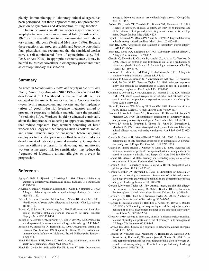
Exposure levels are highly dependent on the number ofanimals in the facility and specific tasks performed by theworkers in the facility (Bush et al. 1998; Harrison 2001).Typical exposure levels found in a laboratory animal facilityare shown in Figure 2. It is important to understand thatnanogram concentrations may elicit symptoms among sen-sitized individuals (Gordon and Newman Taylor 1999).
Recent evidence indicates that for individuals who arenonatopic, the risk of sensitization to rat urinary proteinsincreases with increasing intensity and duration of expo-sure, whereas for atopic subjects, the dose response relationis less steep (Heedrick et al. 1999). For those with atopy, ahistory of respiratory symptoms in the pollen season and thenumber of hours in contact with rodents also are determin-ing factors for the risk of sensitization (Gautrin et al. 2000).Further investigations (working in an animal health facility)indicate that pre-existing lung function, airway hyper-responsiveness, and sensitivity to pets are associated with anincreased risk for the development of occupationally relatedasthma (Gautrin et al. 2001).
Although increasing duration and level of exposure arerisk factors for the development of sensitization and symp-toms in nonatopic individuals, such exposure response re-lations do not exist for those with pre-existing allergy,especially to pets. Currently a threshold level of exposurecannot be precisely determined due to these factors and alsobecause of lack of standardized methods for quantitatingsuch exposure (Gordon 2001).
Occupational Health and Safety in theLaboratory Animal Facility
Prevention of LAA
Ultimately, the best approach to the problem of laboratoryanimal allergy is prevention. It can be achieved as has been
34 ILAR Journal
demonstrated by Fisher and colleagues (1998). At total of159 employees at a pharmaceutical research laboratory wereenrolled in the program to reduce the incidence of LAA.The program consisted of education and training, modifica-tion of work practices such as controlling the animal stackdensity and instituting wet shaving practices, and engineer-ing controls. The engineering controls that were imple-mented included the use of filter-top cages, high-efficiencyparticulate air (HEPA1) filtered room ventilation, increasedroom air exchange, and dust-free bedding. Individuals usedPPE, including the mandatory use of respiratory protection(generally dust mist respirators). A medical surveillanceprogram was included for symptom assessment and detec-tion of sensitivity by RAST for animal allergens. The medi-cal surveillance assessments were performed on an annualbasis. In addition, regular housekeeping routines were es-tablished to ensure that the work place was as clean and freefrom animal allergens as possible. At the time of the insti-tution of this comprehensive program, the annual incidenceof laboratory animal allergy was approximately 10%. At theend of 5 yr, the incidence had been reduced to 0. This studysuggests that laboratory animal allergy is potentially pre-ventable. Subsequently, the annual incidence of LAA at thatcompany has been maintained at close to 0 (Goodno andStave 2002).
A well-designed LAA management program can be use-ful in achieving this goal (Appendices A and B). Important
components of any program designed to reduce the risk oflaboratory animal allergy should include proper training andeducation of the exposed individuals. The individual em-ployed in the laboratory facility must know the proper tech-niques for working with animals and disposing of waste toreduce exposure. Training should include information onthe allergic diseases, the symptoms, and the ways to controland minimize exposures.
Preplacement Evaluation
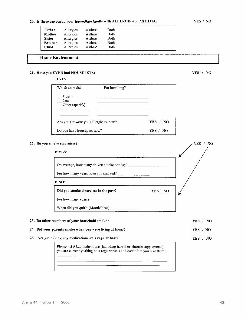
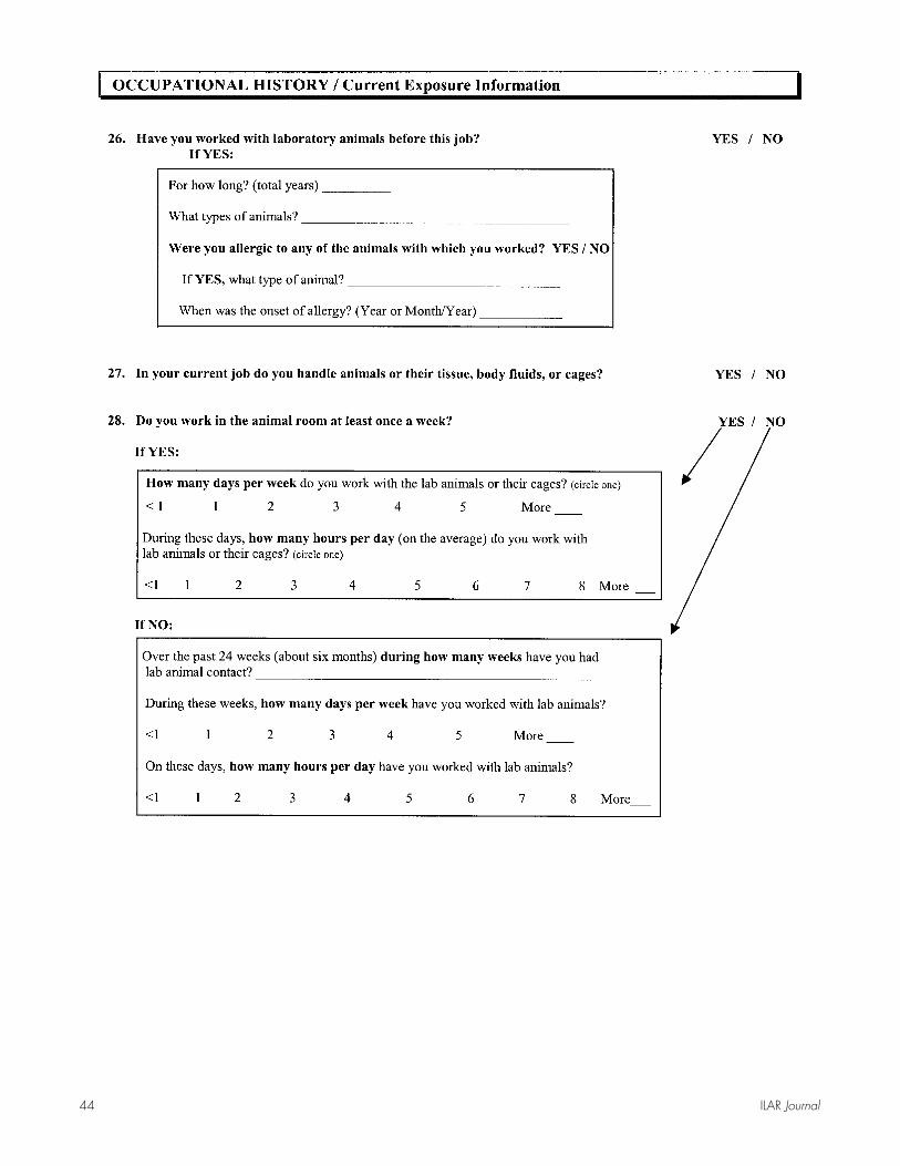
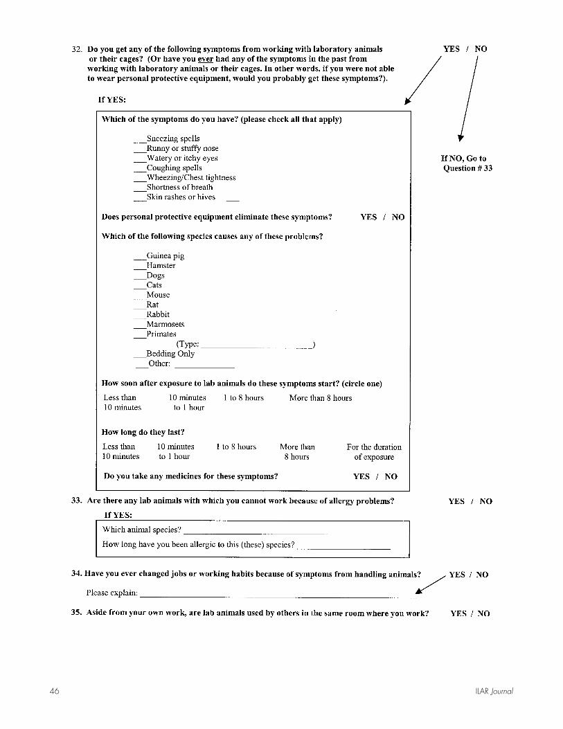
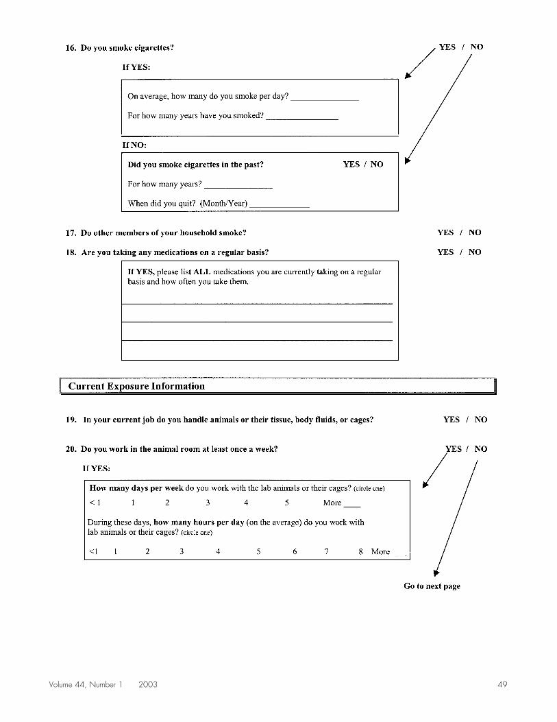
Preplacement medical evaluations may be helpful (Seward2001). The extent of the evaluation depends on the cost andavailability of such services. Questionnaires (e.g., AppendixC) can be used to assist placement of the worker in appro-priate settings that can reduce or minimize the risk of de-veloping laboratory animal allergy (Gordon et al. 2001a). Italso provides the employees the opportunity to learn abouttheir personal health risk and relationship to working withanimals and to establish a baseline against which futurechanges in health can be measured.
Skin testing to animal allergens or in vitro assays suchas the RAST to detect specific IgE antibodies may be per-formed in some instances. This would include testing toallergens with the actual laboratory animals to which theperson will be exposed but may also include other allergenssuch as pollens, molds, and pet danders that if positive,
Figure 2 Rat urinary aeroallergen (RUA) exposure of a pharmaceutical workforce by exposure group. ❍❐, estimated exposure. ��,measured exposure. ❍�, animals housed in conventional cages. ❐�, animals housed in isolators. Reprinted with permission from GordonS, Tee RD, Nieuwenhuijsen MJ, Lowson D, Harris J, Newman Taylor AJ. 1994. Measurement of airborne rat urinary allergen in anepidemiological study. Clin Exp Allergy 24:1070-1077.
Volume 44, Number 1 2003 35
could indicate the employee’s increased risk for sensitiza-tion to laboratory animal allergens (Seward 2001). In oneprospective study, the combined use of RAST plus skintesting to animal allergens was 87.4% predictive of subse-quent development of LAA after 2 yr (Botham et al. 1995).Most other studies have had less predictive value. RASTand skin tests are better utilized as diagnostic tests than asscreening tests in asymptomatic workers.
As part of the medical surveillance, annual or semi-annual evaluations using questionnaires may be con-ducted (e.g., Appendix D). If the worker starts to exhibitsymptoms of laboratory animal allergy, further evaluationmay be necessary.
Exposure Reduction
The techniques to reduce exposure include the followingcategories: (1) substitution, (2) engineering controls, (3) ad-ministrative controls, and (4) PPE. Substitution involvesusing fewer allergenic species (e.g., using female rats asopposed to male rats, using in vitro techniques as opposedto animals, or other methods). Engineering controls includeimprovement in ventilation (reviewed extensively by Har-rison 2001) and improved caging systems (Gordon et al.2001b). Administrative controls include workplace rulesand procedures (e.g., limiting access to the animal care ar-eas) and improved and required handling methods.
Examples of PPE include respirators, eye protectors,gloves, and clothing and footwear designed to reduce ex-posure. Dust mist respirators (not surgical masks, whichprovide no significant protection from LAA) for nonsymp-tomatic workers are relatively comfortable and readily usedby workers. These items can remove up to 98% of rodenturinary allergens from the air (Sakaguchi et al. 1989). Ifemployers choose to use respirators, they should also havea fully developed respiratory protective program that in-cludes quality control, medical approval for use, and testfitting. Only respirators approved by the National Instituteof Occupational Health and Safety should be used.
Gloves, gowns, and coverings are important to keepallergens off the skin. These items should be removed at theend of the work shift, when thorough hand washing andshowering may also be appropriate. Latex gloves may benecessary where infection control is important. However, ifthis risk is minimal, nonlatex gloves can be substituted sincesensitivity to latex is also a risk. Powdered latex glovesshould be avoided because they have the strongest associa-tion with the development of latex allergy.
Prevention of laboratory animal allergy depends on con-trol of the individual’s exposure in the work environment. Inaddition to exposure control technologies, PPE should beconsidered where conditions require. Preplacement evalua-tion and periodic medical surveillance of workers can alsobe an important aspect of the overall occupational healthprogram of the facility. Such medical surveillance programshave been demonstrated to reduce the risk of occupationalasthma in other settings (Tarlo and Liss 2001). The empha-
sis on medical surveillance programs should be on counsel-ing and early disease detection so that progressiveimpairments of the worker’s health do not occur.
Evaluation and Treatment of theIndividual with LAA
When individuals have allergic symptoms related to labo-ratory animal exposure, consultation with appropriate phy-sicians should be considered so that an accurate diagnosisand effective management can be achieved. For animal fa-cility personnel suspected of having allergic problems, thediagnosis of animal sensitivity is largely made on the basisof the history of symptoms in conjunction with exposure.The diagnosis is confirmed by the demonstration of specificIgE antibodies to the allergen in question.
Symptoms of allergic rhinitis include chronic conges-tion and rhinorrhea accompanied by sneezing, itchy nose orthroat, and itchy eyes. To obtain a history of asthma, theindividual should be asked about the following: wheezing,cough, chest tightness, or difficulty breathing that is epi-sodic; colds; exposure to irritants such as cigarette smoke,odors, and cold air; and allergen exposure. Symptoms typi-cally increase with exercise. Symptoms should clearly im-prove if asthma medications have been used.
To confirm a suspected diagnosis of LAA, appropriatelyperformed skin tests or in vitro assays for the presence ofIgE antibodies to laboratory animal allergens should bedone (Bush 2001; Bush et al. 1998). If asthma due to LAAis suspected, it is important to perform lung function mea-surements (Bush 2001), which may include serial peak flowmeasurements obtained in and out of the workplace. Assoon as a diagnosis of LAA or asthma has been confirmed,treatment should be directed toward removing the workerfrom continued exposure. Studies evaluating the clinicalcourse of workers with occupational asthma after removalfrom exposure have shown that persistence of the symptomsfrequently depends on the duration of symptoms before di-agnosis (Bernstein et al. 1996). The longer patients havesymptoms, the less likely they are to recover completely.With early diagnosis, prognosis is much better, lung func-tion is preserved, and the degree of nonspecific bronchialhyper-responsiveness is reduced. In contrast, individualswho remain in the workplace for longer periods of time andexperience deterioration of lung function develop chronicpersistent asthma, which often requires continued medica-tion use.
The use of PPE may be helpful in reducing or prevent-ing symptoms; however, because nanogram concentrationsof allergic exposure can induce symptoms, this approachmay not provide sufficient protection. Pharmacologicaltreatment of acute or chronic symptoms due to LAA issimilar to treatment for individuals who have nonoccupa-tional allergic disease (Bush 2001). Nonetheless, veryhighly sensitized individuals will continue to have symp-toms and must avoid exposure to animal allergens com-
36 ILAR Journal
pletely. Immunotherapy to laboratory animal allergens hasbeen performed, but these approaches may not prevent pro-gression of symptoms and deterioration of lung function.
On rare occasions, an allergic worker may experience ananaphylactic reaction from an animal bite (Teasdale et al.1993) or from needle punctures contaminated with labora-tory animal allergens (Watt and McSharry 1996). Becausethese reactions can progress rapidly and become potentiallyfatal, physicians may recommend that the sensitized workercarry a self-administered form of epinephrine (e.g., Epi-Pen® or Ana-Kit®). In appropriate circumstances, it may behelpful to instruct coworkers in emergency procedures suchas cardiopulmonary resuscitation.
Summary
As noted in Occupational Health and Safety in the Care andUse of Laboratory Animals (NRC 1997), prevention of thedevelopment of LAA should be the aim of all facilitiesengaged in the use of laboratory animals. Cooperation be-tween facility management and workers and the implemen-tation of good industrial hygiene measures aimed atpreventing exposure to inhalant material have the potentialfor reducing LAA. Workers should be educated continuallyabout the importance of adhering to appropriate proceduresthat reduce exposure. Preplacement screening of hiredworkers for allergy to other antigens such as pollens, molds,and animal danders may be considered before assigningemployees to specific jobs in an effort to reduce risks fordevelopment of laboratory animal sensitivity. Comprehen-sive surveillance programs for detecting and monitoringworkers at increased risk for sensitization may reduce thefrequency of laboratory animal allergies or prevent itsprogression.
References
Agrup G, Belin L. Sjostedt L, Skerfving S. 1986. Allergy to laboratoryanimals in laboratory technicians and animal handlers. Br J Indust Med43:192-198.
Aoyama K, Ueda A, Manda F, Matsushita T, Ueda T, Yamauchi C. 1992.Allergy to laboratory animals: an epidemiological study. Br J IndustMed 49:41-47.
Baker J, Berry A, Boscato LM, Gordon S, Walsh BJ, Stuart MC. 2001.Identification of some rabbit allergens as lipocalins. Clin Exp Allergy31:303-312.
Bayard C, Holmquist L, Vesterburg O. 1996. Purification and identifica-tion of allergenic alpha 2�-globulin species of rat urine. BiochemBiophys Acta 1290:129-134.
Beeson MF, Dewdney JM, Edwards RH, Lee D, Orr RG. 1983. Prevalenceand diagnosis of laboratory animal allergy. Clin Allergy 13:433-442.
Bland SM, Evans R III, Rivera JC. 1987. Allergy to laboratory animals inhealth care personnel. Occup Med 2:525-546.
Bland SM, Levine Ms, Wilson PD, Fox NL, Rivera JC. 1986. Occupational
allergy to laboratory animals: An epidemiologic survey. J Occup Med28:1151-1157.
Botham PA, Lamb CT, Teasdale EL, Bonner SM, Tomenson JA. 1995.Allergy to laboratory animals: A follow-up study of its incidence andof the influence of atopy and pre-existing sensitization on its develop-ment. Occup Environ Med 52:129-133.
Bryant D, Boscato LM, Mboloi PN, Stuart MC. 1995. Allergy to laboratoryanimals among animal handlers. Med J Aust 163:415-418.
Bush RK. 2001. Assessment and treatment of laboratory animal allergy.ILAR J 42:55-64.
Charpin C, Zielonka T, Charpin D, Ansaldi JL, Allasia JL, Vervloet D.1994. Effects of castration and testosterone on Fel d 1 production bysebaceous glands of male cats. I. Immunologic assessment. Clin ExpAllergy 12:1169-1173.
Cockcroft A, Edwards J, McCarthy P, Andersson N. 1981. Allergy inlaboratory animal workers. Lancet 1:827-830.
Cullinan P, Cook A, Gordon S, Nieuwenhuijsen MJ, Tee RD, VenablesKM, McDonald JC, Newman Taylor AJ. 1999. Allergen exposure,atopy and smoking as determinants of allergy to rats in a cohort oflaboratory employees. Eur Respir J 13:1139-1143.
Cullinan P, Lowson D, Nieuwenhuijsen MJ, Gordon S, Tee RD, VenablesKM. 1994. Work related symptoms, sensitization and estimated expo-sure in workers not previously exposed to laboratory rats. Occup En-viron Med 51:589-592.
Fisher R, Saunders WB, Murray SJ, Stave GM. 1998. Prevention of labo-ratory animal allergy. J Occup Environ Med 40:609-613.
Fuortes LJ, Weih L, Jones ML, Burmeister LF, Thorne PS, Pollen S,Merchant JA. 1996. Epidemiologic assessment of laboratory animalallergy among university employees. Am J Indust Med 29:67-74.
Fuortes LJ, Weih L, Pomrehn P, Thorne PS, Jones M, Burmeister L,Merchant JA. 1997. Prospective epidemiologic evaluation of laboratoryanimal allergy among university employees. Am J Ind Med 32:665-669.
Gautrin D, Ghezzo H, Infante-Rivard C, Malo J-L. 2000. Incidence anddeterminants of IgE-mediated sensitization in apprentices. A perspec-tive study. Am J Respir Crit Care Med 162:1222-1228.
Gautrin D, Infante-Rivard C, Ghezzo H, Malo J-L. 2001. Incidence andhost determinants of probable occupational asthma in apprentices ex-posed to laboratory animals. Am J Respir Crit Care Med 163:899-904.
Goodno KL, Stave GM 2002. Primary and secondary allergies to labora-tory animals. J Occup Environ Med (In Press).
Gordon S. 2001. Laboratory animal allergy: A British perspective on aglobal problem. ILAR J 42:37-46.
Gordon S, Fisher SW, Raymond RH. 2001a. Elimination of mouse aller-gens in the working environment: Assessment of individually venti-lated cage systems and ventilated cabinets in the containment of mouseallergens. J Allergy Immunol 108:288-294.
Gordon S, Newman Taylor AJ. 1999. Animal, insect, and shellfish allergy.In: Berstein IL, Chan-Yeung M, Malo J, Berstein DI, eds. Asthma inthe Workplace. 2nd ed. New York: Marcell Dekker, Inc. p 399-424.
Gordon S, Tee RD, Stuart MC, Newman Taylor AJ. 2001b. Analysis ofallergens in rat fur and saliva. Allergy 56:563-567.
Gregorie C, Rosinski-Chupin I, Rabillion J, Alzari PM, David B, DandenJ-P. 1996. cDNA cloning and sequencing reveal the major horse aller-gen Equ c1 to be a glycoprotein member of the lipocalin superfamily.J Biol Chem 271:32951-32959.
Gross NJ. 1980. Allergy to laboratory animals: Epidemiologic, chronolog-ical and physiologic aspects, and a trial of cromolyn in its management.J Allergy Clin Immunol 66:158-165.
Harrison DJ. 2001. Controlling exposure to laboratory animal allergens.ILAR J 42:17-35.
Heederik D, Venables KM, Malmberg P, Hollander A, Karlsson A-S,Renstrom A, Doekes G, Nieuwenhuijsen M, Gordon S. 1999. Expo-sure-response relationship for work-related sensitization in workers ex-posed to rat urinary allergens: Results from a pooled study. J AllergyClin Immunol 103:678-684.
Volume 44, Number 1 2003 37
Hollander A, Doekes G, Heederik D. 1996. Cat and dog allergy and totalIgE as risk factors of laboratory animal allergy. J Allergy Clin Immunol98:545-554.
Holt PG. 1999. Development of T-cell memory against inhalant allergens:Risks for future. Clin Exp Allergy 29(Suppl 2):8-13.
Hunskaar S, Fosse RT. 1990. Allergy to laboratory mice and rats: A reviewof the pathophysiology, epidemiology and clinical aspects. Lab Anim34:358-374.
Ichikawa K, Vailes LD, Pomes A, Chapman MD. 2001. Identification of anovel cat allergen-cystatin. Int Arch Allergy Immunol 124:55-56.
Jaffar Z, Roberts K, Pandit A, Linsley P, Djukanovic R, Holgate S. 1999.B7 costimulation is required for IL-5 and IL-13 secretion by bronchialbiopsy tissue of atopic asthmatic subjects in response to allergen stimu-lation. Am J Respir Cell Mol Biol 20:153-162.
Kiekhaefer CM, Kelly EA, Jarjour NN. 2001. Antigen-induced airwaydisease. In: Bush RK, ed, Environmental Asthma. New York: MarcelDekker, p 13-31.
Konieczny A, Morgenstern JP, Bizinkauskas CB, Lilkley CH, Brauer AW,Bond JF. 1997. The major dog allergen, Can f 1 and Can f 2, aresalivary lipocalin proteins. Immunology 92:577-586.
Larson JN, Ford A, Gjesing B, Levy D, Petrunov B, Silvestri L. 1988. Thecollaborative study of the international standard of dog, Canis domes-ticus, hair/dander extract. J Allergy Clin Immunol 82:318-325.
Lutsky IK, Neuman I. 1975. Laboratory animal dander allergy. I. Anoccupational disease. Ann Allergy 35:201-205.
Mancini MA, Majumdar D, Chatterjee B, Roy AK. 1989. �2u-Globulin inmodified sebaceous glands with pheremonal functions. J HistochemCytochem 37:149-157.
Monso E, Malo J-L, Infante-Rivard C, Ghezzo H, Magnan M, LArchev-eque J, Trudeau C, Gautrin D. 2000. Individual characteristics andquitting in apprentices exposed to high-molecular-weight agents. Am JRespir Crit Care Med 161:1508-1512.
Mosmann TR, Cherwinski H, Bond MW, Giedlin MA, Coffman RH. 1986.Two types of murine helper T-cell clones. Definition according toprofiles of lymphokine activities and secreted proteins. J Immunol136:2348-2357.
NRC [National Research Council]. 1997. Occupational Health and Safetyin the Care and Use of Research Animals. Washington DC: NationalAcademy Press.
Ohman JL, Lowell FC, Bloch KJ. 1975. Allergens of mammalian origin.Characterization of allergens extracted from rat, mouse, guinea pig, andrabbit pelts. J Allergy Clin Immunol 55:16-24.
Patel NJ, Olson P, Lumby D, Fine JP, Bush RK. 2000. Laboratory animalallergy. (Abstract). J Allergy Clin Immunol 105:S372.
Price JA, Longbottom J. 1990. Allergy to mice. Further characterization oftwo major mouse allergens (Ag 1 and Ag 3) and immunohistochemicalinvestigations of their sources. Clin Exp Allergy 20:71-77.
Renstrom A, Malmberg P, Larsson K, Sundblad B-M, Larsson PH. 1994.Prospective study of laboratory-animal allergy: Factors predisposing tosensitization and development of allergic symptoms. Allergy 49:548-552.
Robertson DHL, Cox KA, Gaskell SJ, Evershed RP, Benyon RJ. 1996.Molecular heterogeneity in the major mouse urinary proteins on thehouse mouse Mus musculus. Biochem J 316:265-272.
Ruoppi P, Virtanen T, Zeiler T, Rytkonen-Nissinen M, Rautianen J,Juutinen J, Taivainen A. 2001. In vitro and in vivo responses to therecombinant bovine dander allergen Bos d2 and its fragments. Clin ExpAllergy 31:915-919.
Sakaguchi M, Inouye S, Miyazawa H, Kamimura H, Kimura M, YamazakiS. 1989. Evaluation of dust respirators for elimination of mouse aller-gens. Lab Anim Sci 39:63-66.
Schou C, Svendsen VG, Lowenstein H. 1991. Purification and character-ization of the major dog allergen, Can f 1. Clin Exp Allergy 21:321-328.
Schumacher MJ. 1980. Characterization of allergens from urine and peltsof laboratory mice. Mol Immunol 17:1087-1095.
Schumacher MU, Tait BD, Holmes MC. 1981. Allergy to murine allergensin a biological research institute. J Allergy Clin Immunol 68:310-318.
Seward JP. 1999. Occupational allergy to animals. Occup Med 14:285-302.Seward JP. 2001. Medical surveillance of allergy in laboratory animal
handlers. ILAR J 42:47-54.Shearer WT, Fleischer TA. 1998. The immune system. In: Middleton E Jr,
Reed CE, Ellis EF, Adkinson NF Jr, Yunginger JW, Busse WW, eds.Allergy Principles and Practice. St. Louis: Mosby-Yearbook Inc. p1-13.
Siraganian R, Sandberg A. 1979. Characterization of mouse allergens. JAllergy Clin Immunol 63:435-442.
Slovak AJM, Hill RN. 1981. Laboratory animal allergy: A clinical surveyof an exposed population. Br J Indust Med 38:38-41.
Sorrell AH, Gottesman J. 1957. Mouse allergy—A case report. Ann Al-lergy 15:662-663.
Spitzauer S, Schweiger C, Anrather J, Ebner C, Scheiner O, Kraft D. 1993.Characterization of dog allergens by means of immunoblotting. IntArch Allergy Immunol 100:60-67.
Swain SL. 1999. Helper T cell differentiation. Curr Opin Immunol 11:180-185.
Swanson M, Agarwal M, Yunginger J, Reed C. 1984. Guinea pig derivedallergens. Clinicoimmunologic studies. Characterization, airbornequantification and size distribution. Am Rev Respir Dis 129:844-849.
Tarlo SM, Liss GM. 2001. Can medical surveillance measures improve theoutcome of occupational asthma? J Allergy Clin Immunol 107:583-585.
Teasdale EL, Davies EG, Slovak R. 1993. Anaphylaxis after bites byrodents. Br Med J 286:1480.
Tsuyuki S, Tsuyuki J, Einsle K, Kopf M, Coyle AJ. 1997. Costimulationthrough B7-2 (CD86) is required for the induction of a lung mucosal Thelper cell 2 (TH2) immune response and altered airway responsive-ness. J Exp Med 185:1671-1679.
Venables KM, Upton JL, Hawkins ER, Tee RD, Longbottom JL, NewmanTaylor AJ. 1988. Smoking, atopy, and laboratory animal allergy. Br JIndust Med 45:667-671.
Virtanen T. 2001. Lipocalin allergens. Allergy 56(Suppl 67):48-51.Virtanen T, Zeiler T, Mantyjarvi R. 1999. Important animal allergens are
lipocalin proteins: Why are they allergenic? Int Arch Allergy Immunol120:247-258.
Walls A, Longbottom J. 1985. Comparison of rat fur, saliva, and other ratallergen extracts by skin testing, RAST, and RAST inhibition. J AllergyClin Immunol 75:242-251.
Walls A, Taylor A, Longbottom J. 1985. Allergy to guinea pigs. II. Iden-tification of specific allergens in guinea pig dust by crossed radioim-munoelectrophoresis and investigation of possible origin. Clin Allergy15:535-546.
Warner JA, Longbottom J. 1991. Allergy to rabbits. Allergy 46:481-491.Watt AD, McSharry CP. 1996. Laboratory animal allergy: Anaphylaxis
from a needle injury. Occup Environ Med 53:573-574.Whitton JL. 1998. An overview of antigen presentation and its central role
in the immune response. Curr Topics Microbiol Immunol 232:1-14.Wood RA. 2001. Laboratory animal allergens. ILAR J 42:12-16.
38 ILAR Journal
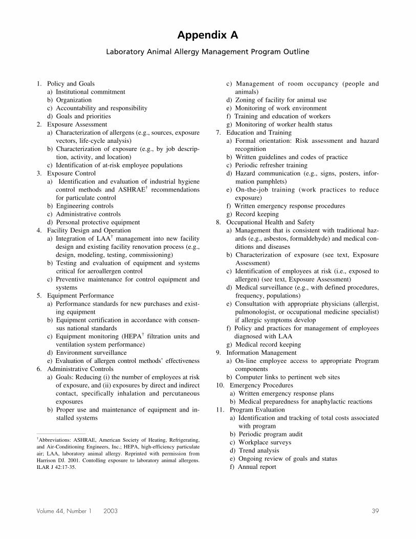
Appendix ALaboratory Animal Allergy Management Program Outline
1. Policy and Goalsa) Institutional commitmentb) Organizationc) Accountability and responsibilityd) Goals and priorities
2. Exposure Assessmenta) Characterization of allergens (e.g., sources, exposure
vectors, life-cycle analysis)b) Characterization of exposure (e.g., by job descrip-
tion, activity, and location)c) Identification of at-risk employee populations
3. Exposure Controla) Identification and evaluation of industrial hygiene
control methods and ASHRAE† recommendationsfor particulate control
b) Engineering controlsc) Administrative controlsd) Personal protective equipment
4. Facility Design and Operationa) Integration of LAA† management into new facility
design and existing facility renovation process (e.g.,design, modeling, testing, commissioning)
b) Testing and evaluation of equipment and systemscritical for aeroallergen control
c) Preventive maintenance for control equipment andsystems
5. Equipment Performancea) Performance standards for new purchases and exist-
ing equipmentb) Equipment certification in accordance with consen-
sus national standardsc) Equipment monitoring (HEPA† filtration units and
ventilation system performance)d) Environment surveillancee) Evaluation of allergen control methods’ effectiveness
6. Administrative Controlsa) Goals: Reducing (i) the number of employees at risk
of exposure, and (ii) exposures by direct and indirectcontact, specifically inhalation and percutaneousexposures
b) Proper use and maintenance of equipment and in-stalled systems
c) Management of room occupancy (people andanimals)
d) Zoning of facility for animal usee) Monitoring of work environmentf) Training and education of workersg) Monitoring of worker health status
7. Education and Traininga) Formal orientation: Risk assessment and hazard
recognitionb) Written guidelines and codes of practicec) Periodic refresher trainingd) Hazard communication (e.g., signs, posters, infor-
mation pamphlets)e) On-the-job training (work practices to reduce
exposure)f) Written emergency response proceduresg) Record keeping
8. Occupational Health and Safetya) Management that is consistent with traditional haz-
ards (e.g., asbestos, formaldehyde) and medical con-ditions and diseases
b) Characterization of exposure (see text, ExposureAssessment)
c) Identification of employees at risk (i.e., exposed toallergen) (see text, Exposure Assessment)
d) Medical surveillance (e.g., with defined procedures,frequency, populations)
e) Consultation with appropriate physicians (allergist,pulmonologist, or occupational medicine specialist)if allergic symptoms develop
f) Policy and practices for management of employeesdiagnosed with LAA
g) Medical record keeping9. Information Management
a) On-line employee access to appropriate Programcomponents
b) Computer links to pertinent web sites10. Emergency Procedures
a) Written emergency response plansb) Medical preparedness for anaphylactic reactions
11. Program Evaluationa) Identification and tracking of total costs associated
with programb) Periodic program auditc) Workplace surveysd) Trend analysise) Ongoing review of goals and statusf) Annual report
†Abbreviations: ASHRAE, American Society of Heating, Refrigerating,and Air-Conditioning Engineers, Inc.; HEPA, high-efficiency particulateair; LAA, laboratory animal allergy. Reprinted with permission fromHarrison DJ. 2001. Contolling exposure to laboratory animal allergens.ILAR J 42:17-35.
Volume 44, Number 1 2003 39
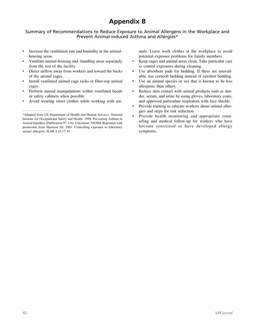
Appendix BSummary of Recommendations to Reduce Exposure to Animal Allergens in the Workplace and
Prevent Animal-induced Asthma and Allergies*
• Increase the ventilation rate and humidity in the animal-housing areas.
• Ventilate animal-housing and -handling areas separatelyfrom the rest of the facility.
• Direct airflow away from workers and toward the backsof the animal cages.
• Install ventilated animal cage racks or filter-top animalcages.
• Perform animal manipulations within ventilated hoodsor safety cabinets when possible.
• Avoid wearing street clothes while working with ani-
mals. Leave work clothes at the workplace to avoidpotential exposure problems for family members.
• Keep cages and animal areas clean. Take particular careto control exposures during cleaning.
• Use absorbent pads for bedding. If these are unavail-able, use corncob bedding instead of sawdust bedding.
• Use an animal species or sex that is known to be lessallergenic than others.
• Reduce skin contact with animal products sush as dan-der, serum, and urine by using gloves, laboratory coats,and approved particulate respirators with face shields.
• Provide training to educate workers about animal aller-gies and steps for risk reduction.
• Provide health monitoring and appropriate coun-seling and medical follow-up for workers who havebecome sensitized or have developed allergysymptoms.
*Adapted from US Department of Health and Human Services, NationalInstitute for Occupational Safety and Health. 1998. Preventing Asthma inAnimal Handlers (Publication 97-116). Cincinnati: NIOSH. Reprinted withpermission from Harrison DJ. 2001. Controlling exposure to laboratoryanimal allergens. ILAR J 42:17-35.