42
Armando Tripodi Angelo Bianchi Bonomi Hemophilia and Thrombosis Center IRCCS Maggiore Hospital and Humanitas University, Milano Italy Laboratory Diagnosis of APS

Armando TripodiAngelo Bianchi Bonomi
Hemophilia and Thrombosis CenterIRCCS Maggiore Hospital andHumanitas University, Milano
Italy
Laboratory Diagnosis of APS

Antiphospholipid SyndromeCondition defined by clinical and lab criteria
• Laboratory criteria
- Lupus anticoagulant and/or solid-phase antiphospholipid antibody positive tests (confirmed on 2 occasions 12 weeks apart)
• Clinical criteria
- Pregnancy complications, venous and/or arterial thrombosis

Solid-phase Antiphospholipid Antibodies
• Which Test(s)
– Anti-cardiolipin
– Anti-β2GPI
– Anti-PS/PT
• Which Isotype(s)
- IgG
- IgM

There are many commercial assays available for aCL or a-ß2GPI
But they are not yet standardized
across laboratories

Assays for aCL and a-ß2GPI
• Many commercial ELISA-based assays
– Poorly standardized. Gross degree of variation across labs
• Chemiluminescence-based assays
– Closed systems (easy handling of reagents and samples)

Issues on LA Detection
• Who should be tested
• Which test(s)
• Diagnostic criteria
• When testing
• Results reporting
• Interpretation

Indications to search for APS
• Occurrence of (accidentally-found) prolongation of the APTT without known etiology
• Patients with venous and/or arterial thrombosis at young age (<50 years)
• Patients with thrombosis at unusual sites, or associated with autoimmune diseases
• Women with pregnancy complications

Issues on LA Detection
• Who should be tested
• Which test(s)
• Diagnostic Criteria
• When testing
• Results reporting
• Interpretation

Which Test
• Two tests based on different principles
- dRVVT
- Sensitive aPTT-based test (low phospholipids and silica as activator)
LA should be considered as positive if at least one of the two tests is positive

Issues on LA Detection
• Who should be tested
• Which test(s)
• Diagnostic Criteria
• When testing
• Results reporting
• Interpretation

Diagnostic Criteria for LA Detection
• Screening
- Prolongation of phospholipid-dependent clotting test
• Mixing
- Evidence that the prolongation is due to the presence of an inhibitor
• Confirmation
- Evidence that the inhibitor is directed against phospholipids

ScreeningHow to determine cut-off values
• Perform testing on plasmas from healthy donors
• Take the cut-off as the value above the 99th percentile of the distribution

MixingHow to determine cut-off values
• Perform testing on plasmas from healthy donors, mixed with PNP at 1:1 proportion
• Take the cut-off as the value above the 99th
percentile of the distribution
• Alternatively, the cut-off may be the value of the ICA defined according to:
ICA = [(CTmix- CTPNP/CTpatient)]x100

ConfirmationHow to determine cut-off values
• Perform testing on plasmas from healthy donors at low (screen) and high (confirm) phospholipid concentrations
• Take the cut-off as the value above the 99th
percentile of the distribution of the individual % corrections calculated according to:
% Corr. = [(screen - confirm)/screen] x 100

• Major variations were observed even within the same platform
• Cut-off values determined in any given lab are not necessarily interchangeable
• Cut-off values for LA detection were calculated in 11 labs each testing plasma from 120 donors with 3 commercial platforms
Essentials

Issues on LA Detection
• Who should be tested
• Which test(s)
• Diagnostic Criteria
• When testing
• Results reporting
• Interpretation

When Testing
• Problem
- Results interpretation is difficult during acute thrombosis and/or during antithrombotic drugs
• Recommendation
- Blood should be collected before starting anticoagulation or after a sufficient period from its discontinuation

Effect of Anticoagulation on LA Testing
• Heparin mimics LA
–Many LA tests do contain heparin neutralizers
• LMWH may mimic LA
– Depending on the brand of LMWH used
– Especially at peak
• VKA give rise to false-positive (or negative) LA
• DOAC give rise to false-positive LA

Approaches to Overcome Anticoagulation
• Dilution (1:1) of patient plasma into pooled normal plasma (PNP)
• Integrated assays (screen and confirm)
• Tests (reportedly) less affected by anticoagulants
• Antidotes or neutralizers to quench in vitro the activity of anticoagulants
• Discontinuation of anticoagulation

Dilution (1:1) of patient plasma into PNP
• Rationale
– Deficiency of coagulation will be corrected by the PNP
• Limitations
– Applicable only to VKA
– Good quality PNP
– Dilution reduces (by 50%) the LA potency
– Correction by PNP is dependent on the APTT or dRVVTused for testing
– No conclusive evidence on the value of the procedure
– False-negative or false-positive LA should be [email protected]

Approaches to Overcome Anticoagulation
• Dilution (1:1) of patient plasma into pooled normal plasma (PNP)
• Integrated assays (screen and confirm)
• Tests (reportedly) less affected by anticoagulants
• Antidotes or neutralizers to quench in vitro the activity of anticoagulants
• Discontinuation of anticoagulation

Low PL
Presence of LAClotting time Prolonged
High PLShortned
Presence of LAClotting time
Schematic representation of Integrated LA Test

Integrated LA Tests
• Earlier reports suggested that screen and confirm integrated tests in the presence of VKA or UFH are proportionally prolonged
• Hence, they are reliable for the majority of patients even in the presence of UFH or VKA
• Later reports showed that screen and confirm in the presence of DOAC are not proportionally prolonged
• Screen tends to be more prolonged than confirm
• Consequently, the ratio screen/confirm tends to be higher than expected and may lead to false-positive LA

Approaches to Overcome Anticoagulation
• Dilution (1:1) of patient plasma into pooled normal plasma (PNP)
• Integrated assays (screen and confirm)
• Tests (reportedly) less affected by anticoagulants
• Antidotes or neutralizers to quench in vitro the activity of anticoagulants
• Discontinuation of anticoagulation

Tests (reportedly) less affected by anticoagulants
• Snake venoms (Taipan & Ecarin) might be useful to detect LA during anticoagulation, as they are able to activate FII
• Taipan is a PL- and calcium-dependent activator, whilst Ecarin is not
• If used in combination they may help detecting LA during anticoagulation
• There is information from literature on their diagnostic efficacy on patients on VKA, but not conclusive evidence

Approaches to Overcome Anticoagulation
• Dilution (1:1) of patient plasma into pooled normal plasma (PNP)
• Integrated assays (screen and confirm)
• Tests (reportedly) less affected by anticoagulants
• Antidotes or neutralizers to quench in vitro the activity of anticoagulants
• Discontinuation of anticoagulation

Antidotes/Neutralizers to Quench Anticoagulants
• Idarucizumab added in vitro to neutralize dabigatran
• Andexanet alfa added in vitro to neutralize anti-FXa drugs
• DOAC-Stop®, or DOAC-Remove®
– Activated charcoal added in vitro to adsorb DOAC

Thromb Res 2019; 180:10-19
EssentialsRivaroxaban caused clotting time prolongation for most LA tests and generated falsely
elevated dRVVT screen/confirm ratio results that mimicked the presence of LA
Rivaroxaban plasma treated with DOAC-Stop showed correction of the clotting time
Prolongation and the screen/confirm ratio for most LA tests
Participants in the study correctly identified the rivaroxaban plasma treated with
DOAC-Stop as LA-negative
Andexanet-alfa corrected the prolonged clotting time induced by rivaroxaban,
but displayed over-correction of the screen/confirm ratio

Approaches to Overcome Anticoagulation
• Dilution (1:1) of patient plasma into pooled normal plasma (PNP)
• Integrated assays (screen and confirm)
• Tests (reportedly) less affected by anticoagulants
• Antidotes or neutralizers to quench in vitro the activity of anticoagulants
• Discontinuation of anticoagulation
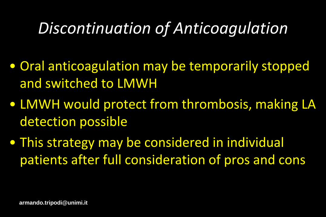
Discontinuation of Anticoagulation
• Oral anticoagulation may be temporarily stopped and switched to LMWH
• LMWH would protect from thrombosis, making LA detection possible
• This strategy may be considered in individual patients after full consideration of pros and cons

Issues on LA Detection
• Who should be tested
• Which test(s)
• Diagnostic Criteria
• When testing
• Results reporting
• Interpretation

Results Reporting
LA detection should be reported with analytical results and an interpretative comment
(i.e., LA yes, or no)

Issues on LA Detection
• Who should be tested
• Which test(s)
• Diagnostic Criteria
• When testing
• Results reporting
• Interpretation

Clinical interpretation of results
• Interpretation should consider the results of all the three tests
- The syndrome is defined if at least one of the tests (LA, aCL or aß2GPI) is positive
- Positivity for all the three tests (triple positivity) identify patients at very high risk

LA DetectionMain unresolved issues
• Standardization of existing procedures
- Application of SSC guidelines
• Urgent need for LA specific tests
- Understanding of pathogenic mechanisms may help
• Tests able to distinguish LA patients who develop clinical events from those who do not
- dRVVT better than APTT-based tests ?
- aβ2-GPI domain I
• Quantification of LA potency
- Establishment of “international standards” ?

Summary & Conclusions
• Accuracy of lab diagnosis is essential as APS patients are candidates for long term anticoagulation
• Diagnosis should be established far from acute events and off therapy
• APS requires one the following
‒ Positive APTT-based or dRVV tests
‒ aCL (aß2GPI-dependent) IgG or IgM above normal limits
‒ aß2GPI, IgG or IgM above normal limits
• Triple positivity identify patients at high risk