Diseases in Asian Aquaculture VII 63 Shaharom-Harrison, F., Seman, N.A. and Suhairi, M. 2011. Laboratory experimental infection of the freshwater snail Gyraulus convexiusculus (Hutton, 1849) and the bighead carp Aristichthys nobilis (Richardson, 1845) with the blood fluke Sanguinicola armata Plehn, 1905 pp. 63-72. In Bondad-Reantaso, M.G., Jones, J.B., Corsin, F. and Aoki, T. (eds.). Diseases in Asian Aquaculture VII. Fish Health Section, Asian Fisheries Society, Selangor, Malaysia. 385 pp. Corresponding author: Faizah Shaharom-Harrison, [email protected]Laboratory experimental infection of the freshwater snail Gyraulus convexiusculus (Hutton, 1849) and the bighead carp Aristichthys nobilis (Richardson, 1845) with the blood fluke Sanguinicola armata Plehn, 1905 FAIZAH SHAHAROM-HARRISON, NOR ASILAH SEMAN and SUHAIRI, M. Institute of Tropical Aquaculture, University Malaysia Terengganu, Kuala Terengganu, Terengganu, Malaysia 21030 ABSTRACT The route of cercarial penetration of Sanguinicola armata into the snail intermediate host Gyraulus convexiusculus and the fish definitive host Aristichthys nobilis was demonstrated through laboratory experimental infection experiments and histopathological studies. For snail infection experiments, fifty definitive host (A. nobilis) infected with the the blood trematode Sanguinicola armata were exposed to 5 naïve G. convexiusculus for 24 hours before snails were moved to another aerated aquarium which was filled with dechlorinated water and Hydrilla sp. Ten snails were sacrificed daily, their carapaces were removed from the tissues and wet smears were done and observed under compound microscope. This procedure was continued until day 17 for 17 replicates. Meanwhile, five snails were fixed in 10% formalin EDTA at days 5, 10, and 15 and snail tissue was processed for histopathology. For production and collection of cercariae, infected G. convexiusculus with S. armata were exposed to light to stimulate emergence of cercariae. Ten collected cercariae were introduced to one fish for 24 hours in 100 l fibreglass tanks. The same procedure was repeated for 240 fish. Three infected fish were examined daily until day 80. For penetration study of S. armata cercariae into A. nobilis, ten groups comprising ten naïve fishes were exposed to 300 cercariae at different time duration of 10, 20, 30, 40, 50 minutes and 1, 3, 6, 12 and 24 hours exposure. Each group was kept in 100 l fibreglass tanks. The fish from each group were divided into two. Some were cut vertically and some horizontally to get sections through all tissues. Then they were fixed in 10% buffered formalin for 48 hours and formalin EDTA for one week before histology studies. Results indicated that the majority of blood fluke adults were found in the bulbous arteriosus and histopathological results
Transcript
Diseases in Asian Aquaculture VII
63
Shaharom-Harrison, F., Seman, N.A. and Suhairi, M. 2011. Laboratory experimental infection of the freshwater snail Gyraulus convexiusculus (Hutton, 1849) and the bighead carp Aristichthys nobilis (Richardson, 1845) with the blood fluke Sanguinicola armata Plehn, 1905 pp. 63-72. In Bondad-Reantaso, M.G., Jones, J.B., Corsin, F. and Aoki, T. (eds.). Diseases in Asian Aquaculture VII. Fish Health Section, Asian Fisheries Society, Selangor, Malaysia. 385 pp.
Laboratory experimental infection of the freshwater snail Gyraulus convexiusculus (Hutton, 1849) and the bighead
carp Aristichthys nobilis (Richardson, 1845) with the blood fluke Sanguinicola armata Plehn, 1905
FAIZAH SHAHAROM-HARRISON, NOR ASILAH SEMAN and SUHAIRI, M.
Institute of Tropical Aquaculture, University Malaysia Terengganu, Kuala Terengganu, Terengganu, Malaysia 21030
ABSTRACT
The route of cercarial penetration of Sanguinicola armata into the snail intermediate host Gyraulus convexiusculus and the fish definitive host Aristichthys nobilis was demonstrated through laboratory experimental infection experiments and histopathological studies. For snail infection experiments, fifty definitive host (A. nobilis) infected with the the blood trematode Sanguinicola armata were exposed to 5 naïve G. convexiusculus for 24 hours before snails were moved to another aerated aquarium which was filled with dechlorinated water and Hydrilla sp. Ten snails were sacrificed daily, their carapaces were removed from the tissues and wet smears were done and observed under compound microscope. This procedure was continued until day 17 for 17 replicates. Meanwhile, five snails were fixed in 10% formalin EDTA at days 5, 10, and 15 and snail tissue was processed for histopathology. For production and collection of cercariae, infected G. convexiusculus with S. armata were exposed to light to stimulate emergence of cercariae. Ten collected cercariae were introduced to one fish for 24 hours in 100 l fibreglass tanks. The same procedure was repeated for 240 fish. Three infected fish were examined daily until day 80. For penetration study of S. armata cercariae into A. nobilis, ten groups comprising ten naïve fishes were exposed to 300 cercariae at different time duration of 10, 20, 30, 40, 50 minutes and 1, 3, 6, 12 and 24 hours exposure. Each group was kept in 100 l fibreglass tanks. The fish from each group were divided into two. Some were cut vertically and some horizontally to get sections through all tissues. Then they were fixed in 10% buffered formalin for 48 hours and formalin EDTA for one week before histology studies. Results indicated that the majority of blood fluke adults were found in the bulbous arteriosus and histopathological results
Shaharom-Harrison et al.
64
showed that adult S. armata caused increased thickness of gill arteries, while infection with eggs of the trematode caused hyperplasia of primary gill lamellae and filaments and secondary gill lamellae were fused resulting in chronic hyperplasia. This study on cercarial penetration of A. nobilis juveniles showed that the most abundant cercarial penetration into A. nobilis occurred at intervals of 30 minutes, 40 minutes and 180 minutes. Most penetration occurred at the caudal fin followed by abdominal and caudal peduncle, whereas least penetration site was the operculum.
Studies on the life cycle of blood flukes have been studied by several authors (Meade and Pratt, 1965; Meade, 1967; Schell, 1974). Kua (1995) showed that Gyraulus convexiusculus is an intermediate host of the blood fluke Sanguinicola armata. Even though Kua (1995) did laboratory experimental infection of the snail, she did not show the route of penetration of the cercaria into the snail and grasscarp fingerlings. In this present, study histopathology studies on snails and fish subjected to laboratory experimental infections proved the route of cercarial penetration into both snail and bighead carp fingerlings as this blood fluke was also found to infect the bighead carp Aristichthys nobilis.
MATERIALS AND METHODS
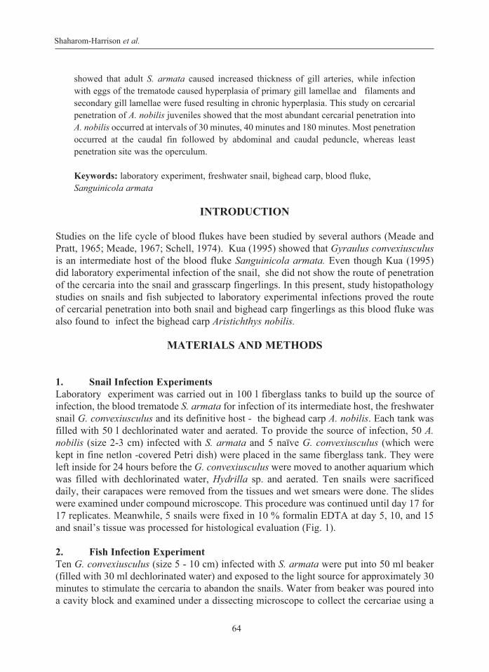
1. Snail Infection Experiments Laboratory experiment was carried out in 100 l fiberglass tanks to build up the source of infection, the blood trematode S. armata for infection of its intermediate host, the freshwater snail G. convexiusculus and its definitive host - the bighead carp A. nobilis. Each tank was filled with 50 l dechlorinated water and aerated. To provide the source of infection, 50 A. nobilis (size 2-3 cm) infected with S. armata and 5 naïve G. convexiusculus (which were kept in fine netlon -covered Petri dish) were placed in the same fiberglass tank. They were left inside for 24 hours before the G. convexiusculus were moved to another aquarium which was filled with dechlorinated water, Hydrilla sp. and aerated. Ten snails were sacrificed daily, their carapaces were removed from the tissues and wet smears were done. The slides were examined under compound microscope. This procedure was continued until day 17 for 17 replicates. Meanwhile, 5 snails were fixed in 10 % formalin EDTA at day 5, 10, and 15 and snail’s tissue was processed for histological evaluation (Fig. 1).
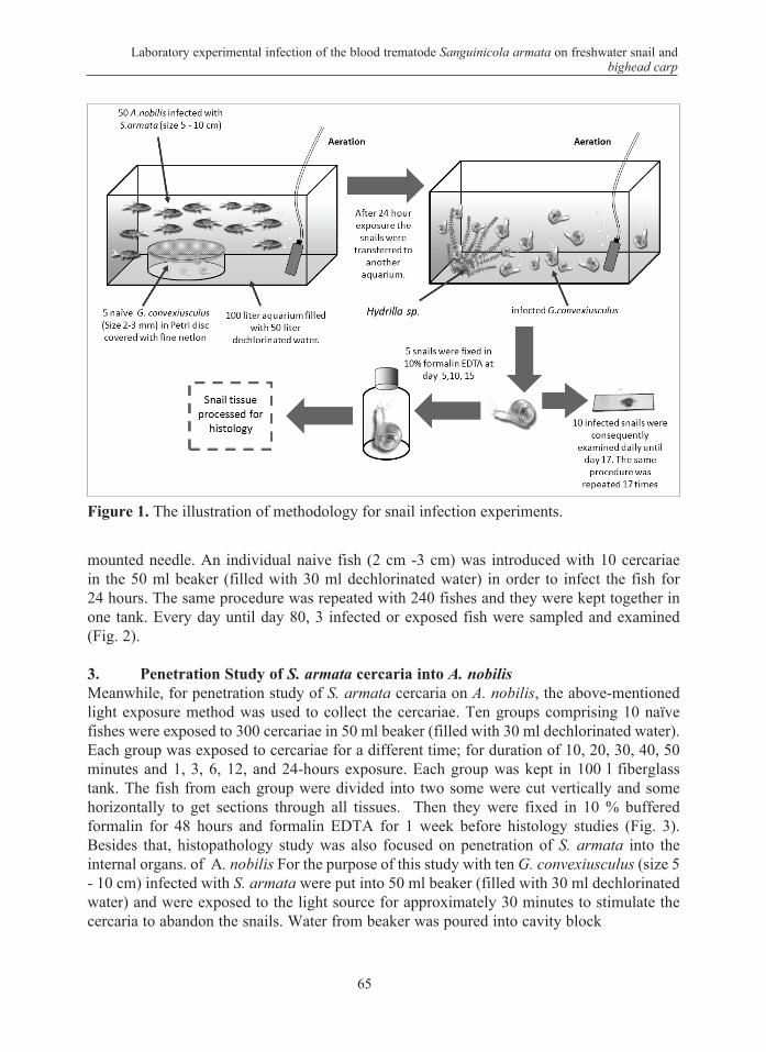
2. Fish Infection Experiment Ten G. convexiusculus (size 5 - 10 cm) infected with S. armata were put into 50 ml beaker (filled with 30 ml dechlorinated water) and exposed to the light source for approximately 30 minutes to stimulate the cercaria to abandon the snails. Water from beaker was poured into a cavity block and examined under a dissecting microscope to collect the cercariae using a
Laboratory experimental infection of the blood trematode Sanguinicola armata on freshwater snail and bighead carp
65
mounted needle. An individual naive fish (2 cm -3 cm) was introduced with 10 cercariae in the 50 ml beaker (filled with 30 ml dechlorinated water) in order to infect the fish for 24 hours. The same procedure was repeated with 240 fishes and they were kept together in one tank. Every day until day 80, 3 infected or exposed fish were sampled and examined (Fig. 2).
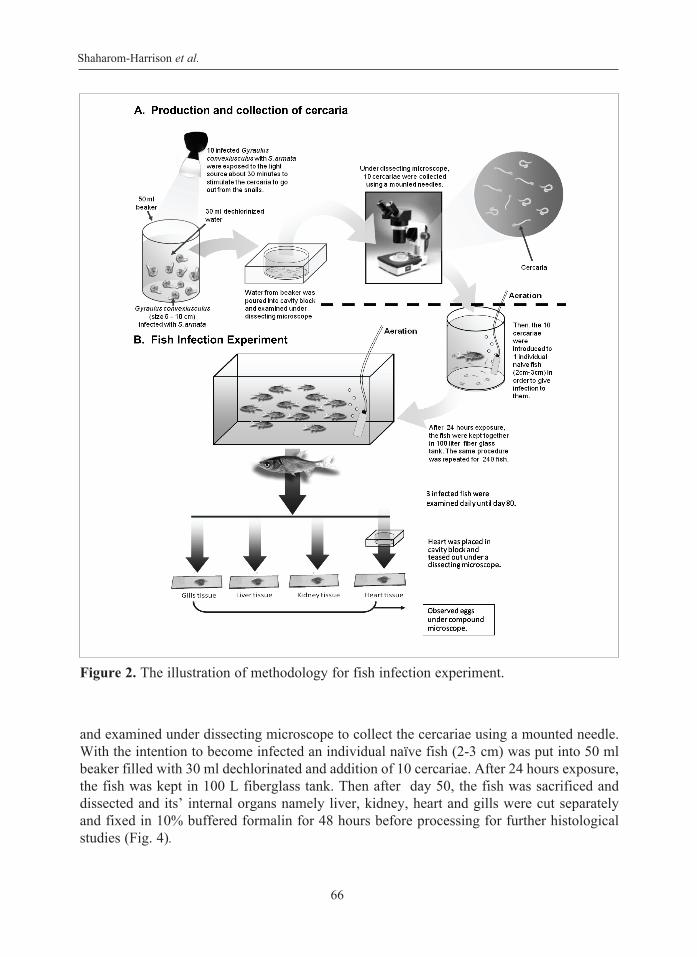
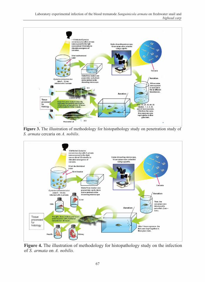
3. Penetration Study of S. armata cercaria into A. nobilisMeanwhile, for penetration study of S. armata cercaria on A. nobilis, the above-mentioned light exposure method was used to collect the cercariae. Ten groups comprising 10 naïve fishes were exposed to 300 cercariae in 50 ml beaker (filled with 30 ml dechlorinated water). Each group was exposed to cercariae for a different time; for duration of 10, 20, 30, 40, 50 minutes and 1, 3, 6, 12, and 24-hours exposure. Each group was kept in 100 l fiberglass tank. The fish from each group were divided into two some were cut vertically and some horizontally to get sections through all tissues. Then they were fixed in 10 % buffered formalin for 48 hours and formalin EDTA for 1 week before histology studies (Fig. 3). Besides that, histopathology study was also focused on penetration of S. armata into the internal organs. of A. nobilis For the purpose of this study with ten G. convexiusculus (size 5 - 10 cm) infected with S. armata were put into 50 ml beaker (filled with 30 ml dechlorinated water) and were exposed to the light source for approximately 30 minutes to stimulate the cercaria to abandon the snails. Water from beaker was poured into cavity block
Figure 1. The illustration of methodology for snail infection experiments.
Shaharom-Harrison et al.
66
and examined under dissecting microscope to collect the cercariae using a mounted needle. With the intention to become infected an individual naïve fish (2-3 cm) was put into 50 ml beaker filled with 30 ml dechlorinated and addition of 10 cercariae. After 24 hours exposure, the fish was kept in 100 L fiberglass tank. Then after day 50, the fish was sacrificed and dissected and its’ internal organs namely liver, kidney, heart and gills were cut separately and fixed in 10% buffered formalin for 48 hours before processing for further histological studies (Fig. 4).
Figure 2. The illustration of methodology for fish infection experiment.
Laboratory experimental infection of the blood trematode Sanguinicola armata on freshwater snail and bighead carp
67
Figure 3. The illustration of methodology for histopathology study on penetration study of S. armata cercaria on A. nobilis.
Figure 4. The illustration of methodology for histopathology study on the infection of S. armata on A. nobilis.
Shaharom-Harrison et al.
68
RESULTS AND DISCUSSION
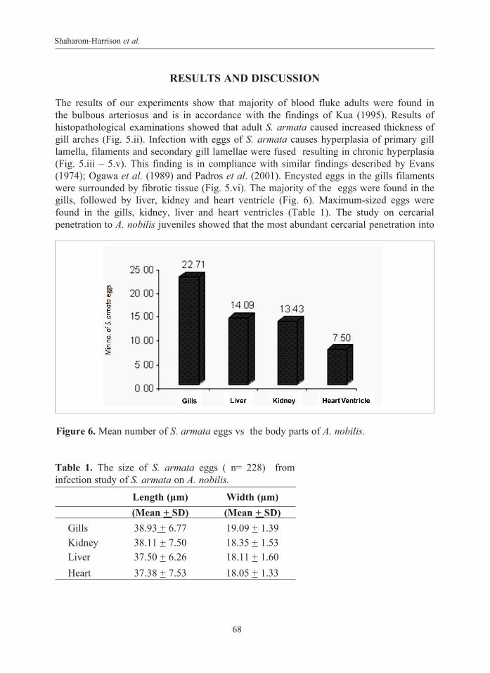
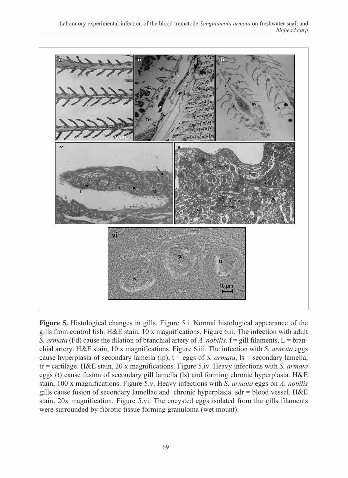
The results of our experiments show that majority of blood fluke adults were found in the bulbous arteriosus and is in accordance with the findings of Kua (1995). Results of histopathological examinations showed that adult S. armata caused increased thickness of gill arches (Fig. 5.ii). Infection with eggs of S. armata causes hyperplasia of primary gill lamella, filaments and secondary gill lamellae were fused resulting in chronic hyperplasia (Fig. 5.iii – 5.v). This finding is in compliance with similar findings described by Evans (1974); Ogawa et al. (1989) and Padros et al. (2001). Encysted eggs in the gills filaments were surrounded by fibrotic tissue (Fig. 5.vi). The majority of the eggs were found in the gills, followed by liver, kidney and heart ventricle (Fig. 6). Maximum-sized eggs were found in the gills, kidney, liver and heart ventricles (Table 1). The study on cercarial penetration to A. nobilis juveniles showed that the most abundant cercarial penetration into
Figure 6. Mean number of S. armata eggs vs the body parts of A. nobilis.
Table 1. The size of S. armata eggs ( n= 228) from infection study of S. armata on A. nobilis.
Laboratory experimental infection of the blood trematode Sanguinicola armata on freshwater snail and bighead carp
69
Figure 5. Histological changes in gills. Figure 5.i. Normal histological appearance of the gills from control fish. H&E stain, 10 x magnifications. Figure 6.ii. The infection with adult S. armata (Fd) cause the dilation of branchial artery of A. nobilis. f = gill filaments, L = bran-chial artery. H&E stain, 10 x magnifications. Figure 6.iii. The infection with S. armata eggs cause hyperplasia of secondary lamella (lp), t = eggs of S. armata, ls = secondary lamella, tr = cartilage. H&E stain, 20 x magnifications. Figure 5.iv. Heavy infections with S. armata eggs (t) cause fusion of secondary gill lamella (ls) and forming chronic hyperplasia. H&E stain, 100 x magnifications. Figure 5.v. Heavy infections with S. armata eggs on A. nobilis gills cause fusion of secondary lamellae and chronic hyperplasia. sdr = blood vessel. H&E stain, 20x magnification. Figure 5.vi. The encysted eggs isolated from the gills filaments were surrounded by fibrotic tissue forming granuloma (wet mount).
Shaharom-Harrison et al.
70
Figure 8. Abundance of cercaria in different sites of penetration.
Figure 7. Number of cercaria vs. time exposure
Laboratory experimental infection of the blood trematode Sanguinicola armata on freshwater snail and bighead carp
71
A. nobilis occurred at intervals of 30 minutes, 40 minutes and 180 minutes (Fig. 8) similar to the findings of Maede and Pratt (1965); Maede (1967); Schell (1974), Sommerville and Iqbal (1991). The part of penetration was the caudal fin, followed by abdominal and caudal peduncle, whereas least penetration site was the operculum (Fig. 8).
CONCLUSIONS
Experimental infection of snails with blood fluke and the histopathological research of its route of penetration into the body of bighead carp A. nobilis gave us the established evidences that freshwater snail G. convexiusculus is the intermediate host of the digenetic blood trematode S. armata. Results of our study confirmed the presence and histopathological lesions in gills, heart, kidney and liver of the host.
ACKNOWLEDGEMENT
We thank the Malaysian Ministry of Science and Technology and University Malaysia Terengganu for funding the project and Kartini Mohammad for technical support.
REFFERENCES
Evans, W. A. 1974. The histopathology of cutthroat trout experimentally infected with the blood fluke Sanguinicola klamathensis. Journal of Wildlife Diseases 10:243-248.
Kua, B. C. 1995. Some aspects of the life cycle of fish blood fluke, Sanguinicola armata Plehn, 1905 (Digenea: Sanguinicolidae) in Grass Carp (Ctenopharyngodon idellus Cuvier and Valenciennes, 1884) fingerlings. Tesis Master Sains. Fakulti Perikanan dan Sains Samudera. Universiti Pertanian Malaysia, Serdang, Selangor. 137 p.
Meade, T. G. and Pratt, J. 1965. Description and life history of Cardicola alseae sp.n. (Trematoda: Sanguinicolidae). The Journal of Parasitology 51(4):575-578.
Meade, T. G. 1967. Life history studies on Cardicola klamathensis (Wales, 1958) Meade and Pratt, 1965 (Trematoda: Sanguinicolidae). Proceeding of the Helminthological Society 34 (2):210-212.
Ogawa, K., Hattori, K., Hatai, K. and Kubota, S. 1989. Histopathology of cultured marine fish Seriola purpurascens (Carangidae) infected with Paradeontacylix spp. (Trematoda: Sanguinicolidae) in its vascular system. Fish Pathology 24( 2): 75-82.
Padros, F, Zarza, C. and Crespo, S. 2001. Histopathology of cultured sea bream Sparus aurata infected with sanguinicolid trematodes. Diseases of Aquatic Organisms 44:47-52.
Schell, S. C. 1974. The life history of Sanguinicola idahoensis sp. n. (Trematoda: Sanguinicolidae), a blood parasite of Steelhead Trout, Salmo gairdneri Richardson. J. Parasitology 60:561-566.
Sommerville, C. and Iqbal, N. A. M. 1991. The process of infection, migration, growth and development of Sanguinicola inermis Plehn, 1905 (Digenea: Sanginicolidae) in carp, Cyprinus carpio L. Journal of Fish Diseases 14: 211-219.