57
Laboratory Procedures
| Date post: | 25-Dec-2015 |
| Category: |
Documents |
| Upload: | gilbert-bruce |
| View: | 218 times |
| Download: | 0 times |

Laboratory Procedures
Bacteria

Culturing Bacteria Culturing: To introduce a specific
microorganism into a solid or liquid broth in order to grow that microorganism for laboratory testing.
Four steps: Swabbing the surface/patient you want to
culture. Streaking the swab onto an agar plate. Staining/testing for identification. Maintaining a stock culture.

Culturing A Patient Proper Method:
Place cotton swab against area being cultured.
Gently spin the swab or move side-to-side to maximize number of microorganisms picked up.
Lift swab away from surface and then remove from area.

Streaking Onto Agar The first step we must take is to learn
about agar.

Agar Agar: A complex polysaccharide medium
used to grow microorganisms. First used by Koch when developing his
postulates, but still used to this day! Begins as a solid, but can be melted into a
liquid. Once melted, it does not solidify until it cools to
40˚C/104˚F. Allows heat-sensitive substances to be added just
before solidification.

Agar Basic agar is made
by combining either chicken or beef broth with a base agar powder. Some types of agar
go father than this! Peptone: A product
of enzyme digestion of proteins easily utilized by microorganisms – a common ingredient!
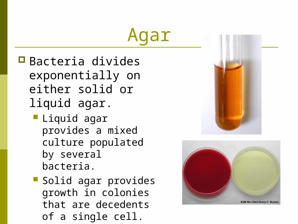
Agar Bacteria divides
exponentially on either solid or liquid agar. Liquid agar provides a
mixed culture populated by several bacteria.
Solid agar provides growth in colonies that are decedents of a single cell.

Streaking Onto Agar The second step is to figure out which type
of agar you want to use, based on patient symptoms.

Classes of Agar Synthetic Media: A medium prepared in
the laboratory from materials with a precise or reasonably well-defined composition.
Complex Media: A medium with a similar base composition as synthetic media, but with slightly differing composition from batch to batch. E.g.: Could contain beef, yeast or soy protein,
in any combination.
Classes of Agar Selective Medium: Encourages the
growth of some bacteria but discourages the growth of others. Typically involves including certain antibiotics
that will not harm the particular bacterium it is designed to grow.
Used to try to grow just one type of organism.
Classes of Agar Differential Medium: Contains a
constituent that causes an observable change. Typically has chemicals that will induce a
chemical reaction and color change when certain organisms are present.
This lets us tell different colonies apart based on color change.
Used to tell things apart!
Classes of Agar Enrichment Medium: Contains special
ingredients to allow a particular organism to flourish. Especially useful when there may not be
enough organisms to be isolated and identified using a normal agar method.
Problem: It does not suppress the growth of other organisms – EVERYTHING goes wild!
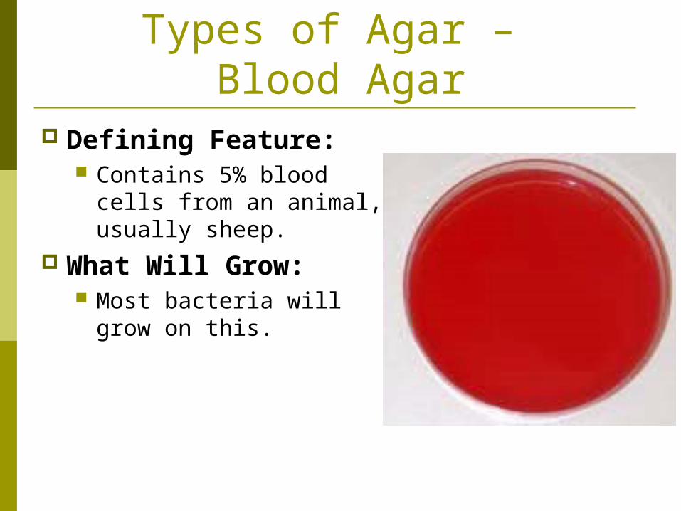
Types of Agar – Blood Agar
Defining Feature: Contains 5% blood cells
from an animal, usually sheep.
What Will Grow: Most bacteria will grow
on this.
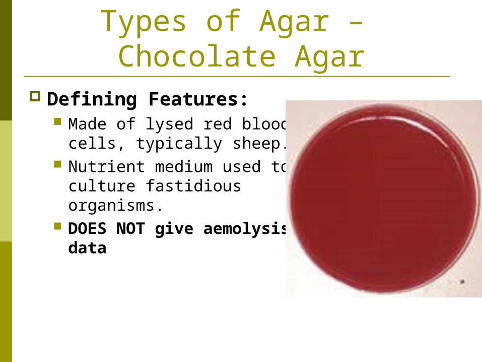
Types of Agar – Chocolate Agar
Defining Features: Made of lysed red blood
cells, typically sheep. Nutrient medium used to
culture fastidious organisms.
DOES NOT give aemolysis data
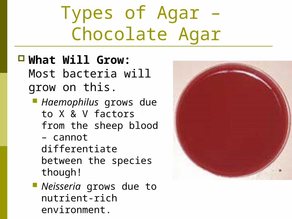
Types of Agar – Chocolate Agar
What Will Grow: Most bacteria will grow on this. Haemophilus grows due
to X & V factors from the sheep blood – cannot differentiate between the species though!
Neisseria grows due to nutrient-rich environment.

Types of Agar – Luria Bertani (LB) Agar
Defining Feature: A subtype of nutrient agar. General medium used for
routine cultivation. What Will Grow:
Does not grow fastidious microorganisms.
Does not preferentially grow one kind of bacteria over another.
Grows everything!
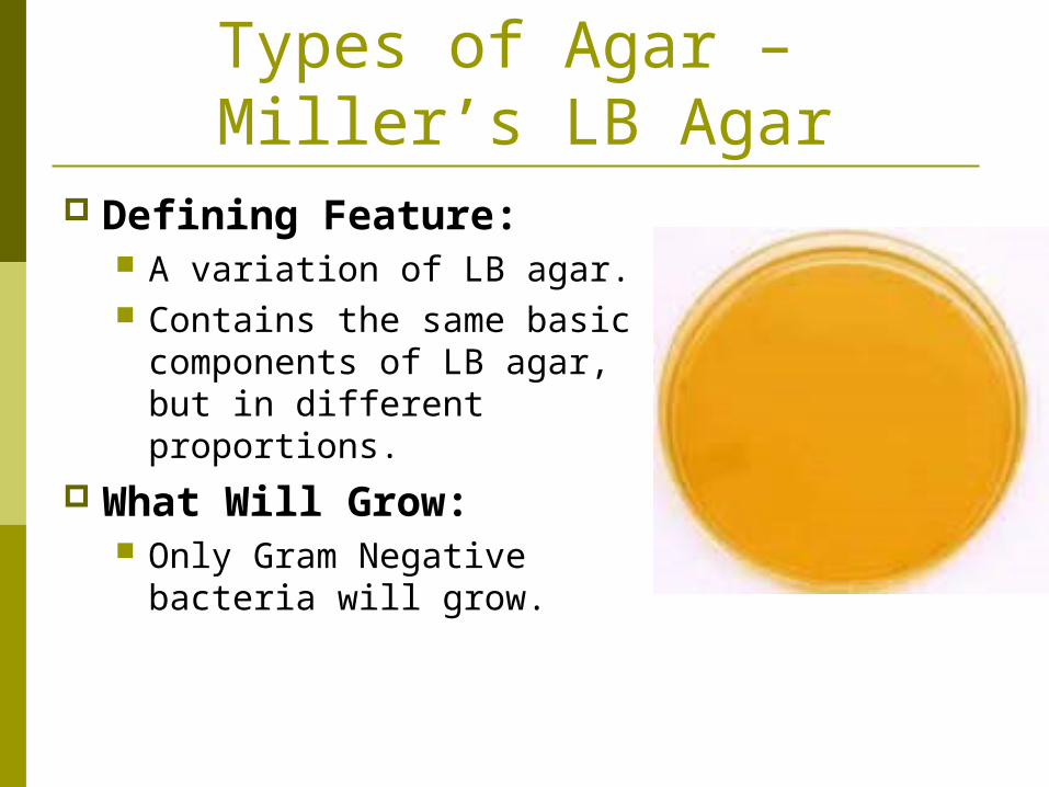
Types of Agar – Miller’s LB Agar
Defining Feature: A variation of LB agar. Contains the same basic
components of LB agar, but in different proportions.
What Will Grow: Only Gram Negative
bacteria will grow.

Types of Agar – MacConkey Agar
Defining Feature: E. coli differentiates by
growing into red colonies due to sugar fermentation.
Can be mixed with or without sugar lactose.
What Will Grow: Will only grow Gram
Negative bacteria.
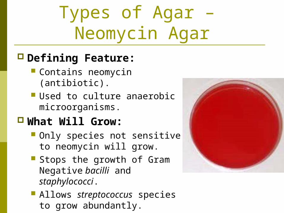
Types of Agar – Neomycin Agar
Defining Feature: Contains neomycin (antibiotic). Used to culture anaerobic
microorganisms. What Will Grow:
Only species not sensitive to neomycin will grow.
Stops the growth of Gram Negative bacilli and staphylococci.
Allows streptococcus species to grow abundantly.
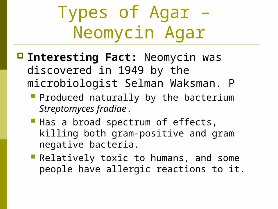
Types of Agar – Neomycin Agar
Interesting Fact: Neomycin was discovered in 1949 by the microbiologist Selman Waksman. P Produced naturally by the bacterium
Streptomyces fradiae. Has a broad spectrum of effects, killing both
gram-positive and gram negative bacteria. Relatively toxic to humans, and some people
have allergic reactions to it.

Types of Agar – Non-Nutrient Agar
Defining Feature: May be used to grow non-
bacterium microorganisms.
What Will Grow: Does not grow bacteria. May be used for growing
other microorganisms.

Types of Agar – Trypticase Soy Agar (TSA)
Defining Feature: Grows the widest variety of
microbes. Uses beef broth. Contains some yeast
extracts. What Will Grow:
Will grow bacteria & fungi. Doesn’t grow all bacterium –
some find it too rich, others find it deficient.
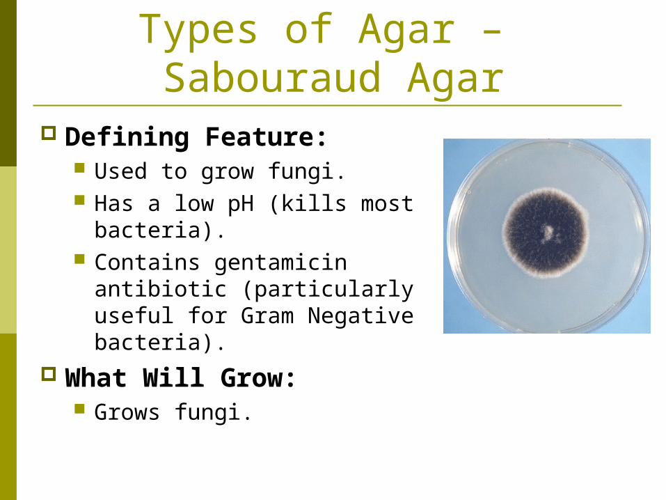
Types of Agar – Sabouraud Agar
Defining Feature: Used to grow fungi. Has a low pH (kills most
bacteria). Contains gentamicin antibiotic
(particularly useful for Gram Negative bacteria).
What Will Grow: Grows fungi.
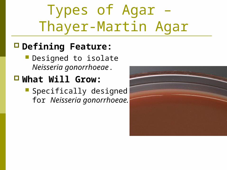
Types of Agar – Thayer-Martin Agar
Defining Feature: Designed to isolate Neisseria
gonorrhoeae. What Will Grow:
Specifically designed for Neisseria gonorrhoeae.

Types of Agar – Tryptic Soy Agar (TSA)
Defining Feature: A basic medium for
general culturing. Mainly used as an initial
growth medium for… Observing colony
morphology. Developing pure cultures. Gaining sufficient growth for
biochemical testing. Culture storage.
What Will Grow: Just about everything!
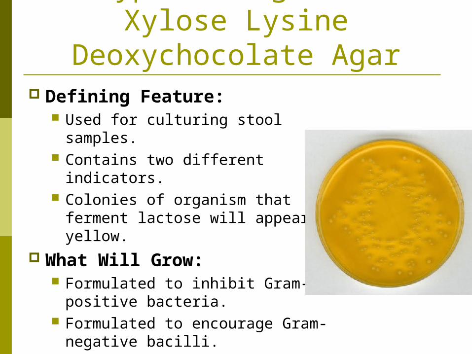
Types of Agar – Xylose Lysine
Deoxychocolate Agar Defining Feature:
Used for culturing stool samples. Contains two different indicators. Colonies of organism that
ferment lactose will appear yellow.
What Will Grow: Formulated to inhibit Gram-
positive bacteria. Formulated to encourage Gram-
negative bacilli.

Streaking
Streaking: The specific technique for isolating bacterium on solid agar.
Streak Plate Method: A method used to obtain colonies from a single parent species.
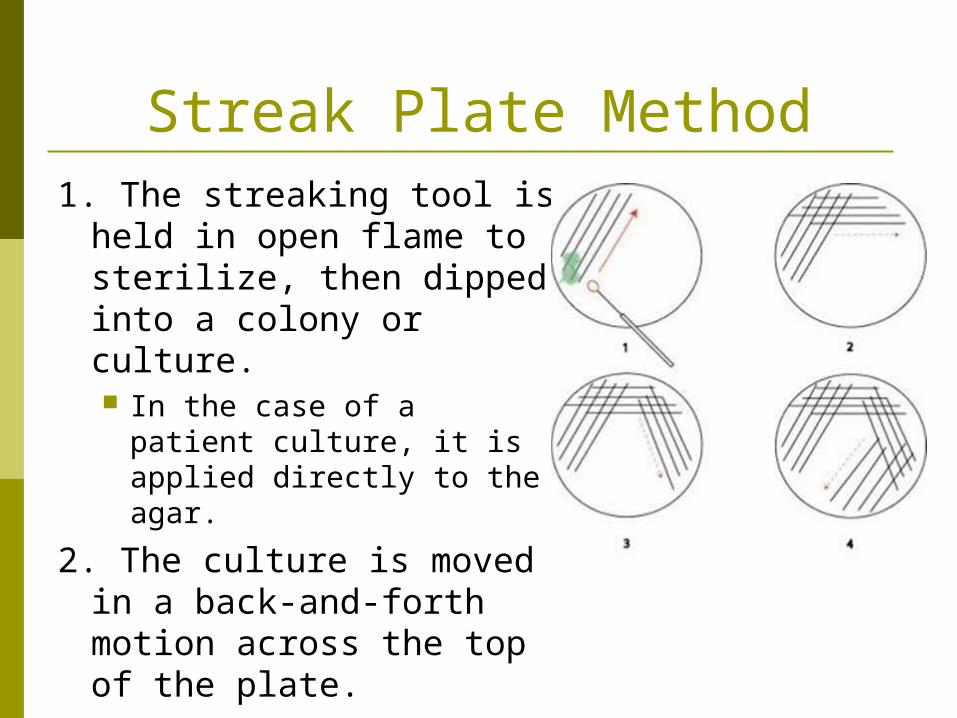
Streak Plate Method1. The streaking tool is
held in open flame to sterilize, then dipped into a colony or culture. In the case of a patient
culture, it is applied directly to the agar.
2. The culture is moved in a back-and-forth motion across the top of the plate.

Streak Plate Method3. The plate is turned 90˚
and a sterile loop is used to streak down from one corner, only touching the previously streaked region once, then continuing up and down the far side of the plate.
4. The plate is turned 90˚ again, and a sterile loop is used to repeat the streaking process.
Staining For Identification
Staining serves several purposes… Tells us information about the cell wall. Helps us to identify the microorganism. Helps us to figure out what chemotherapeutic
agents will kill it.

Types of Stains Stains and dyes are molecules that bind
to a cellular structure to give it color. Cationic Dyes: Basic, positively charged dyes
that are attracted to negatively charged cell components.
Includes methylene blue, crystal violet, safranin, & melachite green.
Anionic Dyes: Negatively charged dyes that are attracted to positively charged material.
Includes eosin & pictric acid.

Methods of Staining Simple Stain: Uses a single dye.
Allows us to determine cell shape and arrangement under a microscope.
Can be done with methylene blue, safranin, crystal violet, and carbolfuchsin.
Differential Stain: Uses two or more dyes. Allows us to distinguish between the different
parts of an organism and between organisms close to each other.
Examples: Gram Stains and Ziel-Neelsen Acid-Fast Stain.

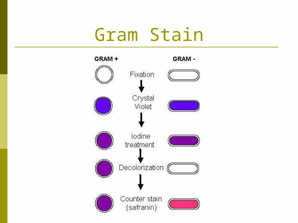
Gram Stain Gram Stain: A staining method that
allows us to determine the type of cell wall a bacterium possesses. The bacteria are streaked onto a glass slide. The slide is passed over a flame to heat-fix the
material. The slide is flooded with crystal violet (purple)
for 15 seconds. The slide is rinsed with iodine, then water. The slide is flooded with safranin (red) for 15
seconds. The slide is light rinsed with water and pat dry.
Gram Stain

Gram Stain Reminder:
Gram-Positive & Acid-Fast bacteria retain the crystal violet due to the thickness of their cell walls.
Gram-Negative bacteria loose the crystal violet and keep the safrinin (since no alcohol rinse is used to remove it!) due to the thinness of their cell wall.
Gram-Variable cells stain purple and red – usually due to the cells being too old when cultured, which decreases their ability to retain stains.
This is a differential stain.

Gram Stain

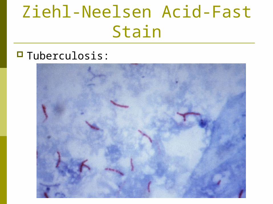
Ziehl-Neelsen Acid-Fast Stain
Ziehl-Neelsen Acid-Fast Stain: Tests for the high lipid content of their cell walls. The same basic procedures are used as in
Gram Staining, except… Crystal violet is replaced with carbolfuchsin (a red
stain). Safranin is replaced with a blue dye.
Acid-Fast bacteria stains red from the carbolfuchsin.
Bacteria that stains blue should be Gram stained.
This is a differential stain.
Ziehl-Neelsen Acid-Fast Stain
Tuberculosis:
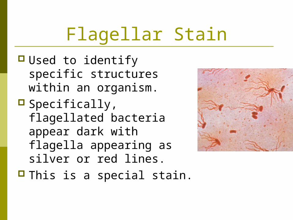
Flagellar Stain Used to identify specific
structures within an organism.
Specifically, flagellated bacteria appear dark with flagella appearing as silver or red lines.
This is a special stain.

Stock Culture Pure Culture: A culture containing only
one species of organism. Streak Plate Method is used to obtain single-
species colonies. Stock Culture: A culture grown in liquid
broth from one organism or one colony of organisms. This can be innoculated onto fresh agar when
the bacteria is needed for study. Aseptic Techniques and other measures
must be used to prevent contamination of the stock culture.
Measuring Bacterial Growth
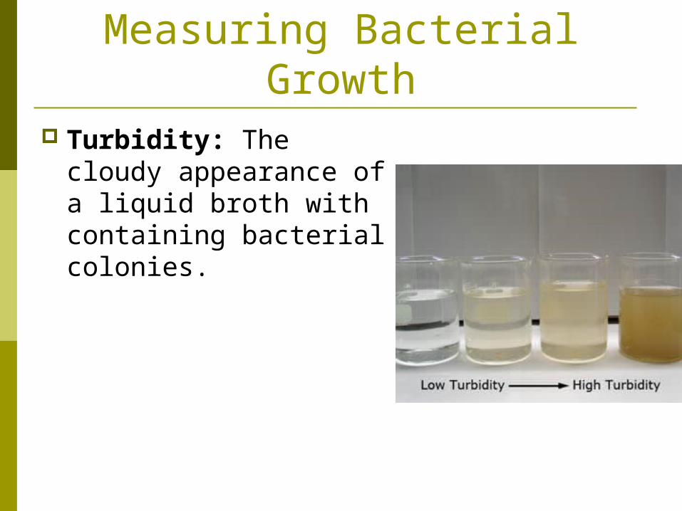
Turbidity: The cloudy appearance of a liquid broth with containing bacterial colonies.
Measuring Bacterial Growth
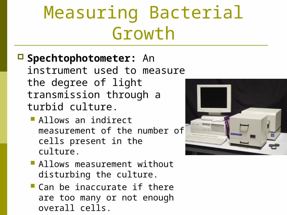
Spechtophotometer: An instrument used to measure the degree of light transmission through a turbid culture. Allows an indirect
measurement of the number of cells present in the culture.
Allows measurement without disturbing the culture.
Can be inaccurate if there are too many or not enough overall cells.
Measuring Bacterial Growth
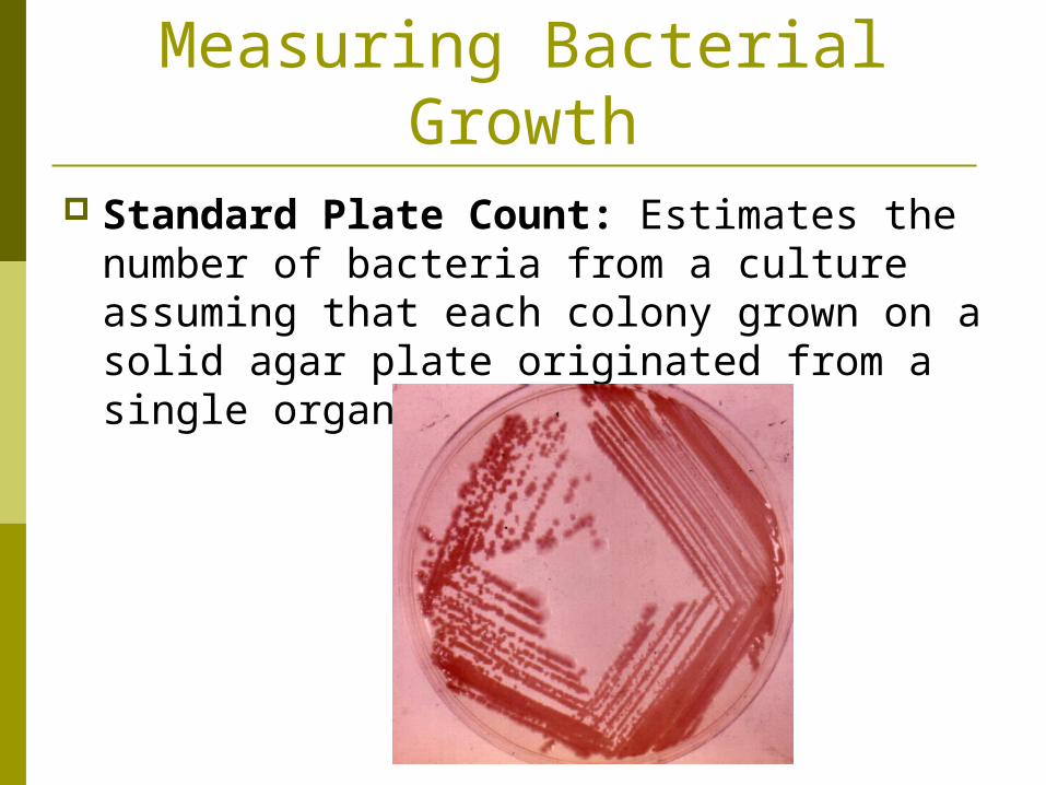
Standard Plate Count: Estimates the number of bacteria from a culture assuming that each colony grown on a solid agar plate originated from a single organism.

Measuring Bacterial Growth
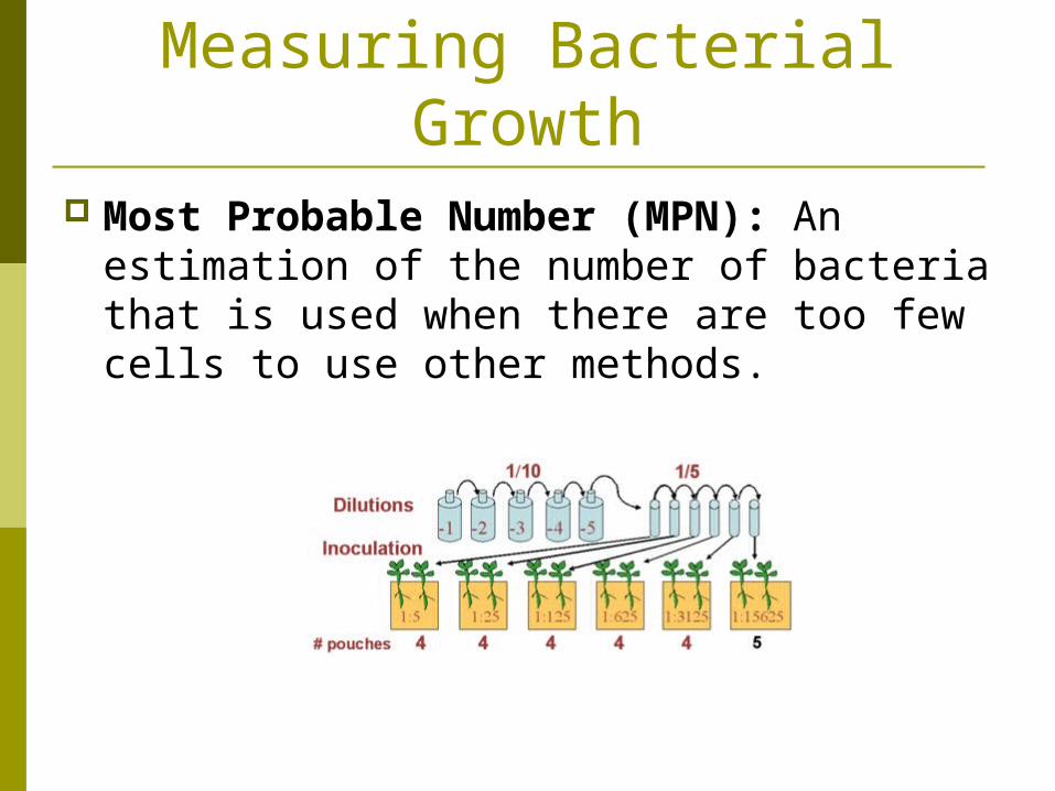
Serial Dilutions: A method of diluting a liquid culture to prevent over-crowding of colonies when grown on solid agar. Multiplying the number of colonies by the ratio
of dilution to estimate the number of bacterial cells in the original culture.
For example: If a microbiologists dilutes a broth to 1:1000 ratio of broth to dilutant, and 50 colonies grow on agar by the next day, he can estimate the 50,000 bacterial cells per one milliliter of original culture.
Measuring Bacterial Growth
Most Probable Number (MPN): An estimation of the number of bacteria that is used when there are too few cells to use other methods.

Measuring Bacterial Growth
Most Probable Number (MPN): Measures the bacterial waste byproducts
produced. A small glass tube is inverted & placed into the
test tube containing liquid culture so that it fills with broth.
Gas produced by the fermentation of bacteria will be trapped in the inverted tube.
The gas can then be compared to a standardized curve to estimate the number of organisms present in the sample.
Filtration Filtration: Forces a known volume of
liquid or air through a filter with pores too small for bacteria to pass through. The filter can then be placed on solid media
and allowed to grow. The resulting colonies can then be counted
using standard plate count. The standard plate count allows an estimate of
the number of organisms per liter of air or water to be estimated.

Fungi
Culturing Fungi Fungi can be grown, cultured and
identified using similar laboratory methods to those used on bacteria!
Acidic, high-sugar media with antibiotics to prevent bacterial growth can be used.
Most pathogenic fungi take two to four weeks to grow as much as bacteria do in a 24-hour period.
This leads to slow identification!
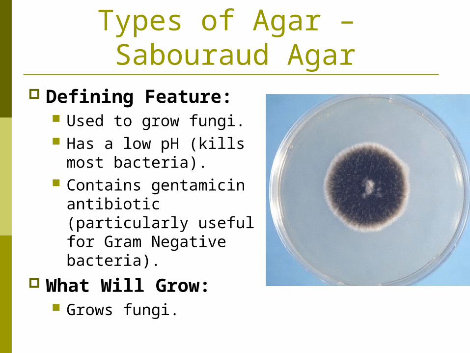
Types of Agar – Sabouraud Agar
Defining Feature: Used to grow fungi. Has a low pH (kills most
bacteria). Contains gentamicin
antibiotic (particularly useful for Gram Negative bacteria).
What Will Grow: Grows fungi.

Viruses

Culturing Viruses Whole animals had to be used to culture
viruses until the 1950’s and 1960’s. Two big discoveries changed this…
The discovery of antibiotics helped with culturing viruses and preventing bacterial contamination of the culture.
The discovery of proteolytic enzymes, especially trypsin, that helps to free animal cells from their surrounding tissue without cell injury – this allowed us to add cells to Petri dishes to grow viruses in!

Culturing Viruses Primary Cell
Cultures: Cultures that come directly from an animal. The younger the source
animal, the longer the cell will survive within the culture.
Typically presents as a mixture of cell types.
Only divide a few times, but can host a wide variety of viruses.

Culturing Viruses Continuous Cell Lines: Cells that will
reproduce for an extended number of generations, allowing long-term study. Typically originate from cancerous cells or cells
that have lost control of cell division rates.

HeLa Cells HeLa Cells: An
immortal cell line used in medical research, particularly with virology.

HeLa Cells Started from a cervical cancer
cell culture taken from Henrietta Lacks.
Mrs. Lacks died on October 4th, 1951.
Her cells are located in just about every laboratory in the U.S.
These cells can divide indefinitely as long as basic conditions for survival are met.
They have branched into several strains as they have mutated over the years.


