+ All Categories
Home > Documents > Laboratory Testing for the Lupus Anticoagulant; Approved ...Laboratory Testing for the Lupus...

Laboratory Testing for the Lupus Anticoagulant; Approved ...Laboratory Testing for the Lupus...

Date post: 24-Jun-2020
Category:
Upload: others
View: 3 times
Download: 0 times
Share this document with a friend
16
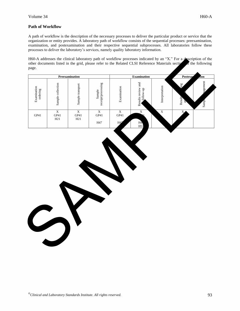
April 2014 H60-A Laboratory Testing for the Lupus Anticoagulant; Approved Guideline This document provides guidance and recommendations regarding the proper collection and handling of the specimen; descriptions and limitations of screening and confirmatory assays, and mixing tests used to identify lupus anticoagulant (LA); determination of cutoff values and calculations associated with the various assays; and interpretation of test results in an LA panel. A guideline for global application developed through the Clinical and Laboratory Standards Institute consensus process. SAMPLE
Transcript

April 2014

H60-ALaboratory Testing for the Lupus Anticoagulant; Approved Guideline

This document provides guidance and recommendations regarding the proper collection and handling of the specimen; descriptions and limitations of screening and confirmatory assays, and mixing tests used to identify lupus anticoagulant (LA); determination of cutoff values and calculations associated with the various assays; and interpretation of test results in an LA panel.

A guideline for global application developed through the Clinical and Laboratory Standards Institute consensus process.

SAMPLE

ISBN 1-56238-959-9 (Print) ISBN 1-56238-960-2 (Electronic) ISSN 1558-6502 (Print) H60-A ISSN 2162-2914 (Electronic) Vol. 34 No. 6

Laboratory Testing for the Lupus Anticoagulant; Approved Guideline

Volume 34 Number 6

Marlies Ledford-Kraemer, MBA, BS, MT(ASCP)SH Gary W. Moore, BSc, DBMS, CSci, FIBMS, CBiol, MSB,

CertMHS Ralph Bottenus, PhD John T. Brandt, MD Donna D. Castellone, MS, MT(ASCP)SH Christine Daniele, MT(ASCP) Philip G. de Groot, PhD François Depasse, PharmD, MSc Jeffrey S. Dlott, MD, FCAP, FASCP Thomas Exner, PhD Emmanuel J. Favaloro, PhD, FFSc (RCPA) Robert C. Gosselin, CLS

Sandra C. Hollensead, MD Piet Meijer, PhD Karen A. Moffat, BEd, MSc, ART, FCSMLS(D) William L. Nichols, MD Thomas L. Ortel, MD, PhD Michael J. Sanfelippo, MS, MT(ASCP) Rosemary Grillo Scott Rita Selby, MBBS, FRCPC, MSc Linda Stang, MLT Perumal Thiagarajan, MD Mark Triscott, PhD Elizabeth M. Van Cott, MD

Abstract Identification of the lupus anticoagulant (LA) by laboratory testing is critical for diagnosing the antiphospholipid syndrome and investigating unexpectedly prolonged activated partial thromboplastin time values. The “anticoagulant” effect of LA is restricted to the prolongation of clotting times when using in vitro, clot-based coagulation assays that are used as surrogates for identifying LA. Clinical and Laboratory Standards Institute document H60—Laboratory Testing for the Lupus Anticoagulant; Approved Guideline provides guidance and recommendations regarding the proper collection and handling of the specimen; descriptions and limitations of screening and confirmatory assays, and mixing tests used to identify LA; determination of cutoff values and calculations associated with the various assays; and interpretation of test results in an LA panel. The guideline is provided for use by laboratorians, physician stakeholders, manufacturers of LA assays, researchers, external quality assessment programs, and accrediting and regulatory agencies. The intent of this guideline is to present information in a practical and easily understandable format; thereby facilitating a standardized approach to LA testing, gaining acceptance in practice, and improving testing quality. Clinical and Laboratory Standards Institute (CLSI). Laboratory Testing for the Lupus Anticoagulant; Approved Guideline. CLSI document H60-A (ISBN 1-56238-959-9 [Print]; ISBN 1-56238-960-2 [Electronic]). Clinical and Laboratory Standards Institute, 950 West Valley Road, Suite 2500, Wayne, Pennsylvania 19087 USA, 2014.

The Clinical and Laboratory Standards Institute consensus process, which is the mechanism for moving a document through two or more levels of review by the health care community, is an ongoing process. Users should expect revised editions of any given document. Because rapid changes in technology may affect the procedures, methods, and protocols in a standard or guideline, users should replace outdated editions with the current editions of CLSI documents. Current editions are listed in the CLSI catalog and posted on our website at www.clsi.org. If you or your organization is not a member and would like to become one, and to request a copy of the catalog, contact us at: Telephone: 610.688.0100; Fax: 610.688.0700; E-Mail: [email protected]; Website: www.clsi.org.

SAMPLE

Number 6 H60-A

ii

Copyright ©2014 Clinical and Laboratory Standards Institute. Except as stated below, any reproduction of content from a CLSI copyrighted standard, guideline, companion product, or other material requires express written consent from CLSI. All rights reserved. Interested parties may send permission requests to [email protected] CLSI hereby grants permission to each individual member or purchaser to make a single reproduction of this publication for use in its laboratory procedure manual at a single site. To request permission to use this publication in any other manner, e-mail [email protected]. Suggested Citation CLSI. Laboratory Testing for the Lupus Anticoagulant; Approved Guideline. CLSI document H60-A. Wayne, PA: Clinical and Laboratory Standards Institute; 2014. Reaffirmed: September 2019 ISBN 1-56238-959-9 (Print) ISBN 1-56238-960-2 (Electronic) ISSN 1558-6502 (Print) ISSN 2162-2914 (Electronic)

SAMPLE

Volume 34 H60-A

v

Contents

Abstract .................................................................................................................................................... i

Committee Membership ........................................................................................................................ iii

Foreword ................................................................................................................................................ ix

1 Scope .......................................................................................................................................... 1

2 Introduction ................................................................................................................................ 1

2.1 Historical Perspective of Antiphospholipid Syndrome and Relationship to Lupus

Anticoagulant Testing ................................................................................................................ 3

3 Standard Precautions .................................................................................................................. 4

4 Terminology ............................................................................................................................... 4

4.1 A Note on Terminology ................................................................................................ 4 4.2 Definitions .................................................................................................................... 5 4.3 Abbreviations and Acronyms ....................................................................................... 8

5 Equipment .................................................................................................................................. 9

6 Validation of Assay Systems ................................................................................................... 10

6.1 Imprecision ................................................................................................................. 10 6.2 Inaccuracy ................................................................................................................... 11 6.3 Establishment of Reference Intervals and Cutoff Values ........................................... 12 6.4 Analytical Specificity ................................................................................................. 13 6.5 Diagnostic Sensitivity and Diagnostic Specificity ...................................................... 14

7 Preexamination Issues (Criterion A) ........................................................................................ 14

7.1 Patient Selection ......................................................................................................... 15 7.2 Specimen Collection and Transport ............................................................................ 17 7.3 Sample Preparation ..................................................................................................... 18

8 Preliminary Examination Issues .............................................................................................. 21

8.1 APTT .......................................................................................................................... 21 8.2 Prothrombin Time-International Normalized Ratio .................................................... 23 8.3 Thrombin Time ........................................................................................................... 24

9 Principles of Lupus Anticoagulant Assays .............................................................................. 25

9.1 Intrinsic Pathway Assays (see Appendix C) ............................................................... 25 9.2 Common Pathway Assays (see Appendix C) ............................................................. 27 9.3 Extrinsic Pathway Assays (see Appendix C) .............................................................. 28 9.4 Overview of Assay Performance ................................................................................ 29

10 Assays to Screen for the Presence of Lupus Anticoagulant (Criterion B) ............................... 31

10.1 Available Screening Assays and Their Usage ............................................................ 31 10.2 APTT .......................................................................................................................... 32 10.3 APTT-based Silica Clotting Time .............................................................................. 33 10.4 Dilute Russell’s Viper Venom Time........................................................................... 34 10.5 Dilute Prothrombin Time ............................................................................................ 35 10.6 Kaolin Clotting Time .................................................................................................. 36

SAMPLE

Number 6 H60-A

vi

Contents (Continued)

11 Assays to Confirm the Presence of Lupus Anticoagulant (Criterion C) .................................. 37

11.1 APTT-based Platelet Neutralization Procedure .......................................................... 38 11.2 APTT-based Hexagonal Phase Phospholipid Neutralization Test .............................. 38 11.3 APTT-based Silica Clotting Time Confirmatory Test ................................................ 39 11.4 Dilute Russell’s Viper Venom Time Confirmatory Test ............................................ 40 11.5 Dilute Prothrombin Time Confirmatory Test ............................................................. 41

12 Mixing Test as Applied to Screening, Confirmatory, and Integrated Assays (Criterion D) .... 42

12.1 Reasons for Performing a Mixing Test ....................................................................... 43 12.2 Normal Pooled Plasma ................................................................................................ 44 12.3 Calculation of Test Results ......................................................................................... 45 12.4 Limitations of Mixing Test ......................................................................................... 46

13 Postexamination Issues ............................................................................................................ 47

13.1 Interpretation of Preliminary Examination Assays ..................................................... 48 13.2 Interpretation of Lupus Anticoagulant Screening Assays

(Criterion/Recommendation B) .................................................................................... 49 13.3 Interpretation of Lupus Anticoagulant Confirmatory Assays

(Criterion/Recommendation C) .................................................................................. 50 13.4 Interpretation of Lupus Anticoagulant Mixing Tests

(Criterion/Recommendation D) .................................................................................. 52 13.5 Interpretation of Lupus Anticoagulant Panel (Criterion/Recommendation F) ........... 53 13.6 How to Report Test Results (Criterion/Recommendation F) ...................................... 54

14 Quality Control and Quality Assurance ................................................................................... 54

14.1 Reagent Lot-to-Lot Number Verification ................................................................... 54 14.2 Internal Quality Control .............................................................................................. 55 14.3 External Quality Assessment ...................................................................................... 55

15 Harmonization of CLSI Lupus Anticoagulant Guideline With Other Guidelines ................... 56

References ............................................................................................................................................. 58

Appendix A. Algorithmic Approach to Lupus Anticoagulant Testing ................................................. 71

Appendix B. Establishing Reference Intervals and Cutoff Values ....................................................... 74

Appendix C. Lupus Anticoagulant Testing in Relationship to Coagulation Pathways ........................ 75

Appendix D. Diagrammatic Principles for Lupus Anticoagulant Tests................................................ 76

Appendix E. Table of Interferences and Limitations for Specific Lupus Anticoagulant Tests ............ 84

Appendix F. Formulas and Examples for Calculating Test Results ..................................................... 88

Appendix G1. Interpretive Comments and Rationale for Comments Based on Patient Examples

(Data) ................................................................................................................................................... 89

Appendix G2. Interpretive Comments and Rationale for Comments Based on Patient Examples

(Comments)........................................................................................................................................... 90

SAMPLE

Volume 34 H60-A

vii

Contents (Continued)

The Quality Management System Approach ........................................................................................ 92

Related CLSI Reference Materials ....................................................................................................... 94

SAMPLE

Volume 34 H60-A

ix

Foreword

Synopsis of Diagnostic Criteria and Testing Recommendations

Criteria for the Laboratory Diagnosis of the Lupus Anticoagulant

In order to make a laboratory diagnosis of the lupus anticoagulant, a sample should identify with the

following:

A. Procurement: adherence to standardized protocols for collection and processing of blood to be used

for testing

B. Screening: prolongation of at least one of two different phospholipid-dependent clotting assays based

on different principles and coagulation pathways

C. Confirmation: evidence that prolongation of the screening test(s) demonstrates phospholipid

dependence by using a similar second test(s) using altered concentrations and/or composition of

phospholipids

D. Mixing: if mixing assays are performed, evidence of inhibitory activity shown by the effect of patient

plasma on an equal volume of normal pooled plasma

E. Exclusion: distinguishing the lupus anticoagulant from other causes of prolonged clotting times that

may mask, mimic, or coexist with the lupus anticoagulant, such as anticoagulant therapies or other

coagulopathies

F. Interpretation and Reporting: numerical results of all testing should be reported, and interpretive

comments that address and integrate these results should be provided

Recommendations Specific to Each Criterion for the Laboratory Diagnosis of the Lupus

Anticoagulant

A. Procurement

1. Testing should preferably be performed in the absence of anticoagulant therapy (except for

antiplatelet therapy).

2. Ideally, samples should not be obtained from vascular access devices.

3. Platelet count of patient-citrated platelet-poor plasma should be < 10 × 109/L.

4. Testing may be performed on fresh or properly frozen/thawed samples.

B. Screening Assays

1. Two tests, representing different principles and coagulation pathways, that are known to be

responsive to the lupus anticoagulant (eg, low phospholipid concentrations) should be used to

screen for the lupus anticoagulant.

2. Lupus anticoagulant–responsive activated partial thromboplastin time and dilute Russell’s viper

venom time tests are recommended as the preferred minimal screening assays.

SAMPLE

Number 6 H60-A

x

3. Other tests for the lupus anticoagulant referenced in this document may supplement the preferred

minimal screening tests.

4. Where test design permits, results should be calculated using the mean of the reference interval

and reported as a normalized ratio.

5. Routine coagulation tests, prothrombin time-international normalized ratio, activated partial

thromboplastin time, and thrombin time, as indicated, may help to characterize anticoagulant

effects (eg, heparin, vitamin K antagonists, direct thrombin inhibitors, factor Xa inhibitors) or

sample suitability (eg, serum sample, improper anticoagulant tube) for lupus anticoagulant

testing and interpretation.

C. Confirmatory Assays

1. Confirmatory assays should use the same assay principle as the screening test that was initially

found to be abnormal (eg, if the dilute Russell’s viper venom time test is abnormal, then a dilute

Russell’s viper venom time test–based confirmatory assay should be used).

2. For paired tests, results should be calculated using the mean of the reference interval for each

screening and confirmatory test and reported as a normalized screen to confirm ratio or

indication of percentage correction of screen ratio by confirm ratio.

3. Solid-phase immunoassays for antibodies against phospholipid (eg, anti-cardiolipin or anti-β2

glycoprotein I) should not be considered as lupus anticoagulant confirmatory procedures.

D. Mixing Test (if performed)

1. The platelet count of the normal pooled plasma should be < 10 × 109/L.

2. A mix ratio of one part plasma sample to one part normal pooled plasma is recommended as the

preferred ratio for a mixing test.

3. The dilution effect of a 1:1 mixing test may mask lupus anticoagulant inhibitory activity. Other

mix ratios (eg, four parts plasma sample to one part normal pooled plasma) can be used, if

validated by the laboratory.

4. Mixing test inhibition is assessed by either comparison of normalized ratios to cutoff values

specific for each lupus anticoagulant screening or confirmatory mixing test or by calculating an

index of circulating anticoagulant.

5. Incubated mixing tests are not recommended for routine lupus anticoagulant testing, but should

be performed when indicated (eg, when a specific factor inhibitor is suspected).

E. Exclusion

1. The lupus anticoagulant should be distinguished from anticoagulant therapies and/or other

coagulation disorders that may interfere with lupus anticoagulant testing and interpretation.

2. If possible, perform factor assays whenever there is suspicion of a specific factor deficiency or

inhibitor, using three or more dilutions of patient plasma and an activated partial thromboplastin

time reagent that is unresponsive to the lupus anticoagulant.

SAMPLE

Volume 34 H60-A

xi

F. Interpretation and Reporting

1. Numerical results of all testing should be reported with the reference interval or cutoff value.

2. Interpretive comments that address and integrate all test results (the lupus anticoagulant panel)

should be provided.

3. The interpretive report should indicate whether the lupus anticoagulant is present, not detected,

or indeterminate.

4. Solid-phase assays for antibodies against cardiolipin and/or anti-β2 glycoprotein I are

recommended as part of an evaluation for antiphospholipid syndrome.

5. If the lupus anticoagulant is present, the test panel should be repeated at or beyond 12 weeks to

determine persistence of the lupus anticoagulant as part of the evaluation for antiphospholipid

syndrome.

Key Words

Antiphospholipid syndrome, lupus anticoagulant, lupus anticoagulant confirmatory assays, lupus

anticoagulant screening assays, mixing test

SAMPLE

Volume 34 H60-A

©Clinical and Laboratory Standards Institute. All rights reserved. 1

Laboratory Testing for the Lupus Anticoagulant; Approved Guideline

1 Scope

This document provides guidance for the performance and interpretation of screening assays,

confirmatory assays, and mixing tests used to identify the lupus anticoagulant (LA). It is intended to assist

in standardization of LA testing and it addresses preexamination issues, examination concerns, and

postexamination matters that pertain to interpretation of individual tests or combinations of assays.

Recommendations from this guideline, when feasible, harmonize with other national and international

guidelines currently in existence. Taken together, standardization and harmonization will permit

laboratories to improve the quality and interpretation of their LA testing.

The intended users of this guideline are laboratory personnel responsible for performing LA testing,

physicians (eg, hematologists, pathologists, rheumatologists), external quality assessment (EQA)

programs, researchers, and manufacturers of reagents used in LA testing.

Two types of methodologies are used for the diagnosis of the antiphospholipid syndrome (APS). This

guideline is limited to clot-based coagulation assays used as surrogates for identifying LA—a strong risk

factor for thrombosis. The guideline will not address solid-phase testing for anti-phospholipid (aPL) (eg,

anti-cardiolipin [aCL] or anti-β2 glycoprotein I [aβ2GPI]), because detection of these specific antibodies

may or may not relate to the laboratory anomaly of a prolonged activated partial thromboplastin time

(APTT).

2 Introduction

Identification of LA by laboratory testing is critical for investigating unexpectedly prolonged APTT

values and diagnosing APS. According to recent consensus classification criteria, two conditions must be

met for defining APS: 1) the persistent presence of circulating aPL in plasma and/or serum, and 2) a

history of thrombosis and/or pregnancy morbidity including fetal loss.1,2

APS is an autoimmune disorder

that occurs in patients who, in general, also show laboratory evidence of antibodies directed against

plasma proteins that have an affinity for anionic phospholipids (PL). The dominant antigenic targets

recognized by aPL in patients with APS are β2GPI or prothrombin.3-5

The terms APS and LA are both misnomers and LA itself is a double misnomer.6,7

The autoantibodies

associated with APS are not directed against PL in general but specifically against proteins that bind to

anionic PL. LA comprises a heterogeneous group of autoantibodies that can develop in individuals with

autoimmune conditions (systemic lupus erythematosus [SLE], APS, or other autoimmune disorders) or

can arise spontaneously.8-10

LA can also be found transiently in plasma from patients with infections or

malignancies, or from patients using certain drugs. The “anticoagulant” effect of LA is restricted to the

prolongation of clotting times (competition between these antibodies and coagulation proteins for PL

surfaces) in clot-based assays (ie, in vitro), whereas in vivo, these antibodies are variably associated with

thrombosis.11,12

Persistent LA is the most important acquired risk factor for thrombosis (or its recurrence)

in APS.8,13

LA may also cause bleeding due to immune-mediated deficiencies of coagulation factors II

(FII) or X (FX) (see Section 7.1.1). Infection- or drug-induced LA tend to be transient, disappearing after

the infection resolves or when the medication is discontinued. Transient LA due to infections have rarely

been reported to increase thrombotic risk in association with immune-mediated protein S deficiency (see

Section 7.1.1). LA due to certain medications may not be transient and can increase thrombotic risk.14

LA

associated with malignancy might also resolve after the malignancy is treated.15,16

SAMPLE

Number 6 H60-A

©

Clinical and Laboratory Standards Institute. All rights reserved. 2

Two types of laboratory testing are used in making a diagnosis of APS:

Solid-phase assays for aCL and aβ2GPI

Liquid-phase (plasma-based) assays for LA

Solid-phase (commonly ELISA) methods are used to identify and quantify the antibody isotypes

(immunoglobulin G [IgG], immunoglobulin M [IgM], immunoglobulin A [IgA]) to β2GPI, cardiolipin

(either dependent or independent of β2GPI), and prothrombin. These assays will not be discussed in this

document.

This guideline will focus on the detection of LA activity (antibodies against β2GPI/PL and/or antibodies

against prothrombin/PL) by surrogate, functional, liquid-phase, clot-based assays. These tests are

associated with all three laboratory pathways of coagulation: intrinsic, common, and extrinsic. Numerous

assays have been designed through the years, as shown in Table 1. Though potentially promising, many

have not been widely used or commercialized.17-20

Commercially available assays, in general, manipulate

PL concentration (or type) that permits LA to either showcase inhibitory action or to have this activity

neutralized. Hence, test systems have been segregated into those having “low” levels of PL to screen or

“tease out” the inhibitory action of LA vs those assays in which high concentrations of PL neutralize the

inhibitory effect. Due to antibody heterogeneity, different clot-based assays can detect different antibody

populations in the same patient.21,22

LA is considered to be more strongly associated with thrombotic risk

than ELISA assays for aCL and aβ2GPI.8,23

A provocative concept is that this stronger association may be

related to antibody titers, ie, a relatively high concentration of antibodies is required to prolong the

clotting time of LA assays, which are analytically relatively insensitive.24

Consensus criteria for the classification (not diagnosis) of APS were initially outlined in 19991 and

updated in 2006.2 Both documents include laboratory criteria for LA as set forth by the Scientific and

Standardization Committee (SSC) on Lupus Anticoagulant/Phospholipid-Dependent Antibodies of the

International Society on Thrombosis and Haemostasis (ISTH). Guidelines for LA testing from the ISTH

were first published in 1983,25

updated in 199126

and 1995,27

and revised in 2009.28

Guidelines are also

available globally from various national and international organizations.9,29,30

Many guidelines have built

upon recommendations and criteria from preceding works. This guideline seeks to continue this building

process while harmonizing with, and adding clarity to, the current guidelines. This will aid laboratories in

the laboratory diagnosis of LA and to assist clinicians in determining the presence of APS.

This guideline also introduces new paradigms for LA testing. The key shift is in the order in which tests

are performed: screening, confirmation, and mixing. This guideline recognizes that many interferences

affect the mixing test as shown in recent publications addressing the potential for false-negative or false-

positive test results when significant reliance is placed on the mixing test. Therefore, this guideline

suggests that less importance be assigned to the mixing test and places it last in the sequence of testing.

Additionally, this guideline stresses the need for and understanding of test principles and their association

with the three coagulation pathways. Thus, screening assays should not only be performed using two test

principles, but each should represent a different coagulation pathway, as recommended by the document

development committee. Finally, this guideline acknowledges the importance of not only performing an

LA panel of tests, but interpreting LA tests in total rather than in isolation. The need for appropriate cutoff

values that differentiate whether LA is or is not detected is inherent in the interpretation of LA tests.

Numerous sections of this guideline strive to reinforce this point.

SAMPLE

Number 6 H60-A

©Clinical and Laboratory Standards Institute. All rights reserved. 92

The Quality Management System Approach Clinical and Laboratory Standards Institute (CLSI) subscribes to a quality management system approach in the

development of standards and guidelines, which facilitates project management; defines a document structure via a

template; and provides a process to identify needed documents. The quality management system approach applies a

core set of “quality system essentials” (QSEs), basic to any organization, to all operations in any health care

service’s path of workflow (ie, operational aspects that define how a particular product or service is provided). The

QSEs provide the framework for delivery of any type of product or service, serving as a manager’s guide. The QSEs

are as follows:

Organization Personnel Process Management Nonconforming Event Management

Customer Focus Purchasing and Inventory Documents and Records Assessments

Facilities and Safety Equipment Information Management Continual Improvement

H60-A addresses the QSE indicated by an “X.” For a description of the other documents listed in the grid, please

refer to the Related CLSI Reference Materials section on page 94.

Org

aniz

atio

n

Cu

stom

er F

ocu

s

Fac

ilit

ies

and

Saf

ety

Per

son

nel

Pu

rchas

ing

and

Inven

tory

Equ

ipm

ent

Pro

cess

Man

agem

ent

Do

cum

ents

an

d

Rec

ord

s

Info

rmat

ion

Man

agem

ent

No

nco

nfo

rmin

g

Ev

ent

Man

agem

ent

Ass

essm

ents

Con

tinual

Imp

rov

emen

t

GP41

M29

GP41

H57

X EP05

EP06

EP09 EP12

EP15

EP24 EP28

GP41

H21

H47

H57

H57

SAMPLE

Volume 34 H60-A

©Clinical and Laboratory Standards Institute. All rights reserved. 93

Path of Workflow

A path of workflow is the description of the necessary processes to deliver the particular product or service that the

organization or entity provides. A laboratory path of workflow consists of the sequential processes: preexamination,

examination, and postexamination and their respective sequential subprocesses. All laboratories follow these

processes to deliver the laboratory’s services, namely quality laboratory information.

H60-A addresses the clinical laboratory path of workflow processes indicated by an “X.” For a description of the

other documents listed in the grid, please refer to the Related CLSI Reference Materials section on the following

page.

Preexamination Examination Postexamination

Ex

amin

atio

n

ord

erin

g

Sam

ple

coll

ecti

on

Sam

ple

tra

nsp

ort

Sam

ple

rece

ipt/

pro

cess

ing

Ex

amin

atio

n

Res

ult

s re

vie

w a

nd

foll

ow

-up

Inte

rpre

tati

on

Res

ult

s re

po

rtin

g

and

arc

hiv

ing

Sam

ple

man

agem

ent

GP41

X GP41

H21

X GP41

H21

X GP41

H47

X GP41

H47

X GP41

H47 H57

X X

SAMPLE

Number 6 H60-A

©Clinical and Laboratory Standards Institute. All rights reserved. 94

Related CLSI Reference Materials

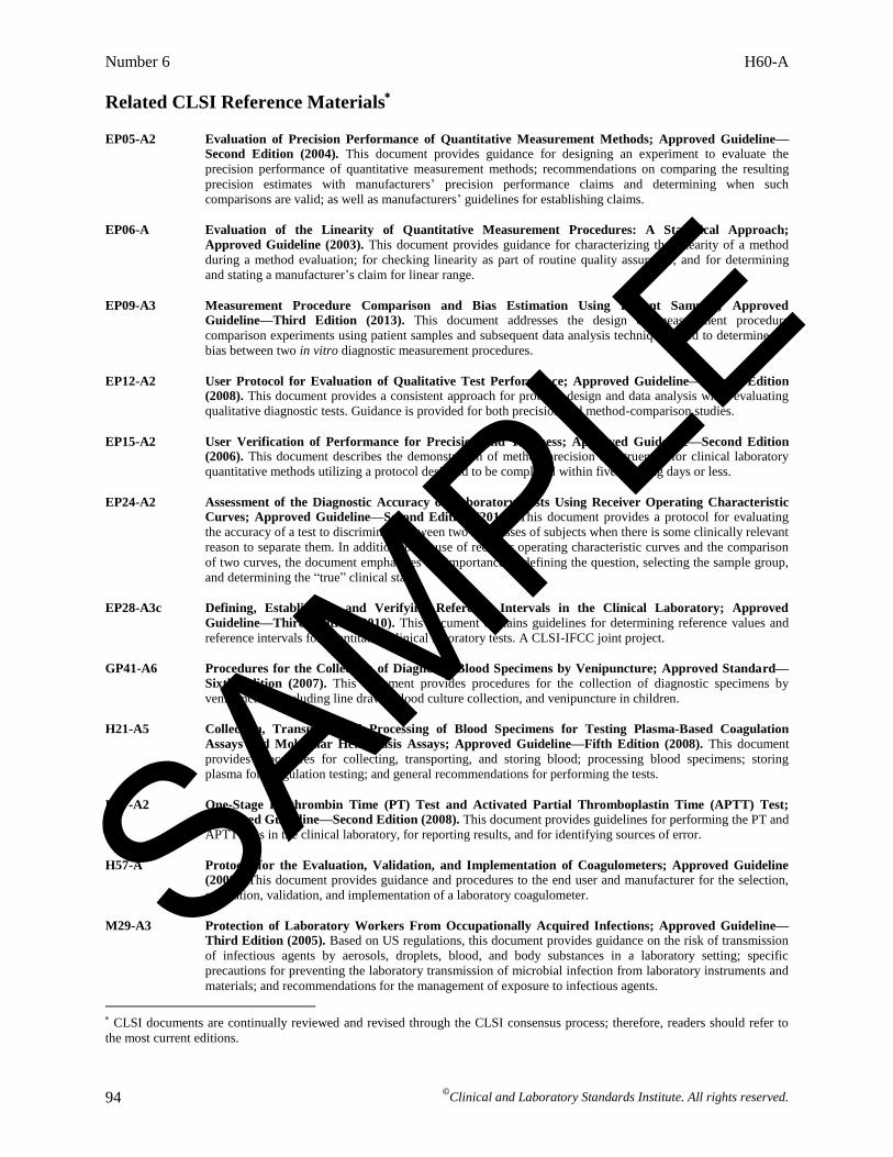
EP05-A2 Evaluation of Precision Performance of Quantitative Measurement Methods; Approved Guideline—

Second Edition (2004). This document provides guidance for designing an experiment to evaluate the

precision performance of quantitative measurement methods; recommendations on comparing the resulting

precision estimates with manufacturers’ precision performance claims and determining when such

comparisons are valid; as well as manufacturers’ guidelines for establishing claims.

EP06-A Evaluation of the Linearity of Quantitative Measurement Procedures: A Statistical Approach;

Approved Guideline (2003). This document provides guidance for characterizing the linearity of a method

during a method evaluation; for checking linearity as part of routine quality assurance; and for determining

and stating a manufacturer’s claim for linear range.

EP09-A3 Measurement Procedure Comparison and Bias Estimation Using Patient Samples; Approved

Guideline—Third Edition (2013). This document addresses the design of measurement procedure

comparison experiments using patient samples and subsequent data analysis techniques used to determine the

bias between two in vitro diagnostic measurement procedures.

EP12-A2 User Protocol for Evaluation of Qualitative Test Performance; Approved Guideline—Second Edition

(2008). This document provides a consistent approach for protocol design and data analysis when evaluating

qualitative diagnostic tests. Guidance is provided for both precision and method-comparison studies.

EP15-A2 User Verification of Performance for Precision and Trueness; Approved Guideline—Second Edition

(2006). This document describes the demonstration of method precision and trueness for clinical laboratory

quantitative methods utilizing a protocol designed to be completed within five working days or less.

EP24-A2 Assessment of the Diagnostic Accuracy of Laboratory Tests Using Receiver Operating Characteristic

Curves; Approved Guideline—Second Edition (2011). This document provides a protocol for evaluating

the accuracy of a test to discriminate between two subclasses of subjects when there is some clinically relevant

reason to separate them. In addition to the use of receiver operating characteristic curves and the comparison

of two curves, the document emphasizes the importance of defining the question, selecting the sample group,

and determining the “true” clinical state.

EP28-A3c Defining, Establishing, and Verifying Reference Intervals in the Clinical Laboratory; Approved

Guideline—Third Edition (2010). This document contains guidelines for determining reference values and

reference intervals for quantitative clinical laboratory tests. A CLSI-IFCC joint project.

GP41-A6 Procedures for the Collection of Diagnostic Blood Specimens by Venipuncture; Approved Standard—

Sixth Edition (2007). This document provides procedures for the collection of diagnostic specimens by

venipuncture, including line draws, blood culture collection, and venipuncture in children.

H21-A5 Collection, Transport, and Processing of Blood Specimens for Testing Plasma-Based Coagulation

Assays and Molecular Hemostasis Assays; Approved Guideline—Fifth Edition (2008). This document

provides procedures for collecting, transporting, and storing blood; processing blood specimens; storing

plasma for coagulation testing; and general recommendations for performing the tests.

H47-A2 One-Stage Prothrombin Time (PT) Test and Activated Partial Thromboplastin Time (APTT) Test;

Approved Guideline—Second Edition (2008). This document provides guidelines for performing the PT and

APTT tests in the clinical laboratory, for reporting results, and for identifying sources of error.

H57-A Protocol for the Evaluation, Validation, and Implementation of Coagulometers; Approved Guideline

(2008). This document provides guidance and procedures to the end user and manufacturer for the selection,

evaluation, validation, and implementation of a laboratory coagulometer.

M29-A3 Protection of Laboratory Workers From Occupationally Acquired Infections; Approved Guideline—

Third Edition (2005). Based on US regulations, this document provides guidance on the risk of transmission

of infectious agents by aerosols, droplets, blood, and body substances in a laboratory setting; specific

precautions for preventing the laboratory transmission of microbial infection from laboratory instruments and

materials; and recommendations for the management of exposure to infectious agents.

CLSI documents are continually reviewed and revised through the CLSI consensus process; therefore, readers should refer to

the most current editions.

SAMPLE

For more information, visit www.clsi.org today.

Explore the Latest Offerings From CLSI!As we continue to set the global standard for quality in laboratory testing, we are adding products and programs to bring even more value to our members and customers.

Find what your laboratory needs to succeed! CLSI U provides convenient, cost-effective continuing education and training resources to help you advance your professional development. We have a variety of easy-to-use, online educational resources that make eLearning stress-free and convenient for you and your staff.

See our current educational offerings at www.clsi.org/education.

When laboratory testing quality is critical, standards are needed and there is no time to waste. eCLIPSE™ Ultimate Access, our cloud-based online portal of the complete library of CLSI standards, makes it easy to quickly find the CLSI resources you need.

Learn more and purchase eCLIPSE at clsi.org/eCLIPSE.

By becoming a CLSI member, your laboratory will join 1,600+ other influential organizations all working together to further CLSI’s efforts to improve health care outcomes. You can play an active role in raising global laboratory testing standards—in your laboratory, and around the world.

Find out which membership option is best for you at www.clsi.org/membership.

SAMPLE

950 West Valley Road, Suite 2500, Wayne, PA 19087 USA

P: 610.688.0100 Toll Free (US): 877.447.1888 F: 610.688.0700

E: [email protected] www.clsi.org

PRINT ISBN 1-56238-959-9

ELECTRONIC ISBN 1-56238-960-2

SAMPLE


Recommended